
The Hip. Pelvic (Hip) Girdle Two hip bones (each also called coxal bone or os coxae) – Attach the...
36
The Hip
-
Upload
mitchell-swim -
Category
Documents
-
view
218 -
download
2
Transcript of The Hip. Pelvic (Hip) Girdle Two hip bones (each also called coxal bone or os coxae) – Attach the...

The Hip
Pelvic (Hip) Girdle• Two hip bones (each also called coxal
bone or os coxae)–Attach the lower limbs to the axial
skeleton with strong ligaments–Transmit weight of upper body to
lower limbs–Support pelvic organs
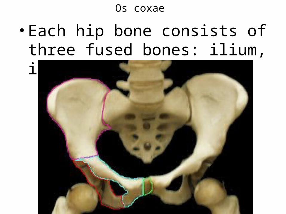
Os coxae
• Each hip bone consists of three fused bones: ilium, ischium, and pubis
• Together with the sacrum and the coccyx, these bones form the bony pelvis
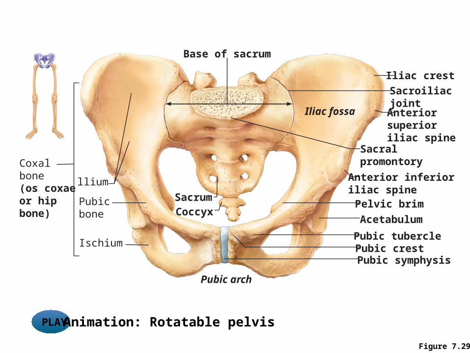
Figure 7.29
Coxalbone(os coxaeor hip bone)
llium
Sacroiliacjoint
Iliac fossa
Pubicbone
Ischium
Sacrum
Base of sacrum
Sacralpromontory
Pelvic brim
Acetabulum
Pubic crestPubic symphysis
Iliac crest
Coccyx
Pubic arch
Anterior inferioriliac spine
Anteriorsuperior iliac spine
Pubic tubercle
PLAY Animation: Rotatable pelvis
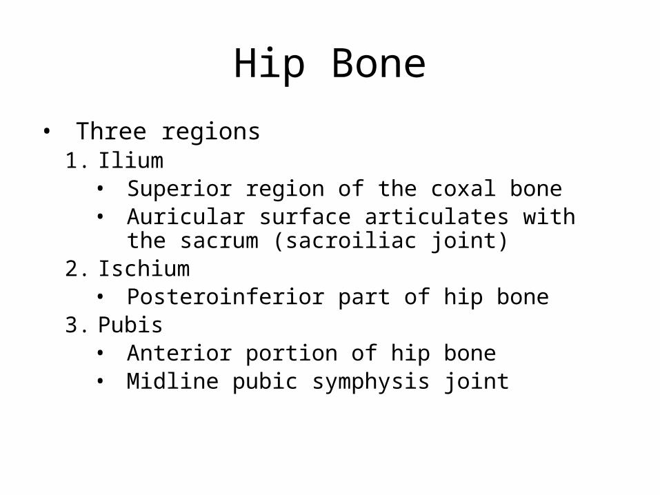
Hip Bone• Three regions
1. Ilium• Superior region of the coxal bone• Auricular surface articulates with the sacrum (sacroiliac
joint)2. Ischium
• Posteroinferior part of hip bone3. Pubis
• Anterior portion of hip bone• Midline pubic symphysis joint
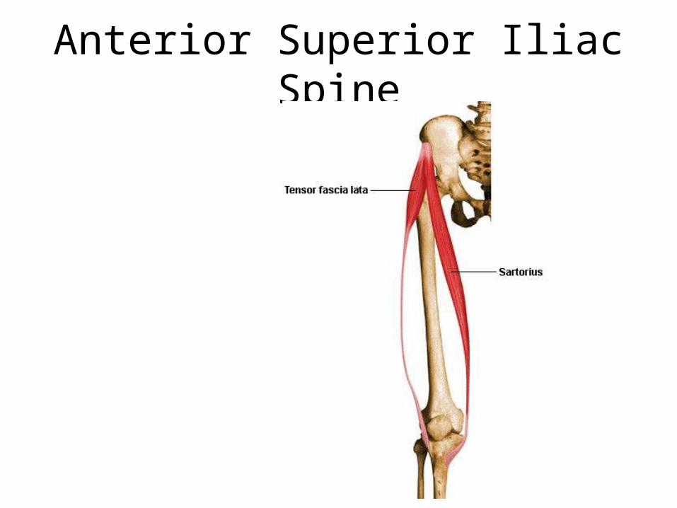
Anterior Superior Iliac Spine
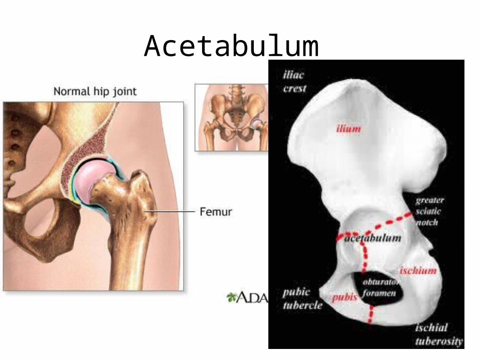
Acetabulum
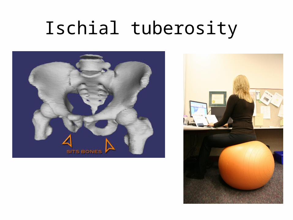
Ischial tuberosity
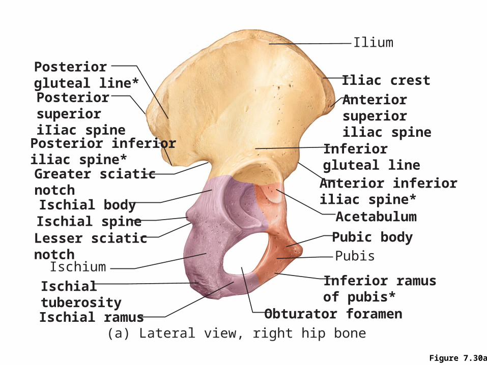
Figure 7.30a
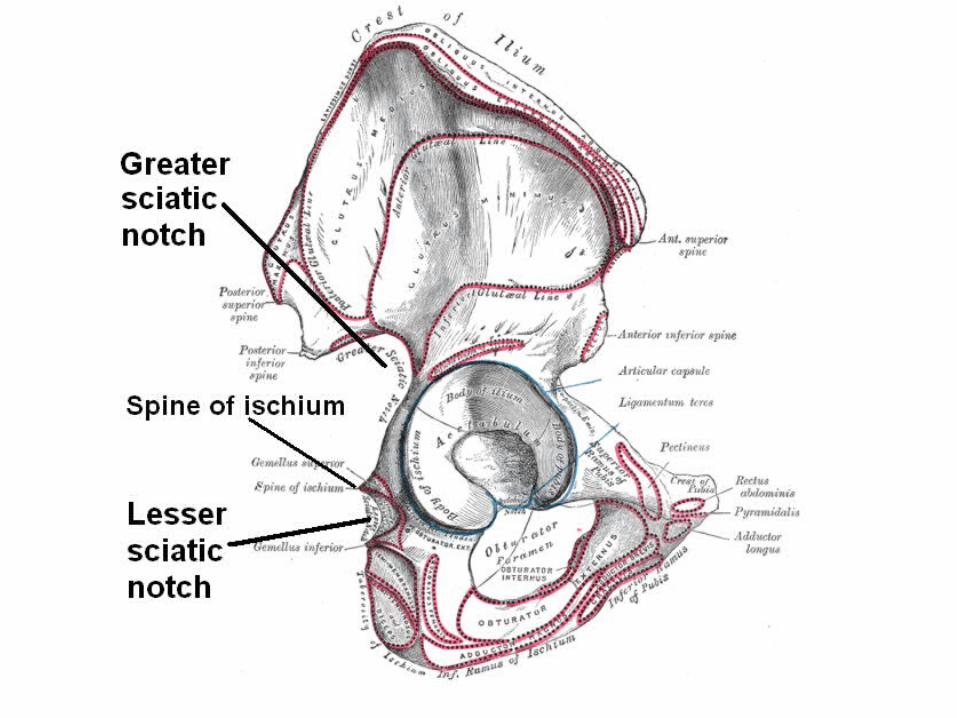
Ilium
Posterior gluteal line*PosteriorsuperioriIiac spine
Greater sciaticnotch
Posterior inferioriliac spine*
Ischial bodyIschial spineLesser sciatic notch
Ischialtuberosity
Ischium
Ischial ramus Obturator foramen
Inferiorgluteal line
Acetabulum
Pubic body
Iliac crest
Anteriorsuperioriliac spine
Anterior inferioriliac spine*
Pubis
Inferior ramusof pubis*
(a) Lateral view, right hip bone
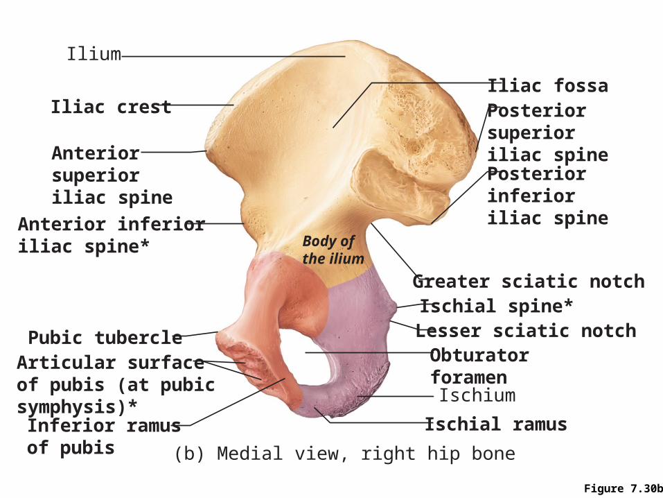
Figure 7.30b
Iliac fossa
Ilium
Iliac crest
Anteriorsuperioriliac spine
Anterior inferioriliac spine*
Pubic tubercle
Inferior ramusof pubis
Posteriorsuperioriliac spine
Obturatorforamen
Body ofthe ilium
Ischium
Ischial ramus
(b) Medial view, right hip bone
Ischial spine*Lesser sciatic notch
Greater sciatic notch
Posteriorinferioriliac spine
Articular surfaceof pubis (at pubic symphysis)*
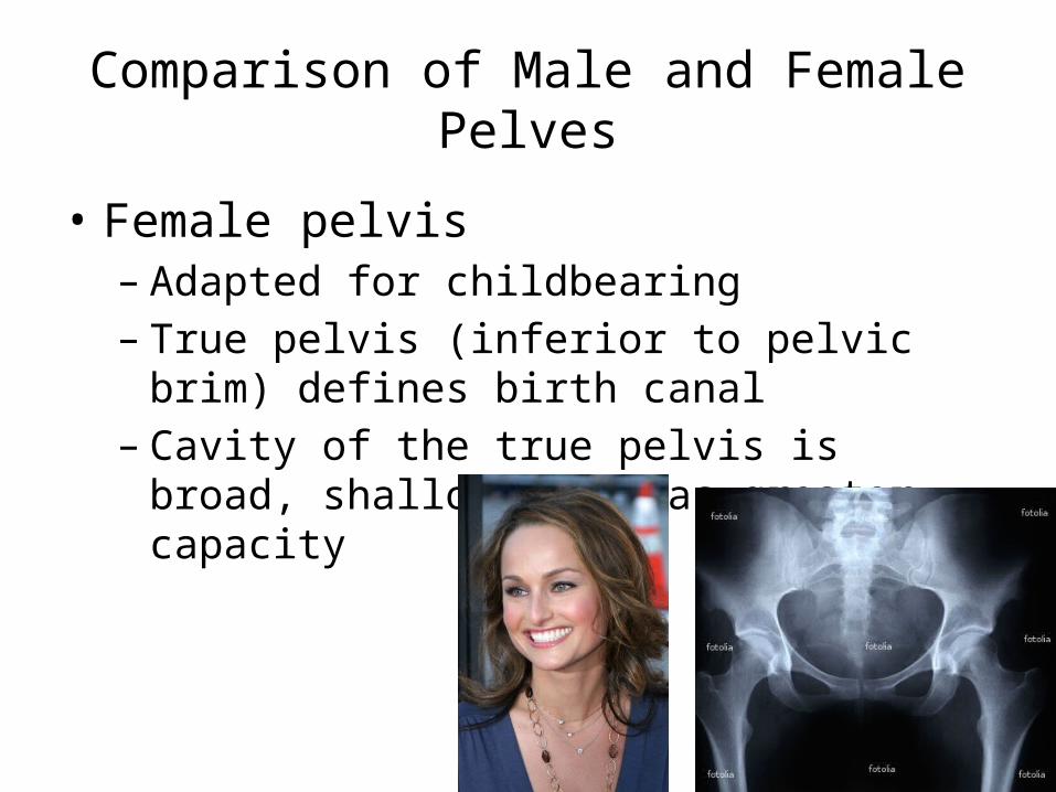
Comparison of Male and Female Pelves
• Female pelvis– Adapted for childbearing– True pelvis (inferior to pelvic brim) defines birth
canal– Cavity of the true pelvis is broad, shallow, and has
greater capacity
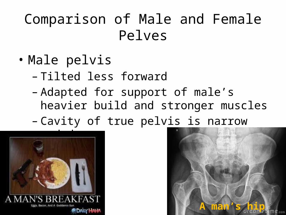
Comparison of Male and Female Pelves
• Male pelvis– Tilted less forward– Adapted for support of male’s heavier build and
stronger muscles– Cavity of true pelvis is narrow and deep
A man’s hip
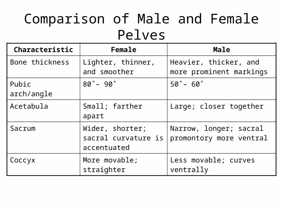
Comparison of Male and Female PelvesCharacteristic Female Male
Bone thickness Lighter, thinner, and smoother
Heavier, thicker, and more prominent markings
Pubic arch/angle 80˚– 90˚ 50˚– 60˚
Acetabula Small; farther apart Large; closer together
Sacrum Wider, shorter; sacral curvature is accentuated
Narrow, longer; sacral promontory more ventral
Coccyx More movable; straighter Less movable; curves ventrally

Table 7.4
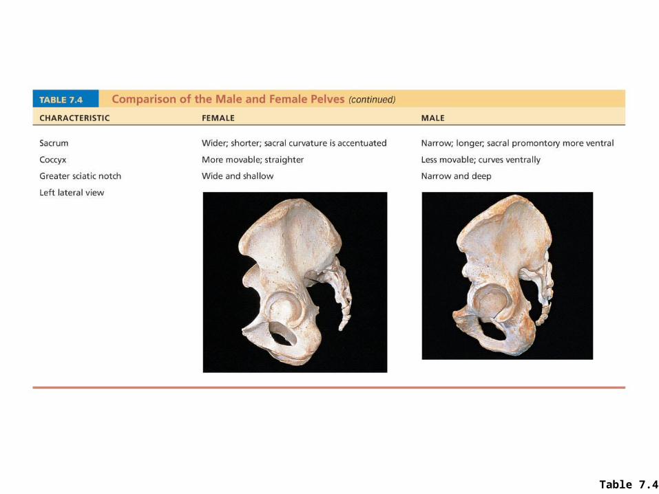
Table 7.4
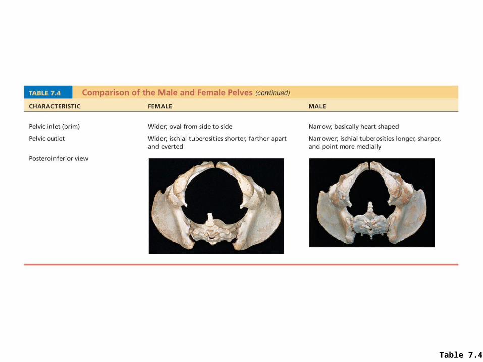
Table 7.4
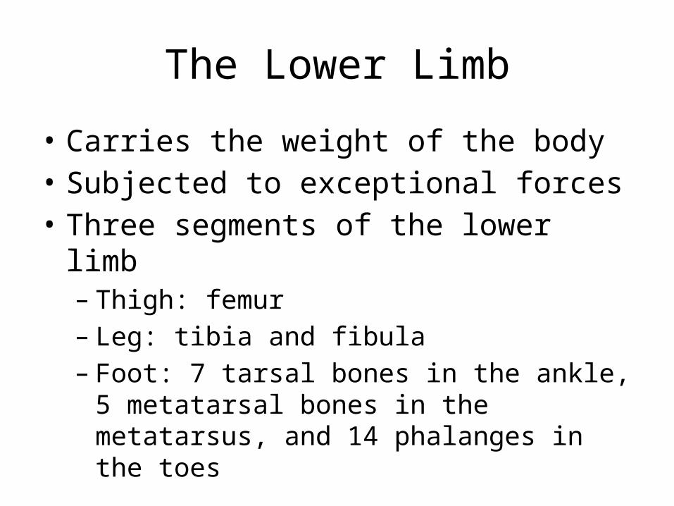
The Lower Limb
• Carries the weight of the body• Subjected to exceptional forces • Three segments of the lower limb
– Thigh: femur– Leg: tibia and fibula– Foot: 7 tarsal bones in the ankle, 5 metatarsal
bones in the metatarsus, and 14 phalanges in the toes
Femur
• Largest and strongest bone in the body• Articulates proximally with the acetabulum of
the hip and distally with the tibia and patella
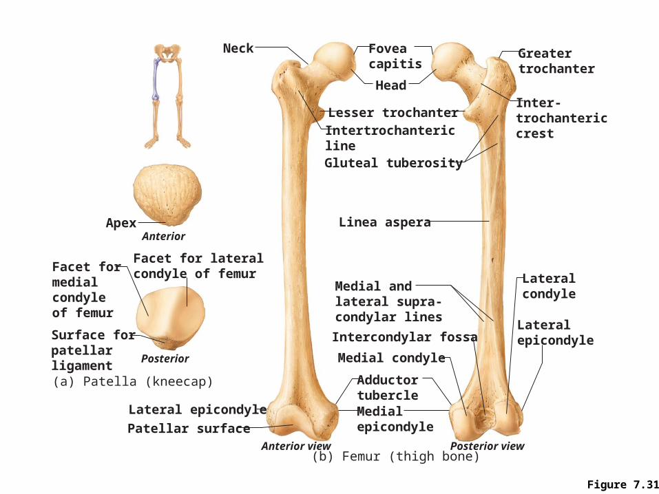
Figure 7.31
Neck Foveacapitis
Greatertrochanter
Inter-trochantericcrest
Head
Intertrochantericline
Lesser trochanter
Gluteal tuberosity
Linea aspera
Lateralcondyle
LateralepicondyleIntercondylar fossa
Medial andlateral supra-condylar lines
Medial condyle
Medialepicondyle
Adductortubercle
Anterior view Posterior view(b) Femur (thigh bone)
Lateral epicondyle
Patellar surface
Posterior
Facet formedialcondyleof femur
Facet for lateralcondyle of femur
Surface forpatellarligament
ApexAnterior
(a) Patella (kneecap)
Bones of the LegTibia• Medial leg bone• Receives the weight of the body from the
femur and transmits it to the foot
Bones of the legFibula• Not weight bearing; no articulation with
femur• Site of muscle attachment • Connected to tibia by interosseous
membrane• Articulates with tibia via proximal and
distal tibiofibular joints
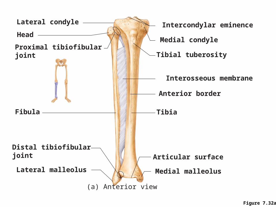
Figure 7.32a
Medial condyle
Articular surface
Tibial tuberosity
Interosseous membrane
Anterior border
Tibia
Medial malleolus
Intercondylar eminence
Proximal tibiofibularjoint
Distal tibiofibularjoint
Lateral malleolus
Lateral condyle
Fibula
Head
(a) Anterior view
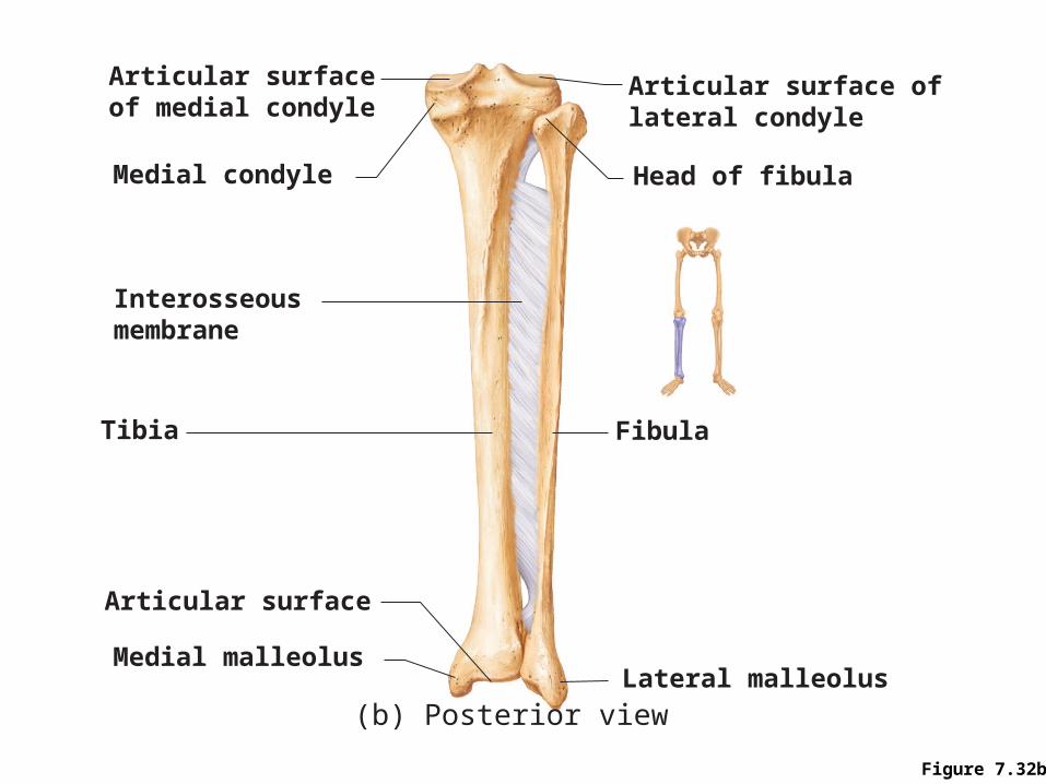
Figure 7.32b
Medial condyle
Articular surface oflateral condyle
Articular surfaceof medial condyle
Articular surface
Interosseousmembrane
Tibia Fibula
Head of fibula
Medial malleolusLateral malleolus
(b) Posterior view
Foot: Tarsals
• Seven tarsal bones form the posterior half of the foot
• Talus transfers most of the weight from the tibia to the calcaneus
• Other tarsal bones: cuboid, navicular, and the medial, intermediate, and lateral cuneiforms
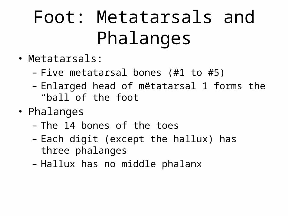
Foot: Metatarsals and Phalanges
• Metatarsals:– Five metatarsal bones (#1 to #5) – Enlarged head of metatarsal 1 forms the “ball of the foot”
• Phalanges– The 14 bones of the toes– Each digit (except the hallux) has three phalanges – Hallux has no middle phalanx
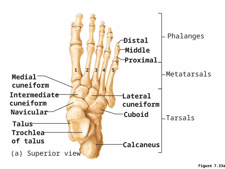
Figure 7.33a
Medialcuneiform
Phalanges
Metatarsals
TarsalsNavicular
Intermediatecuneiform
Talus
Calcaneus(a) Superior view
Cuboid
Lateralcuneiform
Proximal54321
Middle
Distal
Trochleaof talus
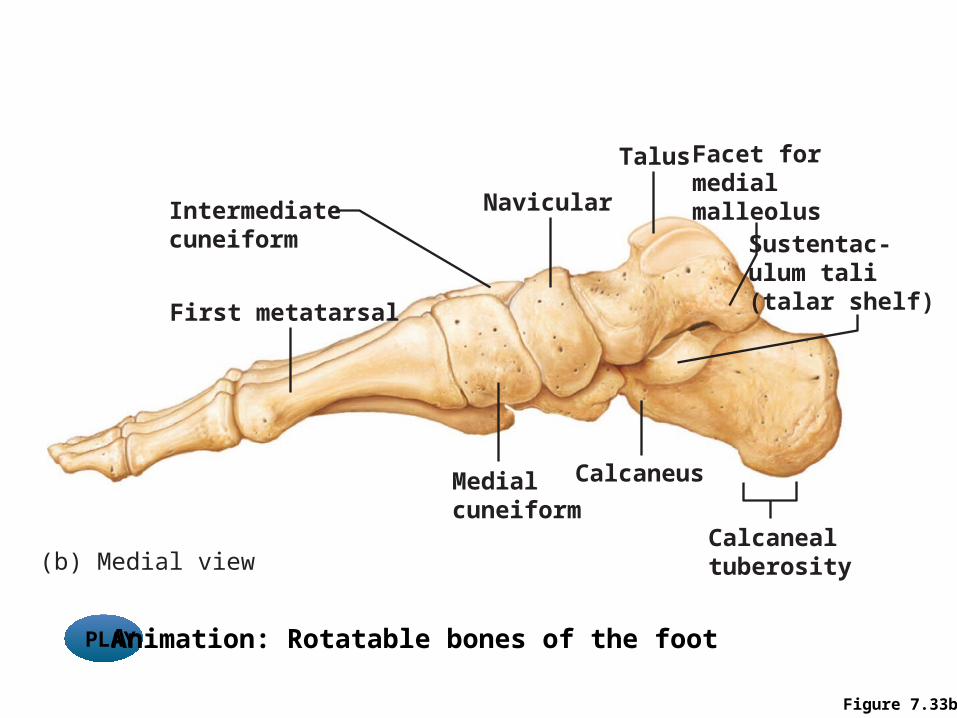
Figure 7.33b
Facet formedialmalleolus
Calcanealtuberosity(b) Medial view
Intermediatecuneiform Sustentac-
ulum tali(talar shelf)
Talus
Navicular
First metatarsal
Medialcuneiform
Calcaneus
PLAY Animation: Rotatable bones of the foot
Arches of the Foot
• Arches are maintained by interlocking foot bones, ligaments, and tendons
• Arches allow the foot to bear weight• Three arches
– Lateral longitudinal – Medial longitudinal – Transverse
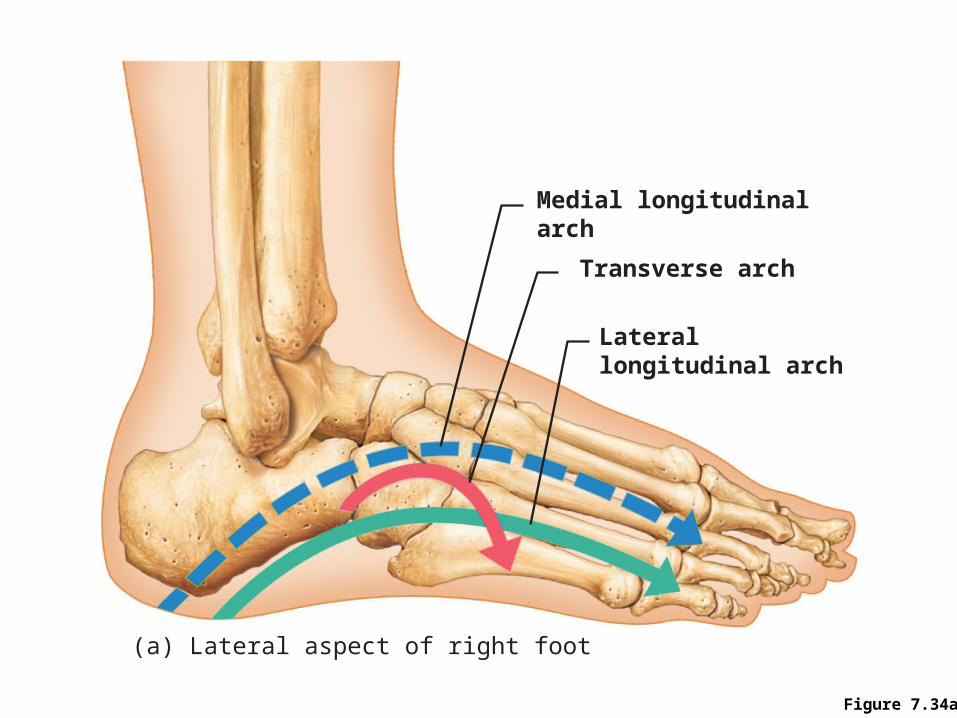
Figure 7.34a
Medial longitudinalarch
Transverse arch
Laterallongitudinal arch
(a) Lateral aspect of right foot
Developmental Aspects: Fetal Skull
• Infant skull has more bones than the adult skull• Skull bones such as the mandible and frontal bones are
unfused • At birth, skull bones are connected by fontanelles
– Fontanelles• Unossified remnants of fibrous membranes between
fetal skull bones• Four fontanelles
– Anterior, posterior, mastoid, and sphenoid