The Hall Technique - heeoe.hee.nhs.uk · The Hall technique is a novel method of managing carious...
19
University of Dundee The Hall Technique A child centred approach to managing the carious primary molar A Users Manual Text copyright Nicola Innes & Dafydd Evans Illustrations copyright Dafydd Evans & Amy McKay
Transcript of The Hall Technique - heeoe.hee.nhs.uk · The Hall technique is a novel method of managing carious...

University of Dundee
The Hall Technique A child centred approach to managing the carious
primary molar
A Users Manual
Text copyright Nicola Innes & Dafydd Evans Illustrations copyright Dafydd Evans & Amy McKay
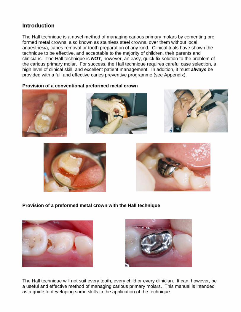
Introduction The Hall technique is a novel method of managing carious primary molars by cementing pre-formed metal crowns, also known as stainless steel crowns, over them without local anaesthesia, caries removal or tooth preparation of any kind. Clinical trials have shown the technique to be effective, and acceptable to the majority of children, their parents and clinicians. The Hall technique is NOT, however, an easy, quick fix solution to the problem of the carious primary molar. For success, the Hall technique requires careful case selection, a high level of clinical skill, and excellent patient management. In addition, it must always be provided with a full and effective caries preventive programme (see Appendix). Provision of a conventional preformed metal crown Provision of a preformed metal crown with the Hall technique
The Hall technique will not suit every tooth, every child or every clinician. It can, however, be a useful and effective method of managing carious primary molars. This manual is intended as a guide to developing some skills in the application of the technique.

Background The technique is named after Dr Norna Hall, a general dental practitioner from Scotland, who developed and used the technique for over 15 years until she retired in 2006. Preformed metal crowns (PMCs) have been used for restoring primary molars since 1950, and have become the accepted restoration of choice for the primary molar with caries affecting more than one surface, with a proven success rate as a restoration. Although popular with specialists, many clinicians find PMCs difficult to fit using the conventional approach, which requires the use of local anaesthetic injections and extensive tooth preparation. There is also an issue of potential damage to the adjacent first permanent molar when preparing a second primary molar for a PMC. For this, and other reasons, PMCs are not widely used in the UK, forming less than 1% of all restorations provided for children. How does the Hall technique get around some of these problems? With the Hall technique, the process of fitting the crown is quick and non-invasive. The crown is seated over the tooth with no caries removal or tooth preparation of any kind, and local anaesthesia is not required. For decades, conventional teaching has been that all carious tooth tissue should be removed before restoring the tooth, unless there is a high risk of pulpal exposure. How can leaving all the caries in the tooth be acceptable? To answer this, it is worth firstly reviewing how and where caries begins. For many years it was assumed that all that was needed was to put a tooth surface, plaque and sugar together, add a little time, and caries would result. This combination can undoubtedly, under the right circumstances, cause caries, but what is remarkable is that so few tooth surfaces seem to be susceptible to carious attack. Clinicians will be aware that, except in extreme cases, the majority of tooth surfaces are relatively immune from caries, despite many of these surfaces often having prolonged coverage by plaque; for example, the labial and buccal cervical margins of teeth as they approach the proximal surfaces.
Low caries susceptibility High caries susceptibility In fact, almost all caries begins at sites which collectively make up only a tiny proportion of the total area of enamel available for colonisation; the base of fissures, and just below the contact point of proximal surfaces. The enamel here is almost identical in composition to that of the labial surfaces, so why the difference? What differs is the ecological niche provided for plaque maturation by the very sheltered environment of these surfaces. Once caries has caused cavitation of the tooth, the availability of sheltered surfaces suitable for plaque colonisation and maturation dramatically increases, and so the caries continues through the tooth.
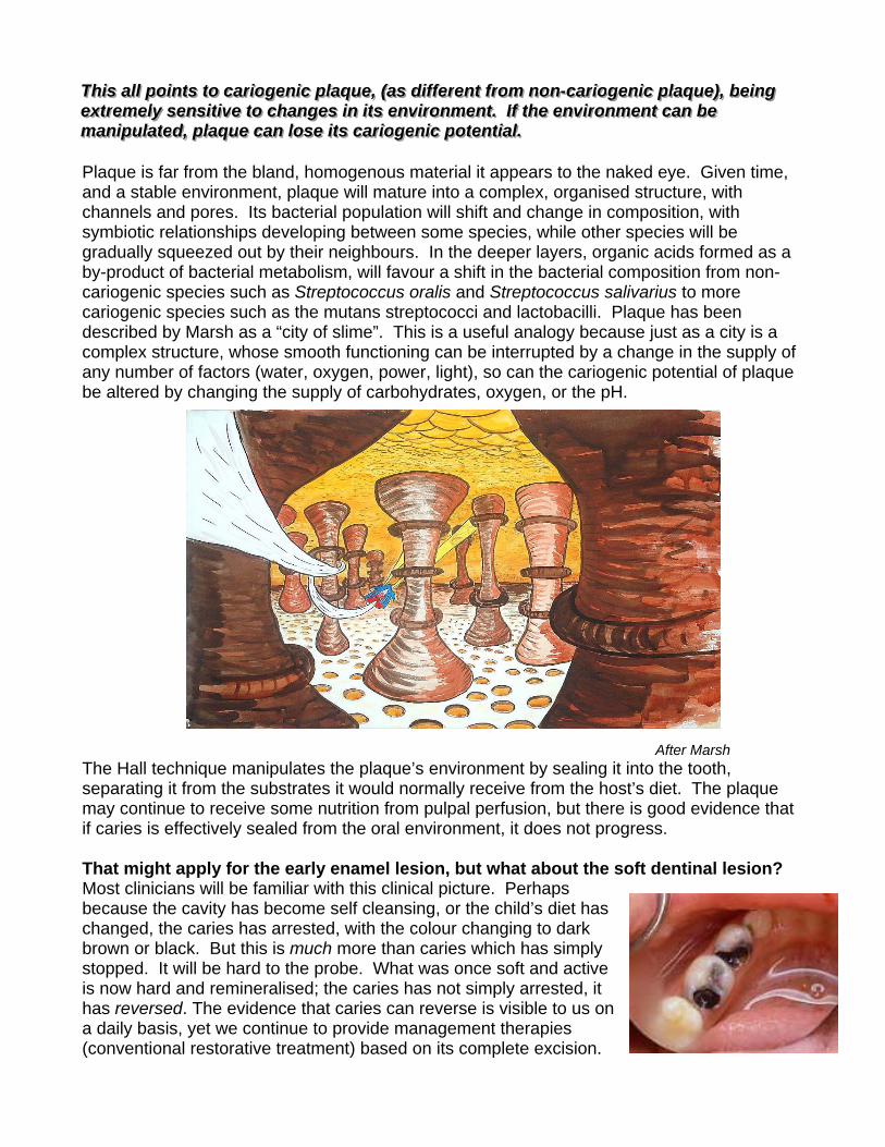
TTThhhiiisss aaallllll pppoooiiinnntttsss tttooo cccaaarrriiiooogggeeennniiiccc ppplllaaaqqquuueee,,, (((aaasss dddiiiffffffeeerrreeennnttt fffrrrooommm nnnooonnn---cccaaarrriiiooogggeeennniiiccc ppplllaaaqqquuueee))),,, bbbeeeiiinnnggg eeexxxtttrrreeemmmeeelllyyy ssseeennnsssiiitttiiivvveee tttooo ccchhhaaannngggeeesss iiinnn iiitttsss eeennnvvviiirrrooonnnmmmeeennnttt... IIIfff ttthhheee eeennnvvviiirrrooonnnmmmeeennnttt cccaaannn bbbeee mmmaaannniiipppuuulllaaattteeeddd,,, ppplllaaaqqquuueee cccaaannn lllooossseee iiitttsss cccaaarrriiiooogggeeennniiiccc pppooottteeennntttiiiaaalll... Plaque is far from the bland, homogenous material it appears to the naked eye. Given time, and a stable environment, plaque will mature into a complex, organised structure, with channels and pores. Its bacterial population will shift and change in composition, with symbiotic relationships developing between some species, while other species will be gradually squeezed out by their neighbours. In the deeper layers, organic acids formed as a by-product of bacterial metabolism, will favour a shift in the bacterial composition from non-cariogenic species such as Streptococcus oralis and Streptococcus salivarius to more cariogenic species such as the mutans streptococci and lactobacilli. Plaque has been described by Marsh as a “city of slime”. This is a useful analogy because just as a city is a complex structure, whose smooth functioning can be interrupted by a change in the supply of any number of factors (water, oxygen, power, light), so can the cariogenic potential of plaque be altered by changing the supply of carbohydrates, oxygen, or the pH. After Marsh The Hall technique manipulates the plaque’s environment by sealing it into the tooth, separating it from the substrates it would normally receive from the host’s diet. The plaque may continue to receive some nutrition from pulpal perfusion, but there is good evidence that if caries is effectively sealed from the oral environment, it does not progress. That might apply for the early enamel lesion, but what about the soft dentinal lesion? Most clinicians will be familiar with this clinical picture. Perhaps because the cavity has become self cleansing, or the child’s diet has changed, the caries has arrested, with the colour changing to dark brown or black. But this is much more than caries which has simply stopped. It will be hard to the probe. What was once soft and active is now hard and remineralised; the caries has not simply arrested, it has reversed. The evidence that caries can reverse is visible to us on a daily basis, yet we continue to provide management therapies (conventional restorative treatment) based on its complete excision.

How does the pulp react to caries? Just as it is becoming increasingly clear that dental caries is a dynamic process, it is also being recognised that the dentine/ pulp complex is far from passive when exposed to dental caries. Instead, these tissues mount an active defence response from the earliest stages of carious lesion formation in the enamel. An increase in pulpal blood flow allows an increased response from the immune system, and odontoblasts are stimulated to lay down a layer of secondary dentine in an effort to distance the pulp from the approaching carious lesion, an effect readily observed, at a gross level, on radiographs.
It seems likely that the dentine/ pulp complex has a greater reparative potential when subject to dental caries than has previously been thought. If the progress of the caries can be halted before the pulp is overwhelmed, then the pulp may well survive.
Summary Not all plaque is cariogenic. Plaque which has matured in a sheltered environment to achieve cariogenic potential can lose that potential if its environment is altered. Effective sealing from the oral environment can cause the necessary alteration, resulting in plaque losing its cariogenic potential for as long as the seal is maintained, and allowing dentine affected by caries to remineralise. The Hall technique is one method of achieving that seal for primary molar teeth. Is the Hall technique effective? To answer this question, a clinical trial set in 9 general dental practices in Tayside, Scotland looked at outcomes at two years for teeth where a Hall crown was fitted, compared to teeth which had undergone conventional restorative treatment. The trial was a split mouth randomised control design, so teeth were matched on each side of the arch for type of lesion and extent of caries. The dentists telephoned a distant operator to be told which tooth to provide a Hall crown and which to fit first, in order to reduce any bias in the trial. 132 children were enrolled in the trial and followed up every year clinically and with bitewing radiographs. The outcomes for the 124 patients seen at 2 years (8 patients failed to return for 2 year appointments) are shown below. Outcomes • pain • major failures (irreversible pulpitis; abscess requiring pulpotomy or extraction; inter-
radicular radiolucency; filling lost and tooth unrestorable)
• minor failures (new/secondary caries; filling/crown worn, lost or requiring other intervention; restoration lost but tooth restorable; reversible pulpitis treated without requiring pulpotomy or extraction)
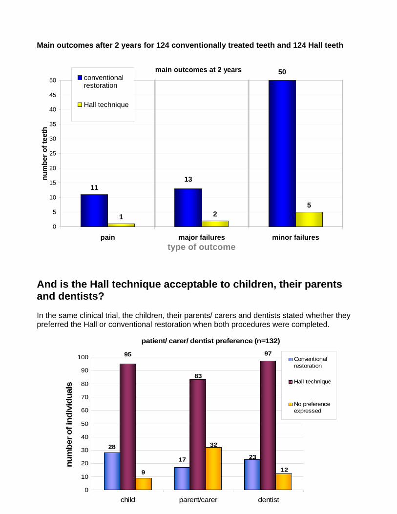
Main outcomes after 2 years for 124 conventionally treated teeth and 124 Hall teeth
main outcomes at 2 years
11
1 25
50
13
0
5
10
15
20
25
30
35
40
45
50
pain major failures minor failurestype of outcome
num
ber o
f tee
th
conventionalrestoration
Hall technique
And is the Hall technique acceptable to children, their parents and dentists? In the same clinical trial, the children, their parents/ carers and dentists stated whether they preferred the Hall or conventional restoration when both procedures were completed.
patient/ carer/ dentist preference (n=132)
28
9
17 23
9795
83
32
12
0
10
20
30
40
50
60
70
80
90
100
child parent/carer dentist
num
ber o
f ind
ivid
uals
Conventionalrestoration
Hall technique
No preferenceexpressed

Using the Hall technique in clinical practice Indications, and some important information The Hall technique is can be used to manage primary molar teeth affected by dental caries. Other management methods are available. As with every treatment decision, clinicians should use their own clinical judgement in deciding which method is appropriate for their patient and themselves, with consent being obtained from the patient, and parent, for that treatment. Although apparently very simple, the Hall technique requires a confident, skilled approach from the operator if the crown is to be successfully fitted. The technique will not suit every clinician, nor every child. In addition, there are some primary molars where, for a combination of reasons, even clinicians very familiar with the Hall technique cannot successfully fit a crown.
For example, should these lower Ds become carious, their unusual morphology would complicate the fitting of a PMC of standard shape.
Again, common to all clinical procedures, it is important that the clinician has a clear understanding of what to do to retrieve a situation which is not proceeding as planned, for example when a Hall crown is not seating properly onto a tooth or appears to be the wrong size or shape and will not fit correctly over the crown of the tooth. These issues are dealt with at the end of this section. To begin with… A full history and clinical examination, including bitewing radiography, should be carried out.
• There should be no clinical or radiographic signs of pulpal involvement • The tooth should have sufficient sound tissue left to retain the crown • Patient co-operation should be such that the clinician should be confident that the
crown can be fitted without endangering the patient’s airway • If the patient is at risk from bacterial endocarditis, the tooth should be managed with a
conventional restoration

The appointment for fitting the crown
Instruments to have ready
Essential: Mirror Straight probe
o to remove separators, if used, and to remove set cement following fitting Excavator
o to remove crown if necessary, and also useful for cement removal Flat plastic
o to load crown with cement Cotton wool rolls
o to wipe away cement Useful:
Orthodontic biting stick o can be useful in seating crowns
Band forming pliers o can be useful for adjusting crowns, particularly where the primary molar
has lost length mesio-distally due to caries Gauze to protect the airway and wipe off excess cement
or Elastoplast to secure the crown for airway protection
Preparation is everything! The child and parent should be briefed on the procedure. Children should be shown a crown, and allowed to handle a spare one if felt beneficial. Young children sometimes respond to the idea of the crown being “a shiny helmet”, just like soldiers wear to protect their heads”, or “a precious, shiny, princess crown” or it being a “twinkle tooth”.
It is important that the child knows that: a) they will have to help, by biting the crown into place when asked to do so b) the cement will taste a bit like Salt & Vinegar crisps

Assess the shape of the tooth and its contacts If the contact points are tight, or there has been loss of mesio-distal width of a tooth due to marginal ridge fracture, placing orthodontic separators through the mesial and distal contacts can be very useful when fitting crowns with the Hall technique, although it does mean the patient will have to make a second visit. Two lengths of dental floss should be threaded through the separator.
The separator should then be stretched taut, and “flossed” through the contact point briskly and firmly until the leading edge only is felt “popping through” the contact point. If the separator is cut
by sharp cavity margins, it may be found helpful to pull the separator “up, into & through” the contact area, approaching from the cervical margin.
The floss should then be removed, and the patient seen between 3-5 days later for removal of the separator. If the separator appears to have
fallen out, the inter-proximal area of the gingiva should be inspected to check that the separator hasn’t worked its way below the contact point. Separators are usually brightly coloured to facilitate this. Oh, the separator has fallen out… No it hasn’t!!

Another difficulty can be placing crowns on Ds where the distal marginal ridge has been lost, and the E has migrated mesially into the cavity.
Here, band forming orthodontic pliers can be useful, both to increase the bucco-lingual width of the crown at the expense of the mesio-distal length (here using the pliers the “right” way around, that is with the curvature of the pliers following the curvature of the crown),
and also, if necessary, to alter the distal margin of the crown from a convex to a concave shape, by gently squeezing the margin with the pliers the “wrong” way around.

The procedure 1. Sit the child upright. A gauze swab square can be used to protect the airway by
placing it between the tongue and the tooth where the crown is to be fitted. It should extend to the palate and round the back of the mouth in front of the fauces. Alternatively, use a clean piece of Elastoplast tape to secure the crown (see below). If you are not confident about being able to control the crown at all stages until it is cemented, then do not use the technique.
Airway protection using a) a gauze square b) Elastoplast Orientation of gauze Positioned in mouth Elastoplast tape securing crown 2. You should aim to fit the smallest size of crown which will seat. Select one which
covers all the cusps, and approaches the contact points, with a slight feeling of “spring back”. Do not attempt to fully seat the crown through the contact points; they can be very difficult to remove for cementation!
3. Dry the crown, and fill with glass-ionomer luting cement, ensuring the crown is well filled, with no air inclusions
4. If possible, the tooth should be dried prior to cementation, but otherwise there is no caries removal or tooth preparation of any kind. No local anaesthetic injection is given.
5. If the cavity is large, some cement may be placed within it, just before placing the crown.
6. Place the crown over the tooth. Fully seating the crown is a critical stage! It is not always easy, and requires a committed, positive approach from the clinician. The child needs to have complete confidence that you know exactly what you are doing; that what you are asking them to do is perfectly reasonable, and that it will not be uncomfortable. Remember that our research found that, surprisingly, most children do not find the procedure painful, and prefer it to conventional fillings. There are two main methods of seating the crowns:
a) the clinician seats the crown by finger pressure b) the child seats the crown by biting on it
A combination of these two methods may be necessary or preferred. Some clinicians will seat the crown with firm finger pressure alone. For mandibular teeth, a useful method is to place your thumb on the occlusal surface of the crown, with the four fingers of your hand placed under the border of the mandible to spread the force as you apply firm pressure with your thumb. For maxillary teeth, the child’s head

may be supported by the back of the dental chair, or sometimes by placing your other forearm gently on the top of their head to balance the force applied by fitting the crown. Often, the child will seat the crown themselves by biting it into place. It can be useful to verbally encourage the child to apply the necessary pressure (“Bite hard, like a Tiger! Grrrrr…..!), and to rehearse this before fitting the crown. If using this method, be aware that some children’s resolve might falter a little, leaving the crown not fully seated. Here, a timely “That was great! Now let me just check it for you! Ooh, well done, and I’ll just give it a little squeeze….., Excellent!” can save the day. Some clinicians partially seat the crown until it engages with the contact points, allowing the finger to be removed without risk of the crown falling off, and the child then being encouraged to bite the crown into place. It must be remembered that your working time with glass ionomer cements is limited, and whatever method is used, you must work smoothly and efficiently. Crowns cannot be seated, no matter how hard you or the child tries, if the cement has started to thicken. It is crucial that the orientation of the crown relative to the tooth is checked either during, or immediately after, seating the crown. If it does not appear to be going on straight, then you must give the crown some physical encouragement to go in the correct direction. If it is not possible to seat it then it should be removed before the cement sets. With either technique, excess cement will be extruded from the crown margins, and the taste of this can upset children. In anticipation of this, as soon as the crown is seated, the child should be asked to open their mouth, and the cement wiped off with a cotton wool roll held ready for this purpose. If a gauze swab has been used to protect the airway, this can be used to wipe away excess cement from the lingual/ palatal side of the tooth as it is being removed. If it is obvious that the crown has not seated, and finger pressure fails to seat it, then it should be removed immediately using the large excavator which you should have placed within easy reach. If you do not work swiftly, you may have to section the crown to remove it (see later).
7. Once excess cement has been removed, the child should be asked to bite firmly on the crown for 2-3 minutes, or the crown should be held down with firm finger pressure as an alternative. This is important, because the crowns can spring back a short way, sucking back the cement form the margins and potentially causing breaches in the seal.
8. Remove excess cement, floss between the contacts and give the child a sticker.

Picture Gallery Working with the child seated upright means that the optimum operator working position has to be compromised. Load crown generously (it should be almost full with cement). Take care to avoid air blows and voids. Some glass ionomer may be wiped on the tooth or placed in any cavitation to improve the seal. The crown is placed evenly over the tooth and engaged in the approximal contact points using finger pressure. This secures its position. The child is instructed to bite down on the crown. Some operators find biting on a cotton wool roll helps the process. Care is taken to ensure the crown seats evenly over the tooth.
Mix the cement to a luting consistency, and rapidly, so as to maximise working time. What happens if you don’t (fortunately this one didn’t seat!). Blanching usually disappears within minutes.
The occlusal discrepancy should resolve in a few weeks. Floss should be used to clear the contacts of any excess cement.

A satisfied customer, with a restored occlusion! If the crown does not seat sufficiently, then remove it using the excavator before the cement sets. If the cement has set, a high speed handpiece can be used to section the crown through the buccal and occlusal surface, following which it can easily be peeled off.

Additional notes 1) The crowns used in the research presented here were Ni-Cro Primary Molar Crowns, cemented with AquaCem, both from 3M/ESPE. Any adjustment of the crowns was minimal, and was limited to re-moulding the crown margins in some cases with orthodontic pliers. No crown had the margin trimmed. 2) Fitting crowns to Ds where loss of the marginal ridge has allowed the E to drift mesially can be particularly tricky. Use of separators, and adjusting the shape of PMCs using band forming pliers has already been mentioned. Sometimes it helps if the PMC is rotated slightly, usually mesio-buccaly. This example is a little extreme! 3) Crowns will try to follow the path of least resistance, and so may tilt towards the “easier” of the contacts, making it almost impossible then to ease the crown through the tight contact. Concentrate on seating the crown through the tight contact, and the easy one should take care of itself. 4) If the crown does not seat sufficiently, then remove it using the excavator before the cement sets. If the cement has set, a high speed handpiece can be used to section the crown through the buccal and occlusal surface, following which it can easily be peeled off. 5) Patients and parents should be reassured that the child will be used to the feeling within 24 hours. It is the authors’ experience that analgesia is not required. The occlusion tends to adjust to give even contact on both sides within weeks. 6) Patients should be reviewed on a normal recall schedule, and the Hall technique should be used in conjunction with a full preventive programme. 7) If fitting crowns to Es, particularly maxillary Es, before the 6s are erupted, keep an eye out for the 6s becoming impacted against the crown margin as they erupt. This can occur even if crowns haven’t been fitted, and there is no evidence from the authors’ clinical trial that there is an increased risk of this. Nevertheless, if it does occur, it can often be managed with orthodontic separators if detected early. 8) If a molar fitted with Hall crown becomes non-vital, a pulpotomy can be carried out through the crown without needing to remove it.

10) In the authors’ experience, it is usually not possible to fit a crown using the Hall technique to a D and an E in the same quadrant at the same appointment; they will need to be fitted at separate appointments. In addition, attempting to fit opposing Hall crowns at the same appointment can prop open the bite beyond the level which a child can find comfortable. 11) Occasionally a crown will wear through occlusally. If this occurs, it can be repaired with composite material.
12) The Hall technique must not be used on teeth with either obvious pulpal involvement clinically, or with insufficient tooth tissue remaining to retain the crown. Final note The field of cariology, and management of the carious primary molar, is rapidly changing. Please let us know of your thoughts and comments regarding the Hall technique, or on any other matter relating to management of the carious primary dentition. Nicola Innes [email protected] Dafydd Evans [email protected]

Some basic prevention… There are several ways of helping children reduce the risk of developing further dental decay. Four very important methods are: Brushing Topical fluoride varnish Fissure sealants Diet advice The following are the key points for each method. For further information regarding the evidence base for these recommendations, look at SIGN Guideline 83, available at www.sign.ac.uk Brushing Fluoridated toothpaste (1000ppmF from 6 months of age if child assessed as being at increased caries risk, then 1,500ppmF when 7 years or older) Twice daily Pea sized amount (smear if < 2 years of age) Spitting out, NOT rinsing, after brushing Supervised if < 7 years of age

Topical fluoride varnish Apply 2 to 3 times a year for children you think are at risk of developing caries Follow manufacturers instructions: Fissure sealants Fissure seal all susceptible pits and fissures in children you think are at risk of developing caries Diet advice Sugar and sugar containing foods and drinks should be restricted to meal times. Between meals, children should snack on fresh fruit or cheese, and drink milk or water

References and further information Sealing in caries 1. Ricketts, D.N., Kidd, E.A., Innes, N. and Clarkson, J., 2006. Complete or ultraconservative
removal of decayed tissue in unfilled teeth. Cochrane database of systematic reviews
(Online), 3.
2. Marsh PD. Dental plaque as a microbial biofilm. Caries Research 2004; 38(3): 204-11.
3. Riberio, C.C.C., Baratieri, L.N., Perdigao, J., Baratieri, N.M.M., Ritter, A.V., 1999 A clinical
and radiographic, and scanning electron micrscopic evaluation of adhesive restorations on
carious dentin in primary teeth. Quintessence International 1999; 30(9):591-9.
4. Paddick, J.S., Brailsford, S.R., Kidd, E.A.M. and Beighton, D., 2005. Phenotypic and
genotypic selection of microbiota surviving under dental restorations. Applied and
Environmental Microbiology, 71(5), pp. 2467-2472.
5. Going RE, Loesche WJ, Grainger DA, Syed SA. The viability of microorganisms in carious
lesions five years after covering with a fissure sealant. Journal of the American Dental
Association. 1978; 97: 455-62.
6. Handelman ,S.L., Leverett, D.H., Espeland, M.A. and Curzon, J.A., 1986. Clinical
radiographic evaluation of sealed carious and sound tooth surfaces. The Journal of the
American Dental Association, 113(5), pp. 751-754.
The Hall Technique 7. Innes, N.P.T., Stirrups, D.R., Evans, D.J.P., Hall, N. and Leggate, M., 2006. A novel
technique using preformed metal crowns for managing carious primary molars in general
practice - A retrospective analysis. British Dental Journal, 200(8), pp. 451-454.
8. Innes N.P.T., Evans D.J.P., Stirrups D.R., 2006. Clinical pulpal responses to sealing caries into
primary molars: 2 year results of an RCT. Caries Research, 40: 327.
9. Evans D.J.P., Innes N.P.T., Stirrups D.R., 2006. Longevity of Hall technique crowns compared
with conventional restoration for primary molars; 2 year results. Caries Research; 40: 327.
10. Evans, D.J.P., Southwick, C.A.P., Foley, J.I., Innes, N.P., Pavitt, S.H. , and Hall, N., 2000. The
Hall technique: a pilot trial of a novel use of preformed metal crowns for managing carious
primary teeth. Tuith http://www.dundee.ac.uk/tuith/Articles/rt03.htm