The German National Registry for Primary...
29
© 2013 British Society for Immunology, Clinical and Experimental Immunology 1 The German National Registry for Primary Immunodeficiencies (PID) 1 First author:Benjamin Gathmann Last author: Bodo Grimbacher, Gerhard Kindle (shared last authorship) Affiliation for the above: Centre of Chronic Immunodeficiency (CCI), University Medical Center Freiburg and University of Freiburg, Germany Further authors: Author Affiliation Sigune Goldacker Centre of Chronic Immunodeficiency (CCI), University Medical Center Freiburg and University of Freiburg, Germany Marion Klima Centre of Chronic Immunodeficiency (CCI), University Medical Center Freiburg and University of Freiburg, Germany Bernd H Belohradsky Children's Hospital, Municipal Hospital 'St Georg', Academic Teaching Hospital of the University of Leipzig, Germany Gundula.Notheis Children's Hospital, Municipal Hospital 'St Georg', Academic Teaching Hospital of the University of Leipzig, Germany Stephan Ehl Centre of Chronic Immunodeficiency (CCI), University Medical Center Freiburg and University of Freiburg, Germany Henrike Ritterbusch Centre of Chronic Immunodeficiency (CCI), University Medical Center Freiburg and University of Freiburg, Germany Ulrich Baumann Immunology Unit, Paediatric Pneumology, Allergology and Neonatology, Hannover Medical School, Germany Almut Meyer- Bahlburg Immunology Unit, Paediatric Pneumology, Allergology and Neonatology, Hannover Medical School, Germany Torsten Witte Department of Clinical Immunology, Hannover Medical School, Germany Reinhold Schmidt Department of Clinical Immunology, Hannover Medical School, Germany Michael Borte Children's Hospital, Municipal Hospital 'St Georg', Academic Teaching Hospital of the University of Leipzig, Germany Stephan Borte Children's Hospital, Municipal Hospital 'St Georg', Academic Teaching Hospital of the University of Leipzig, Germany Richard Linde Children's Hospital, Immunodeficiency Unit, Goethe University, Frankfurt, Germany Ralf Schubert Children's Hospital, Immunodeficiency Unit, Goethe University, Frankfurt, Germany Kirsten Bienemann Pediatric Oncology, Hematology and Clinical Immunology, Medical Faculty, Heinrich Heine University, Düsseldorf, Germany Hans-Jürgen Laws Pediatric Oncology, Hematology and Clinical Immunology, Medical Faculty, Heinrich Heine University, Düsseldorf, Germany Gregor Dueckers HELIOS Kliniken Krefeld, Pediatric Immunology Joachim Roesler Dpt. of Pediatrics, University Hospital Dresden, Fetscherstr. 74, 01307 Dresden Tobias Rothoeft Children's Hospital of the Ruhr-University Bochum, Bochum, Germany Renate Krüger Klinik für Pädiatrie m.S. Pneumologie und Immunologie, Charité, Berlin Eva Christina Scharbatke Zentrum für Innere Medizin (ZIM), Pädiatrische Infektiologie und Immunologie, Universitätsklinikum, Würzburg Katja Masjosthusmann University Children's Hospital Muenster, Department of General Pediatrics, Münster, Germany Jan-Christian Wasmuth Medizinische Klinik und Poliklinik I, Universitätsklinikum Bonn Olga Moser University of Bonn, Department of Pediatric Hematology and This article has been accepted for publication and undergone full peer review but has not been through the copyediting, typesetting, pagination and proofreading process, which may lead to differences between this version and the Version of Record. Please cite this article as doi: 10.1111/cei.12105 Accepted Article
Transcript of The German National Registry for Primary...

© 2013 British Society for Immunology, Clinical and Experimental Immunology 1
The German National Registry for Primary Immunodeficiencies (PID)1 First author:Benjamin Gathmann Last author: Bodo Grimbacher, Gerhard Kindle (shared last authorship) Affiliation for the above: Centre of Chronic Immunodeficiency (CCI), University Medical Center Freiburg and University of Freiburg, Germany
Further authors: Author Affiliation
Sigune Goldacker
Centre of Chronic Immunodeficiency (CCI), University Medical Center Freiburg and University of Freiburg, Germany
Marion Klima Centre of Chronic Immunodeficiency (CCI), University Medical Center Freiburg and University of Freiburg, Germany
Bernd H Belohradsky
Children's Hospital, Municipal Hospital 'St Georg', Academic Teaching Hospital of the University of Leipzig, Germany
Gundula.Notheis Children's Hospital, Municipal Hospital 'St Georg', Academic Teaching Hospital of the University of Leipzig, Germany
Stephan Ehl Centre of Chronic Immunodeficiency (CCI), University Medical Center Freiburg and University of Freiburg, Germany
Henrike Ritterbusch
Centre of Chronic Immunodeficiency (CCI), University Medical Center Freiburg and University of Freiburg, Germany
Ulrich Baumann Immunology Unit, Paediatric Pneumology, Allergology and Neonatology, Hannover Medical School, Germany
Almut Meyer-Bahlburg
Immunology Unit, Paediatric Pneumology, Allergology and Neonatology, Hannover Medical School, Germany
Torsten Witte Department of Clinical Immunology, Hannover Medical School, Germany
Reinhold Schmidt Department of Clinical Immunology, Hannover Medical School, Germany
Michael Borte Children's Hospital, Municipal Hospital 'St Georg', Academic Teaching Hospital of the University of Leipzig, Germany
Stephan Borte Children's Hospital, Municipal Hospital 'St Georg', Academic Teaching Hospital of the University of Leipzig, Germany
Richard Linde Children's Hospital, Immunodeficiency Unit, Goethe University, Frankfurt, Germany
Ralf Schubert Children's Hospital, Immunodeficiency Unit, Goethe University, Frankfurt, Germany
Kirsten Bienemann
Pediatric Oncology, Hematology and Clinical Immunology, Medical Faculty, Heinrich Heine University, Düsseldorf, Germany
Hans-Jürgen Laws
Pediatric Oncology, Hematology and Clinical Immunology, Medical Faculty, Heinrich Heine University, Düsseldorf, Germany
Gregor Dueckers HELIOS Kliniken Krefeld, Pediatric Immunology
Joachim Roesler Dpt. of Pediatrics, University Hospital Dresden, Fetscherstr. 74, 01307 Dresden
Tobias Rothoeft Children's Hospital of the Ruhr-University Bochum, Bochum, Germany
Renate Krüger Klinik für Pädiatrie m.S. Pneumologie und Immunologie, Charité, Berlin
Eva Christina Scharbatke
Zentrum für Innere Medizin (ZIM), Pädiatrische Infektiologie und Immunologie, Universitätsklinikum, Würzburg
Katja Masjosthusmann
University Children's Hospital Muenster, Department of General Pediatrics, Münster, Germany
Jan-Christian Wasmuth Medizinische Klinik und Poliklinik I, Universitätsklinikum Bonn
Olga Moser University of Bonn, Department of Pediatric Hematology and
This article has been accepted for publication and undergone full peer review but has not been through the
copyediting, typesetting, pagination and proofreading process, which may lead to differences between this
version and the Version of Record. Please cite this article as doi: 10.1111/cei.12105
Acc
epte
d A
rticl
e

© 2013 British Society for Immunology, Clinical and Experimental Immunology 2
Oncology,University Children's Hospital Bonn, Germany
Petra Kaiser Prof.- Hess Kinderklinik, Klinikum Bremen - Mitte gGmbH, Bremen, Germany
Ute Groß-Wieltsch
Zentrum für Kinder- und Jugendmedizin - Olgahospital, Pädiatrie 5 (Onkologie, Hämatologie, Immunologie; Allgemeine Pädiatrie, Gastroenterologie, Rheumatologie), Bismarckstr. 8, D-70176 Stuttgart
Carl Friedrich Classen Pädiatrische Onkologie, Univ.-Kinder- und Jugendklinik Rostock, Rostock
Gerd Horneff Zentrum Allgemeine Pädiatrie und Neonatologie, Asklepios Klinik Sankt Augustin GmbH
Veronika Reiser Institute of Medical Biometry and Medical Informatics, University Medical Center Freiburg, Germany
Nadine Binder Institute of Medical Biometry and Medical Informatics, University Medical Center Freiburg, Germany
Sabine M. El-Helou
Centre of Chronic Immunodeficiency (CCI), University Medical Center Freiburg and University of Freiburg, Germany
Christoph Klein Dr v. Haunersches Kinderspital, Ludwig Maximilians University, Munich, Germany
Corresponding author: Benjamin Gathmann Phone: +49 761 270 34450 Fax: +49 761 270 36960 E-Mail: [email protected]
Acc
epte
d A
rticl
e

© 2013 British Society for Immunology, Clinical and Experimental Immunology 3
Abstract
In 2009, a federally funded clinical and research consortium (PID-NET, www.pid-
net.org) established the first national registry for primary immunodeficiencies (PID) in
Germany. The registry contains clinical and genetic information on PID patients and
is set up within the framework of the existing European Database for Primary
Immunodeficiencies, run by the European Society for Primary Immunodeficiencies.
Following the example of other national registries, a central data entry clerk has been
employed to support data entry at the participating centres. Regulations for ethics
approvals have presented a major challenge for participation of individual centres
and have led to a delay of data entry in some cases. Data on 630 patients, entered in
the European registry between 2004 and 2009, were incorporated into the national
registry. From April 2009 to March 2012, the number of contributing centres
increased from seven to 21, and 738 additional patients were reported, leading to a
total number of 1,368 patients, of which 1,232 were alive. The age distribution of
living patients differs significantly by gender, with twice as many males than females
among children, but 15% more women than men in the age group 30 and older. The
diagnostic delay between onset of symptoms and diagnosis has decreased for some
PID over the past 20 years, but remains particularly high at a median of four years in
common variable immunodeficiency (CVID), the most prevalent PID.
Acc
epte
d A
rticl
e
© 2013 British Society for Immunology, Clinical and Experimental Immunology 4
Introduction
Primary immunodeficiency disorders (PID) represent rare inborn errors of the
immune system predisposing to recurrent infections, autoimmunity, and cancer.
Identifying underlying genetic causes and the pathophysiological basis of these rare
diseases is important for the development of innovative gene-based therapeutic
strategies but also has a major impact on the understanding of the more common
immunological disorders. So far, more than 130 phenotypically distinct primary
immunodeficiency diseases have been identified and more than 190 disease-related
genes have been discovered [1].
To gain knowledge about the natural history and approximate the prevalence of PID,
it is mandatory to collect patient data in central electronic patient registries. Over the
past 20 years, patient registries have been set up both on the national [2,3,4] and
international level [5]. These have aimed mainly at questions such as prevalence and
incidence, frequency of symptoms and treatment options. Furthermore, such
registries provide researchers with sufficient numbers of cases for genetic research
and clinical trials.
In Germany, no reliable collection of PID patients had been established before 2009.
Therefore, a consortium of researchers within the German working group for
paediatric immunology (Arbeitsgemeinschaft Pädiatrische Immunologie - API,
www.kinderimmunologie.de) decided to apply for a national registry for PID within a
national consortium for PID. This consortium (PID-NET; www.pid-net.org) is funded
by the German Federal Ministry of Education and Research (BMBF, 01GM0896).
The national registry was set up to analyse the epidemiology and natural course of
primary immunodeficiencies, assess the diagnostic delay for a single PID, identify
factors affecting the clinical course, evaluate the impact of therapeutic strategies and
Acc
epte
d A
rticl
e
© 2013 British Society for Immunology, Clinical and Experimental Immunology 5
to compare and evaluate treatment regimes between medical centres in Germany.
Furthermore, the registry is also intended for establishing links between medical
centres within Germany and beyond. Since the start of the PID-NET project,
participating centres in Germany have contributed their data to several European and
international multi-centre studies, such as a study on chest CT in antibody
deficiencies (www.chest-ct-group.eu) and the PedPAD study on
hypogammaglobulinaemia in children (http://www.esid.org/registry-studies-132-0).
Materials and methods
The PID-NET consortium decided to use the database platform provided by the
European Society for Immunodeficiencies (ESID; http://www.esid.org) for setting up
its national registry. Since 2004, ESID is running this pan-European database for PID
which is also used, among others, by national registries in France, the United
Kingdom, Switzerland, Austria, the Netherlands and the Czech Republic.
The national registry is coordinated at the Centre of Chronic Immunodeficiency (CCI)
at the University Medical Center Freiburg, which also runs the ESID database. The
structure of the ESID Online Database has been described in detail before [6]. Data
is entered using a standard web browser with SSL-protected internet connection and
password-protected access. Data is stored on secure servers at the hospital IT
centre. The database system was approved by data protection authorities in
Germany before the start of the national registry.
The ESID database for PID currently consists of 139 disease-specific registries,
which are grouped within nine main categories and 70 subcategories. The
categorisation is based on the classification established by the IUIS (International
Union of Immunological Societies) [1]. Acc
epte
d A
rticl
e

© 2013 British Society for Immunology, Clinical and Experimental Immunology 6
The PID-NET consortium defined a core dataset which is used for all PID. It is based
on the dataset used in the ESID Database, which also makes it easier to use the
data in international surveys and studies. Furthermore, a set of driving
epidemiological research questions was defined.
There is a large number of paediatric and medical departments that see PID patients
in Germany. Some of these take care of less than ten or even five patients. The aim
of the first funding period was to incorporate all centres (academic and non-
academic) that are specialized in the treatment of PID. Forty-three such centres were
identified. Before the initiation of the PID-NET Registry, only seven of these centres
were reporting patients to ESID, amounting to 630 patients over a period of six years
(2004 to 2009). By March 2012, the number of centres actively contributing data had
increased to 21, and a total of 30 had already received ethics approval. Details on all
active centres can be found on site www.esid.org/documenting-centers. Further
information on the documentation progress is available at www.pid-net.org/registry.
Once the process that focuses on hospitals is complete, the registry will also
incorporate data from community-based local physicians (specialists and general
practitioners who are not based at hospitals) into the registry.
A central asset of the registry is a medical data entry clerk who sets up contacts with
new centres, helps them in applying for ethics approval and provides on-site training
courses introducing users to the PID-NET registry. As many centres have no or little
study personnel to enter the data, the central data entry clerk visits some centres
regularly to enter data into the online system.
The registry contains built-in mechanisms to check data for completeness and
plausibility. In addition, data is monitored manually in particular to check for patients Acc
epte
d A
rticl
e
© 2013 British Society for Immunology, Clinical and Experimental Immunology 7
who have been reported twice by different centres (e.g. due to referral for bone
marrow transplantation).
Due to the complexity of the diseases, the verification of the PID diagnosis according
to the ESID diagnosis criteria is the responsibility of the medical specialist in charge
of each patient.
Data items
For the current analysis, we used a subset of items taken from the core data set that
is common to all diseases: disease, year of birth, year of death, gender, status
(alive/dead/lost to follow-up), current country of living, consanguinity, familial case,
date of clinical diagnosis, date of genetic diagnosis, date of onset, and affected gene.
The onset of disease was defined as the date of first severe infection or characteristic
manifestation of the respective PID. It must be noted that this item represents “soft
data”, as it relies on patients’ and parents’ information and retrospective evaluation.
The date of clinical diagnosis was defined as the date when the patient was
diagnosed based on clinical features and laboratory results. The date of genetic
diagnosis was defined as the date when the genetic diagnosis was confirmed. We
also describe some basic items on therapy, which are current status of therapy, drug
group, route of administration and information on bone marrow or stem cell
transplantation respectively. It must be noted that not all items were completed in all
patients. The respective numbers are indicated in the results section as “patients with
available data”.
Patient distribution by year of birth
In order to analyse the rate of diagnosis, we calculated the patient distribution as a
function of the year of birth. We did so for the most frequent diseases, which were
common variable immunodeficiency (CVID), chronic granulomatous disease (CGD),
Acc
epte
d A
rticl
e
© 2013 British Society for Immunology, Clinical and Experimental Immunology 8
isolated IgG subclass deficiency, agammaglobulinaemias, DiGeorge syndrome and
ataxia telangiectasia. We report the rate of PID patients for 4-year time spans from
1963-2010 to increase readability.
Diagnostic delay
We analysed the time between the onset of the disease and the correct diagnosis,
also known as the diagnostic delay. We examined the development of the diagnostic
delay for patients diagnosed between 1987 and 2010 for the most frequent diseases
(see above).
The date of diagnosis was taken to be either “date of clinical diagnosis” or “date of
genetic diagnosis”, depending on which came first. Data on “year of diagnosis” was
missing in 14% of patients, and the “year of onset” was missing in 29.9%. These
patient datasets were excluded from the analysis. Patients were furthermore grouped
according to the year of diagnosis and then aggregated into four-year groups to
improve the readability of the results. A potential change in diagnostic delay is
quantified by p-values resulting from the Jonckheere–Terpstra test, a non-parametric
test for trends in population medians, which ranks each observation in a current
group according to the number of larger observations in the subsequent group, and
so accounts for the complete distribution of data [7]. We tested to an alpha level of
5% for the alternative hypothesis: median 1 > median 2 > … > median 6. A p-value of
smaller than 0.05 indicates that there is a significant positive trend in diagnostic delay
as time progresses.
Acc
epte
d A
rticl
e

© 2013 British Society for Immunology, Clinical and Experimental Immunology 9
Results
The total number of registered patients was 1,368 (March 9, 2012). Of these, 1,232
were alive, while 44 were deceased and 92 patients were lost to follow-up. 783
(57.2%) of all patients were male and 585 (42.8%) were female.
The affected gene was determined by molecular diagnosis in 414 patients (31.2% of
1329 patients with available data). The proportion of genetic diagnosis varied
considerably between diseases that are by definition genetic defects (such as CD40
ligand deficiency) and diseases where underlying genetic defects have largely not
been determined, such as selective IgA (sIgA) deficiency or common variable
immunodeficiency (CVID). Please see Table 1 for detailed information on each
disease. Consanguinity was reported in 79 of 917 patients with available data (8.6%).
190 of 922 patients with available data were familial cases (20.6%).
Antibody deficiencies formed the largest PID group with 858 patients (62.7%). Within
this group, common variable immunodeficiency (CVID) was by far the most frequent
single disease with 512 patients (37.4% of total patients). 465 of these were reported
to be alive at the last follow-up.
The next more frequent diseases were antibody disorders such as isolated IgG
subclass deficiency (76 patients), agammaglobulinaemias (73 patients) and the
heterogeneous group of other hypogammaglobulinaemias (88 patients). Frequently
reported PID that affect other components of the immune system were chronic
granulomatous disease (CGD) (77 patients), DiGeorge syndrome (53 patients) and
ataxia telangiectasia (51 patients). There is also a considerable group of patients with
an undefined immunodeficiency (54 patients). The complete list of diseases including
information on the number of patients with known genetic mutation, consanguinity
and familial background is given in Table 1.
Acc
epte
d A
rticl
e

© 2013 British Society for Immunology, Clinical and Experimental Immunology 10
Five hundred and fifty-three patients (44.8%) were less than 18 years old. In that age
group, there are significantly more male than female patients (Figure1a). In particular,
in children below 12 years of age, there are more than twice as many boys than girls.
This imbalance diminishes with increasing age, but there are still 28% more men than
women in patients 18 to 29 years old. In contrast, from age 30 onwards, there are
15.7% more women than men (199 men, 236 women). In the 465 CVID patients, the
proportion of children is much smaller, but among these, there are still slightly more
males than females (Figure 1b).
Geographical distribution
Figure 2 shows the geographical distribution of PID centres in Germany, as well as
point markers that are proportional to the number of reported patients. Centres that
are in the process of applying for ethics or have only recently started collecting
informed patient consent have no point marker. It should be mentioned that patients
are often treated in a referral centre far from their place of living. This is in particular
the case for the centre in Freiburg which has become a large national referral centre
through BMBF funding. In addition, patient registration in Freiburg is particularly high
because the registry is run out of Freiburg.
On the other hand, some centres have registered only a fraction of their patients, like
Berlin Charité and Ulm. The Charité has only recently started reporting its patients,
while Ulm differs from all other centres as it is a large transplant centre in Germany to
which many patients are referred for transplantation by other centres. The registry
requires that patients should be reported by the centre where they are usually
followed, which in part explains why Ulm has reported few patients. Acc
epte
d A
rticl
e

© 2013 British Society for Immunology, Clinical and Experimental Immunology 11
Patient distribution by year of birth
The patient distribution as a function of year of birth since 1963 is displayed in Figure
3. The figure shows a steady increase in diagnosed patients for most of the diseases.
This is in particular true for agammaglobulinaemias, DiGeorge syndrome and CGD.
There is a marked drop at the end of the curve for CVID which is due to the fact that
this presents a mainly adulthood-onset disease.
Diagnostic delay
The diagnostic delay for each of the six diseases we analysed reflects the clinical
diversity of PID. Some diseases had a rather short median delay over the whole
observation period from 1987 until 2010, and no statistically significant change could
be observed. This was true for CGD (between one and two years),
agammaglobulinaemias (one year) and DiGeorge syndrome (less than one year; all
values presented are median values).
In contrast, in CVID the diagnostic delay has remained at a relatively high level since
1987; it was at four years for patients diagnosed since 2003, but surprisingly, it was
three years for patients diagnosed from 1991 to 1998 (Figure 4a).
We observed a trend towards a shorter diagnostic delay in ataxia telangiectasia
which was not quite statistically significant (p=0.05); this is most likely due to the
small number of patients (Figure 4b). Three patients diagnosed from 1987 to 1990
had a delay of 10.5 years and in the following four years the delay was three years
for seven patients. In the most recent period (2007-2010), the delay was only one
year, based on two patients. Acc
epte
d A
rticl
e
© 2013 British Society for Immunology, Clinical and Experimental Immunology 12
The only disease that showed a statistically significant positive trend of diagnostic
delay was IgG subclass deficiency (p=0.02). The median delay decreased from eight
years in 1995-1998 to two years in 2007-2010 (Figure 4c).
Treatment
Immunoglobulin (Ig) replacement was the most frequently reported permanent
medication. Five hundred eighty-nine of the living patients (47.8%) received Ig
replacement. Two hundred and twenty-seven (18.4%) received antibiotic prophylaxis.
Eighty-nine patients (7.2%) used bronchodilators, and 72 patients (5.8%) received
steroids. Other treatments such as immunosuppressants were reported in less than
5% of patients.
Seventy-eight (5.77%) out of the total 1368 patients had received one or several
haematopoietic stem cell transplants. A total of 88 transplantations were reported.
Eighty of these were performed after the year 2000. The centres with the most
transplanted patients were Munich, Children’s hospital (30 patients), Freiburg CCI
(23) and Hannover MHH (12). It must be noted that these transplantations were not
necessarily performed at the reporting centre; there is currently no data item that
stores information on the place of transplantation.
Acc
epte
d A
rticl
e

© 2013 British Society for Immunology, Clinical and Experimental Immunology 13
Discussion
The German PID-NET Registry started with 630 patients that had been reported over
six years from 2004 to 2009 into the ESID Database. In the project’s first three years
from 2009 to 2012, 738 additional patients have been reported. This marked increase
in the speed of patient registration suggests that employing a central data entry clerk
who alleviates the participating centres from the burden of documentation is an
efficient way to improve registration. The same effect has been shown by the French
national PID registry CEREDIH (www.ceredih.fr) that has achieved an even faster
reporting rate by employing several data entry clerks to collect patient data [4]. A data
entry clerk is especially helpful for centres that have very limited resources. Larger
centres mostly employ dedicated study nurses who enter data into the registry.
Data protection is regulated at the level of each federal state (“Bundesland”). As data
protection laws differ between states, a huge bureaucratic effort is required to get
such a registry running. There are ethics committees at the Bundesland level which
are responsible for non-academic hospitals, while university hospitals maintain their
own ethics committees. Due to this, almost every centre that intends to join the
registry has to formally apply for ethics approval locally. Although our team supported
the centres in this task, it has caused a delay of years in some cases before
documentation could start. On average, it took centres 10 months from first contact
until receiving ethics approval (total range: 2 to 25 months). Only then could they
start collecting informed consent from their patients and subsequently enter their
patients’ data into the database. This long delay is mainly due to a prolonged
communication with ethics committees and data protection authorities for the
respective responsible physicians. In addition, many ethics committees requested Acc
epte
d A
rticl
e

© 2013 British Society for Immunology, Clinical and Experimental Immunology 14
modifications to the informed patient consent form which had already been approved
at other centres.
The prospect of the additional paper work associated with an ethical approval makes
centres averse to joining the registry and therefore complicates the task to reach a
complete coverage of PID patients. It is certainly desirable to simplify regulations for
non-interventional patient registries in Germany.
It must be noted that other registries exist at the local, national and international level
that also collect data on patients fulfilling the inclusion criteria for the PID-NET
registry. Two of these are the German AID-NET registry [8] that collects data on
autoinflammatory patients and the European SCETIDE registry [9] that collects data
on stem cell transplantations in PID patients. The existence of several possible
registries for the same patient cohort poses a challenge to the reporting centres
because they must complete various report forms for the same patient which
sometimes cover similar items. The associated work load is difficult to manage. For
example, the centre in Ulm continues to report all of its transplanted patients to the
SCETIDE registry, but only some of these to the PID-NET registry because the work
load is too extensive. Therefore, the PID-NET registry has already started
collaborations with these other databases and works on solutions to tackle the issue
of double reporting. A direct transfer of data is no viable solution due to different data
formats as well as data protection laws that make it virtually impossible to match
patient datasets. An interesting perspective for tackling the existence of concurring
registries is the EU initiative for a European Platform for Rare Disease Registries
(http://ec.europa.eu/health/rare_diseases/policy/registries/index_en.htm).
In addition, some diseases are likely being strongly underreported because they are
mostly followed at departments with specialties other than immunology such as Acc
epte
d A
rticl
e

© 2013 British Society for Immunology, Clinical and Experimental Immunology 15
haemophagocytic lymphohistiocytosis (HLH) which is mostly treated by
haematologists, or autoinflammatory diseases which are treated by rheumatologists.
Therefore, a complete coverage of these diseases within the PID-NET registry is not
realistic for the time being.
In March 2012, the PID registry reported 1232 patients alive. Based on a current
population of 81.751 million in Germany, this comes up to 1.51 PID patients per
100,000 living inhabitants. With caution, this number can be interpreted as a first
approximation for PID prevalence. The PID registry is still at an early stage because
some centres have not reported all their patients yet, and some have not even
started reporting. Hence 1.51 PID patients per 100,000 living inhabitants can only be
regarded as a lower limit for PID prevalence in Germany. Determination of
epidemiologic indicators requires a long-term collection of patient data, in particular to
reach a good approximation of the prevalence and incidence of single diseases.
Tackling underreporting and ascertainment biases is therefore first of all a matter of
time and perseverance. Once the large majority of specialized centres have attained
the necessary documents and the registration process is well established, we aim to
include community-based local physicians in the second funding period which starts
in April 2012. Then it will be interesting to compare patient numbers with the results
presented in this paper.
We attribute the high number of PID in boys to diseases that are linked to the X-
chromosome, such as x-linked agammaglobulinaemia (Btk.) and Wiskott-Aldrich
syndrome (WASP). It remains to be explained though why there are more women
than men in the age group aged 30 and older.
As a first informative analysis we presented the diagnostic delay in this paper. Since
only patients who have already been diagnosed are registered, we analysed the Acc
epte
d A
rticl
e

© 2013 British Society for Immunology, Clinical and Experimental Immunology 16
diagnostic delay in a retrospective manner. With a median of four years and singleton
cases with a delay of more than 20 years, the time to diagnosis or diagnostic delay
remains very problematic for CVID.
When discussing the diagnostic delay, it must be noted that by choosing the median,
we eliminate the effect of extreme values. For example, while the median delay was
only one to two years in our CGD group of 60 patients, there was one patient with a
delay of 20 and another with a delay of even 26 years. An analysis of the frequency
of extreme cases over the years could serve as an additional indicator for the
development of the diagnostic delay. We suggest to perform such analyses in future
studies. Efforts to improve the awareness of CVID, antibody deficiencies and PID in
general should certainly be continued and further intensified.
Acc
epte
d A
rticl
e

© 2013 British Society for Immunology, Clinical and Experimental Immunology 17
Acknowledgements
The ESID Online Database was used for collecting data for this study. The ESID
Database is supported by the PPTA (Plasma Protein Theraupeutics Association,
www.pptaglobal.org).
This study was supported by the Federal Ministry of Education and Research
(01GM0896 (PID-NET) and BMBF 01 EO 0803). The authors are responsible for the
contents of this publication.
Acc
epte
d A
rticl
e

© 2013 British Society for Immunology, Clinical and Experimental Immunology 18
References
[1] Al-Herz W, Bousfiha A, Casanova JL, Chapel H, Conley ME, Cunningham-Rundles C, Etzioni A, Fischer A, Franco JL, Geha RS, Hammarström L, Nonoyama S, Notarangelo LD, Ochs HD, Puck JM, Roifman CM, Seger R, Tang ML. Primary immunodeficiency diseases: an update on the classification from the international union of immunological societies expert committee for primary immunodeficiency. Front Immunol. 2011;2:54. Epub 2011 Nov 8. [2] Matamoros N, Llambi JM, Espanol T et al. Primary immunodeficiency syndrome in Spain: first report of the National Registry in children and adults. J Clin Immunol 1997; 17:333–9. [3] Kirkpatrick P, Riminton S. Primary immunodeficiency diseases in Australia and New Zealand. J Clin Immunol 2007; 27:517–524. [4] CEREDIH: The French PID study group. The French national registry of primary immunodeficiency diseases. Clin Immunol. 2010 May;135(2):264-72. [5] Gathmann B, Grimbacher B, Beauté J, Dudoit Y, Mahlaoui N, Fischer A, Knerr V, Kindle G; ESID Registry Working Party. The European internet-based patient and research database for primary immunodeficiencies: results 2006-2008. Clin Exp Immunol. 2009 Sep;157 Suppl 1:3-11. [6] Guzman D, Veit D, Knerr V et al. The ESID online database network. Bioinformatics 2007; 23:654–5. [7] Bewick V, Cheek L, Ball J. Statistics review 10: further nonparametric methods. Crit Care. 2004 Jun;8(3):196-9. Epub 2004 Apr 16. [8] Lainka E, Bielak M, Hilger V, Basu O, Neudorf U, Wittkowski H, Holzinger D, Roth J, Niehues T, Foell D.Translational research network and patient registry for auto-inflammatory diseases. Rheumatology (Oxford). 2011 Jan;50(1):237-42. [9] Gennery AR, Slatter MA, Grandin L, Taupin P, Cant AJ, Veys P, Amrolia PJ, Gaspar HB, Davies EG, Friedrich W, Hoenig M, Notarangelo LD, Mazzolari E, Porta F, Bredius RG, Lankester AC, Wulffraat NM, Seger R, Güngör T, Fasth A, Sedlacek P, Neven B, Blanche S, Fischer A, Cavazzana-Calvo M, Landais P; Inborn Errors Working Party of the European Group for Blood and Marrow Transplantation; European Society for Immunodeficiency. Transplantation of hematopoietic stem cells and long-term survival for primary immunodeficiencies in Europe: entering a new century, do we do better? J Allergy Clin Immunol. 2010 Sep;126(3):602-10.e1-11. Epub 2010 Jul 31 A
ccep
ted
Arti
cle
© 2013 British Society for Immunology, Clinical and Experimental Immunology 19
Tables and Figures
Fig. 1a-b Frequency (number of patients) and plots showing distribution of male and female patients by current age. a: All patients; b: All patients with a diagnosis of CVID.
Figure 2: Geographical distribution of centres in Germany. Centres that are located in the same city are subsumed under the city’s name. Point markers are proportional to the number of reported patients. Cities with an asterisk (*) represent centres that have not yet documented any patient but are in the process of joining the registry. Graphical data from http://www.gadm.org was used to produce this figure.
Fig. 3: Patient distribution as a function of year of birth: number of patients in 4-year time spans.
Fig. 4 a-c: Diagnostic delay by year of diagnosis (grouped in 4-year groups) a. CVID b. Ataxia Telangiectasia (AT) c. IgG subclass deficiency.
Acc
epte
d A
rticl
e

© 2013 British Society for Immunology, Clinical and Experimental Immunology 20
Main category Subcategory Disease Patients Deceased Lost to f/u
Affected gene identified
Con-sangu-inity
Familial case
Predominantly antibody disorders Agammaglobulinemias Agammaglobulinemia 73 0 3 77,5% 5,8% 35,2% Class switch
recombination defects (CSR) /HIGM syndromes
Activation-induced cytidine deaminase deficiency (AID) 5 0 0 100,0% 66,7% 0,0%
CD40 deficiency (TNFRSF5) 1 0 0 100,0% 0,0% 0,0%
CD40 ligand deficiency (CD154) 5 0 0 100,0% 0,0% 40,0%
CSR defects and HIGM syndromes with unknown genetic cause 8 1 0 0,0% 0,0% 25,0%
Hypogammaglobulinemias
Common variable immunodeficiency (CVID) 512 15 32 2,6% 2,4% 12,9%
Deficiency of specific IgG 20 0 0 0,0% 10,5% 25,0% Dystrophia myotonica 3 1 1 100,0% 0,0% 0,0%
IgA with IgG subclass deficiency 7 0 0 0,0% 14,3% 14,3% Isolated IgG subclass deficiency 76 0 0 0,0% 10,9% 15,2% Other Hypogammaglobulinemias 88 1 8 1,1% 13,3% 17,4%
Other immunoglobulin gene deletions 1 0 0 0,0% 0,0% 0,0%
Selective IgA deficiency 34 0 6 0,0% 0,0% 11,8%
Selective IgM deficiency 8 0 1 0,0% 0,0% 0,0% Thymoma with immunodeficiency 2 0 0 0,0% 0,0% 0,0%
Transient hypogammaglobulinemia of infancy 15 0 4 0,0% 0,0% 0,0%
Predominantly T-cell deficiencies CD4-deficiency Selective CD4 cell deficiency 6 0 0 100,0% 0,0% 0,0% Combined
immunodeficiency (CID)
Atypical Severe Combined Immunodeficiency (Atypical SCID) 2 1 1 0,0% 100,0% 0,0%
Calcium channel dysfunction 3 0 0 100,0% 33,3% 66,7%
Cernunnos/XLF deficiency 2 0 0 100,0% 100,0% 100,0%
DNA-ligase 4 ATP-dependent deficiency (LIG4) 5 2 1 100,0% 0,0% 60,0%
DOCK8 deficiency 2 0 0 100,0% 100,0% 0,0%
ITK deficiency 1 0 0 100,0% 0,0% 0,0%
Nucleoside phosphorylase deficiency (NP) 1 0 0 100,0% 0,0% 0,0%
Omenn syndrome Omenn syndrome 1 1 0 100,0% 0,0% 0,0% Other unclassified T-
cell disorders Other unclassified T-cell disorders 4 0 0 0,0% 0,0% 0,0%
T-B+ Severe combined immunodeficiency (SCID)
T-B+ Severe combined immunodeficiency (SCID) 8 1 0 50,0% 25,0% 0,0%
T-B- Severe combined immunodeficiency (SCID)
T-B- Severe combined immunodeficiency (SCID) 25 0 0 78,3% 47,1% 29,4%
Phagocytic disorders
Chronic granulomatous disease (CGD)
Chronic granulomatous disease (CGD) 77 2 0 77,5% 12,1% 26,3%
Cyclic neutropenia Cyclic neutropenia 8 0 1 37,5% 0,0% 20,0% Defects with
susceptibility to mycobacterial infection (MSMD)
Defects with susceptibility to mycobacterial infection (MSMD) 5 0 2 100,0% 0,0% 75,0%
Leukocyte adhesion deficiency (LAD)
Leukocyte adhesion deficiency (LAD) 1 1 0 100,0% 0,0% 0,0%
Myeloperoxidase deficiency (MPO)
Myeloperoxidase deficiency (MPO) 1 0 0 100,0% 0,0% 0,0%
Neutrophil glucose-6-phosphate dehydrogenase
Glucose-6-phosphate dehydrogenase deficiency (G6PD) 1 0 0 100,0% 100,0% 0,0%
Other phagocytic disorders Other phagocytic disorders 2 0 0 0,0% 0,0% 50,0%
Acc
epte
d A
rticl
e

© 2013 British Society for Immunology, Clinical and Experimental Immunology 21
Severe congenital neutropenia Severe congenital neutropenia 11 0 0 40,0% 0,0% 11,1%
Shwachman-Diamond-syndrome Shwachman-Diamond-syndrome 5 0 0 100,0% 20,0% 0,0%
Complement deficiencies
Complement deficiency
Complement component 1 deficiency 1 0 0 100,0% 100,0% 0,0%
Complement component 2 deficiency 5 0 0 100,0% 0,0% 0,0%
Complement component 7 deficiency 1 0 0 100,0% 0,0% 0,0%
Hereditary Angioedema (C1inh) 5 0 0 100,0% 0,0% 100,0%
Properdin P factor complement deficiency (PFC) 1 0 0 0,0% 0,0% 0,0%
Other well defined PIDs Asplenia syndrome
Asplenia syndrome (Ivemark syndrome) 1 0 0 0,0% 0,0% 0,0%
Cartilage hair hypoplasia Cartilage hair hypoplasia 2 0 0 100,0% 0,0% 100,0%
CHARGE syndrome CHARGE syndrome 1 0 0 100,0% 0,0% 0,0%
DiGeorge Syndrome DiGeorge Syndrome 53 0 2 96,2% 0,0% 0,0% DNA-breakage
disorder Ataxia telangiectasia (ATM) 51 12 1 92,0% 18,8% 22,9% Bloom syndrome (RECQ2) 1 0 0 0,0% 0,0% 0,0%
Immunodeficiency centromeric instability facial anomalies syndrome (ICF) 1 1 0 0,0% 0,0% 0,0%
Nijmegen breakage syndrome (NBS1) 7 0 1 100,0% 0,0% 42,9%
Other DNA-breakage disorder 3 0 0 0,0% 0,0% 100,0% Dyskeratosis congenita Dyskeratosis congenita 1 0 0 0,0% 100,0% 0,0% Fc receptor
deficiencies Fc receptor deficiencies 1 0 0 100,0% 0,0% 0,0%
Hyper IgE syndromes Hyper IgE syndrome (HIES) 34 1 4 76,5% 25,0% 43,3% Netherton syndrome Netherton syndrome 1 0 0 100,0% 0,0% 0,0%
Schimke disease Schimke disease 2 0 0 100,0% 50,0% 0,0%
VODI Hepatic venoocclusive disease with immunodeficiency (VODI) 1 1 0 100,0% 100,0% 0,0%
Wiskott-Aldrich syndrome (WAS) Wiskott-Aldrich syndrome (WAS) 9 0 1 77,8% 0,0% 33,3%
X-linked thrombocytopenia with mutations in WASP 2 0 0 100,0% 0,0% 0,0%
Autoimmune and immunedysregulation syndromes
Autoimmune lymphoproliferative syndrome (ALPS)
Autoimmune lymphoproliferative syndrome (ALPS) 19 0 5 33,3% 0,0% 6,7%
Autoimmune polyendocrinopathy candidiasis ectodermal dystrophy (APECED)
Autoimmune polyendocrinopathy candidiasis ectodermal dystrophy (APECED) 4 0 1 100,0% 0,0% 0,0%
Hemophagocytic Lymphohistiocytosis (HLH) Chediak Higashi syndrome 1 0 0 100,0% 0,0% 0,0%
Familial hemophagocytic lymphohistiocytosis syndromes (FHLH) 5 0 0 100,0% 0,0% 25,0%
X-linked lymphoproliferative syndrome (XLP) 14 0 2 100,0% 0,0% 0,0%
IPEX FOXP3 deficiency (IPEX) 2 0 0 100,0% 0,0% 0,0%
IPEX-like disease 1 0 0 0,0% 0,0% 0,0% Autoinflammatory syndromes CINCA syndrome CINCA syndrome 4 0 0 100,0% 25,0% 50,0%
Familial mediterranean fever
Familial mediterranean fever defect 11 0 0 45,5% 20,0% 50,0%
Familial periodic fever Hyper IgD syndrome (MVK) 2 0 0 100,0% 0,0% 0,0%
TNF-receptor associated periodic fever syndrome (TRAPS) 8 0 0 100,0% 0,0% 37,5%
Muckle-Wells syndrome Muckle-Wells syndrome 2 0 0 100,0% 0,0% 0,0%
PFAPA
Periodic fever aphtous stomatitis, pharyngitis and adenopathy (PFAPA) 15 0 0 0,0% 7,1% 0,0%
Defects in innate Chronic Chronic mucocutaneous 10 0 1 22,2% 25,0% 28,6%
Acc
epte
d A
rticl
e
© 2013 British Society for Immunology, Clinical and Experimental Immunology 22
immunity mucocutaneous candidiasis (CMC)
candidiasis (CMC)
Defects of TLR/NFkappa-B signalling
Defects of TLR/NFkappa-B signalling 3 0 0 100,0% 0,0% 33,3%
Unclassified Immunodeficiencies
Immunodeficiencies of unknown cause Unclassified immunodeficiencies 54 3 14 0,0% 7,9% 35,1%
Table 1: Distribution of patients on main categories, sub categories and diseases, number of patients who were deceased or lost to follow-up, and share of patients with known genetic cause, consanguineous background and familial background of PID. Missing values (not answered): genetic cause: 39 patients (2.8%); consanguinity: 462 patients (33.3%); familial case: 455 patients (32.8%).
Acc
epte
d A
rticl
e

Fig1a.tiff
Acc
epte
d A
rticl
e
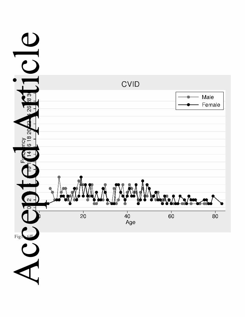
Fig1b.tiff
Acc
epte
d A
rticl
e

Fig2.tiff
Acc
epte
d A
rticl
e

Fig3.tiff
Acc
epte
d A
rticl
e

Fig4a.tiff
Acc
epte
d A
rticl
e
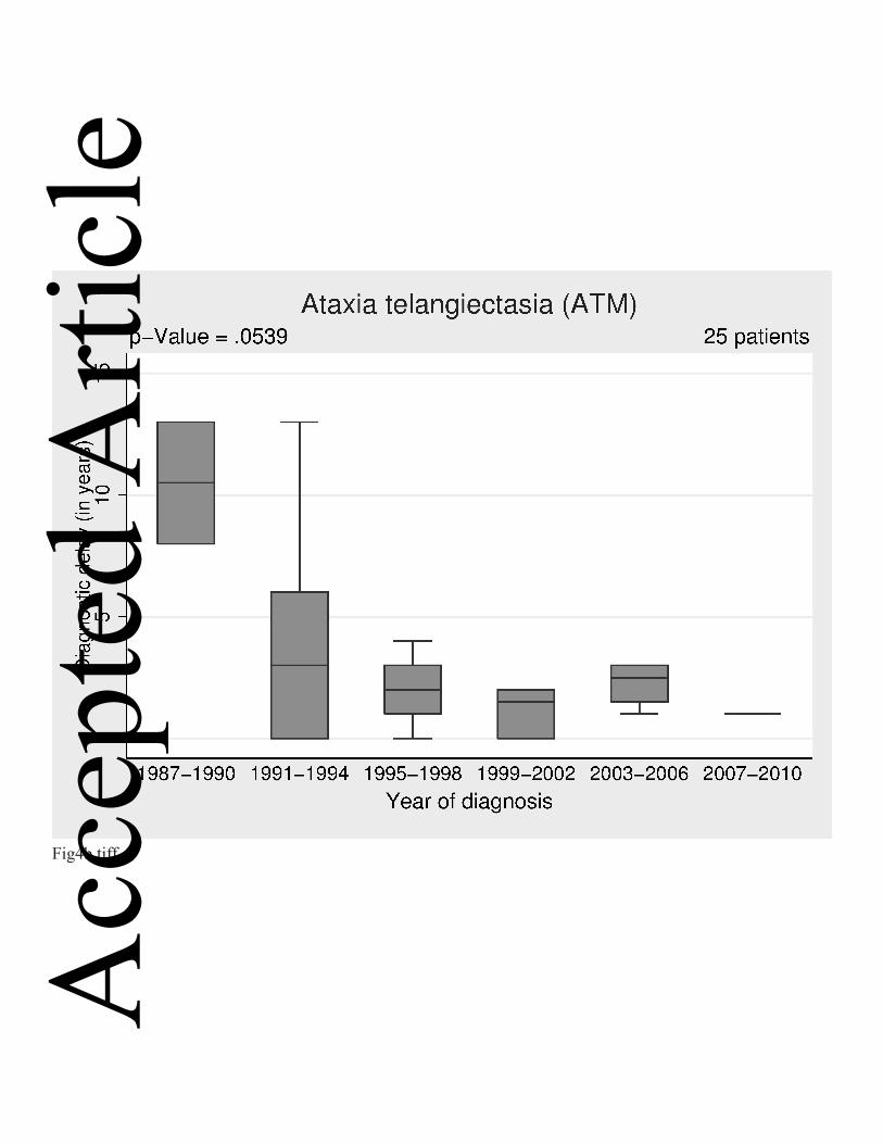
Fig4b.tiff
Acc
epte
d A
rticl
e

Fig4c.tiff
Acc
epte
d A
rticl
e


![[PID] PID Control - Good Tuning - A Pocket Guide](https://static.fdocuments.net/doc/165x107/577d2a661a28ab4e1ea914b1/pid-pid-control-good-tuning-a-pocket-guide.jpg)