The Gene 10 (UL49.5) - Journal of Virology - American Society for
12
JOURNAL OF VIROLOGY, Mar. 2002, p. 2952–2963 Vol. 76, No. 6 0022-538X/02/$04.000 DOI: 10.1128/JVI.76.6.2952–2963.2002 Copyright © 2002, American Society for Microbiology. All Rights Reserved. The Gene 10 (UL49.5) Product of Equine Herpesvirus 1 Is Necessary and Sufficient for Functional Processing of Glycoprotein M Jens Rudolph, 1 Christian Seyboldt, 1 Harald Granzow, 2 and Nikolaus Osterrieder 1 * Institute of Molecular Biology 1 and Institute of Infectology, 2 Friedrich-Loeffler-Institutes, Federal Research Centre for Virus Diseases of Animals, D-17498 Insel Riems, Germany Received 24 September 2001/Accepted 13 December 2001 The functional cooperation of equine herpesvirus 1 (EHV-1) glycoprotein M (gM) and the gene 10 (UL49.5) product was analyzed. Transient-transfection experiments using gM and UL49.5 expression plasmids as well as RK13 cell lines constitutively expressing UL49.5 (RK49.5) or gM (RKgM) demonstrated that the endo-- N-acetylglucosaminidase H (endo H)-resistant mature form of gM was detectable only after coexpression of the two proteins. Deletion of the EHV-1 UL49.5-homologous gene 10 in strain KyA resulted in a small-plaque phenotype and up to 190-fold-reduced virus titers. The growth defects of the mutant KyA49.5 virus, which were very similar to those of a gM-negative KyA virus, could be completely compensated for by growth of the mutant virus on RK49.5 cells or by repairing the deletion of gene 10 in the revertant virus KyA49.5R. Analysis of cells infected with the UL49.5-negative EHV-1 demonstrated that gM was not transported to the trans-Golgi network in the absence of the UL49.5 product. In contrast, gM was efficiently transported and processed to the endo H-resistant mature form in KyA49.5-infected RK49.5 cells. Furthermore, radioimmunoprecipitation experiments demonstrated that gM maturation was observed only if a 10,000-M r protein was coprecipitated with gM in KyA- or KyA49.5R-infected cells or virions. This protein was absent in cells infected with Ky49.5 or KyAgM, suggesting that it was the EHV-1 UL49.5 product. Taken together, our results demonstrate that the expression of the EHV-1 UL49.5 product is necessary and sufficient for gM processing and that it is required for efficient virus replication. The Alphaherpesvirus Equine herpesvirus 1 (EHV-1) encodes a least 13 glycoproteins, most of which share high homology to those of herpes simplex virus type 1 or of its closest relatives suid herpesvirus 1 (pseudorabies virus [PRV]) and bovine her- pesvirus 1 (BHV-1) (23, 29). PRV and BHV-1 together with EHV-1 and Varicella-zoster virus form the Varicellovirus genus within the Alphaherpesvirinae subfamily (30). Whereas the functions of several individual EHV-1 envelope proteins, such as glycoprotein B (gB), gC, gD, gE, and gM, have been ana- lyzed in some detail (4, 18, 20–22), studies on the functions of the putative gE/gI, gH/gL, or gM/UL49.5 complexes have not yet been performed. Complex formation between gM and the UL49.5 product, which is O glycosylated and therefore designated gN in PRV, Epstein-Barr virus (EBV), and human cytomegalovirus (HCMV), has been described for PRV (11), BHV-1 (31), EBV (14, 15), and HCMV (17). In the case of these viruses, gM and the UL49.5 product were shown to physically interact with each other by virtue of disulfide bonds that are probably formed between the first cysteine residue of the UL10 polypep- tide, which is conserved throughout the UL10 homologs of all Herpesviridae sequenced to date, and one of the two equally highly conserved cysteine residues present within the UL49.5 homologs (31). Further, it could be shown for PRV that dele- tion of gM led to an absence of gN in the envelope of mature extracellular virions. In contrast, gM was efficiently incorpo- rated into virions that lacked gN, and no growth defect after deletion of the UL49.5 (gN) gene from PRV was observed (11, 12). Similarly, deletion of the UL49.5 gene from HSV-1 or BHV-1 only marginally reduced virus titers (1, 16). In the case of EBV, gN processing was shown to be dependent on the expression of gM, whereas gM processing appeared to be un- affected in the absence of gN as assessed by experiments using CV-1 cells and T7 promoter-driven expression of gM and gN. Functional studies of the BLRF-1-negative EBV have demon- strated that gN is involved in an entry event following fusion and in virus egress from infected cells. The impaired egress was associated with an accumulation of nucleocapsids in condensed chromatin within the nuclei of infected cells (14). For HCMV, a member of the Betaherpesvirinae, transient-transfection ex- periments using gM and UL73 expression plasmids demon- strated that gM is necessary for processing and transport of the UL73 product, the UL49.5 (gN) homolog of HCMV (17). Recent studies have provided more insight in the roles that gM or the gM/UL49.5 complex and the gE/gI complex play in the life cycle of EHV-1. The EHV-1 UL49.5-homologous pro- tein is encoded by gene 10 of EHV-1. gM, a 50,000- to 55,000-M r component of the virus envelope, is encoded by gene 52 (29). Concomitant deletion of gM together with gE and gI in EHV-1 led to a massive impairment in secondary virus envelopment at membranes of the trans-Golgi network (TGN) and subsequent virus egress which could be repaired by growth on a cell line expressing gM. These results indicated that gM or gM/UL49.5 and the gE/gI complex serve partially overlapping but distinct functions in virus egress (27). The results obtained with EHV-1 gE/I/M triple mutants closely mimicked those with PRV, in which the absence of gE and gM was responsible for massive growth defects affecting egress and * Corresponding author. Mailing address: Institute of Molecular Biology, Friedrich-Loeffler-Institutes, Federal Research Centre for Vi- rus Diseases of Animals, Boddenblick 5a, D-17498 Insel Riems, Ger- many. Phone: 49-38351-7266. Fax: 49-38351-7151. E-mail: klaus [email protected]. 2952 Downloaded from https://journals.asm.org/journal/jvi on 02 January 2022 by 81.198.34.71.
Transcript of The Gene 10 (UL49.5) - Journal of Virology - American Society for

JOURNAL OF VIROLOGY, Mar. 2002, p. 2952–2963 Vol. 76, No. 60022-538X/02/$04.00�0 DOI: 10.1128/JVI.76.6.2952–2963.2002Copyright © 2002, American Society for Microbiology. All Rights Reserved.
The Gene 10 (UL49.5) Product of Equine Herpesvirus 1 Is Necessaryand Sufficient for Functional Processing of Glycoprotein M
Jens Rudolph,1 Christian Seyboldt,1 Harald Granzow,2 and Nikolaus Osterrieder1*Institute of Molecular Biology1 and Institute of Infectology,2 Friedrich-Loeffler-Institutes, Federal Research Centre for
Virus Diseases of Animals, D-17498 Insel Riems, Germany
Received 24 September 2001/Accepted 13 December 2001
The functional cooperation of equine herpesvirus 1 (EHV-1) glycoprotein M (gM) and the gene 10 (UL49.5)product was analyzed. Transient-transfection experiments using gM and UL49.5 expression plasmids as wellas RK13 cell lines constitutively expressing UL49.5 (RK49.5) or gM (RKgM) demonstrated that the endo-�-N-acetylglucosaminidase H (endo H)-resistant mature form of gM was detectable only after coexpression of thetwo proteins. Deletion of the EHV-1 UL49.5-homologous gene 10 in strain KyA resulted in a small-plaquephenotype and up to 190-fold-reduced virus titers. The growth defects of the mutant KyA�49.5 virus, whichwere very similar to those of a gM-negative KyA virus, could be completely compensated for by growth of themutant virus on RK49.5 cells or by repairing the deletion of gene 10 in the revertant virus KyA�49.5R. Analysisof cells infected with the UL49.5-negative EHV-1 demonstrated that gM was not transported to the trans-Golginetwork in the absence of the UL49.5 product. In contrast, gM was efficiently transported and processed to theendo H-resistant mature form in KyA�49.5-infected RK49.5 cells. Furthermore, radioimmunoprecipitationexperiments demonstrated that gM maturation was observed only if a 10,000-Mr protein was coprecipitatedwith gM in KyA- or KyA�49.5R-infected cells or virions. This protein was absent in cells infected with Ky�49.5or KyA�gM, suggesting that it was the EHV-1 UL49.5 product. Taken together, our results demonstrate thatthe expression of the EHV-1 UL49.5 product is necessary and sufficient for gM processing and that it isrequired for efficient virus replication.
The Alphaherpesvirus Equine herpesvirus 1 (EHV-1) encodesa least 13 glycoproteins, most of which share high homology tothose of herpes simplex virus type 1 or of its closest relativessuid herpesvirus 1 (pseudorabies virus [PRV]) and bovine her-pesvirus 1 (BHV-1) (23, 29). PRV and BHV-1 together withEHV-1 and Varicella-zoster virus form the Varicellovirus genuswithin the Alphaherpesvirinae subfamily (30). Whereas thefunctions of several individual EHV-1 envelope proteins, suchas glycoprotein B (gB), gC, gD, gE, and gM, have been ana-lyzed in some detail (4, 18, 20–22), studies on the functions ofthe putative gE/gI, gH/gL, or gM/UL49.5 complexes have notyet been performed.
Complex formation between gM and the UL49.5 product,which is O glycosylated and therefore designated gN in PRV,Epstein-Barr virus (EBV), and human cytomegalovirus(HCMV), has been described for PRV (11), BHV-1 (31), EBV(14, 15), and HCMV (17). In the case of these viruses, gM andthe UL49.5 product were shown to physically interact witheach other by virtue of disulfide bonds that are probablyformed between the first cysteine residue of the UL10 polypep-tide, which is conserved throughout the UL10 homologs of allHerpesviridae sequenced to date, and one of the two equallyhighly conserved cysteine residues present within the UL49.5homologs (31). Further, it could be shown for PRV that dele-tion of gM led to an absence of gN in the envelope of matureextracellular virions. In contrast, gM was efficiently incorpo-
rated into virions that lacked gN, and no growth defect afterdeletion of the UL49.5 (gN) gene from PRV was observed (11,12). Similarly, deletion of the UL49.5 gene from HSV-1 orBHV-1 only marginally reduced virus titers (1, 16). In the caseof EBV, gN processing was shown to be dependent on theexpression of gM, whereas gM processing appeared to be un-affected in the absence of gN as assessed by experiments usingCV-1 cells and T7 promoter-driven expression of gM and gN.Functional studies of the BLRF-1-negative EBV have demon-strated that gN is involved in an entry event following fusionand in virus egress from infected cells. The impaired egress wasassociated with an accumulation of nucleocapsids in condensedchromatin within the nuclei of infected cells (14). For HCMV,a member of the Betaherpesvirinae, transient-transfection ex-periments using gM and UL73 expression plasmids demon-strated that gM is necessary for processing and transport of theUL73 product, the UL49.5 (gN) homolog of HCMV (17).
Recent studies have provided more insight in the roles thatgM or the gM/UL49.5 complex and the gE/gI complex play inthe life cycle of EHV-1. The EHV-1 UL49.5-homologous pro-tein is encoded by gene 10 of EHV-1. gM, a 50,000- to55,000-Mr component of the virus envelope, is encoded bygene 52 (29). Concomitant deletion of gM together with gEand gI in EHV-1 led to a massive impairment in secondaryvirus envelopment at membranes of the trans-Golgi network(TGN) and subsequent virus egress which could be repaired bygrowth on a cell line expressing gM. These results indicatedthat gM or gM/UL49.5 and the gE/gI complex serve partiallyoverlapping but distinct functions in virus egress (27). Theresults obtained with EHV-1 gE/I/M triple mutants closelymimicked those with PRV, in which the absence of gE and gMwas responsible for massive growth defects affecting egress and
* Corresponding author. Mailing address: Institute of MolecularBiology, Friedrich-Loeffler-Institutes, Federal Research Centre for Vi-rus Diseases of Animals, Boddenblick 5a, D-17498 Insel Riems, Ger-many. Phone: 49-38351-7266. Fax: 49-38351-7151. E-mail: [email protected].
2952
Dow
nloa
ded
from
http
s://j
ourn
als.
asm
.org
/jour
nal/j
vi o
n 02
Jan
uary
202
2 by
81.
198.
34.7
1.
cell-to-cell spread in cultured cells (2, 3). More recent work onthe chicken Alphaherpesvirus Marek’s disease virus (MDV) hassuggested that one partner of gE/gI and/or gM/UL49.5 in cell-to-cell spread may be the product of the UL49 homolog(VP22), because MDV lacking gE, gI, or UL49 is nonviable incultured cells (7, 26).
This study was performed to analyze the effect of a deletionof the UL49.5-homologous gene 10 in EHV-1 with respect togrowth of a gE/gI-negative EHV-1 and the maturation of gM.Consistent with the absence of mature gM after infection ofcells with the UL49.5-negative virus, a small-plaque phenotypeand a 190-fold reduction of extracellular virus titers were ob-served in the absence of mature gM. The findings of this studydemonstrate for the first time that a UL49.5-homologous pro-tein is necessary for proper processing of its complex partner,the UL10 product, and for the function of gM and/or thegM/UL49.5 complex in virus egress and cell-to-cell spread.
MATERIALS AND METHODS
Virus and cells. Strain KyA (kindly provided by D. J. O’Callaghan, LouisianaState University, Shreveport) was used in this study. KyA, which lacks the gE andgI genes (9), was grown on RK13 cells propagated in Dulbecco’s minimal essen-tial medium (DMEM) supplemented with 10% fetal calf serum. For isolation ofa gene 10 (UL49.5)-negative KyA (KyA�49.5), 10 �g of recombinant plasmidp�49.5 was cotransfected with 1 to 10 �g of purified KyA DNA into RK13 cells.At 3 to 5 days after transfection, supernatants were harvested and plated on freshcells. Recombinant fluorescent virus plaques were identified under a fluores-cence microscope (Nikon), picked, and purified to homogeneity. Gene 10 rever-tant viruses were obtained after cotransfection of KyA�49.5 DNA and recom-binant plasmid pE49.5 into RK13 cells. Revertant viruses were isolated by plaquepurification of nonfluorescing virus plaques.
Plasmids. For construction of recombinant plasmid pc49.5, a PCR fragment(Table 1; Fig. 1) containing the EHV-1 gene 10 (UL49.5-homologous openreading frame [ORF] [29]) was cloned into the pcDNA3 vector (Invitrogen)under control of the HCMV immediate-early promoter-enhancer. To obtainrecombinant plasmid p�49.5, a 2.2-kbp HindIII-SphI genomic fragment wascloned into vector pTZ18R (Amersham-Pharmacia) (Fig. 1). The SphI site islocated at nucleotide position 146 of the 300-bp EHV-1 UL49.5-homologousgene 10. A 1.6-kbp fragment on the 3� side of the UL49.5 ORF was amplified byPCR (Table 1) and cloned as a KpnI fragment in the vector containing the 5�fragment. Finally, the green fluorescent protein (GFP) gene from plasmidpEGFP-N1 (Clontech) was released as an NspI-PstI fragment and ligated intothe vector cut with SphI and PstI (Fig. 1). Plasmid pE49.5 contains a 3.5-kbpgenomic fragment which encompasses gene 10 and was used for generation ofthe revertant virus (Fig. 1).
Generation of cell lines expressing EHV-1 UL49.5. Ten micrograms of recom-binant plasmid pc49.5 was transfected into RK13 cells, and cell clones developingunder DMEM containing 500 �g of G-418 (Calbiochem) per ml were isolatedand expanded for further analysis by cotransfection with recombinant plasmid
pcgM, which encodes EHV-1 gM under control of the HCMV immediate-earlypromoter-enhancer (22).
Single-step growth kinetics and plaque diameter determinations. Single-stepgrowth kinetics were determined in triplicate by infecting 105 RK13, RK49.5, orRKgM cells (27) at a multiplicity of infection (MOI) of 3. Virus was allowed toattach for 2 h on ice, followed by a penetration period of 2 h at 37°C. At theindicated times after the temperature shift, supernatants of infected cells wereharvested, and viral titers were determined by plating onto RK13 cells (27).Plaque diameters were measured after titration of virus mutants on different celllines and 3 days of incubation at 37°C under a methylcellulose overlay. Cells werefixed with 5% formaldehyde and stained with crystal violet. For each combina-tion of virus and cells, 150 plaques were measured and the average plaquediameters were determined. Values were calculated and compared to KyAplaque diameters, which were set to 100%. Average percentages of plaquediameters and standard deviations were determined from three independentexperiments.
Electron microscopy. For ultrastructural analyses, RK13 or RK49.5 cells wereinfected at an MOI of 1, fixed at different times postinfection (p.i.) (16 to 20 h)for 60 min with 2.5% glutaraldehyde buffered in 0.1 M Na-cacodylate (pH 7.2,300 mosM) (Merck). Cells were then scraped off the flask, pelleted by low-speedcentrifugation, and embedded in LMP agarose (Biozym). Small pieces werepostfixed in 1.0% aqueous OsO4 (Polysciences Europe) and stained with uranylacetate. After stepwise dehydration in ethanol, cells were cleared in propyleneoxide, embedded in Glycid Ether 100 (Serva), and polymerized at 59°C for 4days. Ultrathin sections were counterstained with uranyl acetate and lead saltsand were examined by electron microscopy (Philips EM 400T).
Antibodies. Monoclonal antibodies (MAbs) directed against EHV-1 gM werekindly provided by Lindsey Day and Richard Killington, University of Leeds,Leeds, United Kingdom (5, 27). As a TGN marker, a �-adaptin MAb (Sigma)was used. Polyclonal rabbit anti-gM antisera 047 and 048 were produced afterinjection of the 047 or 048 peptide (Table 1), coupled to keyhole limpet hemo-cyanin (Sigma) and suspended in Freund’s complete (first immunization) orincomplete (booster immunizations) adjuvant, for a total of six times into a NewZealand White rabbit (Charles River). An anti-gD 20-mer antibody (8) (kindlyprovided by Dennis J. O’Callaghan) and the anti-gB 3F8 MAb (20) were used ascontrol antibodies. Secondary peroxidase-conjugated anti-mouse immunoglobu-lin G (IgG) antibodies (Sigma) as well as fluorescein isothiocyanate-conjugatedanti-mouse IgG2b (Sigma) and Cy3-conjugated anti-mouse IgG1 antibodies(Amersham Biosciences) were used according to the manufacturers’ instructions.
IIF, Western blotting, and RIPA. For indirect immunofluorescence (IIF),RK13 cells were grown on six-well plates and subsequently infected at an MOIof 0.01 or transfected with gM or UL49.5 expression plasmids. Cells were fixedwith acetone at 48 to 72 h p.i. or after transfection, and IIF and confocal laserscanning microscopy using a Zeiss LSM510 confocal microscope were doneexactly as described previously (19, 23, 27). For Western blot analyses, cells weretransfected or infected at an MOI of 2 with the various viruses. At 48 h aftertransfection or 16 h after infection, cell lysates were prepared. Extracellularvirions were purified by sucrose gradient centrifugation (19, 22). Cell lysates wereadjusted to equal protein concentrations of 5 mg per ml as determined with abicinchoninic acid kit (Pierce) (24). Samples were heated at 56°C for 2 min in thepresence of 2-mercaptoethanol unless otherwise stated, separated by sodiumdodecyl sulfate-polyacrylamide gel electrophoresis (SDS-PAGE), and trans-ferred to nitrocellulose membranes (Schleicher & Schuell) by the semidry
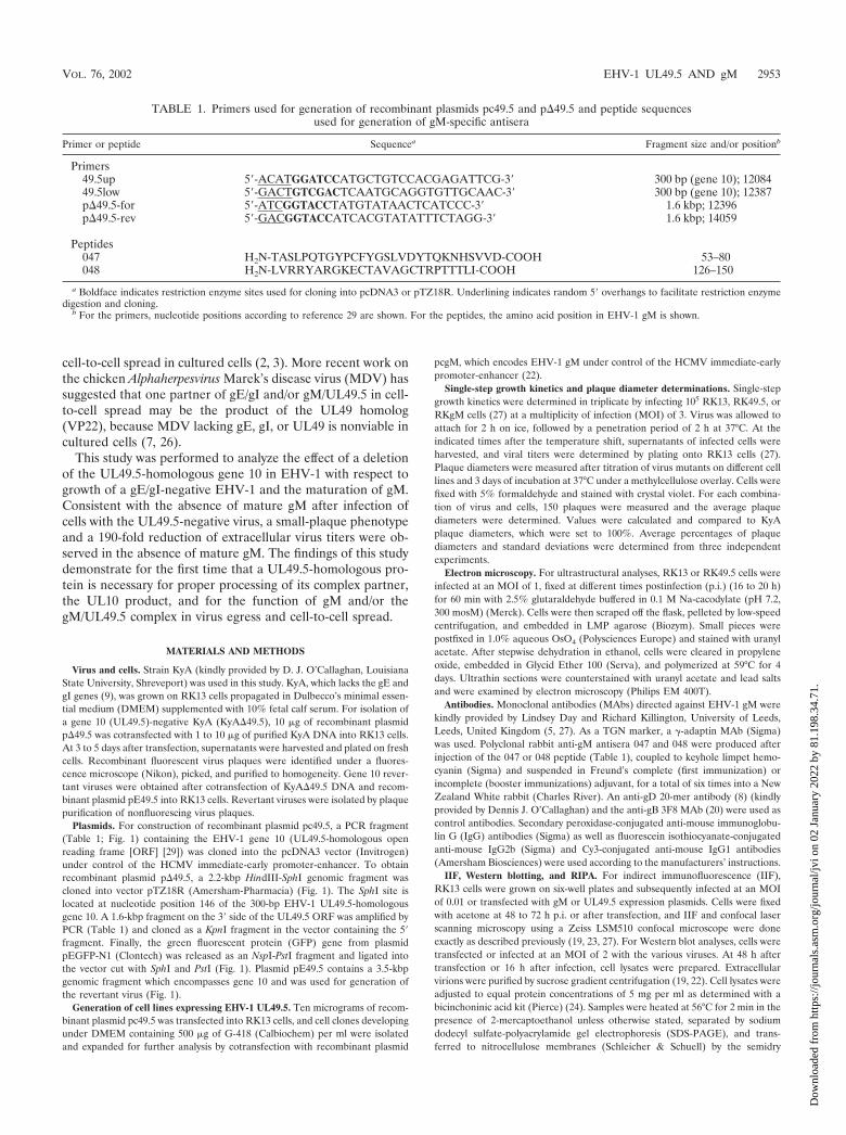
TABLE 1. Primers used for generation of recombinant plasmids pc49.5 and p�49.5 and peptide sequencesused for generation of gM-specific antisera
Primer or peptide Sequencea Fragment size and/or positionb
Primers49.5up 5�-ACATGGATCCATGCTGTCCACGAGATTCG-3� 300 bp (gene 10); 1208449.5low 5�-GACTGTCGACTCAATGCAGGTGTTGCAAC-3� 300 bp (gene 10); 12387p�49.5-for 5�-ATCGGTACCTATGTATAACTCATCCC-3� 1.6 kbp; 12396p�49.5-rev 5�-GACGGTACCATCACGTATATTTCTAGG-3� 1.6 kbp; 14059
Peptides047 H2N-TASLPQTGYPCFYGSLVDYTQKNHSVVD-COOH 53–80048 H2N-LVRRYARGKECTAVAGCTRPTTTLI-COOH 126–150
a Boldface indicates restriction enzyme sites used for cloning into pcDNA3 or pTZ18R. Underlining indicates random 5� overhangs to facilitate restriction enzymedigestion and cloning.
b For the primers, nucleotide positions according to reference 29 are shown. For the peptides, the amino acid position in EHV-1 gM is shown.
VOL. 76, 2002 EHV-1 UL49.5 AND gM 2953
Dow
nloa
ded
from
http
s://j
ourn
als.
asm
.org
/jour
nal/j
vi o
n 02
Jan
uary
202
2 by
81.
198.
34.7
1.

method (13). Free binding sites on the sheets were blocked by addition of 10%skim milk in phosphate-buffered saline containing 0.05% Tween before theantibodies (suspended in phosphate-buffered saline containing 0.05% Tween)were added. Bound antibodies were detected with anti-rabbit (or anti-mouse)IgG–peroxidase conjugates (Sigma) and visualized by enhanced chemilumines-cence (ECL; Pharmacia-Amersham). For radioimmunoprecipitation (RIPA),106 infected or mock-infected cells (MOI � 1) were labeled at 16 h p.i. for 20 minwith 200 �Ci of Tran35S-label (ICN) after incubation for 20 min in Met- andCys-free DMEM (Gibco-BRL). Radiolabeled extracellular virions were pre-pared by continuous labeling of infected RK13 cells with 250 �Ci of Tran35S-label for 36 h and subsequent sucrose gradient purification. Cells or virions werelysed with detergent buffer (24), and precipitations after preclearing of the celllysates were done using a mixture of 2 �l of the 047 peptide serum and 2 �l ofthe 048 peptide serum or with 50 �l of hybridoma supernatant of MAb 3F8.Antigen-antibody complexes were pelleted using Pansorbin cells (Calbiochem)according to the supplier’s instructions. After three thorough washes in RIPAwashing buffer (25), complexes were released from Pansorbin cells by addition ofsample buffer and heating to 56°C for 2 min before SDS-PAGE.
Deglycosylation of cell lysates. Deglycosylation experiments were performedby suspension of infected-cell lysates (from 106 cells) in 100 �l of de-N-degly-cosylation buffer (50 mM K3PO4 [pH 7.2], 50 mM EDTA, 0.6% [vol/vol]CHAPS, 0.1% SDS) and subsequent addition of peptide-N-glycosidase(PNGase) F (0.4 U) or endo H (2 mU) (Roche Biochemicals). Control lysates
were left untreated. After 16 h of incubation at 37°C, lysates were separated bySDS–10% PAGE, transferred to nitrocellulose, and examined by Western blot-ting. De-O-glycosylations were done with neuraminidase and O-glycosidase ex-actly as previously described (12, 19), except that immunoprecipitates with the047 and 048 rabbit antibodies or the anti-gD antibody as a control were used.
RESULTS
Coexpression of EHV-1 UL49.5 and gM results in gM mat-uration. The results of preliminary transfection experimentsusing a gM expression plasmid had suggested that the coex-pression of the EHV-1 UL49.5 gene can result in gM matura-tion. To analyze the putative UL49.5-gM interaction in moredetail, RK13 cells were cotransfected with plasmids expressingeither EHV-1 UL49.5 (pc49.5) or gM (pcgM) (22) or werecotransfected with both expression plasmids. Whereas no re-activity with the gM-specific MAb P18/A8 (5, 27) was observedin cells transfected with pc49.5, the 46,000- to 48,000-Mr endoH-sensitive form of gM was observed in cells transfected withpcgM only (27) (data not shown). In contrast, in RK13 cells
FIG. 1. (A) Schematic illustration of the organization of the approximately 140-kbp KyA genome and the BamHI restriction map. (B) The3.5-kbp EcoRI fragment (pE49.5) which contains the UL49.5-homologous gene 10 of EHV-1. (C) Plasmid pc49.5 was generated by insertion ofthe EHV-1 UL49.5-homologous gene 10 into vector pcDNA3 (Invitrogen). (D) Construction and structure of plasmid p�49.5 used for generationof the UL49.5-negative virus mutant. Restriction enzyme sites: B, BamHI; E, EcoRI; K, KpnI; N, NspI; S, SphI.
2954 RUDOLPH ET AL. J. VIROL.
Dow
nloa
ded
from
http
s://j
ourn
als.
asm
.org
/jour
nal/j
vi o
n 02
Jan
uary
202
2 by
81.
198.
34.7
1.

cotransfected with both pc49.5 and pcgM, both high-mannose-containing gM and, predominantly, the mature 50,000- to55,000-Mr gM moiety were detectable (Fig. 2A). The sameobservation, i.e., maturation of gM to the 50,000- to 55,000-Mr
protein, was made when cell line RKgM (27) was transfectedwith pc49.5 or when a cell line constitutively expressing EHV-1UL49.5 (RK49.5) was transfected with pcgM (Fig. 2A). Incontrast, the endo H-resistant 50,000- to 55,000-Mr gM proteinwas absent in RKgM cells cotransfected with a gB expressionplasmid (pcgB) (20). The expression of gB in pcgB-transfectedRK13 cells but not in those transfected with pcgM or pc49.5was verified by Western blot analysis using MAb 3F8 (20).Especially the di- and oligomeric forms of EHV-1 gB weredetectable in the respective cell lysates (Fig. 2B), because thecell lysates had been heated at 56°C, which leaves most of thegB complexes intact (20). From a number of transfection ex-periments it became apparent that gM processing was muchmore efficient after cotransfection of UL49.5 and gM expres-sion plasmids than after transfection of gM- or UL49.5-ex-pressing cells with the complementary expression plasmid. Anexplanation for this observation may be that efficient process-ing of gM requires cotranslational complex formation, whichcan be impaired if one of the complex partners is preexistent.From the results of the transfection experiments, we concludedthat processing of EHV-1 gM into the mature endo H-resistantform requires the UL49.5 product.
Generation of an RK13 cell line constitutively expressingUL49.5. The full maturation of gM in the presence of theUL49.5 product was exploited to identify and isolate cell clonesconstitutively expressing UL49.5, because an antibody againstthe UL49.5 protein of EHV-1 was not available. After trans-fection of pc49.5 into RK13 cells, G418-resistant cell cloneswere isolated and subsequently transfected with pcgM.UL49.5-expressing cell clones were identified by the detectionof the mature 50,000- to 55,000-Mr gM moiety (Fig. 2A). OneG418-resistant cell clone (clone 4) in which high quantities ofmature gM were observed after transfection of pcgM wastermed RK49.5 and used for all further transcomplementationand deglycosylation experiments (Fig. 2A and C).
Generation and analysis of a UL49.5-negative KyA. Cotrans-fection of RK13 cells with recombinant plasmid p�49.5, inwhich gene 10, the UL49.5-homologous gene, was replacedwith the GFP gene, and KyA DNA resulted in virus progenythat induced the formation of fluorescent plaques. The trans-fection progeny viruses were plaque purified on RK49.5 cellsthree times until all plaques stained homogenously green un-der the fluorescence microscope. Southern blot analysis con-firmed the correct insertion of the GFP ORF instead of theUL49.5-homologous gene 10 in the UL49.5-negative KyA,which was termed KyA�49.5 (Fig. 1). A gene 10 revertantvirus, KyA�49.5R, was generated by cotransfection ofKyA�49.5 DNA with recombinant plasmid pE49.5 (Fig. 1) intoRK13 cells and selection for nonfluorescing plaques. The cor-rect reinsertion of the UL49.5 gene in the revertant virus wasalso confirmed by Southern blot analysis (data not shown).
KyA�49.5, KyA�49.5R, KyA�gM (27), or parental KyAvirus was used to infect RK13 cells and to perform RIPA withthe 047 and 048 anti-gM peptide antisera (Table 1) (27). InRK13 cells, a 10,000-Mr protein was coprecipitated with thetwo gM-specific proteins after infection with the parental KyA
FIG. 2. Western blot analyses of RK13, RK49.5, and RKgM cellstransfected with pc49.5, pcgM, or pcgB. Forty-eight hours after trans-fection, cells were harvested and lysed, and protein extracts wereseparated by SDS–10% PAGE followed by Western blotting usinganti-gM antibody P18/A8 (A and C) or anti-gB antibody 3F8 (B). Cellslines used for the transfections are given. The gM precursor (circle),the high-mannose gM (open arrowhead), and the fully glycosylated gM(solid arrowhead) are indicated. Individual cell clones which constitu-tively expressed the UL49.5 protein were identified by the presence ofprocessed gM after transfection of pcgM (C). Cell line 4 was chosen forfurther studies and termed RK49.5. Mature and immature gM areindicated by solid and open arrowheads, respectively (A and C); gB-specific bands are marked by a bracket (B). The sizes of a prestainedmolecular weight marker (SeaBlue; Novex) are given in thousands.
VOL. 76, 2002 EHV-1 UL49.5 AND gM 2955
Dow
nloa
ded
from
http
s://j
ourn
als.
asm
.org
/jour
nal/j
vi o
n 02
Jan
uary
202
2 by
81.
198.
34.7
1.

or UL49.5 revertant virus, but no such protein was observed inprecipitates of RK13 cells infected with KyA�49.5 or KyA�gM(Fig. 3A). In addition, only the immature form of gM wasdetectable in RK13 cells infected with the UL49.5-negativemutant virus (Fig. 3A), whereas both the immature and matureforms of gM were precipitated with the gM-specific antibody incells infected with KyA or revertant KyA�49.5R virus. The10,000-Mr protein could also be coprecipitated with the gM-specific 047 or 048 antiserum from extracellular KyA orKyA�49.5R virions purified over sucrose gradients, but notfrom KyA�49.5 virions (Fig. 3B). Because the 10,000-Mr pro-tein was present in immunoprecipitates of wild-type KyA orUL49.5 revertant viruses but not in that of KyA�49.5, andbecause the apparent Mr of the putative UL49.5 product cor-responded well to the calculated Mr of 10,801, we concludedthat gM and the UL49.5 protein product form a heterodimer ininfected cells and virions. The fact that the apparent Mr of theUL49.5 product remained unaltered after treatment with neur-aminidase and O-glycosidase and after treatment with PNGaseF (data not shown) suggested that the EHV-1 UL49.5 productis not glycosylated.
The incomplete processing of gM in the absence of theUL49.5 product was also examined by performing deglycosy-lation experiments with RK13 and RK49.5 cells infected witheither KyA or KyA�49.5. Whereas the endo H-resistant50,000- to 55,000-Mr gM moiety was not observed in RK13cells infected with KyA�49.5, the mature glycoprotein wasreadily detected in KyA�49.5-infected complementing RK49.5cells (data not shown). Because EHV-1 gM was not fully pro-cessed in the absence of the UL49.5 product, it was absent inKyA�49.5 virions purified from RK13 cells, whereas the72,000- to 75,000-Mr subunit of gB was readily detected inpurified KyA�49.5 virions by using MAb 3F8 after heating ofthe samples to 95°C (Fig. 3C). In contrast, gM was efficientlyincorporated into UL49.5-negative virions by growth on com-plementing RK49.5 cells (Fig. 3C), demonstrating that incor-poration of gM into extracellular mature virions requires theUL49.5 product.
The intracellular distribution of gM in the absence of theUL49.5 product was also examined by colocalization studieswith IIF and a TGN-specific anti-�-adaptin MAb and anti-gMor gB MAbs, followed by confocal laser scanning microscopy(19, 23). Whereas gB localization to the TGN was readilydetectable in both KyA- and KyA�49.5-infected cells (data notshown), gM was detectable in this compartment in KyA-in-fected cells (Fig. 4A to C) but not in RK13 cells infected withKyA�49.5, where gM exhibited a strong signal around thenuclear rim and was dispersed throughout the cytoplasm, andno colocalization of �-adaptin and gM signals was observed(Fig. 4D to F). The observed gM distribution was highly rem-iniscent of that previously demonstrated for mutant EHV-1gM molecules, which were retained in the endoplasmic retic-ulum (27). Colocalization of gM with �-adaptin signals wasrestored in RK49.5 cells infected with KyA�49.5 (Fig. 4G to I).From these results we concluded that incomplete processing ofEHV-1 gM in the absence of the UL49.5 product was causedby an inhibition of gM transport to the TGN.
KyA�49.5 is impaired in cell-to-cell spread virus and egress.The next series of experiments addressed the growth proper-ties of KyA�49.5 in comparison to parental KyA or revertant
virus. Plating of the different viruses on RK13 and RK49.5 cellsdemonstrated that cell-to-cell spread of the UL49.5-negativemutant was severely impaired in the absence of the protein,and only 37% of the plaque diameters of parental KyA orrevertant virus were reached (Fig. 5). This defect in cell-to-cellspread of infectivity was completely restored by plating ofKyA�49.5 on RK49.5 cells constitutively expressing theUL49.5 protein (Fig. 5) but not on RKgM cells, which consti-tutively express gM and which were able to rescue plaquediameters of the gM-negative KyA mutant (27).
Single-step growth kinetics of mutant KyA�49.5 virus andwild-type KyA or the UL49.5 revertant virus were determined.The individual viruses were used to infect RK13, RK49.5, orRKgM cells at an MOI of 3, and the results of the experimentsare summarized in Fig. 6. KyA�49.5 grown on RK13 cellsexhibited a reduction of virus titers of up to 190-fold comparedto titers of parental KyA or rescuant KyA�49.5R virus (Fig. 6).This defect in virus maturation and egress was virtually com-pletely reversed when KyA�49.5 was grown on complementingRK49.5 cells but not when it was grown on RKgM cells (Fig.6). On RKgM cells, the impaired growth of the gM-negativeKyA�gM virus which was observed on RK13 cells, and wasvirtually indistinguishable from that of the UL49.5-negativeKyA�49.5 virus, could be completely rescued (Fig. 6). Fromthese results we concluded that efficient growth of the gE/gI-negative EHV-1 strain KyA in cultured cells requires theUL49.5 product and that deletion of the gene 10 (UL49.5-homologous) ORF resulted in a massive impairment of cell-to-cell spread and virus egress that was identical to that of agM-negative KyA virus.
KyA�49.5 virus maturation is blocked at the stage of sec-ondary envelopment. To further examine the reason for themassive impairment of virus egress and cell-to-cell spread ofUL49.5-negative KyA, ultrastructural studies were performed.These studies revealed that UL49.5-negative KyA�49.5 exhib-ited a clearly visible defect in secondary envelopment at pre-sumably TGN membranes. Nucleocapsid formation in infect-ed-cell nuclei, primary budding at the inner lamella of thenuclear membrane, and de-envelopment at the outer lamellaof the nuclear membrane, however, did not appear to be dif-ferent from those observed in KyA-infected cells (Fig. 7A andB). Budding of nucleocapsids at membranes of the TGN wasobserved only very rarely, and de-enveloped nucleocapsids ac-cumulated in the Golgi region (Fig. 7C). Higher magnificationof the accumulated nucleocapsids revealed a clearly detectablerim of fuzzy material, presumably tegument proteins, aroundcytoplasmic nucleocapsids which was similar to that observedafter deletion of gM (Fig. 7D) (27). Very rarely, virions re-leased into the extracellular space were observed (Fig. 7E). Incontrast, replication of KyA�49.5 on complementing RK49.5cells was morphologically indistinguishable from that shownfor parental KyA or the wild-type RacL11 virus (10, 27). Afterprimary envelopment by budding at the inner lamella of thenuclear membrane, nucleocapsids were released into the cyto-plasm and received their final envelope by budding at mem-branes in the Golgi region (Fig. 8A and B). Virus-containingvesicles (Fig. 8C) released their content by fusion at the cellmembrane, and a large number of virions were visible in theextracellular space (Fig. 8D and E). Taken together, the elec-tron microscopic studies demonstrated that the impairment of
2956 RUDOLPH ET AL. J. VIROL.
Dow
nloa
ded
from
http
s://j
ourn
als.
asm
.org
/jour
nal/j
vi o
n 02
Jan
uary
202
2 by
81.
198.
34.7
1.

FIG. 3. (A) RIPA analysis of RK13 cells infected with wild-type KyA, the UL49.5- (gene 10)-negative KyA�49.5 mutant, the UL49.5 revertantvirus, or gM-negative KyA�gM (27). (B) RIPA of purified radiolabeled KyA, KyA�49.5, or KyA�49.5R virions The antibodies used forprecipitations were anti-gB antibody 3F8 and the anti-gM polyclonal antibodies 047 and 048. Mature and immature gM are indicated by full andopen arrowheads, respectively; 2-mercaptoethanol-resistant gM dimers (24, 27) are indicated by an asterisk. The putative UL49.5 (gene 10)product is indicated by an arrow. The sizes of a molecular weight marker (14C marker; Gibco-BRL) are given in thousands. (C) Western blotanalysis of purified KyA and KyA�49.5 virions prepared from RK13 or RK49.5 cells by using anti-gM MAb P18/A8 or anti-gB MAb 3F8. Whereasthe 72,000- to 75,000-Mr large subunit of gB could be detected with MAb 3F8 in all virion preparations, gM was detected in KyA virions andKyA�49.5 virions prepared from RK49.5 cells. gM was absent from UL49.5-negative virions prepared on RK13 cells. Samples were heated for 2min at 56°C for detection of gM and for 3 min at 95°C for detection of gB. In the first lane, KyA-infected RK13 cells were loaded. The sizes ofa prestained molecular weight marker (SeaBlue; Novex) are given in thousands.
VOL. 76, 2002 EHV-1 UL49.5 AND gM 2957
Dow
nloa
ded
from
http
s://j
ourn
als.
asm
.org
/jour
nal/j
vi o
n 02
Jan
uary
202
2 by
81.
198.
34.7
1.

KyA�49.5 in virus egress was caused by a defect in secondaryenvelopment of virions. This defect appeared to be ultrastruc-turally identical to that of a gM-negative KyA (27) and couldbe reversed by growth of the virus on a complementing cellline.
DISCUSSION
Experiments to elucidate the function of the EHV-1 UL49.5protein and its cooperation with gM were conducted. It couldbe demonstrated that maturation of gM to the fully glycosy-lated and functional form of the glycoprotein requires theproduct of the UL49.5-homologous ORF of EHV-1, gene 10.This conclusion is drawn from the results of cotransfection
experiments using gM- and UL49.5-expressing plasmids, anal-ysis of glycosylation patterns of gM in cells infected with aUL49.5-negative EHV-1, the growth properties of a UL49.5-negative mutant in cultured cells, and RIPA experimentswhich showed that a 10,000-Mr protein was coprecipitated withgM in infected cells and mature extracellular virions. A func-tional impairment of the larger complex partner gM or thegM/UL49.5 complex was shown for the first time to be depen-dent on the presence of the smaller complex partner, UL49.5.
The Alphaherpesvirinae encode at least six (glyco)proteinswhich form hetero-oligomers in the virus envelope. Thesecomplexes comprise the gE/gI complex, the gH/gL complex,and a complex between gM and the UL49.5 product which is
FIG. 4. Confocal laser scanning microscopy of RK13 (A to F) or RK49.5 (G to I) cells infected with KyA (A to C) or KyA�49.5 (D to I). gMwas detected with MAb P18/A8 and visualized with anti-mouse IgG1–Cy3 conjugate (A, D, and G), and TGN vesicles were stained with �-adaptinMAb followed by anti-mouse IgG2b–fluorescein isothiocyanate antibody (B, E, and H). Merges of the signals recorded separately for each channel(green, 505 to 530 nm; red, �585 nm) are shown in panels C, F, and I. The yellow color in C and G indicates colocalization of gM and �-adaptinsignals. Panels represent views of 60 by 60 �m.
2958 RUDOLPH ET AL. J. VIROL.
Dow
nloa
ded
from
http
s://j
ourn
als.
asm
.org
/jour
nal/j
vi o
n 02
Jan
uary
202
2 by
81.
198.
34.7
1.

held together by disulfide bonds (11, 28, 31). The UL49.5product is O glycosylated in the case of PRV, in which thecomplex formed between the two highly conserved ORFs,present in all herpesviruses identified to date, was first de-scribed. The UL49.5 product does not appear to carry O-glycans in BHV-1 but does so in the members of the Betaher-pesvirinae (HCMV) and Gammaherpesvirina (EBV) (15, 17,31). Our previous studies and the results reported here maysuggest that the UL49.5 product encoded by gene 10 (29) is notglycosylated in EHV-1 but forms a disulfide-linked complexwith gM, as evidenced by altered electrophoretic mobilities ofimmunoprecipitates obtained with anti-gM antibodies undernonreducing conditions (24) and by analysis of immunopre-cipitates by using gM antibodies, which contained a 10,000-Mr
protein which was absent in both gM- and UL49.5-negativeviruses. Attempts to unanimously identify this protein as theUL49.5 product by using a rabbit polyclonal antibody havefailed, and our laboratory currently focuses on the generationof anti-UL49.5 MAbs to define the 10,000-Mr polypeptide co-immunoprecipitated with gM.
The absence of the EHV-1 UL49.5 product had profoundeffects on the maturation of its complex partner, gM, and alsoon virus growth in cultured cells. Using EHV-1 strain KyA,which lacks the gE and gI genes (9), the critical and yet par-tially redundant functions of gM or the gM/UL49.5 complex in
virus egress and cell-to-cell spread could be demonstrated (27).The more pronounced effect of a deletion of gM in a gE/gI-negative background is obviously caused by partially overlap-ping although distinct functions of the glycoproteins in virusegress and cell-to-cell spread, which has been shown not onlyfor EHV-1 but also for PRV (2, 3, 27). Using the same systemof a concomitant absence of gM or the gM/UL49.5 complexand the gE/gI complex, the consequences of a deletion ofUL49.5 in strain KyA were analyzed. Coexpression of UL49.5and gM resulted in complete processing of the multiply hydro-phobic gM, whereas absence of the UL49.5 product led to an
FIG. 5. Plaque diameters of KyA, KyA�49.5, KyA�49.5R, andKyA�gM on RK13 and RK49.5 cells. Shown are means and standarddeviations of diameters of 150 plaques measured for each virus-cellline combination. Plaque diameters of KyA were set to 100%.
FIG. 6. Single-step growth kinetics of KyA (}), KyA�49.5 (■ ),KyA�49.5R (Œ), and KyA�gM (�) on RK13, RK49.5, or RKgM cells.At the indicated times p.i., virus titers were determined on RK13 cellsas described in Materials and Methods. Shown are means and standarddeviations from three independent experiments.
VOL. 76, 2002 EHV-1 UL49.5 AND gM 2959
Dow
nloa
ded
from
http
s://j
ourn
als.
asm
.org
/jour
nal/j
vi o
n 02
Jan
uary
202
2 by
81.
198.
34.7
1.

FIG. 7. Electron micrographs of RK13 cells infected with KyA�49.5 at 16 h p.i. Budding of nucleocapsids of the UL49.5-negative mutant atthe inner lamella of the nuclear membrane and the presence of enveloped virions in the perinuclear space (A), as well as de-envelopment at theouter lamella of the nuclear membrane (B), were visible. Cytoplasmic nucleocapsids released from infected-cell nuclei accumulated in the vicinityof Golgi membranes (C). Higher magnification demonstrated fuzzy material, probably representing tegument proteins, around cytoplasmicnucleocapsids (D). An overview of a section of an infected cell with only few virions in secretory vesicles or the extracellular space is also shown(E). Bars, 250 nm (A, B, and D) and 1 �m (C and E).
2960
Dow
nloa
ded
from
http
s://j
ourn
als.
asm
.org
/jour
nal/j
vi o
n 02
Jan
uary
202
2 by
81.
198.
34.7
1.

FIG. 8. Electron micrographs of RK13 cells infected with KyA�49.5 on complementing RK49.5 cells at 16 h p.i. Various stages of normalEHV-1 morphogenesis can be seen. These stages included budding of nucleocapsids at the inner lamella of the nuclear membrane andde-envelopment at the outer lamella of the nuclear membrane (A), naked nucleocapsids in the cytoplasm (B), and secondary envelopment ofcytoplasmic virions at Golgi membranes leading to enveloped mature virions in secretory vesicles (C) Particles in secretory vesicles were finallyreleased into the extracellular space (D). A lower-magnification overview of a section of an infected cell is also shown (E); note the high numberof released virions at the cell surface. Bars, 500 nm (A to C) or 1 �m (D and E).
2961
Dow
nloa
ded
from
http
s://j
ourn
als.
asm
.org
/jour
nal/j
vi o
n 02
Jan
uary
202
2 by
81.
198.
34.7
1.

incomplete trimming of sugar moieties of gM in infected andstably or transiently transfected cells. These findings and theabsence of gM processing and transport to the TGN in cellsinfected with the UL49.5-negative mutant demonstrated thatexpression of the UL49.5 product is both necessary and suffi-cient for functional maturation of gM to the endo H-resistantglycoprotein. To our knowledge, incomplete gM maturation inthe absence of its complex partner is unprecedented in anyother member of the Herpesviridae. It was previously shown,however, that processing of the smaller complex partner gNwas dependent on gM expression in EBV and HCMV (15) andthat EBV mutants having a gN deletion exhibited reducedgrowth in cultured cells (14). In contrast, deletion of theUL49.5 product in PRV did not lead to altered growth prop-erties of the mutant virus in vitro, whereas deletion of gM ledto an approximately 50-fold decrease of virus titers and re-duced plaque diameters. Moreover, gM appeared to be fullyprocessed and was incorporated into extracellular virus parti-cles, whereas gN was absent from virions which were alsolacking gM (6, 11). From these results it appears that gM butnot gN (UL49.5) or the gM/gN complex of PRV is the func-tional entity. Because EHV-1 gM does not reach the TGN andsecretory vesicles in the absence of its complex partner, it isconsequently absent from extracellular virus particles. There-fore, the situation in EHV-1 is fundamentally different fromthat observed in PRV with regard to the UL49.5 protein, gM,and the complex between the two envelope constituents. Be-sides the structural consequences, deletion of either gM or theUL49.5 product, at least in a gE/gI-negative virus, led to es-sentially identical phenotypes in cultured cells. These findingsleave several ways for interpretation, but it appears that thefunction of the EHV-1 complex partners is more likely toreside in the formation of the complex than in action of theindividual proteins, as is the case in PRV. This interpretationis also based on the ultrastructural investigations, which re-vealed a block of virus egress at the stage of secondary envel-opment in the trans-Golgi region and virus egress for bothKyA�49.5 and KyA�gM. In addition, cell-to-cell spread de-fects of the UL49.5- and gM-negative KyA mutants were vir-tually identical, suggesting that fully processed gM is alsoneeded for direct transmission of infectivity from an infectedcell to a neighboring uninfected cell. Further studies will focuson the physical interaction between the EHV-1 UL49.5 proteinand its complex partner by targeted mutagenesis of cysteineresidues that are predicted to be involved in the complex for-mation between the two virion components. These studies willbe necessary to investigate whether physical interaction is nec-essary for the function of the individual proteins or the com-plex formed between these highly conserved proteins at vari-ous stages of EHV-1 maturation and egress.
ACKNOWLEDGMENTS
The skillful technical assistance of Kerstin Wink and Petra Meyer isgratefully acknowledged. E. Zorn and H. Stephan helped with theartwork. We thank Dennis J. O’Callaghan for providing strain KyAand anti-gD 20-mer antibody, and we thank Lindsey Day and RichardA. Killington for the generous gift of anti-gM MAbs.
This study was supported by DFG grant Os 143/2 to N.O.
REFERENCES
1. Adams, R., C. Cunningham, M. D. Davison, C. A. MacLean, and A. J.Davison. 1998. Characterization of the protein encoded by gene UL49A ofherpes simplex virus type 1. J. Gen. Virol. 79:813–823.
2. Brack, A. R., J. M. Dijkstra, H. Granzow, B. G. Klupp, and T. C. Metten-leiter. 1999. Inhibition of virion maturation by simultaneous deletion ofglycoproteins E, I, and M of pseudorabies virus. J. Virol. 73:5364–5372.
3. Brack, A. R., B. G. Klupp, H. Granzow, R. Tirabassi, L. W. Enquist, andT. C. Mettenleiter. 2000. Role of the cytoplasmic tail of pseudorabies virusglycoprotein E in virion formation. J. Virol. 74:4004–4016.
4. Csellner, H., C. Walker, J. E. Wellington, L. E. McLure, D. N. Love, andJ. M. Whalley. 2000. EHV-1 glycoprotein D (EHV-1 gD) is required forvirus entry and cell-cell fusion, and an EHV-1 gD deletion mutant induces aprotective immune response in mice. Arch. Virol. 145:2371–2385.
5. Day, L. 1999. Characterisation of selected glycoproteins of equine herpesvi-rus-1. Ph.D. thesis. University of Leeds, Leeds, United Kingdom.
6. Dijkstra, J. M., N. Visser, T. C. Mettenleiter, and B. G. Klupp. 1996. Iden-tification and characterization of pseudorabies virus glycoprotein gM as anonessential virion component. J. Virol. 70:5684–5688.
7. Dorange, F., B. K. Tischer, J.-F. Vautherot, and N. Osterrieder. 2002. Char-acterization of Marek’s disease virus serotype 1 (MDV-1) deletion mutantsthat lack UL46 to UL49 genes: MDV-1 UL49, encoding VP22, is indispens-able for virus growth. J. Virol. 76:1959–1970.
8. Flowers, C. C., and D. J. O’Callaghan. 1992. Equine herpesvirus 1 glyco-protein D: mapping of the transcript and a neutralization epitope. J. Virol.66:6451–6460.
9. Flowers, C. C., and D. J. O’Callaghan. 1992. The equine herpesvirus type 1(EHV-1) homolog of herpes simplex virus type 1 US9 and the nature of amajor deletion within the unique short segment of the EHV-1 KyA straingenome. Virology 190:307–315.
10. Granzow, H., B. G. Klupp, W. Fuchs, J. Veits, N. Osterrieder, and T. C.Mettenleiter. 2001. Egress of alphaherpesviruses: comparative ultrastruc-tural study. J. Virol. 75:3675–3684.
11. Jöns, A., J. M. Dijkstra, and T. C. Mettenleiter. 1998. Glycoproteins M andN of pseudorabies virus form a disulfide-linked complex. J. Virol. 72:550–557.
12. Jöns, A., H. Granzow, R. Kuchling, and T. C. Mettenleiter. 1996. The UL49.5gene of pseudorabies virus codes for an O-glycosylated structural protein ofthe viral envelope. J. Virol. 70:1237–1241.
13. Kyhse-Andersen, J. 1984. Electroblotting of multiple gels: a simple appara-tus without buffer tank for rapid transfer of proteins from polyacrylamide tonitrocellulose. J. Biochem. Biophys. Methods 10:203–209.
14. Lake, C. M., and L. M. Hutt-Fletcher. 2000. Epstein-Barr virus that lacksglycoprotein gN is impaired in assembly and infection. J. Virol. 74:11162–11172.
15. Lake, C. M., S. J. Molesworth, and L. M. Hutt-Fletcher. 1998. The Epstein-Barr virus (EBV) gN homolog BLRF1 encodes a 15-kilodalton glycoproteinthat cannot be authentically processed unless it is coexpressed with the EBVgM homolog BBRF3. J. Virol. 72:5559–5564.
16. Liang, X., M. Tang, B. Manns, L. A. Babiuk, and T. J. Zamb. 1993. Identi-fication and deletion mutagenesis of the bovine herpesvirus 1 dUTPase geneand a gene homologous to herpes simplex virus UL49.5. Virology 195:42–50.
17. Mach, M., B. Kropff, P. Dal Monte, and W. Britt. 2000. Complex formationby human cytomegalovirus glycoproteins M (gpUL100) and N (gpUL73).J. Virol. 74:11881–11892.
18. Matsumura, T., T. Kondo, S. Sugita, A. M. Damiani, D. J. O’Callaghan, andH. Imagawa. 1998. An equine herpesvirus type 1 recombinant with a deletionin the gE and gI genes is avirulent in young horses. Virology 242:68–79.
19. Meindl, A., and N. Osterrieder. 1999. The equine herpesvirus 1 Us2 homologencodes a nonessential membrane-associated virion component. J. Virol.73:3430–3437.
20. Neubauer, A., B. Braun, C. Brandmüller, O.-R. Kaaden, and N. Osterrieder.1997. Analysis of the contributions of the equine herpesvirus 1 glycoproteingB homolog to virus entry and direct cell-to-cell spread. Virology 227:281–294.
21. Osterrieder, N., O.-R. Kaaden, and A. Neubauer. 1999. Structure and func-tion of equine herpesvirus glycoproteins—a review, p. 111–118. In Proceed-ings of the 8th International Conference on Equine Infectious Diseases.R&W Publications, Newmarket, United Kingdom.
22. Osterrieder, N., A. Neubauer, C. Brandmüller, B. Braun, O.-R. Kaaden, andJ. D. Baines. 1996. The equine herpesvirus 1 glycoprotein gp21/22a, theherpes simplex virus type 1 gM homolog, is involved in virus penetration andcell-to-cell spread of virions. J. Virol. 70:4110–4115.
23. Osterrieder, N., A. Neubauer, C. Brandmüller, O.-R. Kaaden, and D. J.O’Callaghan. 1998. The equine herpesvirus 1 IR6 protein that colocalizeswith nuclear lamins is involved in nucleocapsid egress and migrates from cellto cell independently of virus infection. J. Virol. 72:9806–9817.
24. Osterrieder, N., A. Neubauer, B. Fakler, C. Brandmüller, C. Seyboldt, O.-R.Kaaden, and J. D. Baines. 1997. Synthesis and processing of the equineherpesvirus 1 glycoprotein M. Virology 232:230–239.
25. Sambrook, J., E. F. Fritsch, and T. Maniatis. 1989. Molecular cloning: a
2962 RUDOLPH ET AL. J. VIROL.
Dow
nloa
ded
from
http
s://j
ourn
als.
asm
.org
/jour
nal/j
vi o
n 02
Jan
uary
202
2 by
81.
198.
34.7
1.
laboratory manual, 2nd ed. Cold Spring Harbor Laboratory, Cold SpringHarbor, N.Y.
26. Schumacher, D., B. K. Tischer, S. M. Reddy, and N. Osterrieder. 2001.Glycoproteins E and I of Marek’s disease virus serotype 1 are essential forvirus growth. J. Virol. 75:11307–11318.
27. Seyboldt, C., H. Granzow, and N. Osterrieder. 2000. Equine herpesvirus 1(EHV-1) glycoprotein M: effect of deletions of transmembrane domains.Virology 278:477–489.
28. Spear, P. G. 1993. Entry of alphaherpesviruses into cells. Semin. Virol.4:167–180.
29. Telford, E. A., M. S. Watson, K. McBride, and A. J. Davison. 1992. The DNAsequence of equine herpesvirus-1. Virology 189:304–316.
30. van Regenmortel, M. H. V., C. M. Fauquet, D. H. L. Bishop, E. B. Carstens,M. K. Estes, S. M. Lemon, J. Maniloff, M. A. Mayo, D. J. McGeoch, and C. R.Pringle, and R. B. Wickner (ed.). 1999. Virus taxonomy. Seventh Report ofthe International Committee on Taxonomy of Viruses. Academic Press, NewYork, N.Y.
31. Wu, S. X., X. P. Zhu, and G. J. Letchworth. 1998. Bovine herpesvirus 1glycoprotein M forms a disulfide-linked heterodimer with the U(L)49.5protein. J. Virol. 72:3029–3036.
VOL. 76, 2002 EHV-1 UL49.5 AND gM 2963
Dow
nloa
ded
from
http
s://j
ourn
als.
asm
.org
/jour
nal/j
vi o
n 02
Jan
uary
202
2 by
81.
198.
34.7
1.