the function of intra-articular fibro- cartilages, with special reference ...
18
THE FUNCTION OF INTRA-ARTICULAR FIBRO- CARTILAGES, WITH SPECIAL REFERENCE TO THE KNEE AND INFERIOR RADIO-ULNAR JOINTS BY M. A. MAcCONAILL Department of Anatomy, University of Sheffield THE structures known as intra-articular fibrocartilages, or menisci, appear with remarkable constancy in the joints to which they are assigned by descrip- tive anatomy; not only in Man but also in other animals. They are therefore to be looked upon as integral parts of such joints, with definite parts to play in their workings. Nevertheless, they are undoubtedly often very variable as regards their size in a particular articulation: a disc stated to divide a cavity into two separate chambers is often found to be perforated to a great extent, and that without any apparent lessening of joint effectiveness. Further, it is a well-known fact that the removal of the internal meniscus of the knee is an operation not necessarily followed by discomfort of the patient in the use of the limb. There exists, then, the paradox that structures clearly of great importance for the normal working of a part can be removed (at least to the extent of one- half) without the production of ill effects. This paper is an attempt to present an account of the working of the intra-articular cartilages in terms of certain simple physical principles which are fundamental in joint mechanism, and which explain the effects of their absence as well. PREVIOUS VIEWS A survey of the literature available shows that attempted explanations of menisci' are morphological and functional. While the two are necessary for a complete understanding of any organ, they may be studied to a great extent independently. It is the second explanation which alone is treated of here. From the morphological side many different interpretations have been ad- vanced: that they are structures sui generis; that they often represent elements of the skeletons of lower forms; and that they stand for tendons or ligaments taken into the synovial cavity (Bland Sutton, 1897). In their less ateleological moments, however, anatomists have paid some attention to possible reasons for the persistence of such remnants of a former glory. A summary may be given of the opinions expressed in the various English text-books. Either in the general or in the special sections of arthrology it is maintained that a 1 The term "meniscus" is employed throughout this paper as a convenient term for an intra- articular fibrocartilage, without regard to the actual shape it may have in a particular instance.
Transcript of the function of intra-articular fibro- cartilages, with special reference ...

THE FUNCTION OF INTRA-ARTICULAR FIBRO-CARTILAGES, WITH SPECIAL REFERENCE TO THEKNEE AND INFERIOR RADIO-ULNAR JOINTS
BY M. A. MAcCONAILL
Department of Anatomy, University of Sheffield
THE structures known as intra-articular fibrocartilages, or menisci, appearwith remarkable constancy in the joints to which they are assigned by descrip-tive anatomy; not only in Man but also in other animals. They are thereforeto be looked upon as integral parts of such joints, with definite parts to playin their workings. Nevertheless, they are undoubtedly often very variable asregards their size in a particular articulation: a disc stated to divide a cavityinto two separate chambers is often found to be perforated to a great extent,and that without any apparent lessening of joint effectiveness. Further, it isa well-known fact that the removal of the internal meniscus of the knee is anoperation not necessarily followed by discomfort of the patient in the use of thelimb. There exists, then, the paradox that structures clearly ofgreat importancefor the normal working of a part can be removed (at least to the extent of one-half) without the production of ill effects. This paper is an attempt to presentan account of the working of the intra-articular cartilages in terms of certainsimple physical principles which are fundamental in joint mechanism, and whichexplain the effects of their absence as well.
PREVIOUS VIEWS
A survey of the literature available shows that attempted explanations ofmenisci' are morphological and functional. While the two are necessary for acomplete understanding of any organ, they may be studied to a great extentindependently. It is the second explanation which alone is treated of here.From the morphological side many different interpretations have been ad-vanced: that they are structures sui generis; that they often represent elementsof the skeletons of lower forms; and that they stand for tendons or ligamentstaken into the synovial cavity (Bland Sutton, 1897). In their less ateleologicalmoments, however, anatomists have paid some attention to possible reasonsfor the persistence of such remnants of a former glory. A summary may begiven of the opinions expressed in the various English text-books. Either inthe general or in the special sections of arthrology it is maintained that a
1 The term "meniscus" is employed throughout this paper as a convenient term for an intra-articular fibrocartilage, without regard to the actual shape it may have in a particular instance.
The Function of Intra-Articular Fibrocartilagesmeniscus (1) allows of dissimilar motions in the same joint; or, (2) abolishes theeffects of incongruity between the male and female surfaces; or, (3) does both.Emphasis is usually laid upon the second of these roles. It is assumed that theflexibility of the cartilage must obviously serve the purpose of fitting the sur-faces together. This is also the opinion of Fick (1904, 1910), and it is seeminglystill unquestioned in Germany (e.g. Broesike-Mair, 1930). (The latter text-book refers to the opinion of the brothers Weber that the menisci of the kneestand to the femur in the relation of wedges placed beneath a carriage wheel.)Goodsir's views on the matter will be taken up later; it is now pointed out thathe adhered to the opinion that the disparity ofthe femoral and the tibial apposedsurfaces was compensated bythe elasticity ofthe interposed cartilages (Goodsir,1858). Veterinary anatomists would appear to share these views. Sisson (1917)adheres to them explicitly, and adds that they help to prevent concussion. Thelast function had previously been ascribed to them by European workers.Wood-Jones (1920) has criticised these explanations. He points out that all
the Mammals (except monotremes) have a meniscus in the temporo-mandibularjoint, even where only one kind of movement is permitted; and adduces thesterno-clavicular articulation as another example where distinct movementscannot be reasonably predicated of the two chambers formed by a meniscus.In regard to the congruence theory he says: "It is difficult to believe thatNature could not fashion the articulating bones to make an accurately fittingjoint, and so adopted a plan of inserting washers to complete the adjustment ";and quotes Poirier's observation on the matter: "Cette explication de l'exis-tence des fibrocartilages interarticulaires est loin de me satisfaire." It is ateleological answer to a teleological argument, and as such is quite pertinentto the discussion. As for the anti-concussional hypothesis, it is vitiated (in thewriter's eyes) by the absence of menisci from such articulations as those of thetalus, at least from the point of first importance.
FUNDAMENTAL PROBLEMS OF JOINT MECHANISM
The attempts which have hitherto been made to assess the working value ofthe menisci have this in common: that they assume them to be primarilyrelated to the articular surfaces. These are large compared with the cartilages.It is here advanced as a principle of analysis, that in the interpretation offunction parts of like dimensions1 should be brought into association before thecorrelations with larger or smaller parts are sought for. There is one element ofa diarthrosis which is of dimensions comparable to those of a meniscus-thesynovial fluid. In practically all cases, the menisci divide the synovial fluidinto roughly equal parts, each of which (by reason of the wedge-like form ofthe disc) is of comparable volume to the cartilage, that is, in the uninflamedjoint. There will, then, be some positions in which the bones will be separatedby a triple layer consisting of two films of synovia with a wedge of fibrocartilage
1 The term " dimensions " may be taken either in the sense of absolute size, or with the meaningassigned to it by theoretical physics: length, mass, or time.
211

212 M. A. MacConaill
between. The first effect of an angular displacement of the laminar part of themeniscus will be an alteration of the relative thicknesses of the films: one will beincreased and the other decreased in like proportion. Hence there will be adefinite change in the character of the layers interposed between the bones; andit is of importance to determine the nature and effects of such an alteration,and the conditions in which it is likely to occur. It is convenient to deal withthe second problem first, for it is merely another way of asking the question:in what circumstances are the bones entirely separated by the interposition ofa synovial film?
In the construction of a diarthrosis two requirements have to be met. Itmust be possible for it to transmit weight through a temporarily stabilisedcouple, the components of which are displaceable, and it must permit of thefree movement of the components on one another by the action of the relatedmuscles (Walmsley, 1928). In a lecture delivered before the British Ortho-paedic Association meeting at Belfast (in 1927) Prof. Walmsley summarisedthe main mechanical features of this class of joint. Following Goodsir, hedeveloped the thesis that in the performance of the first function the jointsurfaces are fully congruent and the articulation is in effect a synarthrosis, thesynovial cavity being obliterated over the area of contact. In other positionsthey are incongruent. In the carrying out of the second function, therefore,a receiving (female) surface is continuously fitted by a male surface until thetwo are wholly congruent (that is, in movement towards the weight-bearingposition: in movement from this position the surfaces will become more andmore incongruent and they will separate to a greater degree). This had alreadybeen demonstrated at the knee (Bruce Young), at the atlanto-axial joints(Fick), and at the elbow (Hultkrantz). It was demonstrated by Walmsley atthe hip, and has since been observed at the radio-ulnar joints (MacConaill,infra). The accessory articular mechanisms which bring about the synar-throidal disposition of diarthroses in the weight-carrying state are inactive inthe function of movement. While in motion the epiphyses are not only rotatedon each other but there is linear advancement of one area on another, so thatthe centre of rotation and the axis of rotation are always changing (Walmsley).
In the analysis just quoted attention was mainly directed to the terminalaspects of intra-articular relations. Further it was pointed out that no directcomparison was to be instituted between artificial and natural joints, inso far as the surfaces of the former tended to a maximal degree of congruencein all positions. There is no doubt that the usual constructions of the wheel-wright and similar artisans do imply that the centres of rotation of the malesurfaces are coincident with the centres of curvature (and, therefore, of rota-tion) of the corresponding female surfaces. The same is true of the modelsdescribed by Fick for the elucidation of articular function (1910, op. cit.). Ithappens, however, that a body of knowledge has been accumulated by engineersconcerning the actual internal state of fully lubricated joints in motion, fromwhich important principles directly applicable to natural joints hav- been

The Function of Intra-Articular Fibrocartilages
derived. These principles follow directly from some simple properties of viscousfluids in contact with moving surfaces, and have been applied with conspicuoussuccess to the solution of many difficulties of the practical engineer. Theirimportance for the present discussion lies in the leading place they assign tothe shape of the lubricant film. They show that it has an active, not a passiverole in the play of forces engendered by movement; they indicate the circum-stances in which a special apparatus is necessary for its effective maintenance;and they explain why this apparatus may sometimes be removed withoutapparent result upon the working of the part.
FUGITIVE ELASTICITY AND ITS EFFECTS
In a classical paper on "The Theory of Lubrication," Osborne Reynolds(1886) showed that the most effective form of lubrication depends upon theformation of a liquid film between the moving surface and its bearing. Whenthis articulation (as an anatomist may call it) is at rest the surfaces are in con-tact so far as their shapes permit, and no fluid intervenes between them. If thebearing be flooded with oil the condition of free movement is that it shall cometo lie between the fixed and moving surfaces in such a way that it can transmitthe thrust from one to the other. This entails the setting up of a pressure in thefluid between the surfaces, of a magnitude equal to the thrust of one towardsthe other, but opposed in direction to it. A film exhibiting such a counter-thrust is known as a pressure film. Prof. Reynolds found that a viscous fluidin such a system displays a characteristic reaction to motion. It develops anelasticity which may be regarded as breaking down at short intervals of time,and is consequently known as "fugitive elasticity." The effect of this reactionupon the movement of the surfaces depends upon their relative inclination.If they be parallel, the forces set up act to oppose the motion and to drag thefixed surface after the moving one. Moreover, the liquid develops no pressureto counter a thrust forcing the surfaces together. Hence, oil tends to be forcedfrom between such surfaces rather than to be carried into the gap which sepa-rates them. Should one of the surfaces be inclined to the other at a small angle(fig. 1), then motion in the direction of the narrow end of the wedge-shapedspace thus formed causes the development of a positive pressure in the fluid'.This pressure begins just beyond the inlet for fluid and attains its maximum onthe outlet side of the centre line (CC', fig. 1). The pressure has a twofold effect.It resists a thrust tending to force the surfaces together, and it forces lubricantthrough the outlet end of the gap. More lubricant will thus be drawn in throughthe inlet and a continuous supply assured so long as motion continues. Thecentre of pressure of the system lies between the area of maximum pressureand the line of centres. The load (weight-bearing) line passes through this point.
1 In this and following descriptions the "horizontal" surface may be taken to be the movingone. Either surface may be in motion relative to the other, however: the important thing is thatthe two be inclined to each other in the direction of motion of the moving one. This motion wouldbe parallel to the moving surface.
213

M. A. MacConaill
0
NO PRESSUREDEVELOPED
O
(a) -w
0
(b)We
. 11) 1.(C
Fig. 1. The effect of the shape of a lubricant film on the forces set up within the liquid. (a) Surfacesparallel. (b) Surfaces inclined at a small angle. (c) Surfaces inclined at the same angle butmovement in the opposite direction. I, inlet of lubricant. 0, outlet of the same. The movingsurface and direction of its motion are indicated by a horizontal arrow. Forces generatedwithin the liquid are indicated by appropriate vertical arrows. In (b): PP', line of greatestpressure; CC', line of centres of surfaces; W W', position assumed by load line (line of resultantof all pressures).
(a)
(b) C,l
U1j 7"4cFig. 2. Beauchamp Tower's experiment. (a) A journal (J) at rest relative to a brass (B) makes
as close contact as possible with the brass, no lubricant intervening over the area of contact.(b) When the journal rotates relatively to the brass it sets itself eccentrically, forming a wedge-shaped space between it and the brass. Observe the direction of motion. 0C', line of centresof B and J.
214
I

The Function of Intra-Articular Fibrocartilages 215
Motion towards the wider end of the cuneiform space tends to draw the surfacesdefinitely together and to force lubricant from between them. The liquid isforced against the motion and lubrication is interfered with.From these facts it follows that movement between plane, or approximately
plane, elements of a joint requires the formation of a wedge of synovia if theyare to transmit weight or other thrusts during movement. The wedge must beconvergent in the direction of motion, so that fluid may be drawn continuouslybetween the surfaces, and the film maintained. It need only be of small angle.In the case of curved surfaces, Reynolds showed that the male surface auto-matically sets itself at the correct angle to the female during rotation, that is,if the bearing be flooded. This entails a shifting of the line of centres, and there-fore of the weight-receiving area of the female surface during motion. Thesepoints are illustrated in fig. 2. It will be observed that the centre of the malesurface-its axis of rotation-travels in the direction of motion. This was firstrecorded by Beauchamp Tower, from whom Reynolds obtained much of hisdata. Engineers usually provide for complete rotations of curved parts, andthere is generally a close approximation between the male and female curva-tures. This one-to-one correspondence is not so necessary in the bearings withwhich the anatomist has to do. In fact, it can be reserved for one position, thesynarthroidal one, in which the apposed elements of the surfaces must fitclosely together. In other phases of movement an incongruence of the parts isa positive advantage in the light of what has just been recorded. It ensuresthe formation of the wedge of synovia without which smooth movements areimpossible. This is the key to the meaning of the inequalities of curvature uponwhich Goodsir laid such stress. Further, the shift in the axis of rotation towhich reference has been made above is seen to be a physical necessity, evenin the case of a well-turned artificial joint. Both features of joint motion areexpressions of thereadjustments necessary to maintain theimportant cuneiformfilm between the acting parts.
So far it has been assumed that the forces set up in the film are sufficient tobalance the forces tending to thrust the surfaces together. The force of fugitiveelasticity varies with the coefficient of viscosity of the liquid and to some extentwith the rate of motion in the bearing. Should the axial thrust exceed a criticalvalue the resistance of the fluid breaks down, and the surfaces are forced intodamaging contact. This is the condition known to motorists as " seizing." It isoften thought that it has but one cause: insufficiency of oil; but it can occur inthe presence of an abundance of it. In the latter circumstance it occurs at highspeeds, at which the longitudinal thrust of the shaft is of great moment. Thesame relative disproportion of oil reaction to thrust appears where a rise oftemperature lowers the viscosity of the medium: that is, it is more prone tooccur with fluids of low viscosity. In the majority of cases the surfaces areparallel. It has been shown above that such a disposition is not favourable tothe formation of a pressure film.
M. A. MacConaill
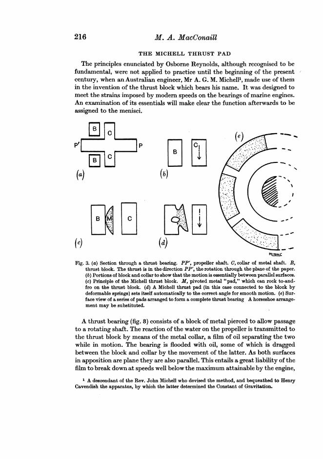
THE MICHELL THRUST PAD
The principles enunciated by Osborne Reynolds, although recognised to befundamental, were not applied to practice until the beginning of the presentcentury, when an Australian engineer, Mr A. G. M. Michell', made use of themin the invention of the thrust block which bears his name. It was designed tomeet the strains imposed by modern speeds on the bearings of marine engines.An examination of its essentials will make clear the function afterwards to beassigned to the menisci.
D C (~~~~~~~~~~~~~e)
(c) (d) _ha ±I.c
Fig. 3. (a) Section through a thrust bearing. PP', propeller shaft. a, collar of metal shaft. B.thrust block. The thrust is in the direction PP', the rotation through the plane of the paper.(b) Portions of block and collar to show that the motion is essentially between parallel surfaces.(c) Principle of the Michell thrust block. M, pivoted metal "pad," which can rock to-and-fro on the thrust block. (d) A Michell thrust pad (in this case connected to the block bydeformable springs) sets itself automatically to the correct angle for smooth motion. (e) Sur-face view of a series of pads arranged to form a complete thrust bearing A horseshoe arrange-ment may be substituted.
A thrust bearing (fig. 8) consists of a block of metal pierced to allow passageto a rotating shaft. The reaction of the water on the propeller is transmitted tothe thrust block by means of the metal collar, a film of oil separating the twowhile in motion. The bearing is flooded with oil, some of which is draggedbetween the block and collar by the movement of the latter. As both surfacesin apposition are plane they are also parallel. This entails a great liability of thefilm to break down at speeds well below the maximum attainable by the engine,
1 A descendant of the Rev. John Michell who devised the method, and bequeathed to HenryCavendish the apparatus, by which the latter determined the Constant of Gravitation.
216

The Function of Intra-Articular Fibrocartilages 2
and demands the provision of many such bearings to divide up the load. Theseadd very much to the dead weight of the ship, an addition not repaid by theincrease in efficiency. Mr Michell interposes a number of metal pads betweenblock and collar. Each of these is sector-shaped in outline, with a plane collarface and a somewhat curved (convex) block face. The curvature of the blockface permits the pad to rock through a small angle during motion; and accord-ingly it sets to the correct angle during movement of the collar, be this aheador astern. Thus is ensured the formation and maintenance of the film wedge.It has been found that the connection between the pads and the block can bemade by a double spring or some equally deformable continuous structure,since the deflection under the load has to be but a slight one. Such a flexibleconnection enables bearings to be simplified by constructing a number of padsintegral with, but flexibly connected to, a common supporting member. Aserious disadvantage is that the flexibility involves more or less risk of fractureofthe flexible part, a danger which is overcome by giving flexibility to a portionof the pad itself (Michell, 1923).
These thrust bearings have proved very successful in practice. Their numbercan often be reduced to one, and other economies are effected by their use.In addition they allow of greater speeds, and are widely used to-day both inmarine and land work in which high thrusts have to be met. Their purposemay be emphasised. They are employed where thrusts are to be carried by a filmof lubricant during motions in which there is a considerable element of gliding,and in which a screw-like movement along the axis of thrust is discouraged.
THE MENISCI AS MICHELL PADS
A sector of a meniscus can be described as a wedge of elastic material, pivotedat its attached base, so that it is capable of some variation in its inclination tothe articular surfaces with which it is in relation. The effect of such variationwill be felt first by the synovial fluid: that is, the synovial film in contact withthe articular surface will be made cuneiform to a greater or a less degreeaccording to the amount of adjustment of the meniscus. Such angular deflec-tions will be of small magnitude. Thus the sector is comparable in every respectto the most up-to-date type of Michell pad. The complete fibrocartilage ismerely the integration of such elements, and may be of ring-, horseshoe-, orplate-like form. By reason of its flexibility, which is increased by the thinningof the central part, such a structure is easily affected by stresses in the synovia.The viscosity of this fluid is certainly not less than that of blood serum. Experi-ments are in progress regarding its actual value: but the table (see p. 218), whichgives the viscosity of serum and of certain oils commonly used in film lubrica-tion, will show that the requisite physical condition of the liquid for the applica-tion of Reynolds' laws to it is certainly present. The matter of the speed of themoving parts is dealt with below. (See further, p. 226.)The synovial fluid, then, is one in which the phenomena of fugitive elasticity
may be expected to appear, and the stresses set up in it by movements at the
217

218 M. A. MacConaill
joint will be of a corresponding nature. The part of the cartilage in relation tothe weight-transmitting surface of the bone (at the moment) must so arrangeitself that a wedge of lubricant, convergent in the direction of movement, isformed between it and that surface. Thus will be brought about the effectivetransmission of weight from the fixed to the moving surface through the fluidduring motion, a transference which demands in certain cases a special ap-paratus that it be maintained without detriment to the continued separationof the surfaces. In a word, the menisci serve to increase the incongruence ofjoint surfaces rather than to decrease it. In this respect they are to be com-pared to the accessory cartilages of the hip and shoulder, although the functionof the latter is not quite that of the menisci.
This statement of their function makes it clear where they are to be expected.They are likely to be found where a thrust is combined with gliding and rota-tion, if the consequent screw-like motion of the parts towards one another isnot of service in the movement: that is, if it tend to put the bones into pre-mature contact. If the surfaces be of large radii of curvature such contact ismore likely to occur than if they be more rounded. A comparison of the carpo-metacarpal joint of the thumb and the sterno-clavicular joint will serve as an
Liquid Coefficient of viscosity(C.G.S. units)
Blood-serum (320 C.) 1-9 (Burns, 1929)Bayonne oil 1-6Rape oil t 200 C. 0 90 Stanton, 1923Sperm oil 0.34J
illustration. The view here put forward approaches that put forward by Parsons(1899-1900): it differs from it in being arrived at from first principles ofmechanics and in defining more rigorously the locus of the structures concerned.Parsons' theory associated intra-articular cartilages with a combination ofgliding and rotatory movements, laying special stress on the rotatory element.The present one rather reverses the emphasis, and takes cognizance of the factthat such combinations are present in practically every diarthrosis in the body,as Goodsir and his followers have justly maintained. They are present in thehip and shoulder. Here, however, they give a helical path to the moving point,at least in the former case. In so far as this tends to force the bones into pre-mature contact, the well-rounded form of the heads of the long bones causesthem to assume a favourable disposition to the female surfaces, as has beenindicated above in the reference to Beauchamp Tower's experiment. It ispossible, however, that the ligamentum teres may play the part of a meniscusin the hip joint, its consistency and disposition being not unfavourable for thatfunction.
It should be added with respect to Parsons' theory, that the form he gave itwas scientifically correct. It was derived from a purely morphological studyand summarised all the facts observed by him in a manner consistent with the
The Function of Intra-Articular Fibrocartilagesphysics of the time. Even yet, the principles of viscous-film lubrication are farfrom being widely known, except to certain classes of engineers; and the usualtext-books of applied mechanics make but passing references to it. Those whoare interested will find a full treatment in the article of Michell quoted above,and in Boswall (1928); while a simple presentation of its basal ideas is given byEdser (1926).So far the treatment ofthe problem has been largely of an aprior nature with
a view to establishing a reasonable hypothesis. It is not proposed to discuss allthe joints which have intra-articular cartilages in what follows. The tibio-femoral and inferior radio-ulnar articulations have been selected as types forthe demonstration and illustration of the lubricant function ofthe menisci. Whatis here proved for them holds for other joints; and in the ensuing discussiononly certain of those others will be referred to as being of special interest.
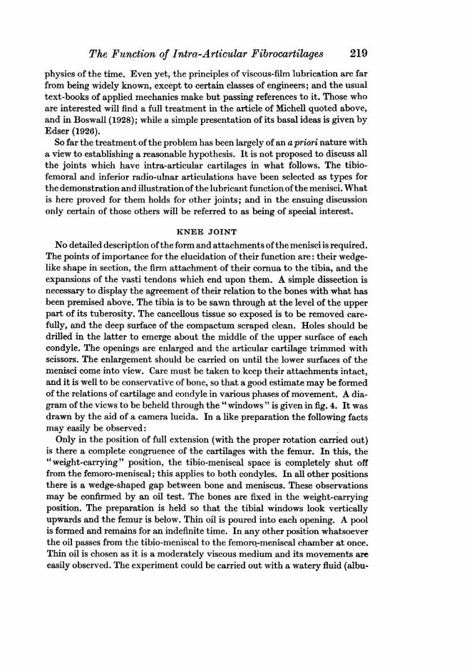
KNEE JOINT
No detailed description ofthe form and attachments ofthe menisci is required.The points of importance for the elucidation of their function are: their wedge-like shape in section, the firm attachment of their cornua to the tibia, and theexpansions of the vasti tendons which end upon them. A simple dissection isnecessary to display the agreement of their relation to the bones with what hasbeen premised above. The tibia is to be sawn through at the level of the upperpart of its tuberosity. The cancellous tissue so exposed is to be removed care-fully, and the deep surface of the compactum scraped clean. Holes should bedrilled in the latter to emerge about the middle of the upper surface of eachcondyle. The openings are enlarged and the articular cartilage trimmed withscissors. The enlargement should be carried on until the lower surfaces of themenisci come into view. Care must be taken to keep their attachments intact,and it is well to be conservative of bone, so that a good estimate may be formedof the relations of cartilage and condyle in various phases of movement. A dia-gram of the views to be beheld through the "windows " is given in fig. 4. It wasdrawn by the aid of a camera lucida. In a like preparation the following factsmay easily be observed:Only in the position of full extension (with the proper rotation carried out)
is there a complete congruence of the cartilages with the femur. In this, the"weight-carrying" position, the tibio-meniscal space is completely shut offfrom the femoro-meniscal; this applies to both condyles. In all other positionsthere is a wedge-shaped gap between bone and meniscus. These observationsmay be confirmed by an oil test. The bones are fixed in the weight-carryingposition. The preparation is held so that the tibial windows look verticallyupwards and the femur is below. Thin oil is poured into each opening. A poolis formed and remains for an indefinite time. In any other position whatsoeverthe oil passes from the tibio-meniscal to the femoro-meniscal chamber at once.Thin oil is chosen as it is a moderately viscous medium and its movements areeasily observed. The experiment could be carried out with a watery fluid (albu-
219

M. A. MacConaill
men), but not in a specimen treated with oil; in this case surface-tensionreactions between water and oil allow the liquid to slip through capillary spaceswhich would normally refuse passage to a viscous medium. The experimentshows that the menisci separate from the femur on the initiation of movementfrom the weight-bearing position in such a way that wedge-shaped spaces areformed between them and the bone, convergent in the direction of motion. Theconvergence will of course be noted for only one gap in each part of the doublejoint. If the corresponding part of the femur be marked it will be found to bethat which is nearest to the tibia at the instant: it is the momentary weight-transmitting area. It has been shown by Michell and his school that the condi-tions of film lubrication are fulfilled if the weight-transmitting area be incontact with a convergent film-the disposition of the film elsewhere is not ofimportance. In extension, therefore, the anterior part of the cartilage is in
A
L M
P
(a) (6) L%2 01-
Fig. 4. Diagrams of knee joint from below, viewed through openings in the tibial condyles.The menisci are stippled. (a) in semi-flexion. (b) in full extension.
action as an aid to weight transmission; and fluid is carried from the patellaraspect of the femur to the under aspect, through the cuneiform gap. In flexionit is the posterior part, and in the rotatory movement, the lateral part thatcomes into effective play.The meniscus, in short, behaves like a series of Michell pads welded into a
generalised form, which is called for by the more complex movements of theanimal joint. They are called for by the compound movement in which thegliding or sliding element is large in proportion to the rotatory. This does notpermit of the self-adjustment which takes place between the components ofa more purely spherical or hinge-like articulation. The " set " of the cartilage islargely automatic, and is conditioned by the forces generated within the fluidby movement. It should here be pointed out that the speed of the moving
220
The. Function of Intra-Articular Fibrocartilages 221
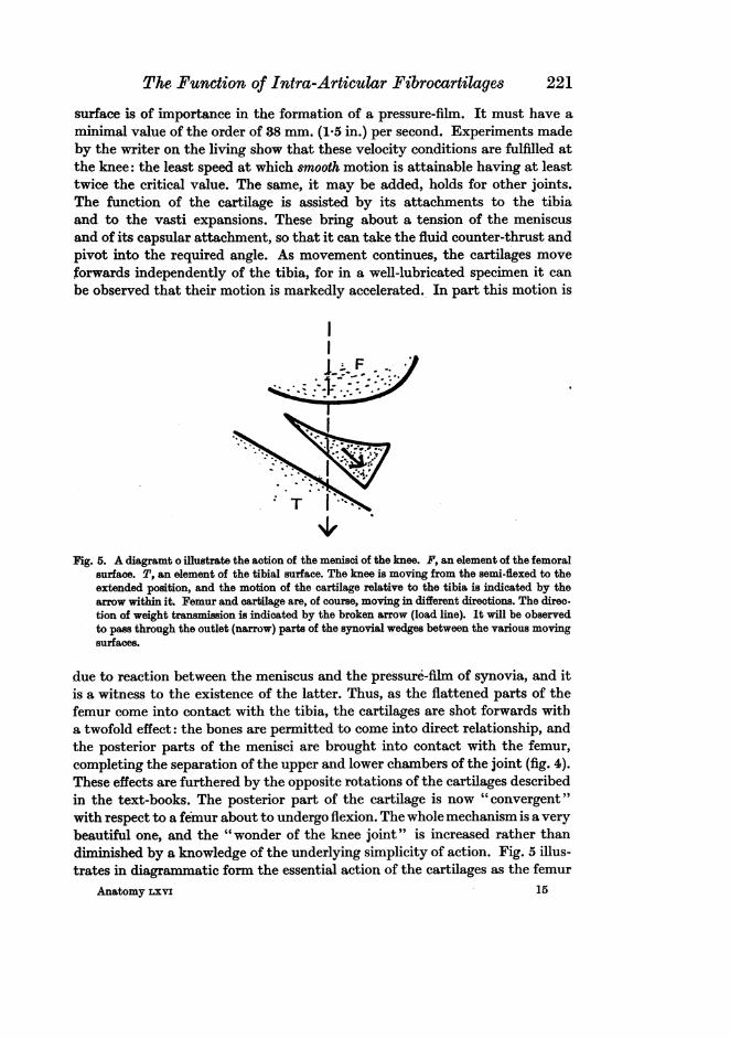
surface is of importance in the formation of a pressure-film. It must have aminimal value of the order of 88 mm. (1.5 in.) per second. Experiments madeby the writer on the living show that these velocity conditions are fulfilled atthe knee: the least speed at which smooth motion is attainable having at leasttwice the critical value. The same, it may be added, holds for other joints.The function of the cartilage is assisted by its attachments to the tibiaand to the vasti expansions. These bring about a tension of the meniscusand of its capsular attachment, so that it can take the fluid counter-thrust andpivot into the required angle. As movement continues, the cartilages moveforwards independently of the tibia, for in a well-lubricated specimen it canbe observed that their motion is markedly accelerated. In part this motion is
; F
T l*"
Fig. 5. A diagramt o illustrate the action of the menisci of the knee. F, an element of the femoralsurface. T, an element of the tibial surface. The knee is moving from the semi-flexed to theextended position, and the motion of the cartilage relative to the tibia is indicated by thearrow within it. Femur and cartilage are, of course, moving in different directions. The direc-tion of weight transmission is indicated by the broken arrow (load line). It will be observedto pass through the outlet (narrow) parts of the synovial wedges between the various movingsurfaces.
due to reaction between the meniscus and the pressure-film of synovia, and itis a witness to the existence of the latter. Thus, as the flattened parts of thefemur come into contact with the tibia, the cartilages are shot forwards witha twofold effect: the bones are permitted to come into direct relationship, andthe posterior parts of the menisci are brought into contact with the femur,completing the separation of the upper and lower chambers of the joint (fig. 4).These effects are furthered by the opposite rotations of the cartilages describedin the text-books. The posterior part of the cartilage is now "convergent"with respect to a femur about to undergo flexion. The whole mechanism is a verybeautiful one, and the "wonder of the knee joint" is increased rather thandiminished by a knowledge of the underlying simplicity of action. Fig. 5 illus-trates in diagrammatic form the essential action of the cartilages as the femur
Anatomy rxvi 15

M. A. MacConaill
moves from the semi-flexed to the extended position, as in walking. Theseparation and angulation of the parts are, of course, exaggerated for the sakeof clearness. The tilt of the cartilage relative to the tibia is to be speciallynoted. This tilt ensures the transmission of pressure from meniscus to tibiathrough the outlet end of the menisco-tibial synovial wedge.With regard to the semilunar form of the menisci, it is of interest to note that
horseshoe arrangements of Michell pads are quite commonly substituted forthe annular disposition. Further, the larger size and more open form of theinternal cartilage are precisely what are demanded by an application" of theMichell principle, assuming that the most efficient lubrication be called for.Again, it has been shown that the total bearing surface of a Michell pad maybe reduced to about half of the total possible area without inefficiency. Thedimensions of the menisci of the knee, relative to the total effective articularsurface at a given instant, are quite in keeping with this standard. Moreover,the occasional failure of the cartilages to perforate is no bar to a reasonablyefficient action, since the elastic plate can function as a whole as it does, forexample, in the temporo-mandibular joint (infra). In this "mass action" itis helped by the attachments of the vasti expansions. It is a corollary of thelast affirmation that perforation of a cartilage which is normally complete isnot necessarily disadvantageous, since the ring can function as a Michell padsystem, provided that the reduction of surface does not appreciably exceedone-half. Rarely, in the writer's experience, does it go beyond that amount.
It remains, in connection with the knee joint, to account for the commonabsence of symptoms following the removal of a semilunar cartilage. The carti-lages are accessories to lubrication, they might almost be called refinementson the part of Nature. Those in whom a violent effort provokes fracture of ameniscus-an accident previously explained to be conditioned by their flexi-bility-are persons of athletic habit. French surgeons, indeed, have noteda great increase of the number of such injuries since the introduction of " Anglo-Saxon" games-notably football (Jeanbrau, 1921). Loss of a meniscus leadsto about 20 per cent. increase in friction. This amount is not usually reachedin any but violent efforts-and the increased strain is likely to be unnoticed ata critical point in the game by a man in training. But it is recognised if ill-health supervene, and is remarked by those who have taken up exercise after aprolonged sedentary period, as the writer has verified. Many surgeons haverecorded the incidence of osteo-arthritic changes in several of their cases, aresult in keeping with the increased liability to premature contact of the arti-cular surfaces of the bones on account of the removal of the counter-pressureset up by the synovial wedge,
222

The Function of Intra-Articular Fibrocartilages
INFERIOR RADIO-ULNAR JOINT
The inferior radio-ulnar joint is distinguished from the superior by thegliding character of the movement associated with it: a distinction which isemphasised by the wholly fleshy structure of its proper pronator. The triangularfibrocartilage is to be reckoned an accessory to this joint. The very full researchesof Parsons (1899) have shown that the degree to which it is developed ispositively correlated with the movement of supination, and that its best repre-sentative is to be found in Man. There are certain details of its structure andattachments which have to do with its function and demand preliminary notice;and some attention must also be given to both joints with regard to the positionin which they assume the relationships characteristic of a synarthrosis, thatis, in which they transmit weight directly from one component to the other.The triangular cartilage follows the rule in that it is thicker circumferentially
than centrally. The thinning is often stated to be central: but is more accuratelydescribed as centroidal-it is nearer the base than the apex of the triangle.When the thin part is perforated the greater part of the remainder bears a greatresemblance to a semilunar cartilage. This likeness is heightened by theroundness of the angles of the cartilage proper. The disc is obliquely disposedbetween radius and ulna, sloping downwards and inwards from base to apex.Both surfaces are moderately concave, the greatest depression being, of course,at the spot already styled the " centroid" of the triangle.
SYNARTHROIDAL POSITION OF JOINTS OF FOREARM
Upper. The head of the radius is not circular but elliptical in outline. In atypical specimen the major axis is 2-4 cm. and the minor, 2-25 cm. This appliesto the cartilage-encrusted bone. The curvature of the corresponding cavity ofthe ulna is equal to that at the extremity of the major axis of caput radii.Elsewhere the curvature of the male (radial) surface is less than that of thefemale (ulnar) surface. If a fresh joint preparation be examined from above,the annular and interosseous ligaments being intact, the synarthroidal positioncan be determined to be nearly half-way between pronation and supination.In this phase of rotation the head of the radius is most accurately adapted tothe ulna, the long axis of its curvature being at right angles to the ulnar shaft,the shafts of the bones are most widely separated, and the interosseous mem-brane is at its greatest tension. The membrane is not quite taut: the significanceof this fact will be discussed in a later paper dealing with the problems of weightcarriage in the forearm. In any other position there is a small, but distinct, gapbetween the lesser sigmoid cavity and the head of the radius; this space isangular, and convergent in the direction of motion at the instant.
Lower. In many cases the triangular fibrocartilage is perforated: wherethisoccurs a view of the relations of the head of the ulna to the radial articularsurface is afforded. It can easily be determined that the head of the ulna ismost closely applied to the radial concavity at the same phase of movement as
15-2
223

M. A. Mac7onaill
brings about maximum congruence of the elements of the upper joint. Eachof the radio-ulnar joints is a virtual synarthrosis at the same instant, aboutmidway between probation and supination; and this is the definition of theweight-carrying position of the forearm.
THE MOVEMENTS OF THE FIBROCARTILAGE
A perforation of the triangular fibrocartilage is, in effect, a window for theinferior radio-ulnar joint similar to those made by dissection for the tibio-femoral. The opening is usually small, but it is large enough for the observationof the varying relationships of the ulnar head and cartilage. Only in one posi-tion does the rounded head of the ulna rest upon the disc in such a way as toocclude the opening-that position is the weight carrying one. In all othersit is separated by a wedge-shaped space from the disc. The oil test is effectivehere, and demonstrates the complete congruence of bone and cartilage in themid-prone stage of motion. If a little fluid be introduced into the joint cavitywhile it is in the fully supinated position it will be observed to be forced throughthat part of the wedge-shaped space which is convergent in the direction ofmotion; small air bubbles make its direction clearer. This shows the identityof the conditions in this joint with those in the knee, and with those set upby a Michell thrust pad.The cartilage is, in fact, such a pad inserted between the hand and ulna. It
enables weight to be transmitted from the hand to the ulna during movementof the radius across the forearm. This joint also illustrates the function of thecongruence of the menisci with their proper bony opposites: it cuts off the flowof fluid and automatically brings the surfaces into direct play. It is to benoted that the path of the lower end of the radius is not a simple turn, but isa compound of sliding and rotation about the ulna of a somewhat helical kind.
COMPARATIVE ANATOMY
The comparative anatomy of these structures is quite well known as theresult of Parsons' researches, and such an authority as Fick accepts them intheir entirety. The writer has been able to verify them in such animals as wereavailable, and the discussion of this part of the subject will be simplified bytaking Parsons' accounts as standard descriptions. Reference need only bemade to the knee, temporo-mandibular, and ankle joints to show the efficiencyof the present explanation in satisfying the postulates laid down above.
Menisci are found in the knee joint as low down in the vertebrate scale asthe amphibia (Bland Sutton). In Mammals the bat is unique in that it has nota trace of them. In this animal the joint is a pure ginglymus, rotation beingprevented by a bony spur. In the English bat (Vespertilio) the tibial spur isvery striking. In one form (Plecotus) a small amount of rotation is allowed,and delicate menisci are developed. More information is required on the landingmovements of these creatures as the weight-carrying parts of their skeletonmust differ both in site and development from those of non-flying Mammals.
224
The Function of Intra-Articular FibrocartilagesIn the lower monkeys the movement of rotation is better developed than inMan, and occurs even in the semi-flexed position. In such animals the semi-lunar cartilages are better marked, especially the outer. This is probably to becorrelated with their swinging habits, which would tend to throw much strainon the knee, unless the latter were made more yielding in its movements thanthose of heavier types.The temporo-mandibular joint is the classical example of the double-move-
ment theory. It is not denied that such a double motion is found there, northat the articular disc is a valuable accessory to it. But the forces which mouldthe flat articular eminence from infancy onward could equally well make onecompatible with the condyle of the jaw, while still retaining its gliding move-ment. The existence of the disc in the carnivore furnishes an anatomicalVoltaire with an excellent text against the "sufficient reason" of the theory.The difficulty disappears if the work of such discs in opposing a counter-pressure to that of the food be borne in mind. Although the trituration factoris absent from the carnivorous mandibular thrust, the tearing stress replaces itin importance; and it is necessary that the bearing surfaces be not prematurelyforced together during the action of the carnassials. Intra-articular cartilagesare not found in the jaw joints of the Monotremata, and they are likewisemissing in certain of the Marsupalia (Dasypus, Dasyurus). Parsons has putforward the theory that the discus is a new mammalian acquisition, and thusaccounts for its absence in these lowly forms. This view is not unacceptable:but further work is needed on the masticatory function of these animals beforethe absence of the structure can be held to be accounted for on functionalgrounds. It may be that such an explanation is unnecessary, as one coveringpositive instances can admit a phylogenetic account of origins.The marsupials have a fibrocartilage between the astragulus and the fibula.
It is to be correlated with the power of the fibula to rotate on its long axis(Parsons). The writer is inclined to associate its presence in the kangaroo withthe strain thrown on the ankle at the termination of the leap; and in all theseforms, the kangaroo and wallaby excepted, the fibula is very movable on thetibia and is liable to be drawn inwards by the tibio-fibular muscle.
DISCUSSION
The facts of human and comparative anatomy, so far as they have beenrecorded, seem to be in agreement with the thesis that the function of intra-articular cartilages is to maintain a convergent film of synovia between thosebones the necessary movement of which is prejudicial to their continuedseparation during motion. This view of their working is in accord with thepostulates of physics with regard to the transference of force between fullylubricated moving surfaces. It provides an explanation of their presence forthe instances in which they occur. The only case of absence from a highermammalian group is reconcilable with considerations of habit; and a primafacie case is recorded for an evolutionary reason of their non-appearance in
225

226 M. A. MacConaill
monotremes and certain marsupials. The congruence hypothesis has beenshown experimentally, as well as theoretically, to be untenable. Goodsirrecognised that the incongruence of articular surfaces of such joints as theknee must leave gaps between the bones. He deduced therefrom that themenisci and synovial pads had the common function of filling up the spaces.He definitely associated the cartilages with localities of pressure, making outthat the other structures were found to occupy spaces free from strain. He wasnot aware of the laws of lubrication-they were not known in his time: never-theless, in correlating the menisci with pressure, he made a good approximationto the truth. It may be said that the present essay is in some sort an extensionof the principles he put forward regarding articular movements. The work ofProf. Parsons has already been referred to in the body of this paper-it ishoped, not unjustly. The difference between the points of view is largely oneof emphasis. Finally, it may be said that an anti-concussional effect is not tobe entirely ruled out. In many cases intra-articular cartilages must serve sucha purpose: but only as a secondary result of their primary action.
SUMMARY
It is shown by reference to the theory of lubrication that intra-articularfibrocartilages are to be related primarily to the synovial fluid rather than tothe articular surfaces of the bones. They act to bring about the formation ofwedge-shaped films of synovia in relation to the weight transmitting parts ofjoints in movement. These wedges are narrowed in the direction of motion, andare necessary for weight transmission. They are to be found in joints wherethrusts are likely to bring about a premature approximation of the jointsurfaces. Preparations are described of knee and inferior radio-ulnar joints,which demonstrate the action of such cartilages. These also show that thecartilages are congruent with articular surfaces only in the " weight-carrying"position of the joints.
I wish to put on record my appreciation of the interest in, and encourage-ment of this work by Prof. Patten; and my thanks to Dr N. K. Adam, lateSorby Fellow of this University, for references to early papers on the theoryof lubrication.
ADDENDUM
The experiments referred to on page 217 above have now been completed.They show that the viscosity of human synovial fluid is of the order of10 C.G.S. units, at 20° C. This value is five times as great as that of blood-serum, but might fall as low as 7 units at body temperature. Acceptingthe latter value, it can be calculated that the thickness of the synovial film(knee) is of the order 50pu. Such a value is in full accordance with theprinciples set out above. Details of the experiments and of the calculationsbased thereon will be given in a future paper.

The Function of Intra-Articular Fibrocartilages 227
REFERENCES
BoswATL (1928). The Theory of Film Lubrication. Longmans, London.BROESIKE-MAIR. Repetitorium Anatomicum, 2nd edn. Fischer, Leipzig.EDSER (1926). General Physics for Students. Macmillan.FioK (1904). Handbuch der Anatomie und Mechanik der G(elenke, Erster Teil. Jena.- (1910). Handbuch der Anatomie und Mechanik der Gelenke, Zweiter Teil. Jena.
GoODSIR (1858). "Mechanism of the knee joint," in Anatomical Memoirs of John Goodsir, vol. II,p. 231. Edinburgh, Adam and Charles Black. See also, "Anatomy of the knee joint," op. cit.pp. 220 ff., and "Curvatures and movements of the acting facets of articular surfaces,"op. cit. pp. 246 ff.
JEANBRAU (1921). Preois de Pathologie Chirurgicale, Tome 4, 3me edn. Paris, Masson et Cie.MIcHELL (1923). "Viscosity and lubrication," in The Mechanical Properties of Fluids (a collective
work). Blackie, London.PARSONS (1899). "The joints of Mammals compared with those of Man. I." Journ. Anat. and
Phys. vol. xxxiv (N.S. vol. xiv), pt. 1.-(1900). "The joints of Mammals compared with those of Man. II." Journ. Anat. and Phys.
vol. xxxiv (N.S. vol. xiv), pt. 3.REYNOLDS (1886). "The theory of lubrication." Phil. Trans. p. 157.SIssoN (1917). The Anatomy of the Domestic Animals. W. B. Saunders Co., Philadelphia.BLAND SUTTON (1897). Ligaments, their Nature and Morphology. Lewis, London.WALMSLEY (1928). "The articular mechanism of the diarthroses." Journ. Bone and Joint Surg.
vol. x, no. 1, pp. 40-45.WOOD-JONES (1920). The Principles of Anatomy as Seen in the Hand. Churchill, London.