
Normal Radiographic Anatomy of the Equine metacarpus/metatarsus, fetlock, and distal extremity
Upload
bertram-hendersonCategory
view
218download
1
The Fetlock and Foot
First Year AnatomyNicholas Urbanek, BVMS, MRCVS

What is the Fetlock?• Fetlock is the common name for the
metacarpophalangeal and metatarsophalangeal joints (MCPJ and MTPJ) of the horses.
• It is formed by the junction of the third metacarpal (forelimb) or metatarsal (hindlimb) bones (cannon bones) proximally and the proximal phalanx (pastern bone) distally.
• Paired proximal sesamoid bones articulate with the palmar or plantar distal surface of the third metacarpal or metatarsal bones and are rigidly fixed to the proximo-palmar /-plantar edge of the proximal phalanx.

Common Views
LateralLateral DMPLO
DP DLPMO
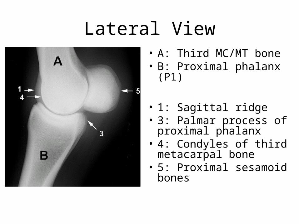
Lateral View• A: Third MC/MT bone• B: Proximal phalanx (P1)
• 1: Sagittal ridge• 3: Palmar process of
proximal phalanx• 4: Condyles of third
metacarpal bone• 5: Proximal sesamoid
bones

Dorso-palmer/-plantar (DP)• A: Third MC/MT bone• B: Proximal phalanx• C: Medial proximal
sesamoid bone• D: Lateral proximal
sesamoid bone• E: Metacarpo(tarso)-
phalangeal joint• J: Depression for medial
collateral ligament attachment
• 1: Sagittal ridge
Obliques – A Review
• The view is named for the projection of the beam.• Typically taken at 45 degree angles off the sagittal axis
of the limb.• Markers must be in place, otherwise unable to
distinguish medial from lateral side.• Dorsomedial-palmarolateral (plantar)oblique (DMPLO)• Dorsolateral-palmaromedial (plantar)oblique (DLPMO)

DLPMO
A: Third MC/MT boneB: Proximal phalanxC: Medial proximal sesamoid boneD: Lateral proximal sesamoid boneE: Metacarpo(tarso)-phalangeal joint1: Sagittal ridge3: Palmar process of proximal phalanx/Lateral palmar tubercle (eminence)6: Medial condyle of third MC/MT bone
DLPMO
Dorsal
Palmar/Pl
MedialLateral

DMPLO
A: Third MC/MT boneB: Proximal phalanxC: Medial proximal sesamoid boneD: Lateral proximal sesamoid boneE: Metacarpo(tarso)-phalangeal joint1: Sagittal ridge2: Lateral condyle of third metacarpal/tarsal bone3: Palmar process of proximal phalanx
Dorsal
Palmar/Pl
MedialLateral
DMPLO

Forelimb vs Hindlimb
DP view - Forelimb DP view - Hindlimb
The proximal sesamoid bones are higher
Bulging
• The MT is convex at its distal aspect
The proximal sesamoid bones are more triangular
Fetlock

Foot or Distal Limb• Composed of four bones
– Proximal, middle, distal (first, second, and third) phalanx– Navicular bone
• Multiple views obtained– Lateral– Dorsopalmar(plantar) view– Dorsoproximal-palmar(plantar)odistal oblique
• “Upright Pedal” or “High coronary”– Palmar(plantar)oproximal-palmar(plantar)odistal
• “Skyline Novicular”– Other oblique views
• The foot should have no shoe, be trimmed, and sulci should be packed with Play-Doh
• Marker always to the lateral side…can not tell laterality otherwise

Lateral view
A: Middle phalanxB: Third phalanxC: Navicular bone1: Proximal interphalangeal joint2: Distal interphalangeal joint3: Extensor process4: Dorsal surface5: Palmar process
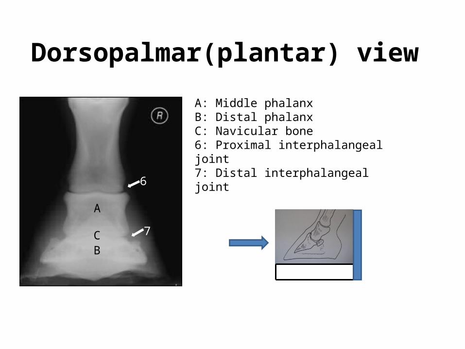
Dorsopalmar(plantar) view
A: Middle phalanxB: Distal phalanxC: Navicular bone6: Proximal interphalangeal joint7: Distal interphalangeal joint
R
A
BC
6
7

Dorsoproximal-palmar(pl)odistal oblique view
A: Middle phalanxB: Third phalanxC: Navicular bone1: Proximal interphalangeal joint2: Distal interphalangeal joint3: Extensor process4: Dorsal surface5: Palmar process6: Vascular channel7: Solar margin
6
55
2
7
B
Dorsoproximal-palmar(plantar)odistal oblique view
R
“Upright pedal”
“High coronary”
A

C: Navicular bone3: Articular surface8: Palmar aspect of middle phalanx9: Nutrient foramen10: Sagittal ridge11: Articulation between navicular bone and middle phalanx
Palmaro(plantar)oproximal-palmar(plantar)odistal oblique view
“Skyline Novicular”


