The FbaBtype fibronectinbinding protein of Streptococcus...
12
The FbaB-type fibronectin-binding protein of Streptococcus pyogenes promotes specific invasion into endothelial cells Silva Amelung, 1 Andreas Nerlich, 1 Manfred Rohde, 1 Barbara Spellerberg, 2 Jason N. Cole, 3† Victor Nizet, 3 Gursharan S. Chhatwal 1 and Susanne R. Talay 1 * 1 Department of Medical Microbiology, Helmholtz Centre for Infection Research, 38124 Braunschweig, Germany. 2 Department of Medical Microbiology and Hygiene, University Hospital Ulm, Ulm, Germany. 3 Department of Pediatrics and Skaggs School of Pharmacy & Pharmaceutical Sciences University of California, San Diego, La Jolla, CA 92093, USA. Summary Invasive serotype M3 Streptococcus pyogenes are among the most frequently isolated organisms from patients suffering from invasive streptococ- cal disease and have the potential to invade primary human endothelial cells (EC) via a rapid and efficient mechanism. FbaB protein, the fibronectin-binding protein expressed by M3 S. pyogenes, was herein identified as a potent invasin for EC. By combining heterologous gene expres- sion with allelic replacement, we demonstrate that FbaB is essential and sufficient to trigger EC inva- sion via a Rac1-dependent phagocytosis-like uptake. FbaB-mediated uptake follows the classi- cal endocytic pathway with lysosomal destination. FbaB is demonstrated to be a streptococcal invasin exhibiting EC tropism. FbaB thus initiates a process that may contribute to the deep tissue tropism and spread of invasive S. pyogenes iso- lates into the vascular EC lining. Introduction Streptococcus pyogenes (group A Streptococcus, GAS) is an important human pathogen that causes superficial infections of the pharyngeal mucosa and the skin (Cun- ningham, 2000). GAS can also produce severe invasive deep tissue infections such as necrotizing fasciitis, which are frequently complicated by toxic shock-like syndrome (TSLS) (Stevens, 2000). Streptococcal TSLS is a life- threatening condition associated with high mortality rates despite intensive medical treatment. In the last few decades, research has focused on the identification of GAS toxins and immune evasion factors involved in the pathogenesis of invasive disease. Less attention has been focused on the diversity and specificity of GAS adhesins and invasins, and how these molecules govern cell and tissue tropism to impact the spectrum of diseases that a particular strain can cause. Fibronectin-binding proteins have been identified as potent adhesins in a variety of pathogenic Gram-positive cocci (Talay, 2005; Schwarz-Linek et al., 2006). The high- affinity binding of this class of covalently linked surface molecules to fibronectin forms the structural basis for their unique biological function (Schwarz-Linek et al., 2003). Epithelial cell adhesion and invasion may be responsible for the localized tissue tropism of the pathogen to the oropharynx. M3 GAS do not possess the genes for SfbI/ PrtF1, or M1, two major epithelial cell adhesins and inva- sins of GAS. However, epidemiological studies revealed that GAS strains harbouring the prtF2 gene, which encodes the fibronectin-binding protein F2/PFBP (Jaffe et al., 1996; Rocha and Fischetti, 1999), are associated with invasive infections (Delvecchio et al., 2002). Within this gene family, two mutually exclusive genotypes exist, the pfbp type and the fbaB type (Ramachandran et al., 2004). The fbaB gene is highly conserved and located within the fibronectin-collagen-T antigen (FCT) region of GAS, a locus harbouring genes coding for adhesins that interact with extracellular matrix molecules and for pilus- forming proteins (Bessen and Kalia, 2002; Mora et al., 2005; Telford et al., 2006). The fbaB gene was first dis- covered in an M3 GAS strain isolated from a TSLS patient and is predominantly expressed by M3 strains isolated from patients suffering from invasive GAS disease (Terao et al., 2002). Serotype M3 GAS have been isolated worldwide from patients with invasive GAS disease (Stevens et al., 1989; Inagaki et al., 2000). How M3 GAS spread from the pha- ryngeal mucosal surface into deep tissue is unknown. A Received 6 January, 2011; revised 24 March, 2011; accepted 5April, 2011. *For correspondence. E-mail: susanne.talay@helmholtz- hzi.de; Tel. (+49) 531 6181 4413; Fax (+49) 531 6181 4499. † Present address: School of Chemistry and Molecular Biosciences, The University of Queensland, Brisbane, St Lucia, Qld 4072, Australia. Cellular Microbiology (2011) doi:10.1111/j.1462-5822.2011.01610.x © 2011 Blackwell Publishing Ltd cellular microbiology
-
Upload
nguyencong -
Category
Documents
-
view
213 -
download
0
Transcript of The FbaBtype fibronectinbinding protein of Streptococcus...

The FbaB-type fibronectin-binding protein ofStreptococcus pyogenes promotes specific invasioninto endothelial cells
Silva Amelung,1 Andreas Nerlich,1 Manfred Rohde,1
Barbara Spellerberg,2 Jason N. Cole,3† Victor Nizet,3
Gursharan S. Chhatwal1 and Susanne R. Talay1*1Department of Medical Microbiology, Helmholtz Centrefor Infection Research, 38124 Braunschweig, Germany.2Department of Medical Microbiology and Hygiene,University Hospital Ulm, Ulm, Germany.3Department of Pediatrics and Skaggs School ofPharmacy & Pharmaceutical Sciences University ofCalifornia, San Diego, La Jolla, CA 92093, USA.
Summary
Invasive serotype M3 Streptococcus pyogenes areamong the most frequently isolated organismsfrom patients suffering from invasive streptococ-cal disease and have the potential to invadeprimary human endothelial cells (EC) via a rapidand efficient mechanism. FbaB protein, thefibronectin-binding protein expressed by M3 S.pyogenes, was herein identified as a potent invasinfor EC. By combining heterologous gene expres-sion with allelic replacement, we demonstrate thatFbaB is essential and sufficient to trigger EC inva-sion via a Rac1-dependent phagocytosis-likeuptake. FbaB-mediated uptake follows the classi-cal endocytic pathway with lysosomal destination.FbaB is demonstrated to be a streptococcalinvasin exhibiting EC tropism. FbaB thus initiates aprocess that may contribute to the deep tissuetropism and spread of invasive S. pyogenes iso-lates into the vascular EC lining.
Introduction
Streptococcus pyogenes (group A Streptococcus, GAS) isan important human pathogen that causes superficialinfections of the pharyngeal mucosa and the skin (Cun-
ningham, 2000). GAS can also produce severe invasivedeep tissue infections such as necrotizing fasciitis, whichare frequently complicated by toxic shock-like syndrome(TSLS) (Stevens, 2000). Streptococcal TSLS is a life-threatening condition associated with high mortality ratesdespite intensive medical treatment. In the last fewdecades, research has focused on the identification ofGAS toxins and immune evasion factors involved in thepathogenesis of invasive disease. Less attention hasbeen focused on the diversity and specificity of GASadhesins and invasins, and how these molecules governcell and tissue tropism to impact the spectrum of diseasesthat a particular strain can cause.
Fibronectin-binding proteins have been identified aspotent adhesins in a variety of pathogenic Gram-positivecocci (Talay, 2005; Schwarz-Linek et al., 2006). The high-affinity binding of this class of covalently linked surfacemolecules to fibronectin forms the structural basis for theirunique biological function (Schwarz-Linek et al., 2003).Epithelial cell adhesion and invasion may be responsiblefor the localized tissue tropism of the pathogen to theoropharynx. M3 GAS do not possess the genes for SfbI/PrtF1, or M1, two major epithelial cell adhesins and inva-sins of GAS. However, epidemiological studies revealedthat GAS strains harbouring the prtF2 gene, whichencodes the fibronectin-binding protein F2/PFBP (Jaffeet al., 1996; Rocha and Fischetti, 1999), are associatedwith invasive infections (Delvecchio et al., 2002). Withinthis gene family, two mutually exclusive genotypes exist,the pfbp type and the fbaB type (Ramachandran et al.,2004). The fbaB gene is highly conserved and locatedwithin the fibronectin-collagen-T antigen (FCT) region ofGAS, a locus harbouring genes coding for adhesins thatinteract with extracellular matrix molecules and for pilus-forming proteins (Bessen and Kalia, 2002; Mora et al.,2005; Telford et al., 2006). The fbaB gene was first dis-covered in an M3 GAS strain isolated from a TSLS patientand is predominantly expressed by M3 strains isolatedfrom patients suffering from invasive GAS disease (Teraoet al., 2002).
Serotype M3 GAS have been isolated worldwide frompatients with invasive GAS disease (Stevens et al., 1989;Inagaki et al., 2000). How M3 GAS spread from the pha-ryngeal mucosal surface into deep tissue is unknown. A
Received 6 January, 2011; revised 24 March, 2011; accepted 5 April,2011. *For correspondence. E-mail: [email protected]; Tel. (+49) 531 6181 4413; Fax (+49) 531 6181 4499.†Present address: School of Chemistry and Molecular Biosciences,The University of Queensland, Brisbane, St Lucia, Qld 4072,Australia.
Cellular Microbiology (2011) doi:10.1111/j.1462-5822.2011.01610.x
© 2011 Blackwell Publishing Ltd
cellular microbiology

hallmark of deep tissue infection and/or systemic spreadof a pathogen is its ability to cross cellular and tissuebarriers, leading to the concept that invasive GAS spreadinto deep tissue as a complication of transient bacter-aemia (Stevens, 2000). We recently reported that M3GAS efficiently invades human primary endothelial cells(EC), a process that may represent a prerequisite fortrans-cellular passage across the endothelial barrier(Nerlich et al., 2009). Here, we identify FbaB as potent ECinvasin of M3 GAS that is necessary and sufficient totrigger a rapid phagocytosis-like uptake of streptococciinto ECs. FbaB triggers uptake via Rac1 recruitment andactivation and follows the endocytic pathway with lysoso-mal destination. FbaB is the first streptococcal factor iden-tified to mediate vascular cell tropism.
Results
FbaB is a potent invasin for EC
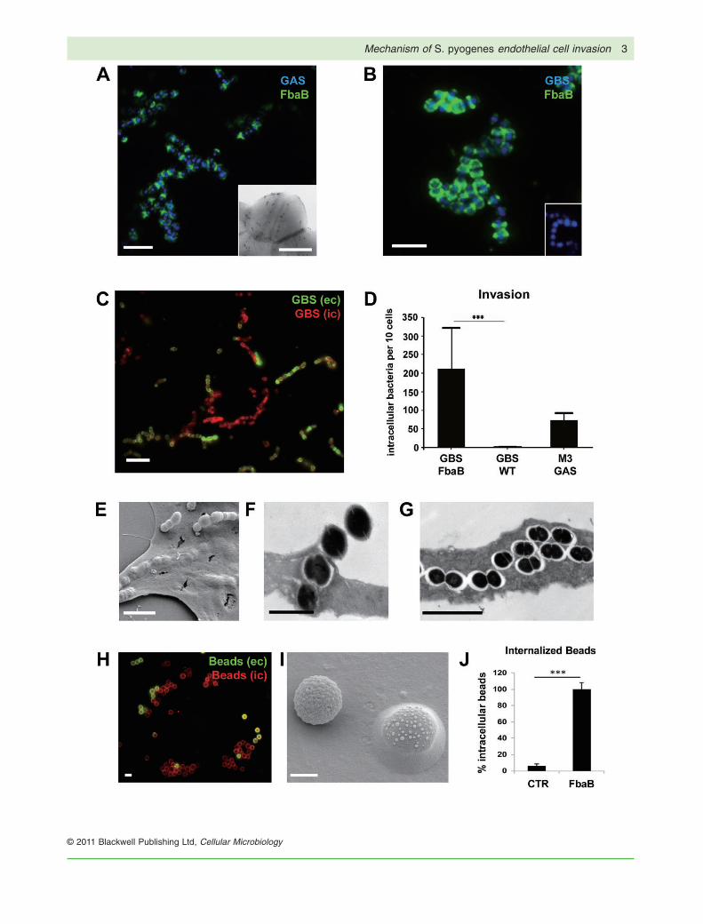
An EC non-invasive strain of Streptococcus agalactiae(GBS) was used as model organism for heterologousexpression of FbaB to study the protein’s role during ECinfection by gain-of-function analysis. The intact full-lengthGAS fbaB gene was cloned into the Escherichia coli–Streptococcus shuttle vector pDCerm generating pfbaB,which was then used for heterologous expression of FbaBon the surface of GBS. Using an FbaB-specific antiserumFbaB expression was visualized on the surface of aninvasive M3 GAS isolate (Fig. 1A) as well as on thesurface of the heterologous GBS strain (Fig. 1B). FbaB-expressing GBS were analysed in an in vitro EC invasionassay using primary human EC. As shown in Fig. 1C,FbaB expressing GBS exhibited a strong invasion poten-tial on EC, yielding 21 [mean � 11.2 standard deviation(SD)] intracellular streptococci per cell after 2 h of infec-tion, whereas the GBS WT strain was not invasive with
less than 3 bacteria per 100 cells (mean � 0.032 SD,Fig. 1D). The parental M3 GAS revealed 7.3 (mean � 1.9SD) intracellular bacteria per cell after 2 h of infection.Electron microscopic images of infected EC also revealedthe presence of extracellular and intracellular streptococcion and within EC (Fig. 1E–G). To prove that GAS FbaB issufficient to promote EC cell entry, an inert latex bead ECinternalization assay (Kaur et al., 2010) was undertaken.Beads coated with GST-FbaB protein were efficientlyinternalized by EC (Fig. 1H–J), but not control beadscoated with GST. Together, our results demonstrate thatFbaB is sufficient to confer efficient uptake of bacteria andinert particles into EC.
FbaB triggers a phagocytosis-like uptake withlysosomal destination
FbaB belongs to a family of fibronectin-binding proteinsharbouring several C-terminal repeats that form high-affinity complexes with fibronectin (Schwarz-Linek et al.,2003). SfbI protein, another member of this protein family,acts as epithelial cell invasin by triggering caveolae-mediated, non-phagocytic uptake of S. pyogenes (Rohdeet al., 2003) resulting in a persistent non-lysosomalintracellular stage. In contrast to this, serotype M3 S.pyogenes invade EC via the zipper mechanism, accom-panied by localized F-actin accumulation, as well as Rac1accumulation and activation (Nerlich et al., 2009) and aredelivered into lysosomes (S.R. Talay, unpublished). It wasthus of interest to determine the mode of uptake andsubsequent trafficking within EC that was promoted byFbaB. FbaB-mediated uptake of bacteria and latex beadsresembles phagocytic uptake via a zipper mechanism(Fig. 2A and B). The molecular basis for the formation ofthe membrane protrusions on the EC membrane is amassive but short-lived F-actin accumulation at the entryport (Fig. 2C).
Fig. 1. FbaB mediates invasion into ECs.A. FbaB surface expression of WT M3 GAS. Immunofluorescent (IF)-labelled FbaB (green) on M3 GAS (blue) and FESEM (insert) image ofimmunolabelled FbaB on M3 GAS.B. Heterologous expression of FbaB on the surface of S. agalactiae (GBS). IF image of immunolabelled FbaB (green) on the surface of theheterologous FbaB-expressing GBS strain (blue), and lack of FbaB expression on WT GBS (insert).C. Invasive potential of GBS-FbaB on EC. HUVEC were infected with GBS-FbaB for 2 h, washed fixed and differentially stained for extra- andintracellular streptococci. IF image of a cell infected with GBS-FbaB; intracellular cocci are red, extracellular green. Figure S1A shows phasecontrast of the same field.D. Quantification of EC invasion. Invasion rates were determined by enumerating intracellular (red) bacteria and expressed as intracellularbacteria per 10 cells. The diagram shows invasion rates of GBS-FbaB, the GBS-WT strain and the parental M3 GAS isolate A60 after 2 h ofinfection (***P < 0.0001).E. FESEM image of adherent and intracellular FbaB-expressing GBS after 2 h of infection.F and G. TEM image of ultrathin sections of GBS-FbaB infected EC after 2 h of infection.H. Uptake of FbaB-coated polystyrene beads into EC. IF image of extracellular (yellow) and intracellular (red) FbaB-coated beads on one ECafter 2 h of co-incubation. Figure S1B shows phase contrast of the same field.I. FESEM image of one adherent and one internalized FbaB-coated bead on EC.J. Quantification of bead internalization into HUVEC. Internalization rates were determined after 1 h co-incubation by enumerating intracellular(red) beads. ‘100% Intracellular beads’ refers to a mean of 5.5 FbaB-coated beads per cell (SD � 0.4). Control beads were coated with GST,revealing a mean of 0.35 intracellular beads per cell (SD � 0.3; ***P < 0.0002).Bars represent 1 mm (H), 5 mm (A, B, C, E, G), 2 mm (F, I) or 0.5 mm (A, insert).
2 S. Amelung et al.
© 2011 Blackwell Publishing Ltd, Cellular Microbiology
Mechanism of S. pyogenes endothelial cell invasion 3
© 2011 Blackwell Publishing Ltd, Cellular Microbiology

Fig. 2. FbaB triggers phagocytosis-like uptake in EC with lysosomal destination.A. Phagocytosis-like uptake of FbaB-expressing GBS into EC. Membrane protrusions along an adherent streptococcal strain characterize theinitial uptake process after 30 min of infection.B. Closure of the EC membrane leads to streptococcal internalization. The insert demonstrates formation of equal structures on EC uponinternalization of FbaB-coated beads.C. IF image showing F-actin (green) accumulation at the entry site of streptococci (red); the inserts show split channels for F-actin and GBSat higher magnification.D–F. FbaB-mediated uptake follows the classical endocytic pathway.D. IF image of FbaB-GBS (red) that accumulate the early endosomal marker protein EEA1 (green) after EC entry (60 min post infection). Theupper insert shows accumulation of the DsRed-labelled PX domain of p40Phox (DsRed-PX, pseudo-coloured in green) around an internalizedstreptococcal chain (pseudo-coloured in red), the lower insert shows circumferential accumulation of EEA1 (green) on an internalizedFbaB-coated bead (red).E. IF image of FbaB-GBS (red) that accumulate the late endosomal/lysosomal marker protein Lamp-1 (green) during the progress of infection(120 min post infection).F. Quantification of inhibition of lysosomal fusion after 2 h of infection using the PI3 kinase (PI3K)-specific inhibitor LY294002. ‘100% Lamp 1association’ refers to a mean of 22.7 Lamp-1 associated cocci per cell (SD � 2.4; ***P < 0.002).G. TEM image of ultrathin sections of GBS-FbaB infected EC. Different stages of the fusion events of GBS-FbaB with BSA-gold-loadedterminal lysosomes are shown. The right image shows gold particles in close association with a bacterial cell, indicating successful fusion ofthe phagosome with a terminal lysosome.Bars indicate 1 mm (A, B, G) and 5 mm (C, D, E).
4 S. Amelung et al.
© 2011 Blackwell Publishing Ltd, Cellular Microbiology

We next assessed whether this zipper-like uptake leadsinto the classical endocytic pathway. GBS-FbaB accumu-late the early endosomal marker protein EEA1 aroundinvading bacterial chains, suggesting early endosomallocalization (Fig. 2D). This finding was further corroboratedby transfecting EC with the DsRed/TagRFP-labelled PXdomain of p40PHOX, which binds to phosphatidylinositol-3-phosphate (PtdIns(3)P) generated by class III PI3K onthe membranes of early endosomes (Ellson et al., 2001).Recognition of membrane incorporated PtdIns(3)P byFYVE and PX domain-containing sorting proteins is anessential event for endosomal fusion and subsequentvesicular trafficking into lysosomes (Vieira et al., 2001).Infection of PX-DsRed/TagRFP-expressing cells demon-strated circumferential accumulation of the PX domain onintracellular streptococcal chains (upper insert in Fig. 2D).FbaB-coated latex beads also followed the endocyticpathway and colocalized with EEA1 (lower insert inFig. 2D) as well as the PX domain (not shown). Since theGBS WT strain was not invasive in HUVEC, there was alsono colocalization with intracellular marker proteins such asF-actin, EEA-1 and PX-domain (data not shown).
The later stages of intracellular trafficking were deter-mined by testing for the acquisition of late endosomal/lysosomal marker protein Lamp-1 in the phagosomalmembrane. Lamp-1 accumulated around intracellularFbaB-expressing GBS after 2 h of infection, indicatingstreptococcal trafficking into the late endosomal/lysosomal compartment (Fig. 2E). Inhibition of lysosomalfusion with LY294002, a PI3 kinase (PI3K)-specific inhi-bitor, substantially reduced the number of Lamp-1-associated streptococci (Fig. 2F) revealing that strepto-coccal entry follows the endocytic pathway. To finallyassess whether FbaB-mediated uptake leads to fusion ofstreptococci-containing phagosomes with terminal lysos-omes, the latter were pre-loaded with BSA-gold particles.Close association of gold particles with FbaB-expressingGBS (Fig. 2G) demonstrates that bacteria fuse with ter-minal lysosomes. The WT GBS strain did not colocalizewith Lamp-1 or BSA-gold particles since it was not inva-sive in HUVEC (data not shown). These data provideevidence that FbaB triggers phagocytic uptake of thebacteria, and engages the classical endocytic pathwayfor subsequent trafficking of bacteria into terminallysosomes.
The small GTPase Rac1 is an essential host factor forFbaB-mediated invasion
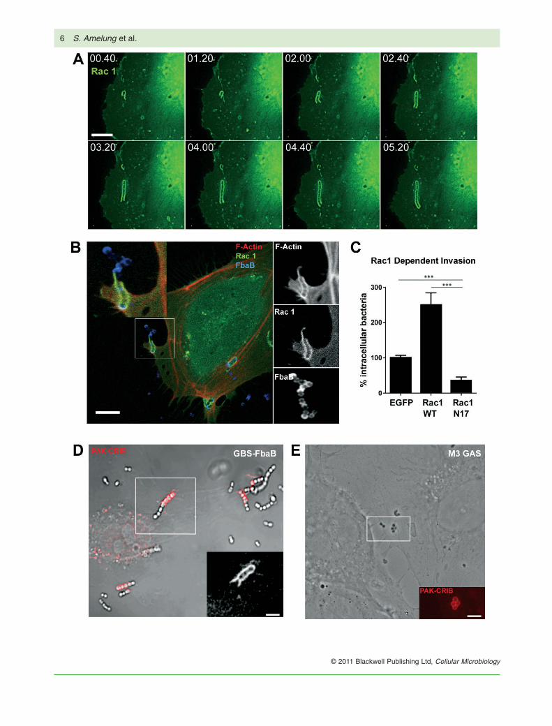
A rapid phagocytosis-like uptake requires whole-scalecytoskeletal rearrangements that are operated by the familyof small GTPases. We therefore assessed the role of Rac1 inthe FbaB-mediated entry process. Transfection of EC withEGFP-fused WT Rac1 and subsequent infection with GBS-
FbaB revealed longitudinal accumulation of Rac1 along aninvading chain (Fig. 3A and Movie S1). These changes areparalleled by simultaneous F-actin accumulation (Fig. 3B).Expression of the dominant negative form of Rac1 (N17)significantly reduced the number of intracellular bacteria,whereas expression of WT Rac1 significantly enhanceduptake of FbaB-expressing GBS in comparison withEGFP-expressing cells (Fig. 3C), clearly demonstratingthat Rac1 is an essential factor for FbaB-mediated uptake.To analyse the activation of Rac1 we used a fluorescentlytagged Cdc42 and Rac1 interactive binding domain (CRIB)fused to TagRFP (CRIB-TagRFP) as a biosensor of Racactivation (Itoh et al., 2002). Infection of transiently trans-fected cells results in accumulation of CRIB-TagRFP at thesite of GBS-FbaB entry (Fig. 3D) as well as accumulationof the CRIB-TagRFP at the site of M3 GAS entry (Fig. 3E).These data indicate that FbaB triggers Rac1 recruitmentand activation during the process of entry.
FbaB is an invasin with EC tropism
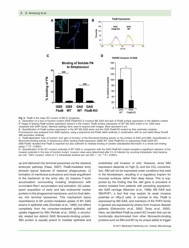
Our gain-of-function analysis demonstrated the potentialfor FbaB to function as a potent invasin for primaryhuman EC. We finally aimed to find out, whether FbaBexhibited a higher affinity to human EC compared withepithelial cells. To assess this, we conducted a mixedcell infection experiment, in which epithelial and endot-helial cells were infected simultaneously in the samewell. FbaB-expressing GBS as well as M3 GAS exhib-ited a strong preference for EC compared with epithelialcells (HEp-2) of human origin (Fig. 4). In the same set ofexperiments, HEp-2 cells were also infected with a sero-type M12 strain expressing the epithelial cell invasin SfbIas positive control (Rohde et al., 2011). Figure S2Eshows quantification of the invasion potential of theHEp-2 invasive M12 GAS isolate in comparison with theM3 GAS strain A60 as well as the FbaB-expressing GBSstrain. The invasion potential of the SfbI protein-expressing M12 strain was more than 20-fold enhancedcompared with the M3 GAS strain and more than 10-foldenhanced compared with the FbaB-expressing GBSstrain. This underlines that in contrast to SfbI protein,FbaB is a poor invasin on HEp-2 cells. In a final set ofexperiments we constructed a loss of function mutant inthe M3 GAS background by allelic replacement anddetermined the invasion potential of the FbaB mutantGAS strain (Fig. 5). In the FbaB mutant strain, a morethan 70% reduction of the EC invasion potential wasobserved. As a control, the fibronectin-binding activity ofthe mutant strain was also monitored and revealed lossof fibronectin-binding activity. These data show that inour representative M3 GAS strain, FbaB is the onlyfibronectin-binding factor and an essential factor for ECtropism and invasion.
Mechanism of S. pyogenes endothelial cell invasion 5
© 2011 Blackwell Publishing Ltd, Cellular Microbiology
6 S. Amelung et al.
© 2011 Blackwell Publishing Ltd, Cellular Microbiology

Discussion
Invasive M3 streptococci have a strong potential to invadeprimary human EC (Nerlich et al., 2009), a feature thatlikely represents an important step towards crossing theendothelial barrier during dissemination to the tissues. M3
GAS do not possess the gene for SfbI/PrtF1, an importantinvasin of GAS, but instead carry the fbaB gene within theFCT region. We here identified FbaB as the major ECinvasin of M3 GAS, and demonstrate that FbaB is neces-sary and sufficient to mediate this function. Phagocytosisis a process by which particles or live bacteria are taken
Fig. 3. Essential role of the small GTPase Rac1 in the FbaB-mediated EC invasion process.A. Time-lapse microscopy of GBS-FbaB entry into EGFP-WT Rac1-transfected EC. Eight frames of Movie S1 show subsequent accumulationof Rac1 along a streptococcal chain during the first minutes of invasion.B. IF image of GFP-Rac1-expressing EC infected with GBS-FbaB for 60 min (red: F-actin, green: Rac1, blue: FbaB). Inserts at the right sideshow split channels for F-actin, Rac1 and FbaB stain (from up to down).C. Quantification of FbaB-mediated invasion into EC that express either wild-type Rac1 (WT Rac1), the dominant-negative form of Rac1(Rac-N17) or EGFP (EGFP) demonstrates that Rac1 is essential for invasion. Invasion rates were determined by enumerating intracellular(red) bacteria after 2 h of infection. EGFP-transfected cells served as control with ‘100% invasion’ referring to 24 intracellular cocci per cell(***P < 0.0001).D. Phase-contrast/fluorescence image of infected EC expressing the PAK-CRIB domain as RFP-fusion protein (CRIB-TagRFP).GBS-FbaB-induced Rac1 activation was visualized by demonstrating accumulation of the PAK-CRIB domain (red) along an invadingstreptococcal chain after 60 min of infection. The insert shows an enlargement of the fluorescence channel of the indicated area.E. Phase-contrast/fluorescence image of M3 GAS-infected EC expressing the PAK-CRIB domain. The insert shows an enlargement of thefluorescence channel of the indicated area.Bars represent 10 mm (A, B) or 3 mm (D, E).
Fig. 4. FbaB is an invasin with endothelialcell tropism. HUVEC and HEp-2 cell (humanepithelial cell line) were seeded on coverslips,placed into the same well and grown toconfluency in EGM2 medium. Cells wereinfected for 2 h with an moi of 25 with GBSWT or GBS-FbaB, or an moi of 50 with M3GAS bacteria. Intracellular bacteria werestained in red, extracellular in green.A. IF image of FbaB-expressing GAS andGBS strains infecting HUVEC (left panel) orHep-2 cells (right panel). Phase-contrastimages are shown in Fig. S2A–D.B. Invasion rates were determined bycounting intracellular (red) bacteria per cell.‘100% invasion’ refers to 19 intracellularbacteria per cell [mean of four independentexperiments (SD � 7.3; ***P < 0.0001)].
Mechanism of S. pyogenes endothelial cell invasion 7
© 2011 Blackwell Publishing Ltd, Cellular Microbiology
up and delivered into terminal lysosomes via the classicalendocytic pathway (Haas, 2007). FbaB-mediated entryshowed typical features of classical phagocytosis: (i)formation of membrane protrusions and close engulfmentof the bacterium at the entry site, (ii) massive F-actinaccumulation surrounding invading streptococci withco-incident Rac1 accumulation and activation, (iii) subse-quent acquisition of early and late endosomal markerproteins in the phagosomal membrane, and (iv) final deliv-ery into terminal lysosomes. This mechanism bearsresemblance to M1 protein-mediated uptake of M1 GASstrains in epithelial cells (Dombek et al., 1999), but differscompletely from the caveolae-mediated epithelial celluptake triggered by SfbI (Rohde et al., 2003), a structur-ally related but distinct GAS fibronectin-binding protein.SfbI protein is equally potent to mediate epithelial and
endothelial cell invasion in vitro. However, since SfbIexpression depends on high O2 and low CO2 concentra-tion, SfbI will not be expressed under conditions that existin the bloodstream, resulting in a regulatory tropism formucosal surfaces rather than deep tissue. This is sup-ported by the finding that the sfbI gene is prevalent instrains isolated from patients with persisting asymptom-atic GAS carriage (Neeman et al., 1998). M3 GAS lackSfbI/PrtF1, a fact that may explain its weak invasivepotential on HEp-2 cells. In contrast to this, FbaB isexpressed by M3 GAS, and members of the PrtF2 familyin general are expressed by strains from invasive diseasepatients (Delvecchio et al., 2002; Terao et al., 2002).Here, we identified FbaB as potent EC invasin that can befunctionally discriminated from other fibronectin-bindingproteins such as SfbI and M1 by its unique ability to trigger
Fig. 5. FbaB is the major EC invasin of M3 S. pyogenes.A. Generation of a loss of function mutant (GAS FbaB-KO) in invasive M3 GAS and lack of FbaB surface expression in the deletion mutant.IF image of lacking FbaB surface expression (green) in the mutant, FbaB surface expression of WT M3 GAS (insert in A). GAS werevisualized with DAPI (blue). Identical settings were used to acquire both images. Bars represent 5 mm.B. Quantification of FbaB surface expression in the WT M3 GAS strain and the GAS FbaB-KO mutant by flow cytometry analysis.Fluorescence was analysed from 5000 bacteria, using a polyclonal anti-FbaB rabbit antibody in combination with an anti-rabbit Alexa Fluor®488 secondary antibody.C. FbaB-dependent ‘loss of function’ and ‘gain of function’ of the fibronectin-binding activity on the surface of GAS and GBS. Quantification offibronectin-binding activity of streptococci either lacking FbaB expression (GBS WT, GAS FbaB-KO) or expressing FbaB (GAS WT,GBS-FbaB) revealed that FbaB is essential but also sufficient to mediate binding of soluble radiolabelled fibronectin in a whole-cell bindingassay (***P < 0.0001).D. Quantification of the EC invasion potential of WT GAS in comparison with the GAS FbaB-KO mutant revealed a significant reduction of theinvasion potential in the loss of function mutant. Invasion rates were determined after 2 h of infection by counting intracellular (red) bacteriaper cell. ‘100% invasion’ refers to 7.4 intracellular bacteria per cell (SD � 1.9; ***P < 0.0001).
8 S. Amelung et al.
© 2011 Blackwell Publishing Ltd, Cellular Microbiology

phagocytic uptake into EC. Moreover, in contrast to otherinvasins from Gram-positive cocci, FbaB shows tropismfor EC and thus may be one of the initial factors respon-sible for the deep tissue tropism of M3 GAS. A furtherrequisite would then be the ability of GAS to leave the ECat the tissue side in a viable form (S.R. Talay, unpub-lished). The question arises, why FbaB (and also M3GAS) has a strong preference for EC. Fibronectin is acommon ligand for both, SfbI protein and FbaB, and is anessential molecule for SfbI-mediated invasion into epithe-lial cells (Talay et al., 2000). One may thus speculate thatFbaB could have a different structural impact on its ligandfibronectin than SfbI protein. This may be due to thelacking element of the UR/Spacer region found in SfbI, orthe presence of putative integrin binding sites as well astwo CnaB domains, which are important structure-defining elements. Interestingly, the CnaB2 domain ofFbaB forms an isopeptide bond that renders the proteinhighly thermostable and acid resistant (Hagan et al.,2010). These additional elements may either constitute orpresent EC-specific binding sites for receptors that are notfound on epithelial cells such HEp-2.
FbaB is the first streptococcal factor identified tomediate EC-specific invasion of GAS and the humanendothelium may thus represent a reservoir for serotypeM3 and other FbaB-expressing group A streptococci.
Experimental procedures
Bacterial strains and culture conditions
Streptococcus pyogenes strain A60 is a serotype M3 GAS iso-lated from blood of a patient suffering from invasive disease(Dinkla et al., 2003). S. pyogenes A40 is an SfbI-expressingepithelial cell invasive M12 GAS and was described previously(Rohde et al., 2011). S. agalactiae serotype Ia strain 102 wasdescribed previously (Kaur et al., 2010) and served as the recipi-ent for the plasmid pDCerm (Zinkernagel et al., 2008) or theFbaB-expressing plasmid pfbaB (this work). Streptococci werecultured in tryptic soy broth (TSB, Oxoid) at 37°C and 5% CO2,containing 2 mg ml-1 erythromycin for selection of plasmidpDCerm and its derivative pfbaB; 2 mg ml-1 erythromycin and1 mg ml-1 chloramphenicol for selection of pKO-fbaB transfor-mants (this work); and 1 mg ml-1 chloramphenicol for selection ofGAS FbaB-KO. E. coli TOP10 (Invitrogen) was grown in Luria–Bertani (LB) broth or on LB plates supplemented with 100 mg ml-1
ampicillin, 500 mg ml-1 erythromycin or 5 mg ml-1 chlorampheni-col, where appropriate. To obtain electrocompetent GAS andGBS cells, strains were grown in Todd–Hewitt broth (THB) con-taining 0.6% glycine. For FACS analysis and infection experi-ments, GAS and GBS strains were grown to logarithmic phase,corresponding to an OD600 of 0.4.
Reagents and antibodies
Rabbit polyclonal antibodies specifically recognizing S. pyogenes(anti-GAS) were generated as described previously (Talay et al.,
2000). Polyclonal anti-GBS antibody was obtained from Acris(Herford, Germany). A mouse monoclonal antibody recognizing aluminal epitope of human Lamp-1 (clone H4A3) was purchasedfrom Pharmingen. Secondary goat anti-rabbit IgG antibodiescoupled to ALEXA Fluor® 488/568, and goat anti-mouse IgGcoupled to ALEXA Fluor® 488 were obtained from Invitrogen;Cy-5-conjugated goat anti-rabbit IgG was purchased from Milli-pore (Schwalbach/Ts, Germany). PI3K inhibitor LY294002 wasobtained from Calbiochem.
Allelic exchange mutagenesis of GAS
An in-frame allelic exchange of fbaB with chloramphenicolacetyltransferase (cat) was obtained in S. pyogenes A60 by usingthe method previously described (Zinkernagel et al., 2008).The primer pair used for amplification of the upstream fragmentflanking the fbaB gene was fbaB-upF (5′-GCGAATTCGATGGATTGTTTGTTGGCAAGTC-3′) and fbaB-upR (5′-GGTGGTATATCCAGTGATTTTTTTCTCCATTATTTTCTCTCTCCACATTCCTAAGCG-3′). The primer pair used for amplification of thedownstream fragment flanking fbaB was fbaB-downF (5′-TACTGCGATGAGTGGCAGGGCGGGGCGTAACTGTTGGTGACAATAGCGAAAAAG-3′) and fbaB-downR (5′-GCGAATTCAGCTTCGATGCTGTTTAGTTCGTAT-3′). The cat gene was ampli-fied using primers catF (5′-ATGGAGAAAAAAATCACTGGATATACCACCGTTGA-3′) and catR (5′-TTACGCCCCGCCCTGCCACTCATCGCAGTACTGTTGTA-3′).
Heterologous expression of FbaB
Streptococcus agalactiae strain 101 was selected as modelorganism for FbaB expression since this WT GBS strain wastested to be non-invasive in HUVEC yielding less than 3 intrac-ellular bacteria per 100 cells at an moi of 25 (S.R. Talay, unpub-lished), and previously used to determine whether SpyCep, theIL-8-degrading cell envelope protease of GAS, acts as invasin(Kaur et al., 2010). The complete M3 S. pyogenes fbaB gene andits flanking regions, starting 68 nucleotides upstream of the startcodon and terminating 100 nucleotides downstream of the stopcodon, was PCR-amplified from the GAS chromosome, clonedinto the TOPO vector (Gateway), and then subcloned intopDCerm (Zinkernagel et al., 2008). Primers for amplification werefbaB-KpnI-F (5′-GCGGGTACCGACAATTGGCCTGTAGTCTTTAGTTTTGGAC-3′) and fbaB-XbaI-R (5′-GCGTCTAGACTTAAAATGACTTATCTAGTGAACCGAGACG-3′). The resulting plasmid,pfbaB, was transformed into electrocompetent S. agalactiae cellsand transformants (GBS-FbaB) were pre-selected for onerythromycin-containing media and further confirmed by restric-tion enzyme analysis and PCR. The empty vector wastransformed in S. agalactiae and the resulting transformant(GBS-pDCerm) used as a negative control. For heterologousexpression and subsequent purification of recombinant FbaBprotein in E. coli, the truncated fbaB gene lacking the sequenceencoding the signal peptide and membrane anchor was clonedinto pGEX-6-P1 (Invitrogen). Primers for amplification were fbaB-BamHI-F (5′-CGGAGGATCCGTAGGACATGCGGAAACAAG-3′)and fbaB-SalI-R (5′-GTTACTGTCGACTTCCTTGTTATCAAAGTGG-3′). Expression of GST-tagged FbaB polypeptide andsubsequent purification using glutathione sepharose affinity chro-matography was conducted according to the manufacturer’s pro-tocol (GE Healthcare Munich).
Mechanism of S. pyogenes endothelial cell invasion 9
© 2011 Blackwell Publishing Ltd, Cellular Microbiology

Cloning of eukaryotic proteins and EC transfection
The PX domain of p40PHOX was cloned into pDsRed-Monomer-N1and pTagRFP-N1, respectively, as described before (Catz et al.,2002) using human p40PHOX full-length cDNAtemplate (imaGenes,Berlin, Germany). To construct CRIB-TagRFP, PCRwas used to amplify the fragment encoding amino acids 60–141 of PAK1B using the primers: CRIB_EcoRI_fwd5′-CGAATTCACACCATGGTACCTGGAGATAAAACAAAT-3′ andCRIB_SalI_rev 5′-CCGTCGACCATTTCTGGCTGTTGGATGTCT-3′, and pGEX-2T-PAK-CRIB as a template. The resulting frag-ment was gel purified and cloned in pTagRFP-N1 using EcoRIand SalI sites, yielding pCRIB-TagRFP. All constructs weresequenced before use. Plasmids were purified with the EndoFreeplasmid purification kit (Qiagen, Hilden, Germany). HUVEC werethen transfected with the Amaxa nucleofection system (AMAXA)applying the standard protocol as specified by the manufacturer.For ectopic expression of GTPase derivatives in HUVEC, wild-type Rac1 and the dominant-negative form of Rac1 (kindly pro-vided by I. Just) were expressed in HUVEC as GFP-fusionproteins as previously described (Nerlich et al., 2009).
Quantification of FbaB surface localization byflow cytometry
Streptococcal strains were grown to mid-exponential phase inTSB medium and washed once in PBS. A total of 1 ¥ 107 bacteriawere suspended in 400 ml of PBS containing 0.5% FCS andincubated with 0.5 mg of anti-FbaB rabbit IgG for 30 min at 37°C.After washing in PBS the bacterial pellet was suspended in100 ml of PBS containing a 1:300 dilution of an anti-rabbitALEXA® Fluor 488 antibody and incubated for 30 min at 37°C.Bacteria were washed in PBS, fixed in PBS containing 3%para-formaldehyde and analysed by flow cytometry using a FAC-SCalibur (Becton Dickinson). Streptococci were detected usinglog-forward and log-side scatter dot plots, and a gating regionwas set to exclude debris and larger aggregates of bacteria. Atotal of 5 ¥ 103 bacteria were analysed for fluorescence usinglog-scale amplification.
EC culture and invasion assay
Primary human large vascular cells isolated from umbilical cord(HUVEC) were purchased from PromoCell. EC were cultured andpropagated in EGM-2 medium (PromoCell) according to the sup-plier’s protocol in a cell incubator at 37°C and 5% CO2. Cells wereseeded on coverslips in multiwell plates (Nunc) and grown to75% confluency. Streptococci were grown in TSB, harvested bycentrifugation, washed and diluted in pre-warmed EGM-2medium containing 5% FCS and used to infect EC with an moi of25 for GBS strains and an moi of 50 for GAS WT strains as wellas the FbaB Knock-out GAS strain. Infection was stopped after amaximum of 2 h of infection by washing the infected monolayerwith EGM-2 medium and fixing with PBS containing 4% PFA.Distinct time points for fixation were used for monitoring earlyentry (60 min post infection) or subsequent trafficking (120 minpost infection). For inhibition of lysosomal fusion events, the PI3kinase inhibitor LY294002 was selected since class III PI3K isessential for endosomal fusion and subsequent trafficking intolysosomes. HUVEC were pre-incubated with 25 mM LY294002for 45 min and infected.
Immunofluorescence and confocal microscopy
After fixation, infected EC monolayers were blocked for 30 minwith PBS containing 10% FCS. Differential staining of streptococciwas performed as previously described (Rohde et al., 2003).According to their respective label, intracellular bacteria appearred and extracellular yellow to green. F-actin, EEA1 and Lamp-1were visualized as described (Kaur et al., 2010). In some experi-ments streptococci were labelled with secondary Cy5-conjugatedantibodies. Following final washing, coverslips were mounted onglass slides using ProLong Gold anti-fade reagent (MolecularProbes). Mounted samples were examined using a confocal laserscanning microscope (LSM 510 Meta, Zeiss). Alternatively,images were recorded using a Zeiss inverted microscope 100 or aZeissAxiophot with an attached ZeissAxiocam HRc digital cameraand Zeiss Axiovision software 7.6. Images were processed forcontrast and brightness using ImageJ (http://rsb.info.nih.gov/ij).
Determination of invasion rates and quantification ofLamp-1 association
Invasion rates of streptococcal strains or beads were determinedby enumerating intracellular (red) bacteria or beads after infectingcells for 2 h with an initial moi of 25 for GBS strains, an moi of 50for GAS strains, and in case of beads with an moi of 100.Invasion rates were expressed as either intracellular bacteria per10 cells or % invasion. For Lamp-1 colocalization, the number ofbacteria (red) residing within a Lamp-1-positive compartmentwas enumerated. A minimum of 100 cells were analysed perassay; assays were conducted in triplicate and repeated for atleast three times on different days. Data represent means of onerepresentative experiment conducted in triplicate.
Time-lapse microscopy
Live cell imaging was performed at 37°C in 5% CO2 with cellsexpressing the GFP fusion of WT Rac1 as described elsewhere(Nerlich et al., 2009).
BSA-gold loading of HUVEC lysosomes
BSA-gold particles with a diameter of 15 nm were prepared byincubating 10 ml of the gold sole (pH 6.0) with 100 mg of BSA for30 min at 20°C, followed by centrifugation at 20 000 r.p.m. in aTLD100 (Beckman) for 15 min, then a wash step with PBS.Lysosomes were pre-loaded by feeding HUVEC with BSA-gold24 h prior to infection as described (Rohde et al., 2003). Thefollowing day, cells were infected and fixed after 120 min postinfection and ultrathin sections analysed by transmission electronmicroscopy (TEM).
Transmission (TEM) and field emission scanningelectron microscopy (FESEM)
For TEM analysis, HUVEC were grown in 6-cm-diameter culturedishes, infected, subsequently washed with EGM-2 medium, andfixed and embedded as described (von Kockritz-Blickwede et al.,2008). Images were recorded digitally with a Slow-Scan CCD-Camera (ProScan, 1024 ¥ 1024) with ITEM-Software (Olympus
10 S. Amelung et al.
© 2011 Blackwell Publishing Ltd, Cellular Microbiology

Soft Imaging Solutions). For FESEM analysis, HUVEC weregrown and infected on coverslips, washed in EGM-2 medium andprepared as described (von Kockritz-Blickwede et al., 2008).Brightness and contrast were adjusted with Adobe PhotoshopCS3.
Whole-cell fibronectin-binding assay
Human fibronectin (Chemicon) was radiolabelled with 125I via theChloramine T method as described (Hunter and Greenwood,1962). Streptococci were grown in TSB, washed in PBS contain-ing 0.05% Tween (PBST) and suspended in PBST to give a finalconcentration of 1 ¥ 108 bacteria per ml. Two hundred and fiftymicrolitres of the bacterial suspension was incubated with 20 ngof radiolabelled fibronectin (specific activity of 2.9 mCi mg-1).After 1 h of incubation, unbound fibronectin was removed bywashing bacteria with PBST and by measuring the amount ofbound fibronectin in a gamma counter. Total counts of radiola-belled fibronectin in the assay were determined in parallel by TCAprecipitation and formed the basis for the 100% value. Bindingactivity was expressed as the percentage of bound fibronectincompared with total fibronectin. Assays were performed in tripli-cate and experiments were repeated three times.
Latex bead internalization assay
Purified recombinant FbaB protein or GST in case was coupled to3 mm latex beads (Sigma) and tested for coupling efficiency asdescribed (Molinari et al., 1997). HUVEC were seeded on cov-erslips as described above and co-incubated with FbaB or GST-coated latex beads with an moi of 100 for 1 h. Cells were thenwashed three times with EGM2 and fixed as described above.Double-immunofluorescent staining of intra- and extracellularbeads was performed as described for streptococci except thatrabbit anti-FbaB and anti-GST antibodies served as primary anti-body. Intracellular (red) beads were enumerated by counting andinternalization rates were expressed as % invasion. A minimum of100 cells were analysed per assay; assays were conducted intriplicate and repeated for at least three times on different days.Data represent means of one representative experiment con-ducted in triplicate.
Statistical analyses
Statistical comparisons were made using the Student’s pairedt-test with Graph Pad Prism software. Values are expressed asmeans � SD.
Acknowledgements
We thank N. Janze and I. Schleicher for excellent technicalassistance and I. Just for providing the Rac1 constructs. S.R.T.gratefully acknowledges funding through the ‘Reentry Program’given by the Helmholtz Association, Germany, and fundingthrough the ‘CAREPNEUMO’ programme given by the EU Com-mission (contract No. 223111). A.N. gratefully acknowledgesfunding through the ‘ASSIST’ programme given by the EU Com-mission (contract No. 032390). J.N.C. was supported by aNational Health and Medical Research Council of Australia grant(514639).
References
Bessen, D.E., and Kalia, A. (2002) Genomic localization of aT serotype locus to a recombinatorial zone encoding extra-cellular matrix-binding proteins in Streptococcus pyo-genes. Infect Immun 70: 1159–1167.
Catz, S.D., Johnson, J.L., and Babior, B.M. (2002) The C2Adomain of JFC1 binds to 3′-phosphorylated phosphoinositi-des and directs plasma membrane association in livingcells. Proc Natl Acad Sci USA 99: 11652–11657.
Cunningham, M.W. (2000) Pathogenesis of group A strepto-coccal infections. Clin Microbiol Rev 13: 470–511.
Delvecchio, A., Currie, B.J., McArthur, J.D., Walker, M.J., andSriprakash, K.S. (2002) Streptococcus pyogenes prtFII,but not sfbI, sfbII or fbp54, is represented more frequentlyamong invasive-disease isolates of tropical Australia. Epi-demiol Infect 128: 391–396.
Dinkla, K., Rohde, M., Jansen, W.T., Kaplan, E.L., Chhatwal,G.S., and Talay, S.R. (2003) Rheumatic fever-associatedStreptococcus pyogenes isolates aggregate collagen.J Clin Invest 111: 1905–1912.
Dombek, P.E., Cue, D., Sedgewick, J., Lam, H.,Ruschkowski, S., Finlay, B.B., and Cleary, P.P. (1999)High-frequency intracellular invasion of epithelial cells byserotype M1 group A streptococci: M1 protein-mediatedinvasion and cytoskeletal rearrangements. Mol Microbiol31: 859–870.
Ellson, C.D., Gobert-Gosse, S., Anderson, K.E., Davidson,K., Erdjument-Bromage, H., Tempst, P., et al. (2001)PtdIns(3)P regulates the neutrophil oxidase complex bybinding to the PX domain of p40(phox). Nat Cell Biol 3:679–682.
Haas, A. (2007) The phagosome: compartment with a licenseto kill. Traffic 8: 311–330.
Hagan, R.M., Björnsson, R., McMahon, S.A., Schomburg, B.,Braithwaite, V., Bühl, M., et al. (2010) NMR spectroscopicand theoretical analysis of a spontaneously formed Lys–Aspisopeptide bond. Angew Chem Int Ed Engl 49: 8421–8425.
Hunter, W.J., and Greenwood, F.C. (1962) Preparation ofiodine 131 labeled human growth hormone of high specificactivity. Nature 194: 495–496.
Inagaki, Y., Myouga, F., Kawabata, H., Yamai, S., andWatanabe, H. (2000) Genomic differences in Streptococ-cus pyogenes serotype M3 between recent isolates asso-ciated with toxic shock-like syndrome and past clinicalisolates. J Infect Dis 181: 975–983.
Itoh, R.E., Kurokawa, K., Ohba, Y., Yoshizaki, H., Mochizuki,N., and Matsuda, M. (2002) Activation of rac andcdc42 video imaged by fluorescent resonance energytransfer-based single-molecule probes in the membrane ofliving cells. Mol Cell Biol 22: 6582–6591.
Jaffe, J., Natanson-Yaron, S., Caparon, M.G., and Hanski, E.(1996) Protein F2, a novel fibronectin-binding protein fromStreptococcus pyogenes, possesses two binding domains.Mol Microbiol 21: 373–384.
Kaur, S.J., Nerlich, A., Bergmann, S., Rohde, M., Fulde, M.,Zahner, D., et al. (2010) The CXC chemokine-degradingprotease SpyCep of Streptococcus pyogenes promotes itsuptake into endothelial cells. J Biol Chem 285: 27798–27805.
von Kockritz-Blickwede, M., Goldmann, O., Thulin, P., Heine-mann, K., Norrby-Teglund, A., Rohde, M., and Medina, E.
Mechanism of S. pyogenes endothelial cell invasion 11
© 2011 Blackwell Publishing Ltd, Cellular Microbiology
(2008) Phagocytosis-independent antimicrobial activity ofmast cells by means of extracellular trap formation. Blood111: 3070–3080.
Molinari, G., Talay, S.R., Valentin-Weigand, P., Rohde, M.,and Chhatwal, G.S. (1997) The fibronectin-binding proteinof Streptococcus pyogenes, SfbI, is involved in the inter-nalization of group A streptococci by epithelial cells. InfectImmun 65: 1357–1363.
Mora, M., Bensi, G., Capo, S., Falugi, F., Zingaretti, C.,Manetti, A.G., et al. (2005) Group A Streptococcus producepilus-like structures containing protective antigens andLancefield T antigens. Proc Natl Acad Sci USA 102:15641–15646.
Neeman, R., Keller, N., Barzilai, A., Korenman, Z., and Sela,S. (1998) Prevalence of internalisation-associated gene,prtF1, among persisting group-A streptococcus strains iso-lated from asymptomatic carriers. Lancet 352: 1974–1977.
Nerlich, A., Rohde, M., Talay, S.R., Genth, H., Just, I., andChhatwal, G.S. (2009) Invasion of endothelial cells bytissue-invasive M3 type group A streptococci requires Srckinase and activation of Rac1 by a phosphatidylinositol3-kinase-independent mechanism. J Biol Chem 284:20319–20328.
Ramachandran, V., McArthur, J.D., Behm, C.E., Gutzeit, C.,Dowton, M., Fagan, P.K., et al. (2004) Two distinct geno-types of prtF2, encoding a fibronectin binding protein, andevolution of the gene family in Streptococcus pyogenes.J Bacteriol 186: 7601–7609.
Rocha, C.L., and Fischetti, V.A. (1999) Identification andcharacterization of a novel fibronectin-binding protein onthe surface of group A streptococci. Infect Immun 67:2720–2728.
Rohde, M., Muller, E., Chhatwal, G.S., and Talay, S.R. (2003)Host cell caveolae act as an entry-port for group A strep-tococci. Cell Microbiol 5: 323–342.
Rohde, M., Graham, R.M., Branitzki-Heinemann, K.,Borchers, P., Preuss, C., Schleicher, I., et al. (2011) Differ-ences in the aromatic domain of homologous streptococcalfibronectin-binding proteins trigger different cell invasionmechanisms and survival rates. Cell Microbiol 13: 450–468.
Schwarz-Linek, U., Werner, J.M., Pickford, A.R., Gurusid-dappa, S., Kim, J.H., Pilka, E.S., et al. (2003) Pathogenicbacteria attach to human fibronectin through a tandembeta-zipper. Nature 423: 177–181.
Schwarz-Linek, U., Hook, M., and Potts, J.R. (2006)Fibronectin-binding proteins of gram-positive cocci.Microbes Infect 8: 2291–2298.
Stevens, D.L. (2000) Streptococcal toxic shock syndromeassociated with necrotizing fasciitis. Annu Rev Med 51:271–288.
Stevens, D.L., Tanner, M.H., Winship, J., Swarts, R., Ries,K.M., Schlievert, P.M., and Kaplan, E. (1989) Severe groupA streptococcal infections associated with a toxic shock-like syndrome and scarlet fever toxin A. N Engl J Med 321:1–7.
Talay, S.R. (2005) Gram-positive adhesins. Contrib Microbiol12: 90–113.
Talay, S.R., Zock, A., Rohde, M., Molinari, G., Oggioni, M.,Pozzi, G., et al. (2000) Co-operative binding of humanfibronectin to Sfbl protein triggers streptococcal invasioninto respiratory epithelial cells. Cell Microbiol 2: 521–535.
Telford, J.L., Barocchi, M.A., Margarit, I., Rappuoli, R., andGrandi, G. (2006) Pili in gram-positive pathogens. Nat RevMicrobiol 4: 509–519.
Terao, Y., Kawabata, S., Nakata, M., Nakagawa, I., andHamada, S. (2002) Molecular characterization of a novelfibronectin-binding protein of Streptococcus pyogenesstrains isolated from toxic shock-like syndrome patients.J Biol Chem 277: 47428–47435.
Vieira, O.V., Botelho, R.J., Rameh, L., Brachmann, S.M.,Matsuo, T., Davidson, H.W., et al. (2001) Distinct roles ofclass I and class III phosphatidylinositol 3-kinases in pha-gosome formation and maturation. J Cell Biol 155: 19–25.
Zinkernagel, A.S., Timmer, A.M., Pence, M.A., Locke, J.B.,Buchanan, J.T., Turner, C.E., et al. (2008) The IL-8 proteaseSpyCEP/ScpC of group A Streptococcus promotes resis-tance to neutrophil killing. Cell Host Microbe 4: 170–178.
Supporting information
Additional Supporting Information may be found in the onlineversion of this article:
Fig. S1. GBS WT infection of HUVEC and phase-contrastimages demonstrating HUVEC monolayer integrity.A. Phase-contrast images underlying the fluorescent image ofFig. 1C, showing GBS-FbaB-infected HUVEC. Bar represents5 mm.B. Phase-contrast image underlying the fluorescent images ofFig. 1H, showing FbaB-coated latex beads on and in HUVEC.Bar represents 1 mm.C. Double immunofluorescent image (left panel) and phase-contrast image (middle panel) of the same section showing extra-cellular (yellow) WT GBS on confluent HUVEC after 2 h ofinfection. WT GBS are not invasive and thus, no intracellular (red)bacteria were detected. A scanning electron micrograph (rightpanel) shows WT GBS attaching to the glass coverslip on asemi-confluent HUVEC monolayer after 2 h of infection. Barsrepresent 10 mm.Fig. S2. Quantification of HEp-2 cell invasion and phase-contrast images demonstrating cell layer integrity.A–D. Phase-contrast images underlying the fluorescent imagesof Fig. 4, showing GBS-FbaB and M3 GAS-infected HUVEC andHEp-2 cells. Bars represent 15 mm.E. Quantification of HEp-2 cell invasion. Epithelial layers weresimultaneously infected with an SfbI-expressing M12 GASisolate, the M3 GAS strain, as well as the GBS-FbaB strain.Invasion rates were determined after 2 h of infection by countingintracellular (red) bacteria per cell. The graph shows meannumbers (� SD) of intracellular bacteria per 10 cells from threeindependent experiments.F. Double immunofluorescent and phase-contrast imageof HEp-2 cells infected with M12 GAS. Note that in the phase-contrast image bacteria are not in focus, due to the thickness ofHEp-2 cells. Bars represent 15 mm.Movie S1. Time-lapse microscopy of Rac1 accumulation alonga HUVEC-invading FbaB-expressing streptococcal chain.
Please note: Wiley-Blackwell are not responsible for the contentor functionality of any supporting materials supplied by theauthors. Any queries (other than missing material) should bedirected to the corresponding author for the article.
12 S. Amelung et al.
© 2011 Blackwell Publishing Ltd, Cellular Microbiology