The family of Peps and their precursors in Arabidopsis … · 2017-04-01 · The family of Peps and...
13
Journal of Experimental Botany, Vol. 64, No. 17, pp. 5309–5321, 2013 doi:10.1093/jxb/ert330 Advance Access publication 22 October, 2013 © The Author 2013. Published by Oxford University Press on behalf of the Society for Experimental Biology. All rights reserved. For permissions, please email: [email protected] Abbreviations: DAMP, damage-associated molecular pattern; GUS, β-glucuronidase; MAMP, microbe-associated molecular pattern; MAPK, mitogen-activated protein kinase; OG, oligogalacturonides; PRR, pattern recognition receptor; PS, prosystemin; PTI, pattern-triggered immunity; YFP, yellow fluorescent protein. RESEARCH PAPER The family of Peps and their precursors in Arabidopsis: differential expression and localization but similar induction of pattern-triggered immune responses Sebastian Bartels 1, *, Martina Lori 1 , Malick Mbengue 2 , Marcel van Verk 3 , Dominik Klauser 1 , Tim Hander 1 , Rainer Böni 1 , Silke Robatzek 2 and Thomas Boller 1 1 Zürich-Basel Plant Science Center, University of Basel, Department of Environmental Sciences, Botany, Hebelstrasse 1, CH-4056 Basel, Switzerland 2 The Sainsbury Laboratory, Norwich Research Park, Norwich NR4 7UH, UK 3 Plant–Microbe Interactions, Department of Biology, Utrecht University, Padualaan 8, 3584 CH Utrecht, The Netherlands * To whom correspondence should be addressed. E-mail: [email protected] Received 29 May 2013; Revised 23 August 2013; Accepted 3 September 2013 Abstract In Arabidopsis thaliana, the endogenous danger peptides, AtPeps, have been associated with plant defences remi- niscent of those induced in pattern-triggered immunity. AtPeps are perceived by two homologous receptor kinases, PEPR1 and PEPR2, and are encoded in the C termini of the PROPEP precursors. Here, we report that, contrary to the seemingly redundant AtPeps, the PROPEPs fall at least into two distinct groups. As revealed by promoter–β- glucuronidase studies, expression patterns of PROPEP1–3, -5, and -8 partially overlapped and correlated with those of the PEPR1 and -2 receptors, whereas those of PROPEP4 and -7 did not share any similarities with the former. Moreover, bi-clustering analysis indicated an association of PROPEP1, -2, and -3 with plant defence, whereas PROPEP5 expres- sion was related to patterns of plant reproduction. In addition, at the protein level, PROPEPs appeared to be distinct. PROPEP3::YFP (fused to yellow fluorescent protein) was present in the cytosol, but, in contrast to previous predic- tions, PROPEP1::YFP and PROPEP6::YFP localized to the tonoplast. Together with the expression patterns, this could point to potentially non-redundant roles among the members of the PROPEP family. By contrast, their derived AtPeps, including the newly reported AtPep8, when applied exogenously, provoked activation of defence-related responses in a similar manner, suggesting a high level of functional redundancy between the AtPeps. Taken together, our find- ings reveal an apparent antagonism between AtPep redundancy and PROPEP variability, and indicate new roles for PROPEPs besides plant immunity. Key words: Arabidopsis, AtPep, DAMP, danger peptide, endogenous elicitor, PROPEP, PTI. Introduction Danger- or damage-associated molecular patterns (DAMPs) are diverse molecules, which trigger the immune system upon perception (Scaffidi et al., 2002; Seong and Matzinger, 2004; Ahrens et al., 2012). Unlike microbe/pathogen-associated molecular patterns (MAMPs/PAMPs), which originate from microorganisms, DAMPs are endogenous molecules of the host (Boller and Felix, 2009). In animals, DAMPs can be produced in the context of damage as degradation products of proteins, DNA, or the cytoskeleton (Ahrens et al., 2012; Pisetsky, 2012), or they are signals associated with danger
Transcript of The family of Peps and their precursors in Arabidopsis … · 2017-04-01 · The family of Peps and...
Journal of Experimental Botany, Vol. 64, No. 17, pp. 5309–5321, 2013doi:10.1093/jxb/ert330 Advance Access publication 22 October, 2013
© The Author 2013. Published by Oxford University Press on behalf of the Society for Experimental Biology. All rights reserved. For permissions, please email: [email protected]
Abbreviations: DAMP, damage-associated molecular pattern; GUS, β-glucuronidase; MAMP, microbe-associated molecular pattern; MAPK, mitogen-activated protein kinase; OG, oligogalacturonides; PRR, pattern recognition receptor; PS, prosystemin; PTI, pattern-triggered immunity; YFP, yellow fluorescent protein.
ReseaRch papeR
The family of Peps and their precursors in Arabidopsis: differential expression and localization but similar induction of pattern-triggered immune responses
Sebastian Bartels1,*, Martina Lori1, Malick Mbengue2, Marcel van Verk3, Dominik Klauser1, Tim Hander1, Rainer Böni1, Silke Robatzek2 and Thomas Boller1
1 Zürich-Basel Plant Science Center, University of Basel, Department of Environmental Sciences, Botany, Hebelstrasse 1, CH-4056 Basel, Switzerland2 The Sainsbury Laboratory, Norwich Research Park, Norwich NR4 7UH, UK3 Plant–Microbe Interactions, Department of Biology, Utrecht University, Padualaan 8, 3584 CH Utrecht, The Netherlands
* To whom correspondence should be addressed. E-mail: [email protected]
Received 29 May 2013; Revised 23 August 2013; Accepted 3 September 2013
Abstract
In Arabidopsis thaliana, the endogenous danger peptides, AtPeps, have been associated with plant defences remi-niscent of those induced in pattern-triggered immunity. AtPeps are perceived by two homologous receptor kinases, PEPR1 and PEPR2, and are encoded in the C termini of the PROPEP precursors. Here, we report that, contrary to the seemingly redundant AtPeps, the PROPEPs fall at least into two distinct groups. As revealed by promoter–β-glucuronidase studies, expression patterns of PROPEP1–3, -5, and -8 partially overlapped and correlated with those of the PEPR1 and -2 receptors, whereas those of PROPEP4 and -7 did not share any similarities with the former. Moreover, bi-clustering analysis indicated an association of PROPEP1, -2, and -3 with plant defence, whereas PROPEP5 expres-sion was related to patterns of plant reproduction. In addition, at the protein level, PROPEPs appeared to be distinct. PROPEP3::YFP (fused to yellow fluorescent protein) was present in the cytosol, but, in contrast to previous predic-tions, PROPEP1::YFP and PROPEP6::YFP localized to the tonoplast. Together with the expression patterns, this could point to potentially non-redundant roles among the members of the PROPEP family. By contrast, their derived AtPeps, including the newly reported AtPep8, when applied exogenously, provoked activation of defence-related responses in a similar manner, suggesting a high level of functional redundancy between the AtPeps. Taken together, our find-ings reveal an apparent antagonism between AtPep redundancy and PROPEP variability, and indicate new roles for PROPEPs besides plant immunity.
Key words: Arabidopsis, AtPep, DAMP, danger peptide, endogenous elicitor, PROPEP, PTI.
Introduction
Danger- or damage-associated molecular patterns (DAMPs) are diverse molecules, which trigger the immune system upon perception (Scaffidi et al., 2002; Seong and Matzinger, 2004; Ahrens et al., 2012). Unlike microbe/pathogen-associated molecular patterns (MAMPs/PAMPs), which originate from
microorganisms, DAMPs are endogenous molecules of the host (Boller and Felix, 2009). In animals, DAMPs can be produced in the context of damage as degradation products of proteins, DNA, or the cytoskeleton (Ahrens et al., 2012; Pisetsky, 2012), or they are signals associated with danger

5310 | Bartels et al.
and thus are actively released (Wang et al., 1999). The lat-ter DAMPs are reminiscent of cytokines such as interleukins, which are processed and released upon an imminent threat, for example the detection of MAMPs (van de Veerdonk et al., 2011). In plants, much less is known about potential DAMPs or cytokine-like proteins. Paradigms of plant DAMPs are cell-wall degradation products such as oligogalacturon-ides (OGs), which trigger pattern-triggered immunity (PTI) upon detection (Rasul et al., 2012). They are released by the activity of microbe-secreted cell-wall-degrading enzymes and perceived by transmembrane pattern recognition recep-tors (PRRs) (D’Ovidio et al., 2004). Besides these prototype DAMPs, endogenous peptides have been identified that trig-ger a PTI-like response as well.
The systemins from the Solanoideae, a subfamily of the Solanaceae that includes tomato (Solanum lycopersicum), pepper (Capsicum annuum), and potato (Solanum tuberosum), were the first plant hormones identified to induce the accu-mulation of proteinase inhibitors, a typical anti-herbivore response, and later connected to the regulation of diverse defence responses (Pearce et al., 1991; McGurl et al., 1992; Ryan and Pearce, 2003). Tomato systemin is an 18 aa peptide processed from a 200 aa precursor protein called prosystemin (PS). Despite many years of systemin research, the systemin receptor is still a matter of debate (Holton et al., 2008; Lanfermeijer et al., 2008; Malinowski et al., 2009). Recently, the PS gene from tomato was shown to be expressed mainly in floral tissues, especially pistils, anthers, and sepals, and only at lower levels in leaves. Treatment of leaves with methyl jasmonate led to a strong induction of PS expression (Avilés-Arnaut and Délano-Frier, 2012). Similar to the expression patterns, PS protein was constitutively found in floral organs including sepals, petals, and anthers, as well as in the vascular phloem parenchyma cells of leaves and stems, where it local-izes to the cytosol and the nucleus (Narváez-Vásquez and Ryan, 2004).
DAMP- or cytokine-like peptides have also been found in Arabidopsis thaliana. Two of these 23 aa peptides, AtPep1 and AtPep5, have been purified from Arabidopsis leaf pro-tein extracts (Huffaker et al., 2006; Yamaguchi and Huffaker, 2011). They belong to a small family of seven homologous peptides, which comprise the C-terminal part of seven small precursor proteins called PROPEPs (Huffaker and Ryan, 2007). It is believed that the PROPEPs are cleaved to release the AtPeps, which in turn are perceived by the two homolo-gous receptor-like kinases PEPR1 and PEPR2 (Yamaguchi et al., 2010; Krol et al., 2010). Upon detection, the PEPRs trigger a set of responses reminiscent of PTI including induced resistance against subsequent infections with viru-lent Pseudomonas syringae bacteria (Huffaker and Ryan, 2007; Krol et al., 2010; Yamaguchi et al., 2010). In addition to the classical PTI-associated responses, recent data show that treatment with AtPep3 led to an increase in cytosolic cGMP, suggesting that AtPeps activate cGMP-dependent signalling pathways (Qi et al., 2010).
To date, little is known about the expression, localization, and function of the PROPEPs. The expression of a num-ber of PROPEPs is induced upon treatment of Arabidopsis
leaves with methyl jasmonate and methyl salicylate, as well as MAMPs and AtPeps (Huffaker and Ryan, 2007). At the cel-lular level, PROPEPs are thought to reside in the cytosol and be exported to the extracellular space via an unconventional secretion system, as the PROPEPs carry no known secretion or subcellular localization signals (Yamaguchi and Huffaker, 2011; Ding et al., 2012).
In this study, we focused on the PROPEPs, including an additional eighth member of the PROPEP family in Arabidopsis, reported here for the first time. Our data dem-onstrated that all eight AtPeps elicited PTI-type responses in a similar manner and depended on the PEPR1/2 receptor pair, revealing great functional redundancy. By contrast, bi-clustering analysis, promoter–β-glucuronidase (GUS) expres-sion and PROPEP::YFP (fused to yellow fluorescent protein) localization studies identified significant tissue-specific differ-ences and subcellular patterns that highlight potentially non-redundant properties of the precursors. Furthermore, our data led to the idea that some PROPEPs might play a role in plant development and reproduction, in addition to their described function in plant immunity.
Materials and methods
Plant materialMature Arabidopsis plants were grown in individual pots at 21 °C and an 10 h photoperiod for 4–5 weeks. For induction of flowering, plants were moved to a 16 h photoperiod. For preparation of sterile seedlings, A. thaliana seeds were surface sterilized with 70% ethanol and plated on half-strength Murashige and Skoog (MS) medium supplemented with 1% sucrose and 0.5% Phytagel (Sigma-Aldrich), stratified for at least 2 d at 4 °C, and then germinated at 21 °C in continuous light (MLR-350; Sanyo). The T-DNA insertion lines SALK_059281 (pepr1) and SALK_098161 (pepr2) were obtained from the Nottingham Arabidopsis Stock Centre (Nottingham, UK) and are in the Col-0 accession background.
Generation of transgenic Arabidopsis linesThe PROPEP and PEPR putative promoter sequences were ampli-fied by PCR from genomic Col-0 DNA with specific primers (see Supplementary Table S4 at JXB online for primers and promoter sequences). The obtained sequences were introduced into the binary destination vector pBGWFS7 (Karimi et al., 2002) using Gateway-based cloning. PROPEPs were cloned from Col-0 cDNA using gene-specific primers (Supplementary Table S4). Introducing PROPEP sequences into the binary destination vector pEarley101 by Gateway-based recombination led to the in-frame fusion of YFP to their C-terminal ends (Earley et al., 2006). Arabidopsis plants were transformed by Agrobacterium tumefaciens using the floral dip method (Clough and Bent, 1998).
PeptidesPeptides of flg22 (QRLSTGSRINSAKDDAAGLQIA), AtPep1 (AT KVKAKQRGKEKVSSGRPGQHN), AtPep2 (DNKAKSKKRD KEKPSSGRPGQTNSVPNAAIQVYKED), AtPep3 (EIKARGKN KTKPTPSSGKGGKHN), AtPep4 (GLPGKKNVLKKSRESSG KPGGTNKKPF), AtPep5 (SLNVMRKGIRKQPVSSGKRGGV NDYDM), AtPep6 (ITAVLRRRPRPPPYSSGRPGQNN), AtPep7 (VSGNVAARKGKQQTSSGKGGGTN), AtPep8 (GGVIVKSK KAARELPSSGKPGRRN) obtained from EZBiolabs were dis-solved in a solution containing 1 mg ml–1 of bovine serum albumin

Characterization of the Arabidopsis PROPEP family | 5311
and 0.1 M NaCl to get peptide stocks of 100 μM. Further dilutions were done with water.
Microarray and data analysisBi-clustering and co-expression analysis was performed as described by van Verk et al. (2011), with the following minor modifications: for bi-clustering, the Euclidean distance measure was used. To obtain separate clusters containing the PROPEPs, the first cluster within the dendogram containing less than 500 genes was selected. For gene annotations into biological categories, the AmiGO Term Enrichment software was employed (Carbon et al., 2009). For cat-egorization of enriched gene ontology (GO) terms, the CateGOrizer tool (Hu et al., 2008) using Plant GO-Slim terms was used, applying the consolidated single occurrences count option. Supplementary Table S2 at JXB online provides a list of the Affymetrix 25K micro-arrays from NASCArrays and AtGenExpress (downloaded from ftp://ftp.arabidopsis.org/).
GUS stainingPlant tissue was fixed in ice-cold 90% acetone for 20 min, washed with water, and then placed in GUS staining buffer [1 mM 5-bromo-4-chloro-3-indolyl β-d-glucuronide (Gold BioTechnology, St Louis, Missouri, USA), 100 mM sodium phosphate (pH 7.5), 0.5 mM potassium ferricyanide, 0.5 mM potassium ferrocyanide, 10 mM EDTA, and 0.1% (v/v) Triton X-100] at 37 °C for 2 h (seedlings) or 24 h (adult leaves). Plant tissue was cleared with 70% (v/v) ethanol and photographed using an Olympus SZX12 binocular microscope in combination with an Olympus DP72 camera and the CellSens imaging software (Olympus America, Pennsylvania, USA).
Fluorescence microscopySeven-d-old seedlings expressing the PROPEP::YFP and Pep1::YFP fusions were stained for 5 min in an aqueous solution containing FM4-64 (SynaptoRed; Sigma-Aldrich) diluted at 5 μg ml–1 and washed for 5 min in water prior to imaging using an SP5 Leica con-focal microscope. YFP (500–560 nm) and FM4-64 (620–650 nm) fluorescence was recorded simultaneously after excitation at 488 nm using a ×63 water-immersion objective. Plasmolysis was achieved by mounting roots in 500 mM NaCl solution for 2 min prior to imaging.
Measurement of ethylene productionFor measurement of ethylene accumulation, five seedlings (5 d after germination) were harvested into a 6 ml glass vial containing 0.1 ml of ddH2O, placed back into the growth chamber, and left overnight (~16 h). Peptides were added to 1 μM final concentration and the vials were closed and made air-tight with rubber septa. After 5 h of incubation on a shaker (100 rpm) at room temperature, ethylene accumulating in the free air space was measured by gas chromatog-raphy (GC-14A Shimadzu).
Mitogen-activated protein kinase (MAPK) phosphorylationTen seedlings (10 d after germination) were placed into 0.5 ml of sterile water and left floating overnight (16 h). Peptides were added to a final concentration of 1 μM. After 15 min, seedlings were shock frozen and ground to a fine powder before the addition of 80 μl of extraction buffer [0.35 M Tris/HCl (pH 6.8), 30% (v/v) glycerol, 10% SDS, 0.6 M dithiothreitol, 0.012% (w/v) bromphenol blue]. After boiling for 5 min, 10 μl of the total cellular protein extract was sepa-rated by 12% SDS-PAGE and electrophoretically transferred to a polyvinylidene fluoride membrane according to the manufacturer’s instructions (Millipore). We used primary monoclonal antibodies against phospho-p44/42 MAP kinase (Cell Signaling Technologies) and actin (Sigma-Aldrich), with alkaline phosphatase-conjugated anti-rabbit and anti-mouse immunoglobulins (Sigma-Aldrich) as
secondary antibodies, as required. Signal detection was performed using CDPstar (Roche).
Growth inhibition assaysAt 5 d after germination, sterile seedlings were transferred to liquid MS medium supplied with the peptides at 1 μM final concentration (one seedling per 500 μl of medium in 24-well plates). The effect of treatment with different peptides on seedling growth was analysed after 10 d by determining fresh weight.
Results
AT5G09976 is a novel member of the Arabidopsis PROPEP family
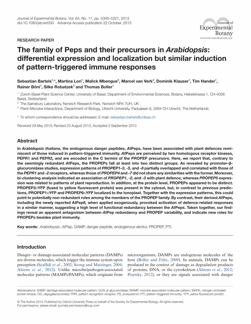
In order to gain insights into the sequence homology of PROPEPs compared with other precursors of plant signal-ling peptides, we searched the Arabidopsis genome and identi-fied AT5G09976 as a new member of the PROPEP family. It clustered with the other seven PROPEPs, despite an overall low sequence homology, and its C terminus contained the conserved AtPep motif SSG-x2-G-x2-N (Fig. 1A). According to sequence similarity, PROPEP4 was the closest homologue of AT5G09976. Moreover, addition of a synthetic peptide based on the last 23 aa of AT5G09976 (Fig. 1A, highlighted with a red bar) triggered similar responses in Arabidopsis plants to the other AtPeps (see below). Thus, we designated AT5G09976 as PROPEP8. Further searches for non-anno-tated sequences with similarity to the PROPEPs did not reveal any further PROPEP in Arabidopsis.
Bi-clustering expression analysis indicates distinct roles for individual PROPEPs
PROPEPs are thought to assist via the release of AtPeps in biotic stress resistance, but their individual roles have not been investigated in detail (Huffaker and Ryan, 2007; Boller and Felix, 2009). Whereas AtPeps are assumed to act rather redundantly, little is known about the spatial and temporal expression of PROPEPs. It has been shown that PROPEPs respond with slight differences to treatments with methyl jas-monate, methyl salicylate, and AtPeps (Huffaker and Ryan, 2007). In order to get a better idea about potential redun-dance as well as specific expression patterns of the PROPEPs in the context of biotic stress resistance, we performed a bi-clustering analysis focusing on 278 biotic stress-related micro-arrays that were downloaded from the TAIR website (ftp://ftp.arabidopsis.org/Microarrays/analyzed_data). Thus, the 22 810 probes (representing genes) present on the Affymetrix 25K arrays used were grouped based on their expression pat-terns over the various biotic stress treatments. Genes with similar expression patterns grouped more closely together, as indicated by the dendrogram, and became part of a subgroup (subclusters). Enrichment of GO terms within one subcluster could be used to get indications about the function of the genes in this subcluster. Moreover, the relative distance of genes within the main cluster showed the diversity of regula-tion of these genes.
5312 | Bartels et al.
PROPEP-containing clusters were selected by setting an individual cut-off within the dendrogram for each PROPEP gene to yield a cluster with <500 genes (Fig. 2). PROPEP7 and PROPEP8 are not spotted on the arrays used, and therefore no bi-cluster analysis could be performed for these precursors. Of the family members PROPEP1–6, only PROPEP2 and -3 clustered together, indicating that most of the PROPEPs are expressed in different ways upon treatment with various biotic stimuli. To get an indication of which processes the individual PROPEPs are involved in, a GO term enrichment analysis was performed on the obtained clusters, the top five terms of which are shown in Fig. 2. Most enriched GO terms within the top five of each cluster represented a relatively broad description of a process. To also provide data on the more specific pro-cesses that are underlying the expression of these clusters, a full overview of all major and minor enriched GO terms for each PROPEP is provided in Supplementary Table S1 at JXB online. As most PROPEPs appeared to be involved in very different processes besides biotic stress resistance, a co-expression analysis followed by a GO term analysis on a set of abiotic or development-related microarrays was also performed (Supplementary Table S2 at JXB online). These results further supported the idea that PROPEP transcrip-tion seems to be regulated individually and does not follow a general pattern valid for all PROPEPs.
The PROPEP that had the most similar global expression pattern compared with PROPEP2 and -3 was PROPEP1, but, besides the shared enriched defence-associated GO terms (Fig. 2), they also had some characteristically differ-ent enriched GO terms. The PROPEP1 cluster revealed an additional enrichment of GO terms related to abiotic stress, hypoxia, and abscisic acid signalling, whereas the PROPEP2 and -3 cluster was also associated with salicylic acid signal-ling, (programmed) cell death, and (transmembrane) ion transport (Supplementary Tables S1 and S2). Distinct from these more defence-associated PROPEPs was the cluster of PROPEP5 that was enriched for processes related to repro-duction and shared the enrichment for gibberellin/terpe-noid biosynthesis and lipid signalling with PROPEP6. The most directly noticeable different PROPEP in Fig. 2 was PROPEP4, whose expression was induced in conditions where all the other PROPEPs were repressed and vice versa.
As the cluster of PROPEP4 contained only 25 genes, it was too small to result in any enriched GO terms. To circumvent this, the genes that were co-expressed with PROPEP4 given a Pearson correlation coefficient cut-off of >0.60 were used to perform a GO term enrichment analysis. This resulted in an enrichment of organismal development-, developmental pro-cesses-, and chromosome/chromatin organization-associated GO terms.
To further diversify the view on PROPEP regulation, we also analysed the type of treatments and/or conditions that had the strongest influence on the expression of each PROPEP; a full overview of treatments, conditions, and their influence on expression is given in Supplementary Table S3 at JXB online. Here we found, in agreement with the bi-clustering analysis, PROPEP1 to be highly induced by abi-otic stress treatments like salt, drought, and osmotic stress, whereas for example, PROPEP5 transcription was highest in certain developmental stages of seeds and flowers.
Overall, our analysis indicated that, in contrast to AtPeps, the transcriptional regulation of PROPEPs is most likely non-redundant. Moreover, based on these findings, we suggest that individual PROPEPs could play a role in very distinct functions in Arabidopsis, as they appear to be associated not only with defence but also with processes ranging from abiotic stress resistance to development and reproduction.
Analysis of PROPEP promoters reveals diverse spatial and temporal expression patterns
To investigate further the potential difference in PROPEP expression at the tissue level, we generated transgenic Arabidopsis lines containing the putative promoter sequences of the PROPEP genes fused to GUS. As shown in Fig. 3, the promoters of PROPEP1, -2, and -3 exhibited similar expression patterns. These promoters conferred expression mainly in the root excluding the root tip. In adult leaves, even after 24 h of staining, nearly no blue pre-cipitate was visible indicating, very low activity of these promoters without stimuli. In contrast, wounding of leaves using forceps led to a clear induction of these PROPEP promoters, which was restricted to the vasculature (Fig. 3, yellow arrows). Besides the great overlap between the
Fig. 1. Alignment of the eight Arabidopsis PROPEPs. ClustalW alignment of the amino acid sequences of all identified Arabidopsis PROPEPs including AT05G09976. Colouring is based in the Clustal colour scheme. (This figure is available in colour at JXB online.)

Characterization of the Arabidopsis PROPEP family | 5313
expression patterns of the promoters of PROPEP1, -2, and -3, the latter also produced GUS staining in the anthers of flowers.
The promoters of PROPEP5 and -8 were also active in the root but restricted to the vascular tissue, reminiscent of the promoter of PEPR2 (see below). They shared with the pro-moters of PROPEP1–3 the wound inducibility in the central vasculature of adult leaves. However, whereas the promoter of PROPEP5 showed strong activity in the leaf veins, the promoter of PROPEP8 did not produce any GUS stain-ing in untreated leaves. In addition, they produced distinct
staining in adult flowers. The promoter of PROPEP5 was highly active in the filaments of flowers (Fig. 3, white arrow), whereas that of PROPEP8 was active in all flower tissues except for the petals. Thus, the promoters of PROPEP5 and -8 partially shared their expression patterns with those of the promoters of PROPEP1–3 but also showed differences from them and among each other.
Intriguingly, the activity of the promoters of PROPEP4 and -7 were restricted to the tips of primary and lateral roots (Fig. 3, red arrows), whereas neither the other PROPEP promoters nor the promoters of PEPR1 and -2 (see below)
Fig. 2. Bi-clustering analysis of PROPEPs based on expression profiles of biotic stress treatments. The similarity in expression pattern of 22 810 different probes (representing genes) was assessed by performing a bi-clustering analysis of the log2-transformed expression values from 278 biotic stress-related microarrays (upregulated genes are represented in yellow, whereas downregulated genes are coloured blue). The different types of treatments within this bi-clustering analysis are colour coded above the clusters, with their details at the bottom of the figure. For multiple treatments, typical examples are given, as each individual treatment could not be colour coded clearly. A full list of all treatments, including the dendrogram and the same colour coding, is given in Supplementary Table S2. Genes that cluster relatively closely are expressed similarly under various biotic stresses and vice versa. Only PROPEP2 and -3 clustered very closely together, suggesting that only these two PROPEPs are involved in similar processes under biotic stress. To obtain an indication of which processes each PROPEP is involved in, GO term enrichment was performed on each subcluster containing a PROPEP (represented as separate clusters). The top five enriched GO terms of the subcluster, indicating the related processes, is shown to the right of each subcluster. Asterisks denote subclusters that showed no enriched GO terms; therefore, co-expressed genes with the PROPEP having a Pearson correlation coefficient of >0.6 were used for GO term enrichment analysis.

5314 | Bartels et al.
conferred any obvious GUS expression. Moreover, expres-sion of the promoters of PROPEP4 and -7 was not detected in flowers and was not induced by wounding.
Taken together, the promoter-mediated expression pat-terns of the PROPEPs fell clearly into two distinct groups. Group 1, which comprised the promoters of PROPEP1, -2, -3, -5, and -8, showed expression in the roots and slightly in the leaf vasculature. They were inducible by wounding. Group 2, containing the promoters of PROPEP4 and -7, was not inducible by wounding and the basal expression was restricted to the root tips.
PROPEP::YFP fusions identify localization to distinct subcellular compartments
Next we generated transgenic Arabidopsis plants con-stitutively expressing PROPEP::YFP fusion proteins to assess their subcellular localization. It has been hypoth-esized that all PROPEPs localize to the cytoplasm based on the predicted function and the lack of an identifiable localization signal (Huffaker et al., 2006). In line with this hypothesis, PROPEP3::YFP localized to the cytoplasm (Fig. 4A). However, our findings with PROPEP1::YFP and
Fig. 3. Spatial and temporal expression patterns of PROPEP promoter–GUS lines. Fusion of putative promoter sequences of indicated PROPEPs to a GUS reporter revealed distinct expression patterns. Pictures show staining (2 h) of untreated 10-d-old seedlings grown in sterile conditions on MS plates and adult leaves, and flowers of soil-grown plants (24 h staining). Wounding of adult leaves was done with forceps and the plants were incubated for 2 h before staining. Red arrows indicate expression in root tips of the primary and lateral roots. Yellow arrows indicate GUS staining in the vasculature after wounding. White arrows highlight flowers with GUS expression. Three independent transgenic lines were analysed for each construct, showing similar results.

Characterization of the Arabidopsis PROPEP family | 5315
PROPEP6::YFP were rather surprising and showed that these precursor proteins were associated with the tonoplast. To clearly distinguish the tonoplast from the plasma mem-brane, we performed a brief FM4-64 staining (Fig. 4A, red), which is often used to image the plasma membrane. The
overlay confirmed that the YFP fluorescence and that emit-ted from FM4-64 did not overlap.
To exclude the possibility that subcellular localization was dependent on the cell type, we imaged epidermal cells of cotelydons as well as root epidermal cells and observed that
Fig. 4. Subcellular localization of PROPEP::YFP fusion proteins. (A, B) Confocal micrographs of Arabidopsis transgenic lines, expressing PROPEP::YFP (A) and Pep1::YFP (B) fusion proteins as indicated under the control of the cauliflower mosaic virus 35S promoter showing single optical sections of cotyledon epidermal cells (top panel) or root epidermal cells (bottom panel). Co-staining with FM4-64 (red channel) highlights the plasma membrane (arrowheads). PROPEP1 and PROPEP6::YFP fusions localize to the tonoplast in both tissues (right panels - arrows) while PROPEP3::YFP (left panel) and Pep1::YFP (B) fusion protein localized to the cytosol in both tissues. Similar results were obtained in two independent transgenic lines for each construct. Bars, 10 μm. (C) Plasmolysis of root cells after 2 min of 500 mM NaCl treatment. DIC, differential interference contrast.

5316 | Bartels et al.
the localization patterns were the same. In contrast, a fusion protein of just the C-terminal part of PROPEP1, which rep-resents AtPep1, with YFP produced a cytosolic localization, indicating that the association of PROPEP1 with the tono-plast seemed to depend on the N-terminal part of PROPEP1 and was not due to binding of AtPep1 to an as yet unidenti-fied tonoplast-localized protein (Fig. 4B).
In order to test further the association of PROPEP1::YFP with the tonoplast, we performed plasmolysis triggered by a brief treatment with 500 mM NaCl. As shown in Fig. 4C, the YFP fluorescence remained at the tonoplast of the shrunken vacuoles.
These findings demonstrated that members of the PROPEP family are present at two different subcellular compartments, the cytosol and the tonoplast. This might indicate non-redun-dant functions between the PROPEPs at the protein level or an as yet not understood level of complexity of their involve-ment in cellular immunity. Moreover, it provides evidence for a potential role of PROPEP1 and -6 associated with the vacuole.
The promoters of PEPR1 and -2 confer overlapping patterns of expression, which resemble those of some but not all PROPEP promoters
AtPeps are known to be detected by two homologous recep-tors, PEPR1 and PEPR2 (Krol et al., 2010; Yamaguchi et al., 2010). To investigate the potential overlap of the expression patterns between the two PEPRs and the PROPEPs, we generated transgenic Arabidopsis lines containing the puta-tive promoter sequences of the PEPR promoters fused to GUS. As shown in Fig. 5, both promoters conferred expres-sion in the vascular tissue of roots and leaves. No PEPR1/2 promoter-mediated GUS expression was observed in root
tips. Focusing on the expression in roots, the activity of the PEPR2 promoter was more restricted to the central cylinder of the root, whereas GUS expression of the PEPR1 promoter was present in most root tissues. Additionally, GUS expres-sion mediated by the PEPR1/2 promoters was detected in stems but was almost absent in flowers.
When comparing the expression patterns between the receptors and the precursors, the PEPR1/2 promoter-medi-ated expression showed partially overlapping patterns with PROPEP1, -2, -3, -5, and -8. By contrast, PROPEP4 and -7 promoter-mediated expression was exclusive to the root tip, a tissue where the PEPRs were not expressed. These results showed that, whereas the promoters of PEPR1 and -2 signifi-cantly overlapped in their conferred expression patterns, they shared only a small overlap with the expression patterns gen-erated by the PROPEP promoters. This indicates potential new, unknown roles for at least PROPEP4 and -7 independ-ent of PEPRs.
PEPR1 and -2, as well as all eight AtPeps, trigger similar defence responses
Previous studies showing that AtPeps triggered alkalinization in cell cultures and induced resistance to P. syringae infection in plants provided evidence that some AtPeps are function-ally redundant (Huffaker and Ryan, 2007; Yamaguchi et al., 2010). To address the extent of functional redundancy among all known AtPeps, we monitored the activation of MAPK, the release of ethylene, and the inhibition of seedling growth stimulated by the eight AtPeps in the single and double pepr1 pepr2 receptor mutants. As shown in Fig. 6, all eight AtPeps activated the stress-related MAPKs MPK3 and -6, induced the production of ethylene, and inhibited seedling growth in a PEPR1- and partially PEPR2-dependent manner. Notably,
Fig. 5. Overlapping expression patterns of PEPR1 and -2 promoter–GUS lines. Putative promoter sequences of PEPR1 and -2 were fused to GUS and stably introduced into Arabidopsis plants. Tissues of transgenic plants were stained for 2 h (roots and seedlings) or 24 h (adult leaves, stems, and flowers). GUS staining revealed a significant overlap in the tissue-dependent expression of PEPR1 and -2. Three independent transgenic lines were analysed for each construct. Pictures show representative samples.

Characterization of the Arabidopsis PROPEP family | 5317
AtPep3–8 were not perceived in the pepr1 mutant, indicat-ing that PEPR2, which is active in this mutant, does not per-ceive these peptides and thus is specific for AtPep1 and -2, whereas the pepr2 mutant responded to all peptides, indicat-ing that PEPR1 recognizes all eight AtPeps in a similar way (Fig. 6). Taken together, all eight AtPeps triggered a similar set of defence responses reminiscent of PTI in a PEPR1- and partially PEPR2-dependent manner. Thus, in contrast to the PROPEPs, the AtPeps as well as PEPRs appear to be highly redundant.
Discussion
Current models discuss PROPEPs and AtPeps as: (i) enhanc-ers of immunity; (ii) damage-signalling peptides; and (iii) elicitors of systemic defence responses, but based on pub-lished data, reliable support for each model is scarce (Boller and Felix, 2009; Yamaguchi and Huffaker, 2011). Previous studies focused primarily on plant responses triggered by the addition of the synthetically produced peptides AtPep1 or -3 and firmly established that treatment with these pep-tides enhances plant immunity via PEPRs (Krol et al., 2010; Yamaguchi et al., 2010). Likewise, the constitutive, ubiqui-tous expression of PROPEP1 or -2 improved plant resistance to an oomycete pathogen (Huffaker et al., 2006). However, these studies did not address the question of the presence or the underlying mechanism of the PROPEP/AtPep/PEPR sys-tem and thus cannot fully answer which (if any) of the cur-rent models is valid.
Recently, two studies involving the pepr1 pepr2 double mutant suggested an interaction of AtPep signalling with the defence hormone ethylene to maintain PTI responses (Liu et al., 2013; Tintor et al., 2013). Thus, the ‘enhancer of immunity’ model appears now to be the most likely one. Here, we investigated the presence and regulation of PROPEPs to either further substantiate the ‘enhancer model’ or to deduce new biological role(s) of the PROPEP/AtPep/PEPR system.
PROPEP1, -2, -3, and maybe -5 and -8 play a role in immunity
In agreement with previous works (Huffaker and Ryan, 2007; Yamaguchi et al., 2010), our bi-clustering showed that PROPEP1, -2, and -3 clustered together with genes implicated in plant defence. Moreover, the almost exclusive expression of these PROPEPs in the roots revealed by promoter::GUS fusions partially overlapped with those of PEPR1 and -2. Thus, PROPEP1, -2, and -3 might play specific roles in the immune response of the root, which is supported by the report that constitutive expression of PROPEP1 led to an induced resistance against the oomycete root pathogen Pythium irregulare (Huffaker and Ryan, 2007). In contrast, these PROPEPs are not or are only weakly expressed in adult leaves but are rapidly induced in wounded leaf veins. Recently, we showed that pre-treatment of leaf tissue with bacterial MAMPs led to enhanced output of reactive oxy-gen species triggered by AtPep perception (Flury et al., 2013).
Since a progressive wave of reactive oxygen species has been discussed as a potential systemic signal, the enhanced expres-sion of PROPEPs in wounded vasculature might contribute to the robustness of this system (Miller et al., 2009; Mittler et al., 2011).
PROPEP5 and -8 displayed expression patterns that partially overlapped with those of PEPR1 and -2, but, in contrast to PROPEP1, -2, and -3, PROPEP5 and -8 were restricted to the root vasculature but were more expressed in the leaf veins (PROPEP5) and the flowers (PROPEP8). However, PROPEP1, -2, -3, -5, and -8 together cover most plant tissues and, since all eight AtPeps triggered redundant responses, PROPEP5 and -8 could play a role in leaves and flowers, respectively, similar to the roles of PROPEP1, -2, and -3 in roots.
It has been hypothesized that PROPEPs are located to the cytoplasm and could be released into the extracellular space in a situation of danger using unconventional protein secre-tion mechanisms (Ding et al., 2012). We indeed found that PROPEP3::YFP was localized in the cytoplasm, but sur-prisingly PROPEP1::YFP as well as PROPEP6::YFP were detected at the tonoplast. Due to the acidic environment of the vacuole negatively impacting on YFP fluorescence, we assume that PROPEP1::YFP and PROPEP6::YFP are asso-ciated with the cytoplasmic side of the vacuolar membrane. Notably, the localization signal that directs the PROPEP to the tonoplast or to a hitherto unidentified interaction domain that could attach the PROPEP to a tonoplast-localized pro-tein, resides in the N terminus of the PROPEP, since a fusion protein of only AtPep1 and YFP localized in the cytoplasm. Therefore, it can be excluded that the AtPep itself binds to a tonoplast-localized receptor-like protein. Recently, it was shown that infection of Arabidopsis with the compatible oomycete Hyaloperonospora arabidopsidis, the causal agent of the downy mildew, triggered rearrangement of intracellu-lar membranes leading to relocation of the tonoplast close to the extra-haustorial membrane (Caillaud et al., 2012). However, neither the involvement of PROPEPs in resistance to H. arabidopsidis nor the necessity of a tonoplast locali-zation of PROPEP1 in the context of resistance to Pythium infection has yet been shown, but it will be interesting to study this potential connection.
Root tip-expressed PROPEP4 and -7 are distinct from the other PROPEPs and may have dual functions
PROPEP4 and -7 are located on chromosome 5 within an ~3.5 kb stretch. Both share specific expression in the tips of primary and lateral roots, which does not overlap with that of PEPR1 and -2. Moreover, they are currently the only PROPEPs that are not induced by wounding. Therefore, they are less likely to enhance plant immune responses locally. However, PROPEP4 and -7 could still be part of a systemic defence response. It has been reported that the systemin peptide is transported via phloem sap (Narváez-Vásquez et al., 1995). Moreover, a plethora of peptide transporters are encoded in the Arabidopsis genome and might facili-tate the transport of AtPeps for systemic signalling (Stacey

5318 | Bartels et al.
Fig. 6. Defence responses activated by all eight AtPeps and both PEPRs. (A) MAPK phosphorylation. Seedlings of the indicated genotypes were treated for 15 min with 1 μM of the indicated elicitor peptide or without any peptide (contr.). MAPK phosphorylation was detected by immunoblotting using an anti-phospho-p44/42-MAPK antibody detecting the pTE-pY motif of MPK6 and -3. The immunoblot was reprobed with anti-actin antibody to determine equal loading. (B) Ethylene production. Seedlings of the indicated genotypes were treated for 5 h with 1 μM of the indicated elicitor peptides or without any peptide (control). Columns represent averages of detected ethylene values of five biological replicates. Error bars indicate SEM.(C) Seedling growth inhibition. Five-d-old seedlings of the indicated genotypes were treated for 5 d with 1 μM of the indicated elicitor or without any peptide. Columns represent the mean weight of 12 seedlings out of six biological replicates. Error bars indicate SEM. Asterisks represent t-test results generated by comparing the labelled value with the respective control **P<0.01; ns, not significant). (This figure is available in colour at JXB online.)

Characterization of the Arabidopsis PROPEP family | 5319
et al., 2002). Thus, PROPEP4 and -7 could be ideal candi-dates to study whether PROPEPs or AtPeps are transported systemically.
The Affymetrix 25K microarrays do not represent PROPEP7. Our bi-clustering analysis produced only a small cluster of 25 genes that contained PROPEP4. Intriguingly, this cluster showed an expression pattern opposite to those of the other PROPEP-containing clusters, meaning that whenever biotic stress treatments lead to an induction of PROPEP4 expression, other PROPEPs are downregulated and vice versa. GO term enrichment indicates biological processes including chromatin and chromosome organization. However, this does not exclude a function in immunity. The mammalian DAMP high-mobility group protein B1 (HMGB1) binds to DNA, modifies the shape, and regulates transcription. In case of danger, it can be secreted by activated monocytes and macrophages, or it is passively released by necrotic or damaged cells. Detection of extracellu-lar HMGB1 by RAGE (receptor for advanced glycation end products) of adjacent cells triggers inflammation (Scaffidi et al., 2002; Sims et al., 2010).
The small PROPEP4-including gene cluster also shows the limitations of the bi-clustering. Most of the arrays used were probed with samples based on seedlings or adult leaves. Genes with tissue-restricted expression patterns like PROPEP4 might only be detected weakly on some of the biotic stress arrays, leading to erroneous expression patterns.
Taken together, PROPEP4 (and PROPEP7) are clearly dis-tinct from the other PROPEPs in terms of tissue expression pattern as well as regulation within the biotic stress treat-ments. A more detailed analysis is needed to uncover their biological roles.
PROPEPs may play roles in plant reproduction and development
Most plant signalling peptides originate from small (~100 aa) proteins, which are processed at the C terminus to release the active signalling peptide. These peptides have various functions, especially in developmental processes such as apical meristem development as well as root growth (Matsubayashi and Sakagami, 2006; Katsir et al., 2011). PROPEPs have been associated with plant innate immunity but share structural similarities (size and presence of signal-ling peptide in the C terminus) with Arabidopsis signalling peptide precursors like RGF1, TDIF, CLV3, PSK1, CEP1, and PSY1. Remarkably, there may be also a functional overlap. In contrast to PROPEP1, -2, and -3, bi-cluster-ing showed that PROPEP5 clusters with genes associated with plant reproduction. Although we did not find this for PROPEP3, GUS analysis revealed expression of both in the stamen. Thus, beside the proposed role in plant immunity, PROPEP5 and maybe also PROPEP3 could be involved in the development of the stamen and therewith in the regula-tion of reproduction. The involvement of small signalling peptides in this process has been demonstrated just recently. RALF (rapid alkalinization factor) signalling peptides regu-late pollen-tube elongation and development of the female gametophyte in Solanaceous species (Covey et al., 2010;
Chevalier et al., 2013). Thus, as a next step, a detailed analy-sis of PROPEP3 and -5 knockout mutants would be needed to investigate a potential role of these PROPEPs in plant reproduction.
Besides the impact of constitutive expression of PROPEP1 on resistance against Pythium infection, it also promoted an increase in root biomass production (Huffaker et al., 2006). It was assumed that PROPEP1 expression somehow generated an advantage for Arabidopsis roots to grow in soil. In con-trast, exogenous application of AtPeps blocked root growth and biomass production similarly to seedling growth inhibi-tion triggered by MAMPs. Notably, application of AtPeps has a more pronounced negative effect on root growth com-pared with MAMPs (Krol et al., 2010).
Whether root growth is also enhanced in sterile condi-tions by constitutive expression of PROPEP1 has been not assessed (Huffaker et al., 2006); thus, this advantage may or may not be based on an increased pathogen resistance of the root. A detailed analysis of propep1 knockout mutants is needed to clarify whether PROPEP1 takes part in additional processes like root development.
Conclusions
Previous studies and our new data reported here show that all eight AtPeps trigger a PTI-like response by binding to either PEPR1 or both PEPR1/2 receptors (Huffaker and Ryan, 2007; Yamaguchi et al., 2010). Interestingly, PEPR2 is spe-cific for AtPep1 and AtPep2, whereas PEPR1 is non-specific and recognizes all eight AtPeps.
In contrast to the AtPeps and the PEPRs, we have pro-vided data indicating that PROPEPs are probably not redun-dant. They show individual spatial and temporal expression patterns and localize to distinct subcellular compartments. Besides their potentially diverse roles in innate immunity, they may additionally be involved in plant development and reproduction. A detailed characterization of each PROPEP, together with an analysis of their processing and release, will be necessary to uncover the full array of functions of PROPEPs in plant biology.
Supplementary data
Supplementary data are available at JXB online.Supplementary Table 1. GO term enrichment for selected
PROPEP-containing clusters in Fig. 2.Supplementary Table 2. GO term enrichment for genes co-
expressed with PROPEPs under biotic, abiotic or develop-mental conditions.
Supplementary Table 3. Conditions and/or treatments influencing the expression of each PROPEP.
Supplementary Table 4. Primer and promoter sequences.
Acknowledgements
We thank Etienne Bucher for help with sequence analy-sis and critical reading of the manuscript. This work was

5320 | Bartels et al.
supported by the Swiss National Science Foundation (grant 31003A_127563 to TB) and by stipends to SB from the European Molecular Biology Organisation (EMBO: ALTF 61–2010) and the Leopoldina Fellowship Programme of the National Academy of Science Leopoldina (LPDS 2009–35). Research in SR’s laboratory is supported by the Gatsby Charitable Foundation and by a grant of the European Research Council (ERC). The research of MvV is supported by a ERC Advanced Investigator Grant (no. 269072). The authors declare that there is no conflict of interests.
References
Ahrens S, Zelenay S, Sancho D, et al. 2012. F-actin is an evolutionarily conserved damage-associated molecular pattern recognized by DNGR-1, a receptor for dead cells. Immunity 36, 635–645.
Avilés-Arnaut H, Délano-Frier JP. 2012. Characterization of the tomato Prosystemin promoter: organ-specific expression, hormone specificity and methyl jasmonate responsiveness by deletion analysis in transgenic tobacco plants. Journal of Integrative Plant Biology 54, 15–32.
Boller T, Felix G. 2009. A renaissance of elicitors: perception of microbe-associated molecular patterns and danger signals by pattern-recognition receptors. Annual Review of Plant Biology 60, 379–406.
Caillaud M-C, Piquerez SJM, Fabro G, Steinbrenner J, Ishaque N, Beynon J, Jones JDG. 2012. Subcellular localization of the Hpa RxLR effector repertoire identifies a tonoplast-associated protein HaRxL17 that confers enhanced plant susceptibility. The Plant Journal 69, 252–265.
Carbon S, Ireland A, Mungall CJ, Shu S, Marshall B, Lewis S, AmiGO Hub, Web Presence Working Group. 2009. AmiGO: online access to ontology and annotation data. Bioinformatics 25, 288–289.
Chevalier E, Loubert-Hudon A, Matton DP. 2013. ScRALF3, a secreted RALF-like peptide involved in cell-cell communication between the sporophyte and the female gametophyte in a solanaceous species. The Plant Journal 73, 1019–1033
Clough SJ, Bent AF. 1998. Floral dip: a simplified method for Agrobacterium-mediated transformation of Arabidopsis thaliana. The Plant Journal 16, 735–743.
Covey PA, Subbaiah CC, Parsons RL, Pearce G, Lay FT, Anderson MA, Ryan CA, Bedinger PA. 2010. A pollen-specific RALF from tomato that regulates pollen tube elongation. Plant Physiology 153, 703–715.
D’Ovidio R, Mattei B, Roberti S, Bellincampi D. 2004. Polygalacturonases, polygalacturonase-inhibiting proteins and pectic oligomers in plant–pathogen interactions. Biochimica Et Biophysica Acta—Proteins and Proteomics 1696, 237–244.
Ding Y, Wang J, Wang J, Stierhof Y-D, Robinson DG, Jiang L. 2012. Unconventional protein secretion. Trends in Plant Science 17, 606–615.
Earley KW, Haag JR, Pontes O, Opper K, Juehne T, Song K, Pikaard CS. 2006. Gateway-compatible vectors for plant functional genomics and proteomics. The Plant Journal 45, 616–629.
Flury P, Klauser D, Schulze B, Boller T, Bartels S. 2013. The anticipation of danger: MAMP perception enhances AtPep-triggered oxidative burst. Plant Physiology 161, 2023–2035.
Holton N, Harrison K, Yokota T, Bishop GJ. 2008. Tomato BRI1 and systemin wound signalling. Plant Signaling & Behavior 3, 54–55.
Hu Z-L, Bao J, Reecy JM. 2008. CateGOrizer: A web-based program to batch analyze gene ontology classification categories. Online Journal of Bioinformatics 9, 108–112.
Huffaker A, Pearce G, Ryan CA. 2006. An endogenous peptide signal in Arabidopsis activates components of the innate immune response. Proceedings of the National Academy of Sciences, USA 103, 10098–10103.
Huffaker A, Ryan CA. 2007. Endogenous peptide defense signals in Arabidopsis differentially amplify signaling for the innate immune response. Proceedings of the National Academy of Sciences, USA 104, 10732–10736.
Karimi M, Inzé D, Depicker A. 2002. GATEWAY vectors for Agrobacterium-mediated plant transformation. Trends in Plant Science 7, 193–195.
Katsir L, Davies KA, Bergmann DC, Laux T. 2011. Peptide signaling in plant development. Current Biology 21, 356–364.
Krol E, Mentzel T, Chinchilla D, et al. 2010. Perception of the Arabidopsis danger signal peptide 1 involves the pattern recognition receptor AtPEPR1 and its close homologue AtPEPR2. Journal of Biological Chemistry 285, 13471–13479.
Lanfermeijer FC, Staal M, Malinowski R, Stratmann JW, Elzenga JTM. 2008. Micro-electrode flux estimation confirms that the Solanum pimpinellifolium cu3 mutant still responds to systemin. Plant Physiology 146, 129–139.
Liu Z, Wu Y, Yang F, Zhang Y, Chen S, Xie Q, Tian X, Zhou J-M. 2013. BIK1 interacts with PEPRs to mediate ethylene-induced immunity. Proceedings of the National Academy of Sciences, USA 110, 6205–6210.
Malinowski R, Higgins R, Luo Y, Piper L, Nazir A, Bajwa VS, Clouse SD, Thompson PR, Stratmann JW. 2009. The tomato brassinosteroid receptor BRI1 increases binding of systemin to tobacco plasma membranes, but is not involved in systemin signaling. Plant Molecular Biology 70, 603–616.
Matsubayashi Y, Sakagami Y. 2006. Peptide hormones in plants. Annual Review of Plant Biology 57, 649–674.
McGurl B, Pearce G, Orozcocardenas M, Ryan CA. 1992. Structure, expression, and antisense inhibition of the systemin precursor gene. Science 255, 1570–1573.
Miller G, Schlauch K, Tam R, Cortes D, Torres MA, Shulaev V, Dangl JL, Mittler R. 2009. The plant NADPH oxidase RBOHD mediates rapid systemic signaling in response to diverse stimuli. Science Signaling 2, 1–10.
Mittler R, Vanderauwera S, Suzuki N, Miller G, Tognetti VB, Vandepoele K, Gollery M, Shulaev V, Van Breusegem F. 2011. ROS signaling: the new wave? Trends in Plant Science 16, 300–309.
Narváez-Vásquez J, Pearce G, Orozco-Cardenas M, Franceschi V, Ryan C. 1995. Autoradiographic and biochemical evidence for

Characterization of the Arabidopsis PROPEP family | 5321
the systemic translocation of systemin in tomato plants. Planta 195, 593–600.
Narváez-Vásquez J, Ryan C. 2004. The cellular localization of prosystemin: a functional role for phloem parenchyma in systemic wound signaling. Planta 218, 360–369.
Pearce G, Strydom D, Johnson S, Ryan CA. 1991. A polypeptide from tomato leaves induces wound-inducible proteinase inhibitor proteins. Science 253, 895–897.
Pisetsky DS. 2012. The origin and properties of extracellular DNA: from PAMP to DAMP. Clinical Immunology 144, 32–40.
Qi Z, Verma R, Gehring C, Yamaguchi Y, Zhao YC, Ryan CA, Berkowitz GA. 2010. Ca2+ signaling by plant Arabidopsis thaliana Pep peptides depends on AtPepR1, a receptor with guanylyl cyclase activity, and cGMP-activated Ca2+ channels. Proceedings of the National Academy of Sciences, USA 107, 21193–21198.
Rasul S, Dubreuil-Maurizi C, Lamotte O, Koen E, Poinssot B, Alcaraz G, Wendehenne D, Jeandroz S. 2012. Nitric oxide production mediates oligogalacturonide-triggered immunity and resistance to Botrytis cinerea in Arabidopsis thaliana. Plant, Cell & Environment 35, 1483–1499.
Ryan CA, Pearce G. 2003. Systemins: a functionally defined family of peptide signal that regulate defensive genes in Solanaceae species. Proceedings of the National Academy of Sciences, USA 100, 14577–14580.
Scaffidi P, Misteli T, Bianchi ME. 2002. Release of chromatin protein HMGB1 by necrotic cells triggers inflammation. Nature 418, 191–195.
Seong S-Y, Matzinger P. 2004. Hydrophobicity: an ancient damage-associated molecular pattern that initiates innate immune responses. Nat Rev Immunol 4, 469–478.
Sims GP, Rowe DC, Rietdijk ST, Herbst R, Coyle AJ. 2010. HMGB1 and RAGE in inflammation and cancer. Annual Review of Immunology 28, 367–388.
Stacey G, Koh S, Granger C, Becker JM. 2002. Peptide transport in plants. Trends in Plant Science 7, 257–263.
Tintor N, Ross A, Kanehara K, Yamada K, Fan L, Kemmerling B, Nürnberger T, Tsuda K, Saijo Y. 2013. Layered pattern receptor signaling via ethylene and endogenous elicitor peptides during Arabidopsis immunity to bacterial infection. Proceedings of the National Academy of Sciences, USA 110, 6211–6216.
van de Veerdonk FL, Netea MG, Dinarello CA, Joosten LAB. 2011. Inflammasome activation and IL-1 and IL-18 processing during infection. Trends in immunology 32, 110–116.
van Verk M, Bol J, Linthorst H. 2011. Prospecting for genes involved in transcriptional regulation of plant defenses, a bioinformatics approach. BMC Plant Biology 11, 88.
Wang H, Bloom O, Zhang M, et al. 1999. HMG-1 as a late mediator of endotoxin lethality in mice. Science 285, 248–251.
Yamaguchi Y, Huffaker A, Bryan AC, Tax FE, Ryan CA. 2010. PEPR2 is a second receptor for the Pep1 and Pep2 peptides and contributes to defense responses in Arabidopsis. Plant Cell 22, 508–522.
Yamaguchi Y, Huffaker A. 2011. Endogenous peptide elicitors in higher plants. Current Opinion in Plant Biology 14, 351–357.