The Expression of Cytokeratins in Bovine Intestinal Microfold
14
1 The Expression of Cytokeratins in Bovine Intestinal Microfold (M) Cells Takashi Kanaya 1 , Tetsuya Hondo 1 , Kohtaro Miyazawa 1 , Michael T. Rose 2 and Hisashi Aso 1 1 Tohoku University 2 Aberystwyth University, 1 Japan 2 UK 1. Introduction The mucosal surface of the gut is exposed to a variety of foreign antigens and microorganisms, some of which are potentially harmful for the host. To protect against the risk of infection, the intestinal mucosa has developed specialized organized lymphoid tissues and epithelial cells. The gut-associated lymphoid tissues (GALT) including Peyer’s patches (PPs) are major inductive sites for intestinal immunity. Different from other peripheral lymphoid tissues, GALT lacks afferent lymphatics, and directly samples mucosal antigens across the epithelial barrier to initiate antigen-specific immune responses. This task is accomplished by specialized epithelial cells within the follicle-associated epithelium (FAE) covering the lymphoid follicles of GALT known as microfold (M) cells. M cells possess a high capacity for phagocytosis and transcytosis, and these functions allow the rapid transport of antigens into the underlying lymphoid tissues, especially antigen- presenting cells. Antigens are then presented to T cells that support B-cell activation, resulting ultimately in the generation of IgA-producing plasma cells. Thus, M-cell-mediated antigen transport is important for the initiation of mucosal immune responses (Kraehenbuhl and Neutra, 2000; Neutra et al., 1996; Neutra et al., 2001). In order to express their specialized functions including phagocytosis and transcytosis, M cells exhibit unique morphologies that differ from the surrounding absorptive enterocytes. M cells lack a dense microvilli brush border structure, though they do possess shorter and irregular microvilli on their apical surface. On the basolateral side, a pocket-like invagination of the plasma membrane is formed to house lymphocytes and antigen-presenting cells (Neutra et al., 1996). These morphological features of M cells have an effect on the composition of the cytoskeletal proteins within the cell, such as actin-containing microfilaments, intermediate filaments and their associated proteins. For example, the lack of a brush border in M cells is reflected in the cellular localization of the actin and villin, resulting in unusual staining patterns of these proteins in M cells of mice and calves, as actin and villin are essential proteins for microvilli formation (Kanaya et al., 2007; Kerneis et al., 1996). In addition to the abnormal cellular localization of actin and actin-related proteins, some investigators have demonstrated that intermediate filament proteins such as vimentin and cytokeratins can be used as www.intechopen.com
Transcript of The Expression of Cytokeratins in Bovine Intestinal Microfold

1
The Expression of Cytokeratins in Bovine Intestinal Microfold (M) Cells
Takashi Kanaya1, Tetsuya Hondo1, Kohtaro Miyazawa1, Michael T. Rose2 and Hisashi Aso1
1Tohoku University 2Aberystwyth University,
1Japan 2UK
1. Introduction
The mucosal surface of the gut is exposed to a variety of foreign antigens and microorganisms, some of which are potentially harmful for the host. To protect against the risk of infection, the intestinal mucosa has developed specialized organized lymphoid tissues and epithelial cells. The gut-associated lymphoid tissues (GALT) including Peyer’s patches (PPs) are major inductive sites for intestinal immunity. Different from other peripheral lymphoid tissues, GALT lacks afferent lymphatics, and directly samples mucosal antigens across the epithelial barrier to initiate antigen-specific immune responses. This task is accomplished by specialized epithelial cells within the follicle-associated epithelium (FAE) covering the lymphoid follicles of GALT known as microfold (M) cells.
M cells possess a high capacity for phagocytosis and transcytosis, and these functions allow the rapid transport of antigens into the underlying lymphoid tissues, especially antigen-presenting cells. Antigens are then presented to T cells that support B-cell activation, resulting ultimately in the generation of IgA-producing plasma cells. Thus, M-cell-mediated antigen transport is important for the initiation of mucosal immune responses (Kraehenbuhl and Neutra, 2000; Neutra et al., 1996; Neutra et al., 2001). In order to express their specialized functions including phagocytosis and transcytosis, M cells exhibit unique morphologies that differ from the surrounding absorptive enterocytes. M cells lack a dense microvilli brush border structure, though they do possess shorter and irregular microvilli on their apical surface. On the basolateral side, a pocket-like invagination of the plasma membrane is formed to house lymphocytes and antigen-presenting cells (Neutra et al., 1996).
These morphological features of M cells have an effect on the composition of the cytoskeletal proteins within the cell, such as actin-containing microfilaments, intermediate filaments and their associated proteins. For example, the lack of a brush border in M cells is reflected in the cellular localization of the actin and villin, resulting in unusual staining patterns of these proteins in M cells of mice and calves, as actin and villin are essential proteins for microvilli formation (Kanaya et al., 2007; Kerneis et al., 1996). In addition to the abnormal cellular localization of actin and actin-related proteins, some investigators have demonstrated that intermediate filament proteins such as vimentin and cytokeratins can be used as
www.intechopen.com
Cytokeratins – Tools in Oncology 4
immunohistochemical markers for M cells. Gebert et al. have shown that CK18 is a sensitive marker for porcine M cells (Gebert et al., 1994) while vimentin is selectively expressed in rabbit M cells (Jepson et al., 1992). M cells in rats are detected by monoclonal antibodies raised against CK8 (Rautenberg et al., 1996). These preferential expressions of intermediate filaments in M cells indicate the substantial roles of CKs and vimentin in M cell morphology and function.
In this chapter, we introduce a number of experiments that we have done with respect to the expression of CK18 in bovine M cells. And we further discuss the relationship between the expressions of CKs and the development and apoptosis of bovine intestinal M cells.
2. Material and methods
In this study, we performed several histological and electron microscopical analyses. The monoclonal antibodies for immunohistochemistry are summarized in Table 1. The detailed procedures have been described in our previous reports (Hondo et al., 2011; Miyazawa et al., 2006).
3. Results
3.1 Localization of M cells in FAE of jejunum and ileum PPs
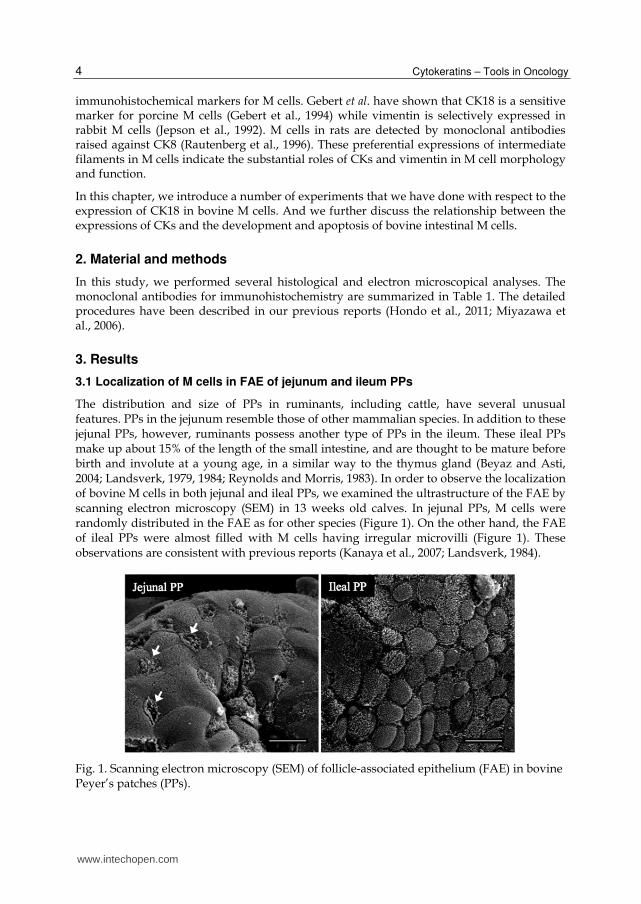
The distribution and size of PPs in ruminants, including cattle, have several unusual features. PPs in the jejunum resemble those of other mammalian species. In addition to these jejunal PPs, however, ruminants possess another type of PPs in the ileum. These ileal PPs make up about 15% of the length of the small intestine, and are thought to be mature before birth and involute at a young age, in a similar way to the thymus gland (Beyaz and Asti, 2004; Landsverk, 1979, 1984; Reynolds and Morris, 1983). In order to observe the localization of bovine M cells in both jejunal and ileal PPs, we examined the ultrastructure of the FAE by scanning electron microscopy (SEM) in 13 weeks old calves. In jejunal PPs, M cells were randomly distributed in the FAE as for other species (Figure 1). On the other hand, the FAE of ileal PPs were almost filled with M cells having irregular microvilli (Figure 1). These observations are consistent with previous reports (Kanaya et al., 2007; Landsverk, 1984).
Fig. 1. Scanning electron microscopy (SEM) of follicle-associated epithelium (FAE) in bovine Peyer’s patches (PPs).
www.intechopen.com

The Expression of Cytokeratins in Bovine Intestinal Microfold (M) Cells 5
The specimens of PPs were fixed with 2.5% glutaraldehyde and coated with platinum-
palladium for SEM analysis. SEM shows the distribution of M cells in the FAE of jejunal PPs
and ileal PPs. Arrows show M cells possessing irregular and sparse microvilli in jejunal PPs.
The FAE of ileal PPs is filled with M cells. Bars = 10 µm.
3.2 Expression of CK18 in bovine M cells
As described above, some intermediate filament proteins, such as CK8, CK18, and vimentin,
are known to be marker for M cells in the intestine. Therefore, we investigated the
expressions of these proteins in bovine PPs (see table 1). As a result of this, we identified
that several monoclonal antibody clones for CK18 were preferentially stained in the FAE
and crypts of both jejunal and ileal PPs (Figure 2, 3A and B). CK20 was detected strongly in
both the villous epithelium and FAE, but not in the crypts. In contrast, CK7, CK8 and CK19
could not be detected in the whole of the small intestinal epithelium, and vimentin was only
detected in the stromal cells of subepithelial tissues (Figure 2 and Table 1). The positive
CK18 signal in the FAE of jejunal and ileal PPs was similar to the distribution of M cells
recognized by SEM. In order to confirm the expression of CK18 in bovine M cells, we
investigated the ultrastructure of CK18-positive cells in the FAE. In jejunal FAE, CK18-
positive cells had irregular and sparse microvilli and pocket-like structures containing
lymphocytes (Figure 3C and E). In the sections of ileal FAE, we clearly observed that CK18-
positive cells had broader microfolds on their apical surface, and CK18-negative cells had
dense microvilli (Figure 3D and F). In addition to the preferential expression of CK18 in M
cells, CK18 was also detected in the crypts (Figure 2). Therefore, we investigated the
proliferative activity of CK18-positive cells in the crypt using the mirror section technique. A
couple of mirror sections revealed that all Ki-67 positive proliferative cells in the crypt were
positive for CK18 (Figure 4). These results suggest that CK18 is a marker for M cells in the
both jejunal and ileal FAE, and proliferative cells in the crypts of the bovine small intestine.
Fig. 2. Expression of cytokeratins and vimentin in bovine PPs.
www.intechopen.com
Cytokeratins – Tools in Oncology 6
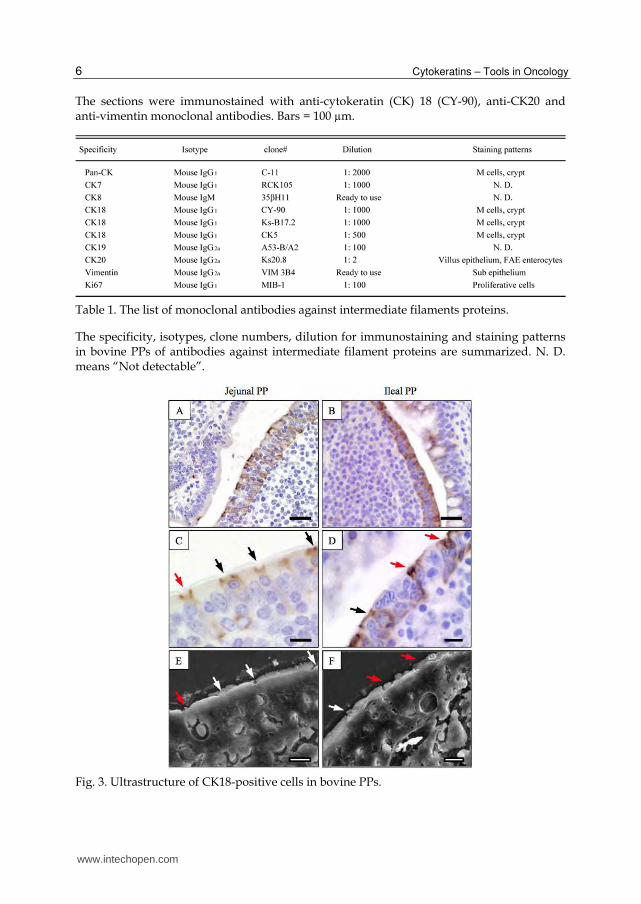
The sections were immunostained with anti-cytokeratin (CK) 18 (CY-90), anti-CK20 and anti-vimentin monoclonal antibodies. Bars = 100 µm.
Table 1. The list of monoclonal antibodies against intermediate filaments proteins.
The specificity, isotypes, clone numbers, dilution for immunostaining and staining patterns in bovine PPs of antibodies against intermediate filament proteins are summarized. N. D. means “Not detectable”.
Fig. 3. Ultrastructure of CK18-positive cells in bovine PPs.
www.intechopen.com

The Expression of Cytokeratins in Bovine Intestinal Microfold (M) Cells 7
CK18-positive cells showed M-cell like distribution in the FAE of jejunal and ileal PPs (A and B). A couple of mirror sections were used for the identification of CK18-positve cells. One section was stained with anti-CK18 (CY-90) monoclonal antibody (C and D). The other was fixed with glutaraldehyde, treated with tannic acid, and coated with platinum-palladium for SEM analysis (E and F), respectively. Arrows show identical cell types. Bars = 30 µm (A and B) and 10 µm (C-F).
Fig. 4. Localization of CK18-positive cells in the crypts
For the identification of CK18-positive cells in the crypts, we prepared a couple of mirror sections of crypts containing villous epithelium (V) and FAE (F), which were immunostained with anti-CK18 monoclonal antibody or anti-Ki67 monoclonal antibody, a marker of proliferation. Arrows show Ki67-positive cells. Dotted lines show the epithelial cells of the crypts. Bars = 30 µm.
3.3 The relationship of CK18 and CK20 in the bovine FAE
CK20 was observed not only throughout the epithelial cells lining villous epithelium, but also in the partial cells of the FAE (Figure 2). These results demonstrate that both CK18 and CK20 are co-expressed in the FAE-crypt axis. Therefore, we investigated the expression of these CKs in the FAE-crypt axis by dual staining of CK18 and CK20. As described above, the preferential expression of CK18 was observed in the M cells of the FAE and proliferative cells in the crypts. On the other hand, CK20 positive signals were exclusively observed in the CK18-negative cells including the partial of the FAE cells and the whole of villous epithelial cells (Figure 5A-D). These results indicate that proliferative cells in the crypts exchange CK18 for CK20 once above the mouths of crypts when they have moved to the villi, whereas M cells continue expressing CK18 during their movement from the crypt to the FAE.
www.intechopen.com

Cytokeratins – Tools in Oncology 8
Fig. 5. Localization of CK18-, CK20-, and TUNEL-positive cells in the jejunum and ileum.
Jejunum and ileum sections were dual immunostained with anti-CK18 (IgG1) and CK20
(IgG2a) monoclonal antibodies. CK18 and CK20 were visualized by goat Alexa 488 anti-
mouse IgG1 (green) and by goat Alexa 594 anti-mouse IgG2a (red) antibodies (A-D).
Apoptotic cells were detected with the Dead End Fluorometric terminal deoxynucleotidyl-
transferase-mediated deoxyuridine-triphosphate-biotin nick-end labeling (TUNEL) system.
The sections of jejunal and ileal FAE were stained using the TUNEL method and
immunostained with anti-CK18 (IgG1) and CK20 (IgG2a) monoclonal antibodies. CK18 and
CK20 were visualized by goat Alexa 546 anti-mouse IgG1 (orange) and by goat Alexa 647
www.intechopen.com

The Expression of Cytokeratins in Bovine Intestinal Microfold (M) Cells 9
anti-mouse IgG2a (magenta) antibodies (E and F). Arrows show TUNEL-positive cells.
Dotted lines show FAE. Bars = 10 µm.
3.4 Apoptosis of bovine M cells
It has been reported that M cells possibly transdifferentiate into enterocytes before exclusion
from the FAE apex in the porcine small intestine (Miyazawa et al., 2006). To investigate
these events for bovine intestinal M cells, we performed a triplicate CK18 and CK20, and
TUNEL staining. The TUNEL-positive apoptotic cells were observed at the apical region of
villi in both the jejunum and ileum (data not shown, see Hondo et al., 2011). We could also
see TUNEL-positive signals in the apex of both jejunal and ileal FAE; however, TUNEL-
positive apoptotic cells were only observed in CK20-positive cells (Figure 5E and F),
indicating that only enterocytes could undergo apoptosis. Moreover, we quantified the cells
that were positive for CK18, CK20 and apoptosis in the crypt-villus axis to evaluate the
possibility that M cells transdifferentiate into enterocytes. The sections containing TUNEL-
positive cells were selected, and the distance from the mouth of the crypt to the apex of half
of the FAE was divided into thirds: lower, peripheral and apical regions. The proportions of
CK18-positive cells in the lower region were 45 and 96% in the jejunal and ileal FAE,
respectively, and these rates decreased to 21 and 57% at the apical regions of the FAE. On
the other hand, the number of CK20-positive cells gradually increased from the lower region
to the apex (Table 2). These data suggest that bovine M cells, positive for CK18, may
transdifferentiate into CK-20 positive enterocytes before they undergo apoptosis at the apex
of the FAE.
Table 2. Configurational comparison of CK18 and CK20-positive cells in jejunal and ileal FAE.
Results for cell no. and terminal deoxynucleotidyl-transferase-mediated deoxyuridine-
triphosphate-biotin nick-end labeling (TUNEL)-positive cells are expressed as means ± SD;
n, no. of cells. Results for cytokeratin (CK) 18/CK20 are expressed as the ratio of the
proportion of CK18-positive cells to that of CK20-positive cells in each region of the FAE.
The sections from jejunal and ileal follicle-associated epithelium (FAE) were stained with
www.intechopen.com

Cytokeratins – Tools in Oncology 10
anti-CK18 and anti-CK20 monoclonal antibodies and TUNEL. The sections containing
TUNEL-positive cells were selected. One-half of the FAE was divided into thirds (lower
region, from the mouth of crypt to the peripheral region; peripheral region, middle third of
the FAE; and apical region, upper third of the FAE).
3.5 The expression of CK18 and CK20 in duodenum and colon
We investigated the expression patterns of CK18 and CK 20 in the duodenum and colon.
In the duodenum, CK18 was also detected in the crypt. These CK18-positive cells moved
to the villi and gradually changed CK18 for CK20 at the mouth of the crypts as observed
in the crypt-villus axis of the jejunum and ileum (Figure 6). Besides this, prominent
expression was also observed in Brunner glands of the duodenum (Figure 6A and B). In
the colon, CK18-positive cells were observed in almost all crypt cells, and this was
changed for the expression of CK20 at the mouth of the crypt (Figure 6C and D). This
observation is similar to that for the mouse, indicating that CK18 and CK20 expression
patterns are conserved across these species, except for the expression of CK18 in bovine
M cells.
Fig. 6. CK18 and CK20 expression in the duodenum and colon.
Sections of duodenum (A and B) and colon (C and D) were dual immunostained with anti-
CK18 (IgG1) and anti-CK20 (IgG2a) monoclonal antibodies. CK18 and CK20 were visualized
by Alexa 488 goat anti-mouse IgG1 (green) and Alexa 594 goat anti-mouse IgG2a (red)
www.intechopen.com
The Expression of Cytokeratins in Bovine Intestinal Microfold (M) Cells 11
antibodies, respectively. B and D are higher magnification of the boxes in A and C,
respectively. Bars = 200 µm (A and C) and 10 µm (B and D).
4. Conclusions
We have demonstrated that CK18 is expressed in bovine M cells, providing a useful tool for the detection of bovine intestinal M cells. Unlike some other mammals, ruminants develop two types of PPs in the jejunum and ileum, respectively, and both PP types possess different phenotypes for FAE and M cells. On the basis of this, we carefully observed the expression of CK18 in these M cells, and identified that both jejunal and ileal M cells were clearly detectable by immunohistochemistry of CK18. This method enabled us to detect an entire set of bovine M cells, and this will contribute to ongoing investigations of bovine M-cell differentiation and function.
Intestinal epithelial cells are well known to derive from stem cells located at the bottom of crypts (Barker and Clevers, 2010). Although M cells are an intestinal epithelial cell type, their origin has not been clarified; M cells directly differentiate from intestinal stem cells, or mature enterocytes, and convert into M cells under the influence of lymphocytes or microorganisms (Kerneis et al., 1997; Savidge et al., 1991). Recent analyses seem to support that M cells directly differentiate from stem cells, for example, Clevers et al. have shown that M cells derive from Lgr5-positive stem cells at the bottom of the crypt in the Lgr5-reporter mouse (Barker and Clevers, 2010). In the bovine intestine, proliferative cells, including the stem cell compartment and M cells, express CK18, indicating that CK18 expressed in immature cells continues into the M-cell lineage. Although these unique expression patterns are bovine specific, this aspect may help with clarifying the biological function of CK18 in intestinal epithelial cells. Our studies also confirm the possibility that M cells may transdifferentiate into enterocytes before apoptosis by examining the expression patterns of CK18 and CK20 in the crypt-FAE axis. Similar phenomena have been observed in murine and porcine M cells (Miyazawa et al., 2006; Sierro et al., 2000), indicating that this transdifferentiation of M cells into enterocytes is conserved for the M-cells of some species.
M cells are thought to be involved in the infections of various pathogens, such as pathogenic
bacteria, viruses or prions (Brayden et al., 2005; Clark et al., 1998; Heppner et al., 2001;
Takakura et al., 2011). In addition, we have recently demonstrated that bovine M cells
possess a higher capacity for transporting the transmissible spongiform encephalopathies
(TSE) agent than enterocytes in vitro (Miyazawa et al., 2010), suggesting a risk of bovine M
cells as the entry site for some pathogens. The detection of bovine M cells by CK18 will
contribute to the in vivo examination of the infectious mechanisms of various pathogens in
the bovine intestine.
It is well known that different types of cells and tissues are characterized by the specific
composition of their intermediate filaments. In the small intestine, CK7, CK8, CK18, CK19
and CK20 are expressed in epithelial cells (Flint et al., 1994; Kucharzik et al., 1998; Zhou et
al., 2003). The subgroup of cytokeratins might serve as potent differentiation markers,
because the diverse expression patterns of cytokeratins are correlated with epithelial
differentiation (Moll et al., 1982). In the murine intestine, several CKs exhibit distinct
expression patterns: CK7 and CK18 are strongly expressed in the crypt region, whereas
www.intechopen.com

Cytokeratins – Tools in Oncology 12
CK20 is expressed in differentiated epithelial cells lining the villi (Zhou et al., 2003). In this
study, we have investigated the expression of various CKs in the bovine intestine, and
demonstrated that CK18 and CK20 are expressed in the bovine intestinal tract. The
expression of CK18 in the crypts and that of CK20 in villi were very similar to the expression
patterns of mice. These conserved expression patterns of CK18 and CK20 indicate that these
CKs are fundamental cytoskeletal proteins in intestinal epithelial cells. In addition, it has
been reported that CK20 is important for keratin filament organization, and that both CK18
and CK20 have functional redundancy (Zhou et al., 2003). We observed that CK18 and CK20
did not co-localize throughout the FAE- or villus-crypt axis, implying important functional
roles for CK18 and CK20 in the keratin filament formation in each compartment.
5. Acknowledgment
This study was supported by a Grant-in-Aid for Scientific Research (21380170) from the Ministry of Education, Culture, Sports, Science and Technology, and BSE Control Project from the Ministry of Agriculture, Forestry and Fisheries.
6. References
Barker, N., and Clevers, H. (2010). Leucine-rich repeat-containing G-protein-coupled receptors as markers of adult stem cells. Gastroenterology 138, 1681-1696.
Beyaz, F., and Asti, R.N. (2004). Development of ileal Peyer's patches and follicle associated epithelium in bovine foetuses. Anat Histol Embryol 33, 172-179.
Brayden, D.J., Jepson, M.A., and Baird, A.W. (2005). Keynote review: intestinal Peyer's patch M cells and oral vaccine targeting. Drug Discov Today 10, 1145-1157.
Clark, M.A., Hirst, B.H., and Jepson, M.A. (1998). M-cell surface beta1 integrin expression and invasin-mediated targeting of Yersinia pseudotuberculosis to mouse Peyer's patch M cells. Infect Immun 66, 1237-1243.
Flint, N., Pemberton, P.W., Lobley, R.W., and Evans, G.S. (1994). Cytokeratin expression in epithelial cells isolated from the crypt and villus regions of the rodent small intestine. Epithelial Cell Biol 3, 16-23.
Gebert, A., Rothkotter, H.J., and Pabst, R. (1994). Cytokeratin 18 is an M-cell marker in porcine Peyer's patches. Cell Tissue Res 276, 213-221.
Heppner, F.L., Christ, A.D., Klein, M.A., Prinz, M., Fried, M., Kraehenbuhl, J.P., and Aguzzi, A. (2001). Transepithelial prion transport by M cells. Nat Med 7, 976-977.
Hondo, T., Kanaya, T., Takakura, I., Watanabe, H., Takahashi, Y., Nagasawa, Y., Terada, S., Ohwada, S., Watanabe, K., Kitazawa, H., et al. (2011). Cytokeratin 18 is a specific marker of bovine intestinal M cell. Am J Physiol Gastrointest Liver Physiol 300, G442-453.
Jepson, M.A., Mason, C.M., Bennett, M.K., Simmons, N.L., and Hirst, B.H. (1992). Co-expression of vimentin and cytokeratins in M cells of rabbit intestinal lymphoid follicle-associated epithelium. Histochem J 24, 33-39.
Kanaya, T., Aso, H., Miyazawa, K., Kido, T., Minashima, T., Watanabe, K., Ohwada, S., Kitazawa, H., Rose, M.T., and Yamaguchi, T. (2007). Staining patterns for actin and villin distinguish M cells in bovine follicle-associated epithelium. Res Vet Sci 82, 141-149.
www.intechopen.com

The Expression of Cytokeratins in Bovine Intestinal Microfold (M) Cells 13
Kerneis, S., Bogdanova, A., Colucci-Guyon, E., Kraehenbuhl, J.P., and Pringault, E. (1996). Cytosolic distribution of villin in M cells from mouse Peyer's patches correlates with the absence of a brush border. Gastroenterology 110, 515-521.
Kerneis, S., Bogdanova, A., Kraehenbuhl, J.P., and Pringault, E. (1997). Conversion by Peyer's patch lymphocytes of human enterocytes into M cells that transport bacteria. Science 277, 949-952.
Kraehenbuhl, J.P., and Neutra, M.R. (2000). Epithelial M cells: differentiation and function. Annu Rev Cell Dev Biol 16, 301-332.
Kucharzik, T., Lugering, N., Schmid, K.W., Schmidt, M.A., Stoll, R., and Domschke, W. (1998). Human intestinal M cells exhibit enterocyte-like intermediate filaments. Gut 42, 54-62.
Landsverk, T. (1979). The gastrointestinal mucosa in young milk-fed calves. A scanning electron and light microscopic investigation. Acta Vet Scand 20, 572-582.
Landsverk, T. (1984). Is the ileo-caecal Peyer's patch in ruminants a mammalian "bursa-equivalent"? Acta Pathol Microbiol Immunol Scand A 92, 77-79.
Miyazawa, K., Aso, H., Kanaya, T., Kido, T., Minashima, T., Watanabe, K., Ohwada, S., Kitazawa, H., Rose, M.T., Tahara, K., et al. (2006). Apoptotic process of porcine intestinal M cells. Cell Tissue Res 323, 425-432.
Miyazawa, K., Kanaya, T., Takakura, I., Tanaka, S., Hondo, T., Watanabe, H., Rose, M.T., Kitazawa, H., Yamaguchi, T., Katamine, S., et al. (2010). Transcytosis of murine-adapted bovine spongiform encephalopathy agents in an in vitro bovine M cell model. J Virol 84, 12285-12291.
Moll, R., Franke, W.W., Schiller, D.L., Geiger, B., and Krepler, R. (1982). The catalog of human cytokeratins: patterns of expression in normal epithelia, tumors and cultured cells. Cell 31, 11-24.
Neutra, M.R., Frey, A., and Kraehenbuhl, J.P. (1996). Epithelial M cells: gateways for mucosal infection and immunization. Cell 86, 345-348.
Neutra, M.R., Mantis, N.J., and Kraehenbuhl, J.P. (2001). Collaboration of epithelial cells with organized mucosal lymphoid tissues. Nat Immunol 2, 1004-1009.
Rautenberg, K., Cichon, C., Heyer, G., Demel, M., and Schmidt, M.A. (1996). Immunocytochemical characterization of the follicle-associated epithelium of Peyer's patches: anti-cytokeratin 8 antibody (clone 4.1.18) as a molecular marker for rat M cells. Eur J Cell Biol 71, 363-370.
Reynolds, J.D., and Morris, B. (1983). The evolution and involution of Peyer's patches in fetal and postnatal sheep. Eur J Immunol 13, 627-635.
Savidge, T.C., Smith, M.W., James, P.S., and Aldred, P. (1991). Salmonella-induced M-cell formation in germ-free mouse Peyer's patch tissue. Am J Pathol 139, 177-184.
Sierro, F., Pringault, E., Assman, P.S., Kraehenbuhl, J.P., and Debard, N. (2000). Transient expression of M-cell phenotype by enterocyte-like cells of the follicle-associated epithelium of mouse Peyer's patches. Gastroenterology 119, 734-743.
Takakura, I., Miyazawa, K., Kanaya, T., Itani, W., Watanabe, K., Ohwada, S., Watanabe, H., Hondo, T., Rose, M.T., Mori, T., et al. (2011). Orally administered prion protein is incorporated by m cells and spreads into lymphoid tissues with macrophages in prion protein knockout mice. Am J Pathol 179, 1301-1309.
www.intechopen.com

Cytokeratins – Tools in Oncology 14
Zhou, Q., Toivola, D.M., Feng, N., Greenberg, H.B., Franke, W.W., and Omary, M.B. (2003). Keratin 20 helps maintain intermediate filament organization in intestinal epithelia. Mol Bio
www.intechopen.com
Cytokeratins - Tools in OncologyEdited by Dr. Gerhard Hamilton
ISBN 978-953-51-0047-8Hard cover, 158 pagesPublisher InTechPublished online 29, February, 2012Published in print edition February, 2012
InTech EuropeUniversity Campus STeP Ri Slavka Krautzeka 83/A 51000 Rijeka, Croatia Phone: +385 (51) 770 447 Fax: +385 (51) 686 166www.intechopen.com
InTech ChinaUnit 405, Office Block, Hotel Equatorial Shanghai No.65, Yan An Road (West), Shanghai, 200040, China
Phone: +86-21-62489820 Fax: +86-21-62489821
The first chapters of the volume "Cytokeratins - Tools in Oncology" discuss multiple functions of cytokeratins inorganization of the intermediary filaments in normal intestine and liver as well as microfold L cells and theusability of cytokeratins 7, 8 and 20 in tumor diagnosis in detail. Epithelial to mesenchymal transition as amechanism important in pathogenesis is touched in another chapter, followed by several articles dealing withthe role of cytokeratins for detection of disseminated tumor cells and as response markers duringchemotherapy. This book is therefore destined to all cancer researchers and therapists who want tounderstand the diagnostic application of cytokeratins in histology and, especially, the use of anti-cytokeratinantibodies to identify viable residual tumor cells accounting for a higher risk of tumor recurrence or cancer cellsresponding to chemotherapy, respectively.
How to referenceIn order to correctly reference this scholarly work, feel free to copy and paste the following:
Takashi Kanaya, Tetsuya Hondo,Kohtaro Miyazawa, Michael T. Rose and Hisashi Aso (2012). The Expressionof Cytokeratins in Bovine Intestinal Microfold (M) Cells, Cytokeratins - Tools in Oncology, Dr. Gerhard Hamilton(Ed.), ISBN: 978-953-51-0047-8, InTech, Available from: http://www.intechopen.com/books/cytokeratins-tools-in-oncology/the-expression-of-cytokeratins-in-bovine-intestinal-microfold-m-cells

© 2012 The Author(s). Licensee IntechOpen. This is an open access articledistributed under the terms of the Creative Commons Attribution 3.0License, which permits unrestricted use, distribution, and reproduction inany medium, provided the original work is properly cited.