THE ENDOPLASMIC RETICULUM OF PURKINJE NEURON BODY ...
11
Neuroscience Vol. 49, No. 2, pp. 467471, 1992 Printed in Great Britain 0306-4522/92 $5.00 + 0.00 Pergamon Press Ltd IBRO THE ENDOPLASMIC RETICULUM OF PURKINJE NEURON BODY AND DENDRITES: MOLECULAR IDENTITY AND SPECIALIZATIONS FOR Ca2+ TRANSPORT A. VILLA,* A. H. SHARP,? G. RACCHETTI,* P. PODINI,* D. G. BoLE,~: W. A. DUNN,$ T. POZZAN,]~S. H. SNYDER? and J. MELDOLPSI*~ *Department of Pharmacology, CNR Cytopharmacology and B. Ceccarelli Centers, S. Raffaele Scientific Institute, Via Olgettina 60, 20132 Milano, Italy tDe.partments of Neuroscience, Pharmacology and Molecular Sciences, Psychiatry and Behavioral Sciences, Johns Hopkins University, School of Medicine, Baltimore, MD, U.S.A. IHoward Hughes Medical Institute Research Laboratory, The University of Michigan Medical Center, Ann Arbor, MI, U.S.A. §Department of Anatomy and Cell Biology, College of Medicine, University of Florida, Gainsville, FL, U.S.A. IIInstitute of General Pathology, CNR Biomembrane Center, University of Padova, Italy Abstract-Immunofluorescence and immunogold labeling, together with sucrose gradient separation and Western blot analysis of microsomal subfractions, were employed in parallel to probe the endoplasmic reticulum in the cell body and dendrites of rat cerebellar Purkinje neurons. Two markers, previously investigated in non-nerve cells, the membrane protein p91 (calnexin) and the lumenal protein BiP, were found to be highly expressed and widely distributed to the various endoplasmic reticulum sections of Purkinje neurons, from the cell body to dendrites and dendritic spines. An antibody (denominated anti-rough-surfaced endoplasmic reticulum), which recognized two membrane proteins, p14 and p40, revealed a similar immunogold labeling pattern. However, centrifugation results consistent with a widespread distribution were obtained for p14 only, while p40 was concentrated in the rough microsome- enriched subfractions. The areas enriched in the inositol 1,4,5&phosphate receptor and thus presumably specialized in Ca*+ transport (stacks of multiple smooth-surfaced cisternae; the dendritic spine apparatus) also exhibited labeling for BiP and ~91, and were positive for the anti-rough-surfaced endoplasmic reticulum antibody (presumably via the p14 antigen). Additional antibodies, that yielded inadequate immunocytochemical signals, were employed only by Western blotting of the microsomal subfractions, while the ryanodine receptor was studied by specific binding. The latter receptor and the Ca*+ ATPase, known in other species to be concentrated in Purkinje neurons, exhibited bimodal distributions with a peak in the light and another in the heavy subfractions. A similar distribution was also observed with another lumenal protein, proteine disulfide isomerase. Taken as a whole, the results that we have obtained suggest the existence in the endoplasmic reticulum of Purkinje neurons of two levels of organization; the first identified by widespread, probably general markers (BiP, ~91, possibly p14 and others), the second by specialization markers, such as the inositol 1,4,5-triphosphate receptor and, possibly, p40, which appear restricted to areas where specific functions appear to be localized. Knowledge about neuron endoplasmic reticulum (ER) is still incomplete. Up until recently, most studies concentrated on the axonal reticulum, investi- gated because of its possible involvement in axonal transport29s49,M and neurotransmitter release (see Ref. 11). In other regions of the neuron, only few studies have been carried out. Except for experiments reveal- ing the distribution of glucose-6-phosphatase activity TTo whom correspondence should be addressed. Abbreoiarions: BSA, bovine serum albumin; EGTA, ethyleneglycolbis(aminoethylether)tetra-acetate; (R)ER, (rough-surfaced) endoplasmic reticulum; HEPES, N-2- hydroxyethylpiperaxine-N’-2-ethanesulfonic acid; Ins-P, and Ins-P,R, inositol 1,4,5_trisphosphate and its receptor; PBS, phosphate-buffered saline; PDI, protein disulfide isomerase; PMSF, phenylsulfonylfluoride; RyR, ryanodine receptor; SDS-PAGE, sodium dodecyl sulfate-polyacrylamide gel electrophoresis. in the entire ER,6 the widespread distribution of concanavalin A and the absence of other lectin binding,‘3v67*68 the information was limited to con- ventional electron microscopy. The latter revealed in most neurons the co-existence and occasional lumenal continuities of the rough- and smooth- surfaced sections of the ER in the cell body up to the dendrite stalk(s). Moreover, the smooth ER was shown to be distributed along the dendrites, up to the tips and (where present) the spines, giving rise to a complex tridimensional network of tubules and longi- tudinal cistemae. Whether at all these sites the ER expresses common molecular markers or includes highly specialized areas, designed to match the struc- tural and functional specializations of the various regions of the cell, is still largely unknown. Recently, interest in these problems has greatly increased in view of the possible key involvement of NSC 49,2--H 461
Transcript of THE ENDOPLASMIC RETICULUM OF PURKINJE NEURON BODY ...

Neuroscience Vol. 49, No. 2, pp. 467471, 1992 Printed in Great Britain
0306-4522/92 $5.00 + 0.00 Pergamon Press Ltd
IBRO
THE ENDOPLASMIC RETICULUM OF PURKINJE NEURON BODY AND DENDRITES: MOLECULAR
IDENTITY AND SPECIALIZATIONS FOR Ca2+ TRANSPORT
A. VILLA,* A. H. SHARP,? G. RACCHETTI,* P. PODINI,* D. G. BoLE,~: W. A. DUNN,$ T. POZZAN,]~ S. H. SNYDER? and J. MELDOLPSI*~
*Department of Pharmacology, CNR Cytopharmacology and B. Ceccarelli Centers, S. Raffaele Scientific Institute, Via Olgettina 60, 20132 Milano, Italy
tDe.partments of Neuroscience, Pharmacology and Molecular Sciences, Psychiatry and Behavioral Sciences, Johns Hopkins University, School of Medicine, Baltimore, MD, U.S.A.
IHoward Hughes Medical Institute Research Laboratory, The University of Michigan Medical Center, Ann Arbor, MI, U.S.A.
§Department of Anatomy and Cell Biology, College of Medicine, University of Florida, Gainsville, FL, U.S.A.
IIInstitute of General Pathology, CNR Biomembrane Center, University of Padova, Italy
Abstract-Immunofluorescence and immunogold labeling, together with sucrose gradient separation and Western blot analysis of microsomal subfractions, were employed in parallel to probe the endoplasmic reticulum in the cell body and dendrites of rat cerebellar Purkinje neurons. Two markers, previously investigated in non-nerve cells, the membrane protein p91 (calnexin) and the lumenal protein BiP, were found to be highly expressed and widely distributed to the various endoplasmic reticulum sections of Purkinje neurons, from the cell body to dendrites and dendritic spines. An antibody (denominated anti-rough-surfaced endoplasmic reticulum), which recognized two membrane proteins, p14 and p40, revealed a similar immunogold labeling pattern. However, centrifugation results consistent with a widespread distribution were obtained for p14 only, while p40 was concentrated in the rough microsome- enriched subfractions. The areas enriched in the inositol 1,4,5&phosphate receptor and thus presumably specialized in Ca*+ transport (stacks of multiple smooth-surfaced cisternae; the dendritic spine apparatus) also exhibited labeling for BiP and ~91, and were positive for the anti-rough-surfaced endoplasmic reticulum antibody (presumably via the p14 antigen). Additional antibodies, that yielded inadequate immunocytochemical signals, were employed only by Western blotting of the microsomal subfractions, while the ryanodine receptor was studied by specific binding. The latter receptor and the Ca*+ ATPase, known in other species to be concentrated in Purkinje neurons, exhibited bimodal distributions with a peak in the light and another in the heavy subfractions. A similar distribution was also observed with another lumenal protein, proteine disulfide isomerase.
Taken as a whole, the results that we have obtained suggest the existence in the endoplasmic reticulum of Purkinje neurons of two levels of organization; the first identified by widespread, probably general markers (BiP, ~91, possibly p14 and others), the second by specialization markers, such as the inositol 1,4,5-triphosphate receptor and, possibly, p40, which appear restricted to areas where specific functions appear to be localized.
Knowledge about neuron endoplasmic reticulum (ER) is still incomplete. Up until recently, most studies concentrated on the axonal reticulum, investi- gated because of its possible involvement in axonal transport29s49,M and neurotransmitter release (see Ref. 11). In other regions of the neuron, only few studies have been carried out. Except for experiments reveal- ing the distribution of glucose-6-phosphatase activity
TTo whom correspondence should be addressed. Abbreoiarions: BSA, bovine serum albumin; EGTA,
ethyleneglycolbis(aminoethylether)tetra-acetate; (R)ER, (rough-surfaced) endoplasmic reticulum; HEPES, N-2- hydroxyethylpiperaxine-N’-2-ethanesulfonic acid; Ins-P, and Ins-P,R, inositol 1,4,5_trisphosphate and its receptor; PBS, phosphate-buffered saline; PDI, protein disulfide isomerase; PMSF, phenylsulfonylfluoride; RyR, ryanodine receptor; SDS-PAGE, sodium dodecyl sulfate-polyacrylamide gel electrophoresis.
in the entire ER,6 the widespread distribution of concanavalin A and the absence of other lectin binding,‘3v67*68 the information was limited to con- ventional electron microscopy. The latter revealed in most neurons the co-existence and occasional lumenal continuities of the rough- and smooth- surfaced sections of the ER in the cell body up to the dendrite stalk(s). Moreover, the smooth ER was shown to be distributed along the dendrites, up to the tips and (where present) the spines, giving rise to a complex tridimensional network of tubules and longi- tudinal cistemae. Whether at all these sites the ER expresses common molecular markers or includes highly specialized areas, designed to match the struc- tural and functional specializations of the various regions of the cell, is still largely unknown.
Recently, interest in these problems has greatly increased in view of the possible key involvement of
NSC 49,2--H 461

the ER in the control of intracellular C‘aL ’ homeo- liquid gelatin in phosphate buffer. After a short trcatmcnt stasis (for reviews, see Refs 37,38.41). A role of at with 1% Na borohydrate, they were washed and exposed l’ot
Ieast part of the system as a dynamic Ca’ ’ store had 30min to a normotonic solution containing O.?“,,, Triton
already been envisaged in the seventies’J.21 and was X-100, 15% filtered goat serum, 0.45 M NaCi. and 10 mM
proposed again more recently,“,4” based on both phosphate buffer. pH 7.4. After washing, the sections were exposed (1 h a& 37°C or overnight at 4°C) to either one o,t
morphological and biochemical results. The appli- the various primary antibodies, to non-immune antihodtes
cation of neurons of Ca” ’ microprobe analysis2 and (all diluted in the Triton X- 100 -goat serum-containmp
high resolution immunocytochemistry35~4”~44~s1~s3 has solution) or to the solution alone. Sections were then washed
focused attention on three specific regions: (i) the again thoroughly and treated with the appropriate rhodamine-labeled goat antibodies ( I : 20 t:40 in the Triton
spine apparatus, which expresses numerous receptors X-100-goat serum solution, 30@60 min, 37 ‘C). washed for the Ca*+ release second messenger, inositol 1,4,5- again, and mounted in glycerol to be examined in a Zeiss
trisphosphate (Ins-P,),4”.s3 and where Ca’+ uptake Photomicroscope III apparatus. Optical sections were
and release was observed following stimulation;2 (ii) obtained using a confocal scanning microscope (series
stacks of parallel, evenly spaced smooth cisternae, ~~~-600; BioRad Laboratories), Digital images stored o,, optical disc were photographed with a Lasergraphic LFR
whose membranes were shown to be highly enriched camera.
with Ins-P, receptors (Ins-P,R):“,53.54 (iii) a popu- lation of vacuoles (calciosomes) containing high ~~~~~~Qg~~~ ~a~e~~~g concentrations of specific, low-affinity high-capacity Samples fixed as described above were processed for
Ca2+ storage protein(s).60.62.04 the preparation of ultrathin cryosections (50.. 100 nm) as
Most of these recent studies were carried out in described in detail elsewhere.” Sectioning was carried out in
Purkinje neurons of the cerebellum which, compared a Reichert Jung (Vienna, Austria) Ultracut ultramicrotome equipped with an FC, apparatus. Cryosections were col-
to the other neurons, are particularly enriched with Iected over nickel grids and covered with 2% gelatin. After
these and also with other markers of Ca*’ stores: treatment with 125 mM phosphate buffer, pH 7.4. sup-
the CaZ + ATPase2+‘.“9,48 and the ryanodine receptor plemented with 0. I M glycine, they were exposed to the first
(RyR), a second intracellular Ca2+ channel that antibody (diluted in phosphate-glyeine buffer) for 1 h at 37’C. then washed with phosphate-glycine buffer and deco-
is apparently distributed differently from the rated with anti-IgG (rabbit or rat)-coated gold particles
Ins-P, R.17.66 Because of these properties, and because (5 nm, dilution 1: 80 in the same buffer). For dual-labeling
of their well-known architecture,45 Purkinje neurons the rabbit anti-Ins-P,R polyclona1 antibodies were applied
are a parti~ulariy interesting model for the molecular together with the anti-BiP monoclonal antibodies, and the
characterization of the ER. in the present study same was eventually done for the targe (15 nm, coated with antibodies anti-rabbit IgGs) and small (5 nm, coated with
immunocvtochemistrv has been emDloved in combi- antibodies anti-rat IgGs) gold particles. The immuno- . I
nation with s&cellular fractionation to try to reveal decorated grids were then washed and processed as
the distribution of various ER markers, believed to be recommended in Ref. 26. Cryosections were examined in a
either of general occurrence or specialized for Ca”. Hitachi H-7000 electron microscope. Pictures were usually taken at x 24.000 maanification. In order to establish
stores. background levels, co&o1 cryosections were processed without exposure to the first antibody. The gold particle densitv (n/urn*) values measured in these preparations (2.5
EXPERIMENTAL PROCEDURES
. ~ I.
and 6.5 with small and large gold) were’similar to those counted over structures of the experimental cryosections
Rats (male, 200 g b.w.) were killed by decapitation and not recognized by the antibodies employed (nuclei. mito-
cerebella rapidly collected. For morphological experiments chondria).
they were immediately immersed in the fixative (4% formal- dehvde-025% ~u~raldehvde in I25 mM nhosphate buffer. ~ubeeli~~~r ~ra~ti~~at~~n
pH 5.4 and 4”CJ then thoroughly sliced to’yield small tissue Cerebellar tissue homogenates were first centrifuged at cubes and thin (1 mm) slices including Purkinje cell bodies 11,000 r.p.m. for 10 min in a Beckman JA20 rotor. The and dendrites. Additional rats were killed by perfusion with pellet was carefully resuspended in the homogenization the above fixative.53 Fixation was pursued for 2 h after solution and spun again as above. The two supernatants which the samples were either processed for conventional were combined and centrifuged at 36,009 r.p.m. for 60 min electron microscopy (posthxation in 2% OsO, followed in the Beckman 50.2Ti rotor. The pellet (totat microsomes) by dehydration, block staining and Epon em~ding; see was resuspended in the homogen~tion solution. Twelve to Ref. 60) or used to prepare cryosections for immuno- fifteen milligrams of protein was loaded on top of a 1 I .O-ml cvtochemistrv. as described below. For cell fractionation linear (0.3-1.9 Mf sucrose gradient in buffer A, and cen- experiments,- three to five cerebella were immersed in trifuged at 23,000r.p.m. overnight in a Beckman SW41 ice-cold 0.32 M sucrose, 5 mM HEPES-KOH, pH 7.4, rotor. Eleven l-ml subfractions were collected from the 0.1 mM phenvlsulfonvlfluoride (PMSF), minced with razor bottom of the centrifuge tube and their protein concen- blades and homogeni&d in 10 ml of the same solution using a glass Dounce homogenizer (loose-fitting pestle, 30 up and down strokes).
Samples fixed as described above were processed for the preparation of cryosections as described in Ref. 60. Sections approximateIy 15pm thick were cut with a conventional cryostat, flattened over glass slides and covered with 2%
tration was measured. Then the subfractions were diluted drop by drop with ice-cold water to 0.32M sucrose. The resulting samples were ~nt~f~ged at ~,~ r.p.m. for 60min in the Beckman TLIOO ultracentrifuge, and the ensuing pellets were either fixed in situ and processed for conventional electron microscopy as described above, or resuspended to yield samples with 0.5 mg/ml protein con- centration. These samples were used for biochemical assays, sodium dodecyl sulfate-polyacrylamide gel electrophoresis (SDS-PAGE) and Western blot analysis.

Endoplasmic reticulum of Purkinje neurons 469
Sodium dodecyl sulfate-polyacrylamide gel electrophoresis and Western blotting
SDS-slab gel electrophoresis was carried out as described in Ref. 63 on S-10% polyacrylamide gradient minigels. High transfer of proteins onto ~~~~~0~ membranes (blots) was carried out at 220 mA for 16-18 h in a buffer containing 25 mM Tris, 192 mM glycine, pH 8.3, and 20% methanol.
Western blots. Blots were processed at room temperature, first with phosphate-buffered saline (PBS) containing 3% bovine serum albumin (BSA) for 6Omin, then for 60min with antibodies dissolved in PBS containing 1% IISA and 0.05% Tween 20. After washing four times for 5 mm with PBS, 0.2% Tween 20, they were incubated for 60 min with either anti-(rabbit Ig) or anti-(mouse Ig) IgGs conjugated with alkaline phosphatase, that were then revealed by staining with S-bromo-Qchloro-3-indolyl phosphate and nitro blue tetrazolium. Densitometry of the relevant antigen bands of immunoblots was carried out using an LKB chromato~nner (X-380.
Biochemical assays [3HJRyanodine binding. 0.5 mg of microsomal protein
was incubated in a medium containing 1 M KCl, 10 mM HEPES. OH 7.4. 150 uM CaCl, 0.1 mM EGTA: 2 mM dithiotreitbl, O.l’mM ‘PMSF, 5‘50 PM ATP and 4 nM [‘Hfryanodine, in the absence and presence of 10&M ryanodine for total and non-specific binding, respectively. After Mrnin at 37°C samples were flltered on 0.45lrM Millipore filters, which were rinsed with 1Oml of ice-cold 1 M NaCl. 10 mM HEPES. OH 7.4. then dried and counted for radioactivity. Under the& conditions, unspecific binding accounted for approximately 35% of the total recovered counts. The & of the specillc binding, measured indepen- dently in total microsomes, was 6.1 nM.
Proteirt. Protein was determined by the BCA procedure (Pierce, Rockford, Ill., U.S.A.) and RNA by a W method following sequential perchloric acid extraction of membrane subfractions.
Materials
The following antibodies employed here have been described elsewhere: antiCa*+ ATPase (a rabbit polyclonal against a fusion protein between the E. co/i type protein and the rabbit slow twitch cardiac enzyme”) anti-p91 and anti-rough-surfaced endoplasmic reticulum (RER) (athnity purified) (rabbit polyclonals against liver ER vesicles15J2); anti-BiP (a rat monoclonal against the purified proteit?); anti-PDI (a rabbit polyclonal against the C terminal KDDD sequence, the KD-DD antibodys). Of the two antibodies against the Ins-P-R. the ~itv-D~fi~ rabbit oolvclonal was described in Ref. 47; the mo&xdonal was dekeloped in the laboratory by injecting the foot-pad of female balb/c mice with the C terminal 21 amino acid peptide of the receptor coupled to keyhole hemocyanin and suspended in complete F&ml’s adjuvant. Lymphocytes of- popliteal lvmphnodes were fused with cells of the 13 x 63 line and c&s‘ were cloned once by limited dilution. The antibodies produced as ascites were partially pm+fred by ammonia sulfate precipitation. The anti-calbindin antibody was pur- chased from Boehringer; [3Hlryanodine from New England Nuclear; rhodamine-labeled anti-rabbit and rat IgGsfrom Teehnogenetica (Milan, Italy); and 5- and 15nm colloidal gold particles, coated with goat IgGs against either rabbit (large and small particles) or rat (small particles only) IgGs from Biocell (Cardll U.K.). The fine chemicals emoloved in this work were reagent grade, purchased from Sigma: St Louis MO, U.S.A.
RESULTS
Western blots of total rat cerebellar microsomes, probed with the antibodies employed in the present
0 2 3@@@ 7
Fig. 1. Western blots of total cerebellar microsomes probed with the antibodies employed in the present work. Fifty micrograms of protein were applied to each lane of an SDS polyacrylamide gradient (515%) gel. Arrows to the right indicate the position of markers: from the top (in mol.wt x lo-‘) 200, 97.4, 69, 46 and 30. Lanes 1 and 2 concern the Ins-P,R (poly and monoclonal antibodies, respectively); lane 3, the Ca*+ ATPase; lanes 4 and 5, the ER membrane proteins (anti-p91 and anti-RER antibodies, respectively); lanes 6 and 7, the ER lumenal proteins (anti-BiP and anti PDI antibodies, respectively). Encircled numbers indicate the antibodies employed also for immuno-
cytochemistry.
work are shown in Fig. 1. Of the two anti-Ins-P,R antibodies, the first, an unity-puffin rabbit polyclonal antibody:’ recognized only the receptor (apparent molwt, 260,000 mol.wt lane 1) whereas the second (a mouse monoclonal antibody against the C-terminal, 21 amino acid sequence) also recognized an additional band, of approximately 185,000 molwt, lane 2. Anti-Ca*+ ATPase (a rabbit poly- clonal antibody against a fusion protein of E. coli tr pE and rabbit slow-twitch cardiac Ca2+ ATPase, Ref. 33) labeled a doublet approximately 99,000 and 105,000 mol. wt. (lane 3). Lanes 4 and 5 show blots decorated with rabbit polyclonal antibodies against purified liver rough microsome membranes. The first30*3~s’ was initially reported to recognize four (apparent mol.wt x 10F3: 29, 58, 66 and 91) major microsomal bands of rat kidney cells. In the cerebel- lum, the slowest migrating of these bands was heavily decorated (Fig. 1, lane 4), while faint bands at appro~mately 66,000 and 29,000 molwt appeared only when staining of the blot was prolonged (not shown). From here on this antibody will be indicated as anti-p91. The second antibody was affinity-purified by binding to stripped RER vesicles from rat liver (anti RER).lS Two of the bands recognized in liver microsomes were also revealed in the cerebellum: the
40,000-mol.wt band visible in Fig. 1, lane 5 as well as a band of 14,000 mol. wt running at the front in that Figure (see Fig. 5). The last two antibodies (lanes 6 and 7) were against proteins resident in the ER lumen, BiP and protein disulfide isomerase, protein disulfide isomerase (PDI), revealed by a rat mono- clonal antibody (single band of 78,000 mol. wt) and a rabbit polyclonal antibody against the C-terminal peptide sequence (55,000 mol. wt band), respect- ively.s.s9 Of the antibodies shown in Fig. I. only four (lane numbers encircled) yielded adequate responses when employed for immunocytochemistry (immuno- fluorescence and immunogold labeling). The others were therefore employed only for Western blot analysis of microsomal subcellular fractions.
Our studies were concentrated on the cerebellar cortex, with special interest for the Purkinje cell body and dendrites. No specific attention was dedicated to the axons in the cerebellar white matter of the folia and cerebellar medulla and to the deep cerebellar nuclei. where Purkinje neuron projections are addressed.
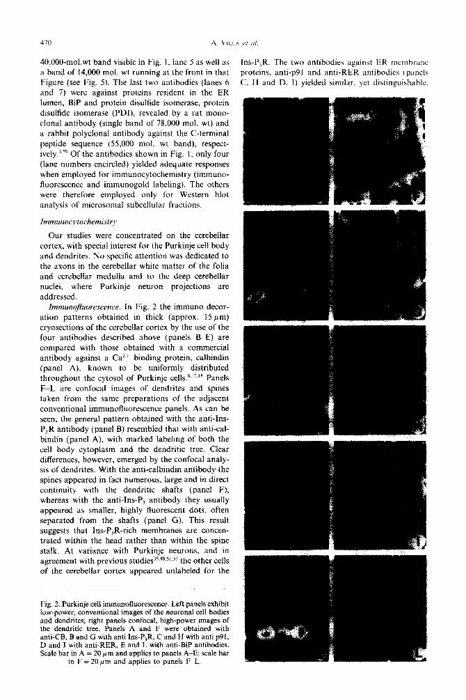
ItnrnunoJuorescence. In Fig. 2 the immuno decor- ation patterns obtained in thick (approx. 15 pm) cryosections of the cerebellar cortex by the use of the four antibodies described above (panels BE) are compared with those obtained with a commercial antibody against a Ca” binding protein, caibindin (panel A), known to be uniformly distributed throughout the cytosol of Purkinje cells.x~‘7.‘8 Panels F--L are confocal images of dendrites and spines taken from the same preparations of the adjacent conventional immuno~uorescence panels. As can be seen, the general pattern obtained with the anti-Ins- P,R antibody (panel B) resembled that with anti-cal- bindin (panel A), with marked labeling of both the cell body cytoplasm and the dendritic tree. Clear differences, however, emerged by the confocal analy- sis of dendrites. With the anti-calbindin antibody the spines appeared in fact numerous, large and in direct continuity with the dendritic shafts (panel F), whereas with the anti-Ins-P, antibody they usually appeared as smaller, highly fluorescent dots, often separated from the shafts (panel G). This result suggests that Ins-P,R-rich membranes are concen- trated within the head rather than within the spine stalk. At variance with Purkinje neurons, and in agreement with previous studies’s.40.5’~53 the other cells of the cerebellar cortex appeared unlabeled for the
Fig. 2. Pnrkinje cell immun~~uores~n~. Left panels exhibit low-power, conventional images of the neuronal cell hodies and dendrites; right panels confocal, high-power images of the dendritic tree. ?anels A and F were obtained- with anti-CB. B and G with anti Ins-P,R. C and H with anti ~91, D and 1 with anti-RER, E and i with anti-BiP antiboGies. Scale bar in A = 20 pm and applies to panels A-E; scale bar
in F = 20 pm and applies to panels F L.
Ins-P,R. The two antibodies against ER membrane: proteins, anti-p91 and anti-RER antibodies t panels C. H and D. I) yielded similar. yet distinguishable

Endoplasmic reticulum of Purkinje neurons 471
patterns. With both antibodies labeling was not restricted to Purkinje neurons but also occurred in cells of the granular (mostly granule neurons) and molecular (stellate and basket neurons) layers.45 The cytoplasm of the Purkinje neuron body showed a widespread, although non-uniform, labeling. In the dendritic tree, the labeling with the anti-p91 anti- body, although less intense than with anti-calbindin and anti-Ins-P,R antibodies, showed up clearly even in the conventional image (panel C), whereas with the anti-RER antibody the signal was distinctly weaker than in the cell body and did not show up above the adjacent structures unless at the level of large dendrites (panel D). In the confocal images, however, the dendrite shafts were revealed by both antibodies (panels H and I). Smaller structures distributed among dendritic shafts were probably accounted for by both dendritic spines and parallel fibers. Finally, the BiP pattern (panels E and L) resembled closely that observed with the anti-p91 antibody, except for the relatively stronger signal in the Purkinje cell body, and for the dendritic spines often also immuno- positive in their stalks, showing their continuity to the dendritic tree.
Zmmunogold. Ultrathin cryosections were studied after labeling with either a single or two antibodies. Organelles moderately positive for BiP were observed within cells and axons of the whole cerebellar cortex, particularly in the granular layer and also within terminals impinging onto Purkinje dendritic spines in the molecular layer (not shown). Stronger labeling was however concentrated in Purkinje neurons, within the lumen of most elements of the entire ER, from the rough-surfaced cistemae of the cell body (not shown) to the longitudinal and tubular elements of dendrites (Fig. 3 A,B), up to the spine apparatus. In contrast, nuclei, mitochondria, the Golgi complex and its adjacent large granules as well as multi- vesicular bodies appeared BiP-negative. By dual labeling, the two ER portions known (see Refs 44,45,54) to be Ins-P,R-rich, i.e. cisternal stacks distributed in both the Purkinje cell body and the dendrites (Fig. 3A) and the spine apparatus (not shown) were always found to also be labeled for BiP, and the same occurred for most of the other ER elements, both rough and smooth-surfaced, labeled to a lower degree for the receptor (inset in Fig. 3A and not shown). In contrast, appreciable Ins-P, label- ing was revealed by only approximately 50% of the profiles positive for BiP.
With the antibodies against the two membrane markers (anti-p91 and anti-RER), immunogold par- ticles were localized at the ER lumenal surface, especially over filaments protruding into the lumen. The general distribution revealed by the anti-p91 antibody resembled that with BiP in both the cell body and dendrites, i.e. labeling was evident on most rough-surfaced (Fig. 3B) and smooth-surfaced (not shown) ER cisternae and not on the other organelles. With the anti-RER antibody, labeling was stronger
on the rough-surfaced cistemae of the body (Fig. 3C) than on the smooth cistemae and tubules of dendrites (not shown). Moreover, with both markers labeling was observed over typical cistemal stacks known to be Ins-P,R-rich (insets in Fig. 3B, C), irrespective of the cell area of localization.
Subcellular fractionation
Microsomal subfractions were isolated by equi- librium continuous sucrose gradient centrifugation and analysed for marker distribution by Western blotting. In the same subfractions the RyR, for which no adequate antibody is available to us, was studied by a specific binding assay. The isolated subfractions were also sedimented as pellets, fixed and studied by conventional electron microscopy. The protein distri- bution and the concentration of RNA and RyR, calculated on a protein basis in the microsomal subfractions is illustrated in Fig. 4. The bulk of the microsomes was recovered in the intermediate 5-9 subfractions of the gradient, whereas RNA exhibited the expected high concentration in the heavy sub- fractions (Fig. 4A, B). Morphologically, the protein- rich subfractions were found to contain primarily smooth-surfaced vacuoles and tubules of various internal electron density, whereas subfractions 10 and 11 exhibited rough-surfaced elements and free ribosomes (not shown). The RyR, on the other hand, was distributed along the gradient with two repro- ducible peaks, one in the light region (rich in mem- brane sheets and cistemae, not shown), the other in the heavy region (Fig. 4B). Ins-P,R, revealed by Western blotting (Fig. 5), appeared rather uniformly distributed from subfractions 3 to 11. On the other hand, bimodal distribution, with peaks in both the heavy and the light microsomes and lower concen- trations in the intermediate fractions was observed with other markers (Fig. 5). Ca*+ ATPase was con- centrated especially in two very light subfractions, 2 and 3, and also showed an enrichment in the heavy subfraction 10. The ER membrane protein p91 and the lumenal protein PDI were enriched in subfrac- tions 3-5 and 9-l 1, while the second lumenal protein, BiP, was more concentrated in the heavy region (peak in subfraction 10) but also showed considerable labeling of the intermediate and of the light sub- fraction 3 (Fig. 5). Only with the last two markers was a clear divergence from the bimodal distribution observed. In fact, ~185 (the protein cross-labeled by our monocional antibody against the Ins-P,R) was recovered in the intermediate 4-8 subfractions. Of the proteins recognized by the anti-RER antibody, one (p40) was indeed concentrated in the RNA-enriched subfractions 10 and 11, while p14 was widely distributed, from subfraction 4 to 11.
DISCUSSION
In the present study, two classical approaches of cell biology, immunocytochemistry and subcellular

Fig. 3. Immunogold labeling of Purkinje neuron ultrathin cryosections. In all panels arrows mark small gold particle immunolabeling. A shows dual immunolabeling with anti-Ins-P,R and anti-BiP antibodies (large and small gold particles, respectively). In the main panel three stacks (st) composed of various smooth-surfaced cisternae located in a dendrite ale
heavily labeled by Ins-P,R-addressed large gold particles distributed over, or in the proximity of the closely apposed membranes. The small gold particle immunola~ling of BiP is concentrated over both the‘clear lumena of the stacked cisternae and adjacent smooth-surfaced vesicles and tubules. Two mitochondria (M) are negative for both markers. Inset of A shows the smooth-surfaced ER network of another dendrite. The large gold Ins-P,R labeling is sparse and restricted to the lower, irregular cisterna, whereas other cisternae and tubules appear negative. Small gold particle clusters labeling BiP are contained within the lumena of both Ins-P,R-positive and negative cisternae. Panel ‘B illustrates the p9 I immunolabeling in two parallel-running RER cisternae and a cisternal stack of the cell body. Notice that the label is concentrated at the lumenal surface of the membranes, particularly over tilamentous structures protruding into the lumen. Panel C illustrates the labeling with the anti-RER antibodies. The field resembles that of panel B (three rough ER cisternae and a cisternal stack in the inset) and the immunola~ling is also similar, except that in the stack the gold particles are
more clustered. Scale bars in all paneis -= 0.1 grn.

Endoplasmic reticulum of Purkinje neurons 473
1 3 5 7 9 II
FK+CtionS
Fig. 4. Distribution of protein, specific ryanodine (Ry) binding and RNA among the subfractions obtained by equilibrium centrifugation of cerebellar microsomes over a
~ntinuous 15-X% sucrose gradient.
fractionation, have been employed in parallel to investigate the distribution of various ER markers. While immunocytochemistry could be precisely focused on Purkinje neurons (cell body and den- drites), easily recognized because of their large size and peculiar morphology, subcellular fractions were by necessity obtained from the entire cell population of the cerebellum. In spite of their small number, and because of their large size and richness in ER, Purkinje neurons are expected to also contribute considerably to the subfractionation rest&s, especially in terms of Ins-P,R, Ca2+-ATPase and
IqR
PQJ
RER
BiP
POI 1 6 11
Fig. 5. Western blots of the microsomal subfractions decor- ated with the antibodies against the antigens indicated to the right. Fifty micrograms protein/subfraction were applied to gels prepared and processed as in Fig. 1. Each strip illus- trates labeling of a single antigen except for that marked RER, where both the p40 and p14 antigens are shown.
Subfraction numbers are shown at the bottom.
RyR, which are highly concentrated in this cell
tYPe* 17~u~35~39~40~48~60 It should be emphasized, however, that the subfractions contained unspecified amounts of su~l~~~ particies originated from other neurons and gha. Thus, the results obtained by this approach cannot be attributed with any certainty to Purkinje neurons unless confirmed by the immunocyto- chemical data.
Up until now, the assignment to the ER of membrane-bound structures expressed by neurons has been based primarily on conventional electron microscopy, particularly on the gross membrane morphology and apparent lmenaf continuity of the various ER sections. Most biochemical analyses have not yielded clear results due to the heterogeneity of the nervous tissues and the ensuing problems of subcellular fractionation (see above). The only enzyme cytochemical study reported so far docu- mented a widespread distribution of a single activity, glucose-6-phosphata~~ In our previous studies on chicken Purkinje neurons, a widespread distribution of the ER lumenal protein, BiP, had already been observed but not studied in detail.60 This result is now confirmed in the rat and extended to the p91 ER membrane marker, which has recently been shown to bind Ca2+ and for which the name of calnexin has thus been proposed. ” This protein had been investi- gated previously only in non-nerve tissues and cells, and is believed to be localized in the rough-surfaced cisternae only”,32~57~65 where it might be involved in the binding of lumenal proteins via Ca2+ bridges.6S Both BiP and p91 were found, however, to be widely distributed in the ER elements of the Purkinje cell body and dendrites, independently of their being rough- or smooth-surfaced, and were never observed in other organelles. These two proteins may thus be considered as the first molecularly identified general ER markers of a neuron. A widespread distribution is also likely for the other ER lumenal protein, PDI, investigated, however, only by subcellular fraction- ation, with recovery in both the heavy and light microsomal subfractions. Within ER elements, com- mon markers apparently co-exist with specialized markers, such as the major, p40 antigen recognized by the anti-RER membrane antibody,” which was recovered only with the rough-surfaced microsome. Even this antibody, however, decorated the smooth ER, possibly by recognizing an additional, smaller (~14) antigen which was found in both the rough and smooth ER subfractions of the gradient. Thus, this antibody cannot be considered as fully RER- specific as previously proposed in hepatocytes.” Pre- liminary immunofluorescence experiments were carried out to probe the differential distribution of p40 and p14 by the use of specific, affinity purified antibodies, without success. On the other hand, Ca2+ ATPase was concentrated especially in very light

microsomal subfractions, with detectable levels in the intermediate and, especially, in those enriched in rough-surfaced vesicles. In Western blots the enzyme appeared as a doublet, suggesting the expression in the rat cerebellum of two isoforms of similar mol.wt., a situation previously observed in non-nerve tissues and cells20,33.46 and recently reported in the cerebellum of the pig.4x The last protein revealed in our blots, ~185 recognized by the monodonal antibody against the Ins-P,R, was apparently absent from the rough microsomes and recovered only in the intermediate subfractions of the gradient. Since, however, the antibody was not adequate for immunocytochem- istry, we do not know whether this antigen is indeed concentrated in the smooth ER or if it is expressed by cells other than Purkinje neurons that are poor in the rough and rich in the smooth ER.
Residenf proteins of the endoplasmic reticulum iumen:
what is their ,finctionY
Interest about BiP and PDI has focused on their function in post-translational modification, preven- tion of stress-induced aggregation, quality control and conformational adjustment of secretory and other lumenal proteins addressed to the Golgi com- plex.‘Y.52 Interaction of BiP with membrane proteins has also been reported, however only during chain elongation and in the case of misfolding of the intralumenal domains.3.‘4,” Because of such a mol- ecular assistance function, BiP has been grouped together with other non-ER proteins under the gen- eral name of chaperones.‘6,‘9 So far, the possible occurrence of these proteins within the smooth ER has been given limited attention except for a few studies in hepatocytes, lymphocytes and the tumoral, adrenocorticotrophic hormone secreting cell line AtT-20.‘,4.56 In the latter work BiP and PDI were shown to be present throughout the entire ER, up to the tip of neurites. Our results demonstrate that BiP is localized throughout the entire ER lumen in Purkinje neurons. This opens the problem of the function of this protein at considerable distances away from the rough ER and the Golgi complex, where its target proteins are synthesized and trans- ported. A conventional chaperone function of BiP in dendrites and spines is hard to envisage unless a fraction of the segregated proteins in transit along the ER-Golgi pathway is diverted to dendrites, possibly by simple overflow along the ER system. Other functions of BiP and PDI, e.g. assistance to either membrane proteins which need continuous control and adjustment in their lumenal domains, or Iumenal proteins translocated post translationally (for yeast, see Refs 10,12,59), aIthough conjectural, cannot be excluded at the present time.
Ca2 + stores
New information has recently been gathered regarding the cytological nature of intracellular Ca*+ stores in Purkinje neurons. Stacks of parallel
smooth-surfaced ER cisternae, separated from each other by evenly spaced bridges (lamellar bodica). W~I-c already described in these neurons in the sixties but considered at that time a fixation artifact”“-i* (sec. however, Ref 28). We and othersU,Ci.r4 have clemon- strated that these structures are not only present in specimens fixed by a variety of procedures, but also greatly (over IO-fold) enriched in Ins-P,R compared to the rest of the ER.““.“,‘4 The accumulation of cisternal stacks composed of molecularly specialized membranes is not unique to Purkinje neurons but has also been observed in Chinese hamster ovary (UT-I ) cells over-expressing the rate-limiting cnzymc of cholesterol synthesis, 3-hydroxy, 3-methylglutaryl CoA reductase (christalloid ER).” An artifactual sorting of individual proteins to yiefd specialized membranes appears unhkely. In contrast, the reverse artifact, i.e. the disassembly of multicisternal stacks to yield a population of smooth-surfaced elements during improper fixation, has been demonstrated in neuronal growth cones.”
Our present results have revealed a new property of the Ins-P,R-rich stacked membranes: their specializ- ation is not absolute inasmuch as their membranes express the general ER markers, p91 and probably also ~14. This observation confirms, at a molecular level, previous conclusions on the ER nature of these structures based on their lumenal continuity with rough-surfaced cisternae.“.53.5’ The presence of BiP within their lumen has been reported in chicken Purkinje neurons.* However, in the previous study dual Ins-P,R-BiP experiments were not carried out and therefore direct proof of the co-existence of the two proteins could not be given6” All the general ER markers (~91. pi4 and BiP) were found to also be expressed in the spine apparatus, another structure rich in Ins-P,R.4”.5”.h6 Because of this latter and other known properties (observed however in the chicken: exclusion of calsequestrin and RyR”.*“.” ) the spine apparatus is suggested to be specialized for transient Ins-P,-triggered Cal+ release responses (for discussion, see Ref. 38).
Within Purkinje neurons, the Ins-P,R is not restricted to the highly enriched regions discussed so far but is also expressed, although at much lower concentrations, by many conventional ER elements, rough-(including the nuciear envelope) and smooth- surfaced.40.44,5’.53 This distribution might explain the widespread recovery of the Ins-P,R along the sucrose gradient, from subfractions 3 to 11. The organization of cisternal stacks and spine apparatus was dis- mantled by homogenization. Thus, these Ins-P,R-rich structures could not be identified by conventional electron microscopy of the subfractions.
A final mention concerns the RyR. Immunocyto- chemistry of chicken Purkinje neurons has revealed a selective distribution of RyR to some ER cisternae,” while subcellular fractionation has suggested local- ization in a population of small vacuoles (calcio- somes6’), characterized by the high concentration of

Endoplasmic reticulum of Purkinje neurons 475
the low-affinity high-capacity Ca2+ binding protein, calsequestrin, and the low concentration of BiP within their lumen.@’ The bimodal distribution of the RyR binding revealed by sucrose gradient centrifu- gation of rat cerebellar microsomes might be con- sistent with such a dual distribution. In the Purkinje neurons of the rat the major low-affinity high- capacity Ca 2+ binding protein is not calsequestrin but a protein recognized by anti-calreticulin anti- bodies (Villa et al., unpublished observations) so far, the intracellular distribution of this protein has not been investigated. Therefore calciosomes have not been identified in these cells, and their relationship to the ER is still unclear (discussion, see Ref. 38). The existence of separate stores endowed with either Ins-P,R or RyR has been documented from the functional point of view in various types of neur- ons,4’*55 including Purkinje neurons.’ The search for the RyR-specific store therefore remains open.
CONCLUSION
The results we have reported identify a group of widespread, probably general markers of the ER in cerebellar Purkinje neurons, as well as particular aspects of the ER subcompartments specialized in
Ca2+ transport. These specialized ER regions could be expressed at different levels and be differently localized within the various neurons. Such a con- clusion is already supported by results in a variety of cells, including cerebellar granule neurons, where [Ca’+], responses sustained by Ins-P, have been observed4’ even if the level of the Ins-P,R is so low as to be undetectable by immunocyto- chemistry. WV’ Moreover, in retinal neurons, in contrast to the widespread distribution of Purkinje neurons, an impressive concentration of the Ins-P,R in the presynaptic compartment has recently been reported.47
Acknowledgemenrs-The generous gift of the anti-p91, anti- Ca*+ ATPase and anti-PDI antibodies (by D. Louvard, Pasteur Institute, Paris, J. Lytton and D. MacLennan, Univ. of Toronto and S. Fuller, EMBL, Heidelberg) is gratefully acknowledged. We are grateful to R. Longhi for the prep- aration of the Ins-P,R C terminal peptide-keyhole hemo- cyanin complex. We thank Lorella Di Giorgio for skilful secretarial assistance and Dr S. Tacchetti of BioRad for the assistance in the use of the confocal microscope. This work was supported by a CNR grant, Target Project Biotechnol- ogy and Bioinstrumentation (to J.M.), a CNR fellowship (to P.P.), the USPHS MH-18501, Research Scientist Award DA-00074 (to S.H.S.), and the Dostdoctoral fellowshiu MH-09953 (to A.H.S.):
L
REFERENCES
1. Akagi S., Yamamoto A., Yoshimori T., Masaki R., Ogawa R. and Tashiro Y. (1988) Distribution of protein disuhide isomerase in rat hepatocytes. J. Hisrochem. Cyrochem. 36, 1533-1542.
2. Andrews S. B., Leapman R. D., Landis D. M. D. and Reese T. S. (1988) Activity-dependent accumulation of calcium in Purkinje cell dendritic spines. Proc. narn. Acud. Sci. U.S.A. 85, 1682-1685.
3. Blount P. and Merlie J. P. (1991) BiP associates with newly synthesized subunits of the mouse muscle nicotinic receptor. J. Cell Biol. 113, 112551132.
4. Bole D. G., Hendershot L. M. and Keamey J. F. (1986) Posttranslational association of immunoglobulin heavy chain binding protein with nascent heavy chains in nonsecreting hybridomas. J. Cefl Biol. 102, 1558-1566.
5. Bole D. G., Dowin R., Doriaux M. and Jamieson J. D. (1989) Immunocytochemical localization of BiP to the rough endoplasmic reticulum: evidence for protein sorting by selective retention. J. Hisrochem. Cylochem. 37, 1817-1823.
6. Broadwell R. D. and Cataldo A.M. (1983) The neuronal endoplasmic reticulum: its cytochemistry and contribution to the endomembrane system. I. Cell bodies and dendrites. J. Hisrochem. Cytochem. 31, 1077-1088.
7. Brorson J. R., Bleakman D., Gibbons S. J. and Miller R. J. (1992) The properties of intracellular Ca*+ stores in cultured rat cerebellar neurons. J. Neurosci. (in press).
8. Celio M. (1990) Calbindin, D-28K and parvalbumin in the rat nervous system. Neuroscience 35, 375-475. 9. Cheng T. P. 0. and Reese T. S. (1985) Polarized compartmentalization of organelles in growth cones from developing
optic tectum. J. Cell Biol. 101, 1473-1480. 10. Chirico W. J., Waters M. G. and Blobel G. (1988) 70K heat stock related proteins stimulate protein translocation into
microsomes. Nature 332, 805-810. 11. De Camilli P. and Jahn R. (1990) Pathways to regulated exocytosis in neurons. A. Rev. Physiol. 52, 625-685. 12. Deshaies R. J., Koch B. D., Werner-Washburne M., Craig E. A. and Schekman R. (1988) A family of stress proteins
facilitaters translocation of secretory and mitochondrial orecursor DOhDeDtideS. Nature 332. 800805. 13.
14.
15.
16. 17.
18.
19. 20.
Dontenwill M., Roussel G. and Zanetta J. P. (1985) Immunohistochet&al localization of a lectin-like molecule, RI, during the postnatal development of the rat cerebellum. Bruin Res. 349, 245-252. Duce I. R. and Keen P. (1978) Can neuronal smooth endoplasmic reticulum function as a calcium reservoir? Neuroscience 3, 837-848. Dunn W. A. Jr (1990) Studies on the mechanisms of autophagy: formation of the autophagic vacuole. J. Cell Biol. 110, 1923-1933. Ellis R. J. and van Der Vies S. M. (1991) Molecular chaperones. A. Rev. Biochem. 60, 321-347. Ellisman M. H., Deerinck T. J., Ouyang Y., Beck C. F., Tanksley S. J., Walton P. D., Airey J. A. and Sutko J. L. (1990) Identification and localization of ryanodine binding proteins in the avian central nervous system. Neuron 5, 135-146. Endo T., Kobayashi S. and Onaya T. (1985) Parvalbumin in rat cerebrum, cerebellum and retina during postnatal development. Neurosci. Lat. 60, 279-282. Gething M.-J. and Sambrook J. (1992) Protein folding in the cell. Nurure 355, 33-45. Gunteski-Hamblin A.-M., Greeb J. and Shull G. E. (1988) A novel Ca*+ pump expressed in brain, kidney, and stomach is encoded by an alternative transcript of the slow-twitch muscle sarcoplasmic reticulum Ca-ATPase gene. J. biol. Chem. 263, 15,032-l 5,040.

21. Henkart M. P., Reese T. S. and Brinley E‘. J. Jr ( 19781 Endoplasmic reticulum sequesters cahum in the sclu~d grant axon. Science 202, 1300&l 303.
22. Henkart M. (1980) Identification and function of intracellular calcium stores in axons and cell body neurons t~bh /‘WC 39, 278332789.
23. Herndon R. M. (1964) Lamellar bodies, an unusual arrangement of the granular endoplasmic reticulum. .i. (‘cl1 Viol’. 20, 3388342.
24. Kaprielian Z., Campbell A. M. and Fambrough D. M. (1989) Identification of a Ca”-ATPase in cerebellar Purkinje cells. Molec. Brain Res. 6, 55-60.
25.
26.
21.
28.
29.
30.
31.
32.
33.
34.
35.
36. 37.
38.
39.
40.
41. 42.
43.
44.
45. 46.
47.
48.
49.
50. 51.
52.
53.
54.
55.
Karlsson U. and Schultz R. L. (1966) Fixation of the central nervous system for electron microscopy by aldehyde perfusion. III. Structural changes after exsanguination and delayed perfusion. J. Ultrasrruct. Res. 14, 47-63. Keller G. A., Tokuyasu K. T., Dutton A. H. and Singer S. J. (1984) An improved procedure for immunoelectron microscopy: ultrathin plastic embedding of immunolabeled ultrathin frozen sections. Proc. natn. Acad. Sci. U.S.A. 81, 5744 -5747. Kochevar D. T. and Anderson R. G. W. (1987) Purified crystalloid endoplasmic reticulum from UT-I ceils contams multiple proteins in addition to 3-hydroxy, 3-methyiglutaryl coenzyme A reductase. J. biol. Chem. 262, 1032tL 10326. Le Beux J. (1972) Subsurface cisterns and lamellar bodies: particular forms of the endoplasmic reticulum in the neurons. Z. Zellforsch. 133, 327- 352. Lindsey J. D. and Ellisman M. H. (1985) The neuronal endomembrane system. III. The origins of the axoplasmic reticulum and discrete axonal cisternae at the axon hillock. J. Neurosci. 5, 313553144. Lippincott-Schwartz J., Donaldson J. G., Schweizer A., Berger E. G., Hauri H. P., Yan L. C. and Klausner R. D. (1990) Microtubule dependent retrograde transport of proteins into the ER in the presence of brefeldin A suggests an ER recycling pathway. Cell 60, 821-836. Lipscombe D., Madison D. V., Poenie M., Reuter H., Tsien R. W. and Tsien R. Y. (1988) Imaging of cytosohc Car + transients arising from CaZ+ stores and Ca2+ channels in sympathetic neurons. Neuron 1, 3555365. Louvard D., Reggio H. and Warren G. (1982) Antibodies to the Golgi complex and rough endoplasmic reticulum. J. Cell Biol. 92, 92-107. Lytton J. and MacLennan D. H. (1988) Molecular cloning of cDNAs from human kidney coding for two alternatively spliced products of the cardiac Ca 2+-ATPase gene. J. biol. Chem. 263, 15,024415,031. Machamer C. E., Doms R. W., Bole D. G., Helenius A. and Rose J. K. (1990) Heavy chain binding protein recognizes incompletely disulfide-bonded forms of vesicular stomatitis virus G protein. J. biol. Chem. 265, 6879-6883. Maeda N., Niinobe M., Inoue Y. and Mikoshiba K. (1989) Developmental expression and intracellular location of P,,, protein characteristic of Purkinje ceils in the mouse cerebellum. Deul Biol. 133, 67.-76. McBurney R. N. (1984) Role for microsomal Ca storage in mammalian neurones? Nafure 309, 158-160. Meldolesi J., Madeddu L. and Pozzan T. (1990) Intracellular Ca l+ storage organelles in non muscle cells: heterogeneity and functional assignment. Biochim. biophys. Acfa 1055, 130-140. Meldolesi J., Villa A., Volpe P. and Pozzan T. (1992) Cellular sites of IP, action. In Inosirol Polyphosphafe.v and CLt’ ’ (ed. Putney J. W.). Raven Press, New York. Michelangeli F., Di Virgilio F., Villa A., Podini P., Meldolesi J. and Pozzan T. (1991) Identification, kinetic properties and intracellular localization of the Cal + -Mg 2+ ATPase from the intracellular stores of chicken cerebellum. Biochem. J. 275, 555-561. Mignery G. A., Sudhof T. C., Takei K. and De Camilli P. (1989) Putative receptor for inositol 1,4,5-trisphosphate similar to ryanodine receptor. Nature 342, 192-195. Miller R. J. (1992) The control of neuronal Ca*+ homeostasis. Prog. Neurobiol. (in press). Neering I. R. and McBurney R. N. (1984) Role for microsomal Ca storage in mammalian neurones? Nature 309. 1588160. Nguyen T. H., Law D. T. S. and Williams D. B. (1991) Binding protein BiP is required for translocation of secretory proteins into the endoplasmic reticulum in Saccharomyces cerevisiae. Proc. nafn. Acad. Sci. U.S.A. 88, 1565-1569. Otsu H., Yamamoto A., Maeda N., Mikoshiba K. and Tashiro Y. (1990) Immunogold localization of inositol 1,4,5-trisphosphate (InsP,) receptor in mouse cerebellar Purkinje cells using three monoclonal antibodies. Cell Struct. Funct. 15, 1633173. Palay S. and Chan-Palay V. (1974) Cerebellur Cortex: Cytology and Organization, p. 348. Springer, Berlin. Papp B., Enyedi A., Kovacs T., Sarkddi B., Wuytack F., Thastrup O., Gardos G., Bredoux R., Levy-Toledano S. and Enouf J. (1991) Demonstration of two forms of calcium pumps by thapsigargin inhibition and radioimmunoblotting in platelet membrane vesicles. J. biol. Chem. 266, 14,593-14,596. Peng Y. W., Sharp A. H., Snyder S. H. and Yau K. W. (1991) Localization of the inositol 1,4,5_trisphosphate receptor in synaptic terminals in the vertebrate retina. Neuron 6, 525-53 I. Plessers L., Eggermont J. A., Wuytack F., Casteels R. (1991) A study of the organelle Ca*+ transport ATPase isoenzymes in pig cerebellar neurons. J. Neurosci. 11, 650-656. Quataker J. (1981) The axonal reticulum in neurons of the superior cervical ganglion of the rat is a direct extension of the Golgi apparatus. Hisrochem. J. 13, 109-124. Rambourg-A. *and Droz B. (1980) Smooth endoplasmic reticulum and axonal transport. J. Neurochem. 35, 16-25. Ross C. A.. Meldolesi J.. Milner T. A.. Satoh T.. Sunattanone S. and Snvder S. H. (1989) Inositol 1,4,5-trisphosphate receptor localized to endoplasmic reticulum in cerebellar*Purkinje neurons. Nufurb 339; 468-470. _ _ Rothman J. E. (1989) Polypeptide chain binding proteins: catalysts of protein folding and related processes in cells. CeN 59, 591-601. Satoh T., Ros C. A., Villa A., Supattapone S., Pozzan T., Snyder S. H. and Meldolesi J. (1990) The inositol 1,4,5-trisphosphate receptor in cerebellar Purkinje cells quantitative immunogold labeling reveals concentration in an ER s&compartment. J. Cell Biol. 111, 6155624. Takei K., Metcalf A., Mignery G., Volpe P., Sudhof T. and De Camilli P. (1990) Ca’+ stores in Purkinje cells: localization of Ins-P, receptor and calsequestrin. Sot. Neurosci. Abstr. 16 (Part II), 1233. Thayer S. A., Hirning L. D. and Miller R. J. (1988) The role of caffeine-sensitive calcium stores in the regulation of the intracellular free calcium concentration in rat sympathetic neurons in Gtro. Molec. Phurmac. 34, 664-673.

Endoplasmic reticulum of Purkinje neurons 417
56. Tooze J., Hollinshead M., Fuller S. D., Tooze S. A. and Huttner W. B. (1989) Morphological and biochemical evidence showing neuronal properties in AtT-20 cells and their growth cones. Eur. J. Cell Biol. 49, 259-273.
57. Tougard C., Louvard D., Picart R. and Tixier-Vidal A. (1984) The rough endoplasmic reticulum and the Golgi apparatus visualized using specific antibodies in normal and tumoral prolactin cells in culture. J. CeN Biol. %, 1197-1207.
58. Van Nimwegen D. and Sheldon H. (1966) Early postmortem changes in cerebellar neurons of the rat. J. Ultrastruct. Res. 14, 36-46.
59. Vaux D. J., Tooze J. and Fuller S. (1990) Identification by anti-idiotype antibodies of an intracellular membrane protein that recognizes a mammalian endoplasmic reticulum retention signal. Nature 345, 495-502.
60. Villa A., Podini P., Clegg D. O., Pozzan T, and Meldolesi J. (1991) Intracellular Ca2+ stores in chicken Purkinje neurons: differential distribution of the low affinity-high capacity Ca 2+ binding protein, calsequestrin, of Ca2+ ATPase and of the ER lumenal protein, BiP J. Cell Biol. 113, 779-791.
61. Vogel J. P., Misra L.M. and Rose M. D. (1990) Loss of BiP/GRP78 function blocks translocation of secretory proteins in yeast. J. Cell Biol. 110, 1885-1895.
62. Volpe P., Krause K. H., Hashimoto S., Zorzato F., Pozzan T., Meldolesi J. and Lew D. P. (1988) “Calciosome”, a cytoplasmic organelle: the inositol 1,4,5-trisphosphate-sensitive Ca 2+ store of nonmuscle cells? Proc. natn. Acad. Sci. U.S.A. 85, 109-1095.
63. Volpe P., Alderson-Lang B. H., Madeddu L., Damiani E., Collins J. H. and Margreth A. (1990) Calsequestrin, a component of the inositol 1,4,5-t&phosphate-sensitive Ca 2+ store of chicken cerebellum. Neuron 5, 713-721.
64. Volpe P., Villa A., Damiani E., Sharp A. H., Podini P., Snyder S. H. and Meldolesi J. (1991) Heterogeneity of microsomal Ca2+ stores in chicken Purkinje neurons. Eur. molec. Biol. Org. J. 10, 3183-3189.
65. Wada I., Rindress D., Cameron P. H., Ou W.-J., Doherty II J.-J., Louvard D., Bell A. W., Dignard D., Thomas D. Y. and Bergeron J. J. M. (1991) SSRa and associated calnexin are major calcium binding proteins of the endoplasmic reticulum membrane. J. biol. Chem. 266, 19,599-19,610.
66. Walton P. D., Airey J. A., Sutko J. L., Beck C. F., Mignery G. A., Sudhof T. C., Deerinck T. J. and Ellisman M. H. (1991) Ryanodine and inositol trisphosphate receptors coexist in avian cerebellar Purkinje neurons. J. Cell Biol. 113, 1145-l 157.
67. Wood J. G., McLaughlin B. J.’ and Barber R. P. (1974) The visualization of concanavalin A binding sites in Purkinje cell somata and dendrites of rat cerebellum. J. Cell Biol. 63, 541-549.
68. Wood J. G., Byrd F. I. and Gurd J. W. (1981) Lectin cytochemistry of carbohydrates on cell membranes of rat cerebellum. J. Neurocytol. ~10, 149-I 59.
(Accepted 25 February 1992)