The Endocrine System - Dr. Scott Croes' · PDF fileThe Endocrine System ... Exocrine glands:...
21
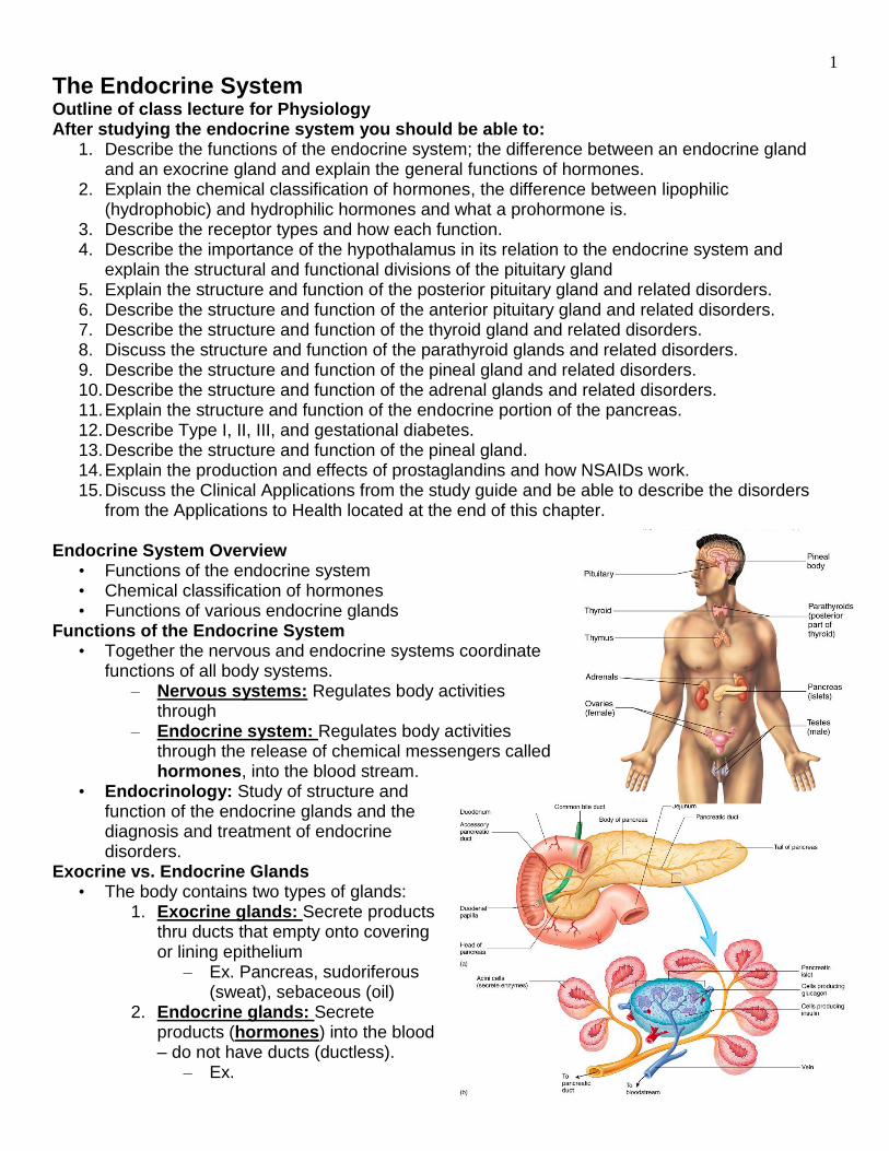
1 The Endocrine System Outline of class lecture for Physiology After studying the endocrine system you should be able to: 1. Describe the functions of the endocrine system; the difference between an endocrine gland and an exocrine gland and explain the general functions of hormones. 2. Explain the chemical classification of hormones, the difference between lipophilic (hydrophobic) and hydrophilic hormones and what a prohormone is. 3. Describe the receptor types and how each function. 4. Describe the importance of the hypothalamus in its relation to the endocrine system and explain the structural and functional divisions of the pituitary gland 5. Explain the structure and function of the posterior pituitary gland and related disorders. 6. Describe the structure and function of the anterior pituitary gland and related disorders. 7. Describe the structure and function of the thyroid gland and related disorders. 8. Discuss the structure and function of the parathyroid glands and related disorders. 9. Describe the structure and function of the pineal gland and related disorders. 10. Describe the structure and function of the adrenal glands and related disorders. 11. Explain the structure and function of the endocrine portion of the pancreas. 12. Describe Type I, II, III, and gestational diabetes. 13. Describe the structure and function of the pineal gland. 14. Explain the production and effects of prostaglandins and how NSAIDs work. 15. Discuss the Clinical Applications from the study guide and be able to describe the disorders from the Applications to Health located at the end of this chapter. Endocrine System Overview • Functions of the endocrine system • Chemical classification of hormones • Functions of various endocrine glands Functions of the Endocrine System • Together the nervous and endocrine systems coordinate functions of all body systems. – Nervous systems: Regulates body activities through – Endocrine system: Regulates body activities through the release of chemical messengers called hormones, into the blood stream. • Endocrinology: Study of structure and function of the endocrine glands and the diagnosis and treatment of endocrine disorders. Exocrine vs. Endocrine Glands • The body contains two types of glands: 1. Exocrine glands: Secrete products thru ducts that empty onto covering or lining epithelium – Ex. Pancreas, sudoriferous (sweat), sebaceous (oil) 2. Endocrine glands: Secrete products (hormones) into the blood – do not have ducts (ductless). – Ex.
Transcript of The Endocrine System - Dr. Scott Croes' · PDF fileThe Endocrine System ... Exocrine glands:...
1
The Endocrine System Outline of class lecture for Physiology After studying the endocrine system you should be able to:
1. Describe the functions of the endocrine system; the difference between an endocrine gland and an exocrine gland and explain the general functions of hormones.
2. Explain the chemical classification of hormones, the difference between lipophilic (hydrophobic) and hydrophilic hormones and what a prohormone is.
3. Describe the receptor types and how each function. 4. Describe the importance of the hypothalamus in its relation to the endocrine system and
explain the structural and functional divisions of the pituitary gland 5. Explain the structure and function of the posterior pituitary gland and related disorders. 6. Describe the structure and function of the anterior pituitary gland and related disorders. 7. Describe the structure and function of the thyroid gland and related disorders. 8. Discuss the structure and function of the parathyroid glands and related disorders. 9. Describe the structure and function of the pineal gland and related disorders. 10. Describe the structure and function of the adrenal glands and related disorders. 11. Explain the structure and function of the endocrine portion of the pancreas. 12. Describe Type I, II, III, and gestational diabetes. 13. Describe the structure and function of the pineal gland. 14. Explain the production and effects of prostaglandins and how NSAIDs work. 15. Discuss the Clinical Applications from the study guide and be able to describe the disorders
from the Applications to Health located at the end of this chapter. Endocrine System Overview
• Functions of the endocrine system • Chemical classification of hormones • Functions of various endocrine glands
Functions of the Endocrine System • Together the nervous and endocrine systems coordinate
functions of all body systems. – Nervous systems: Regulates body activities
through – Endocrine system: Regulates body activities
through the release of chemical messengers called hormones, into the blood stream.
• Endocrinology: Study of structure and function of the endocrine glands and the diagnosis and treatment of endocrine disorders.
Exocrine vs. Endocrine Glands • The body contains two types of glands:
1. Exocrine glands: Secrete products thru ducts that empty onto covering or lining epithelium
– Ex. Pancreas, sudoriferous (sweat), sebaceous (oil)
2. Endocrine glands: Secrete products (hormones) into the blood – do not have ducts (ductless).
– Ex.

2
Hormone Function • Hormones are simply molecular triggers – they do not
carry information – they exert their effect by altering the metabolic activities of cells by changing the type’s activities, or quantities of enzymes and structural proteins.
– Target cells: Cells that have the
– The type and number of receptors in or on a target cell determines its sensitivity to a particular hormone.
Endocrine System Glands and Tissue Include: • Discrete endocrine glands
– Are organs whose primary function is the production and secretion of hormones. – Ex: Pituitary (hypophysis), thyroid, parathyroid, adrenal, and pineal gland.
• Endocrine tissue – Tissue contains hormone producing cells but production of hormones is not their
primary function. – Ex: Kidneys, pancreas,
Chemical Classification of Hormones
• Based on their chemical structure can be divided into 3 chemical classes: 1. Amines: Hormones derived
– Derivatives of tyrosine
include: Thyroid hormones (T3, T4) and the catecholamines (epinephrine, norepinephrine, and dopamine).
– Derivative of tryptophan include: Melatonin
2. Peptides: Hormones made of
– Pituitary gland
hormones, ADH, oxytocin, insulin, glucagon, and many others.
3. Steroids: Hormones are
Are secreted by: – Adrenal cortex: Mineralocorticoids,
glucocorticoids and androgens – Gonads: Androgens, estrogens and progestins. – Kidney: Calcitriol
3
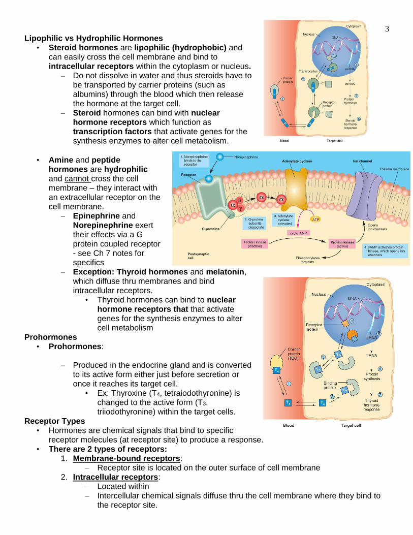
Lipophilic vs Hydrophilic Hormones • Steroid hormones are lipophilic (hydrophobic) and
can easily cross the cell membrane and bind to intracellular receptors within the cytoplasm or nucleus.
– Do not dissolve in water and thus steroids have to be transported by carrier proteins (such as albumins) through the blood which then release the hormone at the target cell.
– Steroid hormones can bind with nuclear hormone receptors which function as transcription factors that activate genes for the synthesis enzymes to alter cell metabolism.
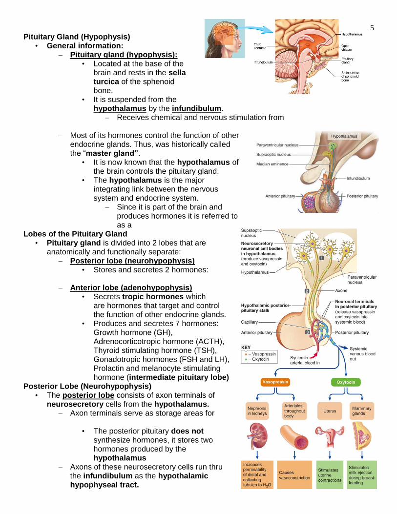
• Amine and peptide
hormones are hydrophilic and cannot cross the cell membrane – they interact with an extracellular receptor on the cell membrane.
– Epinephrine and Norepinephrine exert their effects via a G protein coupled receptor - see Ch 7 notes for specifics
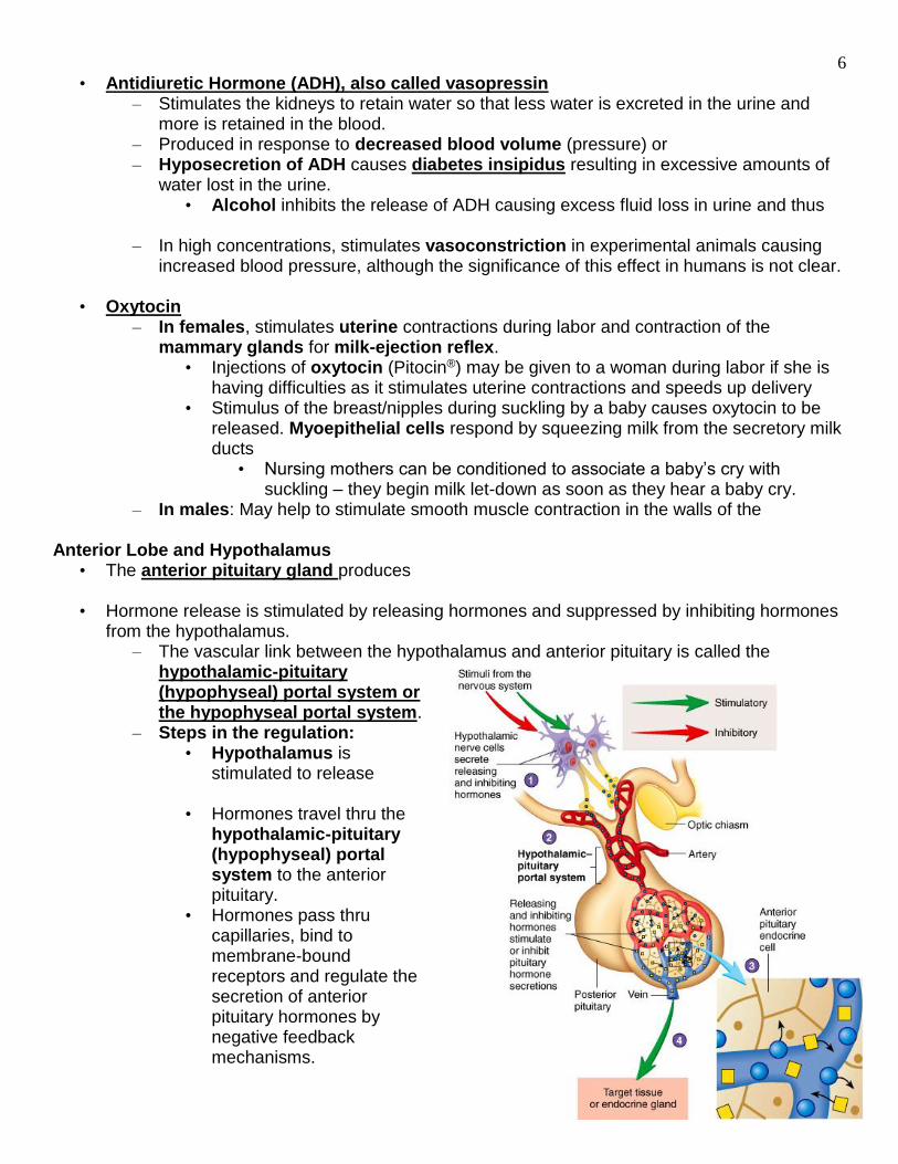
– Exception: Thyroid hormones and melatonin, which diffuse thru membranes and bind intracellular receptors.
• Thyroid hormones can bind to nuclear hormone receptors that that activate genes for the synthesis enzymes to alter cell metabolism
Prohormones • Prohormones:
– Produced in the endocrine gland and is converted
to its active form either just before secretion or once it reaches its target cell.
• Ex: Thyroxine (T4, tetraiodothyronine) is changed to the active form (T3, triiodothyronine) within the target cells.
Receptor Types • Hormones are chemical signals that bind to specific
receptor molecules (at receptor site) to produce a response. • There are 2 types of receptors:
1. Membrane-bound receptors: – Receptor site is located on the outer surface of cell membrane
2. Intracellular receptors: – Located within – Intercellular chemical signals diffuse thru the cell membrane where they bind to
the receptor site.

4
• Membrane-bound receptor response can be divided into three categories based on their mechanism of action.
1. Receptors that directly alter membrane permeability – Chemical signal binds to receptor site causing ion
channels in the cell membrane to
– The movement of ions across the cell membrane is responsible for the cells response
2. Receptors and G proteins – Chemical signal binds to receptor which results in the activation of a complex of
proteins called G proteins located on the inner surface of the cell membrane.
– Once activated, G proteins can: – Open or close membrane channels – –
3. Receptors that directly alter the activity of enzymes. – Chemical signals bind to receptor and directly
increase or decrease the
• Intracellular receptors: Chemical signals diffuse across the cell membrane and bind to intracellular receptors.
– Some intracellular receptors are
– Other intracellular receptors when activated, bind to DNA in the nucleus and increase specific genes to be transcribed and new proteins (structural or enzymes) to be produced.
5
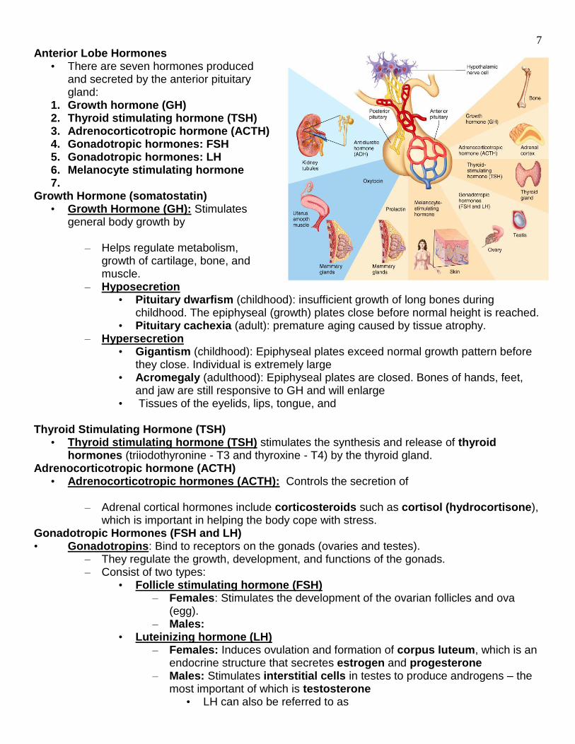
Pituitary Gland (Hypophysis) • General information:
– Pituitary gland (hypophysis): • Located at the base of the
brain and rests in the sella turcica of the sphenoid bone.
• It is suspended from the hypothalamus by the infundibulum.
– Receives chemical and nervous stimulation from
– Most of its hormones control the function of other endocrine glands. Thus, was historically called the “master gland”.
• It is now known that the hypothalamus of the brain controls the pituitary gland.
• The hypothalamus is the major integrating link between the nervous system and endocrine system.
– Since it is part of the brain and produces hormones it is referred to as a
Lobes of the Pituitary Gland • Pituitary gland is divided into 2 lobes that are
anatomically and functionally separate: – Posterior lobe (neurohypophysis)
• Stores and secretes 2 hormones:
– Anterior lobe (adenohypophysis) • Secrets tropic hormones which
are hormones that target and control the function of other endocrine glands.
• Produces and secretes 7 hormones: Growth hormone (GH), Adrenocorticotropic hormone (ACTH), Thyroid stimulating hormone (TSH), Gonadotropic hormones (FSH and LH), Prolactin and melanocyte stimulating hormone (intermediate pituitary lobe)
Posterior Lobe (Neurohypophysis) • The posterior lobe consists of axon terminals of
neurosecretory cells from the hypothalamus. – Axon terminals serve as storage areas for
• The posterior pituitary does not synthesize hormones, it stores two hormones produced by the hypothalamus
– Axons of these neurosecretory cells run thru the infundibulum as the hypothalamic hypophyseal tract.
6
• Antidiuretic Hormone (ADH), also called vasopressin – Stimulates the kidneys to retain water so that less water is excreted in the urine and
more is retained in the blood. – Produced in response to decreased blood volume (pressure) or – Hyposecretion of ADH causes diabetes insipidus resulting in excessive amounts of
water lost in the urine. • Alcohol inhibits the release of ADH causing excess fluid loss in urine and thus
– In high concentrations, stimulates vasoconstriction in experimental animals causing increased blood pressure, although the significance of this effect in humans is not clear.
• Oxytocin – In females, stimulates uterine contractions during labor and contraction of the
mammary glands for milk-ejection reflex. • Injections of oxytocin (Pitocin®) may be given to a woman during labor if she is
having difficulties as it stimulates uterine contractions and speeds up delivery • Stimulus of the breast/nipples during suckling by a baby causes oxytocin to be
released. Myoepithelial cells respond by squeezing milk from the secretory milk ducts
• Nursing mothers can be conditioned to associate a baby’s cry with suckling – they begin milk let-down as soon as they hear a baby cry.
– In males: May help to stimulate smooth muscle contraction in the walls of the
Anterior Lobe and Hypothalamus • The anterior pituitary gland produces • Hormone release is stimulated by releasing hormones and suppressed by inhibiting hormones
from the hypothalamus. – The vascular link between the hypothalamus and anterior pituitary is called the
hypothalamic-pituitary (hypophyseal) portal system or the hypophyseal portal system.
– Steps in the regulation: • Hypothalamus is
stimulated to release • Hormones travel thru the
hypothalamic-pituitary (hypophyseal) portal system to the anterior pituitary.
• Hormones pass thru capillaries, bind to membrane-bound receptors and regulate the secretion of anterior pituitary hormones by negative feedback mechanisms.
7
Anterior Lobe Hormones • There are seven hormones produced
and secreted by the anterior pituitary gland:
1. Growth hormone (GH) 2. Thyroid stimulating hormone (TSH) 3. Adrenocorticotropic hormone (ACTH) 4. Gonadotropic hormones: FSH 5. Gonadotropic hormones: LH 6. Melanocyte stimulating hormone 7.
Growth Hormone (somatostatin) • Growth Hormone (GH): Stimulates
general body growth by
– Helps regulate metabolism, growth of cartilage, bone, and muscle.
– Hyposecretion • Pituitary dwarfism (childhood): insufficient growth of long bones during
childhood. The epiphyseal (growth) plates close before normal height is reached. • Pituitary cachexia (adult): premature aging caused by tissue atrophy.
– Hypersecretion • Gigantism (childhood): Epiphyseal plates exceed normal growth pattern before
they close. Individual is extremely large • Acromegaly (adulthood): Epiphyseal plates are closed. Bones of hands, feet,
and jaw are still responsive to GH and will enlarge • Tissues of the eyelids, lips, tongue, and
Thyroid Stimulating Hormone (TSH)
• Thyroid stimulating hormone (TSH) stimulates the synthesis and release of thyroid hormones (triiodothyronine - T3 and thyroxine - T4) by the thyroid gland.
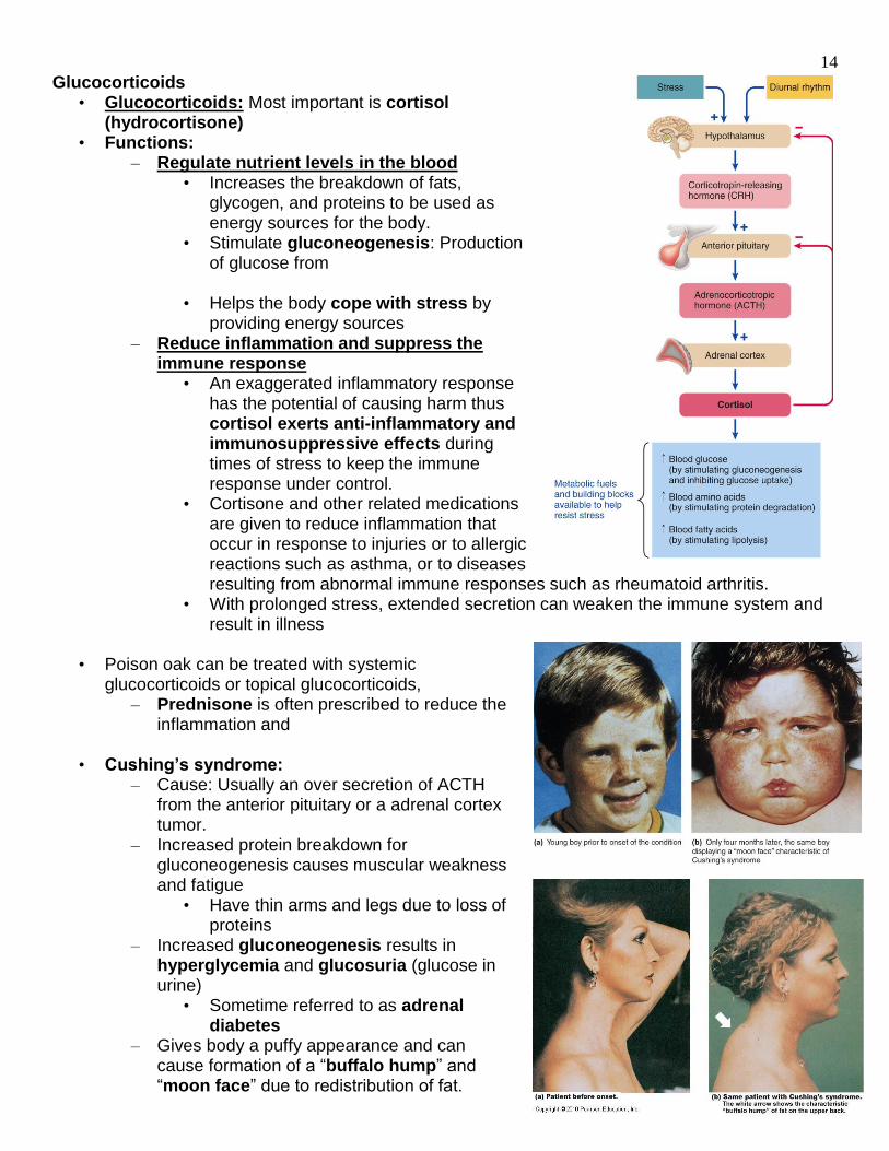
Adrenocorticotropic hormone (ACTH) • Adrenocorticotropic hormones (ACTH): Controls the secretion of
– Adrenal cortical hormones include corticosteroids such as cortisol (hydrocortisone), which is important in helping the body cope with stress.
Gonadotropic Hormones (FSH and LH) • Gonadotropins: Bind to receptors on the gonads (ovaries and testes).
– They regulate the growth, development, and functions of the gonads. – Consist of two types:
• Follicle stimulating hormone (FSH) – Females: Stimulates the development of the ovarian follicles and ova
(egg). – Males:
• Luteinizing hormone (LH) – Females: Induces ovulation and formation of corpus luteum, which is an
endocrine structure that secretes estrogen and progesterone – Males: Stimulates interstitial cells in testes to produce androgens – the
most important of which is testosterone • LH can also be referred to as
8
Prolactin • Prolactin
– Stimulates the development of breasts and mammary glands during pregnancy •
Melanocyte-stimulating Hormone • Melanocyte-stimulating hormone (MSH) has been shown to stimulate the production of
melanin in melanocytes of epidermis in lower vertebrates (fish, amphibians, reptiles, and many mammals), but function is still unclear in humans.
– In excess, can cause darkening of skin by increasing Thyroid Gland
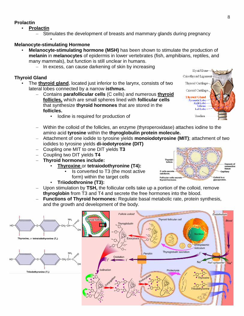
• The thyroid gland, located just inferior to the larynx, consists of two lateral lobes connected by a narrow isthmus.
– Contains parafollicular cells (C cells) and numerous thyroid follicles, which are small spheres lined with follicular cells that synthesize thyroid hormones that are stored in the follicles.
• Iodine is required for production of
– Within the colloid of the follicles, an enzyme (thyroperoxidase) attaches iodine to the amino acid tyrosine within the thyroglobulin protein molecule.
– Attachment of one iodide to tyrosine yields monoiodotyrosine (MIT); attachment of two iodides to tyrosine yields di-iodotyrosine (DIT)
– Coupling one MIT to one DIT yields T3 – Coupling two DIT yields T4 – Thyroid hormones include:
• Thyroxine or tetraiodothyronine (T4): • Is converted to T3 (the most active
form) within the target cells • Triiodothronine (T3):
– Upon stimulation by TSH, the follicular cells take up a portion of the colloid, remove thyroglobin from T3 and T4 and secrete the free hormones into the blood.
– Functions of Thyroid hormones: Regulate basal metabolic rate, protein synthesis, and the growth and development of the body.
9
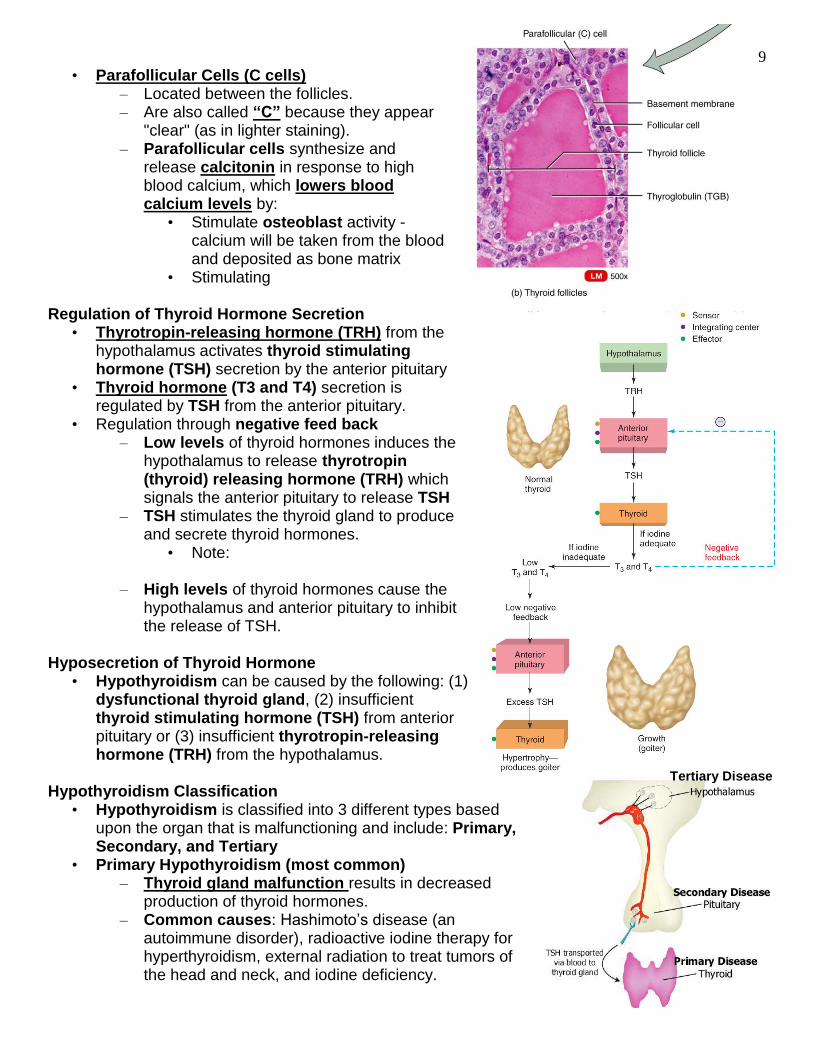
• Parafollicular Cells (C cells) – Located between the follicles. – Are also called “C” because they appear
"clear" (as in lighter staining). – Parafollicular cells synthesize and
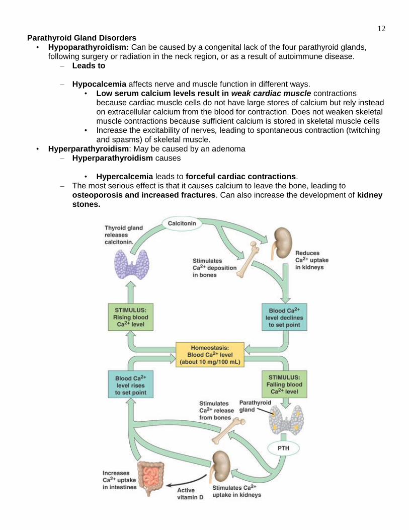
release calcitonin in response to high blood calcium, which lowers blood calcium levels by:
• Stimulate osteoblast activity - calcium will be taken from the blood and deposited as bone matrix
• Stimulating
Regulation of Thyroid Hormone Secretion • Thyrotropin-releasing hormone (TRH) from the
hypothalamus activates thyroid stimulating hormone (TSH) secretion by the anterior pituitary
• Thyroid hormone (T3 and T4) secretion is regulated by TSH from the anterior pituitary.
• Regulation through negative feed back – Low levels of thyroid hormones induces the
hypothalamus to release thyrotropin (thyroid) releasing hormone (TRH) which signals the anterior pituitary to release TSH
– TSH stimulates the thyroid gland to produce and secrete thyroid hormones.
• Note:
– High levels of thyroid hormones cause the hypothalamus and anterior pituitary to inhibit the release of TSH.
Hyposecretion of Thyroid Hormone
• Hypothyroidism can be caused by the following: (1) dysfunctional thyroid gland, (2) insufficient thyroid stimulating hormone (TSH) from anterior pituitary or (3) insufficient thyrotropin-releasing hormone (TRH) from the hypothalamus.
Hypothyroidism Classification
• Hypothyroidism is classified into 3 different types based upon the organ that is malfunctioning and include: Primary, Secondary, and Tertiary
• Primary Hypothyroidism (most common) – Thyroid gland malfunction results in decreased
production of thyroid hormones. – Common causes: Hashimoto’s disease (an
autoimmune disorder), radioactive iodine therapy for hyperthyroidism, external radiation to treat tumors of the head and neck, and iodine deficiency.
Tertiary Disease
10
• Secondary Hypothyroidism – Pituitary gland malfunction. The pituitary gland does not create enough thyroid
stimulating hormone (TSH or Thyrotropin) to induce the thyroid gland to create a sufficient quantity of thyroid hormones.
– Common causes: Pituitary is damaged by a tumor, radiation, or surgery so that it is no longer able to stimulate the thyroid to make enough hormone.
• Tertiary Hypothyroidism – Hypothalamus malfunction. Results in decreased production and/or reduced delivery
of thyrotropin (thyroid) releasing hormone (TRH) from the hypothalamus to the pituitary gland.
– Not very common. This condition can develop years after cranial irradiation.
Hypothyroid Disorders • Hyposecretion of thyroid hormones can result in:
– Low basal metabolic rate, weight gain, fatigue, slowed heart rate, depression, constipation, memory problems, and lethargy.
– A decreased ability to adapt to cold stress • Common Hypothyroid Disorders: Cretinism, myxedema, simple goiter, and Hashimoto’s
disease.
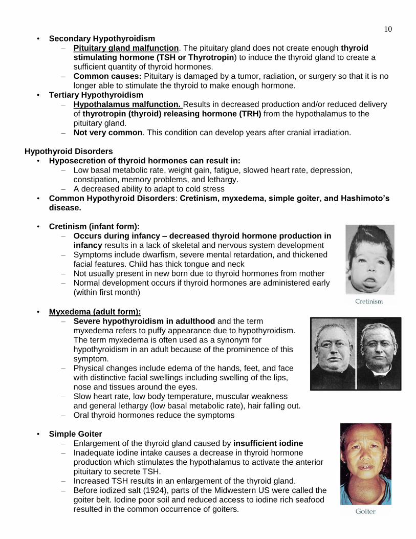
• Cretinism (infant form): – Occurs during infancy – decreased thyroid hormone production in
infancy results in a lack of skeletal and nervous system development – Symptoms include dwarfism, severe mental retardation, and thickened
facial features. Child has thick tongue and neck – Not usually present in new born due to thyroid hormones from mother – Normal development occurs if thyroid hormones are administered early
(within first month)
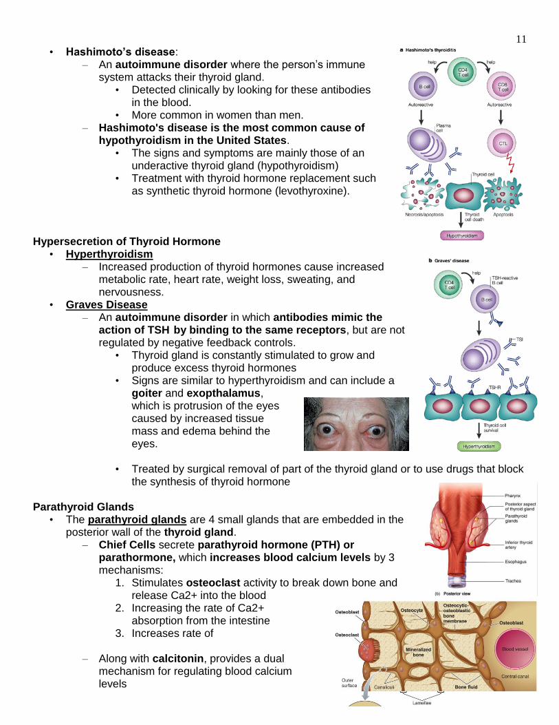
• Myxedema (adult form): – Severe hypothyroidism in adulthood and the term
myxedema refers to puffy appearance due to hypothyroidism. The term myxedema is often used as a synonym for hypothyroidism in an adult because of the prominence of this symptom.
– Physical changes include edema of the hands, feet, and face with distinctive facial swellings including swelling of the lips, nose and tissues around the eyes.
– Slow heart rate, low body temperature, muscular weakness and general lethargy (low basal metabolic rate), hair falling out.
– Oral thyroid hormones reduce the symptoms • Simple Goiter
– Enlargement of the thyroid gland caused by insufficient iodine – Inadequate iodine intake causes a decrease in thyroid hormone
production which stimulates the hypothalamus to activate the anterior pituitary to secrete TSH.
– Increased TSH results in an enlargement of the thyroid gland. – Before iodized salt (1924), parts of the Midwestern US were called the
goiter belt. Iodine poor soil and reduced access to iodine rich seafood resulted in the common occurrence of goiters.
11
• Hashimoto’s disease: – An autoimmune disorder where the person’s immune
system attacks their thyroid gland. • Detected clinically by looking for these antibodies
in the blood. • More common in women than men.
– Hashimoto's disease is the most common cause of hypothyroidism in the United States.
• The signs and symptoms are mainly those of an underactive thyroid gland (hypothyroidism)
• Treatment with thyroid hormone replacement such as synthetic thyroid hormone (levothyroxine).
Hypersecretion of Thyroid Hormone
• Hyperthyroidism – Increased production of thyroid hormones cause increased
metabolic rate, heart rate, weight loss, sweating, and nervousness.
• Graves Disease – An autoimmune disorder in which antibodies mimic the
action of TSH by binding to the same receptors, but are not regulated by negative feedback controls.
• Thyroid gland is constantly stimulated to grow and produce excess thyroid hormones
• Signs are similar to hyperthyroidism and can include a goiter and exopthalamus, which is protrusion of the eyes caused by increased tissue mass and edema behind the eyes.
• Treated by surgical removal of part of the thyroid gland or to use drugs that block
the synthesis of thyroid hormone Parathyroid Glands
• The parathyroid glands are 4 small glands that are embedded in the posterior wall of the thyroid gland.
– Chief Cells secrete parathyroid hormone (PTH) or parathormone, which increases blood calcium levels by 3 mechanisms:
1. Stimulates osteoclast activity to break down bone and release Ca2+ into the blood
2. Increasing the rate of Ca2+ absorption from the intestine
3. Increases rate of – Along with calcitonin, provides a dual
mechanism for regulating blood calcium levels
12
Parathyroid Gland Disorders • Hypoparathyroidism: Can be caused by a congenital lack of the four parathyroid glands,
following surgery or radiation in the neck region, or as a result of autoimmune disease. – Leads to
– Hypocalcemia affects nerve and muscle function in different ways.
• Low serum calcium levels result in weak cardiac muscle contractions because cardiac muscle cells do not have large stores of calcium but rely instead on extracellular calcium from the blood for contraction. Does not weaken skeletal muscle contractions because sufficient calcium is stored in skeletal muscle cells
• Increase the excitability of nerves, leading to spontaneous contraction (twitching and spasms) of skeletal muscle.
• Hyperparathyroidism: May be caused by an adenoma – Hyperparathyroidism causes
• Hypercalcemia leads to forceful cardiac contractions.
– The most serious effect is that it causes calcium to leave the bone, leading to osteoporosis and increased fractures. Can also increase the development of kidney stones.

13
Adrenal Glands • Adrenal glands
(suprarenal glands) are paired glands, located superior to each kidney.
• Have two distinct regions that function as separate endocrine glands:
– Adrenal Cortex: • Hormones
that regulate mineral and energy balance
– Adrenal Medulla: • Hormones that participate in
the “fight or flight” response
Adrenal Cortex • Adrenal cortex
– Divided into three concentric zones with each zone producing different steroid hormones collectively called corticosteroids or corticoids.
• Corticosteriod release is regulated by adrenocorticotropic hormone (ACTH) from the anterior pituitary.
• There are three categories of corticosteroids: 1. Mineralocorticoids 2. Glucocorticoids 3.
Mineralocorticoids • Mineralocorticoids
– Regulate Na+ and K+ balance as well as other electrolyte (mineral) concentrations in the blood.
– Aldosterone is the most potent mineralocorticoid and acts on the kidney’s to retain Na+ and water and increase the rate at which K+ is eliminated in the urine.
• Action: Increases blood volume and pressure and to regulate
14
Glucocorticoids • Glucocorticoids: Most important is cortisol
(hydrocortisone) • Functions:
– Regulate nutrient levels in the blood • Increases the breakdown of fats,
glycogen, and proteins to be used as energy sources for the body.
• Stimulate gluconeogenesis: Production of glucose from
• Helps the body cope with stress by providing energy sources
– Reduce inflammation and suppress the immune response
• An exaggerated inflammatory response has the potential of causing harm thus cortisol exerts anti-inflammatory and immunosuppressive effects during times of stress to keep the immune response under control.
• Cortisone and other related medications are given to reduce inflammation that occur in response to injuries or to allergic reactions such as asthma, or to diseases resulting from abnormal immune responses such as rheumatoid arthritis.
• With prolonged stress, extended secretion can weaken the immune system and result in illness
• Poison oak can be treated with systemic
glucocorticoids or topical glucocorticoids, – Prednisone is often prescribed to reduce the
inflammation and
• Cushing’s syndrome: – Cause: Usually an over secretion of ACTH
from the anterior pituitary or a adrenal cortex tumor.
– Increased protein breakdown for gluconeogenesis causes muscular weakness and fatigue
• Have thin arms and legs due to loss of proteins
– Increased gluconeogenesis results in hyperglycemia and glucosuria (glucose in urine)
• Sometime referred to as adrenal diabetes
– Gives body a puffy appearance and can cause formation of a “buffalo hump” and “moon face” due to redistribution of fat.
15
Gonadocorticoids • Gonadocorticoids (adrenal sex hormones) are produced in small amounts in both males and
females. – Consists of mostly of androgens (male sex hormones) and some estrogens (female
sex hormones) promoting masculine and feminine characteristics respectively. – Androgens contribute to:
• The onset of puberty • The appearance of secondary sex characteristics •
– Their effect is usually masked by the hormones from the testes and ovaries. Adrenogenital Syndrome
• Adrenogenital syndrome is an oversecretion of gonadocorticoids usually due to a tumor or an underproduction of cortisol – genetic disorder.
– Reduced cortisol causes the anterior pituitary to release large amounts of ACTH resulting in the overproduction of mineralocorticoids and gonadocorticoids from the adrenal cortex.
• In females – increased androgens can result in male secondary sex characteristics including body and facial hair patterns, muscle development and an enlarged clitoris.
• In males – enlarged breasts (gynecomastia) and Adrenal Medulla
• Adrenal medulla – Inner layer of the adrenal gland, derived from embryonic neuroectoderm
• Are innervated by sympathetic preganglionic neurons and function as part of the sympathetic nervous system.
• Cells are specialized sympathetic postganglionic neurons that secrete epinephrine (adrenaline) and to a lesser extent, norepinephrine (noradrenaline)
• Gland functions as both – Functions in the “fight or flight” response and causes the following physiological
characteristics: • Increased heart rate, cardiac output, and blood pressure • Increased respiratory rate and dilation of respiratory passageways • Increased rate of
Pancreas • The pancreas has both exocrine and endocrine
functions. – Endocrine function: Pancreatic Islets (Islets
of Langerhans) are clusters of endocrine cells within the pancreas that produce 2 types of hormones. 1. Glucagon: Produced by alpha cells
– Increase blood glucose levels by stimulating the breakdown of glycogen to glucose (glycogenolysis) in the liver
– Stimulates an increase in the breakdown of fats (lypolysis) to fatty acids and proteins to amino acids that circulate in the blood.
16
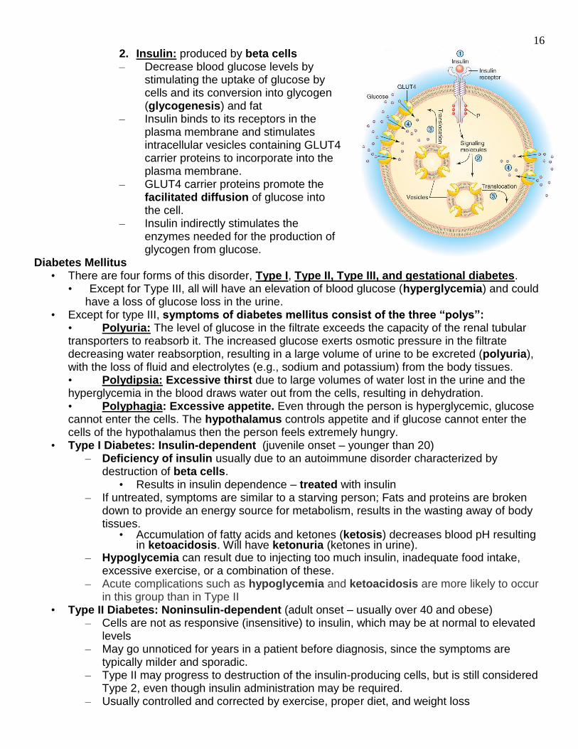
2. Insulin: produced by beta cells – Decrease blood glucose levels by
stimulating the uptake of glucose by cells and its conversion into glycogen (glycogenesis) and fat
– Insulin binds to its receptors in the plasma membrane and stimulates intracellular vesicles containing GLUT4 carrier proteins to incorporate into the plasma membrane.
– GLUT4 carrier proteins promote the facilitated diffusion of glucose into the cell.
– Insulin indirectly stimulates the enzymes needed for the production of glycogen from glucose.
Diabetes Mellitus • There are four forms of this disorder, Type I, Type II, Type III, and gestational diabetes.
• Except for Type III, all will have an elevation of blood glucose (hyperglycemia) and could have a loss of glucose loss in the urine.
• Except for type III, symptoms of diabetes mellitus consist of the three “polys”: • Polyuria: The level of glucose in the filtrate exceeds the capacity of the renal tubular transporters to reabsorb it. The increased glucose exerts osmotic pressure in the filtrate decreasing water reabsorption, resulting in a large volume of urine to be excreted (polyuria), with the loss of fluid and electrolytes (e.g., sodium and potassium) from the body tissues. • Polydipsia: Excessive thirst due to large volumes of water lost in the urine and the hyperglycemia in the blood draws water out from the cells, resulting in dehydration. • Polyphagia: Excessive appetite. Even through the person is hyperglycemic, glucose cannot enter the cells. The hypothalamus controls appetite and if glucose cannot enter the cells of the hypothalamus then the person feels extremely hungry.
• Type I Diabetes: Insulin-dependent (juvenile onset – younger than 20) – Deficiency of insulin usually due to an autoimmune disorder characterized by
destruction of beta cells. • Results in insulin dependence – treated with insulin
– If untreated, symptoms are similar to a starving person; Fats and proteins are broken down to provide an energy source for metabolism, results in the wasting away of body tissues.
• Accumulation of fatty acids and ketones (ketosis) decreases blood pH resulting in ketoacidosis. Will have ketonuria (ketones in urine).
– Hypoglycemia can result due to injecting too much insulin, inadequate food intake, excessive exercise, or a combination of these.
– Acute complications such as hypoglycemia and ketoacidosis are more likely to occur in this group than in Type II
• Type II Diabetes: Noninsulin-dependent (adult onset – usually over 40 and obese) – Cells are not as responsive (insensitive) to insulin, which may be at normal to elevated
levels – May go unnoticed for years in a patient before diagnosis, since the symptoms are
typically milder and sporadic. – Type II may progress to destruction of the insulin-producing cells, but is still considered
Type 2, even though insulin administration may be required. – Usually controlled and corrected by exercise, proper diet, and weight loss
17
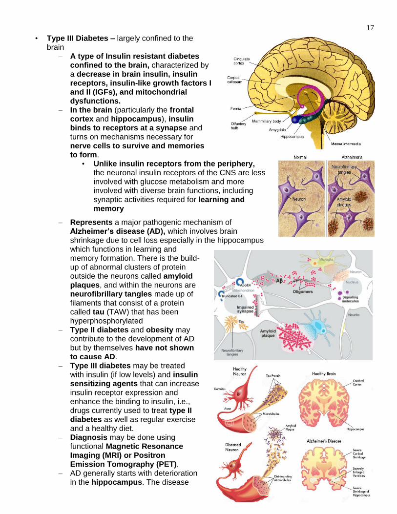
• Type III Diabetes – largely confined to the brain
– A type of Insulin resistant diabetes confined to the brain, characterized by a decrease in brain insulin, insulin receptors, insulin-like growth factors I and II (IGFs), and mitochondrial dysfunctions.
– In the brain (particularly the frontal cortex and hippocampus), insulin binds to receptors at a synapse and turns on mechanisms necessary for nerve cells to survive and memories to form.
• Unlike insulin receptors from the periphery, the neuronal insulin receptors of the CNS are less involved with glucose metabolism and more involved with diverse brain functions, including synaptic activities required for learning and memory
– Represents a major pathogenic mechanism of Alzheimer’s disease (AD), which involves brain shrinkage due to cell loss especially in the hippocampus which functions in learning and memory formation. There is the build-up of abnormal clusters of protein outside the neurons called amyloid plaques, and within the neurons are neurofibrillary tangles made up of filaments that consist of a protein called tau (TAW) that has been hyperphosphorylated
– Type II diabetes and obesity may contribute to the development of AD but by themselves have not shown to cause AD.
– Type III diabetes may be treated with insulin (if low levels) and insulin sensitizing agents that can increase insulin receptor expression and enhance the binding to insulin, i.e., drugs currently used to treat type II diabetes as well as regular exercise and a healthy diet.
– Diagnosis may be done using functional Magnetic Resonance Imaging (MRI) or Positron Emission Tomography (PET).
– AD generally starts with deterioration in the hippocampus. The disease
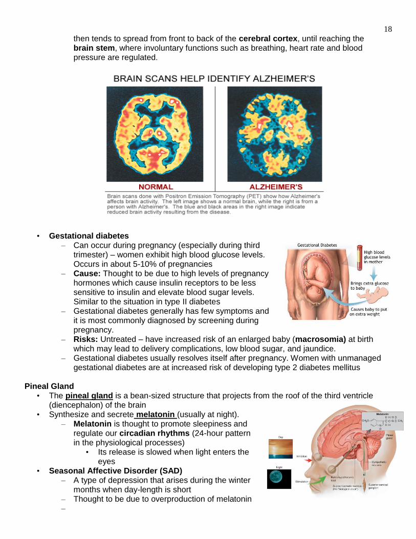
18
then tends to spread from front to back of the cerebral cortex, until reaching the brain stem, where involuntary functions such as breathing, heart rate and blood pressure are regulated.
• Gestational diabetes – Can occur during pregnancy (especially during third
trimester) – women exhibit high blood glucose levels. Occurs in about 5-10% of pregnancies
– Cause: Thought to be due to high levels of pregnancy hormones which cause insulin receptors to be less sensitive to insulin and elevate blood sugar levels. Similar to the situation in type II diabetes
– Gestational diabetes generally has few symptoms and it is most commonly diagnosed by screening during pregnancy.
– Risks: Untreated – have increased risk of an enlarged baby (macrosomia) at birth which may lead to delivery complications, low blood sugar, and jaundice.
– Gestational diabetes usually resolves itself after pregnancy. Women with unmanaged gestational diabetes are at increased risk of developing type 2 diabetes mellitus
Pineal Gland
• The pineal gland is a bean-sized structure that projects from the roof of the third ventricle (diencephalon) of the brain
• Synthesize and secrete melatonin (usually at night). – Melatonin is thought to promote sleepiness and
regulate our circadian rhythms (24-hour pattern in the physiological processes)
• Its release is slowed when light enters the eyes
• Seasonal Affective Disorder (SAD) – A type of depression that arises during the winter
months when day-length is short – Thought to be due to overproduction of melatonin –
19
Testes and Ovaries • Testes: Produce sperm and
– Growth and development of the male reproductive structures, muscle enlargement, growth of body hair, voice changes and the male sexual drive.
• Ovaries produce oocytes (eggs) and secrete estrogen and progesterone which are responsible for:
– The development and function of the female reproductive structures.
– Distribution of fat, which influences the
Autocrine and Paracrine Regulation
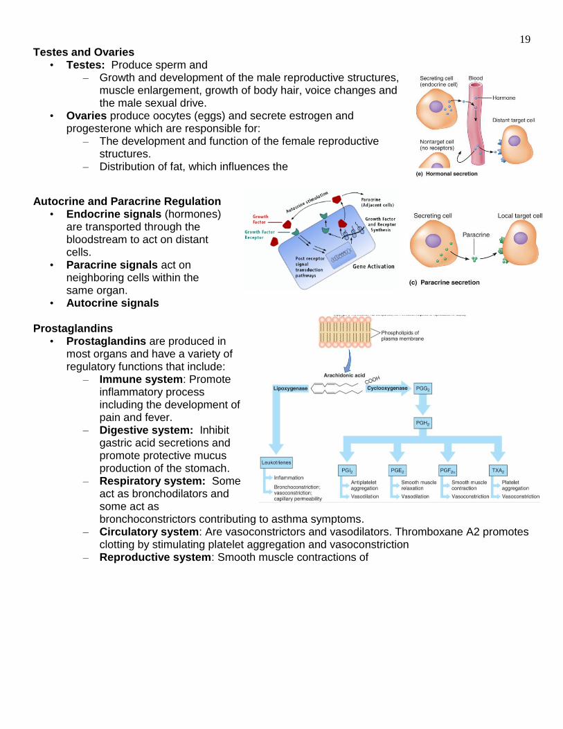
• Endocrine signals (hormones) are transported through the bloodstream to act on distant cells.
• Paracrine signals act on neighboring cells within the same organ.
• Autocrine signals Prostaglandins
• Prostaglandins are produced in most organs and have a variety of regulatory functions that include:
– Immune system: Promote inflammatory process including the development of pain and fever.
– Digestive system: Inhibit gastric acid secretions and promote protective mucus production of the stomach.
– Respiratory system: Some act as bronchodilators and some act as bronchoconstrictors contributing to asthma symptoms.
– Circulatory system: Are vasoconstrictors and vasodilators. Thromboxane A2 promotes clotting by stimulating platelet aggregation and vasoconstriction
– Reproductive system: Smooth muscle contractions of
20
Prostaglandin Production
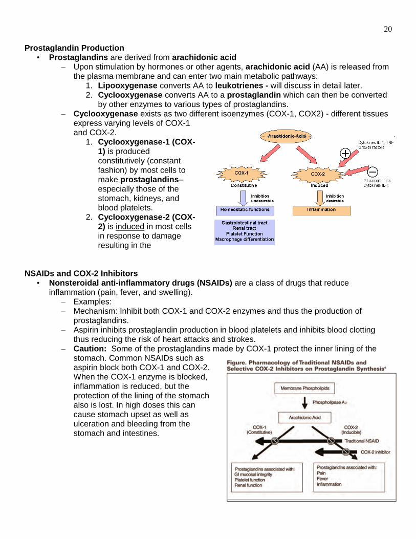
• Prostaglandins are derived from arachidonic acid – Upon stimulation by hormones or other agents, arachidonic acid (AA) is released from
the plasma membrane and can enter two main metabolic pathways: 1. Lipooxygenase converts AA to leukotrienes - will discuss in detail later. 2. Cyclooxygenase converts AA to a prostaglandin which can then be converted
by other enzymes to various types of prostaglandins. – Cyclooxygenase exists as two different isoenzymes (COX-1, COX2) - different tissues
express varying levels of COX-1 and COX-2.
1. Cyclooxygenase-1 (COX-1) is produced constitutively (constant fashion) by most cells to make prostaglandins– especially those of the stomach, kidneys, and blood platelets.
2. Cyclooxygenase-2 (COX-2) is induced in most cells in response to damage resulting in the
NSAIDs and COX-2 Inhibitors
• Nonsteroidal anti-inflammatory drugs (NSAIDs) are a class of drugs that reduce inflammation (pain, fever, and swelling).
– Examples: – Mechanism: Inhibit both COX-1 and COX-2 enzymes and thus the production of
prostaglandins. – Aspirin inhibits prostaglandin production in blood platelets and inhibits blood clotting
thus reducing the risk of heart attacks and strokes. – Caution: Some of the prostaglandins made by COX-1 protect the inner lining of the
stomach. Common NSAIDs such as aspirin block both COX-1 and COX-2. When the COX-1 enzyme is blocked, inflammation is reduced, but the protection of the lining of the stomach also is lost. In high doses this can cause stomach upset as well as ulceration and bleeding from the stomach and intestines.
21
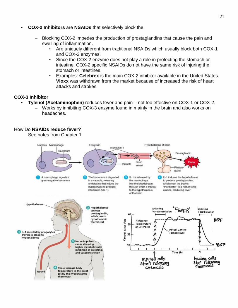
• COX-2 Inhibitors are NSAIDs that selectively block the
– Blocking COX-2 impedes the production of prostaglandins that cause the pain and
swelling of inflammation. • Are uniquely different from traditional NSAIDs which usually block both COX-1
and COX-2 enzymes. • Since the COX-2 enzyme does not play a role in protecting the stomach or
intestine, COX-2 specific NSAIDs do not have the same risk of injuring the stomach or intestines.
• Examples: Celebrex is the main COX-2 inhibitor available in the United States. Vioxx was withdrawn from the market because of increased the risk of heart attacks and strokes.
COX-3 Inhibitor
• Tylenol (Acetaminophen) reduces fever and pain – not too effective on COX-1 or COX-2. – Works by inhibiting COX-3 enzyme found in mainly in the brain and also works on
headaches. How Do NSAIDs reduce fever? See notes from Chapter 1