The Endocrine...
80
PowerPoint ® Lecture Slides prepared by Karen Dunbar Kareiva Ivy Tech Community College © Annie Leibovitz/Contact Press Images Chapter 15 Part A The Endocrine System © 2017 Pearson Education, Inc.
Transcript of The Endocrine...

PowerPoint® Lecture Slides
prepared by
Karen Dunbar Kareiva
Ivy Tech Community College© Annie Leibovitz/Contact Press Images
Chapter 15 Part A
The
Endocrine
System
© 2017 Pearson Education, Inc.

Why This Matters
• Understanding the endocrine system enables
you to monitor and advise patients with
diseases such as diabetes mellitus
© 2017 Pearson Education, Inc.

Video: Why This Matters
© 2017 Pearson Education, Inc.
15.1 Endocrine System Overview
• Endocrine system acts with nervous system to
coordinate and integrate activity of body cells
• Influences metabolic activities via hormones
transported in blood
• Responses slower but longer lasting than
nervous system responses
• Endocrinology: study of hormones and
endocrine organs
© 2017 Pearson Education, Inc.

15.1 Endocrine System Overview
• Endocrine system controls and integrates:
– Reproduction
– Growth and development
– Maintenance of electrolyte, water, and nutrient
balance of blood
– Regulation of cellular metabolism and energy
balance
– Mobilization of body defenses
© 2017 Pearson Education, Inc.

15.1 Endocrine System Overview
• Exocrine glands
– Produce nonhormonal substances (examples:
sweat, saliva)
– Have ducts to carry secretion to membrane
surface
• Endocrine glands
– Produce hormones
– Lack ducts
© 2017 Pearson Education, Inc.
15.1 Endocrine System Overview
• Endocrine glands: pituitary, thyroid, parathyroid,
adrenal, and pineal glands
• Hypothalamus is neuroendocrine organ
• Some have exocrine and endocrine functions
– Pancreas, gonads, placenta
• Other tissues and organs that produce
hormones
– Adipose cells, thymus, and cells in walls of small
intestine, stomach, kidneys, and heart
© 2017 Pearson Education, Inc.
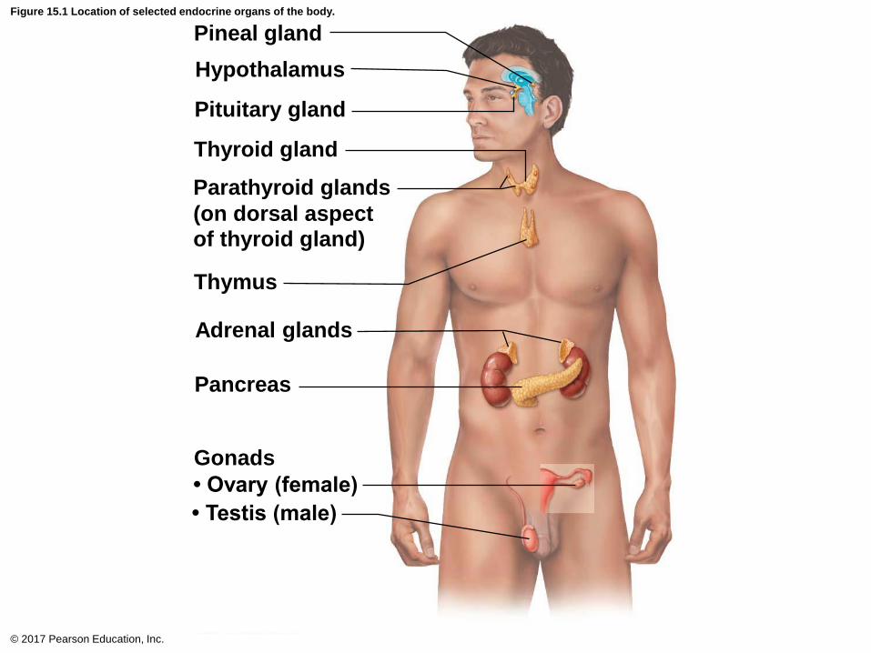
Figure 15.1 Location of selected endocrine organs of the body.
© 2017 Pearson Education, Inc.
Pineal gland
Hypothalamus
Pituitary gland
Thyroid gland
Parathyroid glands
(on dorsal aspect of thyroid gland)
Thymus
Adrenal glands
Pancreas
Gonads
• Ovary (female)
• Testis (male)

15.1 Endocrine System Overview
• Chemical messengers of endocrine system:
– Hormones: long-distance chemical signals;
travel in blood or lymph
– Autocrines: chemicals that exert effects on
same cells that secrete them
– Paracrines: locally acting chemicals that affect
cells other than those that secrete them
– Autocrines and paracrines are local chemical
messengers; not considered part of endocrine
system
© 2017 Pearson Education, Inc.
15.2 Hormone Chemical Structure
• Two main classes of hormones:
– Amino acid–based hormones
• Amino acid derivatives, peptides, and proteins
– Steroids
• Synthesized from cholesterol
• Gonadal and adrenocortical hormones
• A possible third class, eicosanoids, is
considered a hormone by some scientists, but
most classify it as a paracrine
© 2017 Pearson Education, Inc.

15.3 Action of Hormones
• Though hormones circulate systemically, only
cells with receptors for that hormone are
affected
• Target cells: tissues with receptors for a
specific hormone
• Hormones alter target cell activity
© 2017 Pearson Education, Inc.

15.3 Action of Hormones
• Hormone action on target cells may be to:
– Alter plasma membrane permeability and/or
membrane potential by opening or closing ion
channels
– Stimulate synthesis of enzymes or other proteins
– Activate or deactivate enzymes
– Induce secretory activity
– Stimulate mitosis
© 2017 Pearson Education, Inc.

15.3 Action of Hormones
• Hormones act in one of two ways, depending on
their chemical nature and receptor location
1. Water-soluble hormones (all amino acid–based
hormones except thyroid hormone)
• Act on plasma membrane receptors
• Act via G protein second messengers
• Cannot enter cell
2. Lipid-soluble hormones (steroid and thyroid
hormones)
• Act on intracellular receptors that directly activate
genes
• Can enter cell© 2017 Pearson Education, Inc.
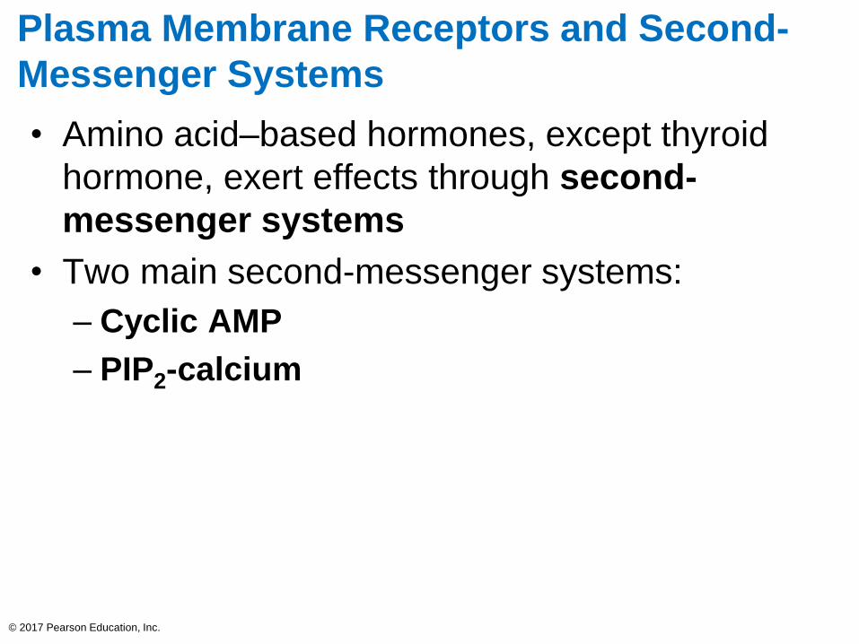
Plasma Membrane Receptors and Second-
Messenger Systems
• Amino acid–based hormones, except thyroid
hormone, exert effects through second-
messenger systems
• Two main second-messenger systems:
– Cyclic AMP
– PIP2-calcium
© 2017 Pearson Education, Inc.

Plasma Membrane Receptors and Second-
Messenger Systems (cont.)
• Cyclic AMP (cAMP) signaling mechanism
1. Hormone (first messenger) binds to receptor
2. Receptor activates a G protein
3. G protein activates or inhibits effector enzyme
adenylate cyclase
4. Adenylate cyclase then converts ATP to cAMP
(second messenger)
5. cAMP activates protein kinases that
phosphorylate (add a phosphate) other proteins
© 2017 Pearson Education, Inc.

Plasma Membrane Receptors and Second-
Messenger Systems (cont.)
• Cyclic AMP (cAMP) signaling mechanism
(cont.)
– Phosphorylated proteins are then either
activated or inactivated
– cAMP is rapidly degraded by enzyme
phosphodiesterase, stopping cascade
– Cascades have huge amplification effect
© 2017 Pearson Education, Inc.

Figure 15.2 Cyclic AMP second-messenger mechanism of water-soluble hormones.
© 2017 Pearson Education, Inc.
G protein signaling mechanisms
are like a molecular relay race.
Hormone
(1st messenger)Receptor G protein Enzyme 2nd
messenger
Extracellular fluidHormone (1st messenger)
binds receptor.
G protein (Gs)
Receptor
GDP GTP
GTP
GTP
ATP
cAMP
Cytoplasm
1
Slide 2

Figure 15.2 Cyclic AMP second-messenger mechanism of water-soluble hormones.
© 2017 Pearson Education, Inc.
G protein signaling mechanisms
are like a molecular relay race.
Hormone
(1st messenger)Receptor G protein Enzyme 2nd
messenger
Extracellular fluidHormone (1st messenger)
binds receptor.
G protein (Gs)
Receptor
GDP GTP
GTP
GTP
ATP
cAMP
Receptor
activates G protein (Gs).
Cytoplasm
1
2
Slide 3

Figure 15.2 Cyclic AMP second-messenger mechanism of water-soluble hormones.
© 2017 Pearson Education, Inc.
G protein signaling mechanisms
are like a molecular relay race.
Hormone
(1st messenger)Receptor G protein Enzyme 2nd
messenger
Adenylate cyclaseExtracellular fluidHormone (1st messenger)
binds receptor.
G protein (Gs)
Receptor
GDP GTP
GTP
GTP
ATP
cAMP
Receptor
activates G protein (Gs).
G protein
activatesadenylatecyclase.
Cytoplasm
1
2 3
Slide 4

Figure 15.2 Cyclic AMP second-messenger mechanism of water-soluble hormones.
© 2017 Pearson Education, Inc.
G protein signaling mechanisms
are like a molecular relay race.
Hormone
(1st messenger)Receptor G protein Enzyme 2nd
messenger
Adenylate cyclaseExtracellular fluidHormone (1st messenger)
binds receptor.
G protein (Gs)
Receptor
GDP GTP
GTP
GTP
ATP
cAMP
Receptor
activates G protein (Gs).
G protein
activatesadenylatecyclase.
Adenylate
cyclase converts
ATP to cAMP(2nd messenger).
Cytoplasm
1
2 3 4
Slide 5

Figure 15.2 Cyclic AMP second-messenger mechanism of water-soluble hormones.
© 2017 Pearson Education, Inc.
G protein signaling mechanisms
are like a molecular relay race.
Hormone
(1st messenger)Receptor G protein Enzyme 2nd
messenger
Adenylate cyclaseExtracellular fluidHormone (1st messenger)
binds receptor.
G protein (Gs)
Receptor
GDP GTP
GTP
GTP
ATP
cAMP
Receptor
activates G protein (Gs).
G protein
activatesadenylatecyclase.
Adenylate
cyclase converts
ATP to cAMP(2nd messenger).
Inactive
protein
kinase
Active
protein
kinase
Triggers responses of
target cell (activatesenzymes, stimulatescellular secretion,opens ion channel, etc.)
Cytoplasm
cAMP activates
protein kinases.
1
2 3 4
5
Slide 6

Plasma Membrane Receptors and Second-
Messenger Systems (cont.)
• PIP2-calcium signaling mechanism
– Hormone-activated G protein activates a
different effector enzyme: phospholipase C
– Activated phospholipase C splits membrane
protein, PIP2, into two second messengers:
• Diacylglycerol (DAG) activates protein kinases
• Inositol trisphosphate (IP3) causes Ca2+ release
from intracellular storage sites
© 2017 Pearson Education, Inc.
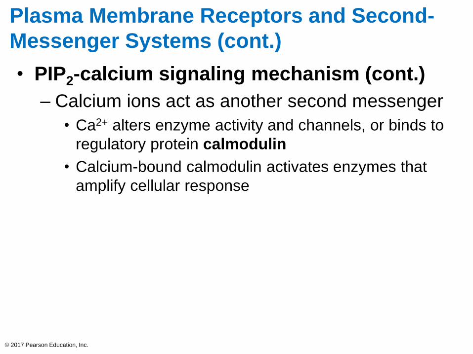
Plasma Membrane Receptors and Second-
Messenger Systems (cont.)
• PIP2-calcium signaling mechanism (cont.)
– Calcium ions act as another second messenger
• Ca2+ alters enzyme activity and channels, or binds to
regulatory protein calmodulin
• Calcium-bound calmodulin activates enzymes that
amplify cellular response
© 2017 Pearson Education, Inc.

Plasma Membrane Receptors and Second-
Messenger Systems (cont.)
• Other signaling mechanisms
– cGMP (cyclic guanosine monophosphate) is
second messenger for selected hormones
– Other hormones work without second
messenger system
• Example: insulin receptor is a tyrosine kinase enzyme
that autophosphorylates upon insulin binding
– Activated tyrosine kinases provide docking sites for
relay proteins that trigger cell responses
© 2017 Pearson Education, Inc.

Intracellular Receptors and Direct Gene
Activation
• Lipid-soluble steroid hormones and thyroid
hormone can diffuse into target cells and bind
with intracellular receptors
• Receptor-hormone complex enters nucleus and
binds to specific region of DNA
• Helps initiate DNA transcription to produce
mRNA
• mRNA is then translated into specific protein
– Proteins synthesized have various functions
– Examples: metabolic activities, structural
purposes, or exported from cell© 2017 Pearson Education, Inc.

Figure 15.3 Direct gene activation mechanism of lipid-soluble hormones.
© 2017 Pearson Education, Inc.
Extracellularfluid
Steroid
hormone Plasmamembrane
Cytoplasm
Receptorprotein
Receptor-hormonecomplex
Nucleus
DNA
mRNA
The steroid hormone diffuses through the plasma membrane and binds an intracellular receptor.
1
Slide 2

Figure 15.3 Direct gene activation mechanism of lipid-soluble hormones.
© 2017 Pearson Education, Inc.
Extracellularfluid
Steroid
hormone Plasmamembrane
Cytoplasm
Receptorprotein
Receptor-hormonecomplex
Nucleus
DNA
mRNA
The steroid hormone diffuses through the plasma membrane and binds an intracellular receptor.
The receptor-
hormone complex
enters the nucleus.
1
2
Slide 3

Figure 15.3 Direct gene activation mechanism of lipid-soluble hormones.
© 2017 Pearson Education, Inc.
Extracellularfluid
Steroid
hormone Plasmamembrane
Cytoplasm
Receptorprotein
Receptor-hormonecomplex
NucleusReceptor
bindingregion
DNA
mRNA
The steroid hormone diffuses through the plasma membrane and binds an intracellular receptor.
The receptor-
hormone complex
enters the nucleus.
The receptor-hormone complex binds a specific DNA region.
1
2
3
Slide 4

Figure 15.3 Direct gene activation mechanism of lipid-soluble hormones.
© 2017 Pearson Education, Inc.
Extracellularfluid
Steroid
hormone Plasmamembrane
Cytoplasm
Receptorprotein
Receptor-hormonecomplex
NucleusReceptor
bindingregion
DNA
mRNA
The steroid hormone diffuses through the plasma membrane and binds an intracellular receptor.
The receptor-
hormone complex
enters the nucleus.
The receptor-hormone complex binds a specific DNA region.
Binding initiates
transcription of the gene to mRNA.
1
2
3
4
Slide 5
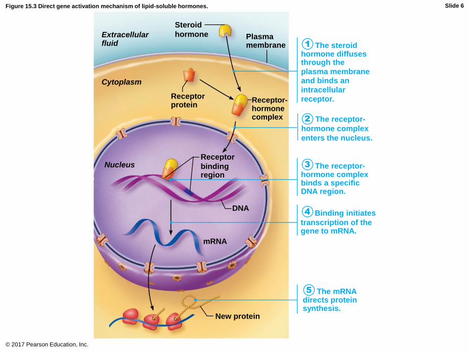
Figure 15.3 Direct gene activation mechanism of lipid-soluble hormones.
© 2017 Pearson Education, Inc.
Extracellularfluid
Steroid
hormone Plasmamembrane
Cytoplasm
Receptorprotein
Receptor-hormonecomplex
NucleusReceptor
bindingregion
DNA
mRNA
New protein
The steroid hormone diffuses through the plasma membrane and binds an intracellular receptor.
The receptor-
hormone complex
enters the nucleus.
The receptor-hormone complex binds a specific DNA region.
Binding initiates
transcription of the gene to mRNA.
The mRNA directs protein synthesis.
1
2
3
4
5
Slide 6
A&P FlixTM: Mechanism of Hormone Action:
Second Messenger cAMP
© 2017 Pearson Education, Inc.
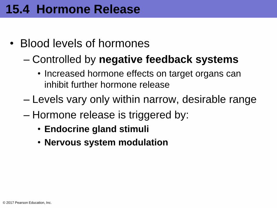
15.4 Hormone Release
• Blood levels of hormones
– Controlled by negative feedback systems
• Increased hormone effects on target organs can
inhibit further hormone release
– Levels vary only within narrow, desirable range
– Hormone release is triggered by:
• Endocrine gland stimuli
• Nervous system modulation
© 2017 Pearson Education, Inc.
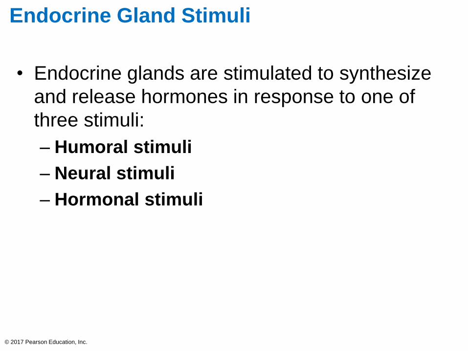
Endocrine Gland Stimuli
• Endocrine glands are stimulated to synthesize
and release hormones in response to one of
three stimuli:
– Humoral stimuli
– Neural stimuli
– Hormonal stimuli
© 2017 Pearson Education, Inc.

Endocrine Gland Stimuli (cont.)
• Humoral stimuli
– Changing blood levels of ions and nutrients
directly stimulate secretion of hormones
– Example: Ca2+ in blood
• Declining blood Ca2+ concentration stimulates
parathyroid glands to secrete PTH (parathyroid
hormone)
• PTH causes Ca2+ concentrations to rise, and stimulus
is removed
© 2017 Pearson Education, Inc.

Figure 15.4a Three types of endocrine gland stimuli.
© 2017 Pearson Education, Inc.
Hormone release caused by
altered levels of certain critical
ions or nutrients.
Humoral Stimulus
Capillary (low Ca2+
in blood)
Parathyroid
glands
Thyroid gland
(posterior view)
Parathyroid
glands
PTH
Stimulus: Low concentration of Ca2+
in capillary blood.Response: Parathyroid glands secreteparathyroid hormone (PTH), whichincreases blood Ca2+.

Endocrine Gland Stimuli (cont.)
• Neural stimuli
– Nerve fibers stimulate hormone release
• Sympathetic nervous system fibers stimulate adrenal
medulla to secrete catecholamines
© 2017 Pearson Education, Inc.

Figure 15.4b Three types of endocrine gland stimuli.
© 2017 Pearson Education, Inc.
Hormone release caused by
neural input.
Neural Stimulus
Stimulus: Action potentials in preganglionicsympathetic fibers to adrenal medulla.Response: Adrenal medulla cells secreteepinephrine and norepinephrine.
CNS (spinal cord)
Preganglionic
sympathetic
fibers
Medulla of
adrenal gland
Capillary
Endocrine Gland Stimuli (cont.)
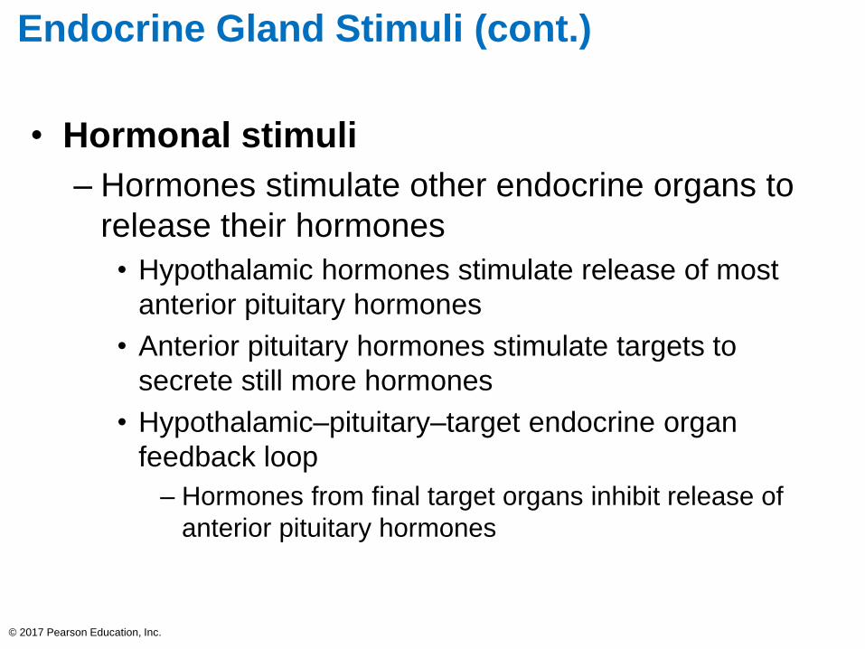
• Hormonal stimuli
– Hormones stimulate other endocrine organs to
release their hormones
• Hypothalamic hormones stimulate release of most
anterior pituitary hormones
• Anterior pituitary hormones stimulate targets to
secrete still more hormones
• Hypothalamic–pituitary–target endocrine organ
feedback loop
– Hormones from final target organs inhibit release of
anterior pituitary hormones
© 2017 Pearson Education, Inc.

Figure 15.4c Three types of endocrine gland stimuli.
© 2017 Pearson Education, Inc.
Hormone release caused by another
hormone (a tropic hormone).
Hormonal Stimulus
Stimulus: Hormones from hypothalamus.
Response: Anterior pituitary gland secreteshormones that stimulate other endocrine glandsto secrete hormones.
Hypothalamus
Anterior
pituitarygland
Thyroid
gland
Adrenal
cortex
Gonad
(Testis)
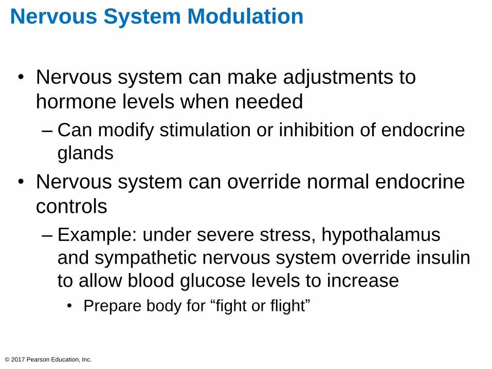
Nervous System Modulation
• Nervous system can make adjustments to
hormone levels when needed
– Can modify stimulation or inhibition of endocrine
glands
• Nervous system can override normal endocrine
controls
– Example: under severe stress, hypothalamus
and sympathetic nervous system override insulin
to allow blood glucose levels to increase
• Prepare body for “fight or flight”
© 2017 Pearson Education, Inc.

15.5 Target Cell Specificity
• Target cells must have specific receptors to
which hormone binds
– Example: ACTH receptors are found only on
certain cells of adrenal cortex, but thyroxin
receptors are found on nearly all cells of body
• Target cell activation depends on three factors:
1. Blood levels of hormone
2. Relative number of receptors on/in target cell
3. Affinity (strength) of binding between receptor
and hormone
© 2017 Pearson Education, Inc.

15.5 Target Cell Specificity
• Amount of hormone can influence number of
receptors for that hormone
– Up-regulation: target cells form more receptors
in response to low hormone levels
– Down-regulation: target cells lose receptors in
response to high hormone levels
• Desensitizes the target cells to prevent them from
overreacting to persistently high levels of hormone
© 2017 Pearson Education, Inc.
Half-Life, Onset, and Duration of Hormone
Activity
• Hormones circulate in blood either free or bound
– Steroids and thyroid hormone are attached to
plasma proteins
– All others circulate without carriers
• Concentration of circulating hormone reflects:
1. Rate of release
2. Speed at which it is inactivated and removed
from body
© 2017 Pearson Education, Inc.

Half-Life, Onset, and Duration of Hormone
Activity (cont.)
• Hormones can be removed from blood by:
– Degrading enzymes or
– Kidneys or
– Liver
• Half-life: time required for level of hormone in blood
level to decrease by half
– Varies anywhere from fraction of a minute to a week,
depending on hormone
© 2017 Pearson Education, Inc.
Half-Life, Onset, and Duration of Hormone
Activity (cont.)
• Hormones have different response times:
– Some responses are immediate
– Some, especially steroid, can take hours to days
– Some are inactive until they enter target cells
• The duration of response is usually limited
– Ranges from 10 seconds to several hours
– Effects may disappear rapidly as blood levels
drop, but some may persist for hours at low
blood levels
© 2017 Pearson Education, Inc.

Half-Life, Onset, and Duration of Hormone
Activity (cont.)
• Half-life, onset, and duration of hormone activity
are dependent on whether the hormone is water
or lipid soluble
© 2017 Pearson Education, Inc.

Table 15.1 Comparison between Lipid- and Water-Soluble Hormones
© 2017 Pearson Education, Inc.
Interaction of Hormones at Target Cells
• Multiple hormones may act on same target at
same time
– Permissiveness: one hormone cannot exert its
effects without another hormone being present
• Example: reproductive hormones need thyroid
hormone to have effect
– Synergism: more than one hormone produces
same effects on target cell, causing amplification
• Example: glucagon and epinephrine both cause liver
to release glucose
© 2017 Pearson Education, Inc.
Interaction of Hormones at Target Cells
(cont.)
– Antagonism: one or more hormones oppose(s)
action of another hormone
• Example: insulin and glucagon
© 2017 Pearson Education, Inc.

15.6 The Hypothalamus
• Hypothalamus is connected to pituitary gland
(hypophysis) via stalk called infundibulum
• Pituitary secretes at least eight major hormones
• It has two major lobes:
– Posterior pituitary: composed of neural tissue
that secretes neurohormones
• Posterior lobe, along with infundibulum make up the
neurohypophysis
– Anterior pituitary: (adenohypophysis) consists
of glandular tissue
© 2017 Pearson Education, Inc.

Pituitary-Hypothalamic Relationships
• Posterior lobe is neural tissue derived from a
downgrowth of brain
– Maintains neural connection to hypothalamus via
hypothalamic-hypophyseal tract
• Tract arises from neurons in paraventricular and
supraoptic nuclei in hypothalamus
• Runs through infundibulum
– Secretes two neurohormones (oxytocin and
ADH)
• Hormones are stored in axon terminals in posterior
pituitary and are released into blood when neurons
fire
© 2017 Pearson Education, Inc.

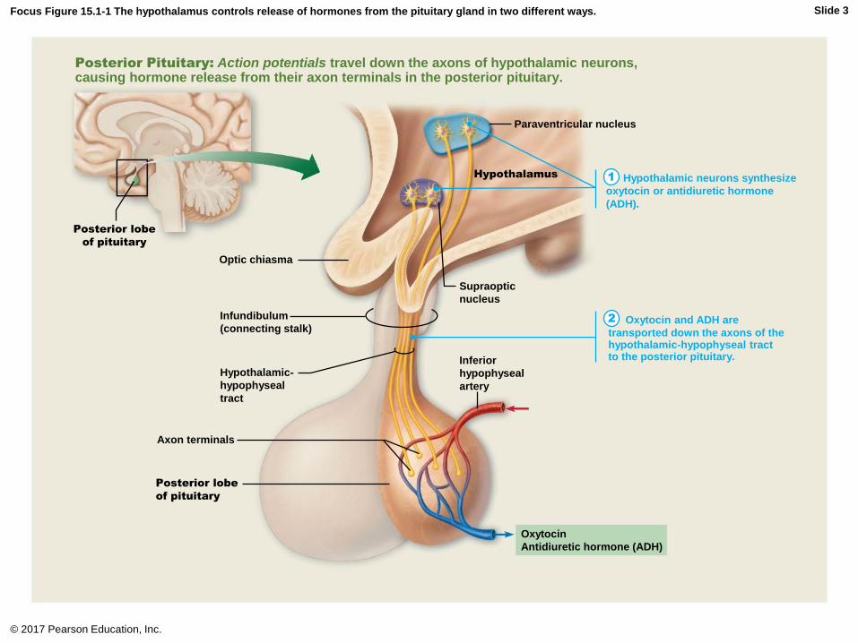
Focus Figure 15.1-1 The hypothalamus controls release of hormones from the pituitary gland in two different ways.
© 2017 Pearson Education, Inc.
Posterior Pituitary: Action potentials travel down the axons of hypothalamic neurons, causing hormone release from their axon terminals in the posterior pituitary.
Paraventricular nucleus
Hypothalamus
Posterior lobe
of pituitary
Supraoptic
nucleus
Optic chiasma
Infundibulum
(connecting stalk)
Hypothalamic-
hypophyseal
tract
Axon terminals
Posterior lobe
of pituitary
Inferior
hypophyseal
artery
Oxytocin
Antidiuretic hormone (ADH)
Hypothalamic neurons synthesize
oxytocin or antidiuretic hormone
(ADH).
1
Slide 2
Focus Figure 15.1-1 The hypothalamus controls release of hormones from the pituitary gland in two different ways.
© 2017 Pearson Education, Inc.
Posterior Pituitary: Action potentials travel down the axons of hypothalamic neurons, causing hormone release from their axon terminals in the posterior pituitary.
Paraventricular nucleus
Hypothalamus
Posterior lobe
of pituitary
Supraoptic
nucleus
Optic chiasma
Infundibulum
(connecting stalk)
Hypothalamic-
hypophyseal
tract
Axon terminals
Posterior lobe
of pituitary
Inferior
hypophyseal
artery
Oxytocin
Antidiuretic hormone (ADH)
Oxytocin and ADH are
transported down the axons of the hypothalamic-hypophyseal tract to the posterior pituitary.
Hypothalamic neurons synthesize
oxytocin or antidiuretic hormone
(ADH).
1
2
Slide 3

Focus Figure 15.1-1 The hypothalamus controls release of hormones from the pituitary gland in two different ways.
© 2017 Pearson Education, Inc.
Posterior Pituitary: Action potentials travel down the axons of hypothalamic neurons, causing hormone release from their axon terminals in the posterior pituitary.
Paraventricular nucleus
Hypothalamus
Posterior lobe
of pituitary
Supraoptic
nucleus
Optic chiasma
Infundibulum
(connecting stalk)
Hypothalamic-
hypophyseal
tract
Axon terminals
Posterior lobe
of pituitary
Inferior
hypophyseal
artery
Oxytocin
Antidiuretic hormone (ADH)
Oxytocin and ADH are
transported down the axons of the hypothalamic-hypophyseal tract to the posterior pituitary.
Oxytocin and ADH are stored in
axon terminals in the posterior
pituitary.
Hypothalamic neurons synthesize
oxytocin or antidiuretic hormone
(ADH).
1
2
3
Slide 4

Focus Figure 15.1-1 The hypothalamus controls release of hormones from the pituitary gland in two different ways.
© 2017 Pearson Education, Inc.
Posterior Pituitary: Action potentials travel down the axons of hypothalamic neurons, causing hormone release from their axon terminals in the posterior pituitary.
Paraventricular nucleus
Hypothalamus
Posterior lobe
of pituitary
Supraoptic
nucleus
Optic chiasma
Infundibulum
(connecting stalk)
Hypothalamic-
hypophyseal
tract
Axon terminals
Posterior lobe
of pituitary
Inferior
hypophyseal
artery
Oxytocin
Antidiuretic hormone (ADH)
Oxytocin and ADH are
transported down the axons of the hypothalamic-hypophyseal tract to the posterior pituitary.
Oxytocin and ADH are stored in
axon terminals in the posterior
pituitary.
When associated hypothalamic neurons fire, action potentials arriving at the axon terminals cause oxytocin or ADH to be released into the blood.
Hypothalamic neurons synthesize
oxytocin or antidiuretic hormone
(ADH).
1
2
3
4
Slide 5

Pituitary-Hypothalamic Relationships (cont.)
• Anterior lobe is glandular tissue derived from an
outpocketing of oral mucosa
– Vascularly connected to hypothalamus via
hypophyseal portal system consisting of:
• Primary capillary plexus
• Hypophyseal portal veins
• Secondary capillary plexus
• Hypothalamus secretes releasing and
inhibiting hormones to anterior pituitary to
regulate hormone secretion
© 2017 Pearson Education, Inc.

Focus Figure 15.1-2 The hypothalamus controls release of hormones from the pituitary gland in two different ways (continued).
© 2017 Pearson Education, Inc.
Anterior Pituitary: Hypothalamic hormones released into special blood vessels
(the hypophyseal portal system) control the release of anterior pituitary hormones.
Anterior lobe
of pituitary
When appropriately stimulated, hypothalamic
neurons secrete releasing or inhibiting
hormones into the primary capillary plexus.
Hypothalamic hormones travel
through portal veins to the anterior pituitary where they stimulate or inhibit release of hormones madein the anterior pituitary.
In response to releasing
hormones, the anterior pituitary
secretes hormones into the secondary capillary plexus. This in turn empties into the general circulation.
Growth hormone (GH)
Thyroid-stimulating hormone (TSH)
Adrenocorticotropic hormone (ACTH)
Follicle-stimulating hormone (FSH)
Luteinizing hormone (LH)
Prolactin (PRL)Anterior lobe
of pituitary
Hypophyseal
portal systemSuperior
hypophyseal
artery
A portal system is two capillary plexuses (beds) connected by veins.
Hypothalamus Hypothalamic neurons
synthesize releasing
and inhibiting hormones
(GHRH, GHIH, TRH,
CRH, GnRH, PIH).
• Primary capillary
plexus
• Hypophyseal
portal veins
• Secondary
capillary plexus
1
2
3

Posterior Pituitary and Hypothalamic
Hormones
• Posterior pituitary consists of axon terminals of
neurons from hypothalamic neurons:
– Paraventricular neurons produce oxytocin
– Supraoptic neurons produce antidiuretic
hormone (ADH)
• Oxytocin and ADH
– Each composed of nine amino acids
– Almost identical but differ in two amino acids
© 2017 Pearson Education, Inc.

Posterior Pituitary and Hypothalamic
Hormones (cont.)
• Oxytocin
– Strong stimulant of uterine contractions released
during childbirth
– Also acts as hormonal trigger for milk ejection
– Both are positive feedback mechanisms
– Acts as neurotransmitter in brain
• Uses PIP2-calcium second messenger system
© 2017 Pearson Education, Inc.

Posterior Pituitary and Hypothalamic
Hormones (cont.)
• Antidiuretic hormone (ADH)
– Hypothalamus contains osmoreceptors that
monitor solute concentrations
– If concentration too high, posterior pituitary
triggered to secrete ADH
– Targets kidney tubules to reabsorb more water
to inhibit or prevent urine formation
– Release also triggered by pain, low blood
pressure, and drugs
© 2017 Pearson Education, Inc.

Posterior Pituitary and Hypothalamic
Hormones (cont.)
• Antidiuretic hormone (ADH) (cont.)
– Inhibited by alcohol, diuretics
– High concentrations cause vasoconstriction, so
also called vasopressin
© 2017 Pearson Education, Inc.
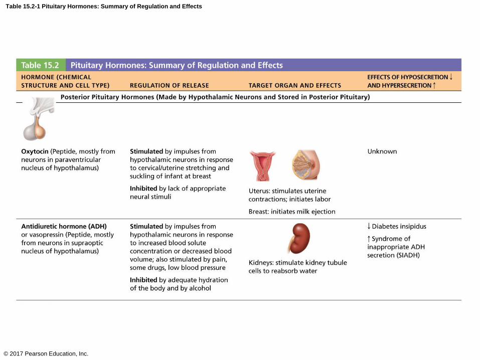
Table 15.2-1 Pituitary Hormones: Summary of Regulation and Effects
© 2017 Pearson Education, Inc.

Clinical – Homeostatic Imbalance 15.1
• Diabetes insipidus
– ADH deficiency due to damage to hypothalamus
or posterior pituitary
– Must keep well hydrated
• Syndrome of inappropriate ADH secretion
(SIADH)
– Retention of fluid, headache, disorientation
– Fluid restriction; blood sodium level monitoring
© 2017 Pearson Education, Inc.
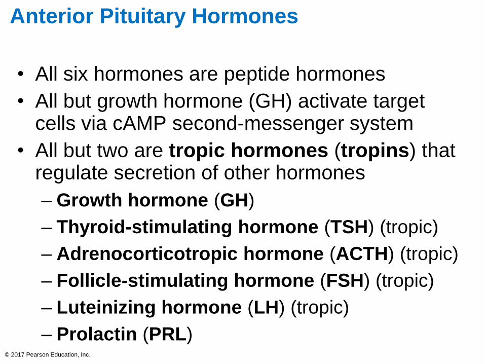
Anterior Pituitary Hormones
• All six hormones are peptide hormones
• All but growth hormone (GH) activate target cells via cAMP second-messenger system
• All but two are tropic hormones (tropins) that regulate secretion of other hormones
– Growth hormone (GH)
– Thyroid-stimulating hormone (TSH) (tropic)
– Adrenocorticotropic hormone (ACTH) (tropic)
– Follicle-stimulating hormone (FSH) (tropic)
– Luteinizing hormone (LH) (tropic)
– Prolactin (PRL)© 2017 Pearson Education, Inc.
Anterior Pituitary Hormones (cont.)
• Growth hormone (GH)
– Also called somatotropin as it is produced by
somatotropic cells
– Has direct actions on metabolism and indirect
growth-promoting actions
– Direct actions on metabolism
• Glucose-sparing actions decrease rate of cellular
glucose uptake and metabolism (anti-insulin effects)
• Triggers liver to break down glycogen into glucose
• Increases blood levels of fatty acids for use as fuel
and encourages cellular protein synthesis
© 2017 Pearson Education, Inc.

Anterior Pituitary Hormones (cont.)
– Indirect actions on growth:
• GH triggers liver, skeletal muscle, and bone to
produce insulin-like growth factors (IGFs)
• IGFs then stimulate:
– Cellular uptake of nutrients used to synthesize DNA
and proteins needed for cell division
– Formation of collagen and deposition of bone matrix
• GH stimulates most cells to enlarge and divide, but
major targets are bone and skeletal muscle
© 2017 Pearson Education, Inc.
Anterior Pituitary Hormones (cont.)
– Regulation of secretion
• GH release or inhibition chiefly regulated by
hypothalamic hormones on somatotropic cells
– Growth hormone–releasing hormone (GHRH)
stimulates GH release
» Triggered by low blood GH or glucose, or high
amino acid levels
– Growth hormone–inhibiting hormone (GHIH)
(somatostatin) inhibits release
» Triggered by increase in GH and IGF levels
• Ghrelin (hunger hormone) also stimulates GH release
© 2017 Pearson Education, Inc.
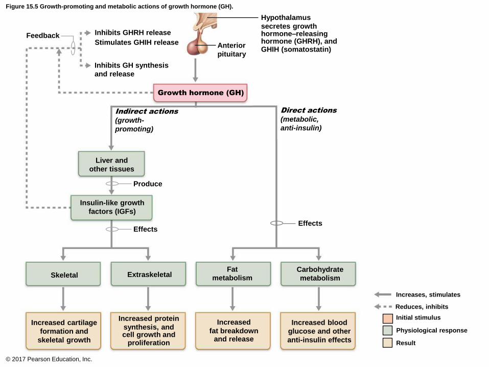
Figure 15.5 Growth-promoting and metabolic actions of growth hormone (GH).
© 2017 Pearson Education, Inc.
Hypothalamus
secretes growthhormone–releasinghormone (GHRH), andGHIH (somatostatin)
Anterior
pituitary
Inhibits GHRH release
Stimulates GHIH release
Inhibits GH synthesis
and release
Growth hormone (GH)
Feedback
Indirect actions
(growth-
promoting)
Liver and
other tissues
Produce
Insulin-like growth
factors (IGFs)
Effects
Skeletal ExtraskeletalFat
metabolismCarbohydrate
metabolism
Increased cartilage
formation and
skeletal growth
Increased protein
synthesis, andcell growth and
proliferation
Increased
fat breakdownand release
Increased blood
glucose and other
anti-insulin effects
Increases, stimulates
Reduces, inhibits
Initial stimulus
Physiological response
Effects
Result
Direct actions
(metabolic,
anti-insulin)

Clinical – Homeostatic Imbalance 15.2
• Hypersecretion of GH is usually caused by
anterior pituitary tumor
– In children results in gigantism
• Can reach heights of 8 feet
– In adults results in acromegaly
• Overgrowth of hands, feet, and face
• Hyposecretion of GH
– In children results in pituitary dwarfism
• May reach height of only 4 feet
– In adults usually causes no problems
© 2017 Pearson Education, Inc.

Figure 15.6 Disorders of pituitary growth hormone.
© 2017 Pearson Education, Inc.
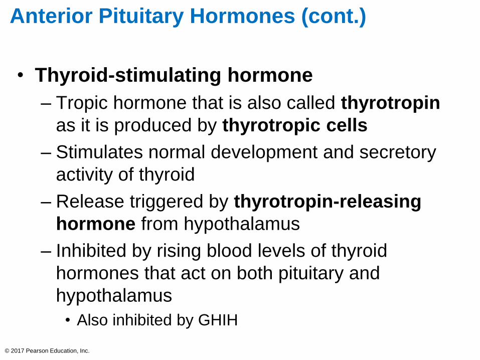
Anterior Pituitary Hormones (cont.)
• Thyroid-stimulating hormone
– Tropic hormone that is also called thyrotropin
as it is produced by thyrotropic cells
– Stimulates normal development and secretory
activity of thyroid
– Release triggered by thyrotropin-releasing
hormone from hypothalamus
– Inhibited by rising blood levels of thyroid
hormones that act on both pituitary and
hypothalamus
• Also inhibited by GHIH
© 2017 Pearson Education, Inc.

Figure 15.7 Regulation of thyroid hormone secretion.
© 2017 Pearson Education, Inc.
Hypothalamus
Anterior pituitary
Thyroid gland
Thyroid
hormones
Target cells
TRH
TSH
Stimulates
Inhibits

Anterior Pituitary Hormones (cont.)
• Adrenocorticotropic hormone (ACTH)
– Also called corticotropin as it is secreted by
corticotropic cells
• Precursor to corticotropin is pro-opiomelanocortin
– ACTH stimulates adrenal cortex to release
corticosteroids
– Regulation of ACTH release
• Triggered by hypothalamic corticotropin-releasing
hormone (CRH) in daily rhythm
– Highest levels in morning
• Internal and external factors that alter release of CRH
include fever, hypoglycemia, and stressors© 2017 Pearson Education, Inc.
Anterior Pituitary Hormones (cont.)
• Gonadotropins (FSH and LH)
– Follicle-stimulating hormone (FSH) and
luteinizing hormone (LH) are secreted by
gonadotropic cells of anterior pituitary
– FSH stimulates production of gametes (egg or
sperm)
– LH promotes production of gonadal hormones
• In females, LH helps mature follicles of egg, triggers
ovulation and release of estrogen and progesterone
• In males, LH stimulates production of testosterone
© 2017 Pearson Education, Inc.

Anterior Pituitary Hormones (cont.)
• Gonadotropins (FSH and LH) (cont.)
– LH and FSH both are absent from blood in
prepubertal boys and girls
– Regulation of gonadotropin release
• Triggered by gonadotropin-releasing hormone
(GnRH) during and after puberty
• Suppressed by gonadal hormones (feedback)
© 2017 Pearson Education, Inc.

Anterior Pituitary Hormones (cont.)
• Prolactin (PRL)
– Secreted by prolactin cells of anterior pituitary
– Stimulates milk production in females; role in
males not well understood
– Regulation primarily controlled by prolactin-
inhibiting hormone (PIH), which is dopamine
– PIH prevents release of PRL until needed, with
decreased levels leading to lactation
© 2017 Pearson Education, Inc.
Anterior Pituitary Hormones (cont.)
• Prolactin (PRL) (cont.)
– Increased estrogen levels stimulate PRL
• Reason behind breast swelling and tenderness during
menstrual cycle
– Blood levels rise toward end of pregnancy
– Suckling stimulates PRL release and promotes
continued milk production
© 2017 Pearson Education, Inc.

Clinical – Homeostatic Imbalance 15.3
• Hypersecretion of prolactin is more common
than hyposecretion
– Hyposecretion not a problem in anyone except
women who choose to nurse
• Hyperprolactinemia is the most frequent
abnormality of anterior pituitary tumors
• Clinical signs include inappropriate lactation,
lack of menses, infertility in females, and
impotence in males
© 2017 Pearson Education, Inc.
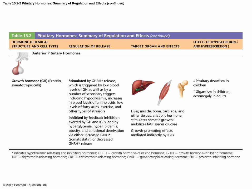
Table 15.2-2 Pituitary Hormones: Summary of Regulation and Effects (continued)
© 2017 Pearson Education, Inc.

Table 15.2-3 Pituitary Hormones: Summary of Regulation and Effects (continued)
© 2017 Pearson Education, Inc.