The Embryonic Development of Trichogramma evanescens, Westw
42
DEVELOPMENT OF TRIOHOGIUMMA EVANESCENS. 149 The Embryonic Development of Trichogramma evanescens, Westw., Monembryonic Egg Parasite of Donacia simplex, Fab. By J. Bronte Gatenby, Exhibitioner of JeauB College, Oxford. Witli Plates 10, 11, and 12. INTRODUCTION. SINCE the description of polyembryony in some parasitic Hymenoptera by Marchal (1) the attention of a few zoologists 1 has been turned to the interesting problems these forms offer. J Sir Ray Lankester has kindly drawn my attention to the writings of the Polish Embryologist, Ganin, whose work on Platygaster, carried out forty years ago, is of great interest. Platygaster is a parasite on the larvse of some Diptera (Cecidomyia), and its larval form is curiously modified in early stages. According to Ganin the larva has neither nervous, vascular, nor respiratory systems (Compare p. 20 of this paper), and its last abdominal segment terminates in a curious caudal organ of a tree-like nature, almost certainly concerned in nutrition. The larva undergoes a number of moults, looses its caudal organ, and gradually becomes vermiform. I have lately noticed that the larva of an Apanteles parasitic on Porthesia has a remarkably modified ultimate abdominal segment, which is very large, vesicular, and formed of hypertrophied cells. The gut of this larva is in all the early stages completely blind, and the animal depends on the swollen abdominal segment for its nutrition. Like Ganin's larvse this form looses the vesicular segment just before pupation. It is a very remarkable fact that the ultimate abdominal segment should be modified for this purpose (Zeit. f. Zool., Bd. xv.).
Transcript of The Embryonic Development of Trichogramma evanescens, Westw
DEVELOPMENT OF TRIOHOGIUMMA EVANESCENS. 149
The Embryonic Development of Trichogrammaevanescens, Westw., Monembryonic EggParasite of Donacia simplex, Fab.
ByJ. Bronte Gatenby,
Exhibitioner of JeauB College, Oxford.
Witli Plates 10, 11, and 12.
INTRODUCTION.
SINCE the description of polyembryony in some parasiticHymenoptera by Marchal (1) the attention of a few zoologists1
has been turned to the interesting problems these formsoffer.
J Sir Ray Lankester has kindly drawn my attention to the writingsof the Polish Embryologist, Ganin, whose work on Platygaster, carriedout forty years ago, is of great interest. Platygaster is a parasite onthe larvse of some Diptera (Cecidomyia), and its larval form is curiouslymodified in early stages. According to Ganin the larva has neithernervous, vascular, nor respiratory systems (Compare p. 20 of this paper),and its last abdominal segment terminates in a curious caudal organ ofa tree-like nature, almost certainly concerned in nutrition. The larvaundergoes a number of moults, looses its caudal organ, and graduallybecomes vermiform. I have lately noticed that the larva of an Apantelesparasitic on Porthesia has a remarkably modified ultimate abdominalsegment, which is very large, vesicular, and formed of hypertrophiedcells. The gut of this larva is in all the early stages completely blind,and the animal depends on the swollen abdominal segment for itsnutrition. Like Ganin's larvse this form looses the vesicular segmentjust before pupation. It is a very remarkable fact that the ultimateabdominal segment should be modified for this purpose (Zeit. f. Zool.,Bd. xv.).
150 J. BROM'ti GATBNBY.
Silvestri (2) has contributed some important papers on thesubject, but these have appeared in an Italian agriculturaljournal only taken by a small number of the scientificlibraries of this country. Considering the vast number ofparasitic Hymenoptera which exist, and their diversity audremarkable iiiotincts, a rich field, only now being explored,is opened to zoologists. But it is a field full of difficulties,for the Trichogrammids, to mention one group alone, are, asPerkins (4) has sdd, among the smallest of known insects.In several other groups of parasitic Hymenoptera there areto be found numbers of forms whose life history and habitsare of absorbing interest. The pure observer finds problemsand instincts of wonderful diversity, and the embryologist isimpressed with the remarkable adaptations for the modusvivendi which these forms follow.
The remarkable oogenesis of some of these parasites hasbeen the subject of some interesting papers by R. Hegner (3).
The parasite, a part of whose embryology I have describedin this paper, is a member of that important family theChalcididse, a numerous and highly interesting assemblage ofminute Hymenoptera. These insects are of great importanceto the economic entomologist, because among them one findsforms which aid the agriculturist, and which often injure.Trichogramma might be said to aid.
It is a pleasant duty to express my thanks to Mr. Goodrichfor his kind interest in this work, and for advice and criticism,•which has been of great value.
THE HOST1 (DONACIA SIMPLEX, FAB.).
Donacia s implex is quite common around Oxfoi'd in theearly summer. Commander Walker informs me that he hasoccasionally taken this Bpecies at Oxford in winter. In theearly summer one can always find the beetles on the water-
1 Kindly identified by Mr. H. Britten, Assistant of the Hope Depart-ment, Oxford Museum.
DEVELOPMENT OF TRIUHOGBAMMA EVANESCENS. 151
reeds which grow from shallow ponds and the sides ofstreams; they may be observed copulating and laying theireggs. The latter are laid in masses in a regular manner, thewhole group forming a rectangular mass containing a varyingnumber of eggs. In one mass eggs of several shades ofbrown may occur in patches, as if a number of beetles hadoviposited in the same place. Whether this is so I do notknow. The egg groups do not adhere very closely to thesurface of the reed, and they are easily removed by bendingthe surface upon which they are laid. Prom the number ofparasitized eggs which one can find there is no doubt thatthis Trichogrannnid must cause a great deal of destructionamong the broods of beetles, and were the Donacia a pest onvaluable plants it would be quite easy and worth while torear batches of parasites. This has been done in the caseof parasites of injurious insects, particularly in America, andsuch methods of attacking pests have so far met with a gooddeal of success. In the case of Donacia almost the entirenumber of eggs laid iu a locality where the parasites arecommon will bo found parasitised. In PL 10, fig. 3, is drawnan enlarged figure of D o n a c i a s i m p l e x ; in A the eggmass (OV.) viewed in profile upon the reed (R.) is shown,and resting on the lower eggs a Trichogramuni {P.P.) is seen,drawn to about the same scale as the beetle.
THE PARASITE (TKICHOGEAMMA EVANESCENS, WESTW.)
I have to thank Commander Walker for drawing myattention to some literature on Trichogramma. The Rev. J.Waterston, B.D., of the Imperial Bureau of Entomology, inkindly identifying this insect, writes that T r i c h o g r a m m ae van esc ens is generally found as a parasite upon the eggsof insects whose habits and place of oviposition are similar tothat of Donacia.
As is common with many of the parasitic Hymenoptera,T r i c h o g r a m m a e v a n e s c e n s has very gaudy colouring.The wings, which are a shiny blue, at once attract attention
152 J. BR0NT6 GATENBY.
to the insect as it walks over the Donacia egg mass. Incollecting my material I found it most convenient to examinethe rushes for Donacia egg-masses from a boat, and thoseupon which parasites were seen were removed from thewater-plant and placed in a box. A most unfortunate circum-stance, unknown to me then, was the fact that the time1 takento walk to the laboratory with the material was just longenough to allow the newly-laid eggs to form polar bodies,segment, and enter upon the blastoderm stage. Except inthe case of a small number of eggs laid in the laboratory, allmy sections begin from the blastoderm stage onwards, andsome important stages are missing. If the insect is takeninto the laboratory ar>d placed with an egg mass of Donacia,it is possible to watch oviposition taking place. The littleparasite may be observed to walk somewhat rapidly over theeggs, continually tapping them with its geniculate antennae.When is satisfied with the egg it has chosen it stops,unsheaths its ovipositor, and moves its abdomen backwardsand forwards with a sawing motion about eight times, untilthe chorion of the Donacia egg is pierced. When thishappens the parasite may be seen to depress its abdomen,thrusting home the ovipositor. It pauses about five secondswhile the egg passes down the evipositor into the Donaciaegg, withdraws its ovipositor, and generally begins on thenext egg in the row. Though the parasite does not seemto work systematically along the rows, in many cases all theeggs in a mass are parasitised, though more often a few areleft untouched.
In cases where all the eggs have been parasitised severalparasites may have laid in one mass, It is quite common toobserve two or three Trichogrammids on one Donacia egg-mass. In very rare cases there are two eggs laid in thesame Donacia egg; one so seldom finds this that it isprobable that a parasite is able to tell whether one of itsfellows has previously given attention to an egg. What
1 Added to the fact that the fixative I uaed does not penetrate thechorion of the beetles' eggs as quickly as desirable.
DEVELOPMENT OV TRICHOGRAMMA EVANESCENS. 153
happens in development when two eggs are laid in the sameDonacia egg I do not know, but one generally finds the twoeggs in different stages of development. Probably the olderembryo succeeds in the end in killing the other, for I have notyeb found more than one insect imerging from one egg.
I am unable to say whether there is more than one broodof parasites during the summer, but it is possible to collectat the same time eggs contaiuing parasites ready to emerge,and some containing newly laid eggs. This points to therebeing more than one brood. There are two or three speciesof Donacia fairly common at Oxford, and they appear oneafter the other, so that this strengthens the view that severalbroods occur in one season. During the winter months Ihave not found the empty egg-cases of Donacia on the stemsof the reeds, and I have not been able to satisfy myself as towhether the parasite hibernate in the egg-cases or whetherthey emerge in summer and creep into crevices with a view towintering there. Nearly every year the reeds upon which theegg-masses are laid are submerged in the floods, and becomewithered and torn, and thorouglily soaked. For this reasonit is unlikely that the parasites would remain in the eggswhich they have destroyed.
In remarking on the parasite and its host, I do not over-look the possibility of T. e v a n e s c e n s being found on theeggs of other insects.1
TECHNIQUE.
The egg of Donacia is covered by a thick chorion which,added to the yolk, makes sectioning a very difficult business.The parasitised egg-masses were generally preserved inPetrunchekewitsch, with a little more nitric acid than usual.This often gave splendid results. A mixture of Petrunche-kewitsch2 and Bouin2 was also tried with about equal results.
1 Prof. Poulton informs me that this Chalcid parasitises the eggs ofDragon flies. I have since been able to observe this interesting factmyself.
3 For these fixatives see Bolls Lee's Microtomists' Vade-Mecuin.
154 J. BRON'rf: GATJENRY.
In some cases the eggs were pricked and the whole throwninto picro-nitric.
After some trials Petrunchekewitsch was almost exclusivelyused, and in most cases it gave a fine fixation, but not always.In using this fixative it is not necessary to prick the eggs.Ordinary preservatives like Bouin, corrosive acetic, orFlemming will not penetrate the chorion. This at oncecauses difficulties, for alcoholic fixatives are not alwaysreliable. The eggs were left over night in the Petrunche-kewitscli and -washed out in 70 per cent, alcohol.
When in xylol the eggs were pricked with a fine needleand placed in the paraffin bath. It was not always possibleto successfully prick the eggs, but unless this was done itwas necessary to leave the masses longer in the bath. Thishnrdens the eggs and makes sectioning a dreadfully difficulttask. The eggs were cut in their groups, 5 u in thickness,on a Yung microtome, each section being painted withcelloidin and ether. One could not be sure that the eggswere not parasitised until after staining, and three or fourbatches would often be cut without finding any stages. Itwas only by staining overnight in Iron Hcematoxylin that aa suitable differentiation could be got. Bhrlich and thecarmines were useless. In some cases alternate slides werecounterstained in orange G. or dilute acid fuchsin.
GENKRAL FACTS CONCERNING THE APPEAHANCE OH THE
MATERIAL IN SECTIONS AND IN WHOLE MOUNTS.
In PI. 11, fig. 8, there is drawn a part of the section of aparasitised egg-mass. The larval parasite (D-P-) lies in theyolk of the Doimcia egg, and a little to the right and loweredge of the larva is the remains of the embryonic gut of thehost (Gr.). At N.S. are the remains of the Donacia larva'snervous system, and below at L is a still recognisable de-generate leg. The parasite has reached the stage just beforeit begins to swallow the yolk in which it lies. Abuttingagainst the chorion of the egg in the middle of the field are
DEVELOPMENT OF TRICHOGRAMMA KVANKSOENS. 1 5 5
the chorions of the neighbouring eggs, all of winch wereparasitised.
It will be seen that when these eggs were attacked thecontained embryos had become far advanced and were almostready to hatch. Though one can observe a Douacia ovi-positing, and a parasite on the same mass piercing anddepositing its eggs in the newly laid beetle's eggs, it ispossible to find eggs parasitised at any stage. If one removesDonacia embryos from their chorion by means of fine needlesand stains them in paracarmine, one can often find thedeveloping parasites as in PI. 10. fig. 2, at D.P. Xowthisembryo lies at the posterior pole of tlie embryo beetle, and istoo far down to have been oviposited there. It may be thatthis egg was an outside one of the mass and that the parasitebored it from the side; but such cases occur too frequentlyin sections of eefg's in the middle of the mass, and I aminclined to think that in those Donacia eggs laid in ahorizontal position the developing Triohogramina embrvomay sink downwards. I cannot otherwise explain howparasites' eggs are found in this position, because the beetle'seggs seem too closely applied to one another to allow theparasite to get its ovipositoi1 between them, and referenceto PI. 10, fig. 1, will show how short the little insect'sovipositor is. (Both fig. 1 and 2 are drawn to the samescale: x 75.)
THE EFFECT or THE DEPOSITION OF THE EGG IN THK
DEVELOPING EMBKYO'S BODY.
Primarily the effect is to arrest further development of thehost, but all life is not killed immediately, for living nucleiare to be found much later on as the parasite develops. Asis well known, the nuclei in the yolk of an insect's egg arevery large, and such vitellophags become larger than theother cells almost from the time they are established. Ibelieve that it is the vitellophags which manage to live longestafter the parasite has oviposited in the beetle's egg, and in
156 J. BUONTE GATENBY.
some cases degenerate, but evidently still living yolk cellscan be found in the gut of the young larval parasite.
In degeneration the nuclei become hyperchromatic, largestainable masses collecting in both nucleus and cytoplasm,the cell finally becoming a black shapeless mass. I aminclined to believe that the large cells forming the serosaalso live longer than the ordinary embryonic cells, after theTrichogramma embryo has been developing some time.
THE OVARIAN EGG WHEN BEADY TO BE LAID.
In PI. 11, fig. 9, a longitudinal section of the nearly matureovarian egg is drawn. The egg is of an elongated ovalshape, the anterior pole (A.) being somewhat broader thanthe posterior, and the cytoplasm appears homogeneous exceptfor the occurrence at the posterior pole of a large darkmass [G.G.D.), the so-called germ-cell, or germ line, deter-minant. The probable nature, mode of appearance, and thefate of this protoplasmic inclusion will be dealt with under aseparate heading. The follicle cells are much drawn out inPI. 11. fig. 9, and it is very difficult to distinguish betweenthe wall of the ovary and the follicular layer. The nucleuslies slightly towards the anterior yolk of the egg in the midline. It consists of a large condensed mass of chromatinsurrounded by a clear nucleoplasmic zone. In the latterminute stainable granules may be found. The manner inwhich this condensed form of nucleus is produced is, as faras I am able to judge from my material of adult insects, thesame as that described by Hegner for Copidosoma (3).
THE NEWLY LAID EGG.
In the eggs at this period I have found a small body nearthe surface, which, I think, is the spermatozoon. In PI. 11,fig. 10, this darkly staining body is seen to be surrounded bya number of small granules. I have been unable to find anysigns of activity around this body as one would expect if it
DEVELOPMENT OF TRICHOGRAMMA EVANESOEXS. 157
were a spermatozoon, though in some insects no dealing ofthe cytoplasm around the male pronucleus, or other event,takes place at this period. In the stage later, during theformation of the polar bodies, the granules which werepresent in PL 11, fig. 10, around the male pronucleus (M.P.N.)cannot be seen, but the latter has penetrated further into theegg. In all the sections of newly laid eggs that I have found,the cytoplasm towards the central region of the egg hasbecome partially vacuolated and thinner, while the germ celldeterminant has become much more faintly staining. Mycollection of newly laid eggs is not complete enough to showwhether this thinning out of the central region of the egg isthe rule, and it should be observed that in PI. 11, fig. 11,which shows the formation of the polar bodies, this vacuolisa-tion was quite absent. In PI. 11. fig. 7, I have drawn atransverse section of an egg which shows the nucleus lyingin a central clear region, and quite close a denser partof the cytoplasm containing a cloud of granules (G.G.).
The egg, when laid, lies almost always towards the top ofthe Donacia ovum, and it never has a definite orientation, forin a section of a group of the host's eggs one cuts across eggsin all directions. In a brood of parasites which I caughtemerging, some had their heads downwards in the Donaciaegg, some their abdomens. At the stage when the larvabegins to feed, it is forced to lie lengthwise in the host's egg*because it has by then become too long to lie in any otherway. It is obvious that the orientation of the pupatingTrichogramma larva in relation to the Donacia egg is notgoverned by any special circumstance. Nevertheless it ispossible, though to my mind unlikely, that the larva may beable to turn around at will within the Donacia egg.
The newly laid egg is provided with a vitelline membraneand a thin chorion (G.H.).
FOKMATION OF THE POLAR BODIES.
In the one egg I found at this stage there were two polarbodies (PL 11, fig. 11). One polar body (P.Z?1.) has been

158 J. BRONTfi GATKNBV.
extruded and lies on the surface of the egg. The spindle ofthe second polar body is in the telophuse and the chromo-somes seem fused. No aster or centrosomes could be seen.Around the neighbourhood of the forming polar body is aclear zone, and a little above the dumb-bell shaped figure aretwo large granules. I do not know exactly how thesegranules arise, but I think that they are possibly extrusionsof the polar figures, for expulsion of granules from the nucleican be observed in later stages. The fate of the polar bodiesis not known. In Oophthora and Bncyrtus they eventuallydegenerate (Hegner, 3«).
THK STAGES BETWEEN FORMATION OF POLAR BODIES AND THE
BLASTODERM.
These are not described in the present paper; through lackof material last spring I have been unable to get the stages.This spring I was able to procure a great deal more material,with which I hope to describe the early segregation of thegerm-cells and the accompanying1 phenomena.
THE BLASTODERM STAGE.
The material from the blastoderm stage onwards to theformation of the young larva is very complete. The germ-celldeterminant at the posterior pole of the egg has, by the timeof formation of the polar bodies, become more faintly staining,and considerably broken up (PI. 11, fig. 20, broken piecesP.P.). Such broken pieces come apart, and the whole deter-minant loses almost all affinity for stains of any kind. Theexact time at which the determinant usually disappears is atpresent unknown, but very rarely one can find rather darklystaining patches in the germ-cells of the blastoderm stage,which may be the remains of the germ-cell determinant(PI. 12, fig. 36 G.)
In PI. 11, fig. 12, the earliest blastoderm stage is seen inlongitudinal section. The number of germ-cells is very diffi-cult to determine, for at about this stage the latter lose almost
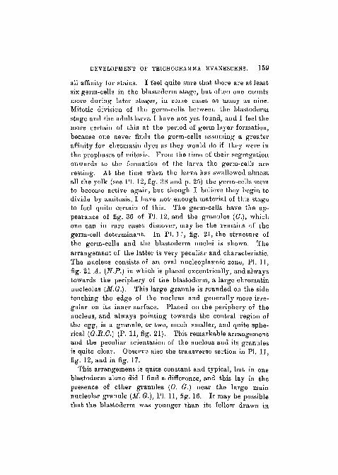
DEVELOPMENT OF TRIOHOGRAMMA EVANESOENS. 159
all affinity for stains. I feel quite sure that there are at leastsix germ-cells in the blastoderm stage, but often one countsmore during later stages, in some cases as many as nine.Mitotic division of the germ-cells between the blastodermstage and the adult larva I have not yet found, and I feel themore certain of this at the period of germ layer formation,because one uevet1 finds the germ-cells assuming a greateraffinity for chroinatin dyes as they would do if they were inthe prophases of mitosis. From the time of their segregationonwards to the formation of the larva the germ-cells areresting. At the time when the larva has swallowed almostall the yolk (see PL 12, fig. 38 and p. 25) the germ-cells seemto become active again, but though I believe they begin todivide by amifcosis, I have not- enough material of this stageto feel quite certain of this. The germ-cells have the ap-pearance of fig. 36 of PL 12, and the granules (G.), whichone can in rare cases discover, may be the remains of thegerm-cell determinant. In PL 11, fig. 21, the structure ofthe germ-cells and the blastoderm nuclei is shown. Thearrangement of the latter is very peculiar and characteristic.The nucleus consists of an oval nucleoplasmic zone, PL 11,fig. 21 A. {N.P.) in which is placed excentrically, and alwaystowards the periphery of the blastoderm, a large chromatinnucleolus (M.G.). This large granule is rounded on the sidetouching the edge of the nucleus and generally more irre-gular on its inner surface. Placed on the periphery of thenucleus, and always pointing towards the central region ofthe egg, is a granule, or two, much smaller, and quite sphe-rical (G.B.G.) (P. 11, fig. 21). This remarkable arrangementand the peculiar orientation of the nucleus and its granulesis quite clear. Observe also the transverse section in PL 11,fig. 12, and in fig. 17.
This arrangement is quite constant and typical, but in oneblastoderm alone did I find a difference, and this lay in thepresence of other granules (0. G.) near the large mainnucleolar granule (M. G.), PI. 11, fig. 16. It may be possiblethat the blastoderm was younger than its fellow drawn in
160 J. KRONTii GATBNBY.
PI. 11, fig. 12, and that the exceptional nucleus is an inter-mediate form.
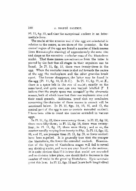
The nuclei at the anterior end of the egg are orientated inrelation to the centre, as are those of the posterior. In thecentral region of the egg are found a number of black masses(Iron Haematoxylin staining) of approximately the same sizeand shape as the excentric nucleolar mass of the blastodermnuclei. That these masses are extrusions from the latter isproved by the fact that all stages in their expulsion can befound. In PI. 11, fig. 12, there were twenty-three in theegg. When the nucleolar mass is shot out towards the centreof the egg the nucleoplasm and the other granules breakapart. The former disappears, the latter may be found inthe egg (PI. 11, fig. 12, G. R. C). In PL 11, fig. 17, at X ,there is a space left in the row of nuclei; exactly on thesame level, and quite near, are two nucleoli labelled Y. Ibelieve that the empty space was occupied by the chromaticmasses, both of which have lost their nucleoplasmic zone andtheir small granule. Additional proof that my conclusionconcerning the character of these masses is correct will bementioned below. In PI. 11, figs. 12, 13, 16, and 17, thecentral part of the egg is seen to contain extruded nucleoli.I have been able to count the number extruded in various
In PL 11, fig. 12, there were twenty-three ; in PL 11, fig. 13,there were fifty-three; in PL 11, fig. 16, there were twenty-four; in PL 11, fig. 17, there were thirty; and so on, thenumber usually varying from twenty to fifty. In PL 11, tigs. 12,16, and 17, are younger than PL 11, fig. 13, so fewer nucleolihave been expelled. It is generally true that the youngerthe blastoderm, the fewer the extruded nucleoli. Examina-tion of the figures of blastoderm stages will fail to revealany dividing nuclei, and none are ever found in the sections.It is quite obvious that if it is true that nuclei are extrudedand no division takes place, one should find a decrease in thenumber of nuclei in the growing blastoderm. Up to a certainpoint this is so. In PL 11, figs. 12 and 14 are both longitudinal
DEVELOPMENT OF TUI0HOGKAJ1MA KVANESCENS. 161
sections through the egg, the former at a time when mostnuclei are present, the latter when fewest are present andjust before multiplication begins ngain. PI. 11, ftg. 12, liasthirty-eight nuclei in the section ; PI. 11, fig. 14, has thirty.Counts of a large number of sections yield similar results,though the total number of nuclei in a number of blastodermstages varies a good deal. From PI. 11, fig. 12 to fig. 14, itwill be noticed that the egg has broadened and contracted inlength a good deal. Measured roughly from the cameralucida drawing, PI. 11, fig. 12, is a centimetre longer thanthe much older stage PI. 11, fig. 14. We then realise thattwo curious processes take place at this time ; one, the expul-sion of as many as fifty nuclei, the other, an obviousshortening and broadening of the egg. Explanations forboth occurrences are difficult to formulate. In cases whereno shortening can be shown to have occurred it is equallytrue that no lengthening has taken place, so that it remainscorrect that the developing egg departs from the proportionswhich it had when laid. A relative shortening always occurs,i. e., in comparison of lengths and breadths of the eggs atdifferent stages, for PI. 11, fig. 8, is one and three-quartertimes as broad again as PI. 11, fig. 12, and a little shorter.PI. 11, fig. 13, is the later stage of the blastoderm. The egghas become relatively broader and shorter, and importantchanges have been taking place in the nuclei. It has alreadybeen remarked that in this egg fifty-three of these have beenextruded. The germ-cells now stain quite faintly, but theirarrangement is still unaltered. Most of the blastodermnuclei in PI. 11, fig. 13, are the same as those in PI. 11,figs. 12 or 21, but others show differences. Many of tliemhave lost their small spherical granule, which was directedcentrally, and in these the large nucleolar mass has shiftedfrom its position in the periphery of the nucleoplasinic wall(PI. 11, fig. 21A) to the middle of the nucleoplasmic zone(PL 11, fig. 13A, 2 and 3). The latter figure is muchenlarged and shows three stages in the alteration of thenuclear arrangement. At a later stage these changes become

162 j . BRONT£ GATBNBY.
widespread, and by the stage in PI. 11, fig. 14, no granulesare left and all nucleolar masses are found in the mid-regionof the nucleoplasmic mass. I have not discovei'ed the earlyblastoderm form of nucleus in any other stages.
PI. 11, fig. 18, is a transverse section of an interestingstage. It shows that the blastoderm nuclei have grown andthat changes have taken place in their disposition, while themass of extruded nuclei which, in PI. 11, figs. 12, 13, 16, and17, was situated in the centre of the egg, appears to beshifting outwards. Now, an examination of all later stagesafter the blastoderm will reveal the fact that the extrudednuclei leave their central position in the egg, and pass to theperiphery (see PI. 11, figs. 14, 15, 18, 19, 24, 25, and 27,E.X.N.).
It is just after the stage drawn in PI. 11, fig. 12, that thisoccurrence takes place, and PI. 11, fig. 18, shows whathappens. The central mass containing the nuclei, as is seenin PL 11, fig. 13, is somewhat vacuolated. Almost the wholeof this central region streams out to the periphery, carryingthe extruded nuclei with it, and breaking through anddisarranging the layer of blastoderm nuclei on one side; inthe process several healthy nuclei are carried out as well(PI. 11, figs. 18 and 19, L. E. N.). The space left by the out-streaming mass is soon closed up, and the disarranged nucleiresume their places; the new membrane appears between there-formed blastoderm and the extruded mass (if. B., in PL 11,fig. 19).
Regarding the position in which this final expulsion of-extruded nuclei takes place, though no absolute regularityexists, it is a fact that the outbreak appears generally towardsthe middle at any place, but more often than not on the futuredorsal side of the embryo. In PL 11, figs. 14, 18, and 19, itwas ventral; in PL 11, figs. 15 and 25, it was dorsal. InPI. 11, fig. 14, it was near the posterior pole; in the othersabout median.
As will be seen in PL 11, figs. 15, 18, 19, and 27, atE. X. N. this extruded mass is quite large and consists of the
DEVELOPMENT OF TBICHOGBAMMA EVANESCENS. 163
wider reticulate central part of the egg. After its expulsionthe widely reticulate central part of the egg (PL 11, fig. 13)disappears (observe PI. 11, figs. 14, 18, and 19). A partialvacuolisation may reappear secondarily, as in PI. 11, fig. 15,but this is rare. Further description of the fate of this•extruded mass will be postponed, but it remains for a goodwhile lying between the chorion (PI. 11, fig. 19, V. M.)and the re-formed blastoderm, often becoming muchflattened.
THE APPEARANCE OF THE GERM LAYERS.
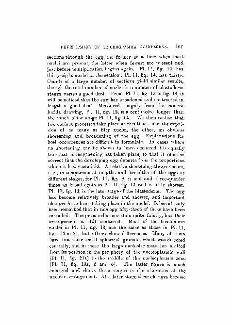
The expulsion of the inner waste mass is a preliminary tothe incipient formation of the germ layers. On what is laterthe dorsal surface of the embryo a longitudinal grooveappears, and beneath this groove the regularity of thearrangement of the blastoderm nuclei becomes disturbed. Oua space oocupied by about five or six nuclei broad and sevenor eight nuclei long a gradual sinking-in begins. In PI. 11,fig. 18 and 19, the groove is marked I. N. V. and the sinkingnuclei N. 8.1. In PI. 11, fig. 14, the egg at this period is seenin longitudinal section.
This process is undoubtedly gastrulation, though in viewof the fact that the representative of the blastula is solid theevent is somewhat disguised. PI. 11, fig. 19, has a strikingresemblance to a gastrulating blastula, though there is nosegmentation cavity or blastoccele. That the groove repre-sents the early blastopore (LN.V.) I have no doubt, and werethe depression to become deeper it would form the mesenteron.As it happens, this never takes place, the cavity of the gutbeing formed in a different way.
It has already been shown that about the stage in PI. 11,figs. 13, 14, the nuclei loose their granule, and the largenucleolus becomes placed in the centre of the nucleoplasmiczone. By the stage in PI. 11, fig. 14, this has taken place inevery nucleuo. In this figure the blastopore (I.N.V.) appears•on the dorsal surface of the anterior end of the egg, but its
VOL. 6 2 , PAET 2 . iNEW SERIES. 12

164 J. BKONTK GATENBY.
position vai'ies little. The row of nuclei which will formmost of the gut, and which are now sinking in (N.S.I.) are,,on the average, a little bigger than the other nuclei. At theanterior pole of the egg, near the letters E.X.N., is seen anextruded nucleus. It is a, fact that though the main expul-sion of nuclei occurs between the stages in PI. 11, fig. 12 andfig. 13, even after the throwing out of the central part of theegg which contains these large granules, sporadic extrusionmay take place. That these later extrusions do really occuris shown by comparing the size of expelled granules. InPI. 11, fig. 14, the granule in the anterior end of the egg istwice as large as those extruded earlier at the posterior region.(Compare also PI. 12, fig. 32.)
The germ-cells in PI. 11, fig. 14, have changed their posi-tion somewhat, becoming arranged towards the ventral edgeof the posterior pole. In this figure the germ-cells are drawna little darker than they should be. PL U, fig. 19, is drawnfrom such a transverse section as tliat through K. in fig. 14.The insinking nuclei (N.S.I.) are shown.
Such an arrangement does not hist long, for as the nucleisink inwards they lose their order. This is caused bythe fact that some lag behind while others penetrate morequickly towards the centre of the egg. This is shown inPI. 11, fig. 15, at N.8.I. By this time these nuclei havebecome very large. The relationship of the various nuclearelements in the egg now becomes more complicated, becauseat intervals around the periphery other nuclei grow larger-and sink inwards (PI. 11, fig. 15, at X.Y.). All these nucleiare quite distinct from those which were the first to beginsinking inwards, and I feel sure that some of them at leastcontribute to the formation of the gut. Others form loosecells lying in the cavity between the gut and tlie ectoderm.Often just before and at this stage amitotic division of nucleiis found taking place. Moreover, the chromatic nrrangementof some of the nuclei changes curiously. In these the largecentrally placed nucleolus becomes ragged at the edges andpieces break off and become arranged around the periphery
DEVELOPMENT OP TR1CH0GRAMMA EVANESCENS. 165
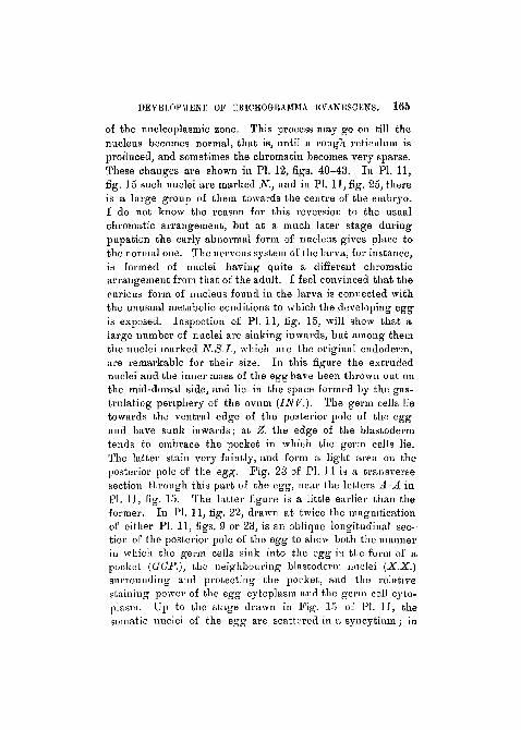
of the nucleoplasmic zone. This process may go on till thenucleus becomes normal, that is, until a rough, retieiilum isproduced, and sometimes the chromatin becomes very sparse.These changes are shown in PI. 12, figs. 40-43. In PI. 11,fig. 15 such nuclei are marked N., and in PI. 11, fig. 25, thereis a large group of them towards the centre of the embryo.I do not know the reason for this reversion to the usualchromatic arrangement, but at a much later stage duringpupation the early abnormal form of nucleus gives place tothe normal one. The nervous system of the larva, for instance,is formed of nuclei having quite a different chromaticarrangement from that of the adult. I feel convinced that thecurious form of nucleus found in the larva is connected withthe unusual metabolic conditions to which the developing eggis exposed. Inspection of PI. 11, fig. 15, will show that alarge number of nuclei are sinking inwards, but among themthe nuclei marked N.8.I., which are the original endoderm,are remarkable for their size. In this figure the extrudednuclei and the inner mass of the egg have been thrown out onthe mid-dorsal side, and lie in the space formed by the gas-trulating periphery of the ovum (INV.). The germ cells lietowards the ventral edge of the posterior pole of the eggand have sunk inwards; at Z. the edge of the blastodermtends to embrace the pocket in which the germ cells lie.The latter stain very faintly, and form a light area on theposterior pole of the egg. Fig. 23 of PI. 11 is a transversesection through this part of the egg, near the letters A-A inPI. 11, fig. 15. The hitter figure is a little earlier than theformer. In PI. 11, fig. 22, drawn at twice the magnificationof either PI. 11, figs. 9 or 23, is an oblique longitudinal sec-tion of the posterior pole of the egg to show both the mannerin which the germ cells sink into the egg iu fclie form of apocket (GOP.), the neighbouring blastoderm nuclei (X.X.)snrrounding and protecting the pocket, and the relativestaining power of the egg cytoplasm and the germ cell cyto-plasm. Up to the stage drawn in Fig. 15 of PI. 11, thesomatic nuclei of the egg are scattered in a syncytium; in
166 3. BRONTE GATENBY.
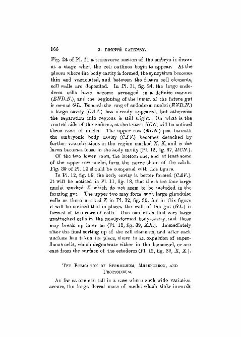
Pig. 24 of PI. 11 a transverse section of the embryo is drawnat a stage when the cell outlines begin to appear. At theplaces where the body cavity is formed, the syncytium becomesthin and vacuolated, and between the future cell elements,cell walls are deposited. In PI. 11, fig. 24, the large eudo-derm cells have become arranged in a definite manner(END.N.), and the beginning of the lumen of the future gutis seen at GL. Beneath the ring of endoderm nuclei (END.N.)a, large cavity (CA.V.) has already appeared, but otherwisethe separation into regions is still slight. On what is theventral side of the embryo, at the letters NGN, will be noticedthree rows of nuclei. The upper row (MGN.) just beneaththe embryonic body cavity (CAV.) becomes detached byfarther vacuolisations in the region marked X, X, and in thelarva becomes loose in the body cavity (PI. 12, fig. 37, MCN.).
Of the two lower rows, the bottom one, and at least someof the upper row nuclei, form the nerve chain of the adult.Pig. 39 of PI. 12 should be compared with this figure.
In PL 12, fig. 39, the body cavity is better formed {CAV.).I t will be noticed in PI. 11, fig. 18, that there are four largenuclei marked Z which do not seem to be included in theforming gut. The upper two may form such large glandularcells as those marked Z in PI. 12, fig. 39, for in this figureit will be noticed that in places the wall of the gut {GL.) isformed of two rows of cells. One can often find very largeunattached cells in the newly-formed body-cavity, and thesemay break up later on (PI. 12, fig. 39, XX.). Immediatelyafter the final sorting up of the cell elements, and after eachnucleus has taken its place, there is an expulsion of super-fluous cells, which degenerate either in the haamocoel, or arecast from the surface of the ectoderm (PI. 12, fig. 39, X, X.).
THE FORMATION OF STOMODiEuar, MESENTERON, AND
PBOCTOD^UM.
As far as one can tell in a case where such wide variationoccurs, the large dorsal mass of nuclei which sinks inwards

DEVELOPMENT OF TKIOHOGJiAMMA UVANESCENS. 167
takes part in the formation of no organ except the mid-gut,but, as I have already pointed out, some of the cells formingthe mesenteron may conceivably be of another origin,namely from nuclei which sporadically wander in from theperiphery on other pai-ts of the surface of the embryo(PI. 11, fig. 15, XT.). From the first the nuclei destined toform the mid-gut are conspicuous by their large size andrapid growth. The lumen of the mesenteron appears justafter the stage drawn in PI. 11, figs. 27 and 28. I t seems tobe formed by an internal delamination of the solid endo-dermal cell mass in some cases, but in others it looks as if,during growth, the l-ing of cells, gradually enlarging, left alumen in their centre, just as the lumen is known to appearin an ordinai-y duct. In any case there is always a residuumleft in the developing lumen (PI. 12, figs. 27 and 28). Afterthe endodermal cells have grouped themselves as shown inPI. 11, fig. '24, the proctodaeum and stomodasum begin to bequite recognisable; and there is no doubt that the latter isformed by a regular imagination (PI. 12, fig. 31, ST.). Themanner in which the proctodasum is formed is a little moredoubtful. In the case of the stomodaeum the imagination isnormal (PI. 11, fig. 27). The inpushing cells meet theroughly disposed endoderin cells, and when the final dis-solving out and disintegration of that part of the embryowhich forms the body-cavity takes place the connectionbetween the stomodseal and mesenteron cells remains unbroken.The same thing applies to a region where the proctodaeum isformed, but it is difficult to be sure of a true invaginationsuch as occurs with the stomodaeum. The latter is formed ofmuch smaller cells than the proctodasum, and is longer, whilethe demarcation between mesenteron and proctodaeum isquite indistinct. In PI. 12, fig. 30, which is a horizontalsection of the front region of a larva of the same age as thatdrawn in PI. 12, fig. 38; the stomodaeum, mouth, andmesenteron are shown. In PI. 12, fig. 34, there is a longi-tudinal section of the proctodaeum of a somewhat youngerlarva, but it serves to show how short the hind gut is. This
168 J. BRONTfi GATENBY.
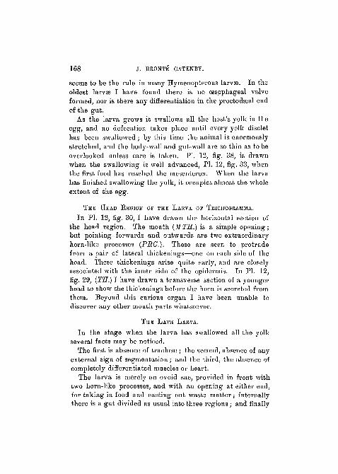
seems to be the rule in many Hymenopterous larvae. In theoldest larvae I have found there is no oesophageal valveformed, nor is there any differentiation in the proctodaeal endof the gat.
As the larva grows it swallows all the host's yolk in theegg, and no defecation takes place until every yolk disclethas been swallowed; by this time the animal is enormouslystretched, and the body-wall and gut-wall are so thin as to beoverlooked unless care is taken. PI. 12, fig. 38, is drawnwhen the swallowing is well advanced, PI. 12, fig. 33, whenthe first food h;is reached the mesenteron. When the larvahas finished swallowing the yolk, it occupies almost the wholeextent of the egg.
THE HEAD REGION OF THE LARVA OF TKICHOGRAMMA.
In PI. 12, fig. 30, I have drawn the horizontal section ofthe head region. The mouth (M TH.) is a simple opening;bat pointing forwards and outwards are two extraordinaryhorn-like pi-ocesses (PRC). These are seen to protrudefrom a pair of lateral thickenings—one on each side of thehead. These thickenings arise quite early, and are closelyassociated with the inner side of the epidermis. In PL 12,fig. 29, {TH.) I have drawn a transverse section of a youngerhead to show the thickenings before the horn is secreted fromthem. Beyond this curious organ I have been unable todiscover any other mouth parts whatsoever.
THE LATE LARVA.
In the stage when the larva has swallowed all the yolkseveral facts may be noticed.
The first is absence of tracheae ; the second, absence of anyexternal sign of segmentation; and the third, the absence ofcompletely differentiated muscles or heart.
The larva is merely an ovoid sac, provided in front withtwo horn-like processes, and with an opening at either end,for taking in food and casting out waste matter; internallythere is a gut divided as usual into three regions; and finally

DEVELOPMENT OF TltlCUOGIiAMMA EVANESCEXS. 169
there is the single median ventral germ-cell pocket, beneaththe proctodteum.
In PI. 12, fig. 30 (CU.), a distinct cuticle could be seen. Itdipped into the pockets from which the horn-like jaw-pro-cesses protruded, and the latter are probably cuticnlar innature. The thickening {TH.) is ectodermal. Cuticle (chitin)was found in the stomodasum, but I am not quite sureof its presence in the proctodamm. It is possible that theprocesses are used for scooping up the yolk of the host as theJarva feeds, and they are probably much modified mandibles.
When the larva has swallowed all the yolk, very often notthe smallest particle can be found outside its gut, and exactlyliow the yolk at the posterior end of the host's egg is workedto its mouth is impossible to say ; but it is probably by meansof movements of the body that the imswallowed parts arebrought forward.
THE FATE OF THE EXTRUDED MATTER.
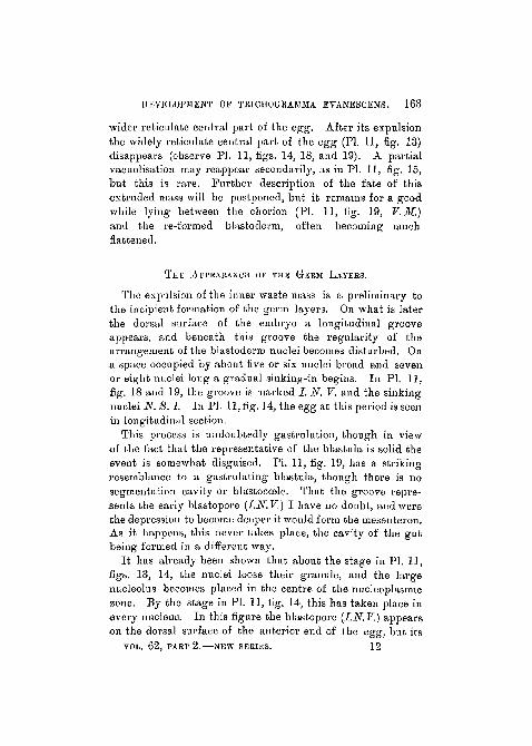
In PI. 11, fig. 15, the extruded mass still lies within thevitclline membrane of the egg. As the larva grows themembrane becomes stretched and the waste mass flattened;but, though it remains intact for a good time, it eventuallybursts. The extruded mass then floats free in the yolk ofthe Donacia egg. In PI. 11, fig. 27, EM., it is shown to theright of the ventral side of the posterior pole of the embryo.In PI. 12, fig. 35, it is seen quite close to the embryo at EM.
Curiously enough these fragments seem to live a goodwhile, and nuclear changes, such as those undergone in theblastoderm, take place in some cases.1 Tlie mass may becomspherical, as in PI. 12, fig. 32, and may resemble the eggitself. Eventually the mass either degenerates outside the
1 One is tempted to entertain the view that this peculiarity may be insome way or other connected with a faculty that culminates in tlieestablishment of polyembryony. Were the extruded mass to containenough live nuclei it might partially follow the development of theembryo.

]70 J. BRONTE OATKNBY.
embryo or is swallowed by the latter. The live nuclei, towhich the temporary persistence of the extraded mass is due,may develop the microsome granule (GBG.) drawn in PI. 11,fig. 21 A. This is the case with the nuclei marked LEN. inPI. 12, fig. 32. (See addendum, p. 30.)
THE NERVOUS SYSTEM.
The nervous system cau be recognised very early; it arisesfrom the multiplication of ectodermal cells in the usualmanner fonnd in insect larvas, but it never becomes properlyseparated off from the ectoderm. Even in late larval life thenervous system seems "coarsely" made; that is to say, it isformed of comparatively few cell elements which are notdifferentiated in the characteristic manner, and there are nosuch things as nerves in the sense of offshoots or twigs toorgans, such as exist in other larvee, such as Vespa. Thenerve-cells do not differ in any w;iy from other cells in the body,always excepting germ-cells. In PL 11, fig. 18, the nerve-chord is seen in a rudimentary condition, and consists of thebottom row of nuclei marked N. C. N., and an unknownnumber of the row above. In PI. 11, fig. 21, the brain (J3R.)and nerve-chord (JSf.C.) are cut longitudinally. In PI. 11,fig. 22, PL 12, figs. 33 and 38, a better view of the chord intransverse section is seen, aud in PL , fig. 29, the brain (J3i?.)is cut transversely, to illustrate its close connection with theepidermis (ISP.) and oesophagus (STD.). No such things asganglia exist, and the chain ends a little before the ererm-pocket; it does not reach the proctodfeurn. In late stages(PL 12, fig. 38, N. C.) it becomes an increasingly difficultmatter to recognise the chain, so stretched does it become,and by the time the larva has swallowed all the yolk in theDonacia egg, the nervous chain is for most of the hinderpart of its length quite unrecognisable. The cesophagealconnectives seem to consist of single cells applied to oneanother (PL 12, fig. 30, (ES. CON.), and are extremelyrough.
DEVELOPMENT OF TKICHOGUAMJIA EVANESOENS. 171
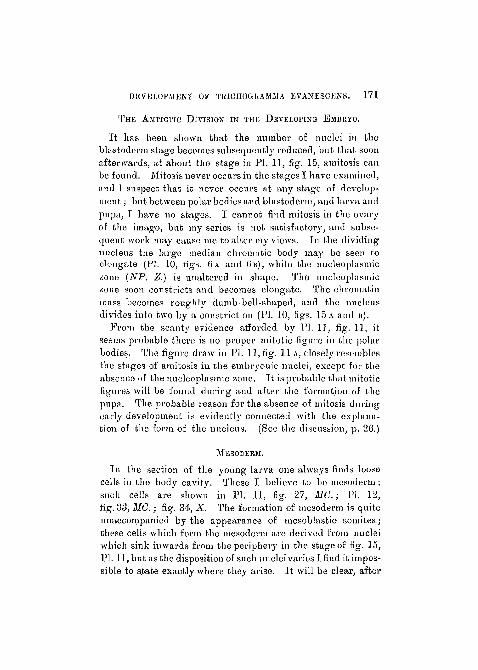
THE AMJTOTIC DIVISION IN TEK DEVELOPING EMBRYO.
It has been shown that the number of nuclei in theblastoderm stage becomes subsequently reduced, but that soonafterwards, at about the stage in PI. 11, fig. 15, amitosis canbe found. Mitosis never occurs in the stages I have examined,and 1 suspect that it never occurs at any stage of develop-ment ; but between polar bodies and blastoderm, and larva andpupa, I have no stages. I cannot find mitosis in the ovaryof the imago, but my series is not satisfactory, and subse-quent work may cause me to alter my views. Iu the dividingnucleus the large median cliroiruitic body may be seen toelongate (PI. 10, figs. 6A and 6B), while the nucleoplasmiczone (NP. Z.) is unaltered in shape. The nucleoplasmiozone, soon constricts and becomes elongate. The chromatinmass becomes roug-hly dumb bell-shaped, and the nucleusdivides into two by a consti-iction (PI. 10, figs. 1 5 A and B).
From the scanty evidence afforded by PI. 1], fig. 11, itseems probable there is no proper mitotic figure in the polarbodies. The figure draw iu PI. 11, fig. 11A, closely resemblesthe stages of amitosis in the embryonic nuclei, excej^t for theabsence of the nucleoplasmic zone. It is probable that mitoticfigures will be found during and after the formation of thepupn. The probable reason for the absence of mitosis duringearly development is evidently connected with the explana-tion of the form of the nucleus. (See the discussion, p. 26.)
MESODERM.
In the section of the young larva one always finds loosecells in the body cavity. These I believe to be mesoderm ;such cells are shown in PI. 11, fig. 27, MG.; PI. 12,fig. 33, MG.; fig. 34, X. The formation of mesoderm is quiteunaccompanied by the appearance of mesoblastic somites;these cells which form the mesoderm are derived from nucleiwhich sink inwards from the periphery in the stage of fig. 15,PI. 11, but as the disposition of such nuclei varies I find it impos-sible to s.tate exactly where they arise. It will be clear, after
172 J . BROVi'E GATENBY.
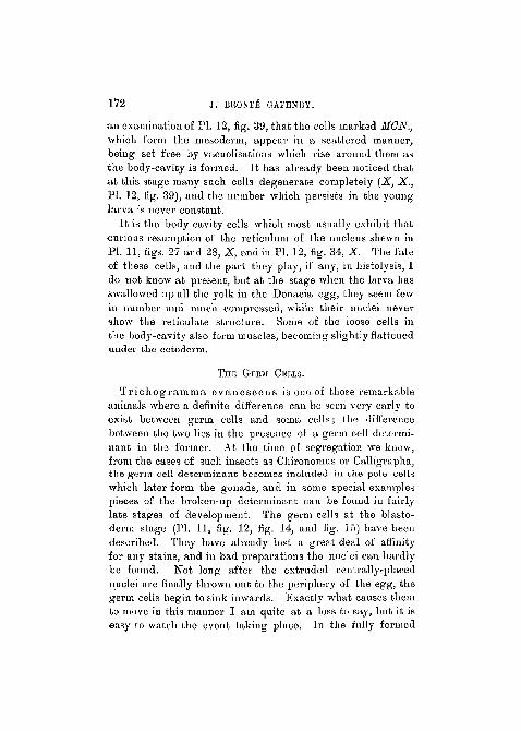
an examination of PI. 12, fig. 39, that the cells marked MCN.,which form the mesoderm, appeal' in a scattered manner,being set free by vacuolisations which rise around them asthe body-cavity is formed. It has already been noticed thatat this stage many such cells degenerate completely (X, X.,PI. 12, fig. 39), and the number which persists in the younglarva is never constant.
It is the body cavity cells which most usually exhibit thatcurious resumption of the reticulum of the nucleus shewn inPI. 11, figs. 27 and 28, X, and in PI. 12, fig. 34, X The fateof these cells, and the part they play, if any, in histolysis, Ido not know at present, but at the stage when the larva hasswallowed up all the yolk in the Donacia egg, they seem fewin number and much compressed, while their nuclei nevershow the reticulate structure. Some of the loose cells inthe body-cavity also form muscles, becoming slightly flattened-under the ectoderm.
THE GERM CELLS.
Trie ho g ramma e van esc ens is one of those remarkableanimals where a definite difference can be seen very early toexist between germ cells and soina cells; the differencebetween the two lies in the presence of a germ cell determi-nant in the former. At the time of segregation we know,from the cases of such insects as Chironomus or Calligraphicthe germ cell determinant becomes included in the pole cells
which later form the gonads, and in some special examplespieces of the broken-up determinant can be found in fairlylate stages of development. The germ cells at the blasto-derm stage (PI. 11, fig. 12, fig. 14, and fig. 15) have beendescribed. They have already lost a great deal of affinityfor any stains, and in bad preparations the nuclei can hardlybe found. Not long after the extruded centrally-placednuclei are finally thrown out to the periphery of the egg, thegerm cells begin to sink inwards. Exactly what causes themto move in this manner I am quite at a loss to say, but it iseasy to watch the event taking place. In. the fully formed
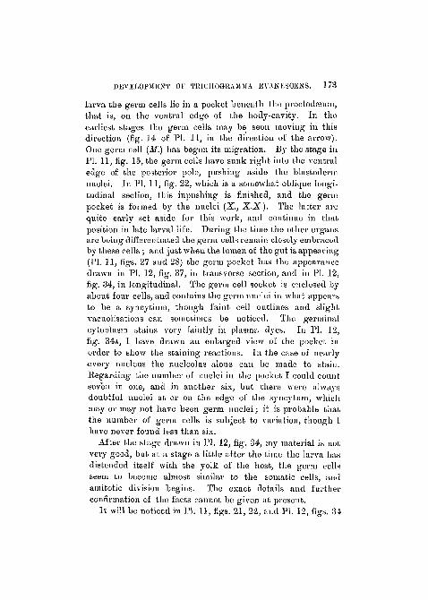
DEVELOPMENT Of TltlCJIOGRAMMA EVANKSCKNS. 1 7 3
larva the germ cells lie in a pocket beneath the pvoctodseuui,that is, on the ventral edge of the body-cavity. In theearliest stages the germ cells may be seen moving in thisdirection (fig. 14 of PI. 11, in the direction of the arrow).One germ cell (M.) has begun its migration. By the stage inPI. 11, fig. 15, the germ cells have sunk right into the ventraledge of the posterior pole, pushing aside the blastodermnuclei. In PI. 11, fig. 22, which is a somewhat oblique longi-tudinal section, this inpushing is finished, and the germpocket is formed by the nuclei (X., X X ) . The latter arequite eavly set aside for this work, and continue in thatposition in late larval life. During the time the other organsarc being differentiated the germ cells remain closely embracedby these cells ; and just when the lumen of the gnt is appearing(PI. 11, figs. 27 and 28) the germ pocket has the appearancedrawn in PI. 12, fig. 37, in transverse section, and in PI. 12,fig. 34, in longitudinal. The germ cell socket is enclosed byabout four cells, and contains the germ, nuclei in what appearsto be a syncytium, though faint cell outlines and slightvacuolisations can sometimes be noticed. The germinalcytoplasm stains very faintly in plasma dyes. In PI. 12,fig. 34A, I have drawn an enlarged view of the pocket inorder to show the staining reactions. In the case of nearly•every nucleus the nucleolus aloue can be made to stain.Regarding the number of nuclei in the pocket I could countseven in one, and in another six, but there were alwaysdoubtful nuclei at or on the edge of the syncytum, whichmay or may not have been germ nuclei; it is probable thatthe number of germ cells is subject to variation, though Ihave never found less than six.
After the stage drawn in PI. 12, fig. 34, my material is notvery good, but at a stage a little after the time the larva hasdistended itself with the yolk of the host, the germ cellsseem to become almost similar to the somatic cells, andamitotic division begins. The exact details and furtherconfirmation of the facts cannot be given at present.
It will be noticed in PI. 11, figs. 21, 22, and PI. 12, figs. 34
174 J. BRONTis GATENBV.
and 35, that tlie germ nuclei gradually lose all stainingpower, except that o£ the chromatic nucleolus, the reticulumdisappearing. In later stages, when the germ cells begin tostain more heavily, only the nucleolus can be made to takeup chromatm dyes.
With regard to the migration of the germ-cells from out-side the embryo inwards (PI. 11, figs. 21 and 22, no polecanal could be recognised. The germ cells seem to sink inpassively, and never become amoeboid as in Oalligrapha(3).
The (xerm Cell D e t e r m i n a n t .
The origin of the germ cell determinant, even in thoseinsects where the eggs are larger and technique easier, isstill in doubt. I have examined several Hymenopterousinsects parasitic upon Aphids, and find that the determi-nants appear as a cloud of granules towards the posteriorpole of the egg.
In Trichogramma the determinant is densest and mostdarkly staining during the period in which it still lies in theovarian tubule, but is just about ready to lay (PL 11, fig. 9)..By the time the egg has been laid and the polar bodies are inprocess of formation the determinant loses a great deal ofits affinity for stains, and begins to break into pieces (PI. 11,fig. 26, P, P.) At the blastoderm stage the determinant hascompletely disappeared, and with the exception of the rarestcases nothing of it remains (PL 12, fig. 36, (?.). Indeed atat this stage the cytoplasm of the germ-cells, instead ofstaining more heavily than that of the somatic syncytium,as one would expect, has lost a great deal of staining power,both in nucleus and cytoplasm. This soon becomes veryaccentuated (PL 11, fig. 22). In the newly laid eggs inPL 11, figs. 10 and 20, the germ cell determinant has becomerather shrunken and faintly staining, though in the case ofPL 11, fig. 11, the determinant has a good deal more affinityfor stains.

DEVELOPMENT OF TK1CHOGRAMMA EVANESCENS. 175
DISCUSSION.
The Significance of the Nuclear Changes duringBlastoderm Stage.—Many nuclei (from twenty-five tofifty-five) are cast out altogether. Others, as far as I can tellall of them, extrude the microsome or sum 11 chromatingranule, marked GRG. in PI. 11, figs. 16 and 21. In somecases there are two granules of the same size, both of whichare expelled into the cytoplasm. No granule can be foundto be extruded from the germ cells, and it might followtherefore that the latter, at this period at least, containmore chromatin than the ordinary- blastoderm nucleus. InMiastor, Kahle (5) and Hegner (3) have described a definitechromatin diminution process whereby the somatic nuclei aredeprived of a part of their chromatin during certain divisions.Though I do not overlook the possibility of a homologousoccurrence taking place in Trichogramma evanescens,I am more inclined to believe that another explanationshould be attached to the remarkable chroniiitin diminutionin the parasite. In the first place the chromatin diminutionin Miastor takes place quite earl}', before the blastoderm isfoi'med completely, and, moreover, the process is brought aboutin a different manner, not by extrusion of a granule, but by thediscarding of the larger part of the chromosomes during themitotic division, only the extreme ends of the chromosomesgoing to the opposite spindles at the telophase. The residualmass in the middle of the spindle undergoes degeneration.
No satisfactory explanation of the occurrence in Miastorhas been advanced, but in Trichogramma evanescens Iwould suggest that the process is connected with the curiousmetabolic influences which must affect the nuclei. It must beremembered that all nourishment which is necessary for thedevelopment of the egg, and which is ordinarily providedby the central mass of yolk of the insect-egg, is, in the caseof this parasite, derived from the yolk of another insect'segg and without the aid of vitellophags. Such nourishment
176 J. HKONTfi GATENBY.
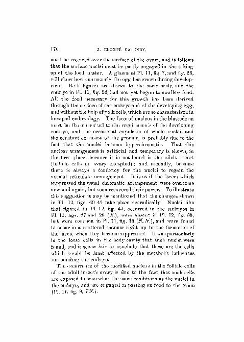
must be received over the surface of the ovum, n,nd it followsthat the surface nuclei must be partly engaged in the takingup of the food matter. A glance at PI. 11, fig. 7, and fig. 28,will show how enormously the egg has grown during develop-ment. Both figures are drawn to the same scale, and theembryo in PI. 11, fig. 28, had not yet begun to swallow food.All the food necessary for this growth has been derivedthrough the surface of the embryo and of the developing egg,and without the help of yolk cells, which are so characteristic inhexapod embryology. The form of nucleus in the blastodermmust be the one suited to the requirements of the developingembryo, and the occasional expulsion of whole nuclei, andthe constant extrusion of the granule, is probably due to thefact that the nuclei become hyperchroniatic. That thisnuclear arrangement is artificial and temporary is shown, inthe first place, because it is not found in the adult insect(follicle cells of ovary excepted) ; and secondly, becausethere is always a tendency for the nuclei to regain thenormal reticulate arrangement. It is as if the forces whichsuppressed the usual chromatic arrangement were overcomenow and again, but soon recovered their power. To illustratethis suggestion it may be mentioned that the changes shownin PI. 12, figs. 40-43 take place sporadically. Nuclei likethat figured in PI. 12, fig. 43, occurred in the embryos inPI. II, figs. _>7 and 28 {X.), were absent in PI. 12, fig. 33,but were common in PI. 11, fig. 15 (N.N.), and were foundto occur in a scattered manner right up to the formation ofthe larva, when they became suppressed. It was particularlyin the loose cells in the body cavity that such nuclei werefound, and it seems fair to conclude that these are the cellswhich would be least affected by the metabolic influencessurrounding the embryo.
The occurrence of the modified nucleus in the follicle cellsof the adult insect's ovary is due to the fact that such cellsare exposed to somewhat the same conditions as the nuclei inthe embryo, and are engaged in passing on food to the ovum(PI. 11, fig. 9, FK.).
DEVELOPMENT OP TKICHOUIfAMMA EVANESCKNS 177
Hypercliromatic nuclei are known to occur in nurse cells-of insects, in various cells of vertebrate fcetal membranes,and in many tissues concerned in nourishment, and wherethese nuclei do not become noticeably h}rperchromatic, theygenerally hypertrophy.
The extruded granules are, therefore, to be regarded assuperfluous chroinatin, which has arisen through the peculiarconditions to which the blastoderm nuclei are exposed.
F o r m a t i o n of t h e Germ L a y e r s . — I n view of the-fact that the egg of Trichogramnia is not provided with yolkthe formation of the germ layers is of great interest, for theyolk profoundly alters the organogeny in the usual hexapoddevelopment. That one would receive a faithful representa-tion of the ancestral mode of development of the insectfrom the case of Trichgramma is too much to expect, becausethe method of development, though primitive in somerespects, is overshadowed by the effects of the parasitic modeof life. The blastoderm stage is without donbt quitenormal, and except for minor nuclear phenomena differs notat all from that of the host or of Miasstor (3), but the eventsleading to the formation of the endoderm are interesting.That the progress figured in PI. 11, figs. 14 and 19, is one ofgastrulation one hardly doubts. In the case of P o l y g n o t u sm i n u t u s Marchal (7) describes how the embryo is formedby a complete mvaginatioii of one side of the hollow blastula,to form a two-layered gastrula. The method of gastrnlationin the parasite treated in this paper is somewhat less distinctthan in the case of Polygnotus, and before the process isfar advanced a secondary insinking of other peripheralnuclei almost completely obscures it (compare PI. 11, figs. 14and 15.)
The manner in which the endoderm is formed in Tricho-gramnia is of very considerable interest in view of the dis-cussions which have been caused by the different opinionsexpressed by several authors (Dohrn, Kowalevsky, andtranin (7) ), but it is not intended here to review their widelydifferent suggestions in the light shed by Trichogramma.

178 J. HHONTE GATKNBY.
G-errn Cell a n d D e t e r m i n a n t .
In the ordinary Hymenopterous larva (e.g.Vespa) thegerm cells lie about two-tliirds way in the length of the bodyand above and resting upon the mid-gut.
In the Trichogramma larva the germ cells are situated atthe posterior pole and ventral to the proctodasmn. In the adultinsect the ovaries occupy the same position as they do in theVespa imago. Migration of germ cells is very small in thedeveloping embryo. In most insect embryos the germ cells•are carried into the tail fold, and may be said to eithermigrate or be passively carried a good distance, but exceptfor the early insinking of the germ cells and the formation ofthe germ pocket in Trichogramma the position of these cellsis hardly altered.
I have looked carefully at my sections of the adult ovary,•and find that the germ cell determinant appears as a cloud ofgranules, which become more and more heavily staining, anddenser and denser, until the determinant resembles a darkspherical ball at the posterior pole of the egg. The wholeliistory of the germ cell determinant, in so far us theovary is concerned, has been exhaustively treated byHegner (3) in more suitable insects. I have examined anumber of sections of the Hymenopterous parasites commonon Aphids, and I am able to substantiate most of his remarks ;but in the nurse cells, as well as in the developing oocyte, Ihave found curious large spherical granules which have nothitherto been mentioned. These seem to appear aftersynezesis in the oocyte, and whether they have anything todo with the germ cell determinant I cannot at present say.If suitable material is procured I hope to examine thispoint.
ADDENDUM.
When this work had been finished I had not had theopportunity of acquainting myself with Prof. Silvestri's•writings, only knowing of them through short reviews in

DEVELOPMENT OF TRICHOGRAMMA EVANESCEXS. 179
other papers more accessible to me. Since then I havebeen enabled, through Mr. Goodrich's kindness, to readSilvestri's valuable articles. I have been impressed by thesimilarity between all stages in the development of Oophthoraand of Trichogramma. To my eye, untrained in the apprecia-tion of small systematic differences in Chalcids, the adultinsects in these species are closely similar, and the peculiarlarvae of both species are structurally identical.
Apart from differences due to different interpretation thereis no doubt that the course of organogeny in these parasitesis parallel.
Silvestri (' Bolletino del Laboratorio de Zoologia Generalee Agraria/ vol. i and iii) identifies the darkly staining massesof the inner region of the blastoderm stage (PI. 11, figs. 12and 13 in my drawings) as a "piccolo numero di nuclei, chein seguito degenereranno," but has overlooked the smallgranule (GRC.) (if really present in Oophthora) which is socharacteristic of stages such as that of PL 11, figs. 12, 13,and 21. In Oophthora the germ cells have sunk into the eggbefore any marked differentiation of the primary germlayers lias taken place (vide Silvestri, vol. iii, p. 78, fig. xxx,vii, 5), for it will be remembered that in the stage drawn in myfig. 15, PI. 11, the germ layers are distinctly forming andthe germ cells still situated at the pole of the egg.
Regarding Silvestri's statement that the extruded massesare nuclei, it might be well to mention that these darklystaining masses are but a part ( i .e . the nucleolus) of theoriginal nuclei (see p. 11, and the figs. 13A and 21 ofPI. 11).
In Trichogramma I have not described the formation of anembryonic membrane, nor do I believe that such exists. InE n c y r t u s aph id ivo rus and in Oophthora Slivestri des-cribes the formation of a " pseudoserosa " from a delaminationof the surface cells of the embryo. He states : " L'involncro•embrionale delF Oophthora e in parte omologo a quello deli'Encyrtus, perche in questo sembra che derivi completamenteper delaminazione delle cellule embrionali, mentre nell'
VOL. 62 , PART 2 . NEW SEEIES. 1 3

180 J. BRONTis GATBNBY.
Oophthora la parte di esso, che prima si forma, deriva dallaparte spugnosa del protoplasma die occupava, a blastodermacouipleto, il centro dell ovo. Intorno a tale differenza ioperd non voglio insistere troppo perclie potrebbe essermisfuggito il primo vero periodo di formazione dell' involucroembrionale nell' Bncyrtus, rnentro ho potuto seguirlo conogni precisione nell' Oophtliora."
In Encyrtus Silvestri gives several figures (vol. iii, 1908,p. 67) of the "inizio della psedoserosa," which I find notunconvincing, but I cannot see any delamination taking placein fig. xxvi, 2, except at P., which I think has little incommon with the "pseudoserosa" drawn in fig. xxv, 3. Iwill leave my comment at this point because Encyrtus is insome ways different from Trichogramma, and will considerOophtliora (vol. iii, pp. 71, 79). Whether Prof. Silvestri's oi-my views concerning these forms are correct, I am convincedthat we have to deal with two species whose development isclosely similar. I find stages such as those drawn by Silvestriin figs, xxxvii and xxxviii, and in almost all others of hisfigures. Not only this, bnt the modified larvte of bothTrichograinma and Oophtliora are similar.
lfe believes that, one part of the pseudoserosa is formedby the extruded inner mass (protoplasma superficiale spug-noso), while the other is formed like that of KncyrtiiSj and ishomologous with this membrane in the latter.
Tn my figs. 15, 18, 19, 24, and 25 of PI. 11, I have drawn atEKN. what Silvestri calls the "pseudoserosa." Since read-ing" the Professor's papers I have very carefully re-examinedmy sections, and find nothing to alter in my interpretations;but I have drawn PI. 10, fig. 4, with a view to the clearerexplanation of my view of the " pseudoserosa " of Silvestri.
The egg when laid is surrounded by a vitelline membraneand a thin chorion, which, however, is quite distinct (PI. 10,fig. 6, CII.) As development goes on the waste nucleolicollect in the centre of the egg, and are soou extruded(PI. 11, figs. 18 and 19). They come to the surface of theegg, and at first form a slight cavity in the ovum. But as
DEVELOPMENT OF TEICHOGBAMMA EVANESCENS. 181
the egg grows rapidly the chorion becomes slightly stretched,and the lump of " protoplasma spugnoso" becomes pressedflat, and mechanically spreads around the egg (PI. 10, fig. 4,X, X, X.). Now should the chorion by any cliance burst,as it sometimes does, the extruded mass is released and liesnear the egg and embryo (PL 11, fig. 27; PL 12, fig. 35,EXN.).
In PL 10, fig. 4, the extruded mass {EXN.) lies inside thechorion, and has been flattened out between the points X,X,X.,on the dorsal surface of the embryo, but on the ventralsurface (V.) the chorion, though somewhat stretched andthinner, is still recognisable, and cannot be confused withuny other structure. The "protoplasma spugnoso" ofSilvestri is an extruded dead mass, and is in no way com-parable or homologous with either the ammon or serosa ofother insects, and since, as Silvestri shows, there is really aliving embryonic membrane around the egg of Encyrtus, it isincorrect, in my humble opinion, to say that " L'involucroombrionale dell' Oophthora e in parte omologo a quello dell'Bncyrtus." In his figure on p. 67 of vol. iii, he depicts amembrane (P.) which has nuclei evenly distributed, and thetout ensemble is far more convincing thini his fig. xxxvii, 6,of Oophthora. In the latter figure there are no nuclei in the" pseudoserosa" except those on one side, which he hadalready declared were " in seguito degenereranno."
I feel convinced that in Trichogramma and Oophthora the" pseudoserosa" of Silvestri is merely an artefact producedby the mechanical flattening out of a waste mass of proto-plasm and chromatin. If the chorion bursts early no" pseudoserosa " can be formed.
I agree with Silvestri's description of the larva except thathis fig. XL., p. 81, which, he says, is a sagittal section, hemarks what I consider to be the longitudinal nerve-chord,as "cellule muscolari M." It is true that no properlydifferentiated muscles seem to exist in the larva of eitherspecies, and the movements of the animal are brought aboutby flattened mesoderm cells lying here and there under the
182 J. BRONTE GATENBY.
ectoderm. These cells only differ from the other somaticcells in that they are more elongate, their nuclei and cyto-plasmic structure being normal.
It is a curious fact that Silvestri, though not paying muchattention to the formation of the germ layers, has not figuredthe invagination of the endoderm (PL 11, figs. 14 and 19).I cannot but believe that this happens in Oophthora, where allour other stages are almost identical.
In Oophthora that remarkable nuclear arrangement ofearly stages (PI. 11, figs. 16 and 21) has not been described,and it possibly is absent; however, Prof. Silvestri does notappear to have paid great attention to the nuclei of earlystages, and it may have been overlooked. I mention thisbecause the early changes in the nuclei of Trichogramma areso striking.
As Silvestri has pointed out, E n c y r t u s aph id ivorus isnot a parasite on aphids, but a hyperparasite on one or twoother true aphid parasites. With regard to the fate of theembryonic membrane which he figures enveloping the larva(on p. 69, fig. xxix) he says : " E in tale stato di sviluppoche la larva allungandosi rompe nella parte anterioi-e e nellaposteriore la serosa e libera comincia a nutrirsi dei tessutidell' ospitatore."
It will be seen that, with the exception of those parts oforgan ogeny which Silvestri has not treated at length, hisadmirable work agrees fairly well with the few remarks Ihave been able to pass on the embryology of Trichogramma,and I have no doubt that when the Professor examines hisstages in greater detail, his results will fall into line withmy own.
SUMMARY.
(1) T r i c h o g r a m m a evanescens lays its eggs on the•egg mass of a beetle, Donacia simplex, a single parasiteemerging from one host's egg.
(2) The ovum has a large germ cell determinant at itsposterior pole, and in segmentation the determinant is
I
DEVELOPMENT OP TRICIHOGRAMMA . EVANESUENS. 183
divided among the large cells at the posterior pole, which arethe germ cells.
(3) In the single case found there were two polar bodies.(4) The blnstula is fairly normal except for the curious
arrangement of the chroinatin in the somatic nuclei.(5) Many nucleoli are cast out into the centre of the egg,
where they collect till from twenty-five to fifty are present ;the mass is then extruded on the periphery of the egg.
(6) As the blastoderm grows it broadens without lengthen-ing up to the stage where the germ layers begin to form.
(7) About thirty-five nuclei sink inwards from the dorsalsurface of the embryo to form endoderm.
(8) From the blastoderm stage to that of the gastrula nonuclear division appears to take place.
(9) Shortly after the formation of the endoderm amitosismay be found, and from this onwards the number of nucleiincreases.
(10) The mesoderm seems to be formed from peripheralnuclei, which sink in sporadically; no somites can be madeout, nor does any segmental method of formation of themesoderm occur.
(11) The nervous system, stomodaeum, and probably procto-deeum, are normally formed.
(12) The germ cells lie in a pocket formed4byj several somaticcells, which embrace them.
(13) Ordinary mouth parts , tracheae, heart , and cesophagealvalve are want ing; the head has two horn-like mandibulavprocesses, which may assist in scooping forwards the food.
(14) The larva does not feed on the food little by little,defecating as it ea t s ; instead, it begins by swallowing all theyolk at once, so tha t its body becomes enormously distendedand stretched.
(15) Metameric external segmentation is absent, the bodyand head being continuous and sac-like.
184 J. BR0NT6 GATENBJT.
BIBLIOGRAPHY.
1. Paul Mai-chal.—" Becherches sur la biologie et le developpementdes Hymenopteres parasites," 'Arch, de Zool. Exp.,' 4e Serie, 2.
2. Silvestri, F., 1906-08.—" Contribuzioni alia conoscenza biologicadegli Imenotteri parasitici,' i-iv, 'Bollet. Scuola sup. Agric,Portici, T. 1 and 3.
3. Robert W. Hegner.*—" Studies on Germ Cells: IV, Protoplasmicdifferentiation in the oocytes of certain Hymenoptera," 'Journalof Morphology,' vol. xxvi, No. 3.
3a. ' The Germ Cell Cycle in Animals,' New York, 1914.4. Perkins.—"Trichograrnma evanescens and Pentarthron," 'Trans.
Ent. Soc.,' 1913.5. Kahle, W.—"Die Psedogense der Cascidomyiden," 'Zoologica,'
1908, Bd. xxi.6. Henneguy.—' Les Insectes,' Paris, 1904.
7. ' The Embryology of the Honey Bee,' J. A. Nelson, PrincetonUniv. Press, 1915, p. 72.* After Hegner's paper (3) a useful bibliography is found.
EXPLANATION OF PLATES 10, 11, AND 12,Illustrating Mr. J. Bronte Gatenby's paper on "Tricho-
gramma evanescens (W.): a Monembryonic EggParasite of Donacia Simplex."
LETTERING.
ANT. Anterior pole. S. 0. Body cavity. BB. Brain. CAV,Developing body cavity. GH. Chorion. GU. Cuticle. D. Dorsalsurface. D. P. Developing parasite. E. Ectoderm. ECD. N. Ecto-dermal nuclei. E. M. Extended mass of cytoplasm and chromatin.END.N. Endodermal nuclei. F. Food. F.N. Follicle nuclei. G.Stainable granule. G. C. Genn cell. G- C. D. Germ cell determinant.G. C. N. Germ cell lmcleuB. G. C. P. Germ cell pocket. G. L. Gutlumen. G. R. C. Minor granule of nucleus. GT. Gut. INV. In-vagination. L. Leg of host. L.E.N. Healthy nucleus extruded.M. Muscle cells. M. G. Cells of body cavity. M. T. H. Mouth.N. Nucleus. N. C. Nerve chord. N. P. Z. Nucleoplasmic zone ofnucleus. JV. S. I. Nuclei sinking inwards (endoderm). CES. (Esophagus.CES. COM. CEsophageal commissure. OV. Eggs of Donacia. P. Brokenpieces of germ cell determinant. P. P. Parasite. P. B. 1st polar body. J

DEVELOPMENT OF TRIOHOGRAMMA. EVANESOENS. 185
PD. Proctodseum. POST. Posterior pole. P. B. C. Frontal processof larva. B. Reed. S.L.N. Mostly vitellophags of host. ST.Stomodeeum. TH. Glandular thickening secreting frontal process.V. Ventral. V. C. Vacuoles in cells. V. M. Vitelline membrane.Y. Yolk.
[In reproduction all figures reduced by one-half.]
Figs. 7, 9, 10, 11, 12, 13, 14, 15, 17, 18, 19, 20, 23, 24, 25, 28, 29,30, 32, 33, 34, 35, 37, drawn with a Zeiss T\j oil immersion and compen.eye-piece 8. A camera lucida was \ised, with drawing-board slightlyinclined towards the microscope, and at table level. Magnificationabout 1,760 diameters.
Figs. 27, 31, and 38 from Zeiss T'¥ and comp. eye-piece 4.Figs 10A, 11A, 13A, 16, 21, 22, 26 enlarged about twice from camera
drawings with O. -/-j- E. 8.Figs. 36, 40, 41, 42, and 43 enlarged in the same way about four times.
Fig. 8 drawn with O. £ B. 2, drawing-board at table level. (Cameralucida).
Fig. 39 was drawn with Zeiss O. F. E. 4.Fig. 4.— X 2400 (Koristka Jjth x Hug. oc. 5.
PLATE 10.
Fig. 1.—Trichogramina evanescens (Westwood), adult female(now x 75.)
Fig. 2.—Donacia embryo X 75 containing at its posterior pole aparasite (1). P.). Whole preparation. The pai'asite was at the Btagedrawn in PI. 11, fig. 24.
Fig. 3.—Donacia simplex (F.) x 4.Fig. 3A.—Egg mass of Donacia, viewed from side with parasite (P.).
All to same scale as beetle.
Fig. 4.—Transverse section in mid region of an embryo when the gutlumen has formed, Shows the flattening out of the extruded mass{.EX. N.) under the chorion (CH.).
Fig. 5.—Stages in amitosis of somatic cells.
Fig. 6.—Part of early blastoderm stage to show chorion (CH.) andextruded nucleolus (EX. N.).
PLATE 11.
Fig. 7.—Part of Donacia egg showing the newly-laid egg of thepai'asite in transverse section.
186 J. BRONTE GA'l'ENBY.
Fig. 8.—Part of egg mass of Donacia in transverse section showing aparasite at tbe stage drawn in fig. 28.
Fig. 9.—Nearly mature ovarian egg of parasite to show nucleus (N.}and germ cell determinant (G. C. 1).).
Fig. 10.—Newly-laid egg, with spermatozoon (M. P. N.).Fig. 11.—Formation of second polar body.Fig. 12.—Typical blastoderm stage showing extruded nuclei (EX. N.}
and germ cells (G. C).Fig. 13.—Later blastoderm to show stages in nuclei and shortening-
of egg.Fig. 14.—Late blastoderm stage showing beginning of formation of
endoderm (N. S. I.).Fig. 15.—Stage after fig. 14 to show beginning of insinking of
peripheral nuclei (x Y.), and penetration and change of positionof germ cells.
Fig. 16.—Part of transverse section of blastoderm stage to showstructure of nuclei.
Fig. 17.—Transverse section of a blastoderm stage to show expulsionof nuclei (Y.).
Fig. 18. —Transverse section showing final extrusion of nuclei{7HX. N.) and beginning of gastrulation.
Fig. 19.—Gastrula stage in transverse section after expulsion ofnuclei (EX. N.). Nuclei in this specimen a little larger than usual.
Fig. 20.—-Transverse section of posterior pole of the same egg asthat in fig. 7, to show germ cell determinant.
Fig. 21.—Posterior pole of blastoderm stage to show germ cells(0. C.) and structure of nuclei.
Fig. 22.—Posterior pole of egg just after sinking inwards of germpocket (G. C. P.) and when the nuclei (X. X.) form a covering forthe pocket.
Fig. 23.—Transverse section of same embryo as that in figs. 24, 25r
and PI. 12, fig. 31, to show germ cells. Such :i section as this isthrough the points A . . . A in PI. . fig. 15.
Fig. 24.—Transverse section of embryo during the formation of gut(END. N.) and nervous system (iV. C), etc.
Fig. 25;—Section through anterior region near stomoda3um.Fig. 26.—Enlarged view of posterior pole of the egg drawn in fig. 11,
to show breaking up germ cell determinant (P. P.).Fig. 27.—Obliquely sagittal section through embryo to show forma-
tion of gut (G. T.), stomodseum (STD.), proctodseuin, brain, and nervechord.

DEVELOPMENT Ob1 TIJICHOGHAMJIA EVANESOENS. 187
Fig. 28.—Section such as that through points x Y X Y in fig. 27.x is a cell whose nucleus has temporarily resumed the usual reticulatearrangement. Compare PL 12, figs. 40-43.
PLATE 12.
Fig. 29.—Transverse section through brain and thickening (27/.)-whicli secretes the horn-like process. Same age as embryo in PI. 11,figs. 27 and 28.
Pig. 30.—Horizontal section through head region of a young larvato show horn-like processes (/'. B. C), niesenteron (MES.), and month(M. T. H.).
Pig. 31—Transverse section through developing stomodseum. Sameembryo as that in PI. 11, figs. 23, 24, and 25.
Fig. 32.—Cast-out mass of cytoplasm with the extruded nuclei. Hasbecome round, and the nuclei still live (L. E. N.).
Fig. 33.—Transverse section of larva near midgut when it begins totake in food (F.).
Fig. 34.—Longitudinal section through germ pocket (G. C. P.) ofsame embryo as that in PI. 11, fig. 27.
Fig. 35.—Part of embryo and the extruded mass (E. M.) with somenuclei still living (L. E. N.).
Fig. 36.—A germ cell of blastoderm stage containing the faintremains (Q.) of the germ cell determinant.
Fig. 37.—Transverse section through germ cell pocket (G. C. P.) insame embryo as that in PL 11, fig. 26.
Fig. 3S.—Larva in transverse section after it has began to swallowyolk (F.), and when the body becomes stretched thereby.
Fig. 39.—Transverse section through embryo after the stage drawnin PL 11, fig. 24, to show formation of body cavity (C. A. V,).
Fig. 40-43.—Stages in the resumption by mesoderm nuclei of thetypical reticulate arrangement. Compare fig. 34 at X.
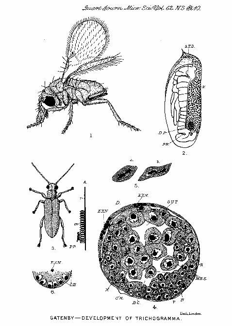
6Z. MS. 0WO.
GATENBY—DEVELOPMENT OF TRICHOGRAMMA.
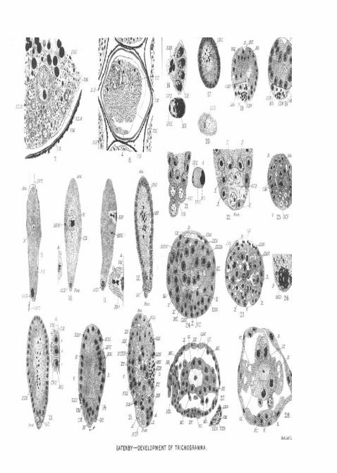
EXN.
Post
GATENBY—DEVELOPMENT OF TRICHOGRAMMA.
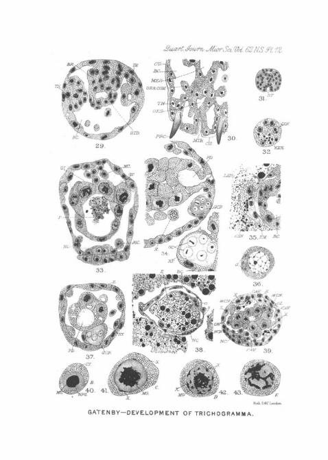
. S2N.S. ft 12.
BR.
Hmli LifcT London.
GATENBY—DEVELOPMENT OF TRICHOGRAMMA.