The Electrical Management of Cardiac Rhythm Disorders Bradycardia Device Course.
Upload
amethyst-nortonCategory
view
40download
0description

The Electrical Management of Cardiac Rhythm Disorders
Bradycardia
Device Course

Pacemaker Components

Leads
● Epicardial
● Endocardial

Goals of Cardiac Pacing● The electrical management of bradyarrhythmias requires
○ Ability to deliver enough energy to consistently depolarize the heart (capture)
○ Ability to correctly sense intrinsic cardiac activity● These functions are affected by many factors
○ Settings of output parameters (pulse amplitude, pulse width)
○ Sensitivity parameter settings○ Impedance○ Electrical concepts

Capture● The capture threshold is defined as the minimum amount
of electrical energy required to consistently depolarize the myocardium
● When a pacemaker output causes a depolarization, that is also called “capture”
● The capture threshold is also called the pacing threshold or the stimulation threshold
● The capture threshold is not constant○ It can change over time (disease, medications, age)○ It can even change over the course of the day!

What Affects the Capture Threshold?
• Activity level• Posture• Time of day• Comorbidity
• Heart failure• Meals• Drugs• Disease progression
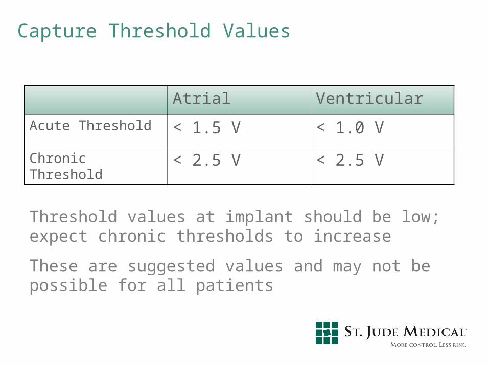
Capture Threshold Values
Atrial Ventricular
Acute Threshold < 1.5 V < 1.0 V
Chronic Threshold < 2.5 V < 2.5 V
Threshold values at implant should be low; expect chronic thresholds to increase
These are suggested values and may not be possible for all patients

Sensing● Sensing refers to how well the pacemaker is able to
“listen to” or perceive intrinsic cardiac events● Important things to consider when talking about sensing
○ Surface ECG○ Intracardiac electrogram (EGM)○ Sensing threshold○ Sense amplifier○ Sensitivity setting and sensitivity safety margin○ Unipolar/bipolar configurations○ Electromagnet interference
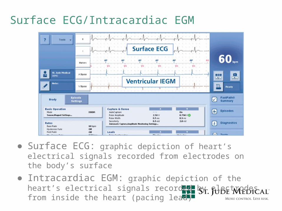
Surface ECG/Intracardiac EGM
● Surface ECG: graphic depiction of heart’s electrical signals recorded from electrodes on the body’s surface
● Intracardiac EGM: graphic depiction of the heart’s electrical signals recorded by electrodes from inside the heart (pacing lead)
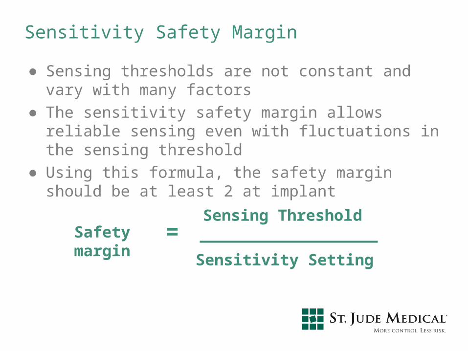
Sensitivity Safety Margin
● Sensing thresholds are not constant and vary with many factors
● The sensitivity safety margin allows reliable sensing even with fluctuations in the sensing threshold
● Using this formula, the safety margin should be at least 2 at implant
Safety margin =Sensing Threshold
Sensitivity Setting

Electromagnetic Interference (EMI)● EMI is defined as electrical signals of nonphysiologic
origin● May interfere with pacemaker (temporarily or
permanently)● Common sources of EMI
○ Cardioversion/defibrillation○ Electrocautery○ MRIs○ Extracorporeal shock wave lithotripsy (ESWL)○ Therapeutic radiation○ Radiofrequency ablation

What About Cardioversion/Defibrillation?● May permanently damage the pulse generator● Can temporarily inhibit or reprogram the pacemaker
○ Backup or noise reversion mode● Myocardial thermal damage secondary to shock which
may result in ventricular fibrillation, myocardial infarction, or both
● Guidelines○ Evaluate potential device interactions○ Place paddles 4 to 6 inches away from implanted
pacemaker○ Orient paddles in anterior/posterior position, if possible

What About Electrocautery?
● May reprogram or permanently damage the pacemaker● May inhibit the pacemaker● May cause the device to go into backup or noise
reversion mode● Myocardial thermal damage secondary to the
transmission of the electrical energy may result in VF, MI, or both
● Guidelines○ Contraindicated

What About MRI?
● The magnet in the MRI device can cause asynchronous pacing (pacing without sensing)
● Guidelines○ Generally contraindicated
● Magna-Safe Study

What About Lithotripsy?
● The vibrations in extracorporeal shock wave lithotripsy can damage the pacemaker (especially pacemakers with sensors, i.e. rate-adaptive units)
● Guidelines○ Program to VVI or VOO mode○ Keep focal point of lithotripter at least 6 inches away
from the implanted pacemaker○ Monitor the heart throughout the procedure

What About Therapeutic Radiation?● Damage depends on dose● Damage is cumulative; monitor device throughout course
of radiation therapy● Transistors may fail● Pacemakers may fail but mode of failure cannot be
predicted● Guidelines
○ Therapeutic ionizing radiation is contraindicated○ If therapeutic radiation is used, pacemaker should be
shielded or moved to a less vulnerable location
What About Radiofrequency Ablation?● RF ablation can temporarily or permanently reprogram
the pulse generator● Guidelines
○ Interrogate the pacemaker following the procedure to verify proper function
○ If necessary, reprogram
Myopotentials● Myopotentials are muscle noises that are sensed by the
pacemaker● Can inhibit pacing
○ The pacemaker senses the myopotential and inhibits the output, thinking the heart has beat on its own!
● Can interfere with sensing● Can cause inappropriate pacing
○ The pacemaker senses myopotential noise and inappropriately “thinks” it is atrial activity; it then tries to pace the ventricle to keep up or track that atrial activity
More EMI Sources
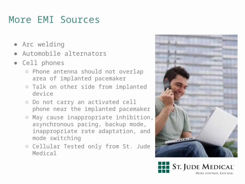
● Arc welding● Automobile alternators● Cell phones
○ Phone antenna should not overlap area of implanted pacemaker
○ Talk on other side from implanted device
○ Do not carry an activated cell phone near the implanted pacemaker
○ May cause inappropriate inhibition, asynchronous pacing, backup mode, inappropriate rate adaptation, and mode switching
○ Cellular Tested only from St. Jude Medical
EMI in the Medical Environment
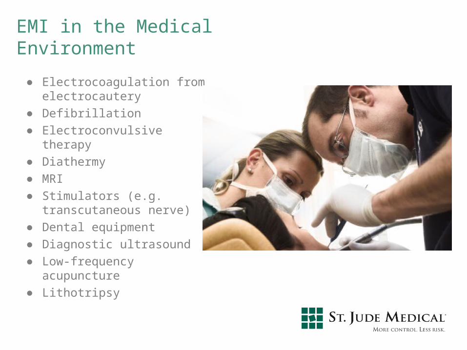
● Electrocoagulation from electrocautery
● Defibrillation● Electroconvulsive therapy● Diathermy● MRI● Stimulators (e.g.
transcutaneous nerve)● Dental equipment● Diagnostic ultrasound● Low-frequency acupuncture● Lithotripsy

EMI in the Industrial Environment
● Arc welding● Power lines● Transformers● Radio and TV transmitters● Static charge● Large metal frames in
magnetic fields● Induction furnaces and
heaters● Electrical switches
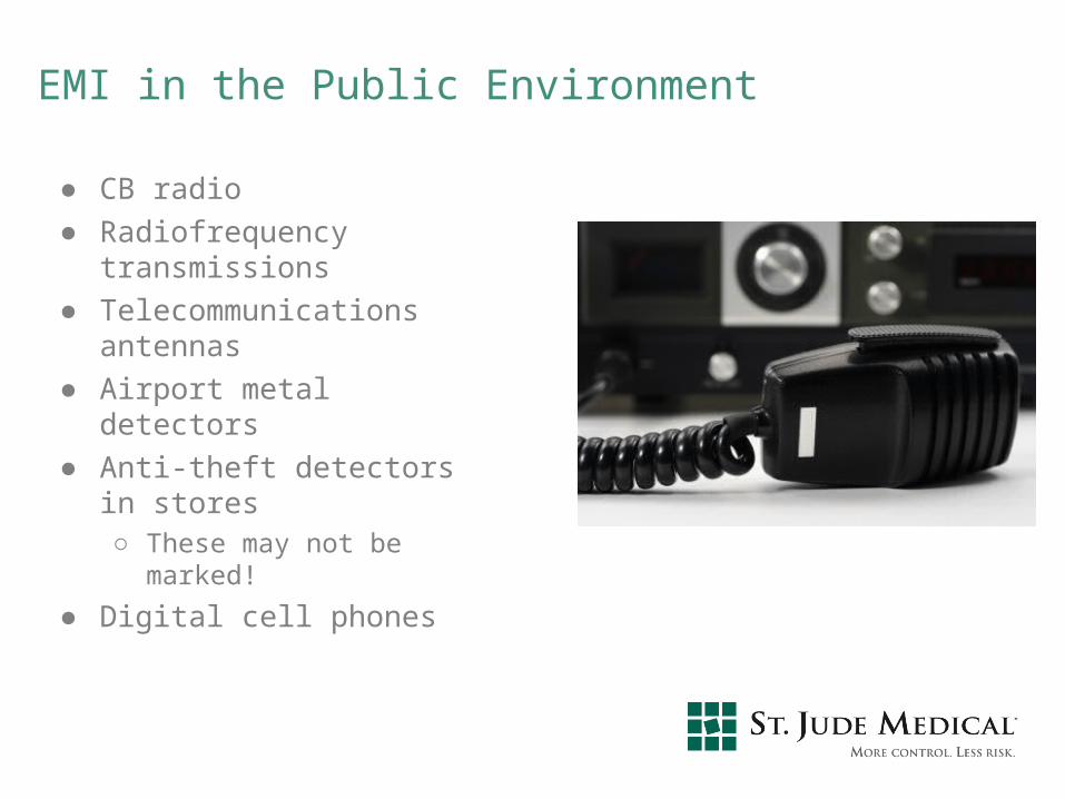
EMI in the Public Environment
● CB radio● Radiofrequency
transmissions● Telecommunications
antennas● Airport metal detectors● Anti-theft detectors in stores
○ These may not be marked!
● Digital cell phones

Effects of EMI● Pacemaker protection
○ Hardware backup circuits (to protect against loss of memory or software errors)
○ Shields● Effects
○ EMI inhibition: pulse-to-pulse interval extends to the point that the pacemaker does not pace as often as it should.
○ Noise reversion: change in mode (typically to asynchronous pacing at the programmed rate) which may require reprogramming.
○ EMI tracking: acceleration of pacing as the pacemaker tries to track electromagnetic signals (“thinking” they are atrial signals)

Pacemaker Overview
NASPE / BPEG (NBG) Pacemaker Code

NAPSE/BPEG Generic (NBG) Code
Position
Category
LettersUsed
I II III IV V
Chamber(s)Paced
Chamber(s)Sensed
Responseto Sensing Rate modulation Multisite Pacing
O-None
R-Rate modulation
O-None
A-Atrium
V-Ventricle
D-Dual(A+V)
S- Single(A or V)
S- Single(A or V)
O-None
A-Atrium
V-Ventricle
D-Dual(A+V)
O-None
T-Triggered
I-Inhibited
D-Dual(T+I)
O-None
A-Atrium
V-Ventricle
D-Dual(A+V)
Manufacturer’s
Designation
Only
Magnet Use● Pacemakers
○ Pace Asynchronously (VOO or DOO) at the given battery rate (Temporarily)
○ Device will revert back to exactly the same parameters it was programmed to once the magnet is removed
● ICD
○ Will disable ICD Shock Therapy (Temporarily)
○ Does not affect pacing
○ Device will revert back to exactly the same parameters it was programmed to once the magnet is removed.
● Magnet must be placed over the device in order for temporary changes to occur.
A Systematic Approach to Diagnosing Rhythm Strips● Measure Base Rate● Measure AV/PV Interval● Verify Atrial capture● Verify Atrial sensing● Verify Ventricular capture● Verify Ventricular sensing● Verify Underlying rhythm● Document

Base Rate 60 ppm
MTR 120 ppm
AVD 200 ms
PVARP 250 ms
ECG # 1
Dual Chamber ECG Analysis
What is the Analysis?

ECG # 2
Dual Chamber ECG Analysis
Base Rate 60 ppm
MTR 120 ppm
AVD 200 ms
PVARP 250 ms
What is the analysis?
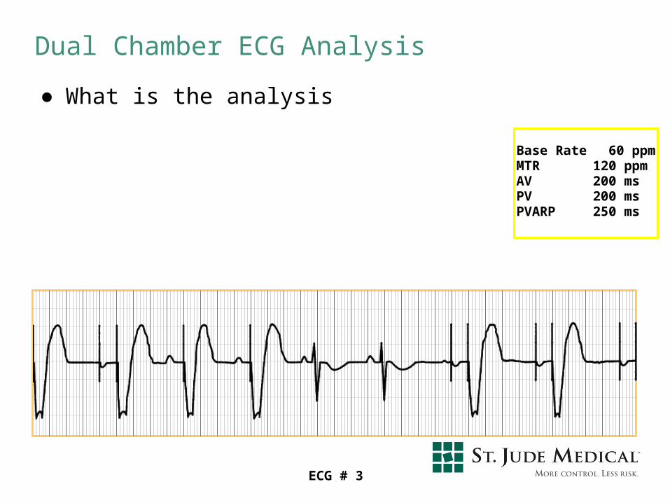
Dual Chamber ECG Analysis
Base Rate 60 ppmMTR 120 ppmAV 200 msPV 200 msPVARP 250 ms
ECG # 3
● What is the analysis
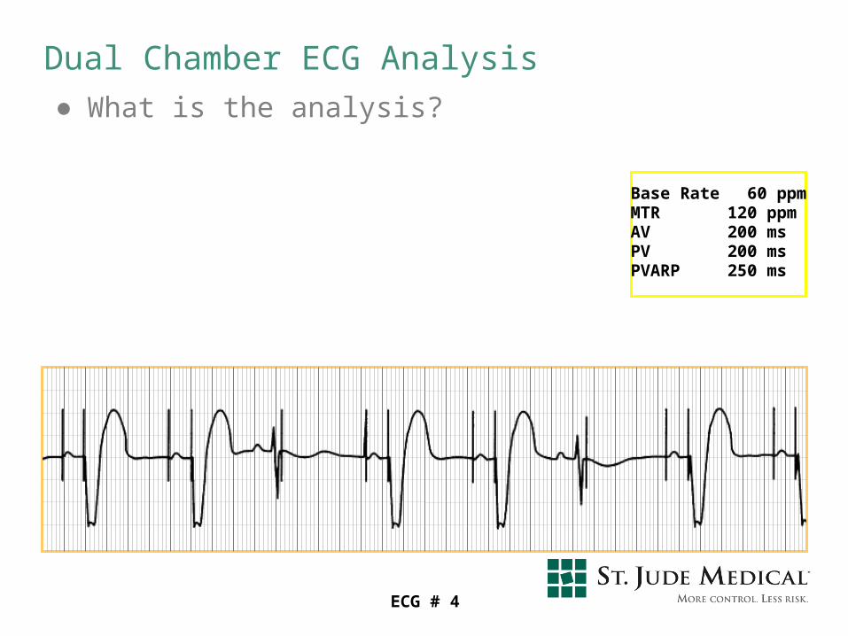
Dual Chamber ECG Analysis● What is the analysis?
Base Rate 60 ppmMTR 120 ppmAV 200 msPV 200 msPVARP 250 ms
ECG # 4
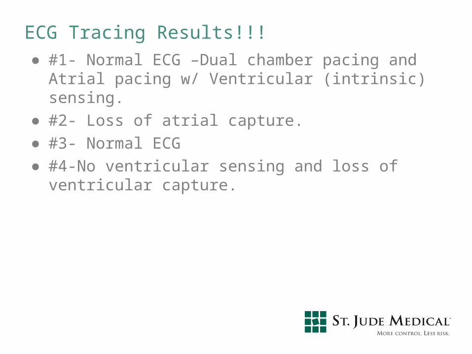
ECG Tracing Results!!!● #1- Normal ECG –Dual chamber pacing and Atrial pacing
w/ Ventricular (intrinsic) sensing.● #2- Loss of atrial capture.● #3- Normal ECG● #4-No ventricular sensing and loss of ventricular capture.
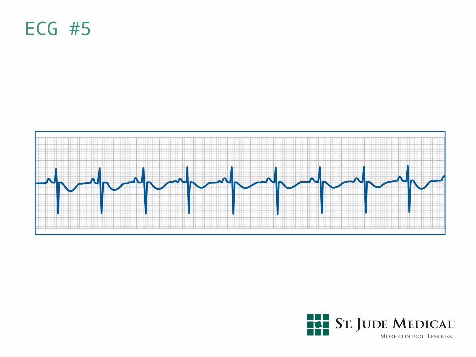
ECG #5
Answer ● Slide #5
○ Normal Sinus Rhythm○ Can not determine any pacemaker function
• Pacers are usually set to pace above 50 or 60 bpm• Single Chamber ICD- Pace above 40bpm
○ Pacemakers only work when?• Native heart rate goes below the base rate• An intrinsic beat does not occur before the set Paced and
Sensed AV Delays.• Set at an Asynchronous Mode (VOO or DOO)

Thank you for your time!!!