The Effects of Static Stretching Versus Dynamic Stretching
127
University of Wisconsin Milwaukee UWM Digital Commons eses and Dissertations August 2013 e Effects of Static Stretching Versus Dynamic Stretching on Lower Extremity Joint Range of Motion, Static Balance, and Dynamic Balance Wenqing Wang University of Wisconsin-Milwaukee Follow this and additional works at: hp://dc.uwm.edu/etd Part of the Kinesiology Commons , and the Medicine and Health Sciences Commons is esis is brought to you for free and open access by UWM Digital Commons. It has been accepted for inclusion in eses and Dissertations by an authorized administrator of UWM Digital Commons. For more information, please contact [email protected]. Recommended Citation Wang, Wenqing, "e Effects of Static Stretching Versus Dynamic Stretching on Lower Extremity Joint Range of Motion, Static Balance, and Dynamic Balance" (2013). eses and Dissertations. Paper 225.
Transcript of The Effects of Static Stretching Versus Dynamic Stretching
University of Wisconsin MilwaukeeUWM Digital Commons
Theses and Dissertations
August 2013
The Effects of Static Stretching Versus DynamicStretching on Lower Extremity Joint Range ofMotion, Static Balance, and Dynamic BalanceWenqing WangUniversity of Wisconsin-Milwaukee
Follow this and additional works at: http://dc.uwm.edu/etd
Part of the Kinesiology Commons, and the Medicine and Health Sciences Commons
This Thesis is brought to you for free and open access by UWM Digital Commons. It has been accepted for inclusion in Theses and Dissertations by anauthorized administrator of UWM Digital Commons. For more information, please contact [email protected].
Recommended CitationWang, Wenqing, "The Effects of Static Stretching Versus Dynamic Stretching on Lower Extremity Joint Range of Motion, StaticBalance, and Dynamic Balance" (2013). Theses and Dissertations. Paper 225.
THE EFFECTS OF STATIC STRETCHING VERSUS DYNAMIC
STRETCHING ON LOWER EXTREMITY JOINT RANGE OF
MOTION, STATIC BALANCE, AND DYNAMIC BALANCE
by
Wenqing Wang
A Thesis Submitted in
Partial Fulfillment of the
Requirements for the Degree of
Master of Science
in Kinesiology
at
The University of Wisconsin-Milwaukee
August 2013
ii
ABSTRACT
THE EFFECTS OF STATIC STRETCHING VERSUS DYNAMIC STRETCHING ON LOWER EXTREMITY JOINT RANGE OF MOTION, STATIC BALANCE,
AND DYNAMIC BALANCE
by
Wenqing Wang
The University of Wisconsin-Milwaukee, 2013 Under the Supervision of Professor Jennifer Earl-Boehm
The purpose of this study was to examine the effects of static stretching (SS)
versus dynamic stretching (SS) on lower extremity joint range of motion (ROM),
static balance, and dynamic balance. Fifteen active subjects with tight hamstring and
calf muscles participated. Hip flexion and knee extension ROM angle was measured
using a fluid inclinometer. A closed-chain method of measuring ankle dorsiflexion
ROM was used. Static balance was assessed in single-leg stance on a force plate using
the time-to-boundary (TTB) measurement. The Star Excursion Balance Test (SEBT)
was used to assess dynamic balance in three directions. These measurements were
assessed before and after each of three interventions: DS, SS or warm-up alone (CN).
The dependent variables included ROM measures (hip flexion, knee extension, and
ankle dorsiflexion), SEBT measures (anterior (ANT), posterior-medial (PM),
posterior-lateral (PL)), and TTB mean in anterior-posterior (AP) and medial-lateral
(ML). Repeated measures ANOVA were used to analyze the data.
There was a significant main effect (p < 0.05) for time. Repeated measures
ANOVA showed that knee extension ROM, hip flexion ROM, ankle dorsiflexion
iii
ROM, the SEBT (ANT, PM, PL) significantly (P<0.05) increased regardless of what
intervention (SS, DS, CN) was performed. There were no significant differences
(p>0.05) for the TTB (ML, AP) and there were also no significant interaction (p>0.05)
between interventions (SS, DS, CN) and time.
The less stiff muscles and more slack connective tissue around the joints
following stretching might attribute to the increased joint ROM. The enhanced ability
to maintain dynamic balance after an increased flexibility might be due to a
desensitized stretch reflex. A less responsive stretch reflex could suppress the postural
deviations, enhance the proprioceptive input, and thus make it easier to establish
equilibrium. Another contributor might be elevated muscle and body temperature,
which enhance nerve conduction velocity. The sensory systems might play a dominant
role in regulating the static postural control. Additional research is needed to more
clearly understand the relationship between altered ROM, balance and stretching.
iv
©Copyright by Wenqing Wang, 2013 All Rights Reserved
v
TABLE OF CONTENTS
LIST OF FIGURES .......................................................................................................vii LIST OF TABLES ...................................................................................................... viii ACKNOWLEDGEMENTS ............................................................................................ ix
CHAPTER 1: INTRODUCTION .................................................................................... 1 Background ............................................................................................................. 1 Purpose .................................................................................................................... 9 Specific Aims .......................................................................................................... 9 Delimitations ........................................................................................................... 9 Assumptions .......................................................................................................... 10 Limitations ............................................................................................................ 10 Significances ......................................................................................................... 11
CHAPTER 2: LITERATURE REVIEW ........................................................................ 12 Introduction ........................................................................................................... 12 Stretching Techniques ............................................................................................ 14 Ballistic Stretching ........................................................................................... 14 Proprioceptive Neuromuscular Facilitation Stretching ...................................... 15 Static Stretching ............................................................................................... 15 Dynamic Stretching .......................................................................................... 17 Physiological Mechanisms Relating to Dynamic Stretching ...................... 20 Static and Dynamic Balance .................................................................................. 22 Time-to-Boundary (TTB) ................................................................................. 24 Star Excursion Balance Test (SEBT) ................................................................ 25 Factors Contributing to SEBT Performance .............................................. 26 Range of Motion ................................................................................. 26 Fatigue ............................................................................................... 28 Interventions ....................................................................................... 29 Stretching and Balance ........................................................................................... 30 Performance ................................................................................................... 30 Mechanism ..................................................................................................... 35 Summary ................................................................................................................ 37
CHAPTER 3: METHODS ............................................................................................. 38 Purpose .................................................................................................................. 38 Participants ............................................................................................................ 38 Instrumentation ...................................................................................................... 39 Protocol ................................................................................................................. 40 Orientation Session ........................................................................................... 41 Range of Motion Tests ...................................................................................... 42
vi
Weight-Bearing Lunge Test ....................................................................... 42 Active Knee Extension Test ....................................................................... 43 Hip Flexion Test ......................................................................................... 45 Deep Squat Test ......................................................................................... 45 Task Practice .................................................................................................... 46 Balance Testing ................................................................................................ 47 Time-to-Boundary ...................................................................................... 47 Star Excursion Balance Test ....................................................................... 48 Warm-up Protocols .......................................................................................... 49 A general warm-up ..................................................................................... 49 Dynamic Stretching .................................................................................... 50 Static Stretching ......................................................................................... 50 Data Analysis......................................................................................................... 51 Statistical Analysis ................................................................................................. 52
CHAPTER 4: RESULTS ............................................................................................... 54 Range of Motion .................................................................................................... 54 Dynamic Balance ................................................................................................... 56 Static Balance ........................................................................................................ 58 Interactions and Stretching Main Effects ................................................................ 59
CHAPTER 5: DISCUSSION ......................................................................................... 61 Knee Extension Range of Motion .......................................................................... 61 Ankle Dorsiflexion Range of Motion ..................................................................... 64 Hip Flexion Range of Motion ................................................................................ 66 Dynamic Balance (SEBT) ...................................................................................... 67 Static Balance (TTB) ............................................................................................. 72 Mechanisms Relating Stretching to Range of Motion and Balance ......................... 75 Limitations and Directions for Future Research ..................................................... 77
Conclusion ............................................................................................................. 79 References ..................................................................................................................... 81 Appendix A: IRB Manager Protocol .............................................................................. 93 Appendix B: Informed Consent Form .......................................................................... 102 Appendix C: Screening & Medical History Questionnaire ........................................... 108 Appendix D: Recruitment Flyer ................................................................................... 110 Appendix E: Data Collection Sheet .............................................................................. 111 Appendix F: Individual Data ........................................................................................ 112 Appendix G: Linear Regression Analysis .................................................................... 116
vii
LIST OF FIGURES
Figure 1. Testing protocol flow-chart ...................................................................... 41
Figure 2. Participant positioning for the weight-bearing lunge test .......................... 43
Figure 3. Participant positioning for the active knee extension test .......................... 44
Figure 4. Participant positioning for the hip flexion test .......................................... 45
Figure 5. Participant positioning for the deep squat test ........................................... 46
viii
LIST OF TABLES
Table 1. Dynamic stretching protocol .................................................................... 50
Table 2. Static stretching protocol ......................................................................... 51
Table 3. Descriptive statistics ................................................................................ 54
Table 4. Means and SD of knee extension ROM measures for interventions .......... 55
Table 5. Means and SD of hip flexion ROM measures for interventions ................ 55
Table 6. Means and SD of ankle dorsiflexion ROM measures for interventions..... 56
Table 7. Means and SD of anterior direction of SEBT for interventions ................. 57
Table 8. Means and SD of posteromedial direction of SEBT for interventions ...... 57
Table 9. Means and SD of posterolateral direction of SEBT for interventions ....... 58
Table 10. Means and SD of anteroposterior TTB for interventions .......................... 58
Table 11. Means and SD of mediolateral TTB for interventions .............................. 58
Table 12. ANOVA table for intervention, time and interaction main effect ............. 60
ix
ACKNOWLEDGEMENTS
I wish to first thank my advisor, Dr. Jennifer Earl. I feel fortunate to have had
the opportunity to be under your guidance in the fields of Biomechanics during the
past two years, thank you for encouraging me when I faced with challenged
difficulties, and thank you for pointing me in the right direction on this long and hard
task. I sincerely appreciate your dedication to assisting me with this journey.
I would like to acknowledge my other committee members, Dr. Kristian
O’Connor and Dr. Kyle Ebersole. Thank you for your valuable perspective and advice
on this project. I would also like to express my thanks to Dr. Stephen Cobb, thank you
for your support on the Time-to-boundary data collection.
Finally I would like to express my gratitude to my parents, who always offer
support to me and believe in me to accomplish to this destination in my life.
1
CHAPTER 1: INTRODUCTION
Background
One of the most common things that individuals are instructed to do prior to
exercise is “warm-up”. A regular warm-up usually consists of three components:
aerobic exercise, stretching, and a rehearsal of the movements that will be used in the
subsequent training exercise or sports competition. Stretching is often utilized for a
wide variety of populations to be an essential part of a warm-up, which includes
ballistic stretching, proprioceptive neuromuscular facilitation (PNF) stretching, static
stretching (SS), and dynamic stretching (DS) (Ranna & Koslow, 1984; Sady, Wortman,
& Blanke, 1982). The benefits of stretching include, but are not limited to improve
joint range of motion (ROM), enhance muscular performance, and reduced risk of
injury (Pasanen, Parkkari, Pasanen, & Kannus, 2009; Shellock & Prentice, 1985; G. J.
Wilson, Murphy, & Pryor, 1994; Witvrouw, Mahieu, Danneels, & McNair, 2004; W. B.
Young & Behm, 2002). However, there was recently doubt over the effectiveness of
SS, as studies have demonstrated that SS decreased an individual’s performance in
force, strength, and power (A. Nelson & Kokkonen, 2001; Power, Behm, Cahill,
Carroll, & Young, 2004). It is therefore increasingly suggested that individuals should
turn to DS warm-up to more closely mimic movements in the subsequent training
exercises or sports competition, and DS has been shown to improve muscular
performance (Fletcher, 2010; Little & Williams, 2006; McMillian, Moore, Hatler, &
Taylor, 2006). Since balance is important for a wide range of populations that include
recreationally active individuals, elite athletes, and elderly to not only produce
2
optimal performance but also to prevent fall or injury, it is critical to understand how
physical intervention affects it. One are that has not been thoroughly investigated is
the effects of stretching on balance. Postural stability, or balance, relies heavily on the
contribution of information from proprioceptive receptors located within the muscle
and connective tissue. Because stretching changes the length of the muscles and
tendons, it is possible that either DS or SS may have an influence on proprioception,
and therefore balance.
Ballistic stretching (BS) is a kind of passive stretch that forces the limb into a
quick and jerking motion, which suddenly produces a bounce beyond a leg or arm’s
normal ROM. Thus, it is recommended that individuals should not perform BS unless
they are high-level athletes or being supervised, otherwise it may cause serious injury
(Sady et al., 1982).
Proprioceptive neuromuscular facilitation (PNF) stretching, defined as a
combination of passive stretch and isometric contractions of the target muscle, is
often utilized to increase the joint ROM, muscular strengthen, and neuromuscular
control in a clinical and rehabilitation environment (Marek et al., 2005). However,
PNF stretching has been proven to decrease vertical jump performance and leg
extension power in recreationally active individuals (Bradley, Olsen, & Portas, 2007;
Marek et al., 2005). Therefore, it is suggested that PNF stretching should not be
performed immediately prior to an explosive movement during physical activity.
Static stretching (SS) is described as gradually lengthening a muscle to an
elongated position as tolerated to a point of discomfort, and holding position for a
3
particular length of time. SS has often been widely uzilized to be a component of a
warm up in the training exercise or sports competition (De Vries, 1962). Traditionally,
SS has been shown to increase the joint ROM, inprove performance, and prevent
injury (Bandy, Irion, & Briggler, 1997; Smith, 1994; W. B. Young & Behm, 2002) .
Increased ROM was one of the greastest benefits derived from SS. This was primarily
due to changes in the length and stiffness of musculotendinous unit (MTU), with
greater ROM generated by a less stiff MTU (G. Wilson, Wood, & Elliott, 1992).
However, there was recently doubt over the effectiveness of SS. Studies have
demonstrated that SS decreased an individual’s performance in force, strength, and
power. These performances included maximal voluntary contraction (MVC) isometric
force, one repetition maximum lifts, vertical jump, sprint, running, and agility effects
(Behm, Bambury, Cahill, & Power, 2004; A. Nelson & Kokkonen, 2001; Power et al.,
2004). Additionally, several studies have concluded that SS had no effect or increased
the risk of injury (Chaouachi et al., 2008; Faigenbaum, Bellucci, Bernieri, Bakker, &
Hoorens, 2005; McHugh & Cosgrave, 2009; McNeal & Sands, 2003). Therefore, the
use of SS remains controversial.
It is increasingly suggested that individuals should turn to dynamic stretching
(DS) designed warm-up due to the close mimic movements in the subsequent training
exercise or sports competition, rather than SS (McMillian et al., 2006; Yamaguchi &
Ishii, 2005). Dynamic stretching is defined as a controlled movement through the joint
active range of motion while moving but not exceeding individual’s extensibility
limits (Fletcher & Jones, 2004). Some studies have demonstrated that DS exhibited
4
similar increases in ROM as SS, while other authors suggested that SS created greater
effects on ROM than DS (Bandy, Irion, & Briggler, 1998; Beedle & Mann, 2007;
Herman & Smith, 2008). Thus, there is no consensus on the effects of DS or SS on
ROM. Additionally, improved muscular performance following DS were seen in the
areas of shuttle run time, medicine ball throw distance, jump and sprint performance,
and leg extension power (Fletcher, 2010; Fletcher & Anness, 2007; Little & Williams,
2006; McMillian et al., 2006; Thompsen, Kackley, Palumbo, & Faigenbaum, 2007;
Yamaguchi & Ishii, 2005; Yamaguchi, Ishii, Yamanaka, & Yasuda, 2007). Several
possible mechanisms by which DS improved muscular performance could be elevated
muscle and body temperature (Fletcher & Jones, 2004), post-activation potentiation
(PAP) in the stretched muscle (Torres et al., 2008), and stimulation of the nervous
system (Yamaguchi & Ishii, 2005). However, these mechanisms have not been fully
explored and the reason behind why DS helps performance is as yet unknown. Since
coaches, athletic trainers, and fitness professinals are increasingly aware of the
advantage of DS in improving muscular performance, the use of DS rather than SS for
the warm-up is increasingly more common. However, we do not yet know the effects
that DS has on balance.
In biomechanics, balance is defined as the ability to maintain the individual’s
center of gravity within their base of support with minimal postural sway
(Shumway-Cook, Anson, & Haller, 1988). Balance can be separated into static
balance and dynamic balance.
Static balance is defined as individual maintaining a stable base of support
5
while minimizing segment and body movement (Bressel, Yonker, Kras, & Heath,
2007). Instruments, such as the Balance Error Scoring System or Berg Balance Scale,
have been widely used to measure static balance (P. Gribble, Hertel, & Denegar,
2007), however they are somewhat subjective. Time-to-boundary (TTB) provides an
objective novel postural control approach to assess static balance. A lower TTB
outcome indicates greater postural instability since the center of pressure (CoP) is
closer in time to reaching the boundary of the base of support (van Emmerik & van
Wegen, 2002). TTB measures can assess CoP excursions in relation to the boundaries
of the base of stability that is not addressed by traditional postural control measures
and has been proven to be more sensitive at detecting improvements in static postural
control compared with traditional CoP-based measures (Hertel & Olmsted-Kramer,
2007; Mckeon et al., 2008). However, stability in static balance might not translate
necessarily to postural control during dynamic movements due to the task and
environmental demands of a dynamic movement being very different from standing
quietly.
Dynamic balance is defined as an individual performing a purposeful
movement around a base of support without compromising the base of support.
Dynamic balance measurements, such as Star Excursion Balance test or wobble board,
have been demonstrated to be more closely to mimic demands of physical activity
than static balance assessments (P. A. Gribble, Hertel, & Plisky, 2012). The Star
Excursion Balance Test (SEBT) is a cost-effective, easy-to-use clinical technique to
measure dynamic balance in the rehabilitation, injury evaluation and prediction, and
6
research applications (Hertel, Miller, & Denegar, 2000; Kinzey & Armstrong, 1998;
Plisky, Rauh, Kaminski, & Underwood, 2006). The SEBT requires individual’s
postural control, strength, range of motion, coordination and proprioceptive abilities.
The farther distance the touching leg reaches, the better dynamic balance it displays
(Hertel et al., 2000). Hertel et al (2006) simplified the SEBT that using three reach
directions (anterior, posteromedial, and posterolateral) from the center of the grid to
identify individuals with chronic ankle instability (CAI) (Hertel, Braham, Hale, &
Olmsted-Kramer, 2006). To make valid comparisons of SEBT, reaching distances
need to be normalized to individual’s limb length (P. A. Gribble & Hertel, 2003). In
addition, several other anthropometric and physiologic factors, such as range of
motion, fatigue, or interventions, have also contributed to SEBT performance. Given
that the interference between dorsiflexion in the ankle, knee flexion, and hip flexion
with the SEBT (P. A. Gribble, Hertel, Denegar, & Buckley, 2004; P. A. Gribble et al.,
2012; M.C. Hoch, Staton, & McKeon, 2011), it is reasonable to assume that alteration
in ROM following stretching could affect the performance of the SEBT, and therefore
dynamic balance.
Postural stability, or balance, relies heavily on the contribution of information
from proprioceptive receptors located within the muscle and connective tissue.
Proprioception includes input from sensory neurons in the inner ear and in the stretch
receptors in the muscles and the joint ligaments, is an important contributor to control
postural stability (Di Giulio, Maganaris, Baltzopoulos, & Loram, 2009). It is possible
that a small change in the activity of a proprioceptor could lead to a greater change in
7
balance (Diener, Dichgans, Guschlbauer, & Mau, 1984).Proprioceptors affect postural
stability through the relationship between sensitivity and muscle stiffness, or the
stretch-reflex response (L. M. Nashner, 1981). Stiffer muscles produce a greater reflex
response (Sinkjaer, Toft, Andreassen, & Hornemann, 1988) which leads to a more
rapid response to slight perturbations of muscle length. A faster response to
perturbation would result in better balance (Petit, Filippi, Emonet-Denand, Hunt, &
Laporte, 1990). Since stretching has the ability to change the muscle stiffness, muscle
length, and increase joint ROM, it is reasonable to postulate that stretching could
affect proprioception and therefore balance (Behm et al., 2004; Chong & Do, 2002;
McHugh & Cosgrave, 2009).
There was little research focusing on the relationship between balance and
stretching. Several studies support that SS enhanced or had no adverse effect on
dynamic balance (P.B. Costa, B.S. Graves, M. Whitehurst, & P.L. Jacobs, 2009;
Handrakis et al., 2010; Lewis, Brismée, James, Sizer, & Sawyer, 2009; A. G. Nelson,
Kokkonen, Arnall, & Li, 2011). Costa et al (2009) evaluated the effects of different
durations of SS on dynamic balance. The results of this study indicated that SS of 45 s
did not adversely affect dynamic balance while SS with 15 s may improve dynamic
balance (P.B. Costa et al., 2009). While Handrakis et al (2010) found that ten minutes
of acute SS enhanced dynamic balance in active middle-aged adults (Handrakis et al.,
2010). Furthermore, Nelson et al (2011) investigated the acute effect of SS on postural
stability in non-balance trained individuals compared with experienced balance
trainers. They found that SS improved balance for non-balance trained individuals,
8
but not for those with greater balance experience (A. G. Nelson et al., 2011). On the
other hand, studies indicated that SS resulted in adverse effects on static balance (Behm
et al., 2004). Behm et al (2004) evaluated the effect of acute lower limb SS on static
balance, force, proprioception, reaction time and movement time. It found that there
was a significant (P < 0.009) decrease in balance scores in the SS condition (decreasing
for 9.2%) compared with the control condition (increasing for 17.3%) (Behm et al.,
2004). This was consistent with Nagano et al (2006)’s finding, which suggested that SS
of the calf muscles increased postural sway, and thus adversely affected static balance
(Nagano, Yoshioka, Hay, Himeno, & Fukashiro, 2006). Since many training exercise
or sports competition requires both types of balance, static and dynamic, it would be
therefore advantageous to incorporate static and dynamic balance task together when
investigating the effect of SS on balance performance in an integrated research
environment.
As discussed above, the benefits of DS on muscular performance have been
distinctly proven and there is a tendency to utilize DS to be a component of a
warm-up rather than SS. However, it is still unclear the effects of DS on static or
dynamic balance, since no research has been conducted in this area. This study will
add preliminary research to reveal the effects of DS on static balance or dynamic
balance.

9
Purpose
The purpose of this study was to examine the effects of static stretching versus
dynamic stretching on lower extremity joint ROM, static balance, and dynamic
balance.
Specific Aims
1. To compare the effects of SS and DS on joint ROM of hip flexion, knee extension,
and dorsiflexion, it was hypothesized that: 1) the SS intervention would have an
increase in joint ROM of the hip, knee, and ankle, 2) the DS intervention would
have an increase in joint ROM of the hip, knee, and ankle, but less than the SS
group, 3) there would be no change in the joint ROM of the control intervention.
2. To compare the effects of SS and DS on static balance (TTB), it was hypothesized
that: 1) the SS intervention would have a decrease in performance of static
balance, 2) the DS intervention would have increased performance of static
balance, 3) there would be no change static balance of the control intervention.
3. To compare the effects of SS and DS dynamic balance (SEBT), it was
hypothesized that: 1) the SS intervention would have decreased dynamic balance,
2) the DS intervention would have increased dynamic balance, 3) there would be
no change in the dynamic balance of the control intervention.
Delimitations
The results of this study were applied to those who are recreationally active
individuals with or without hamstring or calf muscle tightness, both for men and
women ages from 18-45. It was not applied to children, adults older than 45 and
10
anyone who is not recreationally active. The results of this study only applied to static
and dynamic balance, and have limited application to other athletic activities that
require additional skills.
This study only examined balance performance and ROM parameters (TTB
variables, SEBT scores, dorsiflexion, knee extension, and hip flexion ROM). No
conclusion was made with respect to neural activation levels, such as changes in
musculotendinous unit (MTU) stiffness and proprioceptive sense since they were not
being examined.
Assumptions
Some assumptions were made in this study. The first assumption was that
participants honestly completed the questionnaire and accurately reported their
current activity level and injury/surgery history. The second assumption was that
participants continued their recreationally active exercise or sports with no change of
the regular physical activity’s level, but refrained from it 24 hours prior to testing
sessions. The third assumption was that there was no or little learning effect across the
study. The learning effect was controlled by the questionnaire, orientation and data
analysis that calculates different valuables between pre and post balance tests. The
participants completed all trials with maximal effort was the final assumption.
Limitations
The only limitation of this study was learning effect. Although it was
controlled by the questionnaire, orientation and data analysis that calculates different
valuables between pre and post balance tests to a large extent, it is impossible to
11
completely eliminate it.
Significance
The significance of this study was that it will add the body knowledge that will
allow coaches, athletic trainers, and fitness professionals to make evidence based
decisions on how to prepare the individuals with hamstring and calf muscle tightness
for utilizing a proper stretching technique during warm-up session. Additionally, it
will also provide basic scientific evidence on informing future research that focus on
lower extremity functional balance rehabilitation with specific stretching technique.
12
CHAPTER 2: LITERATURE REVIEW
Introduction
A regular warm-up usually consists of three components. The first component
is aerobic exercise, which raises core body and muscle temperature (Bishop, 2003a).
Bishop (2003b) suggests that an aerobic warm-up at 40-60% VO2 max for 5-10
minutes followed by 5 minutes of recovery is optimal to stimulate short-term physical
function and enhance athletic performance (Bishop, 2003b). The second component is
stretching that has been widely proven to enhance neuromuscular performance,
including stimulates core body and muscle temperature, increases the joint range of
motion (ROM), enhances muscle strength, and promotes balance and coordination
(Pasanen et al., 2009; Shellock & Prentice, 1985; Witvrouw et al., 2004; W. B. Young
& Behm, 2002). The third component is a rehearsal of the movements that will be
used in the subsequent training exercise or sports competition (W. B. Young & Behm,
2002). The integrated warm-up components are adopted extensively for a wide of
population, not only for recreationally active individuals, but also for elite athletes.
Various types of stretching technique have been developed to be applied not
only in the training exercise or sports competition, but also in clinical and
rehabilitation environment. These stretching techniques include ballistic stretching
(BS), proprioceptive neuromuscular facilitation (PNF) stretching, static stretching
(SS), and dynamic stretching (DS). Recently, there was doubt over the effectiveness
of SS due to its adverse effect on performance (Chaouachi et al., 2008; Faigenbaum et
al., 2005; McNeal & Sands, 2003). In addition, it is increasingly suggested that
13
individual should turn to DS as a component of an effective warm-up due to its
distinct benefits on muscular performance (McMillian et al., 2006; Yamaguchi & Ishii,
2005).
Impaired balance is a factor to provide negatively effects on athletic
performance (Irrgang, Whitney, & Cox, 1994). In addition, a balance deficit is
attributed to increase the risk of a fall and injury (McGuine, Greene, Best, & Leverson,
2000; Trojian & McKeag, 2006; Tropp, Ekstrand, & Gillquist, 1984). Since balance
plays such an important role in the lifespan, it is critical to understand how physical
interventions affect it. Proprioception was considered as one of the mechanisms to
control balance and is sensitive to muscle tension and length that could be changed by
stretching (Behm et al., 2004; Chong & Do, 2002; McHugh & Cosgrave, 2009).
Therefore, it is reasonable to suppose that stretching could have an influence on
balance.
There was little research focusing on the relationship between balance and
stretching. Several studies support that SS enhanced or had no adverse effects on
dynamic balance (P.B. Costa et al., 2009; Handrakis et al., 2010; Lewis et al., 2009; A.
G. Nelson et al., 2011). However, Behm et al (2004) indicated that SS resulted in
adverse effects on static balance (Behm et al., 2004). Since these studies separated
static balance and dynamic balance task, and many training exercise or sports
competition requires both types of balance, it would be advantageous to incorporate
static and dynamic balance task together in an integrated research. Furthermore, it is
still unclear the effects of DS on static or dynamic balance, since no research has been
14
conducted in this area.
Therefore, the purpose of this literature review is to discuss the effects of
various types of stretching techniques, static and dynamic balance, and the
relationship between stretching and static or dynamic balance.
Stretching Techniques
Various types of stretching techniques have been developed in the training,
sports competition, clinic, and rehabilitation settings in order to gain an increase in
range of motion (ROM), an improvement in muscular performance, and reduce the
risk of injury. These stretches include ballistic stretching (BS), proprioceptive
neuromuscular facilitation (PNF) stretching, static stretching (SS), and dynamic
stretching (DS) (Ranna & Koslow, 1984; Sady et al., 1982).
Ballistic Stretching
Ballistic stretching is a kind of stretch that forces the limb into a quick and
jerking motion, which suddenly produces a bounce beyond a leg or arm’s normal
ROM. Thus, it is recommended that individuals should not perform BS unless they
are high-level athletes or supervised by a personal trainer, otherwise it may cause
serious injury (Bradley et al., 2007; Sady et al., 1982). In addition, it has been
demonstrated that BS resulted a decrease in the jump performance and maximal
strength (Bradley et al., 2007; A. Nelson & Kokkonen, 2001). Bradley et al (2007)
found that there was a decrease in the vertical jump performance (2.7%, p> 0.05)
following a standard cycle warm-up along with 10 minutes BS (Bradley et al., 2007).
Nelson and Kokkonen (2001) also found that BS reduced maximal muscle strength in
15
the knee extension and flexion (A. Nelson & Kokkonen, 2001). Therefore, BS has not
been widely supported in the literature to be a component of a warm-up.
PNF Stretching
PNF stretching, defined as a combination of passive stretch and isometric
contractions of the target muscle, is often utilized to increase the joint ROM, muscular
strengthen, and neuromuscular control by a therapist in clinical and rehabilitation
environment (Marek et al., 2005). Weng et al (2009) found that PNF stretching was
more effective on muscle strength than SS following isokinetic muscle strengthen
exercises in 132 patients with knee osteoarthritis (Weng et al., 2009). However,
Bradley et al (2007) demonstrated that PNF stretching decreased muscular
performance. They found that vertical jump performance was diminished (5.1%) for
15 minutes following a standard cycle warm-up along with PNF stretching (Bradley et
al., 2007). Thus, it is suggested that PNF stretching should not be performed
immediately prior to an explosive movement in the physical activity.
Static Stretching
Static stretching is described as gradually lengthen a muscle to an elongated
position as tolerated and that position is then held for a particular length of time to a
point of discomfort (De Vries, 1962). Traditionally, it had generally been believed that
SS increased the joint ROM, enhanced muscular performance, and prevent injury
(Bandy et al., 1998; O'Sullivan, Murray, & Sainsbury, 2009; Power et al., 2004; Smith,
1994; W. B. Young & Behm, 2002). However, recent studies have demonstrated that
SS reduced force, strength and power production, thus decreased performance
16
(Chaouachi et al., 2008; Faigenbaum et al., 2005; McNeal & Sands, 2003). These
performance included isometric muscular contraction, sprint, and jump performance.
Fowles et al (2000) found that isometric muscular strength in the ankle plantarflexors
has been decreased for up to 1 h after performing 13 static dorsiflexion stretches of
135 s each over 33 minutes in ten young adults. This was interpreted by Kubo et al
(2001) who indicated that tendon structure and connective tissue were inclined to be
more compliant and muscle force was prone to be slack following SS, which led to a
lower rate of force production (Kubo, Kanehisa, Kawakami, & Fukunaga, 2001). In
addition, vertical jump performances diminished followed by SS in the hip and knee
extensors for 100 s (Cornwell, Nelson, Heise, & Sidaway, 2001). The reason behind
this could be that a decrease rate occurred in neural transmission with SS and thus
caused a delay in muscle contraction velocity (Knudson, Bennett, Corn, Leick, &
Smith, 2001). Furthermore, Fletcher and Anness (2007) found that 50-m sprint
performance decreased followed by 800-m jogged warm-up alone with SS compared
with active DS in eighteen experienced sprinters (Fletcher & Anness, 2007). This
could be illustrated that a decreased ability in the musculotendinous unit (MTU)
happened after SS, and then lead to a decrease level in muscle activation and force
production (Cornwell, Nelson, Heise, & Sidaway, 2001). One study combined
running and jump performance following SS. Faigenbaum et al (2005) compared the
acute effects of 3 different warm-up protocols (5 minutes of walking with 5 minutes
of SS, 10 minutes of DS, and 10 minutes of DS plus 3 drop jumps from 15-cm boxes).
They found that long-jump, vertical-jump and shuttle-run performance reduced
17
significantly (p< 0.05) following SS (Faigenbaum et al., 2005).
Since it has been questioned the wisdom of SS on muscular performance, it is
suggested that SS should be avoided as a component of warm-up session.
Dynamic Stretching
Dynamic stretching is defined as a controlled movement through the joint
active range of motion while moving but not exceeding individual’s extensibility
limits (Fletcher & Jones, 2004). The objective of DS is to increase dynamic flexibility
in the target muscle by contracting the antagonist muscle without bouncing
(Yamaguchi & Ishii, 2005). DS has increasingly gained popularity due to a number of
studies showing an increase in high intensity performance in the joint ROM, leg
power output, jump, running, sprint, and agility (Fletcher, 2010; Fletcher & Anness,
2007; Little & Williams, 2006; McMillian et al., 2006; Ranna & Koslow, 1984;
Thompsen et al., 2007; Yamaguchi & Ishii, 2005; Yamaguchi et al., 2007).
Previous study showed that the gain of DS and SS on the ROM was almost
identical. Ranna and Koslow (1984) compared the effects of SS, DS and PNF
stretching on the ROM of hamstring-gastrocnemius muscles. The findings indicated
that all three stretches produced significant improvement (p< 0.001) in the ROM
during the pretest and posttest. No difference was found between all three stretches
condition (Ranna & Koslow, 1984). This was agreed with Herman &Smith (2008)’s
finding (Herman & Smith, 2008).
However, O'Sullivan et al’s (2009) questioned the previous finding. They
investigated the short-term effects of a general warm-up, SS and DS on the
18
hamstrings ROM following assessing passive knee extension test in individuals with
previous hamstrings injury and uninjured controls. It found that passive knee
extension ROM significantly increased after a general warm-up (p < 0.001), further
significantly increased (p = 0.04) after SS, while significantly decreased after DS (p =
0.013). The increased ROM after warm-up and SS reduced significantly (p < 0.001)
after 15 minutes rest and further remained significantly greater than that at baseline (p
< 0.001). The results of this study indicated that the effect of a general warm-up and
SS on ROM was greater in those with hamstrings injured individuals, but not in DS
(O'Sullivan et al., 2009). Therefore, the effect of DS on hamstrings flexibility or ROM
was conflict.
Dynamic stretching has been demonstrated to increase muscular power output
(Yamaguchi & Ishii, 2005; Yamaguchi et al., 2007). Yamaguchi and his colleagues
worked on two studies related to leg power output. For their first study, under various
loads at 5%, 30%, and 60% maximum voluntary contractile (MVC) torque with
isometric leg extension, DS group was significantly (p < 0.05) greater than that in the
no-stretching (NS) condition under each load (5% MVC: 468.4 ± 102.6 W vs. 430.1 ±
73.0 W; 30% MVC: 520.4 ± 108.5 W vs. 491.0 ± 93.0 W; 60% MVC: 487.1 ± 100.6
W vs. 450.8 ± 83.7 W) (Yamaguchi et al., 2007). Another study that measured leg
extension power before and after stretches protocols (DS, SS, and NS) was consistent
with above finding. DS and SS protocols focused on five lower limbs muscle groups,
which were plantar flexors, hip extensors, hamstrings, hip flexors, and quadriceps
femoris. DS group was significantly (0 < 0.01) greater than that in the SS group
19
(2022.3 ± 121.0 W). No significant difference was found between SS (1788.5 ± 85.7
W) and NS (1784.8 ± 108.4 W) condition (Yamaguchi & Ishii, 2005). Yamaguchi and
his colleagues mentioned that post-activation potentiation (PAP) caused by voluntary
contractions of the antagonist of the target muscle was the possible reason behind DS
increased leg power output. Since PAP shortened the time to peak torque and
increased the rate of torque development followed DS.
Besides the benefits in the power output, it has also been proven that DS
increased running, sprint, agility, and jump performance (Fletcher, 2010; Fletcher &
Anness, 2007; Little & Williams, 2006). Little and Williams (2006) found that DS
(1.87 ± 0.09) produced a significantly (p< 0.005) faster 10-m sprint acceleration time
than NS conditions (1.83 ± 0.08 seconds) and significantly (p< 0.005) faster Zig-zag
agility performance (5.14 ± 0.17 seconds) than both SS (5.20 ± 0.16 seconds) and NS
groups (5.22 ± 0.18 seconds). This study informed professional soccer player that DS
was most effective as preparation for the subsequent high-speed performance (Little
& Williams, 2006). Similarly, Fletcher and Anness (2007) notified that active DS
significantly (men p= 0.002; women p= 0.043) decreased 50-m sprint time in
experienced sprinters (Fletcher & Anness, 2007).
One study compared the effects of different DS velocities on jump
performance. Fetcher (2010) found that faster velocity of DS (100 b/min) had a
significant (p< 0.001) greater in all three jump performance, square jump (SJ), drop
jump (DJ), and countermovement jump (CMJ) than both in the slow velocity of DS
(50 b/min) and NS condition, and slow DS also resulted in significant (p<0.001)
20
greater performance in the DJ and SJ than NS condition. The mechanisms behind this
were related to increases in heart rate and core temperature, and also linked to greater
nervous system activation, shown by gastrocnemius in the CMJ significant higher in
EMG output(p<0.005) followed fast DS(Fletcher, 2010).
Given that the BS, PNF stretching, and SS resulted detrimental effects in
muscular performance and thus may increase the incidence of injury, coaches, athletic
trainers, fitness professionals therefore increasingly suggest that individuals should
turn to a designed DS as a component of an effective warm-up due to its higher
benefits on muscular performance (McMillian et al., 2006; Yamaguchi & Ishii, 2005).
Physiological Mechanisms Relating to Dynamic Stretching
Several physiological mechanisms that could explain the advantages of DS on
muscular performance included increased core body and muscle temperature,
alteration in musculotendinous unit (MTU) stiffness, post-activation potentiation
(PAP), and myotatic reflex.
Positive effects of DS could be resulted from increased core body and muscle
temperature within warm-up process (Yamaguchi & Ishii, 2005). This led to stimulate
peripheral blood flow and then enhanced muscle temperature (Smith, 1994), further
resulted in an increase in the nerve receptor sensitivity and nerve impulse velocity,
and then produce a more rapid rate of muscle contraction and power production
(Faigenbaum et al. 2005).
Bishop (2003a) indicated that DS had the ability to alter MTU stiffness. MTU
stiffness incorporating with muscles, tendon, and connective tissue contracts tightly to
21
transmit internal muscle forces to the skeletal system (G. J. Wilson et al., 1994).
Stiffer MTU was required for a faster transmission of muscular force to bones, then
generating a forceful movement (Kubo, Kanehisa, & Fukunaga, 2001). This further
led to favorable changes in the force-velocity relationship (Bishop, 2003a). However,
a compliant MTU allowed less force rate of transmission during muscle contraction
(Kokkonen et al, 1998), less able to store elastic energy (Fletcher & Jones, 2004), and
increase the time of force transmission from the central nervous system (CNS) to the
muscle skeletal system (Fowles, Sale, & MacDougall, 2000).
Post-activation potentiation (PAP) is defined as the process when the
contractile history of muscle holds a role in subsequent muscle contraction (Bishop
2003). This meant that a heavier loading applied to muscle prior to an explosive
movement could cause a higher stimulation of the CNS to allow a forceful muscle
contraction immediately (Chiu et al., 2003). Thus, PAP resulted in more rapid or
forceful muscle contraction, and shortened the time to peak torque and increases the
rate of torque development following DS (Fowles et al., 2000; Yamaguchi et al.,
2007).
Myotatic reflex is defined as muscle contraction in response to stretching
within the muscle. It has been proven that faster stretching speed could cause to
greater action potential of the myotatic reflex (Gollhofer & Rapp, 1993; Gottlieb &
Agarwal, 1979). Fletcher (2010) demonstrated that faster velocity of DS had
significantly faster take-off velocity and vertical jump performance than the slower
velocity of DS (Fletcher, 2010).
22
Although these possible physiological mechanisms provided basic evidence
for DS linked to muscular performance, future research is still required to better
illustrate high intensity muscular performance behind DS.
Static Balance and Dynamic Balance
In biomechanics, balance is defined as the ability to maintain the individual’s
center of gravity within their base of support with minimal postural sway
(Shumway-Cook et al., 1988). Balance can separate into static balance and dynamic
balance (Winter, Patla, & Frank, 1990). Static balance is defined as individual
maintaining a stable base of support while minimizing segment and body movement
(Bressel et al., 2007). Several valid measurements or clinical scales, such as a force
platform, the Balance Error Scoring System (BESS) or Berg Balance Scale (BBS),
can be used to measure static balance (P. Gribble et al., 2007). Although static balance
provide useful clinical information or research outcome, the underlying task of
standing as still as possible, such as postural sway, might not translate necessarily to
movement tasks. Dynamic balance is defined as individual performing expected
movement around a base of support to a new location and immediately attempting to
remain as motionless as possible. Dynamic balance measurements, such as Star
Excursion Balance Test (SEBT), or wobble board, more closely mimic demands of
physical activity than static balance assessments (P. A. Gribble et al., 2012). Since
many training exercise or sports competition requires both types of balance skills, it
should incorporate static balance and dynamic balance together within exercise or
research.
23
Two studies compared static and dynamic balance that was relatively relevant
to the current designed study. Bressel et al (2007) compared static and dynamic
balance among collegiate athletes competing in soccer, basketball, and gymnastics.
BESS was used to assess static balance. Participants performed 3 stance variations
(double leg, single leg, and tandem leg) on 2 surfaces (stiff and compliant). SEBT was
used to assess dynamic balance. Participants performed multidirectional maximal
single-leg reaches from a unilateral base of support. It found that BESS error scores
for the gymnastics group were 55% lower than for the basketball group and SEBT
scores were 7% higher in the soccer group than the basketball group. The results of
this study indicated that gymnasts and soccer players did not differ in terms of static
and dynamic balance. In contrast, basketball players displayed inferior static balance
compared with gymnasts and inferior dynamic balance compared with soccer players
(Bressel et al., 2007). Similarly, Ross & Guskiewicz (2004) determined static and
dynamic postural stability differences with functional ankle instability individuals. A
single leg stance for 20 seconds was used to measure static postural stability, while a
single jump-landing test that required to jump 50% to 55% of participants’ maximum
vertical jump height and maintained motionless for 20 seconds after landing was used
to assess dynamic postural stability. The results indicated that mean sway was not
significantly different between groups in the anterior/posterior (P = 0.28) and
medial/lateral (P = 0.65) directions. The functional ankle instability group took
significantly longer to stabilize in the anterior/posterior (3.27 ± 0.72 seconds vs. 2.33 ±
0.33 seconds; P < 0.001) and medial/lateral (2.48 ± 0.50 seconds vs. 2.00 ± 0.65
24
seconds; P = 0.04) directions. It came to a conclusion that individuals with functional
ankle instability took significantly longer to stabilize than individuals with stable
ankles after a single-leg jump landing, while there was no difference between groups
with mean sway measured during single-leg stance (Ross & Guskiewicz, 2004).
Based on different static balance measurement evaluated above, it is therefore
necessary to examine the effects of static balance through a more sensitive and reliable
tool.
Time-to-Boundary
Postural control is the specific terminology describing static balance. Postural
control plays an important role not only in the injury prevention, but also in the
athletic performance. Increased postural control is generally linked with increased risk
of falling with neurological impairment (Matinolli et al., 2007), unstable ability in
dynamic tasks (Latash, Ferreira, Wieczorek, & Duarte, 2003), and with higher risk for
ankle sprains (McGuine et al., 2000).
Traditionally, maintaining postural control is defined as the amount of postural
sway of the center of mass (COM) or center of pressure (COP) to return the center of
gravity to a centralized position over the base of support (Rietdyk, Patla, Winter, Ishac,
& Little, 1999). The postural sway measures the frequency against time by assessing
medial-lateral and anterior-posterior displacement of the center of pressure (Patla,
1990; Winter et al., 1990). A small amount of COM or COP excursion is considered
as more stable than a larger amount of COM or COP excursion (Woollacott,
Shumway-Cook, & Nashner, 1986).
25
Time-to-boundary (TTB) provides a novel postural control approach to assess
static balance. TTB is defined as estimating the time it would take for the COP to
reach the boundary of the base of support if the COP was to continue on its trajectory
at its instantaneous velocity (Hertel & Olmsted-Kramer, 2007). A lower TTB outcome
indicates greater postural instability since the COP is closer in time to reaching the
boundary of the base of support (van Emmerik & van Wegen, 2002). TTB measures
have been shown to have intrasession reliability with intraclass correlation
coefficients ranging from .34 to .87 (Hertel, Olmsted-Kramer, & Challis, 2006). TTB
measures can assess COP excursions in relation to the boundaries of the base of
stability that is not addressed by traditional postural control measures. TTB has been
proven to be more sensitive at detecting improvements in static postural control
compared with summary COP-based measures (Mckeon et al., 2008), and as well as
in detecting postural control deficits associated with CAI than traditional postural
control measures (Hertel & Olmsted-Kramer, 2007). Therefore, TTB measures were
used in this study rather than traditional postural sway measurement.
Star Excursion Balance Test
The star excursion balance test (SEBT) is a clinical technique to measure
dynamic balance during rehabilitation, injury evaluation, and research applications
(Hertel et al., 2000; Kinzey & Armstrong, 1998). SEBT has been proven to not only
an easy-to-use outcome tool to measure dynamic balance in research, but also a
clinical application to predict the risk of injury to lower extremity (Plisky et al., 2006).
The SEBT usually consists of a series of lower extremity reaching tasks in 8
26
directions (anterior, anteromedial, anterolateral, medial, lateral, posterior,
posteromedial, and posterolateral) from the center of grid that require individual’s
postural control, strength, range of motion, coordination and proprioceptive abilities.
The farther distance the touching leg reaches, the better dynamic balance it displays.
The ability to reach farther with the touching leg also requires a combination ability of
better dynamic balance on the contralateral stance leg (Hertel et al., 2000). Hertel et al
(2006) simplified the SEBT that using three reach directions (anterior, posteromedial,
and posterolateral) to identify individuals with CAI (Hertel, Braham, et al., 2006) .
The SEBT has a strong intratester and intertester reliability. The intraclass correlation
coefficients was ranging from .85 to .96 for intratester reliability and from .81 to .93
for intertester reliability (Hertel et al., 2000; Kinzey & Armstrong, 1998).
Factors Contributing to SEBT Performance
To make valid comparisons of SEBT, reaching distances need to be
normalized to individual’s limb length as measured from the anterosuperior iliac spine
to the medial malleolus (P. A. Gribble & Hertel, 2003). Besides limb length, several
other anthropometric and physiologic factors including ROM, fatigue, and
interventions also potentially contributed to SEBT performance.
Range of Motion
Dorsiflexion range of motion in the ankle was correlated strongly with anterior
reaching distance in the SEBT. Hoch et al (2011) examined the relationships between
maximum dorsiflexion range of motion on the weight-bearing lunge test (WBLT) and
normalized reach distance in three directions (anterior, posteromedial, and
27
posterolateral) on the SEBT. Thirty-five healthy adults performed three trials of the
SEBT in three directions on each limb to assess dynamic balance, and then three trials
of the WBLT to measure maximum dorsiflexion range of motion. It found that only
the anterior direction (79.0 ± 5.8%) of the SEBT was significantly related to the
WBLT (11.9 ± 2.7 cm), r = 0.53 (p = 0.001). The WBLT explained 28% of the
variance in the anterior normalized reach distance (r²= .28). This results indicated that
the anterior direction of the SEBT may be a desired clinical measure to assess the
effects of maximum dorsiflexion range of motion on dynamic balance (M.C. Hoch et
al., 2011).
There are 2 studies related to how kinematic factors (hip and knee flexion) can
affect SEBT performance between participants with and without CAI. Gribble et al
(2007) investigated the influence of CAI on the performance of SEBT after fatiguing
protocol. Thirty subjects completed the SEBT before and after a lunging fatigue
protocol. Pre-post fatigue change scores were measured for sagittal plane kinematics
of the stance leg and the normalized reach distances. When reaching anteriorly after
the lunge fatigue in CAI group, the changes in knee and hip flexion predicted
approximately 49 % of the variance in normalized reach distances (R2 = .487; p
= .001). When reaching medially under lunge fatigue in CAI group, the changes in
knee and hip flexion predicted approximately 20 % of the variance in normalized
reach distances (R2 = .198; p = .014). The results indicated that CAI significantly
affected the variances in normalized reach distances after a fatigue protocol (P.
Gribble et al., 2007). In another similar designed study, Gribble et al (2004) found that
28
the injured side of the CAI subjects displayed significantly smaller reach distance
values and knee flexion angles for all 3 reaching directions compared with the
uninjured side and the healthy group (P. A. Gribble et al., 2004). With 2 studies, the
differences of kinematic pattern in the knee and hip of the sagittal plane after
performing the SEBT suggest that those who with CAI was associated with a
reduction in dynamic balance.
Given that the interference with dorsiflexion in the ankle, knee flexion and hip
flexion in the sagittal plane on the SEBT, this information might be helpful for
clinicians to design specific rehabilitation protocol for patients with dynamic postural
control impairments.
Fatigue
It is widely accepted that fatigue can affect physical performance. Gribble et al
(2009) examined the effects of fatigue on performance measures of the SEBT in three
directions (anterior, posteromedial, and posterolateral).16 healthy young adults
performed the SEBT before and after 4 different fatiguing conditions (isometrically
applied fatigue to the ankle, knee, and hip and continuous lunging). The normalized
reach distances and sagittal-plane kinematics of the knee and hip were recorded. It
found that fatigue produced deficits in normalized reach distances and decreased knee
flexion in all 3 reaching directions (P. A. Gribble, Robinson, Hertel, & Denegar, 2009).
This was consistent with previous two studies, Gribble et al (2004) and Gribble et al
(2007) that suggest that SEBT performance might provide a useful approach for
assessing decline in dynamic balance from fatigue.
29
Interventions
Some studies have examined the effects of SEBT on improvements in
performance and reduce the risk of injury after designed exercise interventions as an
outcome tool, including balance training, core stability training, and neuromuscular
control exercise programs (Filipa, Byrnes, Paterno, Myer, & Hewett, 2010; FitzgeralD,
Trakarnratanakul, Smyth, & Caulfield, 2010; Hale, Hertel, & Olmsted-Kramer, 2007;
Mckeon et al., 2008).
Mckeon et al (2008) investigated the effect of a 4 week balance training
program on static and dynamic postural control in those with CAI. The intervention
consisted of a 4 week supervised balance training program that emphasized dynamic
stabilization in single-limb stance. They found that the balance training group had
significant improvements in reach distances with the posteromedial (P = .01) and the
posterolateral (P = .03) directions of the SEBT (Mckeon et al., 2008). Similarly, Hale
et al (2007) also found differences in the posteromedial (P = .03), posterolateral (P
= .01) reach directions of the SEBT and a composite score of all 8 directions (P = .03)
following a 4 week intervention of strength, ROM, and neuromuscular control
exercises in those who with CAI (Hale et al., 2007).
Kahle and Gribble (2009) focused on a 6 week intervention training program
in healthy and physically active young adults. They found that the exercise group
improved their scores by more than 4 % (P= .001) in the anteromedial direction and
improved 6% from baseline and was more than 6% better than the control group in
the medial direction with moderate to strong effect sizes (Kahle & Gribble, 2009).
30
Fitzgerald et al (2010) revealed improvements of 2.95% to 9.4% in the anterior,
posteromedial, and posterolateral reach directions of SEBT after 12 exercise sessions
of wobble board or postural stability training. Similarly, Filipa et al (2010) found that
8 weeks of neuromuscular control training in young female athletes improved
performance in the same 3 directions by 1.75% to 9.5%. Neuromuscular control
training was provided by mostly moderate to strong effect sizes that ranged from 0.58
to 1.00 (Filipa et al., 2010; FitzgeralD et al., 2010).
Since stretching could affect alteration in ROM and neuromuscular control
that has been associated with the SEBT, it is important to understand the relationship
between stretching and the SEBT, namely dynamic balance.
Stretching and Balance
Balance is important for a wide of population, which includes recreationally
active individuals, elite athletes, and elderly. For the recreationally active individuals
and elite athletes, impaired balance affects optimal athletic performance, and even
cause injury incidence. For the elderly, a balance deficit is prone to the higher risk of a
fall, and then cause osteoporotic fractures (M. E. Nelson et al., 1994). Since balance
plays an important role in the lifespan, it is critical to understand how physical
interventions, especially stretching, affect it.
Performance
Several studies have focused on the relationship between SS and static or
dynamic balance, but no research has concentrated on the effects of DS on either
static or dynamic balance.
31
One study focused on the SS and joint position sense. Ghaffarinejad et al
(2007) investigated the effect of SS in relation to muscle surrounding the knee on the
knee joint position sense (JPS). JPS was measured through the absolute angular error
(AAE) in order to estimate the ability to reach two target positions (20° and 45° of
flexion) in the dominant knee. Thirty-nine healthy students was tested by three 30 s
SS with a 30s rest. AAE values were measured repeated three times before and
immediately after SS trials. They found that the AAE decreased significantly after the
stretching protocols for quadriceps (3.5 (1.3) vs 0.7 (2.4); p<0.001), hamstring (3.6
(2.2) vs 1.6 (3.1); p�=�0.016), and adductors (3.7 (2.8) vs 1.7 (2.4); p�=�0.016) in
45° of flexion. The results suggest that the knee JPS improvement in 45° of flexion
following SS was contributed to the knee joint stability. This was expected to improve
balance since joint position sense was linked to proprioceptive response (Ghaffarinejad,
Taghizadeh, & Mohammadi, 2007).
Three studies examined the effects of SS on dynamic balance, while using
different dynamic balance measurements, stabilometer, Berg Balance Scale (BBS), and
Dynamic Stability Index (DSI), but not the SEBT.
Costa et al (2009) evaluated the effects of different durations of SS on
dynamic balance. The SS protocols consisted of a cycle ergometer warm-up at 70 rpm
and 70 W followed by SS (passive unilateral knee flexion, supine hip flexion, ankle
dorsiflexion with an extended knee, and ankle dorsiflexion with a flexed knee) on the
target muscle groups (quadriceps, hamstring, and plantar flexor). Each stretching
repeated 3 times with 15 seconds rest of periods and the positions were held for 15 or
32
45 seconds to the point of mild discomfort. The control one consisted of the same
cycle ergometer warm-up with a 26-minute rest of period between pretests and
posttests. Dynamic balance was measured using the BBS which was similar to actual
physical activities that resulted in instability. They found that the balance scores were
significantly improved (p<0.01) in the 15-s stretching condition and no significant
was found in the 45 s stretching condition. The results of this study indicated that SS
of 45 s did not adversely affect dynamic balance and SS with 15-second may improve
dynamic balance (P.B. Costa et al., 2009).
Similarly, Handrakis et al (2010) tested ten middle-age subjects (age: 40-60 yr.)
from a martial arts school following 10 minutes SS with 30 seconds hold for session.
Dynamic Stability Index (DSI) was used to test dynamic balance for single-leg stance.
Smaller DSI meant improved dynamic balance while greater DSI indicated opposite
effect. Other dependent variables included distances for broad jump, single hop, triple
hop, and crossover hop; elapsed time for a 6-m timed hop. They found that DSI of SS
group was significantly smaller than that in the NS group (3.5 ± 0.7 vs. 4.3 ± 1.4 DSI,
p < 0.05). No significant difference was found in the other dependent variables in both
two groups. Thus, it came to a conclusion that 10 minutes of acute SS with 30seconds
hold enhanced dynamic balance in active middle-aged adults.(Handrakis et al., 2010).
In comparison with non-balance trained individuals with experienced balance
trainers, Nelson et al (2011) investigated the effects of SS on postural stability in
forty-two college students and ten surfers performed balance testing on a stabilometer
on two separate days following either 30 min of quiet sitting or 30 min of SS
33
protocols. For the dynamic balance, the average time of keeping on the stabilometer
was recorded at 180° for two 30s periods. For the stretching protocol, it consisted of
five different SS exercises (sit-and reach, stretch, the lotus or butterfly stretch, the
heel cord or calf stretch, a standing half lotus stretch, and a quadriceps stretch) for 3
times unassisted and 3 time assisted to the muscles groups of the hip, knee, and ankle.
The results indicated that improved flexibility was significant (p<.05) following the
SS protocols for increasing (6.5 ± 2.7 cm) in sit and reach test. In addition, balance
time for non-balance trained individuals also improved significantly by 11.4% (2.0s
increase), but no significant change in the surfers. Thus, SS improved maintenance of
dynamic balance for non-balance trained individuals, but not for the experienced
balance trainers (A. G. Nelson et al., 2011).
Besides research on the relationship between SS and dynamic balance, three
studies examined the effects of SS on static balance using a wobble board and postural
sway, respectively, but not related to TTB.
Behm et al (2004) evaluated the effect of an acute SS on static balance, force,
proprioception, reaction time and movement time. Sixteen subjects were tested before
and after both with a SS of the quadriceps, hamstring, and plantar flexors or a similar
duration in the control condition. The stretching protocol consisted of a 5-min cycle
warm-up followed three stretches to the point of discomfort of 45s each with 15s rest.
SS included a series of unilateral knee flexion, hip flexion with extended leg in the
supine position, extended leg dorsiflexion in the standing position, and flexed knee
dorsiflexion in the standing position. Measurements included maximal voluntary
34
isometric contraction (MVC) force of the leg extensors, static balance using a wobble
board, reaction and movement time of the dominant lower limb. They found that there
was a significant (P < 0.009) decrease in balance scores with the SS condition
(decreasing for 9.2%) compared with the control condition (increasing for 17.3%).
There was significant difference (P< 0.01) in reaction (decreasing for 5.8%)
and movement (decreasing for 5.7%) time in the control condition and (increasing for
4.0% and 1.9% ) in the SS condition The results indicated that an acute SS adversely
affect performance on static balance and reaction/movement time (Behm et al., 2004).
The finding of Behm et al (2004) was supported by Nagano et al (2006)’s study,
which evaluated the effects of vision and SS of the calf muscles on postural sway
during quiet standing. Participants first stood on a force plate in 30 s for both legs and
the postural sway of the ground reaction force COP was recorded. Participants then
stood quietly on a device incorporating a static ankle joint dorsiflexion stretching in
3 min. After that, postural sway was recorded again. The findings of this study
indicated that postural sway significantly increased after SS in the dependent
variables: sweep speed, sway speed, standard deviation, maximal anteroposterior
range, mean anteroposterior position (Nagano et al., 2006).
Similarly, Lewis et al (2009) investigated the effect of SS on postural sway and
on the kinematic variables in gender. SS and NS groups were tested separately prior
to balance testing with electromyographic (EMG) recordings of muscle responses. In
the SS protocol, the quadriceps, hamstring, and plantar flexors of bilateral were
passively stretched in the supine position with three 45 s and a 15 s rest of period.
35
Testing during the NS condition began after the subject rested quietly for an equivalent
period of time as in the SS condition. Balance testing included the Postural Evoked
Response Test, Adaptation Test, Motor Control Test, Sensory Organization Test, and
Unilateral Stance Test. They found that no significant main effect for SS and 2
significant main effects for gender for the Motor Control Test (P = 0.021) and latency
of tibialis anterior (P = 0.009). The results indicated SS did not affect balance
performance during computerized dynamic posturography both for women and men
(Lewis et al., 2009).
Since many physical activity and rehabilitation interventions requires both
types of balance (static and dynamic), it would be therefore advantageous to
incorporate static and dynamic balance task together when investigating the effect of
SS on performance in an integrated research. In addition, it is also important to
understand how DS would affect on static or dynamic balance since no research has
focused on it.
Mechanism
Keeping balance is described as the ability to maintain the base of support
with minimal movement (Winter et al., 1990). A complex nervous system with
automatic postural responses, volitional motor control and reflexive responses
controls the ability of balance (Bloem, Allum, Carpenter, & Honegger, 2000; Shiratori
& Latash, 2000). This integrated system or mechanism is adjusted mainly by the CNS
as expressing self-promoted postural perturbations (Aruin, Forrest, & Latash, 1998),
and also influence individual’s movement in the ability of coordination, ROM, muscle
36
strength, and power production (Grigg, 1994; L. Nashner, 1976; R. M. Palmieri et al.,
2003; R. Palmieri, Ingersoll, Stone, & Krause, 2002). If function of physiological
mechanism were changed, the performance of balance would be affected, and may
further increase the risk of a fall or injury.
One possible physiological mechanism that affects the ability and performance
of balance in relation to stretching could be proprioception. Proprioception is one of
contributors to control postural stability (Di Giulio et al., 2009). Proprioception is
composed of sense from sensory neurons in the inner ear and in the stretch receptors
in the muscles and the joint ligaments. Proprioceptive sense originating from joint and
muscle receptors plays an integral role in the aimed at preparing, maintaining, and
restoring stability of postural stability of entire body and the joint stability of the
segments (Riemann & Lephart, 2002). It is possible that a small change in the activity
of a proprioceptor, it could lead to a greater change in balance (Diener et al., 1984).
Proprioceptors affect postural stability through the stretch-reflex response (L. M.
Nashner, 1981), which sensitivity could be influenced by muscle stiffness, with stiffer
muscles producing a greater reflex response (Sinkjaer et al., 1988). This was possible
due to the postural control maintained by stiffer muscles through greater or more
rapid responses to slight perturbations in muscle length (Petit et al., 1990). Since
stretching has ability to change the muscle stiffness, muscle length, and increase joint
ROM, it is reasonable to postulate that stretching could affect function of balance
(Behm et al., 2004; Chong & Do, 2002; McHugh & Cosgrave, 2009).
37
Summary
Since SS has been doubted its effectiveness on performance and injury
prevention, DS has been widely accepted to be a component of a warm-up due to its
benefits on muscular performance. In addition, as reliable measure of dynamic
balance, SEBT could be influenced by ROM, fatigue, and balancing training and
neuromuscular control interventions, however, it is still unclear the effects of SS and
DS on the SEBT, and the relationship of the SEBT on static balance. Moreover,
proprioceptors are sensitive to muscle tension and length, it is therefore reasonable to
postulate that stretching could affect balance. Few studies have focused on the
relationship between SS and static or dynamic balance, and no research has
concentrated on the effects of DS on either static or dynamic balance. This study will
provide basic scientific evidence and clinical application for informing future research
that focus on lower extremity muscular performance, injury prevention, and
rehabilitation with regard to altered ROM, balance, and stretching.
38
CHAPTER 3: METHODS
Purpose
The purpose of this study was to examine the effects of static and dynamic
stretching on lower extremity joint ROM, static balance, and dynamic balance.
Participants
Participants were recruited by informational flyers posted at University of
Wisconsin-Milwaukee. The flyers provided the contact information of the investigator
and a brief description of the study including the purpose, and the criteria for
inclusion and exclusion. Classroom visits were made to contact potential participants
who may be interested in participating. These visits met the guidelines of Institutional
Review Board. Both males and females between age of 18 and 45 were eligible for
the study.
The inclusion criteria of the participant was that the individual was: 1) male or
female between the age of 18-45, and 2) recreationally active (engage in some form of
physical activity at least 30mins and 3-4 days per week) (A. G. Nelson et al., 2011).
To maximize the potential effects of the stretching protocols, individuals who
demonstrate muscular tightness in the gastrocnemius/soleus and hamstring muscles
formed the study sample. The assessment procedures were described in the following
“Protocol” section.
The exclusion criteria of the participant was that the individual was: 1) free
from lower extremity pain or injury in the past 6 month or any other physical deficit
that limited them in performing the balance testing and stretching protocols, 2) No
39
history of concussion or balance disorders within the last 6 months, and 3) No history
of participating in a proprioceptive or balance training activity in the past 6 months.
Regardless of current level of physical activity, participants agreed not to change the
intensity or frequency of physical activity during the testing session and refrain from
them 24 hours prior to testing sessions.
Statistical power analysis based on previous studies (Bandy et al., 1997; R.
Gajdosik & Lusin, 1983; Handrakis Bandy & Irion, 1994; Nagano et al., 2006)
concluded that 15 participants would provide sufficient power for the analyses. All
participants provided written informed consent prior to data collection.
Instrumentation
A fluid inclinometer was used to identify maximum hip flexion angle, and
maximum knee extension angle in the Active Knee Extension (AKE) test.
A tape measure was used to measure the furthest distance between the great
toe and the wall in the weight-bearing lung test, an assessment of dorsiflexion ROM.
A light dowel was used for the Deep Squat (DS) test.
The time-to-boundary (TTB) was assessed by an AMTI force plate (Model
OR-6-7-2000, Advanced Mechanical Technology, Inc, Watertown, MA) at a
sampling rate of 100 Hz to measure the functional performance of static balance.
A written program (Matlab, v. 7.6.0, The MathWorks Inc, Natick, MA) was used to
compute a time series of time-to-boundary. Triaxial forces (Fx, Fy, Fz) and moments
(Mx, My, Mz) was recorded at 100 Hz and a time series of 500 Center of Pressure
(COP) data points for each trial was calculated by the Swaywin1 software program
40
(AMTI Corp., Watertown, MA).
Dynamic balance was assessed using the Star Excursion Balance Test (SEBT).
The testing grid consisted of 3 lines, each 120 cm in length extending to anterior,
posteromedial, and posterolateral direction in relation to the stance foot. Standard
athletic tape placed on the surface of grid. The center of the grid was marked with
crosshairs that participants were instructed to stand in the center of the grid during
testing (Hertel, Braham, et al., 2006).
A treadmill was used for a general warm-up. The University of
Wisconsin-Milwaukee Neuromechanics laboratory provided space for participants to
perform stretching interventions.
Protocol
All of the study activities took place at the University of
Wisconsin-Milwaukee Neuromechanics Laboratory. A general testing protocol
overview is provided below in Figure.1.
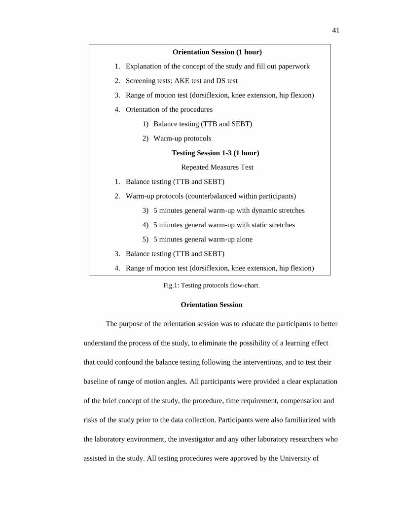
41
Orientation Session (1 hour)
1. Explanation of the concept of the study and fill out paperwork
2. Screening tests: AKE test and DS test
3. Range of motion test (dorsiflexion, knee extension, hip flexion)
4. Orientation of the procedures
1) Balance testing (TTB and SEBT)
2) Warm-up protocols
Testing Session 1-3 (1 hour)
Repeated Measures Test
1. Balance testing (TTB and SEBT)
2. Warm-up protocols (counterbalanced within participants)
3) 5 minutes general warm-up with dynamic stretches
4) 5 minutes general warm-up with static stretches
5) 5 minutes general warm-up alone
3. Balance testing (TTB and SEBT)
4. Range of motion test (dorsiflexion, knee extension, hip flexion)
Fig.1: Testing protocols flow-chart.
Orientation Session
The purpose of the orientation session was to educate the participants to better
understand the process of the study, to eliminate the possibility of a learning effect
that could confound the balance testing following the interventions, and to test their
baseline of range of motion angles. All participants were provided a clear explanation
of the brief concept of the study, the procedure, time requirement, compensation and
risks of the study prior to the data collection. Participants were also familiarized with
the laboratory environment, the investigator and any other laboratory researchers who
assisted in the study. All testing procedures were approved by the University of
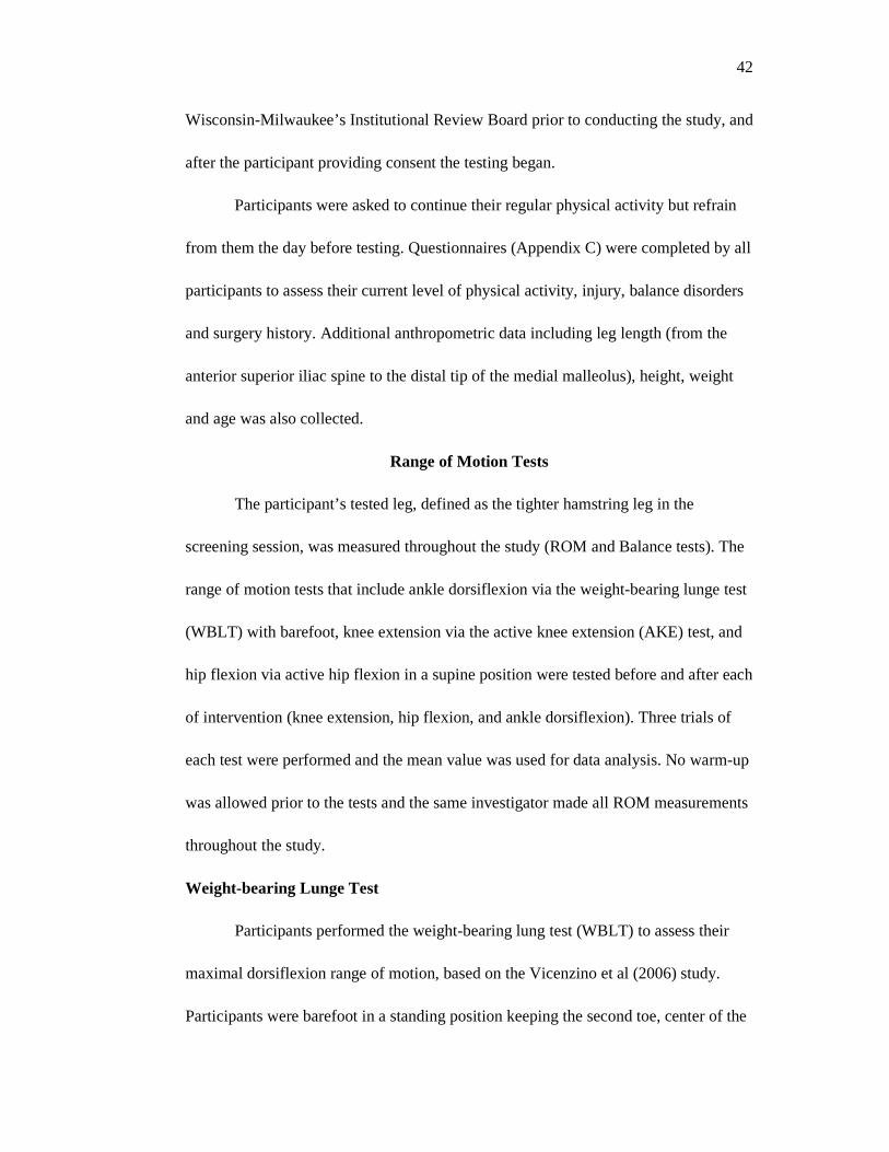
42
Wisconsin-Milwaukee’s Institutional Review Board prior to conducting the study, and
after the participant providing consent the testing began.
Participants were asked to continue their regular physical activity but refrain
from them the day before testing. Questionnaires (Appendix C) were completed by all
participants to assess their current level of physical activity, injury, balance disorders
and surgery history. Additional anthropometric data including leg length (from the
anterior superior iliac spine to the distal tip of the medial malleolus), height, weight
and age was also collected.
Range of Motion Tests
The participant’s tested leg, defined as the tighter hamstring leg in the
screening session, was measured throughout the study (ROM and Balance tests). The
range of motion tests that include ankle dorsiflexion via the weight-bearing lunge test
(WBLT) with barefoot, knee extension via the active knee extension (AKE) test, and
hip flexion via active hip flexion in a supine position were tested before and after each
of intervention (knee extension, hip flexion, and ankle dorsiflexion). Three trials of
each test were performed and the mean value was used for data analysis. No warm-up
was allowed prior to the tests and the same investigator made all ROM measurements
throughout the study.
Weight-bearing Lunge Test
Participants performed the weight-bearing lung test (WBLT) to assess their
maximal dorsiflexion range of motion, based on the Vicenzino et al (2006) study.
Participants were barefoot in a standing position keeping the second toe, center of the
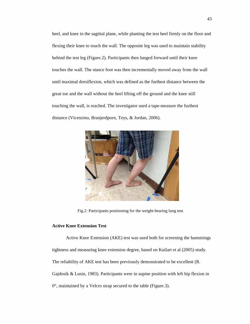
43
heel, and knee in the sagittal plane, while planting the test heel firmly on the floor and
flexing their knee to touch the wall. The opposite leg was used to maintain stability
behind the test leg (Figure.2). Participants then lunged forward until their knee
touches the wall. The stance foot was then incrementally moved away from the wall
until maximal dorsiflexion, which was defined as the furthest distance between the
great toe and the wall without the heel lifting off the ground and the knee still
touching the wall, is reached. The investigator used a tape-measure the furthest
distance (Vicenzino, Branjerdporn, Teys, & Jordan, 2006).
Fig.2: Participants positioning for the weight-bearing lung test.
Active Knee Extension Test
Active Knee Extension (AKE) test was used both for screening the hamstrings
tightness and measuring knee extension degree, based on Kuilart et al (2005) study.
The reliability of AKE test has been previously demonstrated to be excellent (R.
Gajdosik & Lusin, 1983). Participants were in supine position with left hip flexion in
0°, maintained by a Velcro strap secured to the table (Figure.3).
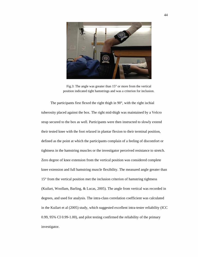
44
Fig.3: The angle was greater than 15° or more from the vertical
position indicated tight hamstrings and was a criterion for inclusion.
The participants first flexed the right thigh in 90°, with the right ischial
tuberosity placed against the box. The right mid-thigh was maintained by a Velcro
strap secured to the box as well. Participants were then instructed to slowly extend
their tested knee with the foot relaxed in plantar flexion to their terminal position,
defined as the point at which the participants complain of a feeling of discomfort or
tightness in the hamstring muscles or the investigator perceived resistance to stretch.
Zero degree of knee extension from the vertical position was considered complete
knee extension and full hamstring muscle flexibility. The measured angle greater than
15° from the vertical position met the inclusion criterion of hamstring tightness
(Kuilart, Woollam, Barling, & Lucas, 2005). The angle from vertical was recorded in
degrees, and used for analysis. The intra-class correlation coefficient was calculated
in the Kuilart et al (2005) study, which suggested excellent intra-tester reliability (ICC
0.99, 95% CI 0.99-1.00), and pilot testing confirmed the reliability of the primary
investigator.
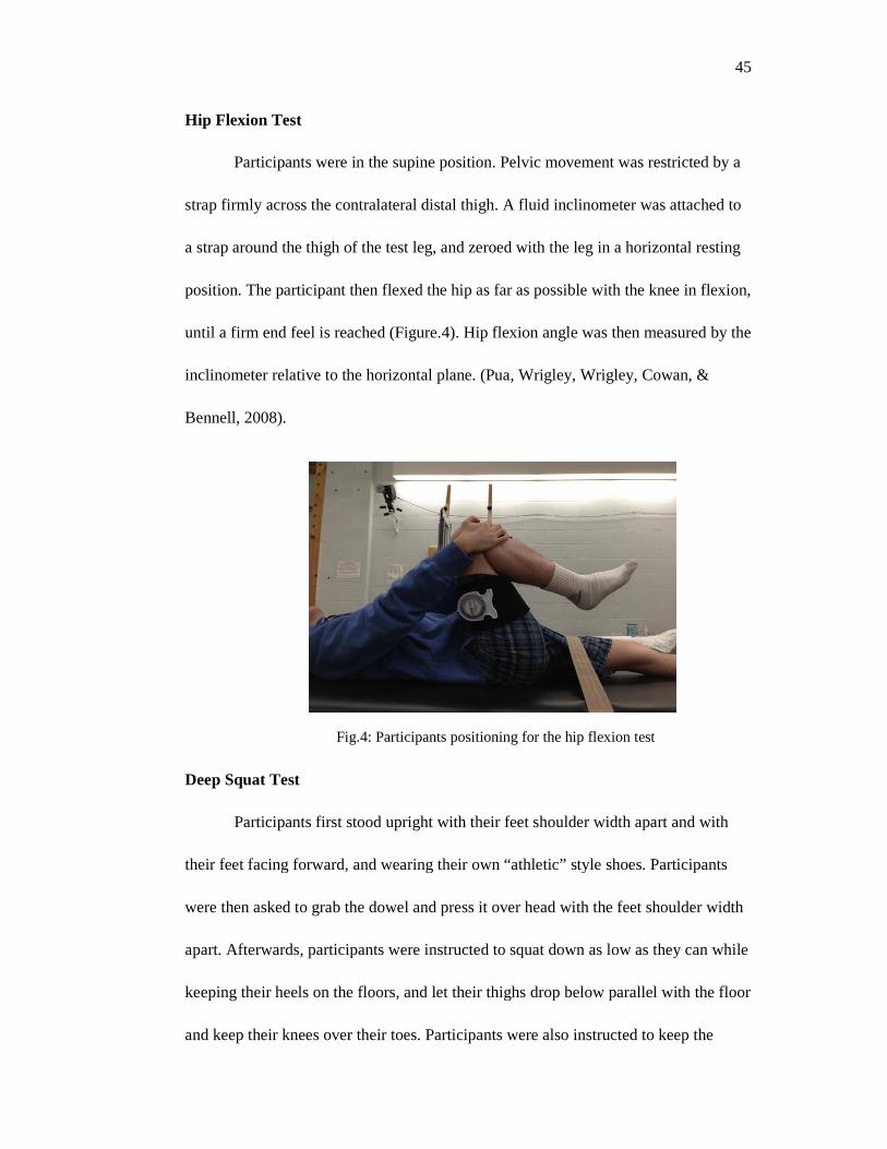
Hip Flexion Test
Participants were
strap firmly across the contralateral distal thigh.
a strap around the thigh of the test leg, and zeroed with the leg in a horizontal resting
position. The participant then
until a firm end feel is reached
inclinometer relative to the horizontal plane.
Bennell, 2008).
Fig
Deep Squat Test
Participants first stood
their feet facing forward, and wearing their own “athletic” style shoes.
were then asked to grab the dowel
apart. Afterwards, participants
keeping their heels on the floor
and keep their knees over their toes
in the supine position. Pelvic movement was restricted by a
strap firmly across the contralateral distal thigh. A fluid inclinometer was
a strap around the thigh of the test leg, and zeroed with the leg in a horizontal resting
then flexed the hip as far as possible with the knee in flexion,
until a firm end feel is reached (Figure.4). Hip flexion angle was then measured by
relative to the horizontal plane. (Pua, Wrigley, Wrigley, Cowan, &
Fig.4: Participants positioning for the hip flexion test
stood upright with their feet shoulder width apart and
their feet facing forward, and wearing their own “athletic” style shoes. Participants
asked to grab the dowel and press it over head with the feet shoulder width
articipants were instructed to squat down as low as they can while
eels on the floors, and let their thighs drop below parallel with
over their toes. Participants were also instructed to keep the
45
restricted by a
was attached to
a strap around the thigh of the test leg, and zeroed with the leg in a horizontal resting
with the knee in flexion,
then measured by the
Pua, Wrigley, Wrigley, Cowan, &
shoulder width apart and with
Participants
and press it over head with the feet shoulder width
to squat down as low as they can while
their thighs drop below parallel with the floor
lso instructed to keep the
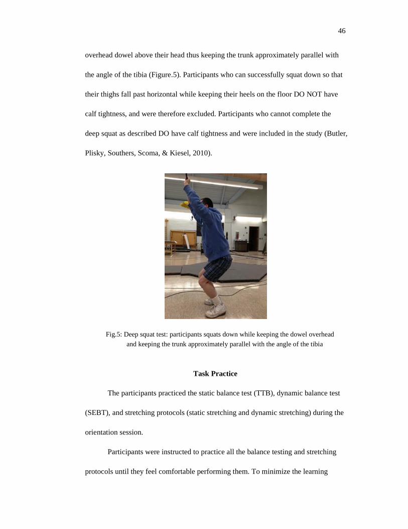
46
overhead dowel above their head thus keeping the trunk approximately parallel with
the angle of the tibia (Figure.5). Participants who can successfully squat down so that
their thighs fall past horizontal while keeping their heels on the floor DO NOT have
calf tightness, and were therefore excluded. Participants who cannot complete the
deep squat as described DO have calf tightness and were included in the study (Butler,
Plisky, Southers, Scoma, & Kiesel, 2010).
Fig.5: Deep squat test: participants squats down while keeping the dowel overhead
and keeping the trunk approximately parallel with the angle of the tibia
Task Practice
The participants practiced the static balance test (TTB), dynamic balance test
(SEBT), and stretching protocols (static stretching and dynamic stretching) during the
orientation session.
Participants were instructed to practice all the balance testing and stretching
protocols until they feel comfortable performing them. To minimize the learning
47
effect of TTB, participants performed 3 practice trails in the single leg stance (30s) for
the test leg with 1 minute rest of periods between each trail in the orientation session
(A. G. Nelson et al., 2011). To minimize the learning effect of SEBT, each participant
performed 6 practice trials in each of the 3 directions on the test leg with 1 minute rest
of periods between each trail in the orientation session (Hertel et al., 2000).
Balance Testing
Participants wore shorts and laboratory sandals during the static balance test.
A standardized sandal method was chosen because it has previously been used to
assess static balance using the time-to-boundary method (Cobb, Joshi, Bazett-Jones,
& Earl-Boehm, 2012). The Star Excursion Balance test was measured with
participant’s barefoot. Balance tests (TTB first, then SEBT) were measured before
and after each of interventions (static stretching, dynamic stretching, control
warm-up).
Time-to-Boundary
Time-to-Boundary was used to assess the static balance. Each participant
performed three trails with 10s of single leg stance as still as possible with eyes closed
on an AMTI force platform (Model OR-6-7-2000, Advanced Mechanical Technology,
Inc, Watertown, MA) to collect ground reaction force data. For all three trials, the
stance foot was meticulously placed in the same position on the force plate that has a
detailed grid on its surface to allow for exact placement. The hands were kept on the
waist, and the opposite leg will be flexed at the hip and knee to approximately 30°.
The data collection began after the participant establishes a stable posture on the force
48
platform. Data was recorded at 100Hz and the mean value of three trails was used for
data analysis. If participants lose their balance and are unable to complete a trial, the
trial will be repeated. A trial will also be repeated if participants open their eyes
during the eye closed condition. Center of Pressure (COP) data was then filtered with
a fourth order zero lag, low pass filter with a cutoff frequency of 5 Hz.
Star Excursion Balance Test
Star Excursion Balance Test (SEBT) was used to assess dynamic balance
based on Hertel et al (2006) study. Athlete tape was placed on the floor to create a “Y”
shaped pattern with 3 lines extending from the center. The 3 lines are named
according to the direction of reach in relation to the stance leg: anterior, posteromedial,
and posterolateral. A crosshairs was drawn at the center of the grid. The most distal
aspect of the great toe was placed at the crosshairs in the center of the grid.
Participants maintained a single-leg stance while the contralateral leg reaches to touch
as far as possible along the each line. Participants touched the furthest point possible
on the line with the most distal part of their reach foot. The reach foot touched the
furthest point on the line as lightly as possible so that the reach leg did not provide
considerable support in the maintenance of upright posture. If it is determined that the
reach leg is used for support or the stable base of support is compromised, the trial
will be performed again. Reach distance was marked with on the tape with a marker
immediately after each trail. Participants then returned to a bilateral stance. The
investigator manually measured the distance in millimeter from the center of the grid
to the touch point with an athletic tape based on the mark. Reach distances were then
49
normalized to participants’ leg length (P. A. Gribble & Hertel, 2003). The order of
reach directions were counterbalanced to avoid order effects from contaminating the
data (Stevens, 2001).
Participants performed 3 trials in each direction and the mean value was used
for data analysis. Ten seconds periods of rest was provided between each trial. Visual
cues and objects on the floor and people in front of the participants were not allowed
in the study to eliminate visual and auditory influences. No encouragement or further
instruction was given to participants throughout the testing (Hertel, Braham, et al.,
2006).
Warm-up Protocols
There were three warm-up interventions (a general warm-up with dynamic
stretching, a general warm-up with static stretching, and a general warm-up alone).
The order of target muscles (quadriceps, hamstrings, and plantar flexors) both for
dynamic stretching and static stretching were randomized. The individual testing
sessions occurred over a three to four week period, with at least 48, but no more than
96 hours between testing. The interventions were counterbalanced to prevent order
bias and learning effect. An attempt was made to test all participants at the same time
of day to be as consistent as possible. During each intervention participants wore their
typical athletic type footwear.
A general warm-up
A general warm-up consisted of 5 minutes of light-jogging on a treadmill at
self-selected comfortable pace was performed by participants before dynamic and

50
static stretching interventions.
Dynamic Stretching
Dynamic stretching consisted of 4 repetitions of bilateral dynamic stretches of
the quadriceps, hamstrings, and plantar flexors (4 repetitions in total x 3 muscle
groups x two limbs) for 30s each and 20s periods of rest. The stretching protocols
based on Behm et al (2011), but dynamic hamstring stretch has been modified to more
directly focus on this muscle group. Participants were asked to achieve the highest
range of motion possible for all dynamic stretches. A description of each dynamic
stretch can be found in Table 1.
Table 1: Dynamic Stretching Protocol
Muscle group Body position Movement Quadriceps Standing Walking “butt kicks”
that causes dynamic
knee flexion and hip
extension
Hamstrings Standing Walking hip flexion
with knee extended that
causes the leg swinging
up to the anterior aspect
of the body
Plantar Flexors Standing facing the wall, hands placed
on the wall at shoulder height. Feet
should be positioned far enough away
from the way to elicit a stretching feeling
in the calf muscles.
Push off or rebound
from the wall to
produce a dynamic
stretch
Static Stretching
Static stretching consisted of 4 repetitions of static stretches for the right and
left quadriceps, hamstrings, and plantar flexors (4 repetitions in total x 3 muscle
groups x two limbs), holding at the point of discomfort for 30s each and 20s periods
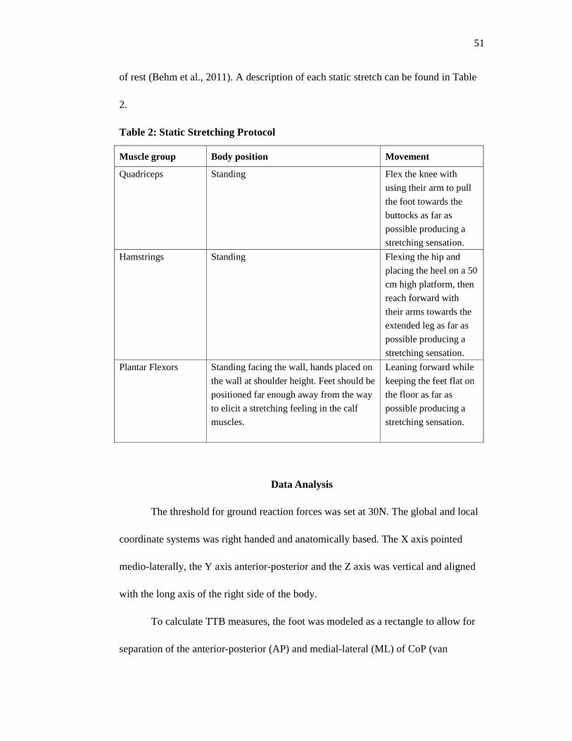
51
of rest (Behm et al., 2011). A description of each static stretch can be found in Table
2.
Table 2: Static Stretching Protocol
Muscle group Body position Movement
Quadriceps Standing Flex the knee with
using their arm to pull
the foot towards the
buttocks as far as
possible producing a
stretching sensation.
Hamstrings Standing Flexing the hip and
placing the heel on a 50
cm high platform, then
reach forward with
their arms towards the
extended leg as far as
possible producing a
stretching sensation.
Plantar Flexors Standing facing the wall, hands placed on
the wall at shoulder height. Feet should be
positioned far enough away from the way
to elicit a stretching feeling in the calf
muscles.
Leaning forward while
keeping the feet flat on
the floor as far as
possible producing a
stretching sensation.
Data Analysis
The threshold for ground reaction forces was set at 30N. The global and local
coordinate systems was right handed and anatomically based. The X axis pointed
medio-laterally, the Y axis anterior-posterior and the Z axis was vertical and aligned
with the long axis of the right side of the body.
To calculate TTB measures, the foot was modeled as a rectangle to allow for
separation of the anterior-posterior (AP) and medial-lateral (ML) of CoP (van
52
Emmerik & van Wegen, 2002). The CoP ML position and velocity was used to
calculate TTB ML. If the CoP ML is moving medially, the distance between CoP ML
and the medial border of the foot will be calculated. This distance was then divided by
the corresponding velocity of CoP ML to calculate the time it would take the CoP ML
to reach the medial border of the foot if it were to continue moving in the same
direction with no acceleration or deceleration. If the CoP ML is moving laterally, the
distance between CoP ML and the lateral border of the foot will be calculated and
divided by the corresponding velocity of CoP ML. Thus, a time series of TTB ML
measures was generated. A time series of corresponding TTB AP measures was
similarly generated by determining the time it would take CoP AP to reach either the
anterior or posterior boundary of the foot (Hertel, Olmsted-Kramer, et al., 2006). The
absolute minimum and mean of minimum samples in the ML and AP direction
represent the temporal margin to the boundary of support and standard deviation of
minimum samples in the ML and AP direction represents its variability (Hertel,
Olmsted-Kramer, et al., 2006).
The distance scores (cm) for each direction of the SEBT was averaged over
the 3 trials and normalized to leg length (reach distance/leg length x 100 = percentage
of leg length).
Statistical Analysis
A 3x2 (warm-up x time) Repeated measures Analysis of Variance (ANOVA)
was used to identify any alteration in the dependent variables. The independent
variables were the three interventions (DS, SS, Control), and time (pre and post).
53
Three separate ANOVA’s was performed on each set of dependent variables: ROM
measures (Hip flexion, knee extension, and ankle dorsiflexion), SEBT measures
(Anterior, posterior-medial, and posterior-lateral), and TTB measures (the absolute
minimum, and standard deviation of minimum in the ML and AP direction). The
alpha level for determining significance was set at ≤ .05 for all calculations and all
statistical analyses were performed with SPSS Version 19.0 (SPSS Inc., Chicago, IL).
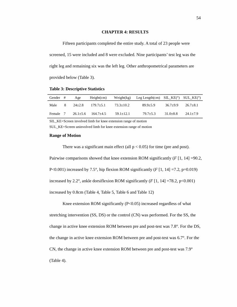
54
CHAPTER 4: RESULTS
Fifteen participants completed the entire study. A total of 23 people were
screened, 15 were included and 8 were excluded. Nine participants’ test leg was the
right leg and remaining six was the left leg. Other anthropometrical parameters are
provided below (Table 3).
Table 3: Descriptive Statistics
Gender # Age Height(cm) Weight(kg) Leg Length(cm) SIL_KE(°) SUL_KE(°)
Male 8 24±2.8 179.7±5.1 73.3±10.2 89.9±5.9 36.7±9.9 26.7±8.1
Female 7 26.1±5.6 164.7±4.5 59.1±12.1 79.7±5.3 31.0±8.8 24.1±7.9
SIL_KE=Screen involved limb for knee extension range of motion
SUL_KE=Screen uninvolved limb for knee extension range of motion
Range of Motion
There was a significant main effect (all p < 0.05) for time (pre and post).
Pairwise comparisons showed that knee extension ROM significantly (F [1, 14] =90.2,
P<0.001) increased by 7.5°, hip flexion ROM significantly (F [1, 14] =7.2, p=0.019)
increased by 2.2°, ankle dorsiflexion ROM significantly (F [1, 14] =78.2, p<0.001)
increased by 0.8cm (Table 4, Table 5, Table 6 and Table 12)
Knee extension ROM significantly (P<0.05) increased regardless of what
stretching intervention (SS, DS) or the control (CN) was performed. For the SS, the
change in active knee extension ROM between pre and post-test was 7.8°. For the DS,
the change in active knee extension ROM between pre and post-test was 6.7°. For the
CN, the change in active knee extension ROM between pre and post-test was 7.9°
(Table 4).
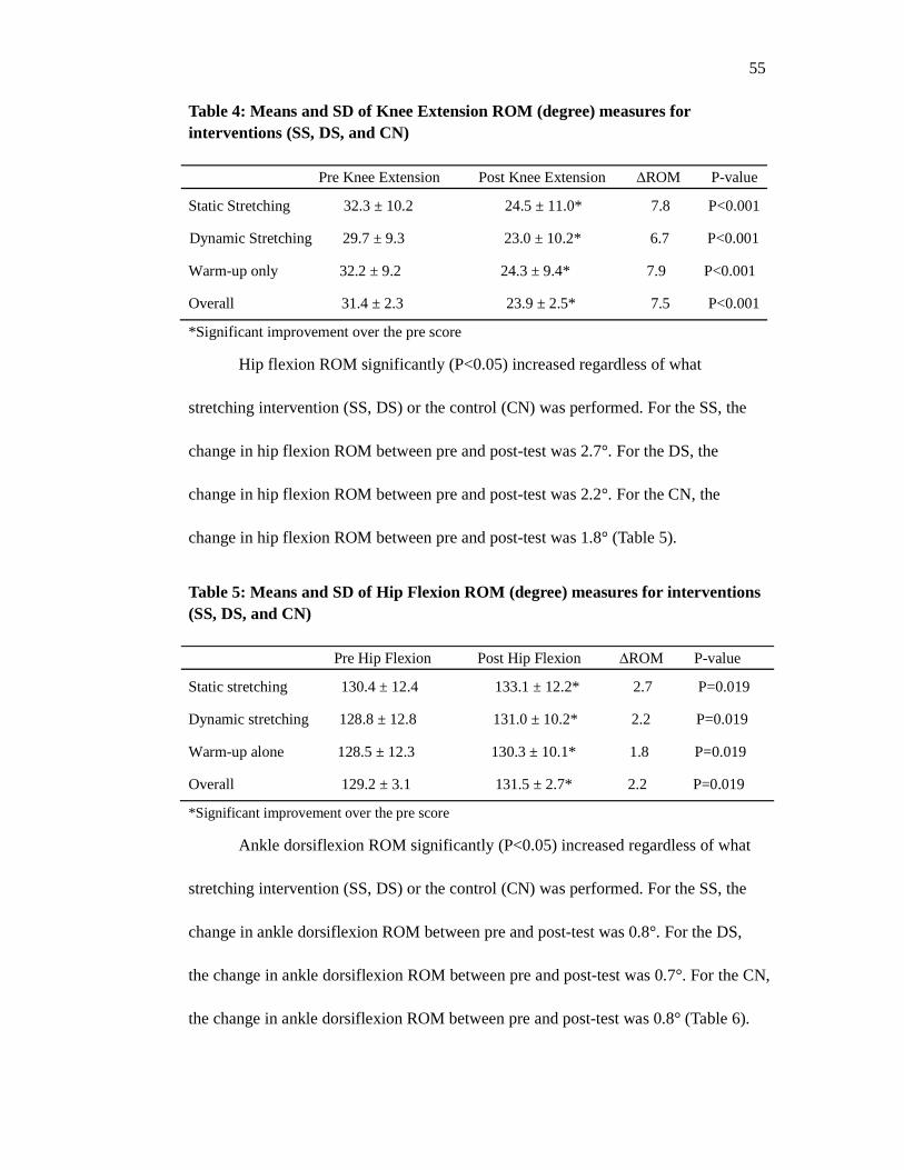
55
Table 4: Means and SD of Knee Extension ROM (degree) measures for interventions (SS, DS, and CN) Pre Knee Extension Post Knee Extension ∆ROM P-value
Static Stretching 32.3 ± 10.2 24.5 ± 11.0* 7.8 P<0.001
Dynamic Stretching 29.7 ± 9.3 23.0 ± 10.2* 6.7 P<0.001
Warm-up only 32.2 ± 9.2 24.3 ± 9.4* 7.9 P<0.001
Overall 31.4 ± 2.3 23.9 ± 2.5* 7.5 P<0.001
*Significant improvement over the pre score
Hip flexion ROM significantly (P<0.05) increased regardless of what
stretching intervention (SS, DS) or the control (CN) was performed. For the SS, the
change in hip flexion ROM between pre and post-test was 2.7°. For the DS, the
change in hip flexion ROM between pre and post-test was 2.2°. For the CN, the
change in hip flexion ROM between pre and post-test was 1.8° (Table 5).
Table 5: Means and SD of Hip Flexion ROM (degree) measures for interventions (SS, DS, and CN) Pre Hip Flexion Post Hip Flexion ∆ROM P-value
Static stretching 130.4 ± 12.4 133.1 ± 12.2* 2.7 P=0.019
Dynamic stretching 128.8 ± 12.8 131.0 ± 10.2* 2.2 P=0.019
Warm-up alone 128.5 ± 12.3 130.3 ± 10.1* 1.8 P=0.019
Overall 129.2 ± 3.1 131.5 ± 2.7* 2.2 P=0.019
*Significant improvement over the pre score
Ankle dorsiflexion ROM significantly (P<0.05) increased regardless of what
stretching intervention (SS, DS) or the control (CN) was performed. For the SS, the
change in ankle dorsiflexion ROM between pre and post-test was 0.8°. For the DS,
the change in ankle dorsiflexion ROM between pre and post-test was 0.7°. For the CN,
the change in ankle dorsiflexion ROM between pre and post-test was 0.8° (Table 6).
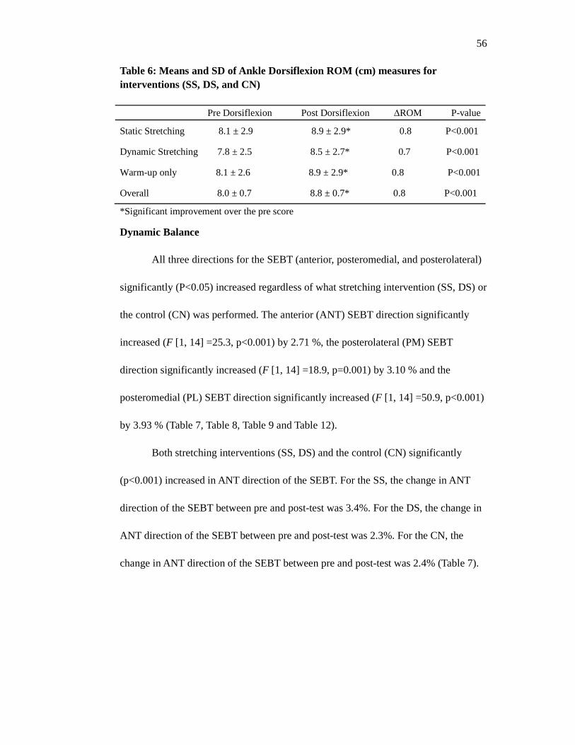
56
Table 6: Means and SD of Ankle Dorsiflexion ROM (cm) measures for interventions (SS, DS, and CN) Pre Dorsiflexion Post Dorsiflexion ∆ROM P-value
Static Stretching 8.1 ± 2.9 8.9 ± 2.9* 0.8 P<0.001
Dynamic Stretching 7.8 ± 2.5 8.5 ± 2.7* 0.7 P<0.001
Warm-up only 8.1 ± 2.6 8.9 ± 2.9* 0.8 P<0.001
Overall 8.0 ± 0.7 8.8 ± 0.7* 0.8 P<0.001
*Significant improvement over the pre score
Dynamic Balance
All three directions for the SEBT (anterior, posteromedial, and posterolateral)
significantly (P<0.05) increased regardless of what stretching intervention (SS, DS) or
the control (CN) was performed. The anterior (ANT) SEBT direction significantly
increased (F [1, 14] =25.3, p<0.001) by 2.71 %, the posterolateral (PM) SEBT
direction significantly increased (F [1, 14] =18.9, p=0.001) by 3.10 % and the
posteromedial (PL) SEBT direction significantly increased (F [1, 14] =50.9, p<0.001)
by 3.93 % (Table 7, Table 8, Table 9 and Table 12).
Both stretching interventions (SS, DS) and the control (CN) significantly
(p<0.001) increased in ANT direction of the SEBT. For the SS, the change in ANT
direction of the SEBT between pre and post-test was 3.4%. For the DS, the change in
ANT direction of the SEBT between pre and post-test was 2.3%. For the CN, the
change in ANT direction of the SEBT between pre and post-test was 2.4% (Table 7).

57
Table 7: Means and SD of normalized anterior (ANT) direction of SEBT for interventions (SS, DS, and CN) Pre ANT SEBT Post ANT SEBT ∆Distance P-value
Static Stretching 77.0 ± 6.6 80.4 ± 8.1* 3.4 P<0.001
Dynamic Stretching 75.8 ± 6.7 78.1 ± 8.1* 2.3 P<0.001
Warm-up only 76.7 ± 8.1 79.1 ± 9.0* 2.4 P<0.001
Overall 76.5 ± 1.7 79.2 ± 2.1* 2.7 P<0.001
*Significant improvement over the pre score
Both stretching interventions (SS, DS) and the control (CN) significantly
(p<0.001) increased in PM direction of SEBT. For the SS, the change in PM direction
of SEBT between pre and post-test was 3.7 %. For the DS, the change in PM direction
of SEBT between pre and post-test was 3.1 %. For the CN, the change in PM
direction of SEBT between pre and post-test was 2.6 % (Table 8).
Table 8: Means and SD of normalized posteromedial (PM) direction of SEBT for interventions (SS, DS, and CN) Pre PM SEBT Post PM SEBT ∆Distance P-value
Static Stretching 112.4 ± 7.5 116.1 ± 7.9* 3.7 P=0.001
Dynamic Stretching 111.5 ± 8.1 114.6 ± 8.6* 3.1 P=0.001
Warm-up only 111.6 ± 7.3 114.2 ± 7.9* 2.6 P=0.001
Overall 111.9 ± 1.8 114.9 ± 2.0* 3.1 P=0.001
*Significant improvement over the pre score
Both stretching interventions (SS, DS) and the control (CN) significantly
(p<0.001) increased in PL direction of the SEBT. For the SS, the change in PL
direction of SEBT between pre and post-test was 5.0 %. For the DS, the change in PL
direction of SEBT between pre and post-test was 3.6 %. For the CN, the change in PL
direction of SEBT between pre and post-test was 3.3 % (Table 9).
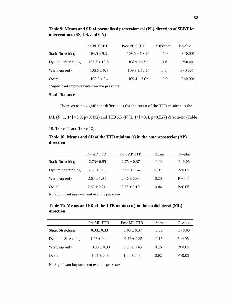
58
Table 9: Means and SD of normalized posterolateral (PL) direction of SEBT for interventions (SS, DS, and CN) Pre PL SEBT Post PL SEBT ∆Distance P-value
Static Stretching 104.5 ± 9.3 109.5 ± 10.4* 5.0 P<0.001
Dynamic Stretching 105.3 ± 10.3 108.9 ± 9.9* 3.6 P<0.001
Warm-up only 106.6 ± 9.4 109.9 ± 10.6* 3.3 P<0.001
Overall 105.5 ± 2.4 109.4 ± 2.6* 3.9 P<0.001
*Significant improvement over the pre score
Static Balance
There were no significant differences for the mean of the TTB minima in the
ML (F [1, 14] =0.8, p=0.402) and TTB AP (F [1, 14] =0.4, p=0.527) directions (Table
10, Table 11 and Table 12).
Table 10: Means and SD of the TTB minima (s) in the anteroposterior (AP) direction Pre AP TTB Post AP TTB ∆time P-value
Static Stretching 2.73± 0.85 2.75 ± 0.87 0.02 P>0.05
Dynamic Stretching 2.69 ± 0.95 2.56 ± 0.74 -0.13 P>0.05
Warm-up only 2.63 ± 1.04 2.86 ± 0.93 0.23 P>0.05
Overall 2.69 ± 0.21 2.73 ± 0.19 0.04 P>0.05
No Significant improvement over the pre score
Table 11: Means and SD of the TTB minima (s) in the mediolateral (ML) direction Pre ML TTB Post ML TTB ∆time P-value
Static Stretching 0.98± 0.33 1.03 ± 0.37 0.05 P>0.05
Dynamic Stretching 1.08 ± 0.44 0.96 ± 0.35 -0.12 P>0.05
Warm-up only 0.95 ± 0.33 1.10 ± 0.43 0.15 P>0.05
Overall 1.01 ± 0.08 1.03 ± 0.08 0.02 P>0.05
No Significant improvement over the pre score
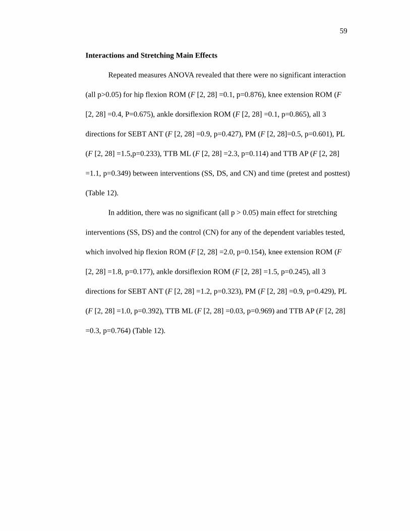
59
Interactions and Stretching Main Effects
Repeated measures ANOVA revealed that there were no significant interaction
(all p>0.05) for hip flexion ROM (F [2, 28] =0.1, p=0.876), knee extension ROM (F
[2, 28] =0.4, P=0.675), ankle dorsiflexion ROM (F [2, 28] =0.1, p=0.865), all 3
directions for SEBT ANT (F [2, 28] =0.9, p=0.427), PM (F [2, 28]=0.5, p=0.601), PL
(F [2, 28] =1.5,p=0.233), TTB ML (F [2, 28] =2.3, p=0.114) and TTB AP (F [2, 28]
=1.1, p=0.349) between interventions (SS, DS, and CN) and time (pretest and posttest)
(Table 12).
In addition, there was no significant (all p > 0.05) main effect for stretching
interventions (SS, DS) and the control (CN) for any of the dependent variables tested,
which involved hip flexion ROM (F [2, 28] =2.0, p=0.154), knee extension ROM (F
[2, 28] =1.8, p=0.177), ankle dorsiflexion ROM (F [2, 28] =1.5, p=0.245), all 3
directions for SEBT ANT (F [2, 28] =1.2, p=0.323), PM (F [2, 28] =0.9, p=0.429), PL
(F [2, 28] =1.0, p=0.392), TTB ML (F [2, 28] =0.03, p=0.969) and TTB AP (F [2, 28]
=0.3, p=0.764) (Table 12).
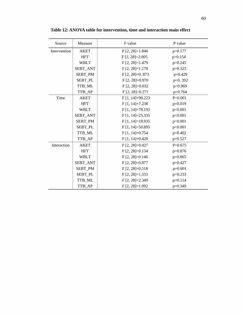
60
Table 12: ANOVA table for intervention, time and interaction main effect
Source Measure F value P value
Intervention
AKET
HFT
WBLT
SEBT_ANT
SEBT_PM
SEBT_PL
TTB_ML
TTB_AP
F [2, 28]=1.846 p=0.177
F [2, 28]=2.005 p=0.154
F [2, 28]=1.479 p=0.245
F [2, 28]=1.178 p=0.323
F [2, 28]=0. 873 p=0.429
F [2, 28]=0.970 p=0. 392
F [2, 28]=0.032 p=0.969
F [2, 28]=0.271 p=0.764
Time
AKET
HFT
WBLT
SEBT_ANT
SEBT_PM
SEBT_PL
TTB_ML
TTB_AP
F [1, 14]=90.223 P<0.001
F [1, 14]=7.238 p=0.019
F [1, 14]=78.193 p<0.001
F [1, 14]=25.335 p<0.001
F [1, 14]=18.935 p=0.001
F [1, 14]=50.895 p<0.001
F [1, 14]=0.754 p=0.402
F [1, 14]=0.420 p=0.527
Interaction
AKET
HFT
WBLT
SEBT_ANT
SEBT_PM
SEBT_PL
TTB_ML
TTB_AP
F [2, 28]=0.427 P=0.675
F [2, 28]=0.134 p=0.876
F [2, 28]=0.146 p=0.865
F [2, 28]=0.877 p=0.427
F [2, 28]=0.518 p=0.601
F [2, 28]=1.533 p=0.233
F [2, 28]=2.349 p=0.114
F [2, 28]=1.092 p=0.349
61
CHAPTER 5: DISCUSSION
The purpose of this study was to examine the effects of static stretching (SS)
versus dynamic stretching (SS) on lower extremity joint range of motion (ROM),
static balance, and dynamic balance. The results of this study indicated that both
stretching interventions (SS, DS), and the control (CN) resulted in a significant
increase on the lower extremity joint ROM (hip flexion, knee extension, and ankle
dorsiflexion) and improvement in dynamic balance in all three directions (anterior,
posteromedial, posterolateral) of the Star Excursion Balance Test (SEBT). There was
no significant difference in static balance, as measured by the Time to boundary (TTB)
measure. In addition, there was no significant interaction between interventions (SS,
DS, and CN) and time (pre and post) meaning that all changes seen in range of motion
and dynamic balance occurred regardless of which intervention (SS, DS, and CN) was
performed. This chapter will first discuss our findings and compare them to the
previous literature, followed by interpretation and explanation of the findings,
limitations, and direction for future research.
Knee Extension Range of Motion
Increased hamstring flexibility is suggested to be an effective way to reduce
the incidence of hamstring strains (Liemohn, 1978), which are one of the most
common injuries experienced in the sports competition or physical activity (Worrell &
Perrin, 1992). In relation to change in ROM of knee extension, our findings showed
that all interventions (SS, DS and CN) resulted in a significant knee extension ROM
increase. It is interesting to note that 13 out of 15 participants would still have been
62
considered “tight” for our inclusion criteria (knee extension ROM >15°) after each
intervention. The less stiff hamstring muscle and more slack connective tissue around
the knee joint following stretching (SS and DS), and improved neuromuscular
performance (enhanced core body temperature and increased muscular activation)
from 5mins jogging warm-up attributed to the increased knee extension ROM.
The observed hamstring flexibility in our results was partly supported by
previous research. Bandy et al (1998) found that both SS (pre 41.9 ± 10.1°, post 39.9
± 6.0°) and DS (pre 30.5 ± 9.1°, post 35.7 ± 6.0°) increased hamstring flexibility
(passive knee extension ROM) but SS increased hamstring flexibility significantly
more than DS (Bandy et al., 1998). This was consistent with our finding to some
extent, which indicated that the change of active knee extension ROM in SS (pre 32.3
± 10.2°, post 24.5 ± 11.0°) was greater than in DS (pre 29.7 ± 9.3°, post 23.0 ± 10.2°),
although no significant difference was found. However, Bandy et al (1998) defined as
tight hamstring as having greater than 30° loss of knee extension, which was greater
than in our study ( >15° met the inclusion criteria). On the other hand, our results
added to the inconclusive findings from previous research. O'Sullivan et al (2009)
revealed that knee extension ROM significantly increased with 5 minutes warm-up,
then further increased with SS but significantly decreased after DS in those with
previous injured hamstring (O'Sullivan et al., 2009). This partly contradicted with our
results that DS significantly increased knee extension ROM rather than a decrease,
and the increase of SS and warm-up alone was almost identical. Moreover, De Weijer
et al (2003) found that warm-up alone only (10 minutes of stair climbing at 70% of
63
maximum heart rate) minimally increase knee extension ROM while the greatest
increase (10.3°) appeared in the warm-up and SS (30s passive static stretches of the
hamstring) group (De Weijer, Gorniak, & Shamus, 2003). Our study found a similar
increase in knee extension ROM following warm-up alone (CN), however, we did not
find any additional increase in knee extension ROM (SS 7.9° vs CN 7.8°) following
SS.
There were some methodological differences between the studies, which may
explain the differences within the results. To begin with, the current study and De
Weijer et al (2003) measured subjects’ knee extension ROM by active knee extension
test (AKET), whereas O'Sullivan et al (2009) and Bandy et al (1998) used passive
knee extension test (PKET). It has been demonstrated that values obtained for knee
extension ROM using PKET and AKET varied by almost 12°, since AKET may only
measure initial hamstrings length whereas PKET measured maximal hamstrings
length (RL Gajdosik, Rieck, Sullivan, & Wightman, 1993). This might have resulted
in the different outcomes. Secondly, the target muscle of stretching was different
within studies. The hamstring was the only main muscle stretched in Bandy et al
(1998), O'Sullivan et al (2009) and De Weijer et al (2003), whereas our study focused
on three main muscle groups: quadriceps, hamstrings, and plantar flexors. Thus, our
stretching protocol might be more effective on the subsequent knee extension ROM
performance after stretching. Thirdly, in order to maximally increase ROM in tight
hamstrings, the duration of SS plays an important role in the subsequent effects.
Bandy & Irion (1994) demonstrated that 30s and 60s of SS were more effective at
64
increasing flexibility of hamstring muscles than stretching for 15s, and 30s of SS was
as effective as the longer duration of 1 minute on the improvement of hamstring
tightness (Handrakis Bandy & Irion, 1994). The duration of SS in our study was 30s,
which was consistent with Behm et al (2011). However, Bandy et al (1998) examined
the effects of hamstring flexibility before and after 6 weeks stretching interventions,
while the current study and other previous research focused on the acute effect of
stretching conditions.
Ankle Dorsiflexion Range of Motion
It has been demonstrated that calf muscle stretching was an effective method
to increase ankle dorsiflexion, which could reduce the symptoms of disorders with
associated with calf muscle tightness (Radford, Burns, Buchbinder, Landorf, & Cook,
2006). Our results showed that all interventions (SS, DS and CN) resulted in a
significant ankle dorsiflexion ROM increase. The increased ankle dorsiflexion ROM
might have been mainly due to the improved calf muscle flexibility resulting from the
stretching interventions (SS, DS) and advantage of faster nerve conduction velocity
following warm-up (jogging). This resulted in more compliant calf muscle and less
ankle joint stiffness. In addition, Samukawa et al (2011) found that a significant distal
displacement of the myotendinous junction was observed by ultrasonography after DS.
Thus, the lengthen ankle plantar flexor muscle–tendon following DS might be another
contributor to increased ankle dorsiflexion (Samukawa, Hattori, Sugama, & Takeda,
2011). Therefore, both factors might be responsible for generating more distance
between the great toe and the wall as measured maximal ankle dorsiflexion ROM in
65
the Weight-bearing Lunge Test.
We chose to use the Weight-bearing Lunge Test (WBLT) to assess dorsiflexion
ROM because it has been thought to more accurately reflect the available ankle
dorsiflexion ROM and more reliable than in a non-weight-bearing position (Bennell et
al., 1998). Most measurement techniques for weight-bearing ankle dorsiflexion ROM
include the use of a standard goniometer (Norkin, 2009), an inclinometer (Cosby &
Hertel, 2011), or a tape measure (Matthew C Hoch & McKeon, 2011). A tape WBLT
measure that was used in our study has been proven to not require the technical
proficiency associated with a goniometer or inclinometer and is more sensitive to
change compared to measures of motion in degrees (Collins, Teys, & Vicenzino,
2004). Therefore comparison of our data to previous studies should be interpreted
with caution.
Previous research found that the combined stretching protocol (running first,
then SS) (pre 18.3 ± 6.2°, post 20.6 ± 5.6°) was more effective than the running only
(pre 18.6 ± 6.6°, post 18.8 ± 6.1°) for increasing ankle dorsiflexion ROM (McNair &
Stanley, 1996), while our study did not find any significant difference within SS
intervention (pre 8.1 ± 2.9 cm, post 8.9 ± 2.9 cm) and the control (warm-up alone)
(pre 8.1 ± 2.6 cm, post 8.9 ± 2.9 cm). The Weight-bearing Lunge test was measured as
electrogoniometer in McNair & Stanley (1996) study, while our study used a tape
measure. In addition, the current study designed a warm-up alone protocol with
self-control comfortable speed jogging on treadmill, which was different with
controlling at 60% maximum heart rate running in McNair & Stanley (1996). This
66
could result in different aerobic metabolism performance. Moreover, Hoch et al (2011)
found that the mean value of the WBLT was 11.9 ± 2.7 cm in healthy population,
which was relatively greater than our finding (SS pre 8.1 ± 2.9 cm, DS pre 7.8 ± 2.5
cm, CN pre 8.1 ± 2.6 cm) (M.C. Hoch et al., 2011). The inclusion criteria of having
tight calf muscle may explain this difference. Youdas and associates indicated that a
30s or 60s per day for 6-weeks SS did not significantly increase active ankle
dorsiflexion ROM in healthy subjects (Youdas, Krause, Egan, Therneau, & Laskowski,
2003). Our study focused on acute effect of SS on ankle dorsiflexion ROM in those
who have tightness in their hamstring and calf muscles, while Youdas et al (2003)
examined a relative longer (6-week) stretching intervention in healthy population
without specific reference to muscle tightness. The effect of SS on a healthy
population ankle dorsiflexion ROM might be not as distinct as in those with tight
muscles.
Hip Flexion Range of Motion
With regard to alternations in ROM of hip flexion, our results showed that all
stretching interventions (SS, DS and CN) resulted in a significant hip flexion ROM
increase. The improved hip ROM was mainly due to the increased hamstring
flexibility following stretching interventions (SS and DS). In addition, enhanced body
temperature after warm-up might also result in a beneficial effect.
The pre value of hip flexion ROM in our finding (SS 130.4 ± 12.4°, DS 130.4
± 12.4°, CN 128.5 ± 12.3°) was all greater than in Pua et al (2008) (118.8±15.9°)
(Pua et al., 2008). However, Pua et al (2008) focused on those with hip osteoarthritis
67
that might have greater limitation of hip flexion ROM than the young healthy subjects
in our study.
Our results were consistent with Godges et al (1989), who compared the two
stretching techniques (Static stretching and Proprioceptive neuromuscular facilitation)
to determine which was most effective for improving hip ROM. SS resulted in
significant improvement in hip flexion ROM (Godges, MacRae, Longdon, Tinberg, &
Macrae, 1989), which was similar to our study. Our finding was also supported by
Cipriani et al (2003)’s research, which demonstrated significant gains in ROM for hip
flexion over the 6 weeks training (2 minutes stretching twice daily), although they
only stretched hamstring muscles (Cipriani, Abel, & Pirrwitz, 2003).
However, there were no significant changes in flexibility as a result of either
warm-up in Young et al (2004), whose protocol involved five minutes of
sub-maximum running followed by seven practice kicks and following 4.5 minutes SS
of the hip flexors and quadriceps. This could be resulted from different warm-up
protocol design. Young et al (2004) added practice kicks while our protocol did not
involve them. In addition, hip ROM in Young et al (2004) was measured in hip
extension using a modified Thomas test, which may not have been sensitive to
estimate the acute change in flexibility from warm-up and stretching (W Young,
Clothier, Otago, Bruce, & Liddell, 2004).
Dynamic Balance (SEBT)
With respect to improvement in dynamic balance, our results showed that all
interventions (SS, DS and CN) resulted in a significant increase in three directions
68
(ANT, PM, and PL) of SEBT, which indicated that dynamic balance performance was
improved despite no difference occurred within interventions. The possible reason
behind this might be due to a desensitized stretch reflex after an increased muscle and
joint flexibility following stretching. As a result, a less responsive stretch reflex could
suppress the postural deviations, thus make it easier to establish dynamic equilibrium
(A. G. Nelson et al., 2011).
For the ANT direction of the SEBT, the pre value of our results (SS pre 77.0 ±
6.6 %, DS pre 75.8 ± 6.7%, and CN pre 76.7 ± 8.1%) was slightly smaller than Hertel
et al (2006) finding in healthy subjects (79 ± 12 %). However, the pre value of the PM
(SS pre 112.4 ± 7.5%, DS pre 111.5 ± 8.1%, CN pre 111.6 ± 7.3%) and PL (SS
pre104.5 ± 9.3%, DS pre 105.3 ± 10.3%, CN pre 106.6 ± 9.4%) directions of the
SEBT was both greater than Hertel et al (2006) finding (PM 90 ± 13%, PL 81 ± 13%)
(Hertel, Braham, et al., 2006). This comparison is interpreted as despite the
participants in our study having tight calf and hamstring muscles, their dynamic
balance performance was similar to previously reported healthy subjects.
To better understand the relationship between increased joint ROM and
increased SEBT performance seen in our study, we conducted a post-hoc correlational
analysis of these variables. None of the pre-test ROM measurements were
significantly correlated with the SEBT reach distance in any direction (Appendix G).
Previously, Hoch et al (2011) examined the relationships between dorsiflexion range
of motion on the WBLT and normalized reach distance in three directions on the
SEBT in healthy subjects (M.C. Hoch et al., 2011). They found that the ANT direction
69
of the SEBT (mean: 79.0 ± 5.8%) was significantly correlated to the WBLT (mean:
11.9 ± 2.7 cm; r = 0.53, r²= 0.28, p = 0.001) and dorsiflexion ROM accounted for an
estimated 28% of the variance in ANT reach, while there were no significant
correlations between the WBLT and the PM direction (mean: 90.0 ± 9.1%; r = 0.21, r²
= 0.04, p = 0.23) or the PL direction (mean: 82.0 ± 13.1%; r = 0.22, r²= 0.05, p =
0.20). However, our results did not find any significant correlation between the
dorsiflexion ROM and 3 normalized reach distances, which was consistent with
previous research (P. A. Gribble & Hertel, 2003). One thing need to be noted that the
subjects in our study were those with tight calf and hamstring muscles, and these
participants may differ from “typical healthy” participants in terms of mechanical
properties of the muscle, muscle-tendon, and connective tissue in the lower extremity.
Therefore, the tight muscle might limit the relationship between dorsiflexion ROM
and the SEBT performance in our study. A new contribution to the literature on SEBT
performance is that it does not appear to be related to available joint ROM in hip
flexion, knee extension, or dorsiflexion.
A second set of post-hoc correlations was performed to determine if there was
a relationship between the amount of ROM gained following the intervention
(∆Pre-Post ROM) and the improvement in SEBT score (∆Pre-Post reach distance).
Results of this analysis indicated that the gained hip flexion ROM was significantly
correlated with the improvement PM direction of the SEBT for the DS intervention (r
= 0.57, r²= 0.32, p = 0.03) (Appendix G). This is not surprise since previous research
has shown that hip flexion alone accounted for 88.6% and 94.5% of the variance in
70
the PM and PL directions, respectively (Robinson & Gribble, 2008). The additional
hip flexion ROM may contribute to the improved SEBT by allowing lower center of
mass to produce greater potential leg reach distance. No significance was found for
the remaining correlation between the increased ROM and the improved SEBT.
Previous research examined the effects of SS on dynamic balance using
different dynamic balance measurements, Biodex Medical System (BBS) (Pablo B
Costa, Barbara S Graves, Michael Whitehurst, & Patrick L Jacobs, 2009) , Dynamic
Stability Index (DSI) , and stabilometer (A. G. Nelson et al., 2011), but none have
used the SEBT. Therefore, comparison of our finding to previous research should be
illustrated with caution.
Our findings agreed with Costa et al (2009) research, who evaluated the
effects of different durations (15s and 45s) of SS on dynamic balance on young
women. The SS protocols was based on Behm et al (2004) but involved with 15s and
45s duration. Dynamic balance was measured as using the Biodex Medical System,
which was similar to actual physical activities that resulted in postural instability. A
warm-up on a cycle ergometer at 70 rpm for 5 minutes was performed before each
condition. The results of this study indicated that SS of 45s did not adversely affect
dynamic balance and 15s of SS improved dynamic balance. This suggested that
shorter duration of SS (15s) might be more effective on dynamic balance
improvement, however, our SS protocol resulted in improvement in the 3 directions of
the SEBT utilizing a 30s duration SS protocol. Future study need to further compare
with the different duration of SS under various dynamic balance measurements. In
71
addition, they did not find any significant change in the control condition (warm-up
alone), although they used a similar cycle warm-up protocol as Behm et al (2004).
Dynamic Stability Index (DSI) was another dynamic balance measurement
that has been used to test dynamic postural control using a single-leg stance. Smaller
DSI meant improved dynamic balance while greater DSI indicated opposite effect
(Handrakis et al., 2010). Handrakis et al (2010) found that DSI of SS group (no
aerobic warm-up, SS alone) was significantly smaller than that in the NS group (no
aerobic warm-up). However, their recruited subjects were from martial arts school,
which was quite different from healthy recreationally active individuals who have not
experienced specific martial or exercise training in the current study.
In comparison with non-balance trained individuals with experienced balance
trainers, Nelson et al (2011) found that balance time for non-balance trained
individuals improved significantly by 11.4% (2s increase), but no significant change
in the experienced balance trainers (surfers). Balance testing was performed on a
stabilometer following either 30 min of quiet sitting or 30 min of SS protocols (20
mins stretching and 10 mins relax, no aerobic warm-up for both groups) (A. G.
Nelson et al., 2011).
All previous studies focused on the effect of SS on dynamic balance, none of
them focused on the effect of DS on dynamic balance. Our study, therefore, added
preliminary data to understand the effects of DS on dynamic balance (SEBT)
performance, and based on these data neither stretching condition had a significant
effect on dynamic balance.
72
Static Balance (TTB)
In relation to modification in static balance, our results showed that none of the
interventions (SS, DS and CN) had a significant effect on the mean of TTB minima in
the anterior-posterior (AP) and in the medio-lateral (ML) directions. This meant that
all three interventions (SS, DS, and CN) had no effect on static balance. The pre
values of the mean of TTB AP (SS 2.73 ± 0.85, DS 2.69 ± 0.95, CN 2.63 ± 1.04) and
TTB ML minima (SS 0.98 ± 0.33, DS 1.08 ± 0.44, CN 0.95 ± 0.33) in our study were
all relatively smaller than from Mckeon et al (2008) finding (TTB AP 5.32 ± 1.77 and
ML 1.84 ± 0.53) in those with a history of chronic ankle instability (CAI) (Mckeon et
al., 2008). The mean of the TTB minima for the ML and the AP directions represents
the measurement of TTB magnitude, which indicates the times where the
sensorimotor system had the least time to make a postural correction to maintain
single leg stance over the base of support (Hertel, Olmsted-Kramer, et al., 2006). Thus,
our results indicated that the subjects with tight calf and hamstring muscle was more
prone to postural instability than those with CAI. One point should be note that the
DS intervention resulted in a tendency to decrease TTB AP (pre 2.69 ± 0.95s vs post
2.56 ± 0.74s) and TTB ML (pre 1.08 ± 0.44s vs post 0.96 ± 0.35s), while SS and CN
increased in TTB AP (SS pre 2.73± 0.85s vs post 2.75 ± 0.87s; CN pre 2.63 ± 1.04s vs
2.86 ± 0.93s) and TTB ML (SS pre 0.98 ± 0.33s vs post 1.03 ± 0.37s; CN pre 0.95 ±
0.33s vs post 1.10 ± 0.43s), although there was no significant difference. Since we
hypothesized a lower TTB measure indicated greater postural instability (Hertel,
Olmsted-Kramer, et al., 2006), DS might negatively affect static balance.
73
The first possible explanation of observed finding on static balance might be
that the current DS protocol (5 min warm-up plus 4 repetitions with 30s of bilateral
DS of the quadriceps, hamstrings, and plantar flexors) might cause fatigue for subjects,
which resulted in a relatively lower TTB value since fatigue has been previously
proven to adversely affect balance (Vuillerme, Burdet, Isableu, & Demetz, 2006).
This is because the slow rate of firing of muscle spindles and reflex receptors caused
by fatigue could result in the slow nerve transmission rate from CNS to maintain the
center of gravity within their base of support, thus static balance. Therefore, the
positive effect of DS on static balance that we hypothesized might be compensated by
fatigue factor. In addition, since sensory systems (vision and vestibular) was thought
to maintain static postural control (L. M. Nashner, 1981), the fact that no significant
difference was found in static balance might be due to the role of sensory systems in
regulating the static postural control greater than improved neuromuscular
performance resulted from stretching interventions (SS, DS) or a general warm-up.
Our finding was supported by Lewis et al (2009), who utilized a
comprehensive balance measurement, consisting of Postural Evoked Response Test,
Adaptation Test, Motor Control Test, Sensory Organization Test, and Unilateral
Stance Test to assess the effect of SS on postural control without any aerobic warm-up
component. No significant effect of lower extremity stretching on postural control
was detected (Lewis et al., 2009). Conversely, after evaluating the effect of an acute
SS on balance, force, proprioception, reaction time and movement time, Behm et al
(2004) found that an acute SS adversely affected static balance performance
74
(decreasing for 9.2%) and reaction/movement time (increasing for 4.0% and 1.9%),
while the control condition (warm-up alone) increased the balance score for 17.3%
and decreased reaction/movement time for 5.8% and 5.7% (Behm et al., 2004). Both
conditions involved a 5-min cycle on a cycle ergometer at 70 rpm with 1-kp resistance
warm-up. Thus, the obtained positive effect might be due to the enhanced body
temperature physiological benefits after cycle warm-up. The static balance was
measured as a computerized 30s wobble board test. Since the wobble board involved
unanticipated perturbations to equilibrium and was multidirectional that could be a
more complex task, it might be more difficulty to maintain static postural control
compared with TTB that participants stood on a stable platform in our study. In
addition, Nagano and his associate also indicated that stretching of the calf muscles
has the effect on increasing postural sway (Nagano et al., 2006). Future research need
to determine if stretching could alter sensory systems, which is vital important in
those sports that static postural control plays in a critical role.
The control group of our study showed no significant improvement in static
balance with SS. The reason might be that the negative effects of SS on static balance
that we hypothesized was diluted by a positive effect of a jogging component of the
warm-up (Warren Young & Elliott, 2001). The jogging that the current study involved
is a common warm-up section. Based on Behm et al (2004), it could be speculated
that in the absence of the 5 min of jogging warm-up, the static balance performance
might have been decreased to a greater extent. Therefore, future studies should
consider avoiding active warm-up influence when designing a stretching protocol.
75
Mechanisms Relating Stretching to Range of Motion and Balance
The results of this study indicated that all interventions (SS, DS, and CN)
resulted in a significant increase on the lower extremity joint ROM (hip flexion, knee
extension, and ankle dorsiflexion) and improvement in dynamic balance. Although
the mechanisms responsible for the increases in balance performance following
stretching have not been thoroughly investigated, several mechanisms based on
previous research will be discussed with the current findings.
One explanation for our findings of both increased ROM and improved
dynamic balance is that all three of the interventions included a general aerobic
warm-up. The observed benefits of improved neuromuscular performance might also
be due to elevated muscle and body temperature (Fletcher & Jones, 2004). The
similarity of warm-up and DS is the aerobic nature of the task, which could allow for
an increase in body temperature. This would positively affect the force-velocity and
length-tension relationships, enhance nerve receptor sensitivity, and nerve conduction
velocity (Morrin & Redding, 2013; Worrell, Smith, & Winegardner, 1994). One
explanation for our findings of both increased ROM and improved dynamic balance is
that all three of the interventions included a general aerobic warm-up. Although body
temperature was not measured, it is possible that even a small change in temperature
led to the positive effects that have been previously described in the literature.
Our results contradicted the previous mechanism that stiffer muscle producing
a greater reflex response resulted in greater or more rapid responses to slight
perturbations in muscle length, thus better dynamic balance performance (Petit et al.,
76
1990), since our data reached the opposite direction that less stiff muscle resulted
from stretching (SS or DS) or jogging warm-up might contribute to the beneficial
effect on dynamic balance. In addition, the current data also questioned Behm et al
(2004)’s view with related to the alteration in musculotendinous (MTU) influence on
static balance. The increase ROM is commonly due to increase the length and
decrease stiffness of MTU, which incorporates the muscle, tendon, and other
associated connective tissue (G. J. Wilson et al., 1994), following stretching (G.
Wilson et al., 1992). A more compliant MTU might decrease the rate of force
transmission and the rate at which changes in muscle length or tension detected by the
Golgi tendon organs (GTO) (Bishop, 2003a). As a result, it might decrease the ability
of stretch receptors to provide proprioceptive input, thus negatively affecting static
balance, reaction and movement times (Behm et al., 2004). However, our results did
not find any change on static balance (TTB) after stretching (SS and DS). Therefore,
our data do not explicitly support either one of these proposed relationship between
stretching and balance.
Our finding that both stretching interventions (SS and DS) and the control
resulted in increased dynamic balance performance agreed with Nelson et al (2011)’s
theory as mentioned before, which suggested that the enhanced ability to maintain
dynamic balance after an increased flexibility resulted from stretching was due to a
desensitized stretch reflex (Nelson et al., 2011). As a result, a less responsive stretch
reflex could suppress the postural deviations, enhance the proprioceptive input, and
thus make it easier to establish dynamic equilibrium. This view was further supported
77
by Ghaffarinejad et al (2007), who suggested that knee joint position sense improved
following SS due to increased proprioceptive sense (Ghaffarinejad et al., 2007).
Our data demonstrate that regardless of the mode of stretching performed
ROM and dynamic balance improved. We did not directly measure MTU stiffness, so
these comparisons are made with caution. Additional research is needed to more
clearly understand the relationship between altered ROM, MTU stiffness, and
balance.
Limitations and Directions for Future Research
One of the limitations of this study was the possibility of a learning effect,
particular for the SEBT measurements. We used a standard protocol that has been
established to minimize the potential for a learning effect (Hertel, Braham, et al.,
2006). Other strategies to control this were the questionnaire, orientation session and
practice trials. Despite these efforts it is possible that participants improved their
SEBT scores from practice alone. The fact that all stretching interventions improved
the SEBT scores similarly could indicate that a learning effect was present.
Previous study indicated that the combined stretching protocol consisting of
SS and DS displayed significantly greater changes in hamstring muscle ROM than DS
and further showed lower COP movement compared to SS and NS (Morrin &
Redding, 2013). However, the current study did not examine the effects of combined
stretching (SS and DS) on ROM and balance. Therefore, the finding of this study was
limited to compare with the effect of the combined stretching protocol.
Our results are also limited to the acute effects of stretching, no conclusion
78
was made in terms of the long-term effect. Further research need to compare the
difference between acute and long-term effect with SS and DS.
Although several mechanisms have been proposed to illustrate the relationship
of stretching on ROM and balance, additional research is needed to further examine
the exact mechanism to thoroughly explain the alternations in ROM and balance
performance after stretching interventions (DS and SS).
The control group of our study showed a significant improvement in ROM and
SEBT performance and there was no additional improvement with SS or DS. The
reason might be that the effects of SS was diluted by a positive effect of a jogging
warm-up (Warren Young & Elliott, 2001). Therefore, future studies should consider
avoiding active warm-up influence.
The subjects in the current study were those who are recreationally active
individuals with hamstring and calf muscle tightness, future research need to
investigate if the finding of this study would apply to general population, athletes, or
patients with specific disorder.
Our study used a practical combination of lower extremity stretches, which
was considered to be a common stretching routine performed before exercising or
participating in an athletic event. The duration and number of repetitions were
consistent with Behm et al (2011). Future research need to comprehensively compare
the effects of different designed stretching protocols.

79
Conclusion
The results of this study indicated that all interventions (SS, DS, and control
(CN)) resulted in a significant increase on the lower extremity joint ROM (hip flexion,
knee extension, and ankle dorsiflexion) and improvement in dynamic balance,
meaning that all alterations observed ROM and dynamic balance occurred regardless
of which stretching intervention was conducted.
Although recent studies have demonstrated that SS reduced force, strength and
power production, the results of our study did not find any negative effect with regard
to SS. In addition, our finding added preliminary data to begin to understand any
potential effects of DS on dynamic balance performance.
The clinical significance of this study will add the body knowledge that will
allow coaches, athletic trainers, and fitness professionals to make evidence based
decisions on how to prepare the individuals for utilizing a proper stretching technique
during warm-up session, especially in those sports that static or dynamic postural
control plays in a critical role. Based on our data, it appears that a general warm-up
period followed by either SS or DS will have a positive effect on joint ROM and
dynamic balance. The findings of the current study also may inform future research
that focus on lower extremity functional balance rehabilitation with specific stretching
technique, particularly for those who with tight hamstring or calf muscle patients. The
scientific impact of this study is that future studies should attempt to consider the
mechanisms behind each intervention separately (i.e. MTU stiffness, body
temperature, proprioception, etc.) in order to more specifically understand the

80
relationship between stretching, balance, and joint ROM. In addition, future studies
should consider avoiding active warm-up influence when designed a stretching
protocol.
81
References
Aruin, A.S., Forrest, W.R., & Latash, M.L. (1998). Anticipatory postural adjustments in conditions of postural instability. Electroencephalography and Clinical Neurophysiology/Electromyography and Motor Control, 109(4), 350-359.
Bandy, W.D., Irion, J.M., & Briggler, M. (1997). The effect of time and frequency of static stretching on flexibility of the hamstring muscles. Physical therapy, 77(10), 1090-1096.
Bandy, W.D., Irion, J.M., & Briggler, M. (1998). The effect of static stretch and dynamic range of motion training on the flexibility of the hamstring muscles. Journal of Orthopaedic and Sports Physical Therapy, 27, 295-300.
Beedle, B.B., & Mann, C.L. (2007). Acomparison of Two Warm-Ups on Joint Range of Motion. The Journal of Strength & Conditioning Research, 21(3), 776-779.
Behm, D.G., Bambury, A., Cahill, F., & Power, K. (2004). Effect of acute static stretching on force, balance, reaction time, and movement time. Medicine and science in sports and exercise, 36, 1397-1402.
Behm, D.G., Plewe, S., Grage, P., Rabbani, A., Beigi, H.T., Byrne, J.M., & Button, D.C. (2011). Relative static stretch-induced impairments and dynamic stretch-induced enhancements are similar in young and middle-aged men. Applied Physiology, Nutrition, and Metabolism, 36(6), 790-797.
Bennell, Kim, Talbot, R, Wajswelner, H, Techovanich, W, Kelly, D, & Hall, AJ. (1998). Intra-rater and inter-rater reliability of a weight-bearing lunge measure of ankle dorsiflexion. Australian Journal of Physiotherapy, 44, 175-180.
Bishop, D. (2003a). Warm up I: potential mechanisms and the effects of passive warm up on exercise performance. Sports Medicine, 33(6), 439-454.
Bishop, D. (2003b). Warm up II: performance changes following active warm up and how to structure the warm up. Sports Medicine, 33(7), 483-498.
Bloem, BR, Allum, JH, Carpenter, MG, & Honegger, F. (2000). Is lower leg proprioception essential for triggering human automatic postural responses? Experimental brain research. Experimentelle Hirnforschung. Expérimentation cérébrale, 130(3), 375.
Bradley, P.S., Olsen, P.D., & Portas, M.D. (2007). The effect of static, ballistic, and proprioceptive neuromuscular facilitation stretching on vertical jump performance. Journal of Strength and Conditioning Research, The, 21(1), 223.
82
Bressel, E., Yonker, J.C., Kras, J., & Heath, E.M. (2007). Comparison of static and dynamic balance in female collegiate soccer, basketball, and gymnastics athletes. Journal of athletic training, 42(1), 42.
Butler, R.J., Plisky, P.J., Southers, C., Scoma, C., & Kiesel, K.B. (2010). Biomechanical analysis of the different classifications of the Functional Movement Screen deep squat test. Sports Biomechanics, 9(4), 270-279.
Chaouachi, A., Chamari, K., Wong, P., Castagna, C., Chaouachi, M., Moussa-Chamari, I., & Behm, DG. (2008). Stretch and sprint training reduces stretch-induced sprint performance deficits in 13-to 15-year-old youth. European journal of applied physiology, 104(3), 515-522.
Chiu, LZ, Fry, A.C., Weiss, L.W., Schilling, B.K., Brown, L.E., & Smith, S.L. (2003). Postactivation potentiation response in athletic and recreationally trained individuals. Journal of strength and conditioning research/National Strength & Conditioning Association, 17(4), 671.
Chong, R.K.Y., & Do, M.C. (2002). Interaction between transient change in local muscle structure and segmented leg motor responses. Perceptual and motor skills, 95(1), 155-162.
Cipriani, Daniel, Abel, Bobbie, & Pirrwitz, Dayna. (2003). A comparison of two stretching protocols on hip range of motion: implications for total daily stretch duration. The Journal of Strength & Conditioning Research, 17(2), 274-278.
Cobb, Stephen C, Joshi, Mukta N, Bazett-Jones, David M, & Earl-Boehm, JE. (2012). The effect of boundary shape and minima selection on single limb stance postural stability. Journal of applied biomechanics, 28(5), 608-615.
Collins, Natalie, Teys, Pamela, & Vicenzino, Bill. (2004). The initial effects of a Mulligan's mobilization with movement technique on dorsiflexion and pain in subacute ankle sprains. Manual therapy, 9(2), 77-82.
Cornwell, A., Nelson, A.G., Heise, G.D., & Sidaway, B. (2001). Acute effects of passive muscle stretching on vertical jump performance. Journal of Human Movement Studies, 40(4), 307.
Cosby, Nicole L, & Hertel, Jay. (2011). Relationships between measures of posterior talar glide and ankle dorsiflexion range of motion. Athl Train Sports Health Care, 3(2), 76-85.
Costa, P.B., Graves, B.S., Whitehurst, M., & Jacobs, P.L. (2009). The acute effects of different durations of static stretching on dynamic balance performance. The Journal of Strength & Conditioning Research, 23(1), 141.
83
Costa, Pablo B, Graves, Barbara S, Whitehurst, Michael, & Jacobs, Patrick L. (2009). The acute effects of different durations of static stretching on dynamic balance performance. The Journal of Strength & Conditioning Research, 23(1), 141-147.
De Vries, H.A. (1962). Evaluation of static stretching procedures for improvement of flexibility. Res Q, 33(2), 222-229.
De Weijer, Volkert C, Gorniak, Gerard C, & Shamus, Eric. (2003). The effect of static stretch and warm-up exercise on hamstring length over the course of 24 hours. The Journal of orthopaedic and sports physical therapy, 33(12), 727-733.
Di Giulio, I., Maganaris, C.N., Baltzopoulos, V., & Loram, I.D. (2009). The proprioceptive and agonist roles of gastrocnemius, soleus and tibialis anterior muscles in maintaining human upright posture. The Journal of Physiology, 587(10), 2399-2416.
Diener, HC, Dichgans, J., Guschlbauer, B., & Mau, H. (1984). The significance of proprioception on postural stabilization as assessed by ischemia. Brain research, 296(1), 103-109.
Faigenbaum, A.D., Bellucci, M., Bernieri, A., Bakker, B., & Hoorens, K. (2005). Acute effects of different warm-up protocols on fitness performance in children. J Strength Cond Res, 19(2), 376-381.
Filipa, A., Byrnes, R., Paterno, M.V., Myer, G.D., & Hewett, T.E. (2010). Neuromuscular training improves performance on the star excursion balance test in young female athletes. The Journal of orthopaedic and sports physical therapy, 40(9), 551.
FitzgeralD, D.D., Trakarnratanakul, N., Smyth, B., & Caulfield, B. (2010). Effects of a wobble board-based therapeutic exergaming system for balance training on dynamic postural stability and intrinsic motivation levels. J Orthop Sports Phys Ther, 40(1), 11-19.
Fletcher, I.M. (2010). The effect of different dynamic stretch velocities on jump performance. European journal of applied physiology, 109(3), 491-498.
Fletcher, I.M., & Anness, R. (2007). The acute effects of combined static and dynamic stretch protocols on fifty-meter sprint performance in track-and-field athletes. The Journal of Strength & Conditioning Research, 21(3), 784-787.
Fletcher, I.M., & Jones, B. (2004). The effect of different warm-up stretch protocols on 20 meter sprint performance in trained rugby union players. Journal of Strength and Conditioning Research, 18, 885-888.
84
Fowles, JR, Sale, DG, & MacDougall, JD. (2000). Reduced strength after passive stretch of the human plantarflexors. Journal of Applied Physiology, 89(3), 1179-1188.
Gajdosik, R., & Lusin, G. (1983). Hamstring Muscle Tightness Reliability of an Active-Knee-Extension Test. Physical Therapy, 63(7), 1085-1088.
Gajdosik, RL, Rieck, MA, Sullivan, DK, & Wightman, SE. (1993). Comparison of four clinical tests for assessing hamstring muscle length. The Journal of orthopaedic and sports physical therapy, 18(5), 614.
Ghaffarinejad, F., Taghizadeh, S., & Mohammadi, F. (2007). Effect of static stretching of muscles surrounding the knee on knee joint position sense. British journal of sports medicine, 41(10), 684-687.
Godges, JJ, MacRae, H, Longdon, C, Tinberg, C, & Macrae, PG. (1989). The effects of two stretching procedures on hip range of motion and gait economy. The Journal of orthopaedic and sports physical therapy, 10(9), 350.
Gollhofer, A., & Rapp, W. (1993). Recovery of stretch reflex responses following mechanical stimulation. European journal of applied physiology and occupational physiology, 66(5), 415-420.
Gottlieb, G.L., & Agarwal, G.C. (1979). Response to sudden torques about ankle in man: myotatic reflex. Journal of Neurophysiology, 42(1), 91-106.
Gribble, P.A., & Hertel, J. (2003). Considerations for normalizing measures of the star excursion balance test. Measurement in physical education and exercise science, 7(2), 89-100.
Gribble, P.A., Hertel, J., Denegar, C.R., & Buckley, W.E. (2004). The effects of fatigue and chronic ankle instability on dynamic postural control. Journal of Athletic Training, 39(4), 321.
Gribble, P.A., Hertel, J., & Plisky, P. (2012). Using the Star Excursion Balance Test to Assess Dynamic Postural-Control Deficits and Outcomes in Lower Extremity Injury: A Literature and Systematic Review. Journal of Athletic Training, 47(3), 339-357.
Gribble, P.A., Robinson, R.H., Hertel, J., & Denegar, C.R. (2009). The effects of gender and fatigue on dynamic postural control. Journal of sport rehabilitation, 18(2), 240.
85
Gribble, PA, Hertel, J., & Denegar, CR. (2007). Chronic ankle instability and fatigue create proximal joint alterations during performance of the Star Excursion Balance Test. International journal of sports medicine, 28(3), 236.
Grigg, P. (1994). Peripheral neural mechanisms in proprioception. J Sport Rehabil, 3(1), 2-17.
Hale, S.A., Hertel, J., & Olmsted-Kramer, L.C. (2007). The effect of a 4-week comprehensive rehabilitation program on postural control and lower extremity function in individuals with chronic ankle instability. The Journal of orthopaedic and sports physical therapy, 37(6), 303.
Handrakis Bandy, W.D., & Irion, J.M. (1994). The effect of time on static stretch on the flexibility of the hamstring muscles. Physical therapy, 74(9), 845-850.
Handrakis, J.P., Southard, V.N., Abreu, J.M., Aloisa, M., Doyen, M.R., Echevarria, L.M., . . . Douris, P.C. (2010). Static stretching does not impair performance in active middle-aged adults. The Journal of Strength & Conditioning Research, 24(3), 825.
Herman, S.L., & Smith, D.T. (2008). Four-week dynamic stretching warm-up intervention elicits longer-term performance benefits. The Journal of Strength & Conditioning Research, 22(4), 1286-1297.
Hertel, J., Braham, R.A., Hale, S.A., & Olmsted-Kramer, L.C. (2006). Simplifying the star excursion balance test: analyses of subjects with and without chronic ankle instability. The Journal of orthopaedic and sports physical therapy, 36(3), 131.
Hertel, J., Miller, SJ, & Denegar, CA. (2000). Intratester and intertester reliability during the Star Excursion Balance Tests. Journal of Sport Rehabilitation, 9(2), 104-116.
Hertel, J., & Olmsted-Kramer, L.C. (2007). Deficits in time-to-boundary measures of postural control with chronic ankle instability. Gait & posture, 25(1), 33-39.
Hertel, J., Olmsted-Kramer, LC, & Challis, JH. (2006). Time-to-boundary measures of postural control during single leg quiet standing. Journal of applied biomechanics, 22(1), 67.
Hoch, M.C., Staton, G.S., & McKeon, P.O. (2011). Dorsiflexion range of motion significantly influences dynamic balance. Journal of Science and Medicine in Sport, 14(1), 90-92.
86
Hoch, Matthew C, & McKeon, Patrick O. (2011). Normative range of weight-bearing lunge test performance asymmetry in healthy adults. Manual therapy, 16(5), 516.
Irrgang, J.J., Whitney, S.L., & Cox, E.D. (1994). Balance and proprioceptive training for rehabilitation of the lower extremity. J Sport Rehabil, 3(1), 68-83.
Kahle, NL, & Gribble, PA. (2009). Core stability training in dynamic balance testing among young, healthy adults. Athl Train Sports Health Care, 1(2), 65-73.
Kinzey, S.J., & Armstrong, C.W. (1998). The reliability of the star-excursion test in assessing dynamic balance. The Journal of orthopaedic and sports physical therapy, 27(5), 356.
Knudson, D., Bennett, K., Corn, R., Leick, D., & Smith, C. (2001). Acute effects of stretching are not evident in the kinematics of the vertical jump. Journal of strength and conditioning research/National Strength & Conditioning Association, 15(1), 98.
Kubo, K., Kanehisa, H., & Fukunaga, T. (2001). Is passive stiffness in human muscles related to the elasticity of tendon structures? European journal of applied physiology, 85(3), 226-232.
Kubo, K., Kanehisa, H., Kawakami, Y., & Fukunaga, T. (2001). Influence of static stretching on viscoelastic properties of human tendon structures in vivo. Journal of Applied Physiology, 90(2), 520-527.
Kuilart, K.E., Woollam, M., Barling, E., & Lucas, N. (2005). The active knee extension test and Slump test in subjects with perceived hamstring tightness. International Journal of Osteopathic Medicine, 8(3), 89-97.
Latash, M.L., Ferreira, S.S., Wieczorek, S.A., & Duarte, M. (2003). Movement sway: changes in postural sway during voluntary shifts of the center of pressure. Experimental brain research, 150(3), 314-324.
Lewis, N.L., Brismée, J.M., James, C.R., Sizer, P.S., & Sawyer, S.F. (2009). The effect of stretching on muscle responses and postural sway responses during computerized dynamic posturography in women and men. Archives of physical medicine and rehabilitation, 90(3), 454-462.
Liemohn, W. (1978). Factors related to hamstring strains. The Journal of sports medicine and physical fitness, 18(1), 71.
87
Little, T., & Williams, A.G. (2006). Effects of differential stretching protocols during warm-ups on high speed motor capacities in professional soccer players. Journal of strength and conditioning research, 20(1), 203-207.
Marek, S.M., Cramer, J.T., Fincher, A.L., Massey, L.L., Dangelmaier, S.M., Purkayastha, S., . . . Culbertson, J.Y. (2005). Acute effects of static and proprioceptive neuromuscular facilitation stretching on muscle strength and power output. Journal of Athletic Training, 40(2), 94.
Matinolli, M., Korpelainen, J.T., Korpelainen, R., Sotaniemi, K.A., Virranniemi, M., & Myllylä, V.V. (2007). Postural sway and falls in Parkinson's disease: a regression approach. Movement Disorders, 22(13), 1927-1935.
McGuine, T.A., Greene, J.J., Best, T., & Leverson, G. (2000). Balance as a predictor of ankle injuries in high school basketball players. Clinical Journal of Sport Medicine, 10(4), 239.
McHugh, M.P., & Cosgrave, CH. (2009). To stretch or not to stretch: the role of stretching in injury prevention and performance. Scandinavian journal of medicine & science in sports, 20(2), 169-181.
Mckeon, P., Ingersoll, C., Kerrigan, D.C., Saliba, E., Bennett, B., & Hertel, J. (2008). Balance training improves function and postural control in those with chronic ankle instability. Medicine+ Science in Sports+ Exercise, 40(10), 1810.
McMillian, D.J., Moore, J.H., Hatler, B.S., & Taylor, C. (2006). Dynamic vs. static-stretching warm up: the effect on power and agility performance. Journal of Strength and Conditioning Research, 20(3), 492.
McNair, Peter J, & Stanley, Stephen N. (1996). Effect of passive stretching and jogging on the series elastic muscle stiffness and range of motion of the ankle joint. British journal of sports medicine, 30(4), 313-317.
McNeal, J.R., & Sands, W.A. (2003). Acute static stretching reduces lower extremity power in trained children. Pediatric Exercise Science, 15(2), 139-145.
Morrin, Niamh, & Redding, Emma. (2013). Acute Effects of Warm-up Stretch Protocols on Balance, Vertical Jump Height, and Range of Motion in Dancers. Journal of Dance Medicine & Science, 17(1), 34-40.
Nagano, A., Yoshioka, S., Hay, D.C., Himeno, R., & Fukashiro, S. (2006). Influence of vision and static stretch of the calf muscles on postural sway during quiet standing. Human movement science, 25(3), 422-434.
88
Nashner, Lewis M. (1981). Analysis of stance posture in humans. Handbook of behavioral neurobiology, 5, 527-565.
Nashner, LM. (1976). Adapting reflexes controlling the human posture. Experimental Brain Research, 26(1), 59-72.
Nelson, A.G., Kokkonen, J., Arnall, D.A., & Li, L. (2011). Acute stretching increases postural stability in non-balance trained individuals. The Journal of Strength & Conditioning Research.
Nelson, AG, & Kokkonen, J. (2001). Acute ballistic muscle stretching inhibits maximal strength performance. Research quarterly for exercise and sport, 72(4), 415.
Nelson, M.E., Flatarone, MA, Morganti, C.M., Trice, I., Greenberg, R.A., & Evans, W.J. (1994). Effects of high-intensity strength training on multiple risk factors for osteoporotic fractures: a randomized controlled trial.
Norkin, White. (2009). Measurement Of Joint Motion: A Guide To Goniometry (MEASUREMENT OF JOINT MOTION: A GDE TO GONIOMETRY) Autho.
O'Sullivan, K., Murray, E., & Sainsbury, D. (2009). The effect of warm-up, static stretching and dynamic stretching on hamstring flexibility in previously injured subjects. BMC Musculoskeletal Disorders, 10(1), 37.
Palmieri, R.M., Ingersoll, C.D., Cordova, M.L., Kinzey, S.J., Stone, M.B., & Krause, B.A. (2003). The effect of a simulated knee joint effusion on postural control in healthy subjects. Archives of physical medicine and rehabilitation, 84(7), 1076-1079.
Palmieri, RM, Ingersoll, CD, Stone, MB, & Krause, BA. (2002). Center-of-pressure parameters used in the assessment of postural control. Journal of Sport Rehabilitation, 11(1), 51-66.
Pasanen, K., Parkkari, J., Pasanen, M., & Kannus, P. (2009). Effect of a neuromuscular warm-up programme on muscle power, balance, speed and agility: a randomised controlled study. British journal of sports medicine, 43(13), 1073-1078.
Patla, A. (1990). Assessment of balance control in the elderly: major issues. Physiotherapy Canada, 42(2), 89-97.
Petit, J, Filippi, GM, Emonet-Denand, F, Hunt, CC, & Laporte, Y. (1990). Changes in muscle stiffness produced by motor units of different types in peroneus longus muscle of cat. Journal of neurophysiology, 63(1), 190-197.
89
Plisky, P.J., Rauh, M.J., Kaminski, T.W., & Underwood, F.B. (2006). Star Excursion Balance Test as a predictor of lower extremity injury in high school basketball players. The Journal of orthopaedic and sports physical therapy, 36(12), 911.
Power, K., Behm, D., Cahill, F., Carroll, M., & Young, W. (2004). An acute bout of static stretching: effects on force and jumping performance. Medicine and science in sports and exercise, 36(8), 1389.
Pua, Y.H., Wrigley, TV, Wrigley, TW, Cowan, SM, & Bennell, KL. (2008). Intrarater test-retest reliability of hip range of motion and hip muscle strength measurements in persons with hip osteoarthritis. Archives of physical medicine and rehabilitation, 89(6), 1146.
Radford, Joel A, Burns, Joshua, Buchbinder, Rachelle, Landorf, Karl B, & Cook, Catherine. (2006). Does stretching increase ankle dorsiflexion range of motion? A systematic review. British journal of sports medicine, 40(10), 870-875.
Ranna, C.L., & Koslow, R. (1984). Comparative study of static, dynamic, and proprioceptive neuromuscular facilitation stretching techniques on flexibility. Perceptual and motor skills, 58(2), 615-618.
Riemann, B.L., & Lephart, S.M. (2002). The sensorimotor system, part II: the role of proprioception in motor control and functional joint stability. Journal of Athletic Training, 37(1), 80.
Rietdyk, S., Patla, AE, Winter, DA, Ishac, MG, & Little, CE. (1999). Balance recovery from medio-lateral perturbations of the upper body during standing. Journal of biomechanics, 32(11), 1149-1158.
Robinson, Richard, & Gribble, P. (2008). Kinematic predictors of performance on the Star Excursion Balance Test. Journal of sport rehabilitation, 17(4), 347.
Ross, Scott E, & Guskiewicz, Kevin M. (2004). Examination of static and dynamic postural stability in individuals with functionally stable and unstable ankles. Clinical Journal of Sport Medicine, 14(6), 332-338.
Sady, S.P., Wortman, M., & Blanke, D. (1982). Flexibility training: ballistic, static or proprioceptive neuromuscular facilitation. Arch Phys Med Rehabil, 63(6), 261-263.
Samukawa, Mina, Hattori, Masaki, Sugama, Naoko, & Takeda, Naoki. (2011). The effects of dynamic stretching on plantar flexor muscle-tendon tissue properties. Manual therapy, 16(6), 618-622.
90
Shellock, F.G., & Prentice, W.E. (1985). Warming-up and stretching for improved physical performance and prevention of sports-related injuries. Sports medicine (Auckland, NZ), 2(4), 267.
Shiratori, T., & Latash, M. (2000). The roles of proximal and distal muscles in anticipatory postural adjustments under asymmetrical perturbations and during standing on rollerskates. Clinical neurophysiology, 111(4), 613-623.
Shumway-Cook, A., Anson, D., & Haller, S. (1988). Postural sway biofeedback: its effect on reestablishing stance stability in hemiplegic patients. Archives of physical medicine and rehabilitation, 69(6), 395.
Sinkjaer, Thomas, Toft, E, Andreassen, Steen, & Hornemann, Birte C. (1988). Muscle stiffness in human ankle dorsiflexors: intrinsic and reflex components. Journal of Neurophysiology, 60(3), 1110-1121.
Smith, CA. (1994). The warm-up procedure: to stretch or not to stretch. A brief review. The Journal of orthopaedic and sports physical therapy, 19(1), 12.
Stevens, J.P. (2001). Applied multivariate statistics for the social sciences: Lawrence Erlbaum.
Thompsen, A.G., Kackley, T., Palumbo, M.A., & Faigenbaum, A.D. (2007). Acute effects of different warm-up protocols with and without a weighted vest on jumping performance in athletic women. Journal of Strength and Conditioning Research, The, 21(1), 52.
Torres, Earlando M, Kraemer, William J, Vingren, Jakob L, Volek, Jeff S, Hatfield, Disa L, Spiering, Barry A, . . . Anderson, Jeffrey M. (2008). Effects of stretching on upper-body muscular performance. The Journal of Strength & Conditioning Research, 22(4), 1279.
Trojian, T.H., & McKeag, D.B. (2006). Single leg balance test to identify risk of ankle sprains. British journal of sports medicine, 40(7), 610-613.
Tropp, H., Ekstrand, J., & Gillquist, J. (1984). Stabilometry in functional instability of the ankle and its value in predicting injury. Medicine and science in sports and exercise, 16(1), 64.
van Emmerik, R.E.A., & van Wegen, E.E.H. (2002). On the functional aspects of variability in postural control. Exercise and sport sciences reviews, 30(4), 177-183.
91
Vicenzino, B., Branjerdporn, M., Teys, P., & Jordan, K. (2006). Initial changes in posterior talar glide and dorsiflexion of the ankle after mobilization with movement in individuals with recurrent ankle sprain. Journal of Orthopaedic and Sports Physical Therapy, 36(7), 464-471.
Vuillerme, Nicolas, Burdet, Cyril, Isableu, Brice, & Demetz, Sylvain. (2006). The magnitude of the effect of calf muscles fatigue on postural control during bipedal quiet standing with vision depends on the eye–visual target distance. Gait & posture, 24(2), 169-172.
Weng, M.C., Lee, C.L., Chen, C.H., Hsu, J.J., Lee, W.D., Huang, M.H., & Chen, T.W. (2009). Effects of different stretching techniques on the outcomes of isokinetic exercise in patients with knee osteoarthritis. The Kaohsiung Journal of Medical Sciences, 25(6), 306-315.
Wilson, G.J., Murphy, A.J., & Pryor, J.F. (1994). Musculotendinous stiffness: its relationship to eccentric, isometric, and concentric performance. Journal of Applied Physiology, 76(6), 2714-2719.
Wilson, GJ, Wood, GA, & Elliott, BC. (1992). The Relationship Between Stiffness of the Musculature and Static Flexibility: An Alternative Explanation for the Occurrence of Muscular Injury. Clinical Journal of Sport Medicine, 2(2), 150.
Winter, D.A., Patla, A.E., & Frank, J.S. (1990). Assessment of balance control in humans. Med Prog Technol, 16(1-2), 31-51.
Witvrouw, E., Mahieu, N., Danneels, L., & McNair, P. (2004). Stretching and injury prevention: an obscure relationship. Sports Medicine, 34(7), 443-449.
Woollacott, M.H., Shumway-Cook, A., & Nashner, L.M. (1986). Aging and posture control: changes in sensory organization and muscular coordination. The International Journal of Aging and Human Development, 23(2), 97-114.
Worrell, T.W., & Perrin, DH. (1992). Hamstring muscle injury: the influence of strength, flexibility, warm-up and fatigue. J Orthop Sports Phys Ther, 16(1), 12-18.
Worrell, T.W., Smith, T.L., & Winegardner, J. (1994). Effect of hamstring stretching on hamstring muscle performance. The Journal of orthopaedic and sports physical therapy, 20(3), 154.
Yamaguchi, T., & Ishii, K. (2005). Effects of static stretching for 30 seconds and dynamic stretching on leg extension power. The Journal of Strength and Conditioning Research, 19(3), 677-683.
92
Yamaguchi, T., Ishii, K., Yamanaka, M., & Yasuda, K. (2007). Acute effects of dynamic stretching exercise on power output during concentric dynamic constant external resistance leg extension.
Youdas, James W, Krause, David A, Egan, Kathleen S, Therneau, Terry M, & Laskowski, Edward R. (2003). The effect of static stretching of the calf muscle-tendon unit on active ankle dorsiflexion range of motion. The Journal of orthopaedic and sports physical therapy, 33(7), 408.
Young, W, Clothier, P, Otago, L, Bruce, L, & Liddell, D. (2004). Acute effects of static stretching on hip flexor and quadriceps flexibility, range of motion and foot speed in kicking a football. Journal of Science and Medicine in Sport, 7(1), 23-31.
Young, W.B., & Behm, D.G. (2002). Should static stretching be used during a warm-up for strength and power activities? Strength and Conditioning Journal, 24(6), 33-37.
Young, Warren, & Elliott, Simon. (2001). Acute effects of static stretching, proprioceptive neuromuscular facilitation stretching, and maximum voluntary contractions on explosive force production and jumping performance. Research quarterly for exercise and sport, 72(3), 273-279.
93
Appendix A IRB Manager Protocol
SECTION A: Title
A1. Full Study Title: The Effects of Static Stretching versus Dynamic Stretching on Lower
Extremity Joint Range of Motion, Static Balance, and Dynamic Balance
SECTION B: Study Duration
B1. What is the expected start date? 03/15/2013
B2. What is the expected end date? 12/31/2014
SECTION C: Summary
C1. Write a brief descriptive summary of this study in Layman Terms (non-technical language): The area of the research is in biomechanics in the field of Kinesiology. This study will
investigate the effects of two stretching techniques on joint range of motion and balance
performance. Healthy individuals who demonstrate muscular tightness in the hamstring and
calf muscle will be recruited to participate. All participants will have their hip, knee, and
ankle range of motion, and balance measured before and after they complete two different
stretching protocols. The first stretching protocol uses static stretches (holding the muscle in a
stretched position for about 30 seconds) and will stretch the hamstring, quadriceps, and calf
muscles. The second stretching protocol uses dynamic stretches (the participant actively
moves the leg through functional movements to stretch the muscle) and focuses on the same
muscle groups as above. The range of motion and balance tests will be done before and after
the stretching, on two different days.
C2. Describe the purpose/objective and the significance of the research: The purpose of this study is to examine the effects of two stretching techniques on range of
motion in the ankle, knee and hip, and balance performance. This study may help establishing
favorable stretching technique on how to prepare the individuals with hamstrings and calf
muscle tightness during warm-up session. Also, the findings of this study could be used to
design better rehabilitation protocol on functional balance.
C3. Cite any relevant literature pertaining to the proposed research: Static stretching (SS) and dynamic stretching (DS) are often utilized for a wide variety of
populations to be an essential part of a warm-up. The benefits of stretching include, but are
not limited to, improved joint range of motion (ROM), enhanced muscular performance, and
reduced risk of injury. However, there was recently doubt over the effectiveness of SS.
Studies have demonstrated that SS decreased an individual’s performance in force, strength,
and power. These performances included maximal voluntary contraction (MVC) isometric
force, one repetition maximum lifts, vertical jump, sprint, running, and agility effects. It is
94
therefore increasingly suggested that individuals should turn to DS warm-up to more closely
mimic movements in the subsequent training exercise or sports competition. DS has been
shown to improve muscular performance including shuttle run time, medicine ball throw
distance, jump and sprint performance, and leg extension power.
Postural stability, or balance, relies heavily on the contribution of information from
proprioceptive receptors located within the muscle and connective tissue. Because stretching
changes the length of the muscles and tendons, it is possible that either DS or SS may have an
influence on proprioception, and therefore balance. There has been little research focusing on
the relationship between stretching and balance. Balance can be further divided into static
balance (maintaining stability in a single leg stance position) or dynamic balance (maintaining
stability during movement). Several studies support that SS enhanced or had no adverse effect
on dynamic balance. Costa et al evaluated the effects of different durations of SS on dynamic
balance. The results of this study indicated that SS of 45s did not adversely affect dynamic
balance while SS with 15s may improve dynamic balance. Handrakis et al found that ten
minutes of acute SS enhanced dynamic balance in active middle-aged adults. Furthermore,
Nelson et al found that SS improved maintenance of balance for non-balance trained
individuals, but not for the experienced balance trainers. For static balance, Behm et al found
that there was a significant (P < 0.009) decrease in balance scores in the SS condition
(decreasing for 9.2%) compared with the control condition (increasing for 17.3%). This was
consistent with Nagano et al’s finding, which suggested that SS of the calf muscles increased
postural sway, and thus adversely affected static balance. It is still unclear what effects DS has
on static or dynamic balance, since no research has been conducted in this area. Since
individuals with hamstring and calf muscle tightness are likely to have a more robust response
to stretching it is necessary to examine how stretching and balance are related in this
population.
SECTION D: Subject Population
D1. Identify any population(s) that you will be specifically targeting for the study None Describe the subject group and enter the total number to be enrolled for each group.
15 healthy adults with muscular tightness in the calf and hamstring muscles will be enrolled
in this study.
D3. List any major inclusion and exclusion criteria Inclusion Criteria
1) Male or female
2) Age 18-45 years
3) Recreationally active (engage in some form of physical activity at least 30mins and 3-4
days per week)
4) Tightness in the hamstring muscles (assessed via the Active Knee Extension Test, described
later)
5) Tightness in the calf muscles (assessed via a deep squat test, described later) Healthy,
active males and females are being recruited for this study. To maximize the potential effects
95
of the stretching protocols individuals with tightness in their hamstrings and calf are forming
the sample.
Exclusion Criteria
1) Lower extremity pain or injury in the past 6 months
2) Any other physical deficit that will limit them to perform the balance testing and stretching
protocols
3) History of concussion or balance disorders within the last 6 months
4) History of participating in a proprioceptive or balance training in the past 6 months
Factors such as pain, injury, or other conditions that impair balance or stretching are being
excluded as they may potentially influence the measurements. To create a more homogenous
sample, individuals with specific balance or proprioceptive training are also being excluded.
SECTION E: Informed Consent
E1. Describe how the subjects will be recruited Participants will be recruited by informational flyers (Appendix A) posted at University of
Wisconsin-Milwaukee. The flyers will provide the contact information of the investigator and
a brief description of the study including the purpose, and the criteria for inclusion and
exclusion. The primary investigator will also request permission from course instructors to
make announcements prior to classes (i.e. KIN 270, 320, 460) offered in the Department of
Kinesiology.
E2. Describe the forms that will be used for each subject group Standard adult informed consent will be used for each subject.
Recruitment flyer- to be posted on the UWM campus
Screening Questionnaire- to determine study eligibility
Data collection form- to record the measurements
E3. Describe who will obtain consent and where and when consent will be obtained The Co-PI (Wang) will obtain the consent during orientation session at Neuromechanics
Laboratory (Enderis Hall, Room 132A) at University of Wisconsin-Milwaukee. All
participants will have the opportunity to ask questions in person prior to giving their written
consent to participate. The consent process will continue informally throughout the study and
participants will be reassured that they are free to withdraw penalty or harm.
SECTION F: Data Collection and Design
F1. In the table below, chronologically describe all study activities where human subjects are involved.
96
A. Activity Name:
B. Activity Description: C. Activity Risks and Safeguards:
Recruiting Participants will be recruited by informational flyers
(Appendix A) posted at University of Wisconsin-Milwaukee.
The flyers will provide the contact information of the
investigator and a brief description of the study including the
purpose, and the criteria for inclusion and exclusion. The
primary investigator will also request permission from course
instructors to make announcements prior to classes (i.e. KIN
270, 320, 460) offered in the Department of Kinesiology.
Recruitment will begin in March 2013 after IRB approval is
received, and continue until May 2013
No risk
Consent All study activities will occur in the Neuromechanics
Laboratory (Enderis 132).
All participants will be informed of the study equipment and
procedures and will provide written consent in accordance with
institutional guidelines.
The consenting process should take no longer than 10 minutes
No risk
Testing
sessions
There will be 4 testing days each occurring between 48-96
hours apart.
Day 1- Screening and practice of balance tests and stretching
protocols (60 minutes)
Day 2,3,4 (60 minutes)
o Range of motion tests
o Balance assessment
o Stretching protocols (each performed on a different
day: Dynamic Stretch, Static Stretch, Warm-up
only(control))
o Balance assessment
o Range of motion tests
Minimal risk-
participants will be
given instructions
and allowed to
practice each test
until they feel
comfortable.
Screening • All screening and data collection will occur in the
Neuromechanics Laboratory (Enderis 132)
• Only the dominant leg (defined as the leg with which the
participant would kick a ball) will be screened and tested.
• The Screening Questionnaire (Appendix B) will be
completed.
• Two screening tests (active knee extension (AKE) test and
deep squat (DS) test) will be provided for participants to
meet the inclusion criteria:
AKE Test
Participants will be in supine position with the non-test leg in a
straight resting position on an examination table. A small bench
will be placed under the test leg with the hip and knee flexed to
90º. A strap will be placed around the non-test leg and the table
Minimal risk-
participants will be
given instructions
and allowed to
practice each test
until they feel
comfortable.
97
at the mid-thigh position to prevent movement of this leg
during testing. A second strap will be placed around the test leg
thigh and bench to maintain the hip in a vertical position. A
fluid inclinometer will be placed on the lateral mid-shin and
lateral mid-thigh of the test leg and used to measure the knee
extension angle. Participants will be instructed to actively
straighten their test leg as far as possible. The inclinometer
will measure the angle between the shin and vertical. The
measured angle greater than 15° from the vertical position will
meet the inclusion criterion of hamstring tightness.
Deep Squat Test (DS)
Participants will be standing and be asked to hold a light
wooden dowel and press it over their head with the feet
shoulder width apart. Participants will be instructed to squat
down as low as they can while keeping their heels on the floor,
keeping the dowel above their head. Participants who can
successfully squat down so that their thighs fall past horizontal
while keeping their heels on the floor DO NOT have calf
tightness, and will therefore be excluded. Participants who
cannot complete the deep squat as described DO have calf
tightness and will be included in the study.
• Height and weight will be measured using a standard scale
and stadiometer.
• Leg length will be measured from the Anterior Superior
Iliac Spine to the most distal point of the medial malleolus.
The screening tasks should take no longer than 10 minutes
Participants will be
assigned a unique
code that will not
be identifiable.
The only document
that links
participant’s
information with
the code will be
kept by the primary
investigator in a
locked cabinet.
This document will
be destroyed upon
completion of the
study.
Task practice Height, weight, and leg length will be measured and recorded
on the Data Collection Form (Appendix C). During the
screening session (Day 1) participants will be instructed on
each of the balance assessments and stretching protocols as
described below. All participants will be required to practice
each test/stretch 3-5 times to minimize the learning effect and
ensure proper performance of each task. The task practice
session should take no longer than 60 minutes.
Minimal risk-
participants will be
given instructions
and allowed to
practice each test
until they feel
comfortable.
Range of
motion tests
• Laboratory sandal and tight-fitting shorts will be provided
to participants for the testing session. The range of motion
test should take no longer than 15 minutes
• Range of motion tests will be performed before and after
each balance assessments.
o Hip Flexion: participants will be in the supine
position. A fluid inclinometer will be attached to a
strap around the thigh of the test leg, and zeroed with
the leg in a horizontal resting position. The
Minimal risk-
participants will be
given instructions
and allowed to
practice each test
until they feel
comfortable.
98
investigator will then flex the hip with the knee in
flexion, until a firm end feel is reached. Hip flexion
angle will be then measured by the fluid inclinometer
relative to the horizontal plane.
o Ankle Dorsiflexion: participants will be in standing
position facing a wall approximately 3 inches away
from the wall. The opposite leg will be used to
maintain stability behind the test leg. Keeping the
second toe, center of the heel and knee in line, and
keeping the test heel firmly planted on the floor,
participants will lunge forward to touch the wall with
their knee. If successful, the stance foot will then be
incrementally moved away from the wall until the
knee can no longer touch the wall while keeping the
heel on the ground. This will be defined as maximal
dorsiflexion, and measured as the distance between
the great toe and the wall. The investigator will use a
tape-measure the furthest distance.
o Knee Extension: The AKE test, as described in the
screening section, will be used to assess the knee
extension ROM. This test will not be repeated, as the
measurement was made during the screening.
Balance
assessments
• Static Balance Test (Time-to-boundary): Participants will
place the dominant leg on the center of the force plate.
The hands will be kept on the waist, while the opposite leg
will be flexed at the hip and knee to approximately 30°.
After the participant feels stable in their single leg stance,
they will be asked to close their eyes, and data collection
will begin. A computer and software program will be used
to record the movement of the Ground Reaction Force,
which will be used for data analysis. Participants will
perform three, 10s trails.
• Dynamic Balance Test (Star Excursion Balance test):
Participants will stand in the center of a “Y” shaped grid
marked on the floor. The great toe will be placed at a mark
in the center of the grid. Standing on the test leg,
participants will be instructed to maintain a single-leg
stance while the contralateral leg reaches as far as possible
along each of the 3 lines extended from the center of the
“Y” (anterior, posteromedial, and posterolateral) and
touches the line as lightly as possible with distal part of
their reach foot then will return to a bilateral stance. The
reach distance will be marked with a pencil on the floor
immediately after each trail. Participants will complete 3
Minimal risk-
minor muscle
soreness similar to
mild physical
activity
99
trials in each direction with 30s rest between each trial.
The investigator will manually measure the distance in
centimeters from the center of the grid to each touch point
with a tape measure, and use these data for analysis.
The balance assessments should take no longer than 15
minutes.
Stretching • Dynamic Stretch: A general warm-up consisted of 6
minutes of light-jogging on a treadmill at self-selected
comfortable pace will be performed by participants before
the DS intervention. DS will consist of bilateral dynamic
stretches on the quadriceps, hamstrings, and plantar
flexors. Each dynamic stretching movement will last for
30 seconds, with 20 seconds of rest in between, and 4 sets
will be performed. Participants will be asked to achieve
the highest range of motion possible for all dynamic
stretches. For the quadriceps, participants will walk “butt
kicks” that perform dynamic knee flexion and hip
extension. For the hamstrings, participants will walk with
high hip flexion with knee extended that causes the leg out
in front of the body. For plantar flexors, participants stand
facing a wall with their hands placed on the wall, and will
push off or rebound from the wall to give the plantar
flexors a dynamic stretch.
• Static Stretch: The SS will also target the quadriceps,
hamstrings, and plantar flexors. Each static stretching
position will be held for 30 seconds, with 20 seconds of
rest in between, and 4 sets will be performed. The SS will
then be repeated on the opposite leg. For quadriceps,
participants will flex the knee with using their arm to pull
the foot towards the buttocks. For hamstrings, participants
will flex the hip and place the heel on a 50 cm high
platform, then reach forward with their arms towards the
extended leg. For plantar flexors, participants will extend
dorsiflexion while standing with keeping the feet flat on
the floor and then leaning, supporting their body against a
wall.
• Control (Warm-up only): For the control session, only
the general warm-up consisting of 6 minutes of
light-jogging on a treadmill at self-selected comfortable
pace will be performed.
Each stretching protocol should take no longer than 15
minutes
Minimal risk-
minor muscle
soreness similar to
mild physical
activity
100
F2. Explain how the privacy and confidentiality of the participants' data will be maintained after study closure: All data will be stored in a locked filing cabinet in a locked room. All data will be given a
letter and number that is uniquely associated with participants. This code will not contain any
partial identifiers (i.e. last four digits of your SSN) and will be stored in a separate locked
office in a locked filing cabinet. No identifiers will be stored with the research data. Only
those individuals with an active role in this study will have access to the research data and
only the PI and Co-PI will have access to identifying information. When all participants
complete active participants in the study and data collection is completed, the code will be
destroyed. All appropriate measures to protect your private information will be taken.
F3. Explain how the data will be analyzed or studied and how the data will be reported Data Analysis
• A written program (Matlab, v.7.6.9, The MathWorks Inc, Natick, MA) will be used to calculate the time-to-boundary (TTB) data. To calculate TTB measures, the foot will be
modeled as a rectangle to allow for separation of the anterior-posterior (AP) and
medial-lateral (ML) of center of pressure (COP). The COP ML position and velocity will
be used to calculate TTB ML. If the COP ML is moving medially, the distance between
COP ML and the medial border of the foot will be calculated. This distance will be then
divided by the corresponding velocity of COP ML to calculate the time it would take the
COP ML to reach the medial border of the foot if it were to continue moving in the same
direction with no acceleration or deceleration. If the COP ML is moving laterally, the
distance between COP ML and the lateral border of the foot will be calculated and
divided by the corresponding velocity of COP ML. Thus, a time series of TTB ML
measures will be generated. A time series of corresponding TTB AP measures will be
similarly generated by determining the time it would take COP AP to reach either the
anterior or posterior boundary of the foot.
• The distance scores (cm) for each direction of the star excursion balance test (SEBT) will be averaged over the 3 trials and normalized to leg length (reach distance/leg length x 100
= percentage of leg length). The normalized distances in each direction will then be
summed for the test leg.
Statistical Analysis
A 3x2 (warm-up x time) Repeated measures Analysis of Variance (ANOVA) will be used in
SPSS for Windows (version 16.0, Chicago, IL, USA) to identify any alteration in the
dependent variables. The independent variables will be the three interventions (a general
warm-up with dynamic stretching, a general warm-up with static stretching, and a general
warm-up alone), and time (pre and post). Three separate ANOVA’s will be performed on each
set of dependent variables: range of motion measures (hip flexion, knee extension, and ankle
dorsiflexion), SEBT measures (anterior, posterior-medial, and posterior-lateral), and TTB
measures (the absolute minimum, mean of minimum samples, and standard deviation of
minimum samples in the ML and AP direction). Post-hoc will be used to further evaluate any
significant findings. The alpha level for determining significance will be set at ≤ .05 for all
calculations. Data will only be reported in aggregate form.

101
SECTION G: Benefits and Risk/Benefit Analysis
G1. Describe any benefits to the individual participants. There are no benefits to you other than to further research.
G2. Risks to research participants should be justified by the anticipated benefits to the participants or society 1 Physical risks: Muscle soreness as the result of the testing (unlikely)
Musculoskeletal injuries such as muscle strain (unlikely)
2 Psychological, social risks: None
3 Protection of Physical Risks: to reduce the above risks, tasks practice will be performed
prior to data collection to allow participants more familiar with each test. If participants feel
any soreness or strain while participating in this study, please tell the investigators as soon as
possible. Participants will you initial be provided care by investigators, who are all certified in
first aid and CPR, and will then be referred to the Norris Health Center (student) for
follow-up care or participants’ personal physician (no-students) for follow-up care.
SECTION H: Subject Incentives/ Compensations
H1. Does this study involve incentives or compensation to the subjects? [X] Yes
H2. Explain what (a) the item is, (b) the amount or approximate value of the item, and (c) when it will be given. For extra credit, state the number of credit hours and/or points The awarding of extra credit and its amount is dependent upon your instructor. Please contact
your instructor before participating if you have any questions. If extra credit is awarded and
you choose to not participate, the instructor will offer an equitable alternative. Participants
who complete all visits will receive $30 in gift card.
H3. If extra credit is offered as compensation/incentive, Student may be compensated in the form of coursework extra credit if an instructor deems the
research an extra credit opportunity
SECTION I: Deception/ Incomplete Disclosure (INSERT “NA” IF NOT APPLICABLE)
I1. Describe (a) what information will be withheld from the subject (b) why such deception/ incomplete disclosure is necessary, and (c) when the subjects will be debriefed about the deception/ incomplete disclosure. NA
102
Appendix B Informed Consent Form
UNIVERSITY OF WISCONSIN – MILWAUKEE CONSENT TO PARTICIPATE IN RESEARCH
1. General Information
Study title: The Effects of Static Stretching versus Dynamic Stretching on Lower Extremity Joint Range
of Motion, Static Balance, and Dynamic Balance
Person in Charge of Study (Principal Investigator): The Principal Investigator (PI) for this study is Jennifer Earl-Boehm, PhD, LAT. Dr.
Earl-Boehm is a faculty member in the Department of Kinesiology and is the Director of the
Athletic Training Education Program. The Co-PI on this study is Wenqing Wang. Wenqing is
a Master’s student in the Department of Kinesiology.
2. Study Description
You are being asked to participate in a research study. Your participation is completely
voluntary. You do not have to participate if you do not want to.
Study description: The purpose of this study is to examine the effects of two stretching techniques on range of
motion in the ankle, knee and hip, and balance performance.
This study will help us learn more about which stretching technique might be best to prepare
the individuals with hamstrings and calf muscle tightness for exercise. Also, the results could
be used to design better rehabilitation protocols for improving balance.
The study is being done in the Neuromechanics Laboratory (Enderis 132A) University of
Wisconsin-Milwaukee.
There will be 15 participants in this study and each participant. There will be 4 visits to the
laboratory, each lasting about an hour.
3. Study Procedures
What will I be asked to do if I participate in the study? If you agree to participate you will be asked to go to the Neuromechanics Laboratory (Enderis
Hall, Room 132A) at University of Wisconsin-Milwaukee for 4 testing sessions.
• You will need to wear appropriate shorts and sandals, which are both provided by the laboratory. There will be 4 testing days each occurring between 48-96 hours apart.
103
Screening Session: You will be asked some questions about your history of previous leg
injuries and your physical activity. We will measure your leg length, weight, and height.
After that, there will be two screening tests:
Deep Squat Test: You will be asked to grab the dowel and press it over head with
the feet shoulder width apart. Then you will be instructed to squat down as low as you
can while keeping your heels on the floors. If you are unable to squat low while
keeping your heels on the floor it means that you have tight hamstring and calf
muscles, and you will be able to continue in the study. If you are able to squat low
and keep your heels on the floor, it means you do not have tightness, and you are not
able to continue in the study.
Active Knee Extension Test: You will lie on your back on an exam table with your
hip bent and your leg resting on top of a bench. You will then try to straighten your
knee all the way. A device called a fluid inclinometer will be used to measure the
knee angle. If he knee angle is greater than 15° from the vertical position you will be
included for the study. If it is not, it means you do not have hamstring tightness and
you will not be included in the study (20 minutes)
Range of Motion: You will be measured the bilateral leg range of motion in the ankle
by a tape measure and hip by a fluid inclinometer before and after balance tests. (10
minutes)
o Hip Flexion: You will lie on your back on an exam table. A tool to
measure joint angle (fluid inclinometer) will be attached to a strap around
your thigh. The investigator will then bend your hip with your knee
bent, until a firm end feel is reached. Hip flexion angle will be then
measured by the fluid inclinometer.
o Ankle Dorsiflexion: You will stand facing a wall approximately 3
inches away from the wall. One leg will be placed behind the other and
used to maintain stability. Keeping the second toe, center of the heel and
knee in line, and keeping the test heel firmly planted on the floor, you
will lunge forward to try and touch the wall with your knee. If successful,
you will move the foot you are standing on away from the wall until the
knee can no longer touch the wall while keeping the heel on the ground.
The investigator will use a tape-measure the furthest distance between
your toe and the wall.
o Knee Extension: The AKE test, as described in the screening section,
will be used to assess the knee extension range of motion. This test will
not be repeated, as the measurement was made during the screening.
Balance Tests: Two balance tests will be performed before and after the stretching
routine on each day.
o Static Balance: You will stand as still as possible on a force plate on
one leg with your eyes closed for 10 seconds. You will be able to
practice, and then we will collect 3 trials.
104
o Dynamic Balance: You will stand in the center of a “Y” shaped grid
marked on the floor. You will need to maintain your balance on one leg
while your opposite leg reaches to touch as far as possible along the 3
lines that extend from the center of the “Y”. You will perform 3 trials in
each direction with 30s rest between each trail. (10 minutes)
Stretching Protocols: You will perform three different stretching protocols during the
study. Each one will be done on a different day that is 48-96 hours apart. Each day
you will start with 6 minutes of light jogging at a self-selected pace on a treadmill.
Then you will be stretching your quadriceps (front of your thigh), hamstrings (back of
your thigh), and calf muscles during each different routine. Each stretching protocol
should take no longer than 15 minutes
• Dynamic Stretch: For the quadriceps, you will perform walking “butt kicks” that include dynamically bending your hip and knee. For the
hamstrings, you will walk with “high kicks” that bends the leg out in
front of the body while keeping your knee straight. For your calf muscles,
you will stand facing a wall with your hands placed on the wall, and will
push off or rebound from the wall to give the calf muscles a dynamic
stretch. Each stretching movement will last for 30 seconds, with 20
seconds of rest in between, and 4 sets will be performed. You will be
asked to achieve the highest range of motion possible for all dynamic
stretches
• Static Stretch: For the quadriceps, you will bend your knee using your arm to pull the foot towards the buttocks. For the hamstrings, you will
bend the hip and place the heel on a 50 cm high platform, then reach
forward with your arms towards your toes. For the calf, you will keep
your feet flat on the floor and then lean in towards a wall. Each static
stretching position will be held for 30 seconds, with 20 seconds of rest in
between, and 4 sets will be performed. The SS will then be repeated on
the opposite leg.
• Control (Warm-up only): For the control session, only the general warm-up consisting of 6 minutes of light-jogging on a treadmill at
self-selected comfortable pace will be performed.
4. Risks and Minimizing Risks
What risks will I face by participating in this study? Physical risks: Muscle soreness as the result of the testing (unlikely)
Musculoskeletal injuries such as muscle strain (unlikely)
Psychological, social risks: None

105
Protection of Physical Risks: To reduce the above risks, you will be allowed to practice all tests prior to data collection until
you feel comfortable with the task. If you feel any soreness or strain while participating in this
study, please tell the investigators as soon as possible. You will you initial be provided care by
investigators, who are all certified in first aid and CPR, and will then be referred to the Norris
Health Center (student) for follow-up care or your personal physician (non-students) for
follow-up care.
Risks to Privacy and Confidentially: Since your private information will be collected for this study, there is always a risk of breach
of confidentiality (less than 1%)
Protection of Risks to Privacy and Confidentially: All data will be stored in a locked filing cabinet in a locked room. All data will be given a
letter and number that is uniquely associated with you. This code will not contain any partial
identifiers (i.e. last four digits of your SSN) and will be stored in a separate locked office in a
locked filing cabinet. No identifiers will be stored with the research data. Only those
individuals with an active role in this study will have access to the research data and only the
PI and Co-PI will have access to identifying information. When all participants complete
active participants in the study and data collection is completed, the code will be destroyed.
All appropriate measures to protect your private information will be taken.
5. Benefits
Will I receive any benefit from my participation in this study? There are no benefits to you other than to further research
6. Study Costs and Compensation
Will I be charged anything for participating in thi s study? You will not be responsible for any of the costs from taking part in this research study
Are subjects paid or given anything for being in the study? You may be able to earn extra credit in some of your courses. Participants who complete all
visits will receive $30 in gift card.
7. Confidentiality
What happens to the information collected? All information collected about you during the course of this study will be kept confidential to
the extent permitted by law. We may decide to present what we find to others, or publish our
results in scientific journals or at scientific conferences. Only the PI and Co-PI, will have
access to the information. However, the Institutional Review Board at UW-Milwaukee or
appropriate federal agencies like the Office for Human Research Protections may review this
study’s records.

106
The confidentiality of your data and information will be safeguarded as outlined in “Risks &
Minimizing Risks” section under the “Protection of Risks to Privacy and Confidentiality”
header.
8. Alternatives
Are there alternatives to participating in the study? There are no known alternatives available to you other than not taking part in this study.
9. Voluntary Participation and Withdrawal
What happens if I decide not to be in this study? Your participation in this study is entirely voluntary. You may choose not to take part in this
study. If you decide to take part, you can change your mind later and withdraw from the
study. You are free to not answer any questions or withdraw at any time. Your decision will
not change any present or future relationships with the University of Wisconsin Milwaukee.
If you choose to withdraw, we will use the information collected about you to that point. If
you are a student, your refusal to take part in the study will not affect your grade or class
standing.
10. Questions
Who do I contact for questions about this study? For more information about the study or the study procedures or treatments, or to withdraw
from the study, contact:
Jennifer Earl-Boehm, PhD, LAT
Athletic Training Education Program
Pavilion, 367 PO Box 413 Milwaukee, WI 53201
414-229-3227
Who do I contact for questions about my rights or complaints towards my treatment as a research subject? The Institutional Review Board may ask your name, but all complaints are kept in confidence.
Institutional Review Board
Human Research Protection Program
Department of University Safety and Assurances
University of Wisconsin – Milwaukee
P.O. Box 413
Milwaukee, WI 53201
(414) 229-3173

107
11. Signatures
Research Subject’s Consent to Participate in Research: To voluntarily agree to take part in this study, you must sign on the line below. If you choose to
take part in this study, you may withdraw at any time. You are not giving up any of your legal
rights by signing this form. Your signature below indicates that you have read or had read to you
this entire consent form, including the risks and benefits, and have had all of your questions
answered, and that you are 18 years of age or older.
________________________________________________
Printed Name of Subject/ Legally Authorized Representative
________________________________________________ ______________________
Signature of Subject/Legally Authorized Representative Date
Principal Investigator (or Designee) I have given this research subject information on the study that is accurate and sufficient for
the subject to fully understand the nature, risks and benefits of the study.
________________________________________________ ______________________
Printed Name of Person Obtaining Consent Study Role
________________________________________________ ______________________
Signature of Person Obtaining Consent Date

108
Appendix C Screening & Medical History Questionnaire
Screening Criteria
Yes No Are you between the ages of 18 and 45 years old?
Yes No Are you current recreationally active (engage in some form of physical activity at least 30 minutes a day, 3-4 days of the week for the past 6 months)?
(Above questions must be YES, for participants) Screening Exclusion Criteria
Yes No Do you have a medical condition that may impair your balance performance (i.e. concussion, neurological impairments, etc.)?
Yes No Do you participate in any of a proprioceptive or balance training in the past 6 months?
Yes No Do you have lower extremity pain or injury in the past 6 month Yes No Do you have any surgery in the lower extremity in the past 6
month? Yes No Evidence or history of head injury or vestibular disorder within
the last 6-months (Above questions must be NO for all participants) Exercise/Sporting Activity: _____________________________________________ Average weekly participation (hours): ___________

109
Active Knee Extension test Left leg: _______ _______ _______ Right leg: _______ _______ _______ (The angle is greater than 15° or more from the ver tical position)
Deep Squat test _______ Can squat down so the things are below horizontal while keeping the arms above the head and the trunk straight � Exclude _______ Can NOT squat down so the things are below horizontal while keeping the arms above the head and the trunk straight � Include
Comments/Notes:
110
Appendix D Recruitment Flyer
DO YOU HAVE TIGHT HAMSTRINGS AND CALF MUSCLES?
University of Wisconsin –Milwaukee
Neuromechanics Laboratories, END 132
Title: The Effects of Static Stretching versus Dynamic Stretching on Lower Extremity Joint
Range of Motion, Static Balance, and Dynamic Balance
Purpose: The purpose of this study is to examine the effects of two stretching techniques on
joint range of motion in the ankle, knee and hip, and balance performance.
Who can participant?
• Male and female (Ages 18 to 45)
• Recreationally active (30mins of moderate exercise 3-4 days /week)
• Feel tight in your hamstrings and calf muscles
• No lower extremity injury, concussion or balance disorders within the last 6 months
• No history of participating in balance training activities within the last 6 months
What will I do?
• Initial Screening: Active Knee Extension and Deep Squat tests (~5 min) Joint Range of Motion assessments (~5 min)
• Visit 1~3 (In 3 separate days): Balance assessments (~15 min) Stretching Protocol 1~3 (~15 min)
Balance assessments (~15 min)
Joint Range of Motion assessments (~10 min)
Compensation? You may be able to earn extra credit in some of your courses.
Participants who complete all visits will receive $30 in gift card.
Questions? Principal Investigator: Principal Investigator:
Jennifer Earl-Boehm, PhD, LAT Wenqing Wang
414-229-3227 414-520-5298
This research project has been approved by the University of Wisconsin-Milwaukee
Institutional Review Board for the Protection of Human Subjects (IRB Protocol Number
13.309, approved on 03/06/2013)
111
Appendix E
Data Collection Sheet
University of Wisconsin –Milwaukee Neuromechanics Laboratories, END 132
Gender: _______ Shoe Size _________________
Age: _______ _____Condition 1- Dynamic stretches
Height: _______ _____Condition 2- Static stretches
Weight: _______ _____Condition 3- Control, warm-up only
Leg length: _______
Knee Extension (Active Knee Extension test)
PRE POST
Trial 1 Trial 2 Trial 3 Trial 1 Trial 2 Trial 3
Left
Right
Hip Flexion test
PRE POST
Trial 1 Trial 2 Trial 3 Trial 1 Trial 2 Trial 3
Left
Right
Ankle Dorsiflexion (Weight-bearing Lunge test) PRE POST
Trial 1 Trial 2 Trial 3 Trial 1 Trial 2 Trial 3
Left
Right
Star Excursion Balance test
PRE POST
Trial 1 Trial 2 Trial 3 Trial 1 Trial 2 Trial 3
Anterior
Posteromedial
Posterolateral
Time-to-boundary PRE POST
Trial 1 Trial 2 Trial 3 Trial 1 Trial 2 Trial 3
Mediolateral
Anteroposterior
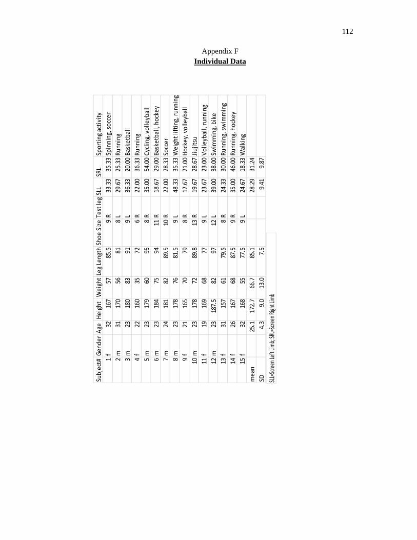
112
Appendix F Individual Data
Sub
ject
#G
en
de
rA
geH
eig
ht
We
igh
tLe
g Le
ngt
hSh
oe
Siz
eTe
st le
gSL
LSR
LSp
ort
ing
acti
vity
1f
3216
757
85.5
9R
33.3
335
.33
Spin
nin
g, s
occ
er
2m
3117
056
818
L29
.67
25.3
3R
un
nin
g
3m
2318
083
919
L36
.33
20.0
0B
aske
tbal
l
4f
2216
035
726
R22
.00
36.3
3R
un
nin
g
5m
2317
960
958
R35
.00
54.0
0C
ycli
ng,
vo
lle
ybal
l
6m
2318
475
9411
R18
.67
29.0
0B
aske
tbal
l, h
ock
ey
7m
2418
182
89.5
10R
22.0
028
.33
Socc
er
8m
2317
876
81.5
9L
48.3
335
.33
We
igh
t li
ftin
g, r
un
nin
g
9f
2116
570
798
R12
.67
21.0
0H
ock
ey,
vo
lle
ybal
l
10m
2317
872
89.8
13R
19.6
728
.67
Jiu
jits
u
11f
1916
968
779
L23
.67
23.0
0V
oll
eyb
all,
ru
nn
ing
12m
2318
7.5
8297
12L
39.0
038
.00
Swim
min
g, b
ike
13f
3115
761
79.5
8R
24.3
330
.00
Ru
nn
ing,
sw
imm
ing
14f
2616
768
87.5
9R
35.0
046
.00
Ru
nn
ing,
ho
cke
y
15f
3216
855
77.5
9L
24.6
718
.33
Wal
kin
g
me
an25
.117
2.7
66.7
85.1
28.2
931
.24
SD4.
39.
013
.07.
59.
419.
87
SLL=
Scre
en Le
ft Li
mb;
SRL
=Scr
een
Righ
t Lim
b
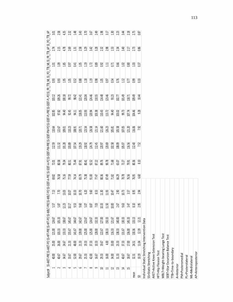
113
Subj
ect#
SS-A
KET-
PRE
SS-A
KET-
POSS
-HFT
-PRE
SS-H
FT-P
OSS
-WBL
T-PR
ESS-
WBL
T-PO
SS-S
EBT-
A-PR
ESS
-SEB
T-A-
POSS
-SEB
T-PM
-PRE
SS-S
EBT-
PM-P
OSS
-SEB
T-PL
-PRE
SS-S
EBT-
PL-P
OSS
_PR_
TTB_
ML
SS_P
O_TT
B_M
LSS_
PR_T
TB_A
PSS
_PO_
TTB_
AP
140
.00
25.0
012
1.00
124.
675.
577.
1369
.82
72.0
111
2.59
110.
6410
2.85
103.
120.
931.
012.
743.
01
240
.67
26.0
010
3.33
105.
335.
877.
7076
.58
80.5
811
1.32
122.
6797
.82
109.
260.
931.
092.
152.
58
334
.67
24.6
713
3.33
138.
6712
.23
13.0
375
.16
79.3
410
1.28
109.
0194
.40
100.
181.
951.
854.
784.
35
433
.67
21.3
315
5.67
145.
6710
.17
10.0
777
.59
80.5
110
3.89
110.
2392
.45
99.7
21.
050.
972.
692.
32
552
.00
48.0
014
1.67
144.
679.
5310
.33
74.1
475
.44
107.
5410
8.91
94.1
198
.42
0.52
0.41
1.31
1.48
629
.67
21.3
313
9.00
142.
679.
6010
.70
83.7
987
.91
119.
2912
0.71
118.
0512
2.41
0.88
1.05
2.58
3.43
725
.33
19.3
312
5.00
123.
675.
076.
2075
.38
80.4
111
8.92
123.
5411
3.93
120.
041.
181.
193.
293.
70
842
.00
37.3
312
5.00
124.
678.
879.
6091
.00
92.5
212
4.79
126.
5812
0.94
124.
461.
191.
723.
423.
67
916
.67
10.3
312
8.00
132.
337.
038.
5377
.47
87.2
211
2.45
121.
1410
3.38
110.
550.
980.
893.
282.
49
1029
.33
19.6
712
2.67
127.
0010
.33
11.3
080
.36
89.7
612
0.97
121.
6011
0.43
114.
481.
050.
922.
321.
98
1116
.00
4.00
138.
3315
0.33
11.5
011
.93
87.4
990
.78
120.
6912
6.23
113.
7212
3.46
0.97
1.11
2.98
3.17
1234
.00
33.0
011
6.33
121.
6710
.00
10.4
771
.82
71.6
810
2.58
105.
8194
.40
93.2
30.
540.
711.
531.
30
1331
.33
23.3
313
8.33
140.
002.
672.
9066
.29
65.0
710
8.09
105.
5810
3.02
102.
770.
760.
662.
342.
23
1440
.67
37.3
313
1.67
130.
339.
6310
.73
70.7
872
.27
105.
0710
7.05
99.7
310
1.49
1.12
1.02
3.40
3.44
1518
.67
17.0
013
6.00
145.
333.
473.
5776
.56
79.9
111
6.56
121.
2010
7.74
118.
710.
770.
802.
172.
18
mea
n32
.31
24.5
113
0.36
133.
138.
108.
9576
.95
80.3
611
2.40
116.
0610
4.46
109.
490.
991.
032.
732.
75
SD10
.19
11.0
412
.39
12.2
12.
952.
946.
608.
107.
527.
929.
2810
.44
0.33
0.37
0.86
0.87
Ind
ivid
ua
l Sta
tic
Stre
tch
ing
Inte
rve
nti
on
Da
ta
SS=S
tati
c St
retc
hin
g
AK
ET=
Act
ive
Kn
ee
Ext
en
sio
n T
est
HF
T=H
ip F
lexi
on
Te
st
WB
LT=
We
igh
t-b
ear
ing
Lun
ge
Te
st
SEB
T=S
tar
Exc
urs
ion
Ba
lan
ce T
est
TTB
=Ti
me
-to
-bo
un
dar
y
A=
An
teri
or
PM
=Po
ste
rom
ed
ial
PL=
Po
ste
rola
tera
l
ML=
Me
dio
late
ral
AP
=A
nte
rop
ost
eri
or
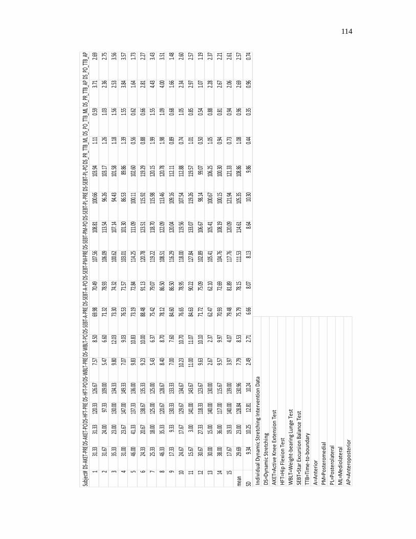
114
Subj
ect#
DS-A
KET-
PRED
S-AK
ET-P
ODS
-HFT
-PRE
DS-H
FT-P
ODS
-WBL
T-PR
EDS-
WBL
T-PO
DS-S
EBT-
A-PR
EDS
-SEB
T-A-
PODS
-SEB
T-PM
-PRE
DS-S
EBT-
PM-P
ODS
-SEB
T-PL
-PRE
DS-S
EBT-
PL-P
ODS
_PR_
TTB_
ML
DS_P
O_TT
B_M
LDS
_PR_
TTB_
APDS
_PO_
TTB_
AP
131
.33
31.3
312
0.33
126.
677.
578.
5069
.98
70.4
910
7.56
108.
8110
0.66
103.
941.
110.
593.
712.
69
231
.67
24.0
097
.33
109.
005.
476.
6071
.32
78.9
310
6.09
113.
5496
.26
103.
171.
261.
032.
362.
75
335
.33
23.0
013
0.00
134.
339.
8012
.03
73.3
074
.32
100.
6210
7.14
94.4
310
1.58
1.18
1.56
2.53
3.56
431
.00
23.6
714
7.00
149.
337.
079.
0376
.53
71.5
710
3.01
101.
3086
.53
89.8
61.
391.
553.
843.
57
546
.00
41.3
313
7.33
136.
009.
8310
.83
73.1
972
.84
114.
2511
1.09
100.
1110
2.60
0.56
0.62
1.64
1.73
624
.33
20.6
713
8.67
135.
339.
2310
.00
88.4
891
.13
120.
7812
3.51
115.
9211
9.29
0.88
0.66
2.81
2.27
725
.33
18.0
012
5.00
125.
005.
436.
3775
.42
79.0
711
9.22
118.
7011
5.98
120.
151.
991.
554.
433.
43
846
.33
35.3
312
0.67
128.
678.
408.
7078
.12
86.5
010
8.51
122.
0911
3.46
120.
781.
981.
094.
003.
51
917
.33
9.33
130.
3313
3.33
7.00
7.60
84.6
086
.50
116.
2912
0.04
109.
1611
2.11
0.89
0.68
1.66
1.48
1024
.67
17.6
712
9.67
134.
6710
.23
10.7
076
.65
78.9
511
8.00
119.
5610
7.54
112.
880.
741.
052.
342.
60
1115
.67
3.00
141.
0014
3.67
11.0
011
.07
84.6
390
.22
127.
8413
3.07
119.
2611
9.57
1.01
0.85
2.97
2.57
1230
.67
27.3
311
8.33
123.
679.
6310
.10
71.7
275
.09
102.
8910
6.67
98.1
499
.07
0.50
0.54
1.07
1.19
1330
.00
15.0
014
0.00
130.
002.
672.
3762
.47
62.1
010
5.41
105.
4110
0.67
106.
251.
050.
882.
282.
37
1438
.00
36.0
011
7.00
115.
679.
579.
9770
.93
72.6
910
4.76
108.
1910
0.15
100.
300.
940.
812.
672.
21
1517
.67
19.3
314
0.00
139.
003.
974.
0779
.48
81.8
911
7.76
120.
0912
1.94
121.
330.
730.
942.
062.
61
mea
n29
.69
23.0
012
8.84
130.
967.
798.
5375
.79
78.1
511
1.53
114.
6110
5.35
108.
861.
080.
962.
692.
57
SD9.
3410
.25
12.8
110
.24
2.49
2.71
6.66
8.07
8.13
8.64
10.3
09.
860.
440.
350.
960.
74
Ind
ivid
ual
Dyn
amic
Str
etc
hin
g In
terv
en
tio
n D
ata
DS=
Dyn
amic
Str
etc
hin
g
AK
ET=A
ctiv
e K
ne
e E
xte
nsi
on
Te
st
HFT
=Hip
Fle
xio
n T
est
WB
LT=W
eig
ht-
be
arin
g Lu
nge
Te
st
SEB
T=St
ar E
xcu
rsio
n B
alan
ce T
est
TTB
=Tim
e-t
o-b
ou
nd
ary
A=A
nte
rio
r
PM
=Po
ste
rom
ed
ial
PL=
Po
ste
rola
tera
l
ML=
Me
dio
late
ral
AP
=An
tero
po
ste
rio
r
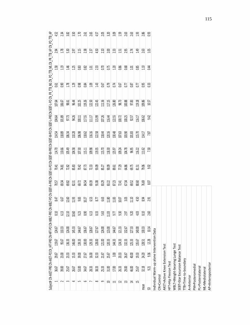
115
Su
bjec
t#CN
-AKE
T-PR
ECN-
AKET
-PO
CN_H
FT-P
RECN
-HFT
-PO
CN-W
BLT-
PRE
CN-W
BLT-
POCN
-SEB
T-A-
PREC
N-SE
BT-A
-PO
CN-S
EBT-
M-P
RECN
-SEB
T-M
-PO
CN-S
EBT-
L-PRE
CN-S
EBT-
L-PO
CN_P
R_TT
B_M
LCN_
PO_T
TB_M
LCN_
PR_T
TB_A
PCN
_PO_
TTB_
AP
136
.6729
.6711
9.67
124.6
78.1
08.4
770
.57
70.41
114.7
011
4.54
104.6
410
7.64
0.61
1.58
2.94
4.52
239
.0023
.0095
.67
110.3
36.1
36.6
769
.34
74.81
110.6
611
8.89
101.8
910
6.67
0.90
1.19
1.78
2.34
325
.6723
.3313
6.33
124.0
012
.10
12.60
69.8
272
.8210
5.49
106.3
497
.73
98.61
1.78
1.46
5.30
3.82
426
.6715
.3314
6.00
145.3
310
.60
11.93
82.3
185
.8310
4.58
110.2
894
.26
96.44
1.29
1.25
2.97
3.50
553
.0039
.0013
9.33
144.6
79.2
39.8
369
.72
70.42
107.3
010
6.98
100.1
110
2.25
0.98
0.83
2.15
1.70
634
.6727
.6713
0.00
136.6
78.9
010
.2784
.54
87.73
115.1
111
8.62
117.5
511
9.26
0.84
0.82
2.38
2.61
728
.3316
.0012
9.33
128.6
76.1
36.5
369
.39
72.33
109.9
811
6.16
111.1
712
2.50
1.09
1.01
2.37
2.65
838
.3332
.3312
8.00
127.6
78.3
38.7
791
.08
93.09
119.3
512
2.58
115.9
912
2.45
1.43
2.33
4.50
4.57
925
.3321
.6712
1.00
124.0
05.6
36.6
383
.29
85.27
115.7
011
8.44
107.2
611
1.56
0.67
0.70
2.10
2.05
1031
.0025
.6712
0.33
123.0
011
.03
11.80
83.2
290
.0911
8.30
120.1
611
6.44
117.
150.7
90.7
52.0
03.
20
1117
.003.0
014
4.33
144.6
79.7
711
.8387
.23
89.61
125.9
713
0.48
122.5
512
6.80
0.74
1.20
2.33
3.09
1226
.3320
.3312
4.33
121.3
39.3
010
.0772
.41
77.29
109.2
410
7.63
100.7
298
.730.6
70.8
61.5
11.
59
1329
.0026
.6713
2.33
132.6
72.9
73.0
367
.34
64.86
104.7
810
3.90
100.2
510
0.88
0.77
0.68
2.95
2.63
1446
.0040
.0012
5.33
127.3
39.2
011
.1069
.52
69.75
96.95
101.8
392
.57
97.83
0.87
1.01
2.74
2.63
1525
.6719
.3313
5.67
140.0
04.0
34.5
080
.60
81.51
116.2
211
5.70
116.1
711
9.18
0.77
0.85
1.49
1.93
mea
n32
.1824
.2012
8.51
130.3
38.1
08.9
476
.69
79.06
111.6
211
4.17
106.6
210
9.86
0.95
1.10
2.63
2.86
SD9.2
19.3
612
.28
10.14
2.60
2.91
8.07
9.02
7.30
7.87
9.42
10.57
0.33
0.44
1.05
0.93
Ind
ivid
ua
l War
m-u
p a
lon
e In
terv
en
tio
n D
ata
CN
=Co
ntr
ol
AK
ET=A
ctiv
e K
ne
e E
xte
nsi
on
Te
st
HF
T=H
ip F
lexi
on
Te
st
WB
LT=W
eig
ht-
be
arin
g Lu
nge
Te
st
SEB
T=S
tar
Excu
rsio
n B
ala
nce
Te
st
TTB
=Ti
me
-to
-bo
un
dar
y
A=
An
teri
or
PM
=Po
ste
rom
ed
ial
PL=
Po
ste
rola
tera
l
ML=
Me
dio
late
ral
AP
=An
tero
po
ste
rio
r
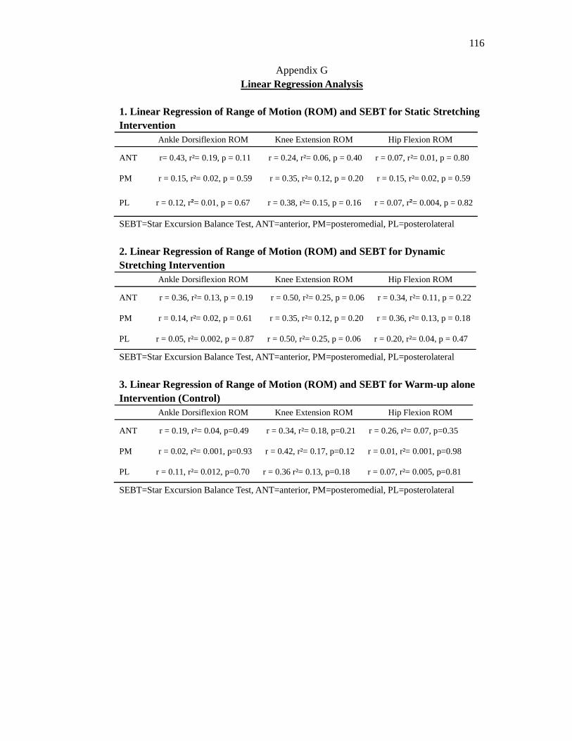
116
Appendix G Linear Regression Analysis
1. Linear Regression of Range of Motion (ROM) and SEBT for Static Stretching Intervention Ankle Dorsiflexion ROM Knee Extension ROM Hip Flexion ROM
ANT r= 0.43, r²= 0.19, p = 0.11 r = 0.24, r²= 0.06, p = 0.40 r = 0.07, r²= 0.01, p = 0.80
PM r = 0.15, r²= 0.02, p = 0.59 r = 0.35, r²= 0.12, p = 0.20 r = 0.15, r²= 0.02, p = 0.59
PL r = 0.12, r²= 0.01, p = 0.67 r = 0.38, r²= 0.15, p = 0.16 r = 0.07, r²= 0.004, p = 0.82
SEBT=Star Excursion Balance Test, ANT=anterior, PM=posteromedial, PL=posterolateral
2. Linear Regression of Range of Motion (ROM) and SEBT for Dynamic Stretching Intervention
Ankle Dorsiflexion ROM Knee Extension ROM Hip Flexion ROM
ANT r = 0.36, r²= 0.13, p = 0.19 r = 0.50, r²= 0.25, p = 0.06 r = 0.34, r²= 0.11, p = 0.22
PM r = 0.14, r²= 0.02, p = 0.61 r = 0.35, r²= 0.12, p = 0.20 r = 0.36, r²= 0.13, p = 0.18
PL r = 0.05, r²= 0.002, p = 0.87 r = 0.50, r²= 0.25, p = 0.06 r = 0.20, r²= 0.04, p = 0.47
SEBT=Star Excursion Balance Test, ANT=anterior, PM=posteromedial, PL=posterolateral
3. Linear Regression of Range of Motion (ROM) and SEBT for Warm-up alone Intervention (Control)
Ankle Dorsiflexion ROM Knee Extension ROM Hip Flexion ROM
ANT r = 0.19, r²= 0.04, p=0.49 r = 0.34, r²= 0.18, p=0.21 r = 0.26, r²= 0.07, p=0.35
PM r = 0.02, r²= 0.001, p=0.93 r = 0.42, r²= 0.17, p=0.12 r = 0.01, r²= 0.001, p=0.98
PL r = 0.11, r²= 0.012, p=0.70 r = 0.36 r²= 0.13, p=0.18 r = 0.07, r²= 0.005, p=0.81
SEBT=Star Excursion Balance Test, ANT=anterior, PM=posteromedial, PL=posterolateral
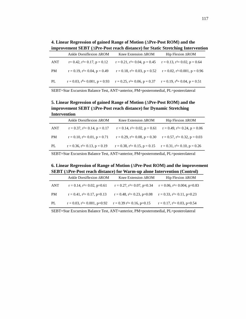
117
4. Linear Regression of gained Range of Motion (∆Pre-Post ROM) and the improvement SEBT (∆Pre-Post reach distance) for Static Stretching Intervention Ankle Dorsiflexion ∆ROM Knee Extension ∆ROM Hip Flexion ∆ROM
ANT r= 0.42, r²= 0.17, p = 0.12 r = 0.21, r²= 0.04, p = 0.45 r = 0.13, r²= 0.02, p = 0.64
PM r = 0.19, r²= 0.04, p = 0.49 r = 0.18, r²= 0.03, p = 0.52 r = 0.02, r²<0.001, p = 0.96
PL r = 0.03, r²= 0.001, p = 0.93 r = 0.25, r²= 0.06, p = 0.37 r = 0.19, r²= 0.04, p = 0.51
SEBT=Star Excursion Balance Test, ANT=anterior, PM=posteromedial, PL=posterolateral
5. Linear Regression of gained Range of Motion (∆Pre-Post ROM) and the improvement SEBT (∆Pre-Post reach distance) for Dynamic Stretching Intervention
Ankle Dorsiflexion ∆ROM Knee Extension ∆ROM Hip Flexion ∆ROM
ANT r = 0.37, r²= 0.14, p = 0.17 r = 0.14, r²= 0.02, p = 0.61 r = 0.49, r²= 0.24, p = 0.06
PM r = 0.10, r²= 0.01, p = 0.71 r = 0.29, r²= 0.08, p = 0.30 r = 0.57, r²= 0.32, p = 0.03
PL r = 0.36, r²= 0.13, p = 0.19 r = 0.38, r²= 0.15, p = 0.15 r = 0.31, r²= 0.10, p = 0.26
SEBT=Star Excursion Balance Test, ANT=anterior, PM=posteromedial, PL=posterolateral
6. Linear Regression of Range of Motion (∆Pre-Post ROM) and the improvement SEBT (∆Pre-Post reach distance) for Warm-up alone Intervention (Control)
Ankle Dorsiflexion ∆ROM Knee Extension ∆ROM Hip Flexion ∆ROM
ANT r = 0.14, r²= 0.02, p=0.61 r = 0.27, r²= 0.07, p=0.34 r = 0.06, r²= 0.004, p=0.83
PM r = 0.41, r²= 0.17, p=0.13 r = 0.48, r²= 0.23, p=0.08 r = 0.33, r²= 0.11, p=0.23
PL r = 0.03, r²= 0.001, p=0.92 r = 0.39 r²= 0.16, p=0.15 r = 0.17, r²= 0.03, p=0.54
SEBT=Star Excursion Balance Test, ANT=anterior, PM=posteromedial, PL=posterolateral