The Effects of Comvita Manuka Honey on Oral Mucositis in ...
176
The Effects of Comvita Manuka Honey on Oral Mucositis in Patients treated with Radiation Therapy to the Head and Neck Aubrey Begley A thesis submitted in fulfilment of BHealSc(Hons) University of Otago 2009/2010 Principal Investigator Dr Patries Herst
Transcript of The Effects of Comvita Manuka Honey on Oral Mucositis in ...
The Effects of Comvita Manuka Honey
on Oral Mucositis in Patients treated with
Radiation Therapy to the Head and Neck
Aubrey Begley
A thesis submitted in fulfilment of
BHealSc(Hons)
University of Otago
2009/2010
Principal Investigator
Dr Patries Herst

Page | 2
Abstract
Mucositis is a common side effect of radiation therapy to the head and neck region and one
that causes considerable discomfort for patients. Mucositis compromises patients’ ability to
eat, drink and talk thus affecting patient health and quality of life. Currently there is no
worldwide standard for the prevention or treatment of oral mucositis; care is limited to
symptom control. Honey has anti-bacterial and potential anti-inflammatory properties and
three trials overseas investigated its effect on radiation-induced oral mucositis. The three
trials conducted in Malaysia, Iran and Egypt found that honey did reduce the incidence of
severe mucositis in head and neck patients. This New Zealand mucositis trial aimed to verify
the results from the three overseas trials by comparing the effects of manuka honey with
current best practice on oral mucositis in head and neck patients.
This report analyses a sub-set of the patients recruited to the trial; those from the Wellington
and Dunedin departments. A total of 14 patients were recruited to the trial from these
departments, nine recruited to the honey arm and five to the control arm. Four honey patients
withdrew from the trial due to issues with the honey application and one patient withdrew
from the control arm. Honey arm patients were given manuka honey and instructed to swirl
20mL (amended to 10mL) around their oral cavity and then swallow three times a day, these
patients also had access to the standard of care. The control arm patients were treated with
the standard of care alone.
Patient assessment involved three times weekly mucositis scoring using the TROG multi-site
mucositis scoring system, weekly weight and fortnightly quality of life assessment using a 65
question form adapted from the European organisation for Research and Treatment of Cancer
questionnaires (EORTC QLQ-C30 and EORTC QLQ HN35). Patients were also asked to fill
in food and drug diaries to assess changes in food intake and pain medication. Results
showed that manuka honey was not well tolerated by our patient cohort. Patients complained
of extreme nausea and stinging sensations in the oral cavity. The honey had to be diluted to
be better tolerated (1:3 with another liquid). Contradictory to previous studies, preliminary
analysis showed that manuka honey did not affect the extent of oral mucositis in the small
cohort of New Zealand head and neck patients when taken in addition to current best practice.
Page | 3
Acknowledgements
The health and wellbeing of our patients is our number one priority, and to ensure we give
them the best standard of care we need to practice evidence-based medicine and participate in
clinical trials. I would like to acknowledge all the patients who participated in the New
Zealand manuka honey trial. These patients were all undergoing intense cancer treatment but
put time and energy into adhering to the trial requirements.
A big thank you to the staff at the Wellington Blood and Cancer Centre for supporting the
trial and allowing me to combine the roles of research assistant and radiation therapist.
Thank you to those who helped with the mucositis scoring and collection of the quality of life
forms and food diaries. I would also like to acknowledge the Christchurch Oncology
Department for allowing me time to complete this report.
The University of Otago Research Fund allowed the trial to go ahead financially and the
Comvita Company supplied the manuka honey free of charge. Thank you.
My supportive family and friends deserve acknowledgement, thank you for the support,
encouragement and for your proofreading!
Dr Patries Herst is the most enthusiastic scientist I have met and her passion for research is
infectious. A huge thank you for being the driving force behind this trial and giving me the
opportunity to get involved in research. I truly appreciate all your encouragement and advice.
Page | 4
Table of Contents Chapter 1 INTRODUCTION ................................ ................................ ............................... 8 1.1 Head and neck cancer ................................ ................................ ................................ ..... 8 1.2 Head and neck cancer treatment ................................ ................................ ................... 14 1.3 Radiation-induced side effects ................................ ................................ ...................... 19 1.4 Management of head and neck patients................................ ................................ ......... 30 1.5 Honey ................................ ................................ ................................ .......................... 34 1.6 Aim and objectives of the study................................ ................................ .................... 37 Chapter 2 METHODOLOGY................................ ................................ ............................. 39 2.1 Survey of New Zealand Oncology Departments ................................ ........................... 39 2.2 Oral Mucositis Trial ................................ ................................ ................................ ..... 39 Chapter 3 RESULTS................................ ................................ ................................ .......... 53 3.1 Review of current New Zealand oncology department head and neck care.................... 53 3.2 Mucositis trial: Patient results................................ ................................ ....................... 57 3.3 Mucositis trial: Individual patient experiences ................................ .............................. 65 3.4 Mucositis trail: Comparison between the treatment and control arms.......................... 137 Chapter 4 DISCUSSION................................ ................................ ................................ .. 143 4.1 Interpretation of results................................ ................................ ............................... 143 4.2 Limitations of the current study ................................ ................................ .................. 145 4.3 Recommendations for future clinical trials................................ ................................ .. 154 4.4 Conclusion ................................ ................................ ................................ ................. 156 References ................................ ................................ ................................ ........................ 157 Glossary................................ ................................ ................................ ............................ 163 Appendices ................................ ................................ ................................ ....................... 165

Page | 5
List of Figures Figure 1. Diagram illustrating structures of the head and neck region ................................ ... 8 Figure 2. Diagram illustrating factors determining an individual’s choice of treatment for head and neck cancer ................................ ................................ ................................ .......... 15 Figure 3. Diagram of mucositis pathophysiology................................ ................................ 21 Figure 4. Diagram showing the synergistic effect of chemotherapy and radiation therapy in the oral cavity ................................ ................................ ................................ ..................... 25 Figure 5. Diagram illustrating the fourteen oral sites that were assessed for mucositis ........ 42 Figure 6. Trial timeline................................ ................................ ................................ ....... 46 Figure 7. Consort diagram illustrating patient flow through the oral mucositis trial ............ 59 Figure 8. Number of patients with each tumour stage ................................ ......................... 63 Figure 9. Number of patients with each nodal stage................................ ............................ 63 Figure 10. WHN3 oral cavity sites at risk ................................ ................................ ........... 66 Figure 11. WHN3 anterior view of the treatment area................................ ......................... 67 Figure 12. WHN3 lateral view of the treatment area ................................ ........................... 67 Figure 13. WHN3 transverse slices................................ ................................ ..................... 68 Figure 14. WHN3 mean mucositis score (TS/NSR) vs dose................................ ................ 69 Figure 15. WHN3 change in weight vs dose ................................ ................................ ....... 69 Figure 16. WHN3 change in perception of QoL score vs dose ................................ ............ 70 Figure 17. WHN3 negative change in QoL category scores vs dose................................ .... 71 Figure 18. WHC4 oral cavity sites at risk ................................ ................................ ........... 76 Figure 19. WHC4 anterior view of the treatment area................................ ......................... 77 Figure 20. WHC4 lateral view of the treatment area ................................ ........................... 77 Figure 21. WHC4 transverse slices................................ ................................ ..................... 78 Figure 22. WHC4 mean mucositis scores (TS/NSR) vs dose ................................ .............. 78 Figure 23. WHC4 change in weight vs dose ................................ ................................ ....... 79 Figure 24. WHC4 change in perception of QoL score vs dose ................................ ............ 80 Figure 25. WHC4 negative change in QoL category scores vs dose................................ .... 80 Figure 26. WHN5 oral cavity sites at risk ................................ ................................ ........... 85 Figure 27. WHN5 anterior view of the treatment area................................ ......................... 86 Figure 28. WHN5 lateral view of the treatment area ................................ ........................... 86 Figure 29. WHN5 transverse slices................................ ................................ ..................... 87 Figure 30. WHN5 mean mucositis score (TS/NSR) vs dose................................ ................ 87 Figure 31. WHN5 change in weight vs dose ................................ ................................ ....... 88 Figure 32. WHN5 change in perception of QoL score vs dose ................................ ............ 89 Figure 33. WHN5 negative change in QoL category scores vs dose................................ .... 89 Figure 34. WHC6 oral cavity sites at risk ................................ ................................ ........... 93 Figure 35. WHC6 anterior view of treatment area ................................ .............................. 94 Figure 36. WHC6 lateral view of treatment area................................ ................................ . 94 Figure 37. WHC6 transverse slices................................ ................................ ..................... 95 Figure 38. WHC6 mean mucositis score (TS/NSR) vs dose................................ ................ 95 Figure 39. WHC6 change in weight vs dose ................................ ................................ ....... 96 Figure 40. WHC6 change in perception of QoL score vs dose ................................ ............ 97 Figure 41. WHC6 negative change in QoL category scores vs dose................................ .... 97 Figure 42. DHC1 oral cavity sites at risk ................................ ................................ .......... 101 Figure 43. DHC1 mean mucositis score (TS/NSR) vs dose................................ ............... 102 Figure 44. DHC1 change in weight vs dose ................................ ................................ ...... 102 Figure 45. DHC1 change in perception of QoL score vs dose ................................ ........... 103
Page | 6
Figure 46. DHC1 negative change in QoL categories vs dose................................ ........... 104 Figure 47. WHN1 oral cavity sites at risk ................................ ................................ ......... 108 Figure 48. WHN1 the anterior view of the treatment area................................ ................. 109 Figure 49. WHN1 lateral view of the treatment area ................................ ......................... 109 Figure 50. WHN1 transverse slices................................ ................................ ................... 110 Figure 51. WHN1 mean mucositis score (TS/NSR) vs dose................................ .............. 110 Figure 52. WHN1 change in weight vs dose ................................ ................................ ..... 111 Figure 53. WHN1 change in perception of QoL score vs dose ................................ .......... 112 Figure 54. WHN1 negative changes in QoL category scores vs dose ................................ 112 Figure 55. WHC2 oral cavity sites at risk ................................ ................................ ......... 115 Figure 56. WHC2 anterior view of treatment area ................................ ............................ 116 Figure 57. WHC2 lateral view of the treatment area ................................ ......................... 116 Figure 58. WHC2 transverse slices................................ ................................ ................... 117 Figure 59. WHC2 mean mucositis score (TS/NSR) vs dose................................ .............. 117 Figure 60. WHC2 change in weight vs dose ................................ ................................ ..... 118 Figure 61. WHN4 oral cavity sites at risk ................................ ................................ ......... 122 Figure 62. WHN4 anterior view of the treatment area................................ ....................... 123 Figure 63. WHN4 lateral view of the treatment area ................................ ......................... 123 Figure 64. WHN4 transverse slices................................ ................................ ................... 124 Figure 65. WHN4 mean mucositis score (TS/NSR) vs dose................................ .............. 124 Figure 66. WHN4 change in weight vs dose ................................ ................................ ..... 125 Figure 67. WHN4 change in perception of QoL score vs dose ................................ .......... 126 Figure 68. WHN4 negative change in QoL category scores vs dose................................ .. 126 Figure 69. WHC7 oral cavity sites at risk ................................ ................................ ......... 130 Figure 70. WHC7 anterior view of treatment area ................................ ............................ 131 Figure 71. WHC7 lateral view of treatment area................................ ............................... 131 Figure 72. WHC7 transverse slices at the isocentre................................ ........................... 132 Figure 73. WHC7 mean mucositis score (TS/NSR) vs dose................................ .............. 132 Figure 74. WHC7 change in weight vs dose ................................ ................................ ..... 133 Figure 75. WHC7 change in the perception of QoL score vs dose................................ ..... 134 Figure 76. WHC7 negative change in QoL category scores vs dose................................ .. 134 Figure 77. A comparison of mean average oral mucositis scores ................................ ...... 137 Figure 78. Comparison of weights between patients in control and honey arm.................. 139 Figure 79. Percentage weight loss of all trial patients ................................ ....................... 139 Figure 80. Average percentage weight loss of control vs honey patients ........................... 140 Figure 81. Average change in QoL score for the control patientsvs honey ........................ 142 Figure 82. Change in patients’ perceived QoL vs dose ................................ ..................... 142
Page | 7
List of Tables Table 1. Stage grouping of oral cancer ................................ ................................ ............... 11 Table 2. Acute and chronic radiation-induced side effects ................................ .................. 20 Table 3. Risk factors associated with developing severe oral mucositis .............................. 23 Table 4. The TROG mucositis scoring system................................ ................................ .... 26 Table 5. Standard of care for New Zealand departments ................................ ..................... 53 Table 6. Treatment management of trial patients ................................ ................................ 61 Table 7. Tumour sites................................ ................................ ................................ ......... 62 Table 8. Tumour stage................................ ................................ ................................ ........ 63 Table 9. Dose at onset of oral mucositis ................................ ................................ ........... 138 Table 10. Percentage of patients in each trial arm requiring PEG/NG feeding, hospitalisation and analgesics ................................ ................................ ................................ ................... 141 Table 11. Number of QoL forms filled in by each patient ................................ ................. 141
Page | 8
Chapter 1 INTRODUCTION
This research project describes a case series conducted at the Wellington Blood and Cancer
Centre which investigated the effect of manuka honey on mouth ulcers in patients treated
with radiation therapy for head and neck cancer.
1.1 Head and neck cancer
Cancer is a malignant disease which is characterised by a series of cellular and genetic
changes that lead to abnormal cell proliferation with the potential to invade surrounding
tissues and metastasize to distant locations. Cancer cells can originate from many areas of
the body and cause symptoms specific to the region where the tumour manifests. The head
and neck is an important region of the body because it is essential for many physiological
functions and is critical for a person's appearance, expression and social interactions. Cancer
within the head and neck region can cause structural deformities and disrupt the functions of
this region which can lead to a significant decrease in patients’ quality of life (Bomford &
Kunkler, 2003).
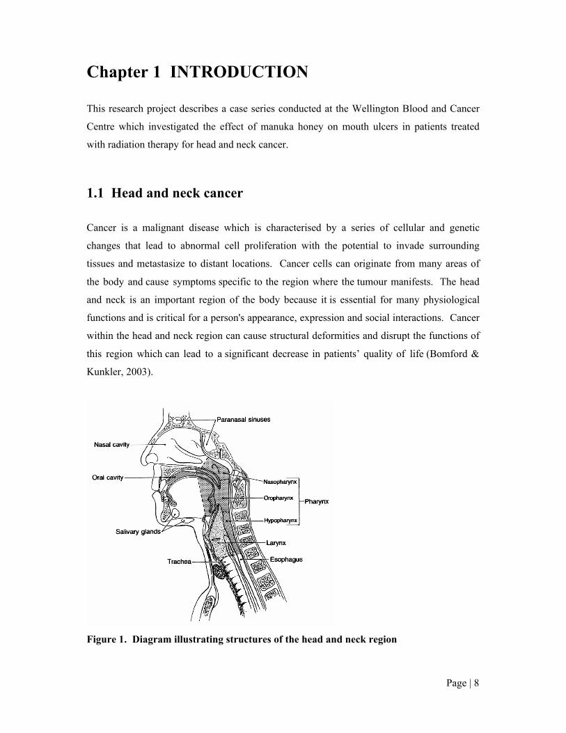
Figure 1. Diagram illustrating structures of the head and neck region
Page | 9
Due to the large number of structures present in the head and neck region there are many
potential cancer sites including the lip, base of tongue, gum, floor of mouth, palate, parotid,
tonsil, nasal cavity, para-nasal sinuses, oropharynx, nasopharynx, hypopharynx, larynx and
thyroid gland.
Head and neck tumours vary not only by site but also by pathophysiology, biological
behaviour and their sensitivity to treatment modalities. Squamous cell carcinoma (SCC) is
the most common type of cancer in the head and neck region. It accounts for more than 90%
of all malignant lesions in the mouth. The most common sites for oral SCC are the tongue
and floor of mouth then at a lower frequency the soft palate, gingiva and buccal mucosa
(Cabral et al., 2010).
Head and neck tumours can invade local structures, spread to lymph nodes and metastasise to
other organs of the body. The lymph drainage of the head and neck region is complex and is
categorised by levels (Appendix A). Due to the ability of tumours to readily metastasise via
lymph, the assessment and treatment of the neck lymph nodes is of utmost importance in
disease management (Rosenthal, 2009). The most common site of distant metastasis is the
lungs but other sites include the mediastinal lymph nodes, liver, brain and bones (Washington
& Leaver, 2006).
1.1.1 Incidence
The incidence of head and neck cancer varies around the world, however most authors report
that head and neck cancers account for between three and five percent of all cancers (Yarbro
et al., 2004). Head and neck cancer has been described as the sixth most common cancer
worldwide and its incidence is increasing in most parts of the world (Evans et al., 2003).
Each year the New Zealand Ministry of Health (MOH) publishes a detailed report on the
incidence of cancer; Cancer: New Registrations and Deaths. In 2007 the report showed there
were 711 head and neck cancer registrations in New Zealand with 191 people dying of the
disease. Head and neck cancer made up 3.6% of all cancers registered that year (NZ Health
and Information Service, 2009).

Page | 10
Head and neck tumours are reported as being more common in males than females and are
historically more common in older people although they are becoming increasingly common
in younger people (Toner & O’Regan, 2009).
1.1.2 Predisposing factors
A number of predisposing factors for cancers of the head and neck region have been
identified with alcohol and tobacco being the most common.
Alcohol and tobacco
Tobacco smoke contains more than 300 carcinogens that can damage DNA. Alcohol is an
independent risk factor but also increases tobacco related carcinogenesis. The synergistic
effect of alcohol and tobacco was described by Evans et al. (2003) as resulting from increased
mucosal absorption of carcinogens (from both tobacco and alcohol), from the chronic
inflammation and the increased solubility of carcinogens in alcohol compared to saliva.
Alcohol independent of tobacco shows an increased risk of cancer especially in the oral
cavity. Alcoholism may also lead to impaired metabolism resulting from liver dysfunction
and nutritional deficiencies that may promote carcinogenesis (Evans et al., 2003).
Environmental and occupational exposure
Ultraviolet light exposure has been described as a common risk factor for head and neck
cancer. Other environmental risk factors can relate to an individual’s occupation. Evans et
al. (2003) highlighted that people involved in nickel refining, woodworking, metal work and
work with textile fibres have an increased risk of head and neck cancer, as do car mechanics.
Diet
Dietary factors may also have an influence on the incidence of head and neck cancer as iron
and vitamin A, C and E deficiencies have been linked to cancer. Nasopharyngeal cancer is
also associated with the consumption of salted fish (Vokes et al., 1993).
Viral infection
Increasing evidence suggests that viruses contribute to the cause of head and neck cancer.
The Human Papilloma Virus and Epstein-Barr Virus are associated with a specific type of
head and neck cancer; nasopharyngeal cancer (Myers et al., 2003).
Page | 11
1.1.3 Symptoms
The symptoms of head and neck cancer vary with the location of the primary tumour and the
stage of the cancer. Patients with early stage cancer frequently have only vague symptoms
and minimal physical abnormalities. The vague symptoms include pain, ulcers that do not
heal and hoarseness. Often early stage cancers of the oropharynx and hypopharynx do not
produce any symptoms so are commonly diagnosed in later stages. Cancers in later stages
have easily detectable signs and symptoms which include pain, airway obstruction, cranial
neuropathies, trismus, dysphasia and decreased mobility of the tongue (Vokes et al., 1993).
1.1.4 Diagnosis
Disease diagnosis follows a number of procedures including a complete physical examination
involving inspection of the oral mucosa, palpation of floor of mouth and all aspects of the
tongue as well as thorough palpation of the neck. Further investigations may include
examination with a flexible fibre-optic nasopharygoscope, radiologic evaluation to assess the
extent of local and regional spread of the tumour and its depth of invasion. Biopsies are
taken to define the primary tumour and identify possible sites of secondary tumours. Fine
needle aspiration (FNA) biopsies are often done first and then excisional biopsies are used for
further investigation (Vokes et al., 1993).
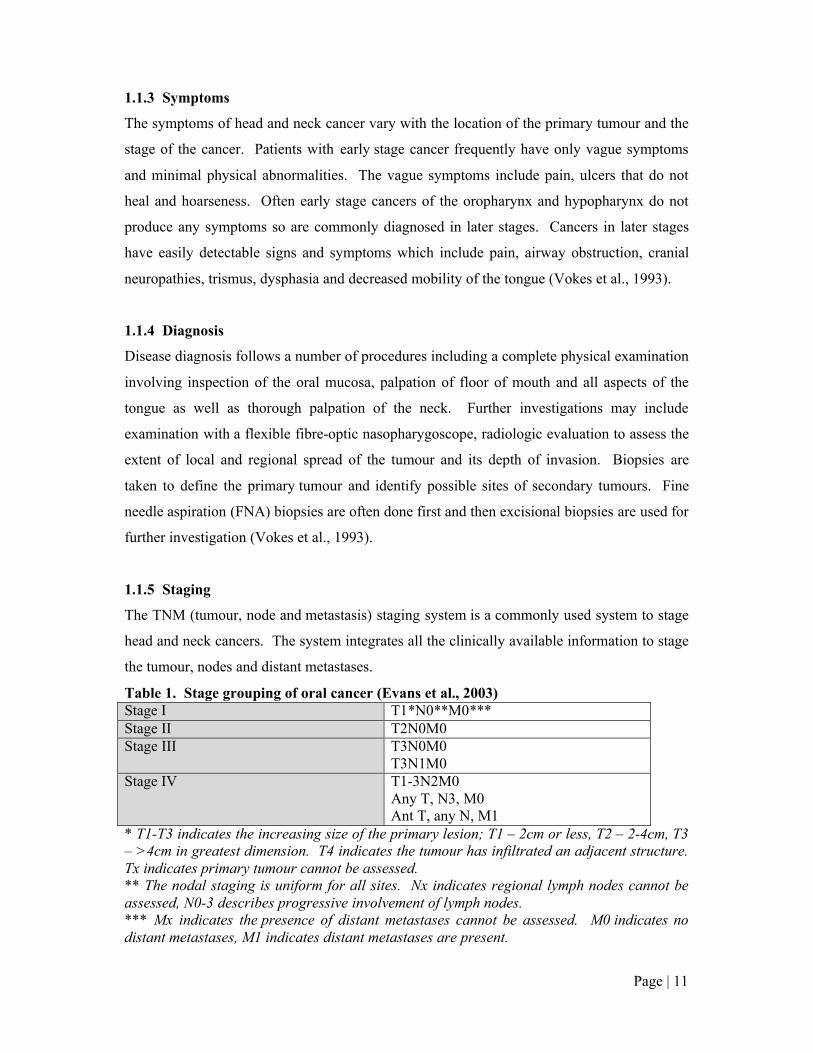
1.1.5 Staging
The TNM (tumour, node and metastasis) staging system is a commonly used system to stage
head and neck cancers. The system integrates all the clinically available information to stage
the tumour, nodes and distant metastases.
Table 1. Stage grouping of oral cancer (Evans et al., 2003) Stage I T1*N0**M0*** Stage II T2N0M0 Stage III T3N0M0
T3N1M0 Stage IV T1-3N2M0
Any T, N3, M0 Ant T, any N, M1
* T1-T3 indicates the increasing size of the primary lesion; T1 – 2cm or less, T2 – 2-4cm, T3 – >4cm in greatest dimension. T4 indicates the tumour has infiltrated an adjacent structure. Tx indicates primary tumour cannot be assessed. ** The nodal staging is uniform for all sites. Nx indicates regional lymph nodes cannot be assessed, N0-3 describes progressive involvement of lymph nodes. *** Mx indicates the presence of distant metastases cannot be assessed. M0 indicates no distant metastases, M1 indicates distant metastases are present.
Page | 12
The risk of distant metastasis correlates with nodal stage, the risk is less than 10 percent with
N0 or N1. In patients with T4 lesions the presence of distant metastases in lungs, liver or
bones should be ruled out before aggressive local or regional therapy is commenced.
1.1.6 Head and neck cancer sites
As outlined above there are many potential sites of head and neck cancer, this report later
describes a trial in which patients had cancers of seven different sites (maxillary gingiva,
tongue, tonsil, palate, nasal cavity, floor of mouth and Waldeyer's ring). Each of these sites
has their own symptoms and preferred method of disease management:
Maxillary gingiva lesions often mimic common inflammatory lesions but are normally
painless. Primary treatment is surgery with a radical neck dissection to treat metastatic
lymph nodes. Radiotherapy is used as adjuvant post-operative treatment and chemotherapy is
sometimes used for adjuvant or palliative treatment (Cabral et al., 2010).
The tongue is a large muscular organ (made of six paired muscles both intrinsic and extrinsic)
which has a mobile anterior portion and a non-mobile portion (base of tongue) (Evans et al.,
2003). Tongue tumours are often asymptomatic and can enlarge considerably before
producing symptoms. They often present with a painless mass or ulcer that does not heal and
often appear on the lateral borders of the tongue. Treatment can involve surgery
(glossectomy) and/or radiation therapy. Post-operative radiation therapy can be used to target
the primary site and nodal involvement. Lymph from the tongue drains to the sub-mental and
sub-mandibular glands (Appendix A) on both sides so tumours of the tongue require bilateral
neck treatment (Myers et al., 2003).
Tonsils are small structures that are part of the immune system. Tonsillar tumours commonly
present in advanced stages as an ulcer or lump or with symptoms of pain, bleeding or
difficulty swallowing. Primary treatment is commonly surgery depending on the stage of the
tumour and the patient’s state of health. Radiation therapy and chemotherapy are also
commonly used.
The palate or roof of mouth separates the mouth from the nasal cavity and consists of a bony
hard palate and a muscular soft palate at the rear. Tumours of the hard palate are rare and
Page | 13
usually present with a painless mass which can spread into the nasal cavity or skull base
(Myers et al., 2003). The lymph from the roof of mouth drains to the retropharyngeal and
then deep cervical nodes (Evans et al., 2003).
The nasal cavity is divided in the midline by the nasal septum (cartilage and bone) and opens
anteriorly through the nares and posteriorly into the nasopharynx. Tumours in this area are
rare and have similar presenting symptoms to those of more common benign conditions.
They have the propensity for early spread and involvement of surrounding critical structures
which means that most patients present with advanced-stage disease. Lymph drainage from
the nasal cavity is to the sub-mandibular nodes, upper jugular and retropharyngeal lymph
nodes.
The floor of mouth (FOM) is a horseshoe-shaped space from the lower alveolar ridge to the
under surface (ventral) of the tongue. The FOM contains openings for sub-mandibular and
sub-lingual salivary gland ducts (Evans et al., 2003). Cancers arise on the anterior surface
and can spread to the bone and ventral aspect of the tongue and also the sub-mandibular duct
(Myers et al., 2003). Tumours are often asymptomatic and can arise in regions of leukoplakia
or erythroplakia. Surgical resection and radiation therapy are often used in conjunction. Bite
blocks are commonly used when treating with radiation therapy to spare the roof of mouth.
The lymph drainage for the floor of mouth is to the sub-mental and sub-mandibular lymph
nodes.
Waldeyer's ring or pharyngeal lymphoid ring is a ring of lymphoid tissue located in the
pharynx at the back of the oral cavity. The ring consists of the pharyngeal tonsil, the tubal
tonsil, the palatine tonsils and the lingual tonsils. The Waldeyer's ring is a potential site of
lymphoma which is a lymphoproliferative disorder. Lymphoma often presents with painless
lymphadenopathy but the masses can become more painful with rapid growth. Lymphomas
are sensitive to chemotherapy and radiation therapy (Myers et al., 2003).
Page | 14
1.2 Head and neck cancer treatment
The control of the primary tumour and the regional nodal metastases is of upmost importance
for treatment of head and neck cancers regardless of the type, site or stage of the disease.
While achieving disease control to reduce mortality treatment also needs to reduce deformity
and maintain tissue and organ function and minimise side effects.
A multidisciplinary approach is the best way of achieving cure and structure, function and
aesthetic preservation. The multidisciplinary team consists of a large number of medical
professionals including radiation oncologists, medical oncologists, dentists, maxillofacial
prosthodontists, nutritionists, head and neck surgeons, neurosurgeons, plastic surgeons, oral
surgeons, pathologists, oncology nurses, radiologists, social workers, radiation therapists,
speech therapists and pain intervention teams. The specific team that is involved in each
individual case depends on the treatment modality choice.
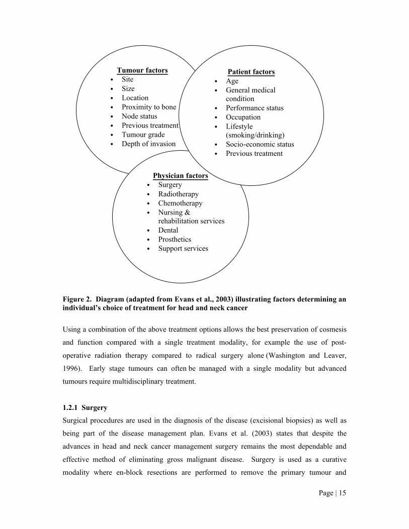
The main treatment options are surgery, chemotherapy, radiation therapy or a combination of
these. The choice is based on factors relating to the disease such as the tumour type, site and
stage and also patient related factors such as previous treatment, co-morbidities and the
impact on quality of life (Figure 2.). The availability of treatment modalities and expertise of
the medical team may also impact on the decision but with all these factors taken into account
the decision is ultimately the patient’s.
Page | 15
Figure 2. Diagram (adapted from Evans et al., 2003) illustrating factors determining an individual’s choice of treatment for head and neck cancer
Using a combination of the above treatment options allows the best preservation of cosmesis
and function compared with a single treatment modality, for example the use of post-
operative radiation therapy compared to radical surgery alone (Washington and Leaver,
1996). Early stage tumours can often be managed with a single modality but advanced
tumours require multidisciplinary treatment.
1.2.1 Surgery
Surgical procedures are used in the diagnosis of the disease (excisional biopsies) as well as
being part of the disease management plan. Evans et al. (2003) states that despite the
advances in head and neck cancer management surgery remains the most dependable and
effective method of eliminating gross malignant disease. Surgery is used as a curative
modality where en-block resections are performed to remove the primary tumour and
Tumour factors • Site • Size • Location • Proximity to bone • Node status • Previous treatment • Tumour grade • Depth of invasion
Physician factors • Surgery • Radiotherapy • Chemotherapy • Nursing &
rehabilitation services • Dental • Prosthetics • Support services
Patient factors • Age • General medical
condition • Performance status • Occupation • Lifestyle
(smoking/drinking) • Socio-economic status • Previous treatment
Page | 16
involved neck lymph nodes. Elective neck dissections are also effective for disease control in
patients with no neck nodes involved but there is potential for spread (Hosal et al., 2000).
Depending on the amount of involved and normal surrounding tissue that needs to be
removed reconstructive procedures can be carried out which may involve grafts. Surgical
procedures are also used for palliative salvage therapy.
Surgery is beneficial for early stage cancers as it reduces the risk of salivary deficiencies
often resulting from radiation therapy. A recent review by Gil & Fliss (2009) describes that
surgical advances now allow minimally invasive treatment using reliable microscopic and
endoscopic procedures. The advances in these surgical procedures have improved patients’
quality of life and prolonged survival.
1.2.2 Chemotherapy
Chemotherapy is a systemic cytotoxic treatment which causes a range of side effects
throughout the body depending on the combination of drugs administered. Toxicities include
renal impairment, hearing impairment, peripheral neuropathy and neutropenia. Due to these
side effects and the fact that head and neck cancer patients often have a poor health (co-
morbidities) and nutritional status the use of chemotherapy as a front-line modality has not
been favourable (Washington & Leaver, 1996). In the past chemotherapy was used for
patients with metastatic disease, patients who had locally recurrent disease and as a form of
salvage therapy when surgery and radiation therapy were no longer options. Now
chemotherapy is commonly used in conjunction with surgery and/or radiation therapy. The
timing of chemotherapy delivery can be neo-adjuvant, adjuvant, concomitant or rapidly
alternating (Evans et al., 2003). Chemotherapy is commonly used concomitantly with
radiation therapy in patients with advanced disease. The chemotherapy agent (often
Cisplatin) is used to increase the sensitivity of cancer cells to radiation. Cisplatin inhibits
DNA synthesis by forming DNA cross-links and Cisplatin combinations have been shown to
produce the highest overall survival and complete remission rates (Evans et al., 2003).
R-CHOP is a chemotherapy regime used in lymphoma patients. The combination of
rituximab, cyclophosphamide, doxorubicin, vincristine and prednisone followed by involved
field radiation therapy is effective with regard to progression free survival and overall
survival (Pfreundschuh et al., 2006).
Page | 17
1.2.3 Radiation therapy
Radiation therapy (RT) uses ionising beams to eradicate tumour cells by damaging the cells’
deoxyribonucleic acid (DNA). The ionising radiation which is measured in absorbed dose
(amount of ionising radiation energy absorbed per unit mass of tissue, Gray Gy) initiates a
chain of events when it enters tissue. First the radiation ionises water molecules to produce
ions and high energy elections which cause a cascade of further ionisation events. As the
electrons lose their energy they are captured by molecules in the cell (mainly water
molecules) to produce free radicals (reactive oxygen species ROS) such as superoxide,
hydrogen peroxide and free hydroxyl radicals. These metabolites damage chemical bonds in
the cell’s macromolecules, the most important in terms of cell death is DNA damage. DNA
damage includes single stranded breaks (SSBs), double stranded breaks (DSBs), base damage
(BD) and strand cross-linking (CL). SSBs, BD and CL are relatively easy and quick to repair
successfully but DSB repair is more difficult due to the lack of a complimentary undamaged
template to direct the repair process. The irreparable breaks lead to chromosome damage and
damage to the cells mitotic apparatus resulting in mitotic catastrophe when the cell attempts
to divide (Joiner & van der Kogel, 2009). Damaged cells can also die by apoptosis but this is
less frequent than the mitotic death (Myers et al., 2003).
RT can be delivered in the form of external beam or brachytherapy, external beam being
more common for head and neck cancers. RT has been used in combination with surgery
since the 1920s (Evans et al., 2003). RT is used post-operatively when there have been
adverse pathologic features such as inadequate margins, microscopic nodal disease or peri-
neural invasion. The majority of head and neck patients will receive radiation therapy as part
of their treatment.
Radiation therapy is customised for each individual patient and there is a variety of tools and
techniques used in designing a treatment plan. Proton beams of 6 or 10 megavolt (MV)
energy are delivered by a linear accelerator (LINAC) and are commonly used to treat head
and neck patients. The beam energy determines the depth of penetration of the beams; the
higher the energy the more penetrating the beam. Beam design depends on the isodose
distribution that is required to encompass the target volume (the depth of the tumour and the
areas of potential spread).
Page | 18
Recent developments
Advances in technology in both imaging and treatment in the past decade have seen a
significant improvement in loco-regional control and disease-free survival. The significant
improvements in imaging (CT, PET and MRI) and the increased accuracy of three-
dimensional conformal treatment and on-treatment imaging allow higher prescribed doses to
be delivered to the tumour or tumour bed and lower doses to the surrounding healthy tissue
minimising side effects. Intensity modulated radiation therapy (IMRT) uses computer-
assisted multi-leaf collimators with real-time portal imaging to deliver multiple beams of
differential intensity with minimal human intervention. Increased accuracy of imaged guided
radiation therapy (IGRT) allows for tighter margins, limiting dose to adjacent healthy tissue.
Radiation therapy delivery
A radical course of radiation therapy is normally given as a prescribed number of fractions
delivered daily Monday to Friday over a period of three to seven weeks. The fractionation
increases the differential effect of radiation on tumour compared with normal tissues.
Conventional fractionation regimes use 1.8-2Gy per fraction but hypo- and hyper-
fractionation regimes exist. A recently published paper (Rusthoven et al., 2008) reports that
altered fractionation regimes, namely hyper-fractionation and accelerated radiation therapy
have been associated with improved loco-regional control and disease-free survival.
The accuracy of radiation treatment delivery depends on the reproducibility of patient set-
ups. This is an important factor when considering the number of critical structures in close
proximity to the treatment area. Most oncology departments use masks to stabilise patients
during the CT planning scan and treatment delivery. Generally patients are positioned supine
with their neck extended and shoulders displaced inferiorly to allow better access to the
lymph nodes of the neck without treating through the shoulders. Mouth-bites or tongue
depressors may also be used to displace the tongue or palate from the treatment volume.
Often curative plans use a ‘shrinking field’ technique in which the primary site receives the
highest dose and the peripheral areas at risk for microscopic tumour spread receive a lower
dose.
Radiation therapy is often used with concurrent systemic therapy to improve loco-regional
control and overall survival in patients with more advanced disease (Rusthoven et al., 2008).
Page | 19
Combining radiation with chemotherapy allows improved tumour control, organ preservation
and increased survival rates but it has been recently reported that with longer patient follow-
up there is evidence emerging that the survival improvements may be at the expense of
increased late toxicity (fibrosis and dysphagia leading to feeding tube dependency) (Myers et
al., 2003).
1.3 Radiation-induced side effects
Radiation is unable to distinguish between tumour cells and normal cells. Although normal
cells are better able to minimise free radical damage and are able to repair their DSBs more
efficiently RT leads to acute and chronic side effects (Joiner & van der Kogel, 2009).
The cells that are in the process of dividing are particularly sensitive to radiation so cell
populations with high division rates are more quickly affected by radiation. Tissues with high
division rates in the head and neck region include mucosa which lines the oral cavity and
throat, the salivary glands and the taste buds. Damage to these structures results in the more
common side effects experienced by patients.
RT to the head and neck region puts a number of organs at risk (Figure 1.). Some sensitive
head and neck structures have relatively low tolerance doses and excess dose to these
structures may result in chronic side effects for the patient. These structures are dose-limiting
and include the oral cavity (mucositis), spinal cord (myelopathy), lens of eyes (cataracts),
brain (necrosis), retina (blindness), ear (deafness) and thyroid and pituitary glands (hormonal
imbalance). Side effects such as severe mucositis and oral fungal infection (candidiasis)
disrupt the function and integrity of the mouth.
Side effects can be acute or chronic depending on the total dose a specific tissue receives over
the course of treatment (Table 2.). The severity of radiation induced side effects depends
upon treatment factors (total dose, the size of the fields and the specific site of treatment) and
patient related factors (alcohol and tobacco use and increasing age). The use of concomitant
chemotherapy (described above) increases the risk of serious side effects (Trotti, 2000).
Page | 20
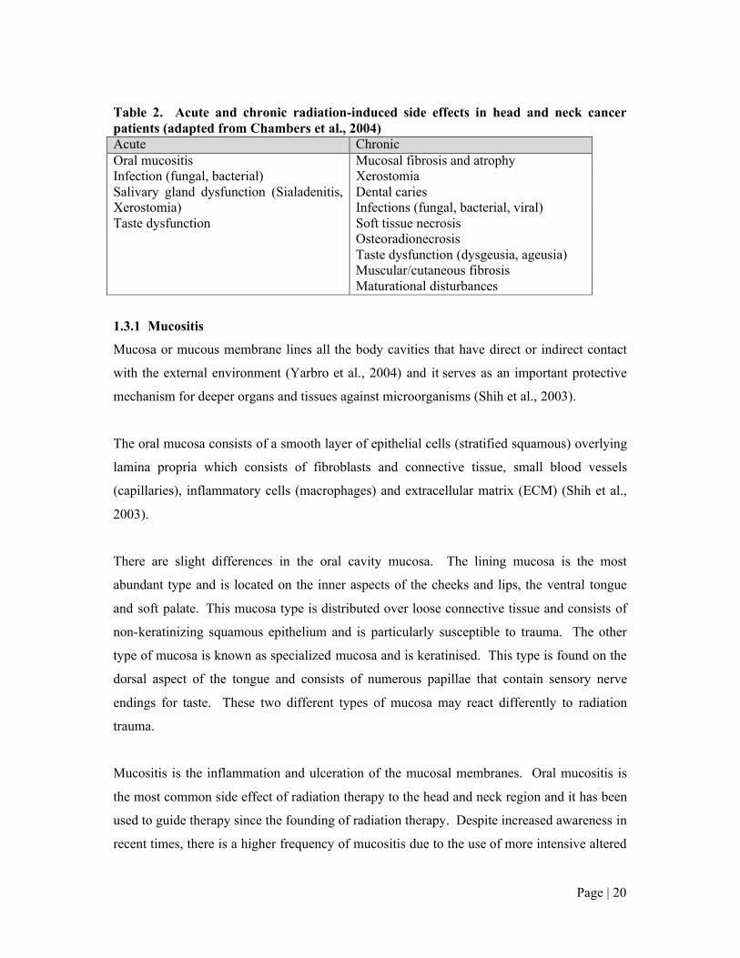
Table 2. Acute and chronic radiation-induced side effects in head and neck cancer patients (adapted from Chambers et al., 2004) Acute Chronic Oral mucositis Mucosal fibrosis and atrophy Infection (fungal, bacterial) Xerostomia Salivary gland dysfunction (Sialadenitis, Xerostomia)
Dental caries Infections (fungal, bacterial, viral)
Taste dysfunction Soft tissue necrosis Osteoradionecrosis Taste dysfunction (dysgeusia, ageusia) Muscular/cutaneous fibrosis Maturational disturbances
1.3.1 Mucositis
Mucosa or mucous membrane lines all the body cavities that have direct or indirect contact
with the external environment (Yarbro et al., 2004) and it serves as an important protective
mechanism for deeper organs and tissues against microorganisms (Shih et al., 2003).
The oral mucosa consists of a smooth layer of epithelial cells (stratified squamous) overlying
lamina propria which consists of fibroblasts and connective tissue, small blood vessels
(capillaries), inflammatory cells (macrophages) and extracellular matrix (ECM) (Shih et al.,
2003).
There are slight differences in the oral cavity mucosa. The lining mucosa is the most
abundant type and is located on the inner aspects of the cheeks and lips, the ventral tongue
and soft palate. This mucosa type is distributed over loose connective tissue and consists of
non-keratinizing squamous epithelium and is particularly susceptible to trauma. The other
type of mucosa is known as specialized mucosa and is keratinised. This type is found on the
dorsal aspect of the tongue and consists of numerous papillae that contain sensory nerve
endings for taste. These two different types of mucosa may react differently to radiation
trauma.
Mucositis is the inflammation and ulceration of the mucosal membranes. Oral mucositis is
the most common side effect of radiation therapy to the head and neck region and it has been
used to guide therapy since the founding of radiation therapy. Despite increased awareness in
recent times, there is a higher frequency of mucositis due to the use of more intensive altered
Page | 21
radiation fractionation and concurrent chemotherapy regimens. Mucositis represents a
significant clinical and economic burden in oncology (Gibson et al., 2008).
The epithelial cells of the mucosa are particularly sensitive to radiation and due to their high
turnover rate (life span of three to five days) will show radiation damage within a few weeks
of treatment. When exposed to radiation the epithelial cells become inflamed and die and
then they are sloughed off. There are large numbers of stem cells in the mucosa that are able
to differentiate and replace the lost cells. Confluent mucositis develops when the cell death
exceeds the cell renewing process i.e. there is inadequate replacement of the lost cells (Joiner
& van der Kogel, 2009).
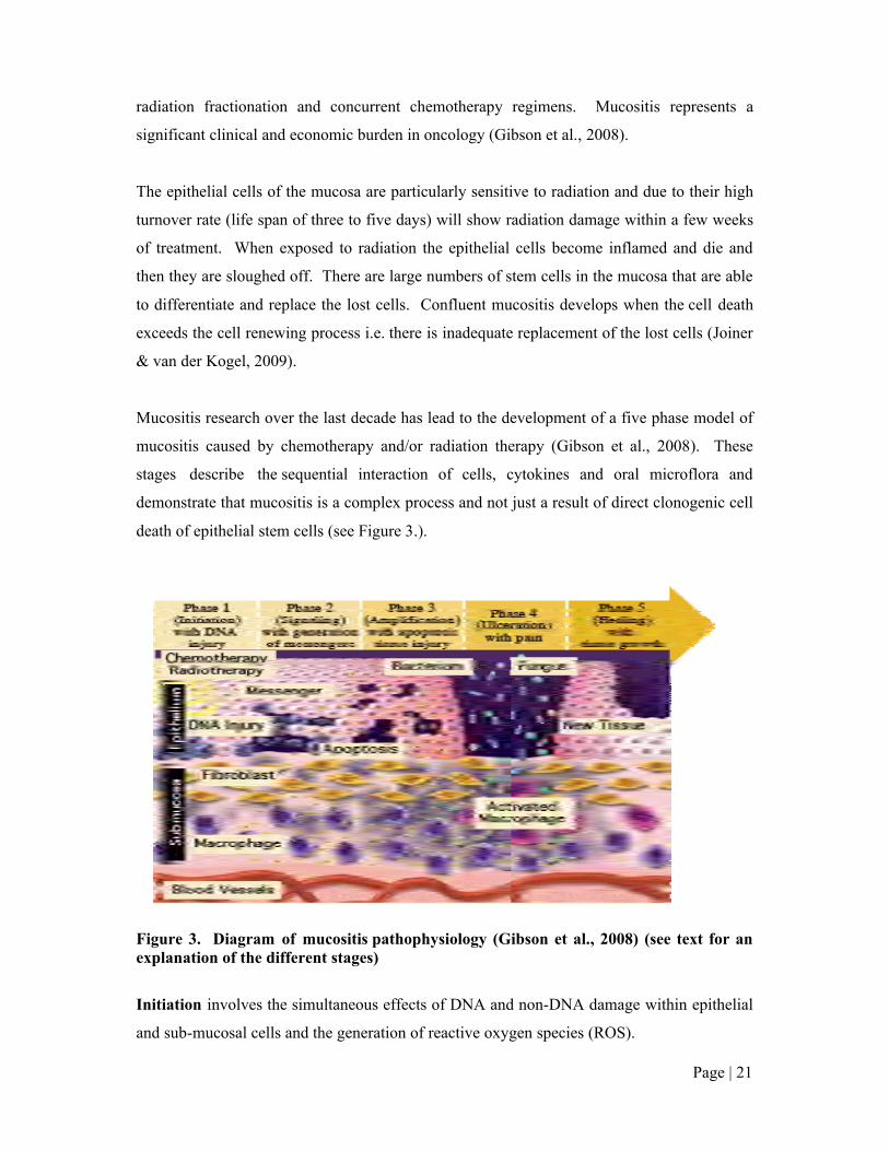
Mucositis research over the last decade has lead to the development of a five phase model of
mucositis caused by chemotherapy and/or radiation therapy (Gibson et al., 2008). These
stages describe the sequential interaction of cells, cytokines and oral microflora and
demonstrate that mucositis is a complex process and not just a result of direct clonogenic cell
death of epithelial stem cells (see Figure 3.).
Figure 3. Diagram of mucositis pathophysiology (Gibson et al., 2008) (see text for an explanation of the different stages)
Initiation involves the simultaneous effects of DNA and non-DNA damage within epithelial
and sub-mucosal cells and the generation of reactive oxygen species (ROS).

Page | 22
Up-regulation and message generation. In the next stage the DNA strands break and the
ROS activate multiple transcription factors. A series of events leads to cell cycle arrest, DNA
repair or apoptosis.
Signalling and amplification follows where pro-inflammatory cytokines (interleukin (IL)-1
and IL-6) and tumour necrosis factor (TNF-α) accumulate and target the tissues of the sub-
mucosa causing a positive feedback signal to amplify the reaction.
Ulceration (most clinically significant stage) occurs when mucosal integrity is lost and
lesions develop which are vulnerable to superficial bacterial colonisation. Bacterial cell wall
products induce immune cells to produce cytokines leading to further inflammation and
apoptosis.
Healing generally only occurs once treatment has ceased. This stage involves epithelial cell
migration, proliferation and differentiation of healing tissue. Integrity of the mucosa is
restored.
Other authors have described a similar process of mucositis with slight variations in the
descriptions of events at each particular phase (Biron et al., 2000). Sonis (1998) and Shih et
al. (2003) described the process as a four phase process; inflammatory, epithelial,
ulcerative/bacterial and healing phases.
The duration of mucositis is proportional to the degree of mucosal stem cell depletion; it can
take weeks or months to heal depending on stem cell recovery. Excessive stem cell depletion
may result in chronic open wounds known as soft tissue necrosis. Other late effects include
mucosal scarring and loss of mucosal compliance contributing to chronic dysphagia
(Rosenthal & Trotti, 2009).
Risk of developing mucositis
Not all patients are at equal risk of developing mucositis. The incidence and severity of
mucositis varies depending on the type of cancer, its management and patient related factors
(Barasch & Peterson, 2003). Some of these individual and treatment related factors are listed
in Table 3.
Page | 23
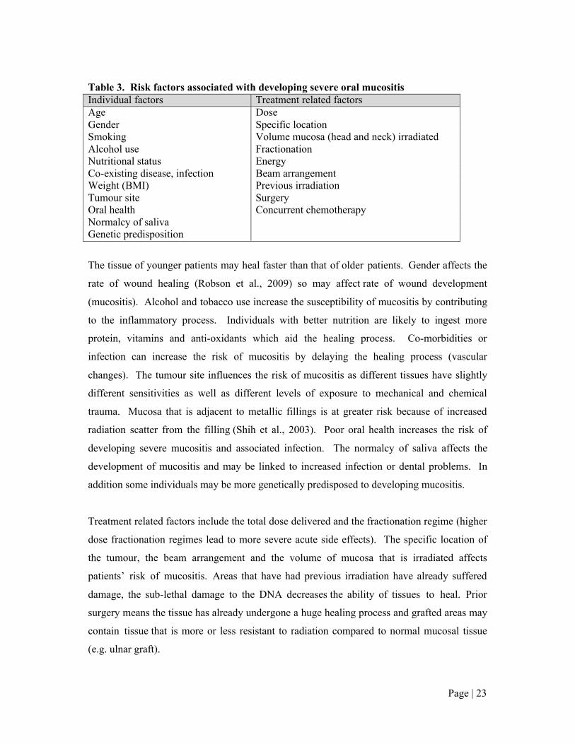
Table 3. Risk factors associated with developing severe oral mucositis Individual factors Treatment related factors Age Dose Gender Specific location Smoking Volume mucosa (head and neck) irradiated Alcohol use Fractionation Nutritional status Energy Co-existing disease, infection Beam arrangement Weight (BMI) Previous irradiation Tumour site Surgery Oral health Concurrent chemotherapy Normalcy of saliva Genetic predisposition
The tissue of younger patients may heal faster than that of older patients. Gender affects the
rate of wound healing (Robson et al., 2009) so may affect rate of wound development
(mucositis). Alcohol and tobacco use increase the susceptibility of mucositis by contributing
to the inflammatory process. Individuals with better nutrition are likely to ingest more
protein, vitamins and anti-oxidants which aid the healing process. Co-morbidities or
infection can increase the risk of mucositis by delaying the healing process (vascular
changes). The tumour site influences the risk of mucositis as different tissues have slightly
different sensitivities as well as different levels of exposure to mechanical and chemical
trauma. Mucosa that is adjacent to metallic fillings is at greater risk because of increased
radiation scatter from the filling (Shih et al., 2003). Poor oral health increases the risk of
developing severe mucositis and associated infection. The normalcy of saliva affects the
development of mucositis and may be linked to increased infection or dental problems. In
addition some individuals may be more genetically predisposed to developing mucositis.
Treatment related factors include the total dose delivered and the fractionation regime (higher
dose fractionation regimes lead to more severe acute side effects). The specific location of
the tumour, the beam arrangement and the volume of mucosa that is irradiated affects
patients’ risk of mucositis. Areas that have had previous irradiation have already suffered
damage, the sub-lethal damage to the DNA decreases the ability of tissues to heal. Prior
surgery means the tissue has already undergone a huge healing process and grafted areas may
contain tissue that is more or less resistant to radiation compared to normal mucosal tissue
(e.g. ulnar graft).
Page | 24
Chemotherapy agents have differing mucosal toxicity and as described above some act
synergistically with radiation (Gibson et al., 2008). Chemotherapy and RT affect the oral
mucosa differently and cause mucositis via different processes. All oral mucosa is
susceptible to radiation-induced mucositis but only the movable mucosa develops
chemotherapy-induced injury. Chemoradiation patients often develop mucositis earlier and
to a more severe degree.
Understanding the different factors that affect the incidence of mucositis can help determine
high risk patients and enable healthcare providers to initiate prophylactic measures to
minimize the incidence and severity of oral mucositis.
Page | 25
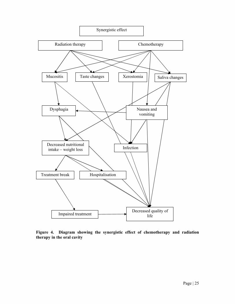
Figure 4. Diagram showing the synergistic effect of chemotherapy and radiation therapy in the oral cavity
Mucositis
Dysphagia
Radiation therapy Chemotherapy
Synergistic effect
Decreased nutritional intake – weight loss
Decreased quality of life
Saliva changes Taste changes
Infection
Treatment break Hospitalisation
Impaired treatment
Xerostomia
Nausea and vomiting
Page | 26
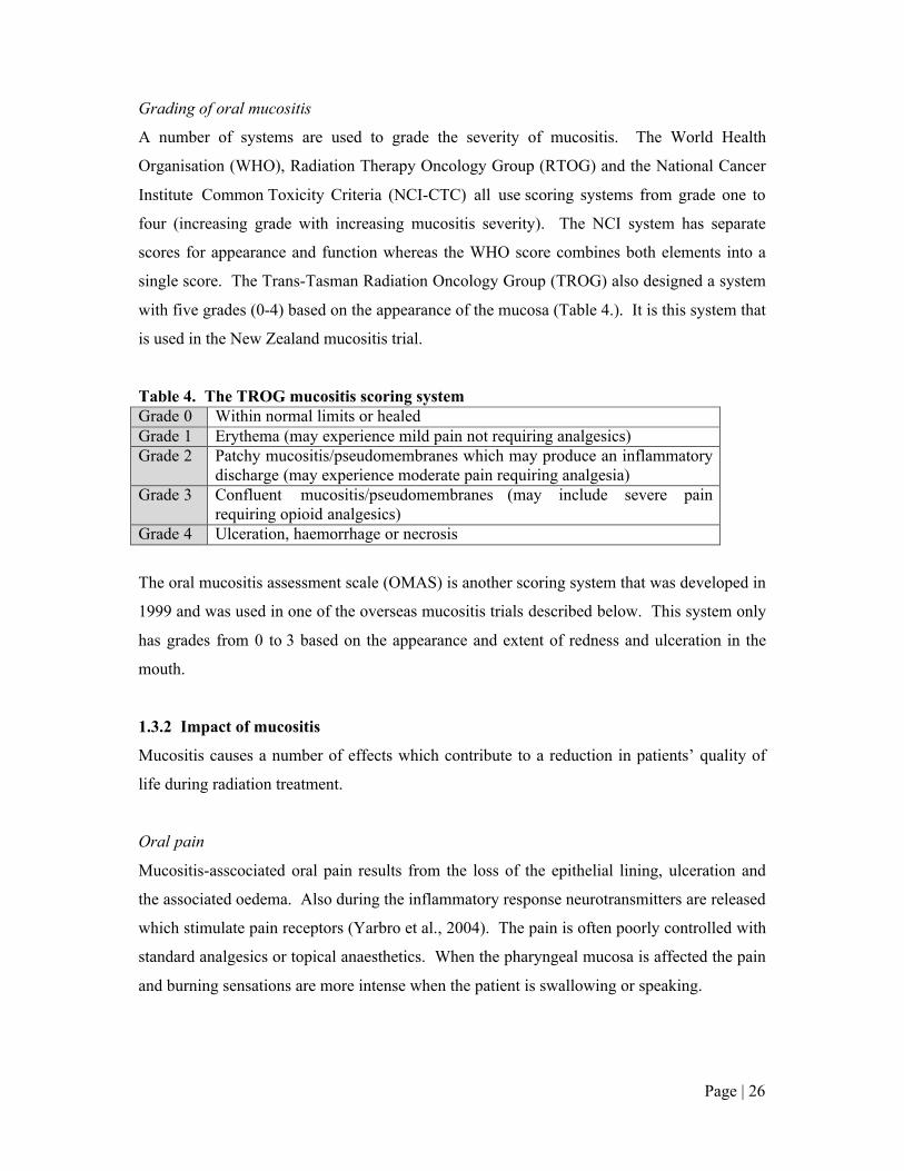
Grading of oral mucositis
A number of systems are used to grade the severity of mucositis. The World Health
Organisation (WHO), Radiation Therapy Oncology Group (RTOG) and the National Cancer
Institute Common Toxicity Criteria (NCI-CTC) all use scoring systems from grade one to
four (increasing grade with increasing mucositis severity). The NCI system has separate
scores for appearance and function whereas the WHO score combines both elements into a
single score. The Trans-Tasman Radiation Oncology Group (TROG) also designed a system
with five grades (0-4) based on the appearance of the mucosa (Table 4.). It is this system that
is used in the New Zealand mucositis trial.
Table 4. The TROG mucositis scoring system Grade 0 Within normal limits or healed Grade 1 Erythema (may experience mild pain not requiring analgesics) Grade 2 Patchy mucositis/pseudomembranes which may produce an inflammatory
discharge (may experience moderate pain requiring analgesia) Grade 3 Confluent mucositis/pseudomembranes (may include severe pain
requiring opioid analgesics) Grade 4 Ulceration, haemorrhage or necrosis
The oral mucositis assessment scale (OMAS) is another scoring system that was developed in
1999 and was used in one of the overseas mucositis trials described below. This system only
has grades from 0 to 3 based on the appearance and extent of redness and ulceration in the
mouth.
1.3.2 Impact of mucositis
Mucositis causes a number of effects which contribute to a reduction in patients’ quality of
life during radiation treatment.
Oral pain
Mucositis-asscociated oral pain results from the loss of the epithelial lining, ulceration and
the associated oedema. Also during the inflammatory response neurotransmitters are released
which stimulate pain receptors (Yarbro et al., 2004). The pain is often poorly controlled with
standard analgesics or topical anaesthetics. When the pharyngeal mucosa is affected the pain
and burning sensations are more intense when the patient is swallowing or speaking.
Page | 27
Weight loss/malnutrition
Head and neck cancer patients commonly have poor nutritional statues prior to treatment
which diminishes further through a number of mucositis related side effects such as
dysphagia and the loss of taste and saliva. Low mood also decreases appetite (Mueller et al.,
1995). Inadequate nutrition leads to weight loss and the possible requirement of feeding tube
insertion. Some patients’ ability to talk can be affected by mucositis and many report sleep
and mood disturbances. Decreased energy levels result directly from the radiation therapy
but also from the lack of nutrition and from the negative mood.
Xerostomia
The three pairs of major salivary glands in the head and neck region; the parotid, sub-
mandibular and sub-lingual glands are responsible for 90% of all salivary secretions which
are normally in excess of one litre per day (Chambers et al., 2004). Irradiation of these
salivary glands during treatment of head and neck cancer leads to a decrease in the amount
and quality of saliva produced causing patients to suffer dryness of the mouth. Dryness of
the mouth is known as xerostomia and causes oral discomfort, altered taste, nutritional
impairment (difficulty with mastication and swallowing) and dental decay. Xerostomia can
be temporary or permanent depending on the total dose the salivary glands receive, it has
been reported at as little as 6Gy and can be permanent at greater than 30Gy. Xerostomia
ranges from slight dryness, moderate dryness (poor response on stimulation) to complete
dryness of the mouth (no response on stimulation) depending on the volume and dose of the
glands irradiated. Xerostomia may also be caused or exacerbated by the concomitant or
sequential use of chemotherapy agents (Chambers et al., 2004). Different RT fractionation
regimes may also impact on the degree of xerostomia; patients treated with continuous
hyperfractionated accelerated radiotherapy (CHART) experienced less dryness of the mouth
compared to conventional fractionation regimes (Leslie & Dische, 1991).
Rosenthal et al. (2008) reported that the problem of excessive, viscid mucus in the mouth and
throat is a burdensome symptom for many patients although it is rarely reported. This
mucous may be caused by changes in saliva composition (pH level, electrolyte and
immunoglobulin (Ig) content) or be a product of mucositis or could result from a combination
of these two factors (Chambers et al., 2004).

Page | 28
Infection
Damaged mucosal tissues can easily develop infections caused by microorganisms including
fungi, viruses (herpes) and a wide variety of bacteria due to the loss of normal tissue
responses. The loss of the normal immune responses is due to decreased saliva volume,
alterations in saliva quality and decreased levels of immunity. The reduction in IgA, IgM and
IgG causes a change in the oral microbial flora which leads to a decrease in oral hygiene and
increased susceptibility to opportunistic infections when pathogens colonize the inflamed
mucosal surfaces (Yarbro et al., 2004). Some patients are at risk of developing life
threatening infection (septicaemia) (Worthington et al., 2009).
An infection in a region of mucositis will exacerbate the existing mucositis making the
patient’s condition more severe and prolonging the duration of mucositis. An example of this
is a bacterial infection where the bacteria release endotoxins which are mediators of the
inflammatory process in the oral mucosa (Yarbro et al., 2004).
Candidiasis infection (Candida albicans) is a common yeast infection in head and neck
patients. It is commonly known as oral thrush, is painful and presents with erythema or
discrete white plaques so can often be confused with mucositis. Treatment involves the use
of antifungal drugs such as the topical application of nystatin (Silverman, 1999).
Taste changes
Taste buds line the tongue and oral cavity mainly in the circumvallate and fungiform papillae
and are sensitive to radiation (Silverman, 1999). They are radiosensitive due to their high
cell turnover and are in the radiation fields for most treatments. It is the microvilli and outer
surfaces of the taste buds that suffer radiation damage causing changes in taste sensations
(Yarbro et al., 2004). The incidence of these changes is dose dependant and commonly
noticed above 10Gy (one week of treatment). Partial loss of taste is known as hypogeusia
and complete loss ageusia. Distorted taste sensation is known as dysgeusia and it has been
reported that the sweet taste is affected more than the salty taste. Taste changes are probably
linked with saliva changes as Silverman (1991) described that saliva probably has a
modulating effect on the acuity of some tastes (sour, bitter, salt, sweet) through biochemical
interactions and saliva probably provides an ionic environment in signal transduction.
Page | 29
The duration of taste changes varies between patients; recovery may occur within two to four
months after treatment but some patients may experience a permanent decrease in taste
perception (Yarbro et al., 2004). Taste is closely linked to appetite so reduction or alteration
in taste perception affects nutritional status and quality of life. The olfactory senses are not
often affected so patients should smell food to partially counteract the loss of taste.
Nausea and vomiting
In head and neck patients nausea and vomiting generally only occur in those receiving
chemotherapy. These are two of the most feared toxicities and usually happen rapidly after
chemotherapy delivery. Anticipatory nausea and vomiting is a conditioned response that
occurs before chemotherapy administration due to inadequately controlled emesis from a
previous treatment. Nausea and the taste changes impact on patients’ ability to tolerate
certain foods (honey).
Hospitalisation and treatment delays
Patients with severe mucositis have increased resource requirements and are often admitted to
hospital causing a substantial burden on the health care system (Gibson et al., 2008). In
severe cases of mucositis treatment (chemotherapy and radiation therapy) may have to be
delayed to allow for healing. Treatment delays have the potential to compromise local
tumour control through tumour cell repopulation and therefore they risk patient survival. The
planned radiation dose may have to be decreased which also leads to reduced survival.
Duncan and colleagues (1996) found an increase in risk of relapse and cancer-related
mortality in laryngeal cancer patients following a gap in radiation treatment. They suggest
that if treatment is prolonged (there is a gap longer than four days) additional radiation dose
should be prescribed to compensate for increased cell proliferation (Duncan et al., 1996).
The increase in prescribed total dose is likely to lead to more severe acute side effects and a
greater probability of late tissue complications for the patient.
Page | 30
1.4 Management of head and neck patients
Oral mucositis is the most prevalent side effect of radiation treatment to the head and neck
area which severely affects patient quality of life. Management of oral mucositis is critical to
maintain the patients food pathway, avoid interruption in the delivery of radiation treatment
and to avoid hospitalisation and the need for parenteral or tube feeding.
Currently there is no standard treatment for oral mucositis in head and neck patients
worldwide and there is a lack of clinical data to direct patient care. The Food and Drug
Administration (FDA) have no approved intervention for prevention of radiation induced
mucositis (Rosenthal & Trotti, 2009). Current management of oral mucositis is limited to
symptom control including pain relief and maintenance of good oral hygiene.
1.4.1 Oral hygiene
Patients require education about the use of mouthwashes to keep the oral cavity clean and
about avoiding irritants including spicy and course foods, smoking and alcohol (including
mouthwashes that contain alcohol). Teeth and gums should be cleaned frequently with soft
toothbrushes and fluoride toothpaste (Biron et al., 2000). Oral mouthwashes currently
advocated are salt and baking soda, hydrogen peroxide or benzydamine hydrochloride
(difflam). Dodd et al. (2003) reported that baking soda is effective in reducing the pain of
radiation induced mucositis and can help slough off cellular debris in the oral cavity. Regular
review of the oral cavity should be conducted to highlight any areas of concern. A study
investigating an antimicrobial intervention suggested that emphasis of strict oral hygiene may
significantly reduce the incidence of mucositis (Trotti et al., 2004).
1.4.2 Dental management
Head and neck patients are referred for dental assessments prior to radiation treatment for
prophylactic elimination of teeth at risk of decay with the aim of decreasing oral
complications (Ang & Garden, 2002).
1.4.3 Pain relief
Topical anaesthetics and mixtures containing anaesthetics allow a temporary reduction in oral
cavity pain, however the pain relief action of these agents also causes a reduction in patients’
Page | 31
taste and thermal perception. After applying topical anaesthetic patients are at risk of burning
or biting parts of their oral cavity which can lead to further ulceration and pain. Bongela and
xylocaine viscous are two commonly used topical anaesthetics. Yarbro et al. (2004)
described the use of oral bandages and gels that congeal in the oral cavity to form a layer on
the ulcerated surfaces providing pain relief.
Many patients experience mucositis that is severe enough to require systemic analgesia.
There are a number of medications (opioid or non-opioid) available but carry side effects
(nausea, dizziness and constipation). Most of these medications are taken orally but can be
given intravenously (IV). Common pain relief includes paracetamol (non-opioid drug),
ibuprofin (non-steroidal anti-inflammatory drug), codeine, oxycodone, morphine (opioid
drugs) and amitriptyline (a tricyclic antidepressant).
1.4.4 Nutritional management
The risk of weight loss is high for head and neck patients so nutritional support is imperative.
A dietician makes regular assessments and gives advice about diet modification including
texture changes, high calorie, high protein intake and nutritional supplements
(Fortisip/Ensure plus).
Enteral feeding
Used to enable patients to meet nutritional requirements when they are no longer able to
ingest via their oral cavity.
• Nasogastric (NG) feeding is generally a short term solution (less than four weeks) in
which fine bore tubes are placed through the patient’s nasal cavity, down their throat
and into their stomach (Evans et al., 2003).
• Gastrostomy feeding is a longer term approach (used for periods longer than four
weeks) which is more cosmetically acceptable to patients as they can be covered
when not being used and they avoid some of the complications that may arise with the
long-term use of NG tubes. The tubes for gastrostomy feeding are inserted via
endoscopy so are called percutaneous endoscopic gastrostomies (PEG). PEGs are
retained in the stomach through the use of a flange or balloon and occasionally have
complications (infection, leakage or bleeding) (Evans et al., 2003).
Page | 32
1.4.5 Infection
Infections are managed with antibiotic and antifungal ointments or lozenges, regular cleaning
and salt water rinses. Some radiation therapy departments use antifungal medication
prophylactically; patients are prescribed antifungal medication when treatment commences.
Antibiotic lozenges may be used to try and prevent bacterial colonisation and reduce
inflammation of damaged mucosa (Sutherland & Browman, 2001).
1.4.6 Specific treatment of oral mucositis
Identifying agents which can be used to prevent or treat radiation induced mucositis is a
primary concern for researchers and clinicians. Recent research has focussed on therapies
that interfere with the causative factors of mucositis with the aim of diminishing its incidence
and severity. These therapies protect normal mucosa through direct radioprotection or
manipulation of growth factors and cytokines that are involved in mucosal repopulation
(Garden, 2003). Recent studies have shown that amifostine is effective in reducing mucositis
by protecting healthy tissue from radiation (Antonadou et al., 2002). Other therapies attempt
to inhibit inflammation or infection and promote healing (Garden, 2003). One report
describes the use of lasers to decrease mucositis through increased cell division and synthesis
of myofibroblasts (Biron et al., 2000).
Humidification
Humidifiers can reduce the incidence and severity of mucositis by keeping the mucosa moist
(37°, 100% humidity), an extension of the general principal of moist wound care in wound
management (Morton et al., 2008). A randomised phase II clinical trial is currently being
conducted in New Zealand and Australia through the trans-Tasman Radiation Oncology
Group (TROG) investigating the effectiveness of humidification compared to the standard of
care.
Benzydamide hydrochloride
Difflam is a non-steroidal drug commonly used to treat the symptoms of mucositis through
anti-inflammatory mechanisms (Epstein et al., 2001). It has been described as the most
successful mouthwash to date and is recommended by the American Cancer society.
Research has more recently focussed on plants as sources of biologically active compounds
to combat treatment reactions. Extracts such as chamomile and aloe vera have been trialled

Page | 33
in the past with mixed outcomes. An ideal oral care solution or product for head and neck
cancer patients needs to reduce oral flora, have an acceptable taste, reduce oral pH, assist in
re-epithelialisation of the mucosa and be non-toxic and non-irritating to the oral tissue.
Page | 34
1.5 Honey
1.5.1 Composition
Honey is a saturated solution of sugar that is made from nectar collected from flowers by
bees of the genera Apis and Meliponinae (Namias, 2003). The nectar is mixed with enzymes
in the beehives and is placed in wax cells (honeycombs). A ripening process follows where
the enzymes (invertase) convert the sucrose into glucose and fructose and the overall water
content is reduced. As well as sugars honey contains small quantities of enzymes, amino
acids, vitamins, minerals (calcium, iron, zinc, potassium, phosphorous, magnesium, selenium,
chromium, manganese) and organic acids. The exact composition of a specific honey is
variable and depends on the geographical and floral source of the nectar (Robson et al.,
2009).
1.5.2 Medical purposes
Honey had been used for medical purposes since ancient Egyptian civilisation and has been
referenced in ancient medical writings from Greece and India. It is considered the oldest
wound dressing. The use of honey in medicine declined in the 20th Century with the
discovery of antibiotics but as some bacteria became antibiotic resistant honey has been
reconsidered as an attractive alternative (Robson et al., 2009). In recent times honey has been
used to treat burns, surgical wounds, gastric and diabetic ulcers as well as many skin
conditions including eczema, psoriasis, ringworm, athletes foot and acne (Molan, 2006).
1.5.3 Antibacterial properties
There is a large body of evidence demonstrating the antibiotic nature of honey. Robson et al.
(2009) assessed 105 patients with open wounds in a randomised clinical trial to compare
conventional dressings with a honey dressing. The trial found that the healing times for those
patients with the honey dressing were reduced compared to those with conventional
treatment. The reduction in the healing times was thought to be due to the anti-bacterial and
anti-inflammatory action of the honey.
The antibacterial property of honey is due to its acidic nature (pH ranging from 3.2 to 4.5
(Bardy et al., 2007)), its high sugar content and thus low water activity (honey is able to draw
water out of bacteria) and the production of hydrogen peroxide. Manuka honey is a specific
Page | 35
type of honey derived from the manuka tree (Leptospermum scoparium), a New Zealand
indigenous plant (Maddocks-Jennings et al., 2009). This type of honey has an additional
antibacterial component previously referred to as the unique manuka factor (UMF), now
known to be methylglyoxal (MGO) (Stephens, 2009). Several studies have reported that
manuka honey has a significantly higher level of antibacterial activity compared to other
honeys. Mavric et al. (2008) found this activity originates from MGO.
1.5.4 Physical barrier
Honey can prevent infection by forming a physical protective barrier. The barrier stops tissue
oxygenation by sealing damaged tissue from air and it allows a moist healing environment for
new cells to grow. Honey also controls odour by destroying the bacteria that cause it. There
have been reports of reductions in scar formation in areas applied with honey (Al-Waili &
Saloom, 1999).
1.5.5 Immunomodulatory function
Molan (2001) reported that honey has an anti-inflammatory effect which has been
demonstrated in histological studies of wounds in animals where there was no infection
involved. Honey contains phenolic compounds which have anti-oxidant and free radical
scavenging ability. Free radicals cause damage and prevent healing in areas of prolonged
inflammation. Molan described the way honey has visibly reduced inflammation and oedema
surrounding wounds and that pain (a feature of inflammation) is reduced with the application
of honey.
Recent research supported an anti-inflammatory activity of certain honeys in acute
inflammation (Van der Berg et al., 2008: Leong et al., manuscript in preparation). Tonks et
al. (2003) reported that honey (three different varieties; manuka, pasture and jelly bush)
modulates the action of monocytic cells. Honey affects the release of anti-inflammatory
agents and growth factors from these cells (Tonks et al., 2003).
The anti-inflammatory properties of manuka honey may be relevant to the management of
radiation-induced oral mucositis (decrease the incidence and/or severity). With respect to
this three small clinical trials have shown that different types of honey were able to decrease
the extent of radiation induced oral mucositis by at least 50%. These trials had very similar

Page | 36
methodology and were conducted in Malaysia (Biswal et al., 2003), Iran (Motallebnejed et
al., 2008) and Egypt (Rashad et al., 2008).
All three trials used 20mL of undiluted honey three times a day as a topical application in 20
patients with an additional 20 patients receiving the standard care. Biswal et al. (2003) and
Rashad et al. (2008) excluded chemoradiation patients and used a single site scoring system
to measure the extent of mucositis. In the trial by Motallebnejad et al. (2008) a multiple site
scoring system (OMAS) was used where the oral cavity was divided into nine areas and each
area was scored separately. Standard care differed between the trials with control patients in
the trial by Biswal et al. (2003) taking water, those in the trial by Motallebnejad et al. (2008)
rinsing with saline solutions and control patients in the trial by Rashad et al. (2008) having
access to analgesics, benzydamide hydrochloride, antibiotics and antifungal medication if
necessary. The three trials are described in Appendix B.
Page | 37
1.6 Aim and objectives of the study
The three previous honey studies showed that different types of honey significantly reduced
the extent of severe oral mucositis in their patient cohorts. The trials did however
recommend that further multi-centre randomised trials were conducted to validate their
findings.
New Zealand manuka honey produced by Comvita LTD is well known for its strong
antibacterial activity based on its high MGO content. We hypothesized that Comvita manuka
honey would be superior to best practice of care in decreasing radiation induced oral
mucositis in head and neck patients in the New Zealand setting. The New Zealand manuka
honey trial was designed to investigate the effects of manuka honey on radiation induced
mucositis to validate the findings of the three overseas trials.
1.6.1 Aim
To determine whether medical grade manuka honey from Comvita LTD is superior to
standard best practice in decreasing the extent of radiation-induced oral mucositis in a small
cohort of New Zealand head and neck cancer patients.
The trial aimed to address the hypothesis by following head and neck cancer patients through
their radiation treatment and comparing the effects of those receiving the department standard
of care or manuka honey intervention.
1.6.2 Objectives
The primary objective of the trial was to determine the difference in the total mean mucositis
scores for the treatment and control groups.
The secondary objectives were to determine the differences between the treatment and
control groups with regard to:
• Percent of patients developing oral mucositis
• Time of onset and dose necessary for developing mucositis
• Weight loss/nutritional intake
• Quality of life (QoL)

Page | 38
• Changes in the oral microflora composition.
1.6.3 Study design
The trial was originally designed to be a stage II randomised single blinded study in which
the research assistant responsible for patient assessment would not know which arm the
patients were randomized to (control or treatment arm). Early on a number of protocol
amendments were made (described in the methodology) including the removal of blinding
and a change in the volume and application of the honey. It was originally calculated the trial
would require a total of 120 patients in order to obtain statistical power. Preliminary analysis
proved that it was not possible to obtain the required patient numbers and the required level
of compliance. The most important amendment was a down-staging of the trial to a stage I
clinical trial due to the adverse effects experienced by the patients on the honey arm.

Page | 39
Chapter 2 METHODOLOGY
2.1 Survey of New Zealand Oncology Departments
Currently there is no standard protocol for the care of head and neck cancer patients treated
with radiation therapy in New Zealand. All eight radiotherapy departments (two private and
six public departments) have slightly different recommendations and instructions that are
given to patients to manage side effects including oral mucositis. Review of the current
practice in the public departments was conducted to highlight the common recommendations
between these centres. A staff member (the clinical tutor or head of department) from each
of the six public departments was contacted via email and questioned about their
department’s current practice for the care of head and neck patients. Each centre responded
with a brief description of their current care which is reported in the results section.
2.2 Oral Mucositis Trial
This research study was set up as a single blinded multi-centre stage II clinical trial to
determine the effect of Comvita manuka honey on the extent of oral mucositis in head and
neck cancer patients. Only data from 13 patients from Wellington Regional Hospital and one
patient from Dunedin Regional Hospital has been analysed in this thesis.
2.2.1 Participants
Participant numbers. This thesis analyses the results of 14 patients entered into the larger
multi-centre clinical trial. From March 2009 to December 2009 13 patients who received
radiation treatment at the Wellington Blood and Cancer Centre and one patient who received
treatment at the Dunedin Oncology Centre were entered into the trial.
Inclusion/exclusion. Patients with head and neck cancer were eligible for enrolment if
radiation or chemoradiation treatment was part of their disease management plan. It was a
trial requirement that patients’ radiation plans delivered a minimum of 40Gy to at least two
oral anatomical sites (as illustrated below in the mucositis assessment section). Patients were
Page | 40
excluded if they had received previous radiotherapy to the head and neck area, there was
evidence of systemic disease or they had a history of diabetes or an allergy to honey. Each
patient considered for the trial was given adequate information about the trial; its purpose and
patient requirements all of which were included in the patient information sheet (Appendix
C). Patients also had to understand the concept of the randomisation process to enable the
oncologist or research assistant (RA) to obtain informed consent.
Randomisation. Originally patients were to be informed about the trial at their first specialist
appointment (FSA) with the oncologist however this was amended and most patients were
informed about the trial and referred for enrolment at their pre-treatment planning CT
appointment. Patients who fitted the inclusion criteria and gave written informed consent had
their details (initials, age, radiotherapy dose, chemotherapy) faxed to the University of Otago
Wellington campus (Appendix D). The patient details were received by either the trial’s
principal investigator (PI) Dr Patries Herst or the department physicist Ivan Luketina who
randomised the patient using computer generated random numbers supplied by the University
of Otago Statistician Gordon Purdie. The patients were randomized to either the control or
honey arm. Randomisation was important to the trial protocol to avoid potential confounding
from the various factors that influence mucositis. The two individuals involved in the
randomisation process were not directly involved in patient care.
Patient protocol
All patients were educated on good oral hygiene, use of soft tooth brushes, fluoride
toothpaste, avoiding spicy food, alcohol and tobacco. Patients were encouraged to rinse with
water after each meal and sip small quantities of water when necessary.
Honey arm
From day one of radiation treatment patients in the honey arm were required to swirl 20mL
(later amended to 10mL) of Comvita Active UM F30+ manuka honey around their mouth for
one minute then swallow slowly to coat the oral cavity and pharynx. On treatment days
patients were required to take the honey immediately before and after RT and six hours after
RT. On non-treatment days patients were instructed to take the honey morning, afternoon
and evening.

Page | 41
Control arm
Patients received the current standard of care as per their department protocol (see NZ
departments current standard of care). The protocols differed slightly but most departments
instructed their patients to use regular mouthwashes of bicarbonate soda and salt daily.
Adverse reactions
As stipulated in the ethics application if a patient developed an adverse reaction to the honey
they had to stop using it immediately and this was to be recorded. All patients were seen on a
daily basis (Monday to Friday) by radiation therapists and three times a week by the research
assistant. Each patient enrolled was also seen by other allied health professionals who form
the multi-disciplinary team; the clinical oncologist, oncology nurse, dietician and speech
language therapist. Any adverse reactions were picked up and dealt with efficiently. Any
breaks in patients’ treatment schedules due to reactions were recorded. A patient’s radiation
oncologist could remove them from the trial if their treatment side effects were such that the
use of the honey was causing harm.
Ethics Approval
The trial obtained ethics approval from the M ulti-Region Ethics Committee in March 2009
(MEC/08/11/142). It was registered in the Australian New Zealand Clinical Trial Registry
(ACTRN12608000180314).
Funding
The salary of Dr Patries Herst was covered by the University of Otago. The salaries of Dr
David Hamilton and the other radiation oncologists were covered by the Capital and Coast
District Health Board. The manuka honey was supplied by Comvita NZ LTD free of cost.
There was no known conflict of interest on behalf of the study researchers or the Oncology
department of Wellington or Dunedin hospital.
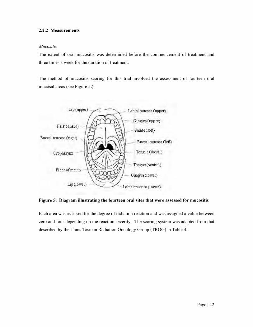
Page | 42
2.2.2 Measurements
Mucositis
The extent of oral mucositis was determined before the commencement of treatment and
three times a week for the duration of treatment.
The method of mucositis scoring for this trial involved the assessment of fourteen oral
mucosal areas (see Figure 5.).
Figure 5. Diagram illustrating the fourteen oral sites that were assessed for mucositis
Each area was assessed for the degree of radiation reaction and was assigned a value between
zero and four depending on the reaction severity. The scoring system was adapted from that
described by the Trans Tasman Radiation Oncology Group (TROG) in Table 4.
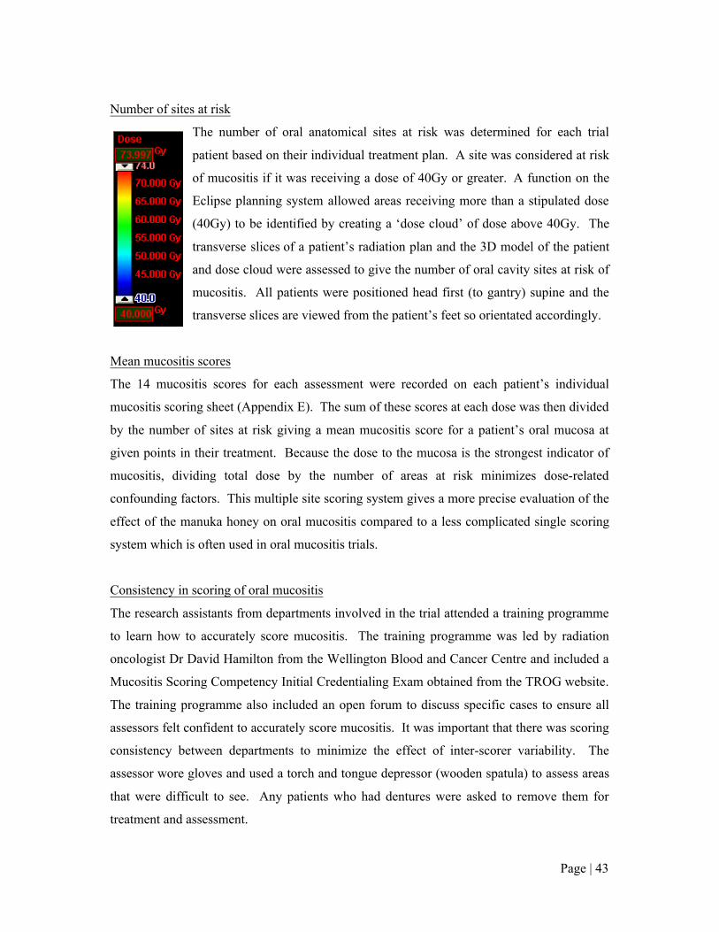
Page | 43
Number of sites at risk
The number of oral anatomical sites at risk was determined for each trial
patient based on their individual treatment plan. A site was considered at risk
of mucositis if it was receiving a dose of 40Gy or greater. A function on the
Eclipse planning system allowed areas receiving more than a stipulated dose
(40Gy) to be identified by creating a ‘dose cloud’ of dose above 40Gy. The
transverse slices of a patient’s radiation plan and the 3D model of the patient
and dose cloud were assessed to give the number of oral cavity sites at risk of
mucositis. All patients were positioned head first (to gantry) supine and the
transverse slices are viewed from the patient’s feet so orientated accordingly.
Mean mucositis scores
The 14 mucositis scores for each assessment were recorded on each patient’s individual
mucositis scoring sheet (Appendix E). The sum of these scores at each dose was then divided
by the number of sites at risk giving a mean mucositis score for a patient’s oral mucosa at
given points in their treatment. Because the dose to the mucosa is the strongest indicator of
mucositis, dividing total dose by the number of areas at risk minimizes dose-related
confounding factors. This multiple site scoring system gives a more precise evaluation of the
effect of the manuka honey on oral mucositis compared to a less complicated single scoring
system which is often used in oral mucositis trials.
Consistency in scoring of oral mucositis
The research assistants from departments involved in the trial attended a training programme
to learn how to accurately score mucositis. The training programme was led by radiation
oncologist Dr David Hamilton from the Wellington Blood and Cancer Centre and included a
Mucositis Scoring Competency Initial Credentialing Exam obtained from the TROG website.
The training programme also included an open forum to discuss specific cases to ensure all
assessors felt confident to accurately score mucositis. It was important that there was scoring
consistency between departments to minimize the effect of inter-scorer variability. The
assessor wore gloves and used a torch and tongue depressor (wooden spatula) to assess areas
that were difficult to see. Any patients who had dentures were asked to remove them for
treatment and assessment.

Page | 44
Weight Assessment
All head and neck patients receiving radiation therapy were weighed weekly. Regular
recording of weight ensures there is not excessive variation which can impact on the accuracy
of the radiation therapy plan (weight changes can alter the dose distribution). It is also
important that patients maintain a relatively stable weight to cope with the energy sapping
side effects of treatment. A radiation therapist or oncology dietician weighed each patient
weekly.
Assessment of food intake and use of systemic analgesics
Patients were given a food/drug diary and were asked to record their food/fluid/medication
intake on a daily basis. Recording later became weekly or when there was a significant
change to the patient’s diet or medications, for example the move to NG or PEG feeding.
Patients were asked to record the quantity and type of food and fluid they were consuming.
All head and neck patients had regular consultations with the oncologist and the oncology
dietician throughout their treatment. The documentation from these appointments was also
reviewed with regard to changes in the patient’s diet or medications. Discussion with the
patient and review of their oncology notes revealed if they had a PEG inserted prior to
treatment, during treatment or not at all.
Assessment of Quality of Life
The European Organisation for Research and Treatment of Cancer (EORTC) established a
quality of life department which deals with the design, implementation and analysis of quality
of life study within clinical trials. The department has input from a multidisciplinary team
(oncologists, radiation therapists, surgeons, psychiatrists, palliative care specialists,
psychologists, social workers and research methodologists) and has designed questionnaires
to assess health-related quality of life (QoL) of cancer patients participating in international
clinical trials. Two of these questionnaires were adapted for use in this oral mucositis trial;
the EORTC QLC-C30 and the EORTC QLQ HN35. A four page QoL questionnaire was
constructed and patients were asked to complete one every two weeks over their course of
treatment. The 65 questions had slightly different scoring; questions 1 to 28 and 31 to 60 had
a Likert scale of one to four (one indicating ‘not at all’, two ‘a little’, three ‘quite a bit’, four
‘very much’). Questions 29 and 30 had a Likert scale of 1 to 7 (1 being very poor and 7
being excellent). Questions 61 to 65 were yes/no questions.
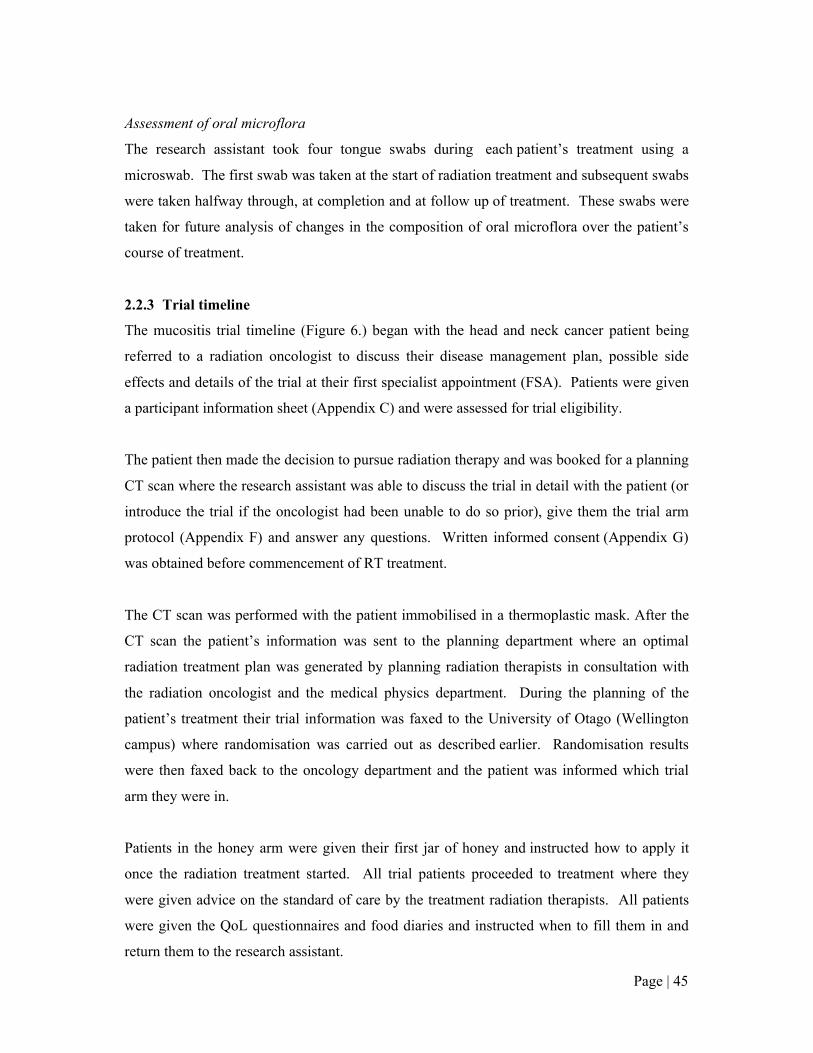
Page | 45
Assessment of oral microflora
The research assistant took four tongue swabs during each patient’s treatment using a
microswab. The first swab was taken at the start of radiation treatment and subsequent swabs
were taken halfway through, at completion and at follow up of treatment. These swabs were
taken for future analysis of changes in the composition of oral microflora over the patient’s
course of treatment.
2.2.3 Trial timeline
The mucositis trial timeline (Figure 6.) began with the head and neck cancer patient being
referred to a radiation oncologist to discuss their disease management plan, possible side
effects and details of the trial at their first specialist appointment (FSA). Patients were given
a participant information sheet (Appendix C) and were assessed for trial eligibility.
The patient then made the decision to pursue radiation therapy and was booked for a planning
CT scan where the research assistant was able to discuss the trial in detail with the patient (or
introduce the trial if the oncologist had been unable to do so prior), give them the trial arm
protocol (Appendix F) and answer any questions. Written informed consent (Appendix G)
was obtained before commencement of RT treatment.
The CT scan was performed with the patient immobilised in a thermoplastic mask. After the
CT scan the patient’s information was sent to the planning department where an optimal
radiation treatment plan was generated by planning radiation therapists in consultation with
the radiation oncologist and the medical physics department. During the planning of the
patient’s treatment their trial information was faxed to the University of Otago (Wellington
campus) where randomisation was carried out as described earlier. Randomisation results
were then faxed back to the oncology department and the patient was informed which trial
arm they were in.
Patients in the honey arm were given their first jar of honey and instructed how to apply it
once the radiation treatment started. All trial patients proceeded to treatment where they
were given advice on the standard of care by the treatment radiation therapists. All patients
were given the QoL questionnaires and food diaries and instructed when to fill them in and
return them to the research assistant.
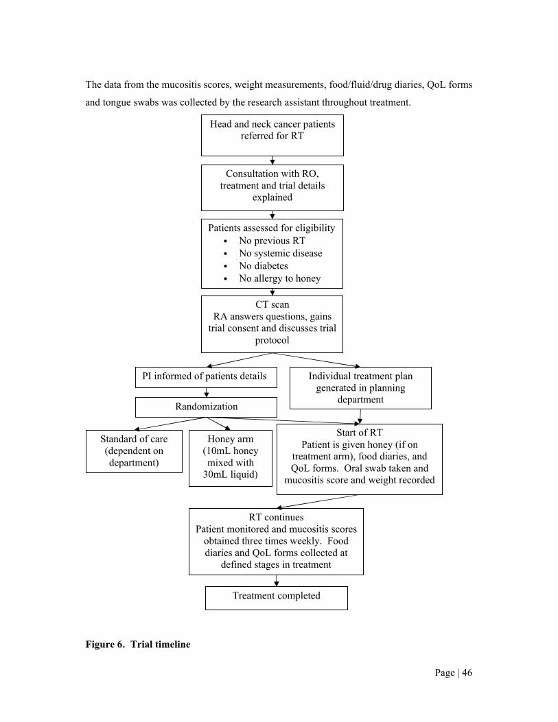
Page | 46
The data from the mucositis scores, weight measurements, food/fluid/drug diaries, QoL forms
and tongue swabs was collected by the research assistant throughout treatment.
Figure 6. Trial timeline
Head and neck cancer patients referred for RT
Consultation with RO, treatment and trial details
explained
Patients assessed for eligibility • No previous RT • No systemic disease • No diabetes • No allergy to honey
CT scan
RA answers questions, gains trial consent and discusses trial
protocol
PI informed of patients details
Randomization
Start of RT Patient is given honey (if on
treatment arm), food diaries, and QoL forms. Oral swab taken and
mucositis score and weight recorded
RT continues Patient monitored and mucositis scores
obtained three times weekly. Food diaries and QoL forms collected at
defined stages in treatment
Treatment completed
Honey arm (10mL honey mixed with
30mL liquid)
Standard of care (dependent on department)
Individual treatment plan generated in planning
department
Page | 47
2.2.4 Amendments
Several amendments were made to the original trial design and were approved by the Multi-
region Ethics Committee.
1) The first amendment pertained to the blinding of the research assistant (person who
recorded the mucositis scores) to the randomisation arm of each patient. The research
assistant was the main point of contact for patients regarding trial information so the
assistant had to be able to answer questions about the application of honey and to
address patient concerns. There was no feasible way to maintain the blinded nature of
the relationship.
2) An amendment was made to the timing of the trial introduction. Originally the trial
was to be introduced by the oncologist at the patient’s initial consultation (informed
consent could then be gained at the same appointment or by the oncologist at the CT
scan). Oncologists reported that patients were often faced with ‘information
overload’ at their first consultation (it was often at this consultation that the patients
were finding out the severity of their disease) and so the trial was not mentioned.
This resulted in head and neck patients arriving for their planning CT scan with no
prior knowledge of the trial. The amendment allowed the oncologist to introduce the
trial at the planning CT scan appointment and informed consent to be obtained by the
research assistant in some cases.
3) Application of the honey was an area of concern for the patients randomised to the
honey arm so changes were made to find a more tolerable method. Three treatment
arm patients reported that 20mL of honey was too much to hold in the mouth so the
amount to be taken three times daily was reduced to 10mL. It was then reported that
due to the thick nature of manuka honey it was not easy to spread and swirl around
the oral cavity. The honey was then diluted with water by a ratio of 1:3. The
increased volume of liquid was applied over two mouthfuls and patients were
instructed to hold the liquid in their mouth for as long as possible (ideally a minute).
To combat the intense sweetness of the honey three patients diluted it with liquids
other than water; organic apple juice, lemon and barley cordial and flat lemonade.
Page | 48
2.2.5 Head and neck RT setups
All head and neck patients are scanned and treated with an immobilisation mask which is
made at the time of the CT appointment. These masks are made with spacers underneath the
head rests to allow for the slight shrinkage that occurs between CT and treatment. The masks
are reasonably tight for patients and can contribute to a feeling of restriction in the throat.
Patients are able to swallow with these masks on although it can be more difficult than
normal. Mouth-bites (also made at the time of the CT scan) are used in some instances with
the aim of sparing certain peri-oral tissues and they allow the reproducible positioning of the
intraoral tissues. These pieces of equipment allow reproducible setups to help minimise dose
to normal tissues.
2.2.6 Data collection, entry and analysis
For each participant data from the oral mucositis scoring sheets (Appendix E) and weight
measurements were entered into a Windows Excel ’03 worksheet. From here common
information and trends in the initial assessment form were analysed as described in the results
section. Statistical analysis included determining averages and standard errors. All data was
recorded and stored carefully to ensure patient confidentiality.
Descriptive analysis
The experiences of the nine patients who completed the trial were documented and reviewed
to give a qualitative analysis of their experiences. The descriptions were based on verbal
exchanges between patients and the research assistant at the three times weekly assessments
and documentation in the clinical notes by other health professionals involved in the patients
care (dietician, speech language therapist, clinical nurse specialist and the oncologist). The
descriptive analyses allowed the identification of common experiences between the head and
neck patients.
Mucositis analysis
Each patient’s mucositis scores were recorded manually (three times weekly as per the trial
protocol) then entered into an Excel spreadsheet corresponding to the dose at each
assessment. The sum of the mucositis grades for the 14 oral cavity sites was divided by the
number of sites at risk giving the mean mucositis score for each assessment (TS/NSR). The
mean mucositis score was graphed against dose. The individual patient results were

Page | 49
combined to give an overall picture of the mucosal changes for the honey and control groups
and to illustrate the difference between the two groups. The mucositis scores were later
assessed with regard to the percentage of patients who developed certain levels of mucositis,
the dose at which mucositis first became apparent and the number of treatment delays related
to mucositis.
Weight analysis
Graphs were produced for each patient to illustrate the changes in weight throughout
treatment. The overall weight loss was recorded as was the percentage weight loss compared
to their original weight. Graphs were produced to illustrate weight change of the honey
patients compared to the control patients.
Diet/medication analysis
The food/fluid/drug diaries filled in by patients were reviewed and each patient’s diet at the
beginning of treatment was compared to that at the end. There were four main descriptions
of a patient’s diet; normal (regular food including solids), soft (only soft foods eaten, for
example mashed potato, eggs, fish and well-cooked vegetables), fluids (oral or intravenous)
and PEG/NG tube feeding (no oral foods). A diagram was used to illustrate the main changes
in the patient’s diet from the beginning of treatment through to completion.
Interpretation and analysis of the QoL questionnaires
The QoL questionnaires contained 65 questions addressing various aspects of the patient’s
health and wellbeing. For analysis seven categories (fatigue, pain, swallowing, oral cavity
changes, nutrition, mood, psychosocial wellbeing and perception of QoL) were created with
relevant questions being pooled under each category. The sum of the scores in each category
gave the overall score for that category at that point in the patient’s treatment. All pooled
questions had a scoring system of one to four (one indicating ‘not at all’ and four indicating
‘very much’).
A description of each category follows with the questions that were pooled to give the overall
category score. Each category has a range and a description of what a greater score means.
Page | 50
Fatigue
Three questions were pooled under the fatigue category to indicate the impact of fatigue in
the patient’s life at that point in time:
• Question 4 – Do you need to stay in bed or a chair during the day?
• Question 10 – Did you need to rest?
• Question 18 – Were you tired?
The patient reported score for fatigue ranges from three to 12, the greater the score the greater
the experience of fatigue.
Pain
Six questions were pooled under the pain category to indicate the degree of pain the patient
was experiencing at that point in time:
• Question 9 – Have you had pain?
• Question 19 – Did pain interfere with your daily activities?
• Question 31 – Have you had pain in your mouth?
• Question 32 – Have you had pain in your jaw?
• Question 33 – Have you had soreness in your mouth?
• Question 34 – Have you had a painful throat?
The patient reported score for pain ranges from six to 24, the greater the score the greater the
experience of pain.
A yes/no question relating to the use of pain medication was also included in the pain section.
• Question 61 – Have you used pain-killers?
Swallowing
Three questions were pooled under the swallowing category to indicate the degree of
swallowing difficulty the patient was experiencing at that point in time:
• Question 35 – Have you had problems swallowing liquids?
• Question 36 – Have you had problems swallowing pureed food?
• Question 37 – Have you had problems swallowing solid food?
The patient reported score for swallowing ranges from three to 12, the greater the score the
more difficulty the patient had swallowing.

Page | 51
Oral cavity changes
Three questions were pooled under the oral cavity changes category to indicate the extent of
negative changes experienced by the patient at that point in time.
• Question 41 – Have you had dry mouth?
• Question 42 – Have you had sticky saliva?
• Question 44 – Have you had problems with your sense of taste?
The patient reported score for oral cavity changes ranges from three to 12, the greater the
score the more negative oral cavity changes.
Nutrition - nausea/vomiting/appetite
Five questions were pooled under the nutrition category to indicate the negative changes in
nutrition that the patient experienced at that point in time:
• Question 13 – Have you lacked appetite?
• Question 14 – Have you felt nauseated?
• Question 15 – Have you vomited?
• Question 52 – Have you had trouble enjoying your meals?
• Question 49 – Have you had trouble eating?
The patient reported score for nutrition ranges from five to 20, the greater the score the more
negative changes in nutrition.
Mood
Four questions were pooled under the mood category to indicate the patient’s perceived mood
at that point in time:
• Question 21 – Did you feel tense?
• Question 22 – Did you worry?
• Question 23 – Did you feel irritable?
• Question 24 – Did you feel depressed?
The patient reported score for mood ranges from four to 16, the greater the score the more
negative the mood.
Page | 52
Psychosocial
Three questions were pooled for the psychosocial category to indicate the negative impact on
the patient’s psychosocial situation at that point in time:
• Question 26 – Has your physical condition or medical treatment interfered with you
family life?
• Question 27 – Has your physical condition or medical treatment interfered with your
social activities?
• Question 28 – Has your physical condition or medical treatment caused you financial
difficulties?
The patient reported score for their psychosocial situation ranges from three to 12, the greater
the score the more negative the patient’s psychosocial situation.
Perception of quality of life
Question 30 had a scoring system of one to seven (one indicated ‘very poor’ and seven
indicated ‘excellent’), the higher the score the better the QoL. This question was included to
give an indication of the patient’s perceived quality of life.
• Question 30 – How would you rate your overall quality of life during the past week?
Each patient’s QoL forms were analysed after the patient had completed treatment.
Questions relating to each of the seven categories were combined as described above and a
visual interpretation of the changes in each patient’s quality of life was made in the form of
two graphs; the perception of quality of life versus dose (increasing slope indicated
improving QoL) and the negative change in the seven QoL categories versus dose (increasing
slope indicates more negative change).

Page | 53
Chapter 3 RESULTS
3.1 Review of current New Zealand oncology department head and neck care
Review of the nationwide management of head and neck patients showed the lack of a
standard protocol and highlights the importance of continued research to ensure the current
standard of care is the gold standard for this vulnerable cohort of patients. The inter-
department differences depend on department resources and staff (oncologist preference) as
well as trial participation, for example the humidification trial. All six public departments
recommend patients use a baking soda and salt based mouthwash to be used several times a
day (the timing differed between departments).
Table 5. Standard of care for New Zealand departments Auckland Waikato PN # Wellington ChCh ^ Dunedin BS & S * √ √ √ √ √ √ Oral hygiene √ √ √ √ √ √ Humidification √ (trial) √ (trial) √ (trial) Dietician input √ √ √ √ √ √ Analgesia √ √ √ √ √ √ * Baking soda and salt mouthwash # PN – Palmerston North ^ Chch – Christchurch
3.1.1 Auckland DHB
The Auckland Regional Oncology Unit gives each head and neck patient their own care diary
(patient personal record). These are designed to help patients through their experience of
cancer treatment and give patients a sense of control. The diary has different sections;
contact information, side-effects information, swallowing and nutrition management, exercise
instructions to maximise the neck and shoulder movement and information about the use of
home humidifiers. Patients’ nutrition is carefully monitored by Auckland staff and dieticians
are involved with head and neck patients to try and maintain patients’ weight over the course
of treatment. Weight and diet details are recorded. Oral care advice is given and baking soda
mouthwashes recommended. Systemic and topical pain relief is prescribed as required.

Page | 54
3.1.2 Waikato DHB
The Waikato DHB encourages head and neck patients to maintain good oral hygiene from
start of treatment, including regular tooth brushing. Advice on social habits and foods to
avoid is given at the beginning of patients’ treatment. The advice includes the cessation of
smoking and alcohol consumption and avoidance of spicy foods. Mouthwashes such as
difflam and sodium bicarbonate (1/2 teaspoon of salt and baking soda and water) are
commonly used, as are analgesics and nystatin drops for prevention of candidiasis.
3.1.3 Palmerston North DHB (MidCentral DHB)
The oncology staff at the Palmerston North DHB ensure head and neck patients have a dental
assessment prior to their radiation therapy. Patients are advised to use soft toothbrushes and
fluoride toothpaste. They have regular assessment by a dietician and PEG tubes are inserted
for patients who have significant weight loss. Patients are also advised to avoid smoking and
drinking alcohol. Mouthwash instructions are given at the beginning of treatment; either
baking soda (one teaspoon in a cup of lukewarm water four times a day) or difflam (10-20mL
four times a day). The consultant oncologist may start patients on prophylactic anti-fungal
medication (nystatin 1mL drop on the tongue four times a day) at start of treatment or when
there is evidence of candida infection. Lip and gum care involves water-based moisturiser
applied to lips and gums (mouth moisturiser or oral balance gel). The Palmerston North
protocol details actions to be taken at each grade of mucositis;
Grade 1-2 mucositis: mouthwashes are to be continued as above every 2 hours. Baking soda
swabs to be used to clean around teeth and staff are to check for the presence of infections
and treat accordingly. Xylocaine viscous analgesia can be applied (5-10mLs swirled round
mouth then swallowed) four times per day before eating to provide local anaesthetic effect.
Analgesia is to be commenced as required, starting with paracetamol (tablets or liquid) and/or
ibuprofen (tablets or liquid) before adding in opiate medication.
Grade 3 mucositis: the oncologist may decide the patient is to be admitted to the ward as they
are likely to need IV fluids in addition to PEG feeding. Assessment of the oral cavity to
identify any infections that are high risk at this grade of mucositis. Hourly mouthwashes are
to be continued.
Page | 55
3.1.4 Wellington (Capital and Coast DHB)
Nursing care is focused on promoting oral hygiene, assessment, patient education and
supportive care. Mouth care regimens are individualised according to patient needs with
evidence based principals used to govern patient management. Patients have dental
assessments prior to treatment. They have education on the likelihood of ulcers and fungal
infections during treatment and how to reduce the risk of these developing: check the mouth
twice daily for any sores, areas of pain, bleeding or white film. Salt and baking soda
mouthwashes are encouraged four times a day. To combat a dry mouth patients are
instructed to drink fluids often, chew sugar-free gum and to avoid hot, spicy or acidic foods
and alcohol. The dietician is involved early in treatment and patients are advised on a high
calorie soft food plan. Anti-nausea advice is also given including small frequent meals,
regular water, avoiding fatty/spicy foods, sucking ice, peppermints or barley sugars.
Avoiding food smells is beneficial if these trigger nausea. Good personal and oral hygiene,
electric shaving (not blades) and the use of aqueous cream or aloe vera gel is encouraged.
PEGs are inserted prophylactically in high risk patients while others may have them inserted
part way through treatment if required.
3.1.5 Christchurch (Canterbury DHB)
Skin care advice (aqueous cream or pure aloe vera gel, salt water bathing, and dressings if
required) is given as are instructions regarding oral care; salt water and baking soda washes
after early meal. Soft toothbrushes or cotton buds are used to brush sensitive teeth/gums.
Topical (xylocaine viscous) or oral analgesia is prescribed as required. The patient’s
nutritional status is monitored regularly; soft food diet, supplements and weekly review by
the dietician and/or speech language therapist. Most head and neck patients will have
prophylactic PEGs inserted. Oral thrush is treated as required with antifungal medication and
patients are encouraged to sip water regularly for dry mouth. The Christchurch department is
involved in the TROG humidification trial so eligible patients may receive humidification as
part of their treatment.
3.1.6 Dunedin (Southern DHB)
The Dunedin department has registered nurses assigned to assess head and neck patients
(twice weekly). Patients are encouraged to commence strict oral hygiene at the earliest
opportunity. This advice about mouthwashes and teeth cleaning is often given to patients
Page | 56
when they come for their planning CT scan and gives patients a good foundation for
treatment. The baking soda and salt mouthwashes (made fresh each day) are to be used four
times a day at the beginning of treatment and then hourly once significant side effects start to
manifest. Patients are referred to the dental school for dental review if required. Increased
water intake is encouraged, preferably two litres per day. Pain control consists of systemic
pain relief and topical analgesics (xylocaine viscous). Oralife peppermint lip balm is
recommended to combat erythema of the lips.
Page | 57
3.2 Mucositis trial: Patient results
The New Zealand mucositis trial was conducted through the University of Otago and was a
multi-centre trial with three oncology centres participating; Palmerston North, Wellington
and Dunedin. This analysis includes the data of patients from the Wellington oncology
department and one patient from the Dunedin oncology department.
3.2.1 Patient recruitment
Trial recruitment began in March 2009 with the aim of recruiting as many eligible head and
neck patients as possible. Recruitment continued until December 2009 when it was decided
after preliminary analysis that undiluted honey was not well tolerated and that diluted honey
in the form of a mouthwash did not affect the extent of oral mucositis.
Recruitment was slow in all three centres. The Wellington department saw 27 patients
referred for radiation treatment for their head and neck cancer. When combined with the one
Dunedin patient there was a total of 28 trial potential patients seen between March and
December 2009. Of these patients only 50% (n=14) were recruited to the trial (see Figure 7.).
The other 50% of head and neck patients (n=14) were unable to participate in the trial; three
were not willing, five patients were excluded because they did not meet the selection criteria
and six potential trial head and neck patients were missed due to time and resource
constraints and a lack of communication. Of the five patients who did not meet the selection
criteria three patients had received previous radiation treatment, one had a history of diabetes
and the other had a mental health problem and did not understand the concept of
randomisation meaning informed consent could not be obtained.
The 14 patients that were recruited to the trial were randomised; nine to the treatment arm
and five to the control arm. The patients in the manuka honey arm commenced the honey
application on day one of their radiation treatment adhering to the trial instructions.
36% of patients (n=5) recruited to the trial pulled out within one week of commencing the
radiation treatment. Four of these patients were from the treatment (manuka honey) arm and

Page | 58
pulled out because they could not tolerate the manuka honey. One control patient pulled out
after being admitted to hospital with pneumonia.
The four patients on the honey arm who pulled out all experienced problems with the
application of the honey. One patient received concurrent chemotherapy and did not have
sufficient antiemetics. He therefore suffered severe nausea and vomiting after the first day of
chemotherapy. Because he also took the honey as instructed on day one he built a strong
association between nausea and vomiting and honey so he could no longer tolerate the honey.
This patient attempted to apply the honey several times after the initial incident but the
association meant he suffered a gag reflex with each attempt. Another one of the four
patients who pulled out had a strong dislike for honey and also suffered a gag reflex when
trying to apply the honey.
An amendment was made to the treatment arm protocol and was approved by the Multi-
region Ethics Committee. The change allowed the patients in the treatment arm to use less
honey (10mL instead of 20mL) and dilute the honey (1:3) in water or other liquids.
Patient accrual was slow due in part to time and resource constraints. The oncologists were
responsible for identifying potential trial patients when they met them at their FSA (first
specialist appointment). Two oncologists reported that they found it difficult to deliver the
information about the trial at the patient’s first appointment due to all the other information
they were required to deliver. The oncologists advised that it would be better to mention the
trial at a later date once the initial information regarding the patient’s disease management
had time to sink in. It then became the research assistant’s responsibility to identify the
potential patients before their CT planning scan but due to time constraints and a lack of
communication six potential patients were missed. Another change to the original study
design was made so the research assistant could gain trial patient consent at the patient’s
planning CT scan (i.e. the oncologist did not have to).

Page | 59
Figure 7. Consort diagram illustrating patient flow through the oral mucositis trial
Nine patients randomised to honey arm
14 patients unable to recruit to trial
14 patients were recruited to the trial and randomised
Six patients were missed due to resource constraints
Four honey patients pulled out
Five honey patients completed the trial
One control patient pulled out
28 patients referred for RT to head and neck area, potential trial participants
Three patients not willing to participate
Five patients randomised to the control arm
Five patients did not meet trial inclusion criteria
Four control patients completed the trial
Page | 60
3.2.2 Personal construct
These results apply to the nine patients who completed to the trial.
Age
The average age of all patients recruited to the trial was 57.3 years (range 41 to 78). Overall
patients in the honey group were younger than those in the control group. The average age of
the control patients was 61 years (range 52 to 78) whilst for the honey patients the average
was 54.4 years (range 41 to 69). The difference in the average age between the two groups
was 6.6 years.
Gender
The trial recruited four (44.4%) female (three control, one honey) and five (55.6%) male
(four honey, one control) patients. There were more males in the honey arm (80%) than the
control arm, only 25% of controls were males.
Ethnicity
Seven trial patients (77.8%) were of European descent, one was a Pacific Islander and the
other of Cook Island and Maori descent. The non-European patients were both in the control
group.
General health
One of the trial inclusion criteria was that patients needed to be in good health therefore none
of the patients had significant co-morbidities that impacted directly on their cancer treatment
or participation in the trial. The trial patients were not questioned with regard to their
smoking status or alcohol intake at the time of trial inclusion. The patient clinical oncology
notes were reviewed to ascertain information relating to this. No patients had a reported
history of alcohol or drug dependency and only one was documented as being a smoker
(control patient).
3.2.3 Therapy construct
Radiation therapy
All patients included in the trial were receiving radical radiation treatment with the aim of
local control. Each radiation plan was individualised to treat a specific volume of tissue

Page | 61
critical for each patient’s management. The dose and number and orientation of beams, the
shielding and patient set-up varied significantly to account for the variability in each patient’s
disease and contour. Plans ranged from simple conformal parallel opposed fields delivering
dose over one phase to the more complex conformal eleven field three phase plans. Four
patients received a total dose of 70Gy, one received 66Gy, two received 60Gy, one a total of
40.8Gy (despite being prescribed 48Gy) and the other a total of 40Gy.
The volume of tissue included in the treatment field impacts on the amount of mucosa that is
at risk of radiation induced inflammation. The mean mucositis scores account for this
variation by dividing the total mucositis score by the number of oral cavity sites at risk of
mucositis. All radiation plans were prescribed with the standard fractionation of 1.8-2Gy per
fraction aside from one control patient who received 2.4Gy per fraction and one honey
patient who in phase two received 1.3Gy per fraction. The dose per fraction affects the acute
radiation reactions; the higher the dose per fraction the greater the risk of an acute reaction
occurring.
Table 6. Treatment management of trial patients Patient Arm Dose (Gy) Phases Surgery Chemo Humidification WHN1 Control 40.8 (48) One √ X √ WHN3 Honey 66 Three √ X √ WHN4 Control 60 One √ X X WHN5 Honey 60 One √ X X WHC2 Control 70 Two X √ (Cisplatin) X WHC4 Honey 70 Two √ √ (Cisplatin) X WHC6 Honey 40 Two √ √ (R-CHOP) X WHC7 Control 70 Three X √ (Cisplatin) √ DHC1 Honey 69.5 Two √ √ (Cisplatin) X
Surgery
Previous surgery may result in scar tissue which has been reported as being more susceptible
to acute radiation-induced side effects. Seven (77.8%) patients had surgery prior to radiation
treatment. The two who had not were control patients.
Chemotherapy
55.6% (n=5) of trial patients had chemotherapy as part of their treatment regime. Four had
concurrent Cisplatin chemotherapy, the other patient had received R-CHOP chemotherapy
prior to radiation treatment. Of the five patients who received chemotherapy three were
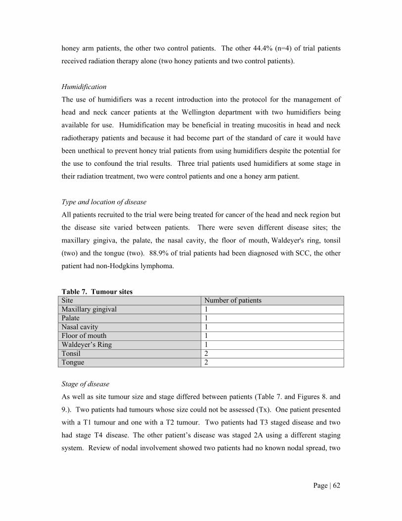
Page | 62
honey arm patients, the other two control patients. The other 44.4% (n=4) of trial patients
received radiation therapy alone (two honey patients and two control patients).
Humidification
The use of humidifiers was a recent introduction into the protocol for the management of
head and neck cancer patients at the Wellington department with two humidifiers being
available for use. Humidification may be beneficial in treating mucositis in head and neck
radiotherapy patients and because it had become part of the standard of care it would have
been unethical to prevent honey trial patients from using humidifiers despite the potential for
the use to confound the trial results. Three trial patients used humidifiers at some stage in
their radiation treatment, two were control patients and one a honey arm patient.
Type and location of disease
All patients recruited to the trial were being treated for cancer of the head and neck region but
the disease site varied between patients. There were seven different disease sites; the
maxillary gingiva, the palate, the nasal cavity, the floor of mouth, Waldeyer's ring, tonsil
(two) and the tongue (two). 88.9% of trial patients had been diagnosed with SCC, the other
patient had non-Hodgkins lymphoma.
Table 7. Tumour sites Site Number of patients Maxillary gingival 1 Palate 1 Nasal cavity 1 Floor of mouth 1 Waldeyer’s Ring 1 Tonsil 2 Tongue 2
Stage of disease
As well as site tumour size and stage differed between patients (Table 7. and Figures 8. and
9.). Two patients had tumours whose size could not be assessed (Tx). One patient presented
with a T1 tumour and one with a T2 tumour. Two patients had T3 staged disease and two
had stage T4 disease. The other patient’s disease was staged 2A using a different staging
system. Review of nodal involvement showed two patients had no known nodal spread, two

Page | 63
had N1 staged involvement, one had N2 and three had N3 staged involvement. The
remaining patient was not staged using the TNM system.
Table 8. Tumour stage Patient Arm Tumour stage Nodal stage WHN1 Control Tx N0 WHN3 Honey T1 N1 WHN4 Control Tx N1 WHN5 Honey T4 N0 WHC2 Control T3 N2 WHC4 Honey T3 N3 WHC6 Honey WHC7 Control T2 N3 DHC1 Honey T4 N3
Figure 8. Number of patients with each tumour stage as described by the TNM staging system
Figure 9. Number of patients with each nodal stage as described by the TNM staging system
Page | 64
Honey tolerance
Only 40% of the honey patients (n=2) finished treatment still applying the manuka honey and
only one continued taking it three times a day, the other had reduced to two times due to the
pain and fatigue. The other three honey patients applied the manuka honey for over two
thirds of their radiation treatment but stopped the application prior to treatment completion.
One stopped the honey at 28.8Gy (six fractions before treatment completion) due to severe
pain and nausea (chemo patient), another stopped the honey application at 64Gy (one fraction
before treatment completion) but had reduced the honey to once a day at 62Gy (two fractions
before treatment completion), she was a non-chemo patient. The fifth patient who received
concurrent chemotherapy stopped using the honey seven fractions prior to treatment
completion (after 60.4Gy) due to severe pain. 60% of honey patients found they had to mix
the honey with a liquid other than water to reduce the sweetness. These liquids included
lemonade, organic apple juice and lemon and barley cordial.
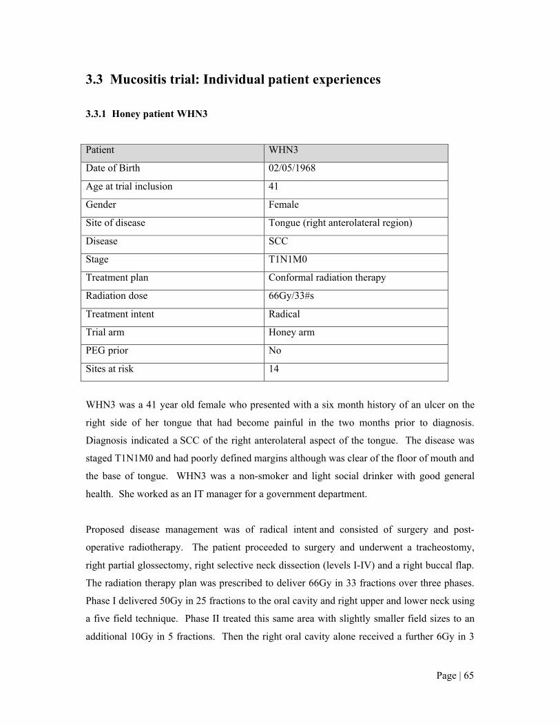
Page | 65
3.3 Mucositis trial: Individual patient experiences
3.3.1 Honey patient WHN3
Patient WHN3
Date of Birth 02/05/1968
Age at trial inclusion 41
Gender Female
Site of disease Tongue (right anterolateral region)
Disease SCC
Stage T1N1M0
Treatment plan Conformal radiation therapy
Radiation dose 66Gy/33#s
Treatment intent Radical
Trial arm Honey arm
PEG prior No
Sites at risk 14
WHN3 was a 41 year old female who presented with a six month history of an ulcer on the
right side of her tongue that had become painful in the two months prior to diagnosis.
Diagnosis indicated a SCC of the right anterolateral aspect of the tongue. The disease was
staged T1N1M0 and had poorly defined margins although was clear of the floor of mouth and
the base of tongue. WHN3 was a non-smoker and light social drinker with good general
health. She worked as an IT manager for a government department.
Proposed disease management was of radical intent and consisted of surgery and post-
operative radiotherapy. The patient proceeded to surgery and underwent a tracheostomy,
right partial glossectomy, right selective neck dissection (levels I-IV) and a right buccal flap.
The radiation therapy plan was prescribed to deliver 66Gy in 33 fractions over three phases.
Phase I delivered 50Gy in 25 fractions to the oral cavity and right upper and lower neck using
a five field technique. Phase II treated this same area with slightly smaller field sizes to an
additional 10Gy in 5 fractions. Then the right oral cavity alone received a further 6Gy in 3
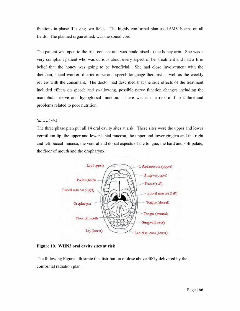
Page | 66
fractions in phase III using two fields. The highly conformal plan used 6MV beams on all
fields. The planned organ at risk was the spinal cord.
The patient was open to the trial concept and was randomised to the honey arm. She was a
very compliant patient who was curious about every aspect of her treatment and had a firm
belief that the honey was going to be beneficial. She had close involvement with the
dietician, social worker, district nurse and speech language therapist as well as the weekly
review with the consultant. The doctor had described that the side effects of the treatment
included effects on speech and swallowing, possible nerve function changes including the
mandibular nerve and hypoglossal function. There was also a risk of flap failure and
problems related to poor nutrition.
Sites at risk
The three phase plan put all 14 oral cavity sites at risk. These sites were the upper and lower
vermillion lip, the upper and lower labial mucosa, the upper and lower gingiva and the right
and left buccal mucosa, the ventral and dorsal aspects of the tongue, the hard and soft palate,
the floor of mouth and the oropharynx.
Figure 10. WHN3 oral cavity sites at risk
The following Figures illustrate the distribution of dose above 40Gy delivered by the
conformal radiation plan.
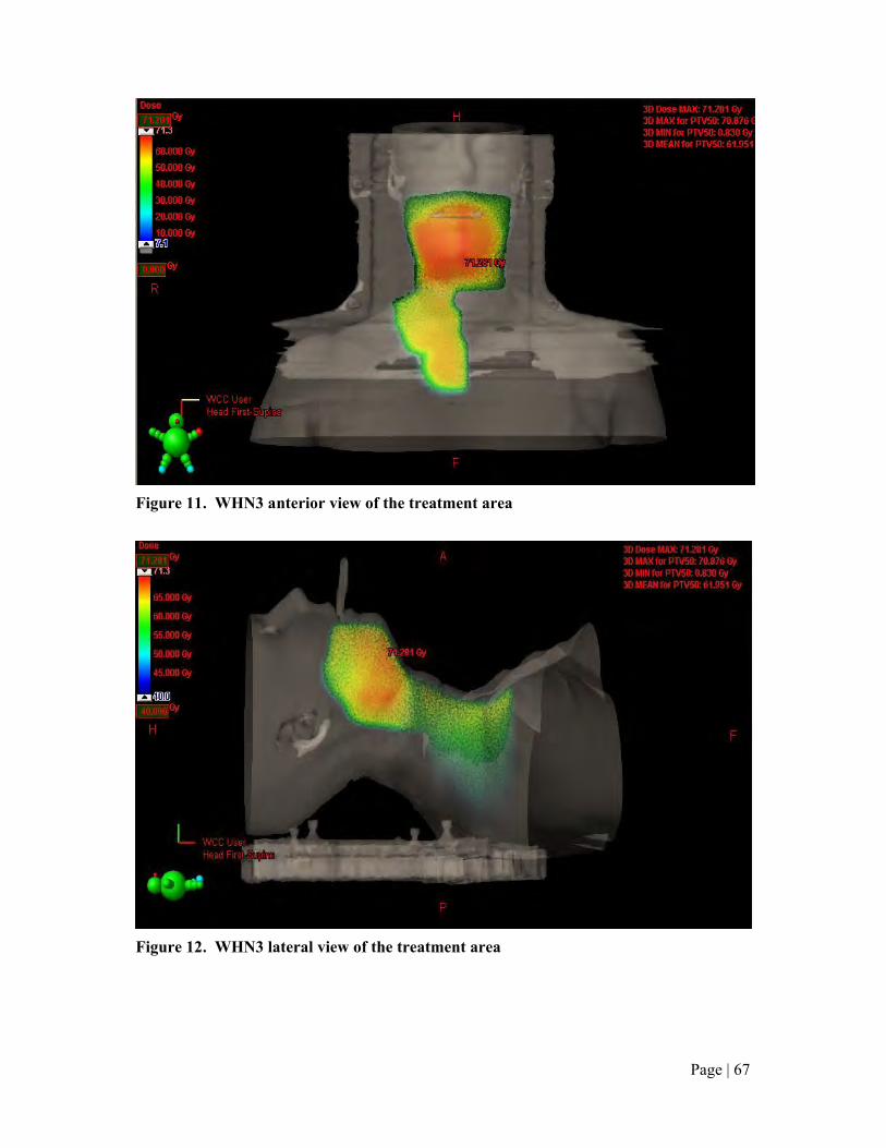
Page | 67
Figure 11. WHN3 anterior view of the treatment area
Figure 12. WHN3 lateral view of the treatment area
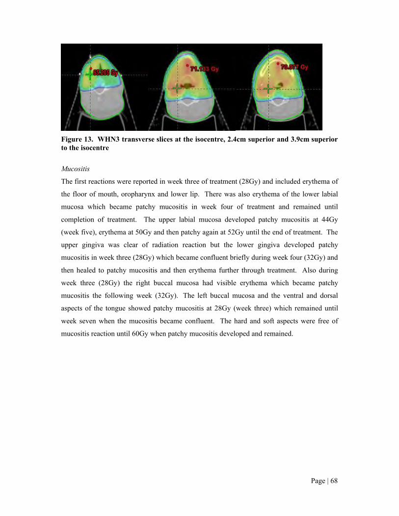
Page | 68
Figure 13. WHN3 transverse slices at the isocentre, 2.4cm superior and 3.9cm superior to the isocentre
Mucositis
The first reactions were reported in week three of treatment (28Gy) and included erythema of
the floor of mouth, oropharynx and lower lip. There was also erythema of the lower labial
mucosa which became patchy mucositis in week four of treatment and remained until
completion of treatment. The upper labial mucosa developed patchy mucositis at 44Gy
(week five), erythema at 50Gy and then patchy again at 52Gy until the end of treatment. The
upper gingiva was clear of radiation reaction but the lower gingiva developed patchy
mucositis in week three (28Gy) which became confluent briefly during week four (32Gy) and
then healed to patchy mucositis and then erythema further through treatment. Also during
week three (28Gy) the right buccal mucosa had visible erythema which became patchy
mucositis the following week (32Gy). The left buccal mucosa and the ventral and dorsal
aspects of the tongue showed patchy mucositis at 28Gy (week three) which remained until
week seven when the mucositis became confluent. The hard and soft aspects were free of
mucositis reaction until 60Gy when patchy mucositis developed and remained.

Page | 69
Figure 14. WHN3 mean mucositis score (TS/NSR) vs dose
Weight
WHN3 experienced a weight gain over the course of her treatment which is unusual in a head
and neck patient. The overall gain was 1.3kg which was a 2% gain compared to her initial
weight.
Figure 15. WHN3 change in weight vs dose
Diet
The patient had been on a soft food diet since her surgical resection and remained on this
throughout treatment with the addition of Fortisip supplements. Midway through treatment

Page | 70
she pureed her food and towards the end had a NG tube inserted and was commenced on IV
fluids to treat her dehydration.
QoL
WHN3 was a very compliant patient who completed five forms.
Figure 16. WHN3 change in perception of QoL score vs dose
Soft diet supplemented with Fortisip (x1 daily)
NG tube and IV fluids
Soft pureed diet
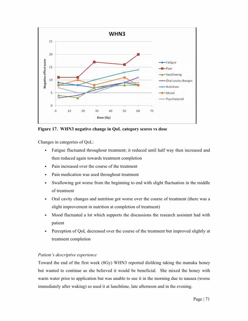
Page | 71
Figure 17. WHN3 negative change in QoL category scores vs dose
Changes in categories of QoL:
• Fatigue fluctuated throughout treatment; it reduced until half way then increased and
then reduced again towards treatment completion
• Pain increased over the course of the treatment
• Pain medication was used throughout treatment
• Swallowing got worse from the beginning to end with slight fluctuation in the middle
of treatment
• Oral cavity changes and nutrition got worse over the course of treatment (there was a
slight improvement in nutrition at completion of treatment)
• Mood fluctuated a lot which supports the discussions the research assistant had with
patient
• Perception of QoL decreased over the course of the treatment but improved slightly at
treatment completion
Patient’s descriptive experience
Toward the end of the first week (8Gy) WHN3 reported disliking taking the manuka honey
but wanted to continue as she believed it would be beneficial. She mixed the honey with
warm water prior to application but was unable to use it in the morning due to nausea (worse
immediately after waking) so used it at lunchtime, late afternoon and in the evening.
Page | 72
During the first week the patient found the treatment had heightened her sense of smell which
triggered her nausea. The patient reported finding the biotene mouthwash “much easier” to
use than the honey because the honey was difficult to fully dissolve and “dregs” would be
left in the bottom of the glass. At the beginning of week two WHN3 began mixing the honey
with organic apple juice to make it more palatable. Later in week two (14Gy) there were no
pain issues and the patient’s nausea was controlled with ondansetron. The QoL form filled
out at this stage (16Gy) showed that the perceived QoL score remained the same compared to
the beginning of treatment and the seven QoL categories had not changed dramatically.
The mouth had become more sensitive (she felt like she had “scrubbed too hard” on her
gums) and dry especially on the right side. Regular paracetomol (especially before meals)
was taken for the pain and the patient tried to eat bigger meals less often to prevent
aggravation of the mucosa. She was also using a smaller, softer toothbrush. All these
changes caused the patient a lot of stress and anxiety.
At the beginning of week three (22Gy) the patient used liquid panadol and codeine for pain
relief. She was reluctant to talk because of the pain and had noticed a change in her voice.
She avoided acidic foods and found that the source of pain was the area of previous surgery
(the flap taken from the right upper gingiva).
The patient was referred to an immunologist after experiencing ongoing nausea and
diarrhoea, she was advised to limit her fruit and vegetable intake (due to a possible fructose
intolerance). However she continued mixing the honey with apple juice as she thought the
honey was more important. The honey application continued to cause pain especially over
the back molar area and her lips felt like they had “been in the sun”. She also complained of
a mild sore throat.
Despite using morphine three times a day at the end of week three (28Gy) WHN3 still
noticed pain when chewing and swallowing and found that eating took a long time. As a
result of the pain and the development of thick saliva the patient’s food intake had decreased
and she was taking one Fortisip supplement per day.
Page | 73
Grade two mucositis was evident after three weeks and was more prominent on the left side.
There was significant pain and loss of taste. It was interesting to note that the mucositis on
left side of the tongue was a definite line one day and only patchy the next indicating a degree
of healing overnight.
Towards the end of week four (36Gy) the pain was better managed however there was an
increase in nausea. The patient’s morphine prescription was decreased because of the nausea
and she was instructed to increase her Fortisip use to three per day. The level of mucositis
had decreased and the patient reported that the honey stung less but tasted like salt.
After four weeks of treatment (40Gy) the patient was emotionally fragile. She was
introduced to humidification use and was instructed to use the humidifier at night to help
moisturise her dry mouth. Later in the week (44Gy) the patient reported significant fatigue
and new areas of mucositis were developing.
Five weeks of treatment resulted in high levels of pain especially where the back molars
touched the tongue. The patient also experienced pain that radiated up to her left ear and she
found that all meals required puree consistency. There was a decrease in the perceived QoL
score and a negative change in six of the seven QoL categories at this point in the treatment.
Interestingly, the pain category did not increase.
In the following fractions the patient’s pain and mucositis increased, she could no longer eat
solid food and had moved on to a completely pureed diet. She had further loss of saliva and a
croaky voice. The humidifier was used every night from 10:30pm to 6:30am. The patient
also used biotene dry mouth toothpaste and was started on nystatin for possible oral thrush
infection.
After six weeks of treatment (60Gy) the patient was still using the honey despite it causing a
bad stinging sensation. The pain had increased again despite being on severodol (morphine)
and panadol for pain, diazepam for insomnia and anxiety, difflam for pain and inflammation
and pilocarpine for dry mouth. During the final week of treatment (62Gy) the patient
suffered from pain, reflux, fatigue, constipation and nausea. There were areas of grade three
mucositis and as a result of these significant reactions she was admitted to hospital for NG
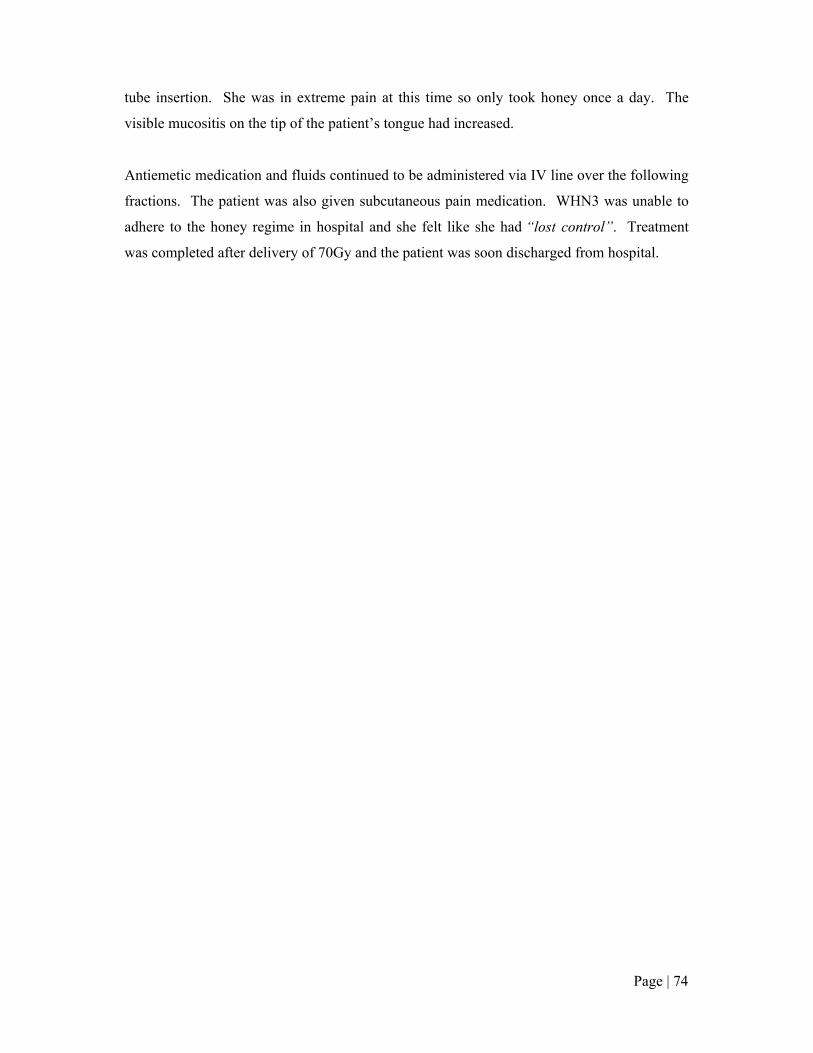
Page | 74
tube insertion. She was in extreme pain at this time so only took honey once a day. The
visible mucositis on the tip of the patient’s tongue had increased.
Antiemetic medication and fluids continued to be administered via IV line over the following
fractions. The patient was also given subcutaneous pain medication. WHN3 was unable to
adhere to the honey regime in hospital and she felt like she had “lost control”. Treatment
was completed after delivery of 70Gy and the patient was soon discharged from hospital.

Page | 75
3.3.2 Honey patient WHC4
Patient WHC4
Date of Birth 27/08/1952
Age at trial inclusion 56
Gender Male
Site of disease Palate
Disease SCC
Stage T3N3M0
Treatment plan Chemoradiation
Radiation dose 70Gy
Treatment intent Radical
Trial arm Honey
PEG prior No (inserted at 20Gy)
Sites at risk 10
WHC4 was 56 years of age when he was diagnosed with SCC of the soft palate and was
referred for radical post operative chemoradiation. The disease was staged T3N3M0 and the
recommended chemoradiation involved three weekly Cisplatin and a radiation dose of 70Gy
with the aim of local control.
The delivery of the radiation was split over two phases. Phase I used an 11 field conformal
beam arrangement to treat the bilateral upper and lower neck to 50Gy. A simpler three field
plan was used in phase II to treat the upper neck alone (primary site and involved nodes) to
an additional 20Gy. The oncologist required the spinal cord dose to be kept below 45Gy and
the brain stem below 54Gy to avoid late complications. A combination of 6 and 18 MV
beams were used.
This patient was very open to the trial concept and despite disliking the taste of standard
honey was willing to participate. He was randomised to the honey arm. Conversation with
the patient indicated he believed the manuka honey was going to be beneficial.
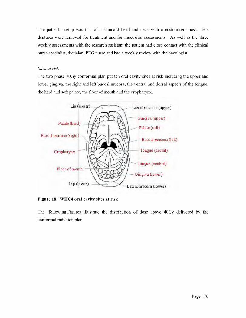
Page | 76
The patient’s setup was that of a standard head and neck with a customised mask. His
dentures were removed for treatment and for mucositis assessments. As well as the three
weekly assessments with the research assistant the patient had close contact with the clinical
nurse specialist, dietician, PEG nurse and had a weekly review with the oncologist.
Sites at risk
The two phase 70Gy conformal plan put ten oral cavity sites at risk including the upper and
lower gingiva, the right and left buccal mucosa, the ventral and dorsal aspects of the tongue,
the hard and soft palate, the floor of mouth and the oropharynx.
Figure 18. WHC4 oral cavity sites at risk
The following Figures illustrate the distribution of dose above 40Gy delivered by the
conformal radiation plan.
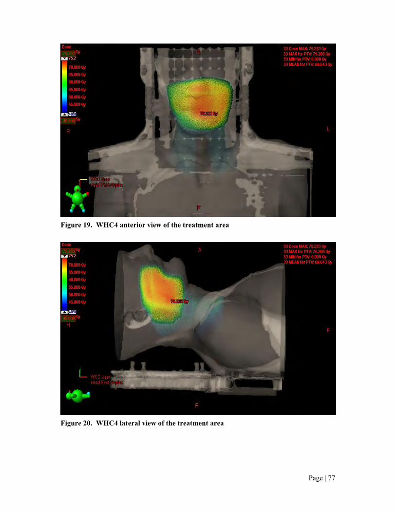
Page | 77
Figure 19. WHC4 anterior view of the treatment area
Figure 20. WHC4 lateral view of the treatment area
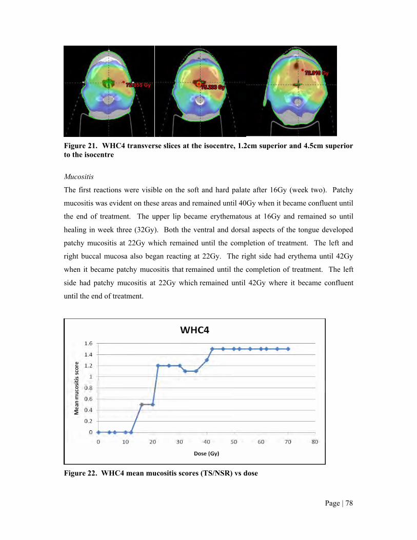
Page | 78
Figure 21. WHC4 transverse slices at the isocentre, 1.2cm superior and 4.5cm superior to the isocentre
Mucositis
The first reactions were visible on the soft and hard palate after 16Gy (week two). Patchy
mucositis was evident on these areas and remained until 40Gy when it became confluent until
the end of treatment. The upper lip became erythematous at 16Gy and remained so until
healing in week three (32Gy). Both the ventral and dorsal aspects of the tongue developed
patchy mucositis at 22Gy which remained until the completion of treatment. The left and
right buccal mucosa also began reacting at 22Gy. The right side had erythema until 42Gy
when it became patchy mucositis that remained until the completion of treatment. The left
side had patchy mucositis at 22Gy which remained until 42Gy where it became confluent
until the end of treatment.
Figure 22. WHC4 mean mucositis scores (TS/NSR) vs dose
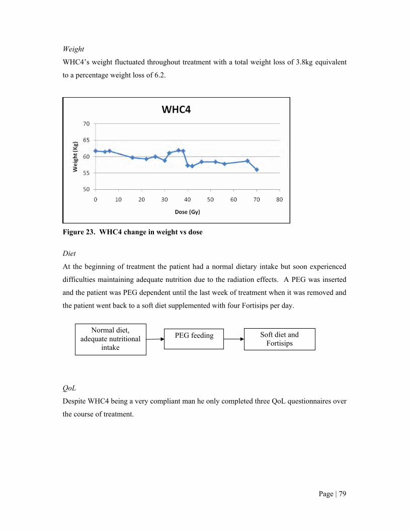
Page | 79
Weight
WHC4’s weight fluctuated throughout treatment with a total weight loss of 3.8kg equivalent
to a percentage weight loss of 6.2.
Figure 23. WHC4 change in weight vs dose
Diet
At the beginning of treatment the patient had a normal dietary intake but soon experienced
difficulties maintaining adequate nutrition due to the radiation effects. A PEG was inserted
and the patient was PEG dependent until the last week of treatment when it was removed and
the patient went back to a soft diet supplemented with four Fortisips per day.
QoL
Despite WHC4 being a very compliant man he only completed three QoL questionnaires over
the course of treatment.
Normal diet, adequate nutritional
intake
Soft diet and Fortisips
PEG feeding

Page | 80
Figure 24. WHC4 change in perception of QoL score vs dose
Figure 25. WHC4 negative change in QoL category scores vs dose
Changes in categories of QoL:
• Fatigue decreased
• Pain increased from beginning to halfway and then decreased towards the end of
treatment back to the initial level
• Pain medication was used the whole way through the treatment
• Swallowing difficulties increased
• Oral cavity changes got worse up to halfway and then improved slightly
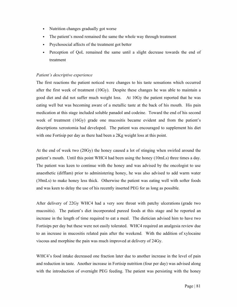
Page | 81
• Nutrition changes gradually got worse
• The patient’s mood remained the same the whole way through treatment
• Psychosocial affects of the treatment got better
• Perception of QoL remained the same until a slight decrease towards the end of
treatment
Patient’s descriptive experience
The first reactions the patient noticed were changes to his taste sensations which occurred
after the first week of treatment (10Gy). Despite these changes he was able to maintain a
good diet and did not suffer much weight loss. At 10Gy the patient reported that he was
eating well but was becoming aware of a metallic taste at the back of his mouth. His pain
medication at this stage included soluble panadol and codeine. Toward the end of his second
week of treatment (16Gy) grade one mucositis became evident and from the patient’s
descriptions xerostomia had developed. The patient was encouraged to supplement his diet
with one Fortisip per day as there had been a 2Kg weight loss at this point.
At the end of week two (20Gy) the honey caused a lot of stinging when swirled around the
patient’s mouth. Until this point WHC4 had been using the honey (10mLs) three times a day.
The patient was keen to continue with the honey and was advised by the oncologist to use
anaesthetic (difflam) prior to administering honey, he was also advised to add warm water
(30mLs) to make honey less thick. Otherwise the patient was eating well with softer foods
and was keen to delay the use of his recently inserted PEG for as long as possible.
After delivery of 22Gy WHC4 had a very sore throat with patchy ulcerations (grade two
mucositis). The patient’s diet incorporated pureed foods at this stage and he reported an
increase in the length of time required to eat a meal. The dietician advised him to have two
Fortisips per day but these were not easily tolerated. WHC4 required an analgesia review due
to an increase in mucositis related pain after the weekend. With the addition of xylocaine
viscous and morphine the pain was much improved at delivery of 24Gy.
WHC4’s food intake decreased one fraction later due to another increase in the level of pain
and reduction in taste. Another increase in Fortisip nutrition (four per day) was advised along
with the introduction of overnight PEG feeding. The patient was persisting with the honey
Page | 82
application despite the associated pain which had not decreased with the introduction of
difflam prior to application.
At the end of week three of treatment (30Gy) the patient had lost 3Kg. His pain medication
had to be increased to include morphine elixir two to three times per day. The QoL form
filled in after delivery of 32Gy illustrates the negative effect treatment had on six of the seven
QoL categories when compared to the beginning of treatment. At this stage the patients
perceived QoL score remained the same.
At 36Gy WHC4 was in a lot of pain especially on the left upper gingiva area and found the
pain more intense when using the honey. By mixing the honey with warm water he was able
to soften it enough to be able to swirl it around his mouth and coat the mucosa. The patient
was very persistent with the honey, he was reassured he could stop if the pain became too
intense and to report any changes to the research assistant.
The level of mucositis had increased throughout the mouth after 40Gy. At 42Gy there was
severe confluent mucositis and suspected candida infection. The patient’s mouth was very
sore and his morphine use had increased. Nutritional intake had decreased and the weight
loss since the start of treatment was now 4.5Kg. Soft meals, Fortisip supplements and
overnight PEG feeding advice was reiterated.
At the end of week five (50Gy) the patient reported vomiting and a severe headache. Grade
two mucositis was obvious in the oral cavity. The main issue for the patient at this stage was
swallowing and trying to maintain his weight with the use of Fortisips after experiencing
problems with his PEG (placement and infection).
After week six (60Gy) the mucositis appeared to have stabilised. Patchy mucositis was noted
on the soft and hard palate and on the left buccal mucosa. The patient found that all food,
fluids and medications stung but he reported that things were not getting worse. After the
initial problems were addressed WHC4 was able to use his PEG frequently with additional
oral intake occasionally. His weight had also stabilised at this point and the patient looked
better and sounded more cheerful. The QoL questionnaire filled in after six weeks of
treatment reported that all the QoL categories had improved aside from the nutrition category.
Page | 83
The overall perceived QoL score had, however, got worse compared to the score after three
weeks of treatment.
Fatigue had increased after 62Gy (early in week seven) and the patient had begun to mix the
honey with flat lemonade to reduce the sweetness and thickness of the honey. The patient’s
PEG was taken out after 68Gy due to difficulties feeding through it and severe nausea.
WHC4 continued with Fortisip nutritional supplements and soft foods through to treatment
completion.
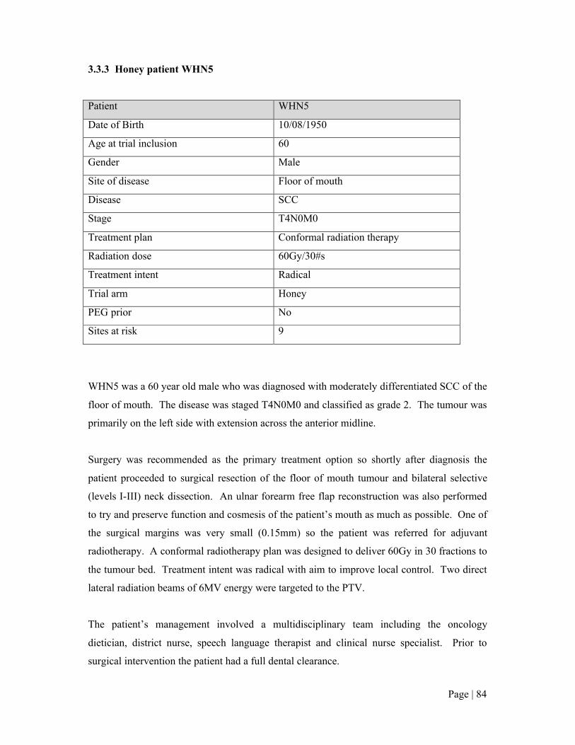
Page | 84
3.3.3 Honey patient WHN5
Patient WHN5
Date of Birth 10/08/1950
Age at trial inclusion 60
Gender Male
Site of disease Floor of mouth
Disease SCC
Stage T4N0M0
Treatment plan Conformal radiation therapy
Radiation dose 60Gy/30#s
Treatment intent Radical
Trial arm Honey
PEG prior No
Sites at risk 9
WHN5 was a 60 year old male who was diagnosed with moderately differentiated SCC of the
floor of mouth. The disease was staged T4N0M0 and classified as grade 2. The tumour was
primarily on the left side with extension across the anterior midline.
Surgery was recommended as the primary treatment option so shortly after diagnosis the
patient proceeded to surgical resection of the floor of mouth tumour and bilateral selective
(levels I-III) neck dissection. An ulnar forearm free flap reconstruction was also performed
to try and preserve function and cosmesis of the patient’s mouth as much as possible. One of
the surgical margins was very small (0.15mm) so the patient was referred for adjuvant
radiotherapy. A conformal radiotherapy plan was designed to deliver 60Gy in 30 fractions to
the tumour bed. Treatment intent was radical with aim to improve local control. Two direct
lateral radiation beams of 6MV energy were targeted to the PTV.
The patient’s management involved a multidisciplinary team including the oncology
dietician, district nurse, speech language therapist and clinical nurse specialist. Prior to
surgical intervention the patient had a full dental clearance.
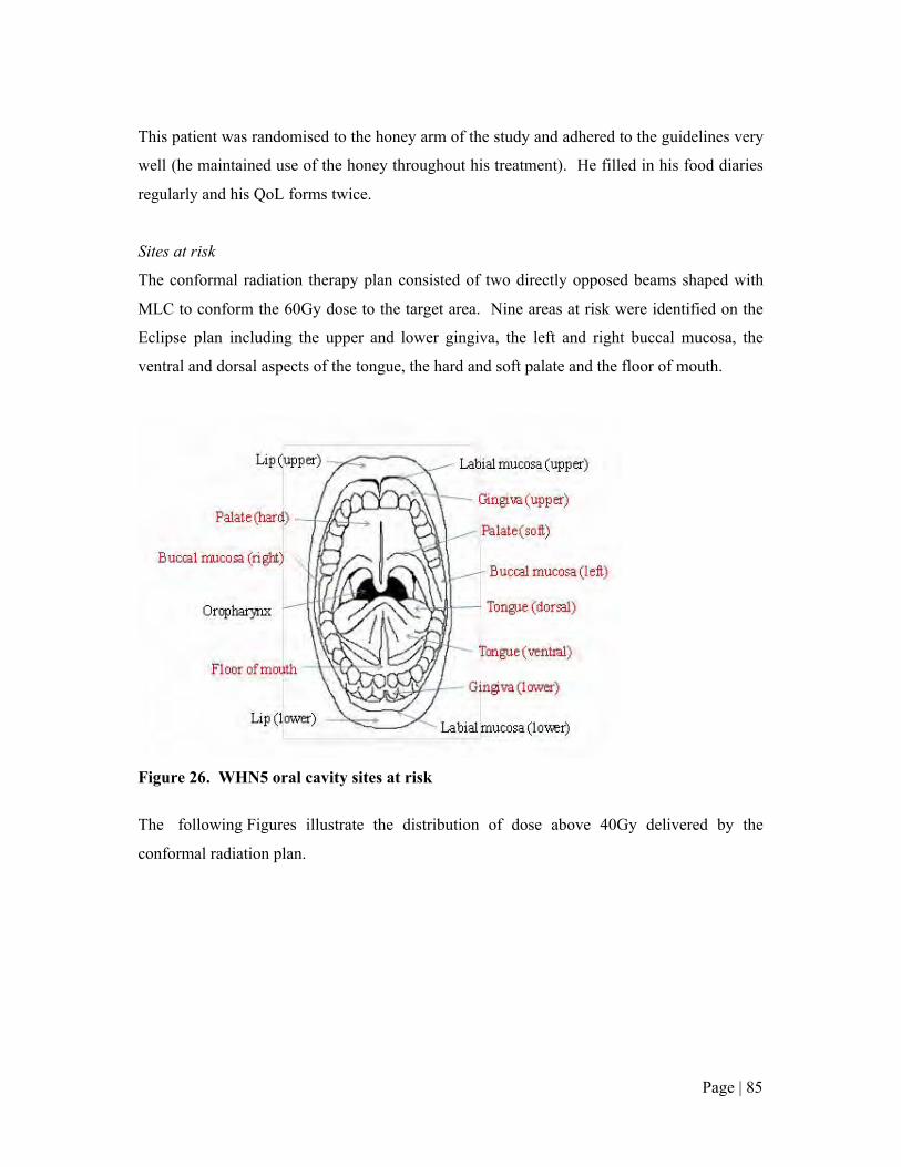
Page | 85
This patient was randomised to the honey arm of the study and adhered to the guidelines very
well (he maintained use of the honey throughout his treatment). He filled in his food diaries
regularly and his QoL forms twice.
Sites at risk
The conformal radiation therapy plan consisted of two directly opposed beams shaped with
MLC to conform the 60Gy dose to the target area. Nine areas at risk were identified on the
Eclipse plan including the upper and lower gingiva, the left and right buccal mucosa, the
ventral and dorsal aspects of the tongue, the hard and soft palate and the floor of mouth.
Figure 26. WHN5 oral cavity sites at risk
The following Figures illustrate the distribution of dose above 40Gy delivered by the
conformal radiation plan.

Page | 86
Figure 27. WHN5 anterior view of the treatment area
Figure 28. WHN5 lateral view of the treatment area
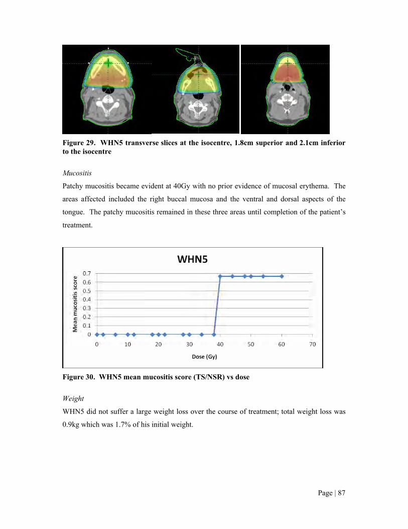
Page | 87
Figure 29. WHN5 transverse slices at the isocentre, 1.8cm superior and 2.1cm inferior to the isocentre
Mucositis
Patchy mucositis became evident at 40Gy with no prior evidence of mucosal erythema. The
areas affected included the right buccal mucosa and the ventral and dorsal aspects of the
tongue. The patchy mucositis remained in these three areas until completion of the patient’s
treatment.
Figure 30. WHN5 mean mucositis score (TS/NSR) vs dose
Weight
WHN5 did not suffer a large weight loss over the course of treatment; total weight loss was
0.9kg which was 1.7% of his initial weight.
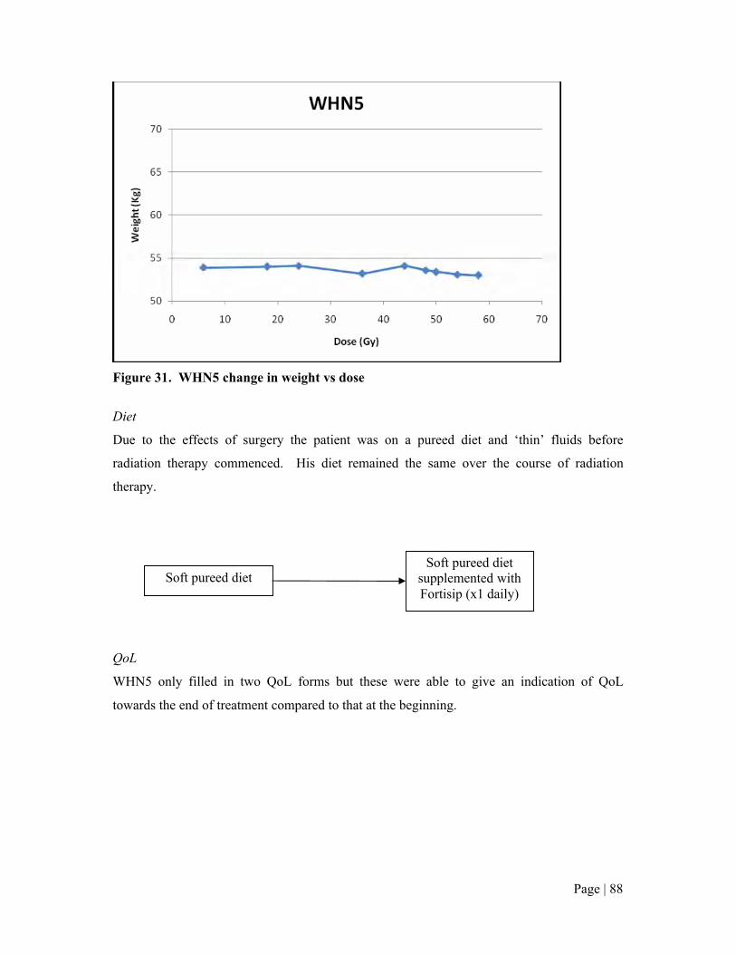
Page | 88
Figure 31. WHN5 change in weight vs dose
Diet
Due to the effects of surgery the patient was on a pureed diet and ‘thin’ fluids before
radiation therapy commenced. His diet remained the same over the course of radiation
therapy.
QoL
WHN5 only filled in two QoL forms but these were able to give an indication of QoL
towards the end of treatment compared to that at the beginning.
Soft pureed diet Soft pureed diet
supplemented with Fortisip (x1 daily)

Page | 89
Figure 32. WHN5 change in perception of QoL score vs dose
Figure 33. WHN5 negative change in QoL category scores vs dose
Changes in categories of QoL:
• Fatigue reduced over the course of treatment
• Pain level increased slightly
• There was no requirement for pain medication over the course of the treatment
• Swallowing ability remained the same
• Oral cavity changes improved
Page | 90
• Nutrition improved slightly
• Mood improved over the course of treatment
• Psychosocial effects and perceived QoL also improved
A lot of these results were unusual, usually a head and neck patient experiences more
negative changes the further through treatment they progress. It may be that this patient
experienced a reduction in anxiety, got used to the treatment and the side effects or it is
possible that the effects of the previous surgery diminished the more time that passed causing
a perceived improvement in QoL.
Patient’s descriptive experience
The patient found the mouth-bite and mask very uncomfortable and early in treatment the
mask had to be loosened and assessed by the oncologist. Treatment proceeded and the
patient started using the soda and salt mouthwash every two hours as well as the honey three
times daily. The honey was diluted with warm water to make the honey easier to apply to the
oral mucosa.
During the first week of treatment the patient handled the honey and mouthwashes well and
there was no evidence of mucositis and no descriptions of pain. Since his surgery WHN5 had
maintained a good diet, he knew the importance of maintaining a high calorie and high
protein diet throughout treatment and was well-equipped with a blender to puree his food.
The first reaction the patient noticed toward the end of the first week was a slightly dry
mouth, mainly at night.
Towards the end of week two (18Gy) no further radiation effects were evident; the mouth
was clear of mucositis and infection and the patient had made no changes to his eating or
drinking. The dryness of mouth became more evident after 38Gy (week four) but this did not
cause any alterations in oral intake. The patient had been prescribed bongela and was using it
sparingly.
During week five (44Gy) the patient noticed pain in the right buccal area although it was not
severe enough to warrant pain medication. He found that the honey stung more on the right
side of the mouth and more so than the other mouthwash he was using. The oral pain did not
Page | 91
extend to the throat. At the end of week five (48Gy) an area of ulceration developed on the
right side of the patient’s mouth but it still did not require oral analgesia. At this stage the
patient reported a good appetite with no nausea; he was drinking without pain and was trying
to supplement his diet with one Fortisip per day.
After 50Gy patchy mucositis was evident and the right ulcer was clearly visible but “not too
painful”. WHN5’s QoL form reported his pain level was worse than the beginning of
treatment, his swallowing had got slightly worse but the other five categories had marginally
improved which was interesting given the cumulative effect of the radiation dose.
At this stage in his treatment the patient was continuing with the honey three times a day. He
was instructed to continue coating all the oral mucosa by moving his tongue although his
tongue movement was limited by his graft. It was interesting to note that despite an increase
in pain his perceived QoL score had improved.
For the rest of the treatment the patient maintained the same pureed diet with no other
changes in the oral cavity; the level of mucositis remained the same. Over the whole course
of treatment delivery of 60Gy the patient only suffered 0.9kg weight loss which results from
his good oral intake throughout treatment.
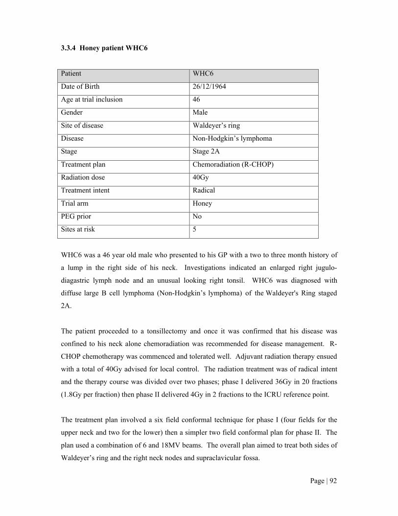
Page | 92
3.3.4 Honey patient WHC6
Patient WHC6
Date of Birth 26/12/1964
Age at trial inclusion 46
Gender Male
Site of disease Waldeyer’s ring
Disease Non-Hodgkin’s lymphoma
Stage Stage 2A
Treatment plan Chemoradiation (R-CHOP)
Radiation dose 40Gy
Treatment intent Radical
Trial arm Honey
PEG prior No
Sites at risk 5
WHC6 was a 46 year old male who presented to his GP with a two to three month history of
a lump in the right side of his neck. Investigations indicated an enlarged right jugulo-
diagastric lymph node and an unusual looking right tonsil. WHC6 was diagnosed with
diffuse large B cell lymphoma (Non-Hodgkin’s lymphoma) of the Waldeyer's Ring staged
2A.
The patient proceeded to a tonsillectomy and once it was confirmed that his disease was
confined to his neck alone chemoradiation was recommended for disease management. R-
CHOP chemotherapy was commenced and tolerated well. Adjuvant radiation therapy ensued
with a total of 40Gy advised for local control. The radiation treatment was of radical intent
and the therapy course was divided over two phases; phase I delivered 36Gy in 20 fractions
(1.8Gy per fraction) then phase II delivered 4Gy in 2 fractions to the ICRU reference point.
The treatment plan involved a six field conformal technique for phase I (four fields for the
upper neck and two for the lower) then a simpler two field conformal plan for phase II. The
plan used a combination of 6 and 18MV beams. The overall plan aimed to treat both sides of
Waldeyer’s ring and the right neck nodes and supraclavicular fossa.
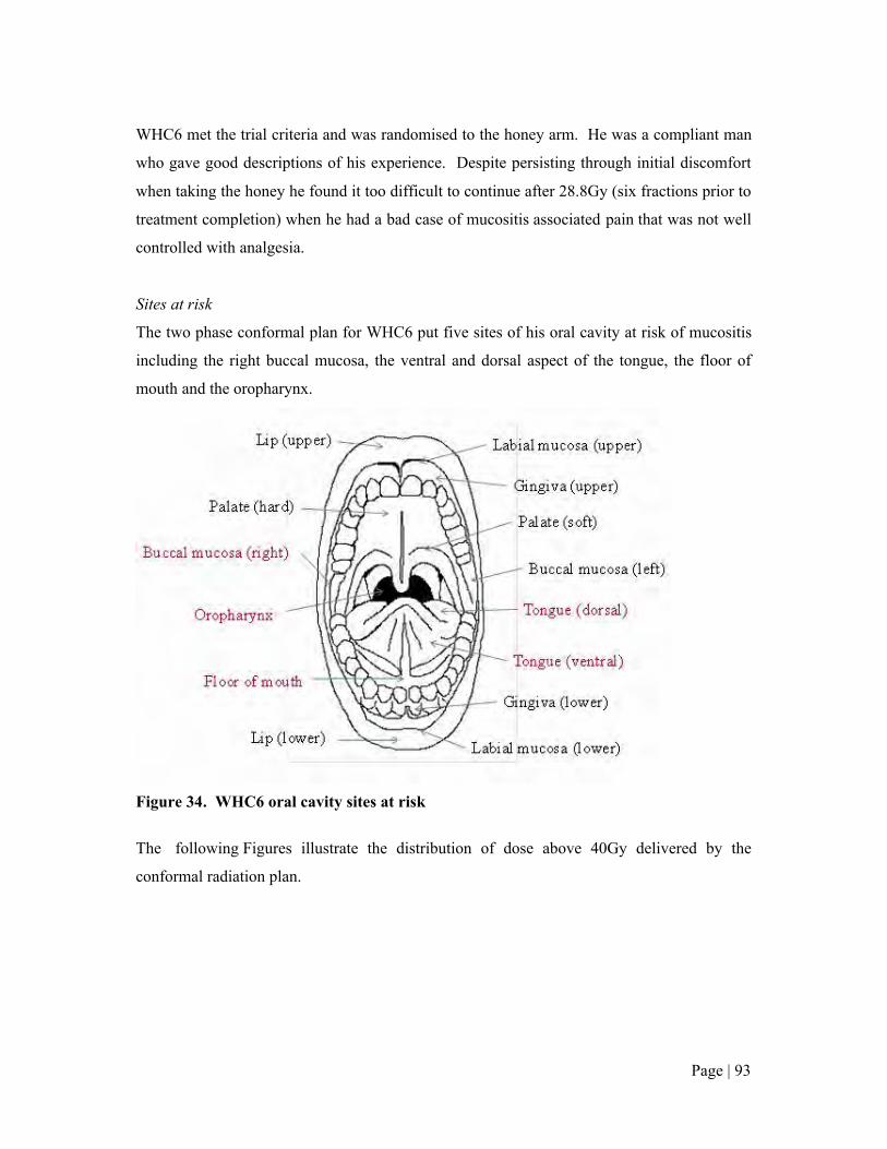
Page | 93
WHC6 met the trial criteria and was randomised to the honey arm. He was a compliant man
who gave good descriptions of his experience. Despite persisting through initial discomfort
when taking the honey he found it too difficult to continue after 28.8Gy (six fractions prior to
treatment completion) when he had a bad case of mucositis associated pain that was not well
controlled with analgesia.
Sites at risk
The two phase conformal plan for WHC6 put five sites of his oral cavity at risk of mucositis
including the right buccal mucosa, the ventral and dorsal aspect of the tongue, the floor of
mouth and the oropharynx.
Figure 34. WHC6 oral cavity sites at risk
The following Figures illustrate the distribution of dose above 40Gy delivered by the
conformal radiation plan.
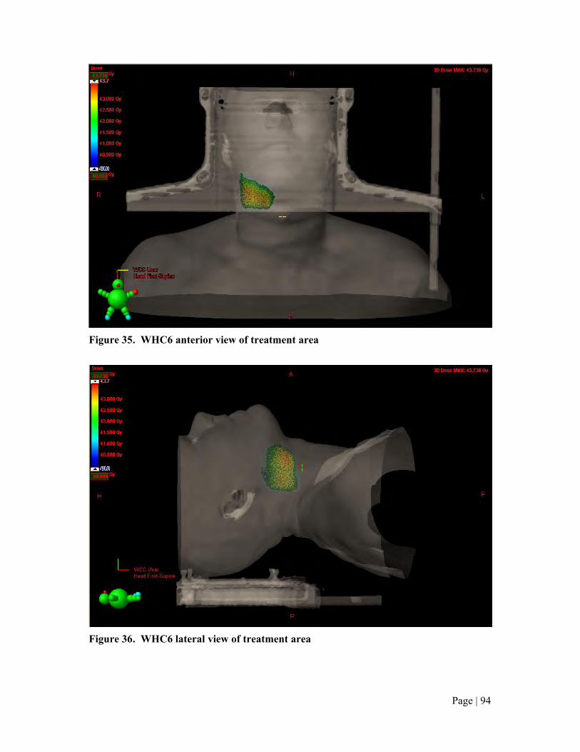
Page | 94
Figure 35. WHC6 anterior view of treatment area
Figure 36. WHC6 lateral view of treatment area
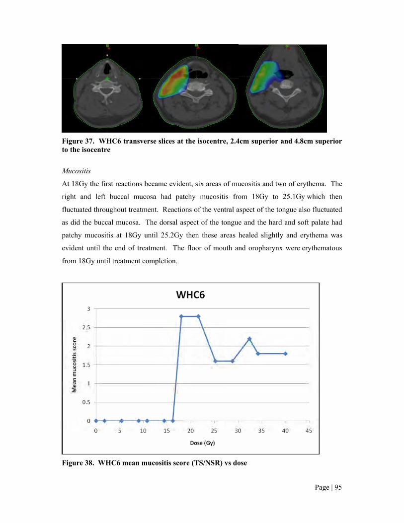
Page | 95
Figure 37. WHC6 transverse slices at the isocentre, 2.4cm superior and 4.8cm superior to the isocentre
Mucositis
At 18Gy the first reactions became evident, six areas of mucositis and two of erythema. The
right and left buccal mucosa had patchy mucositis from 18Gy to 25.1Gy which then
fluctuated throughout treatment. Reactions of the ventral aspect of the tongue also fluctuated
as did the buccal mucosa. The dorsal aspect of the tongue and the hard and soft palate had
patchy mucositis at 18Gy until 25.2Gy then these areas healed slightly and erythema was
evident until the end of treatment. The floor of mouth and oropharynx were erythematous
from 18Gy until treatment completion.
Figure 38. WHC6 mean mucositis score (TS/NSR) vs dose
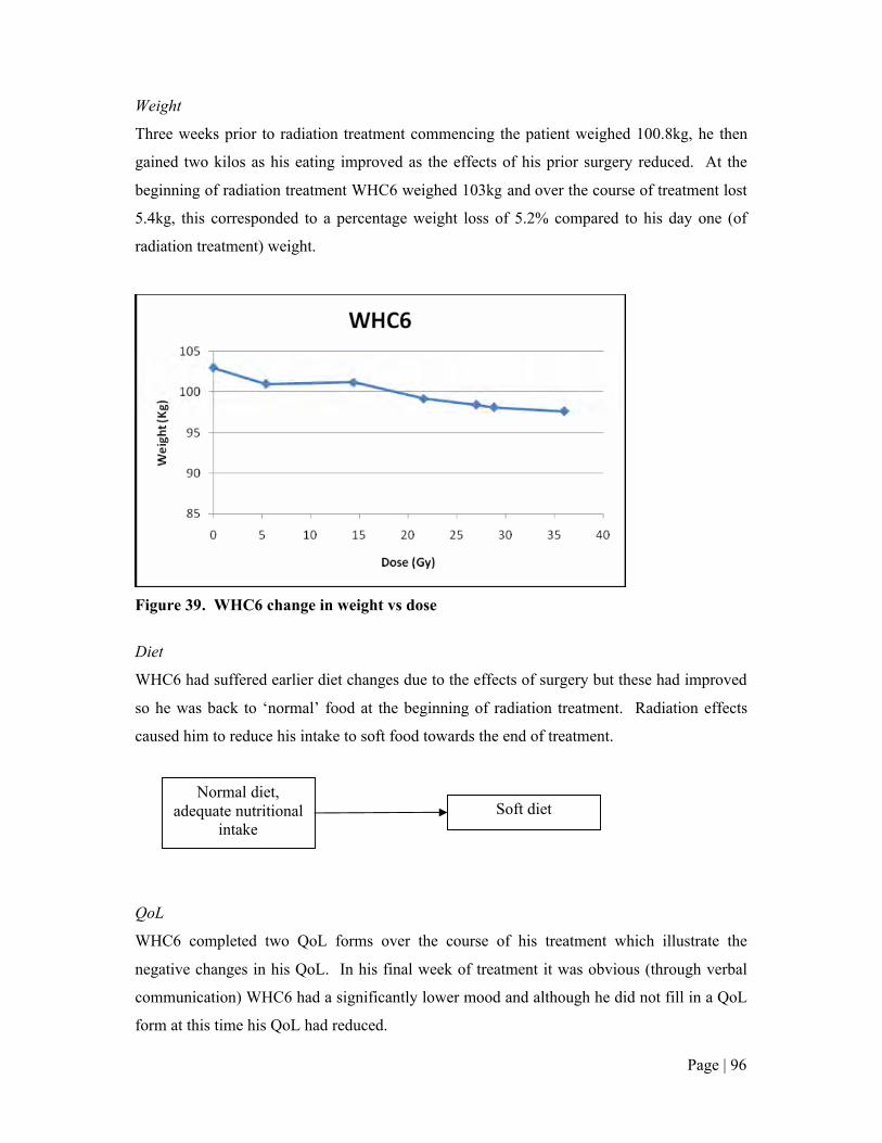
Page | 96
Weight
Three weeks prior to radiation treatment commencing the patient weighed 100.8kg, he then
gained two kilos as his eating improved as the effects of his prior surgery reduced. At the
beginning of radiation treatment WHC6 weighed 103kg and over the course of treatment lost
5.4kg, this corresponded to a percentage weight loss of 5.2% compared to his day one (of
radiation treatment) weight.
Figure 39. WHC6 change in weight vs dose
Diet
WHC6 had suffered earlier diet changes due to the effects of surgery but these had improved
so he was back to ‘normal’ food at the beginning of radiation treatment. Radiation effects
caused him to reduce his intake to soft food towards the end of treatment.
QoL
WHC6 completed two QoL forms over the course of his treatment which illustrate the
negative changes in his QoL. In his final week of treatment it was obvious (through verbal
communication) WHC6 had a significantly lower mood and although he did not fill in a QoL
form at this time his QoL had reduced.
Normal diet, adequate nutritional
intake Soft diet

Page | 97
Figure 40. WHC6 change in perception of QoL score vs dose
Figure 41. WHC6 negative change in QoL category scores vs dose
Changes in categories of QoL:
• Fatigue and pain increased
• The patient used pain medication the whole way through treatment
• Swallowing became more difficult
• The oral cavity changes became more negative
• The patient reported nutrition level remained the same
• Mood became more negative
Page | 98
• Psychosocial aspects of treatment remained the same
• There was a decrease in perception of QoL
Patient’s subjective experience
The patient had lost a small amount of weight after his tonsillectomy then gained eight
kilograms during his course of chemotherapy. Upon commencement of radiotherapy he had
a good appetite and was eating a normal diet with three meals a day. After 7.2Gy had been
delivered (week one) the patient had noticed mild tiredness and nausea but no skin reaction.
He was using the manuka honey three times a day with no problems. After 9Gy the patient
began to find the sweetness of the honey difficult to tolerate, it was contributing to his
nausea. To avoid ceasing the honey completely he was advised to mix it 1:3 with lemon and
barley juice. There were no reports of mucositis or erythema. The patient’s first QoL
questionnaire showed a low perceived QoL score of three.
Oral changes continued as the dose delivered increased. After 10.8Gy (week two) the patient
reported no taste; “everything tastes like cardboard”. He reported a cotton wool ball or flour
feeling in his mouth but no pain. WHC6 was continuing with the honey but only managing it
twice a day and had to spit it out after swirling it around his mouth because it made him
nauseous if he swallowed it.
At 16.2Gy (end of week two) WHC6 reported his mouth was uncomfortable and very dry.
He was attempting to eat normal food but found it “very very” difficult to swallow. The
patient reported drinking water constantly and feeling very tired. At 18Gy the pain in his
mouth increased and there was visible mucositis causing difficulty with eating, he was only
managing jelly and ice-cream. WHC6 persisted with the honey and followed a stringent
mouth wash regime with baking soda, difflam, nystatin and the honey mixture between two
and four times a day. The patient also monitored his codeine intake to maintain a good level
of pain control. The doctor described “quite a marked radiation reaction and thickened
saliva”. The patient’s perceived QoL score at this point had decreased and six of the seven
QoL categories were reported as worse compared to earlier in his treatment.
At 28.8Gy the patient stopped taking honey after finding it too much to tolerate. The initial
mucositis had subsided but the inflammation caused the buccal mucosa to puff out and press

Page | 99
against teeth causing a painful ridge of mucosal tissue. The patient began taking biotene
mouthwash to combat the constant dry mouth. After another fraction a metallic taste had
developed.
At completion of the course of treatment there was grade two mucositis, a dry mouth and
pain that was managed with codeine. The patient’s nutrition had been maintained through
oral intake.
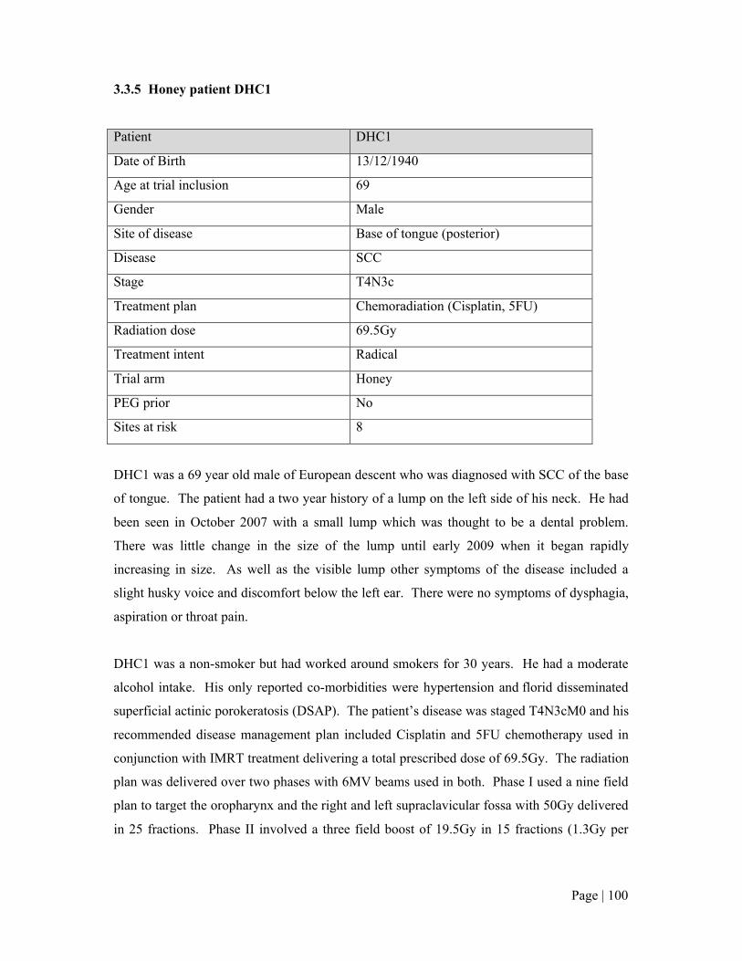
Page | 100
3.3.5 Honey patient DHC1
Patient DHC1
Date of Birth 13/12/1940
Age at trial inclusion 69
Gender Male
Site of disease Base of tongue (posterior)
Disease SCC
Stage T4N3c
Treatment plan Chemoradiation (Cisplatin, 5FU)
Radiation dose 69.5Gy
Treatment intent Radical
Trial arm Honey
PEG prior No
Sites at risk 8
DHC1 was a 69 year old male of European descent who was diagnosed with SCC of the base
of tongue. The patient had a two year history of a lump on the left side of his neck. He had
been seen in October 2007 with a small lump which was thought to be a dental problem.
There was little change in the size of the lump until early 2009 when it began rapidly
increasing in size. As well as the visible lump other symptoms of the disease included a
slight husky voice and discomfort below the left ear. There were no symptoms of dysphagia,
aspiration or throat pain.
DHC1 was a non-smoker but had worked around smokers for 30 years. He had a moderate
alcohol intake. His only reported co-morbidities were hypertension and florid disseminated
superficial actinic porokeratosis (DSAP). The patient’s disease was staged T4N3cM0 and his
recommended disease management plan included Cisplatin and 5FU chemotherapy used in
conjunction with IMRT treatment delivering a total prescribed dose of 69.5Gy. The radiation
plan was delivered over two phases with 6MV beams used in both. Phase I used a nine field
plan to target the oropharynx and the right and left supraclavicular fossa with 50Gy delivered
in 25 fractions. Phase II involved a three field boost of 19.5Gy in 15 fractions (1.3Gy per
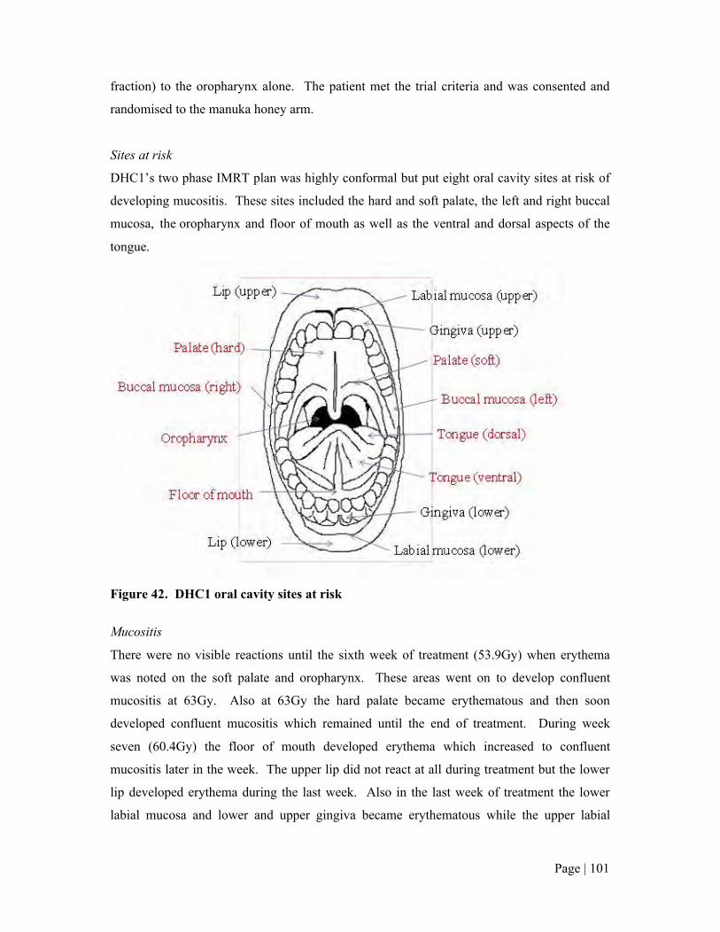
Page | 101
fraction) to the oropharynx alone. The patient met the trial criteria and was consented and
randomised to the manuka honey arm.
Sites at risk
DHC1’s two phase IMRT plan was highly conformal but put eight oral cavity sites at risk of
developing mucositis. These sites included the hard and soft palate, the left and right buccal
mucosa, the oropharynx and floor of mouth as well as the ventral and dorsal aspects of the
tongue.
Figure 42. DHC1 oral cavity sites at risk
Mucositis
There were no visible reactions until the sixth week of treatment (53.9Gy) when erythema
was noted on the soft palate and oropharynx. These areas went on to develop confluent
mucositis at 63Gy. Also at 63Gy the hard palate became erythematous and then soon
developed confluent mucositis which remained until the end of treatment. During week
seven (60.4Gy) the floor of mouth developed erythema which increased to confluent
mucositis later in the week. The upper lip did not react at all during treatment but the lower
lip developed erythema during the last week. Also in the last week of treatment the lower
labial mucosa and lower and upper gingiva became erythematous while the upper labial
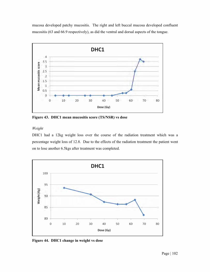
Page | 102
mucosa developed patchy mucositis. The right and left buccal mucosa developed confluent
mucositis (63 and 66.9 respectively), as did the ventral and dorsal aspects of the tongue.
Figure 43. DHC1 mean mucositis score (TS/NSR) vs dose
Weight
DHC1 had a 12kg weight loss over the course of the radiation treatment which was a
percentage weight loss of 12.8. Due to the effects of the radiation treatment the patient went
on to lose another 6.5kgs after treatment was completed.
Figure 44. DHC1 change in weight vs dose

Page | 103
Diet
At the beginning of treatment the patient had a normal diet. As the treatment reactions began
to manifest DHC1’s diet became predominantly soft and then towards the end of treatment
the patient required NG tube insertion and significant Fortisip supplementation.
QoL
DHC1 was a motivated individual who was keen to comply with the trial requests and he
filled in five QoL questionnaires over the course of his treatment. Review of these
questionnaires gives a good illustration of his experience.
Figure 45. DHC1 change in perception of QoL score vs dose
Normal diet NG tube and four Fortisips per day Soft diet
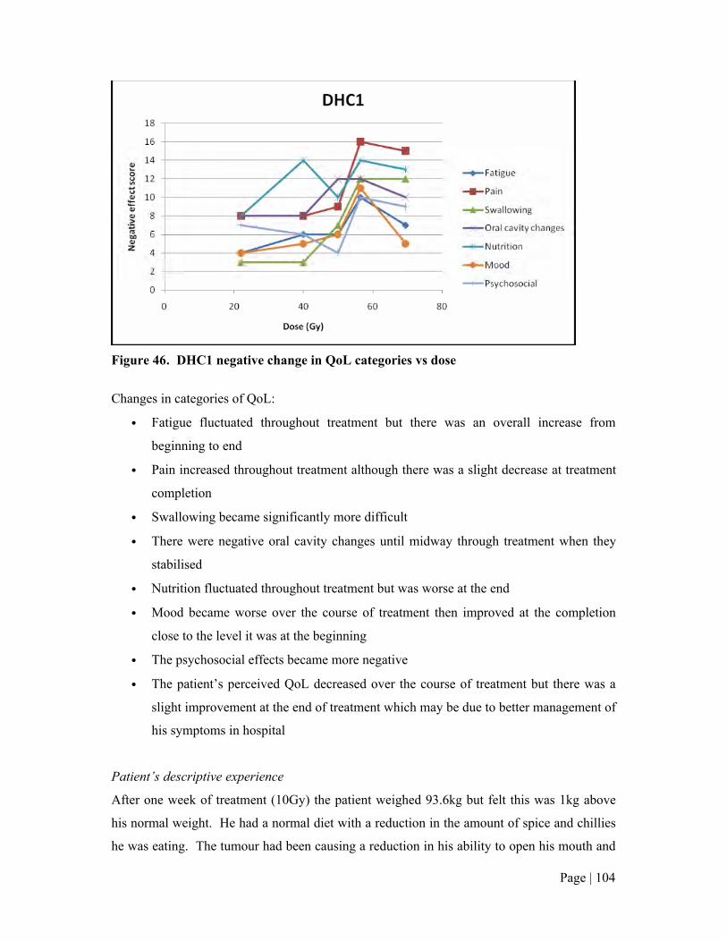
Page | 104
Figure 46. DHC1 negative change in QoL categories vs dose
Changes in categories of QoL:
• Fatigue fluctuated throughout treatment but there was an overall increase from
beginning to end
• Pain increased throughout treatment although there was a slight decrease at treatment
completion
• Swallowing became significantly more difficult
• There were negative oral cavity changes until midway through treatment when they
stabilised
• Nutrition fluctuated throughout treatment but was worse at the end
• Mood became worse over the course of treatment then improved at the completion
close to the level it was at the beginning
• The psychosocial effects became more negative
• The patient’s perceived QoL decreased over the course of treatment but there was a
slight improvement at the end of treatment which may be due to better management of
his symptoms in hospital
Patient’s descriptive experience
After one week of treatment (10Gy) the patient weighed 93.6kg but felt this was 1kg above
his normal weight. He had a normal diet with a reduction in the amount of spice and chillies
he was eating. The tumour had been causing a reduction in his ability to open his mouth and
Page | 105
move his tongue and there had also been reports of occasional haemoptysis. During week
two (16Gy) the patient vomited despite taking antiemetic medication, his saliva was thick and
he had lost his taste. The dietician advised him to increase his fluid intake at this point
Completion of week three saw DHC1 eating a good range of foods despite food “tasting
terrible”. The left lymph node was visibly smaller and the patient described the skin in the
area feeling “less tight” and he was able to open his mouth wider than last week. The patient
was a little more nauseous because he was not taking his medications routinely. Despite his
dry mouth and thick saliva there was no visible mucositis.
A week later (40Gy) there was another noticeable reduction in the palpable neck node and
there was still no mucositis visible in the patient’s oral cavity. The patient was experiencing
bloody discharge from his nose which was thought to be related to his chemotherapy. At this
point the patient reported that his taste had improved but he still had a reduced appetite and
worked hard to eat a variety of foods (small amounts often).
The patient was continuing with the honey application but found it was irritating his lips. He
was advised to dilute the honey, maintain regular teeth cleaning and was given advice on how
to avoid irritation to the skin in the treatment area which had become red and itchy.
There were still no signs of mucositis halfway through week five (46Gy). At the end of this
week the patient’s weight had decreased and food was no longer enjoyable, he found eating a
“chore”. These nutritional changes were due to lack of taste, thick saliva and pain when
swallowing. DHC1 was advised to take two Fortisips per day to help with nutritional
maintenance and he was given advice on a high energy, high protein diet. Panadol was
prescribed for pain. The patient’s oral mucosa was intact at this stage.
Over the following weekend (52.6Gy) the patient did not eat or drink due to pain so
oxycontin was commenced. One fraction later the patient experienced a difficult evening in
which he woke with difficulty breathing due to the thick mucous in his throat. His pain
control had improved with the addition of xylocaine viscous.
At the end of week six (56.6Gy) morphine was required to enable eating. The patient
maintained a soft diet and one Fortisip per day, he found slightly thicker fluids easier to
Page | 106
swallow. The patient tried pure aloe vera juice to soothe the inflammation in the throat and to
aid swallowing because he was becoming dehydrated (had only had 1.5L over the previous
three days). At this stage in the treatment hiccups were also a frequent problem and the
application of the honey was very painful.
After 60.4Gy the patient was admitted to hospital and reported feeling miserable. There were
areas of confluent mucositis but the oropharynx could not be assessed due to the level of pain
when the patient opened his mouth. DHC1 stopped using the honey at this stage.
There was further increase in the level of pain during week seven, medicine was difficult to
swallow and sweet foods and Fortisips led to a “burning feeling”. A NG tube was inserted to
try and stabilise the patient’s nutritional status.
Treatment was completed after delivery of 69.5Gy and at this stage the patient was having
three or four Fortisips per day via his NG tube including overnight pump feeding. He had no
taste and was on regular morphine pain relief. The patient filled in a questionnaire about the
honey application. He reported that he had tried to adhere to the guidelines for applying the
honey but as the treatment progressed he found he could not always manage due to the
burning feeling. He also reported the honey was not easy to use and was messy.

Page | 107
3.3.6 Control patient WHN1
Patient WHN1
Date of Birth 17/03/1931
Age at trial inclusion 78
Gender Female
Site of disease Maxillary gingiva
Disease SCC
Stage TxN0M0
Treatment plan Conformal radiation therapy
Radiation dose 40.8Gy (48 prescribed)
Treatment intent Radical
Trial arm Control
PEG prior No
Sites at risk 7
A 78 year old female, WHN1 was the eldest of the trial patients. She was referred for
radiation therapy after presenting with a recurrent SCC of the maxillary gingiva. Initial
disease management had involved surgical resection of the tumour and after the disease
recurred WHN1 had another surgical procedure to remove the local recurrence. Two months
after the local recurrence resection radical radiation therapy was commenced with the
prescription of 48Gy in 20 fractions, 2.4Gy per fraction.
The treatment plan utilised 6MV parallel opposed (POP) conformal fields to target the mouth
alone. A mouth bite and mask were used to improve accuracy and remove the tongue from
the treatment area as much as possible. WHN1 was randomised to the control arm of the trial
so received the current standard of care at the Wellington department. WHN1 had close
dietician and district nurse involvement throughout her course of treatment. Although the
initial plan prescription was 48Gy the treatment was completed prematurely after delivery of
40.8Gy (17 fractions) due to severe confluent mucositis across the labial gingiva with poorly
controlled pain.
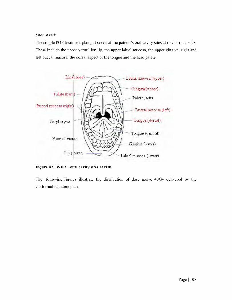
Page | 108
Sites at risk
The simple POP treatment plan put seven of the patient’s oral cavity sites at risk of mucositis.
These include the upper vermillion lip, the upper labial mucosa, the upper gingiva, right and
left buccal mucosa, the dorsal aspect of the tongue and the hard palate.
Figure 47. WHN1 oral cavity sites at risk
The following Figures illustrate the distribution of dose above 40Gy delivered by the
conformal radiation plan.
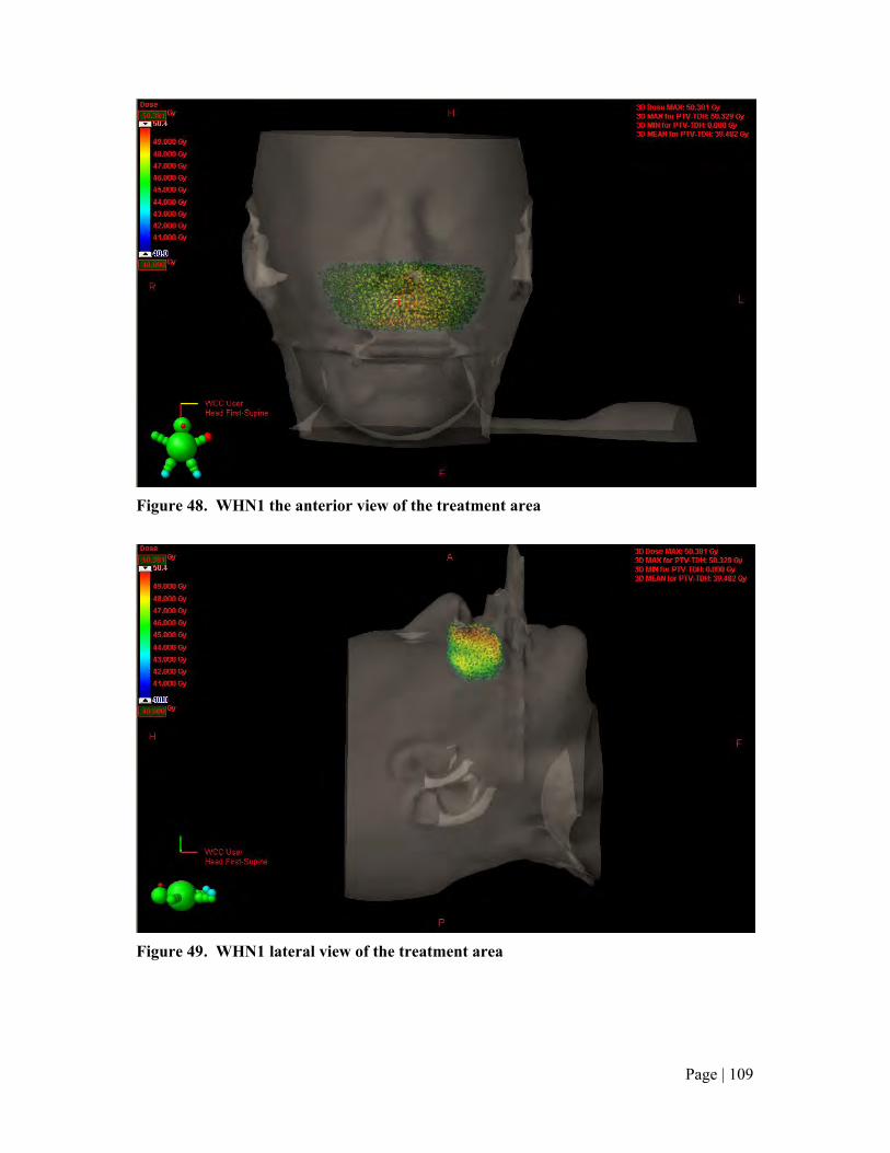
Page | 109
Figure 48. WHN1 the anterior view of the treatment area
Figure 49. WHN1 lateral view of the treatment area

Page | 110
Figure 50. WHN1 transverse slices at the isocentre and 1.2cm superior to the isocentre
Mucositis
The first reactions were evident at the beginning of week three (28.8Gy); erythema on the
upper gingiva and right buccal mucosa. These two sites progressed to patchy mucositis at
31.2Gy (later in week three) then confluent mucositis shortly after (36Gy and 40.8Gy
respectively). The left buccal mucosa developed patchy mucositis at the end of week three
(36Gy) which became confluent at the end of treatment (40.8Gy). The upper lip although
painful over the whole course of treatment first developed erythema at 31.2Gy then patchy
mucositis at treatment completion. Patchy mucositis was evident on the upper labial mucosa
at 31.2Gy and went on to become confluent at 36Gy.
Figure 51. WHN1 mean mucositis score (TS/NSR) vs dose
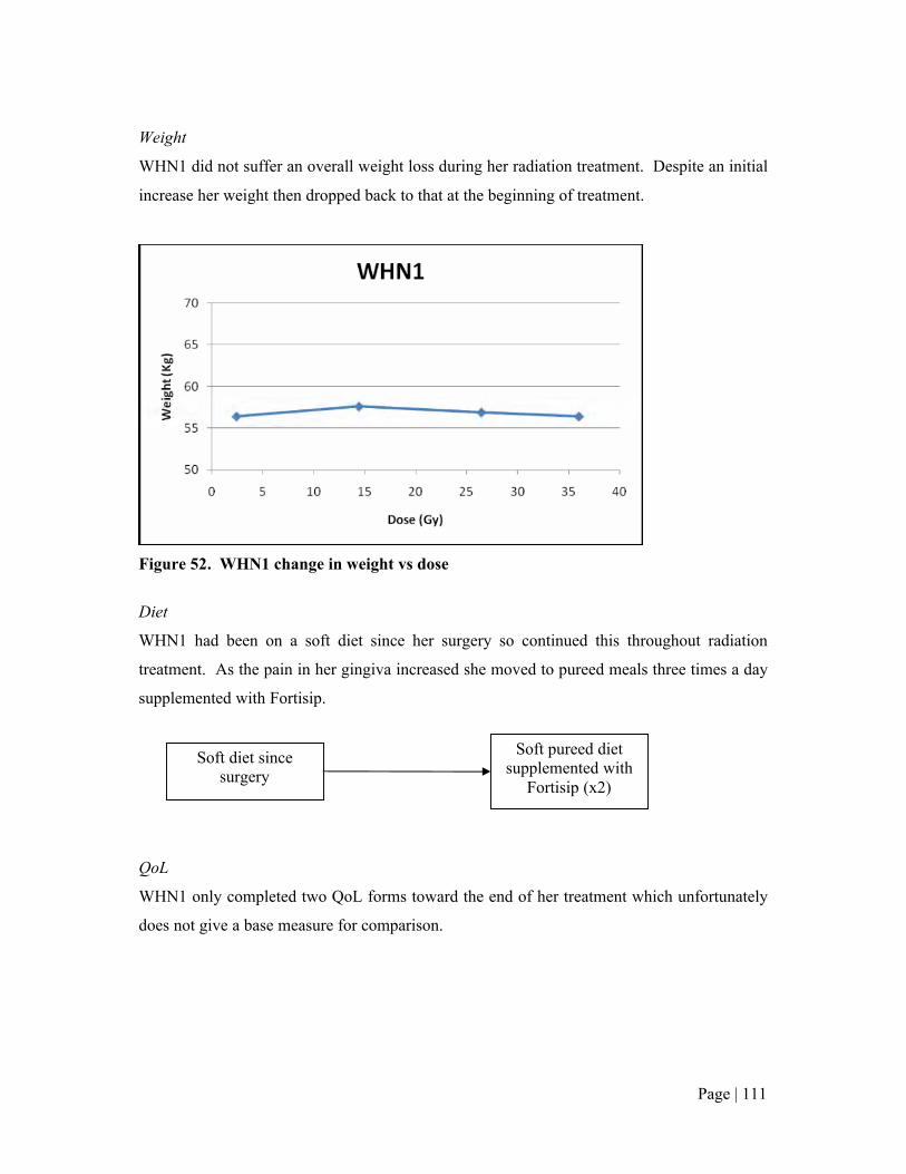
Page | 111
Weight
WHN1 did not suffer an overall weight loss during her radiation treatment. Despite an initial
increase her weight then dropped back to that at the beginning of treatment.
Figure 52. WHN1 change in weight vs dose
Diet
WHN1 had been on a soft diet since her surgery so continued this throughout radiation
treatment. As the pain in her gingiva increased she moved to pureed meals three times a day
supplemented with Fortisip.
QoL
WHN1 only completed two QoL forms toward the end of her treatment which unfortunately
does not give a base measure for comparison.
Soft diet since surgery
Soft pureed diet supplemented with
Fortisip (x2)
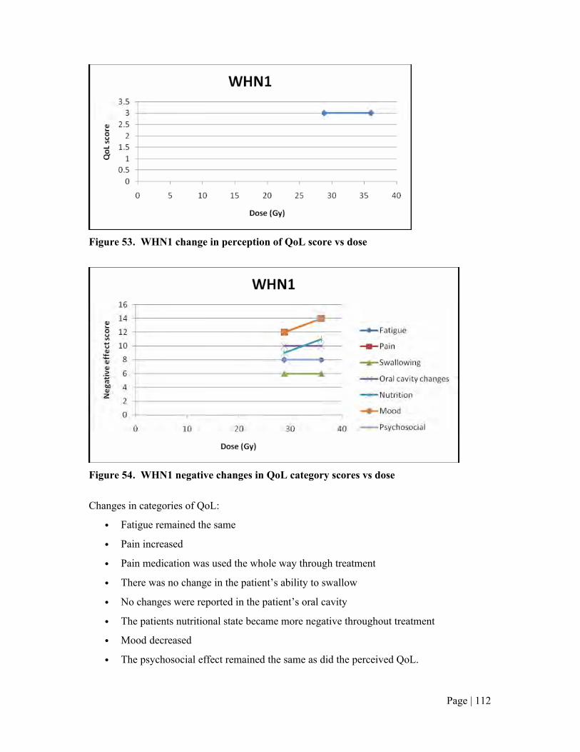
Page | 112
Figure 53. WHN1 change in perception of QoL score vs dose
Figure 54. WHN1 negative changes in QoL category scores vs dose
Changes in categories of QoL:
• Fatigue remained the same
• Pain increased
• Pain medication was used the whole way through treatment
• There was no change in the patient’s ability to swallow
• No changes were reported in the patient’s oral cavity
• The patients nutritional state became more negative throughout treatment
• Mood decreased
• The psychosocial effect remained the same as did the perceived QoL.
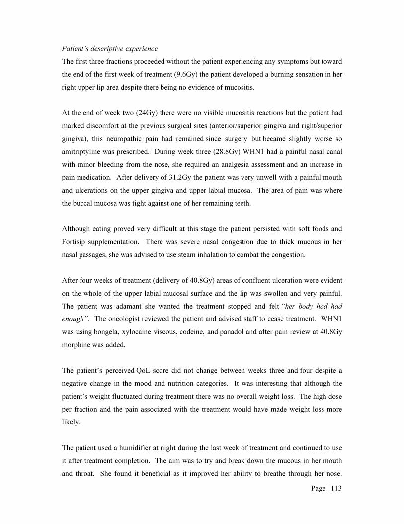
Page | 113
Patient’s descriptive experience
The first three fractions proceeded without the patient experiencing any symptoms but toward
the end of the first week of treatment (9.6Gy) the patient developed a burning sensation in her
right upper lip area despite there being no evidence of mucositis.
At the end of week two (24Gy) there were no visible mucositis reactions but the patient had
marked discomfort at the previous surgical sites (anterior/superior gingiva and right/superior
gingiva), this neuropathic pain had remained since surgery but became slightly worse so
amitriptyline was prescribed. During week three (28.8Gy) WHN1 had a painful nasal canal
with minor bleeding from the nose, she required an analgesia assessment and an increase in
pain medication. After delivery of 31.2Gy the patient was very unwell with a painful mouth
and ulcerations on the upper gingiva and upper labial mucosa. The area of pain was where
the buccal mucosa was tight against one of her remaining teeth.
Although eating proved very difficult at this stage the patient persisted with soft foods and
Fortisip supplementation. There was severe nasal congestion due to thick mucous in her
nasal passages, she was advised to use steam inhalation to combat the congestion.
After four weeks of treatment (delivery of 40.8Gy) areas of confluent ulceration were evident
on the whole of the upper labial mucosal surface and the lip was swollen and very painful.
The patient was adamant she wanted the treatment stopped and felt “her body had had
enough”. The oncologist reviewed the patient and advised staff to cease treatment. WHN1
was using bongela, xylocaine viscous, codeine, and panadol and after pain review at 40.8Gy
morphine was added.
The patient’s perceived QoL score did not change between weeks three and four despite a
negative change in the mood and nutrition categories. It was interesting that although the
patient’s weight fluctuated during treatment there was no overall weight loss. The high dose
per fraction and the pain associated with the treatment would have made weight loss more
likely.
The patient used a humidifier at night during the last week of treatment and continued to use
it after treatment completion. The aim was to try and break down the mucous in her mouth
and throat. She found it beneficial as it improved her ability to breathe through her nose.
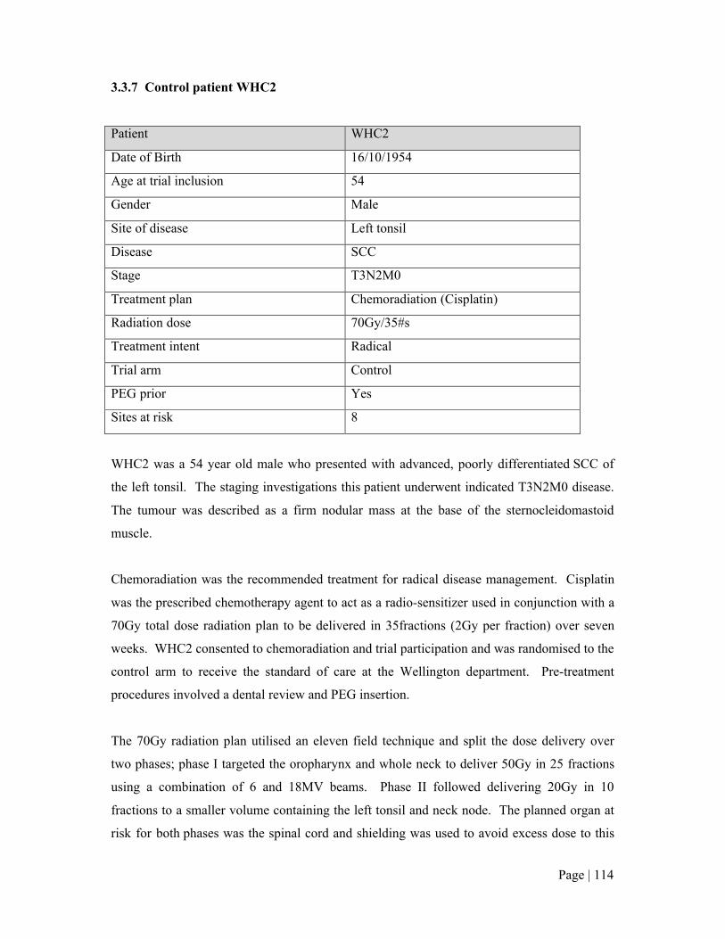
Page | 114
3.3.7 Control patient WHC2
Patient WHC2
Date of Birth 16/10/1954
Age at trial inclusion 54
Gender Male
Site of disease Left tonsil
Disease SCC
Stage T3N2M0
Treatment plan Chemoradiation (Cisplatin)
Radiation dose 70Gy/35#s
Treatment intent Radical
Trial arm Control
PEG prior Yes
Sites at risk 8
WHC2 was a 54 year old male who presented with advanced, poorly differentiated SCC of
the left tonsil. The staging investigations this patient underwent indicated T3N2M0 disease.
The tumour was described as a firm nodular mass at the base of the sternocleidomastoid
muscle.
Chemoradiation was the recommended treatment for radical disease management. Cisplatin
was the prescribed chemotherapy agent to act as a radio-sensitizer used in conjunction with a
70Gy total dose radiation plan to be delivered in 35fractions (2Gy per fraction) over seven
weeks. WHC2 consented to chemoradiation and trial participation and was randomised to the
control arm to receive the standard of care at the Wellington department. Pre-treatment
procedures involved a dental review and PEG insertion.
The 70Gy radiation plan utilised an eleven field technique and split the dose delivery over
two phases; phase I targeted the oropharynx and whole neck to deliver 50Gy in 25 fractions
using a combination of 6 and 18MV beams. Phase II followed delivering 20Gy in 10
fractions to a smaller volume containing the left tonsil and neck node. The planned organ at
risk for both phases was the spinal cord and shielding was used to avoid excess dose to this
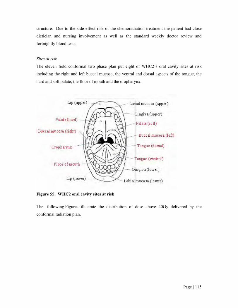
Page | 115
structure. Due to the side effect risk of the chemoradiation treatment the patient had close
dietician and nursing involvement as well as the standard weekly doctor review and
fortnightly blood tests.
Sites at risk
The eleven field conformal two phase plan put eight of WHC2’s oral cavity sites at risk
including the right and left buccal mucosa, the ventral and dorsal aspects of the tongue, the
hard and soft palate, the floor of mouth and the oropharynx.
Figure 55. WHC2 oral cavity sites at risk
The following Figures illustrate the distribution of dose above 40Gy delivered by the
conformal radiation plan.
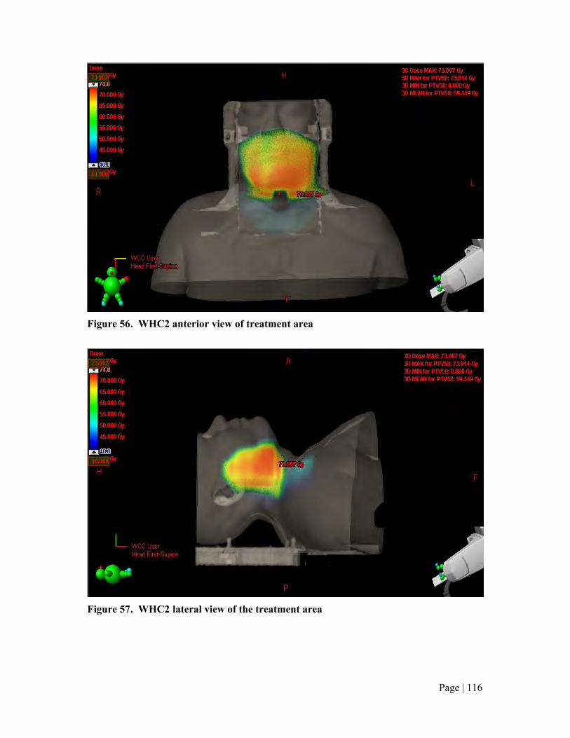
Page | 116
Figure 56. WHC2 anterior view of treatment area
Figure 57. WHC2 lateral view of the treatment area
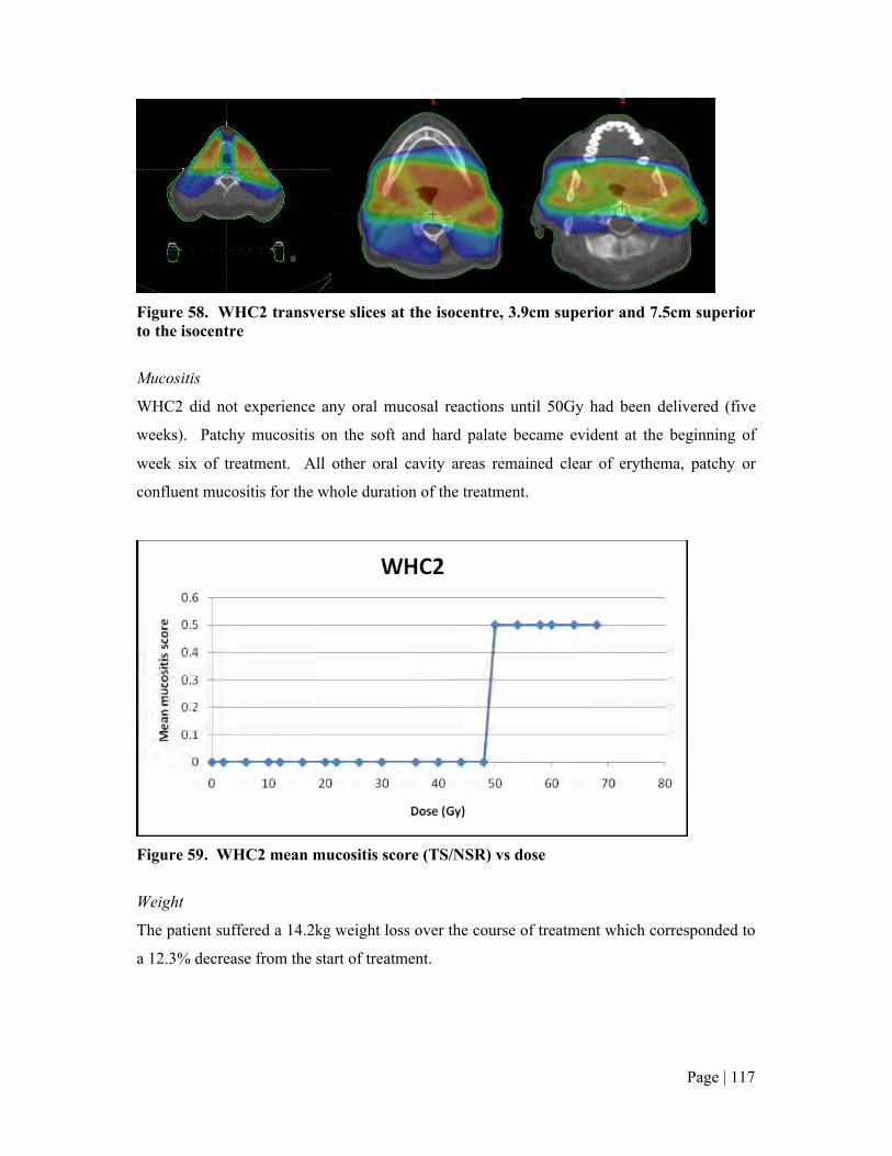
Page | 117
Figure 58. WHC2 transverse slices at the isocentre, 3.9cm superior and 7.5cm superior to the isocentre
Mucositis
WHC2 did not experience any oral mucosal reactions until 50Gy had been delivered (five
weeks). Patchy mucositis on the soft and hard palate became evident at the beginning of
week six of treatment. All other oral cavity areas remained clear of erythema, patchy or
confluent mucositis for the whole duration of the treatment.
Figure 59. WHC2 mean mucositis score (TS/NSR) vs dose
Weight
The patient suffered a 14.2kg weight loss over the course of treatment which corresponded to
a 12.3% decrease from the start of treatment.
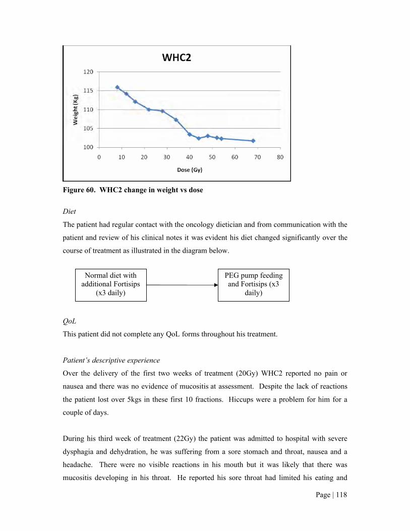
Page | 118
Figure 60. WHC2 change in weight vs dose
Diet
The patient had regular contact with the oncology dietician and from communication with the
patient and review of his clinical notes it was evident his diet changed significantly over the
course of treatment as illustrated in the diagram below.
QoL
This patient did not complete any QoL forms throughout his treatment.
Patient’s descriptive experience
Over the delivery of the first two weeks of treatment (20Gy) WHC2 reported no pain or
nausea and there was no evidence of mucositis at assessment. Despite the lack of reactions
the patient lost over 5kgs in these first 10 fractions. Hiccups were a problem for him for a
couple of days.
During his third week of treatment (22Gy) the patient was admitted to hospital with severe
dysphagia and dehydration, he was suffering from a sore stomach and throat, nausea and a
headache. There were no visible reactions in his mouth but it was likely that there was
mucositis developing in his throat. He reported his sore throat had limited his eating and
Normal diet with additional Fortisips
(x3 daily)
PEG pump feeding and Fortisips (x3
daily)
Page | 119
described the sensation of “a lump in the throat”. Despite his reluctance to pump feed via
his PEG he was instructed to increase his Fortisip PEG delivery to four per day.
At the end of his third week (30Gy) there was written report of a visible decrease in the size
of the left neck mass. WHC2’s throat was still sore and he was started on morphine. At
36Gy there were still no visible oral cavity changes although the sore throat descriptions
indicated significant oropharngeal mucosal changes. The patient’s taste has decreased and he
was maintaining nutritional intake primarily through PEG use with some additional oral
intake. WHC2 reported feeling very nauseated from the chemotherapy, would often try to eat
or use the PEG and then vomit soon after. He was unable to swallow tablets orally so they
were administered through his PEG for the remainder of his treatment (any oral medications
had to be crushed). He was also started on IV fluids at this point.
After delivery of 40Gy (4 weeks) the patient had to be admitted via ambulance to the short
stay unit due to severe nausea and vomiting. Immediately prior to admission he had no oral
intake and no Fortisips via his PEG. He had lost over 10kgs since the first week of his
treatment. The throat pain was still severe but there was still no visible mucositis. The
patient felt better after staying overnight in hospital.
Partway through week five of treatment (44Gy) WHC2 had better pain control and his nausea
and vomiting had settled so he was able to be discharged from hospital two fractions later.
Back at home after 48Gy the patient reported no nausea or vomiting and had recommenced
PEG feeding. He felt a lot better when not on chemotherapy.
During the sixth week of treatment (50Gy) the patient had a very dry mouth and throat
describing it like “lava” or “chillies” in his throat. He was using the salt and baking soda
mouthwashes which he found eased the pain temporarily. At this stage his nutritional intake
was maintained via PEG use and Fortisip supplements. Most of the oral cavity was clear
although the roof of the mouth had patchy mucositis.
There was another increase in the level of dysphagia described by the patient at 52Gy, on
examination there was evidence of thick saliva and patchy mucositis. Liquid pain medication
was administered for the sore throat. At 54Gy the mucositis level had decreased and after
68Gy the patient felt “like things were picking up”. WHC2 was an admitted to hospital for

Page | 120
his chemotherapy delivery and for better antiemetic control. At the completion of treatment
the patient had lost a total of 14kgs since the first week. At this point he was pump feeding
over night and taking three Fortisips per day to prevent further weight loss.
The lack of visible mucositis on all oral cavity areas aside from the soft and hard palate was
unusual. The soft and hard palate were two of eight oral cavity sites that were considered at
risk of mucositis.
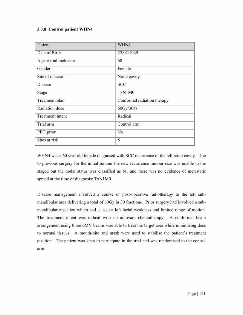
Page | 121
3.3.8 Control patient WHN4
Patient WHN4
Date of Birth 22/02/1949
Age at trial inclusion 60
Gender Female
Site of disease Nasal cavity
Disease SCC
Stage TxN1M0
Treatment plan Conformal radiation therapy
Radiation dose 60Gy/30#s
Treatment intent Radical
Trial arm Control arm
PEG prior No
Sites at risk 8
WHN4 was a 60 year old female diagnosed with SCC recurrence of the left nasal cavity. Due
to previous surgery for the initial tumour the new recurrence tumour size was unable to the
staged but the nodal status was classified as N1 and there was no evidence of metastatic
spread at the time of diagnosis; TxN1M0.
Disease management involved a course of post-operative radiotherapy to the left sub-
mandibular area delivering a total of 60Gy in 30 fractions. Prior surgery had involved a sub-
mandibular resection which had caused a left facial weakness and limited range of motion.
The treatment intent was radical with no adjuvant chemotherapy. A conformal beam
arrangement using three 6MV beams was able to treat the target area while minimising dose
to normal tissues. A mouth-bite and mask were used to stabilise the patient’s treatment
position. The patient was keen to participate in the trial and was randomised to the control
arm.

Page | 122
Sites at risk
The three field conformal plan put eight oral cavity sites at risk including the lower labial
mucosa and lower gingiva, the left buccal mucosa, and the ventral and dorsal aspects of the
tongue, the soft palate, the floor of mouth and the oropharynx.
Figure 61. WHN4 oral cavity sites at risk
The following Figures illustrate the distribution of dose above 40Gy delivered by the
conformal radiation plan.

Page | 123
Figure 62. WHN4 anterior view of the treatment area
Figure 63. WHN4 lateral view of the treatment area
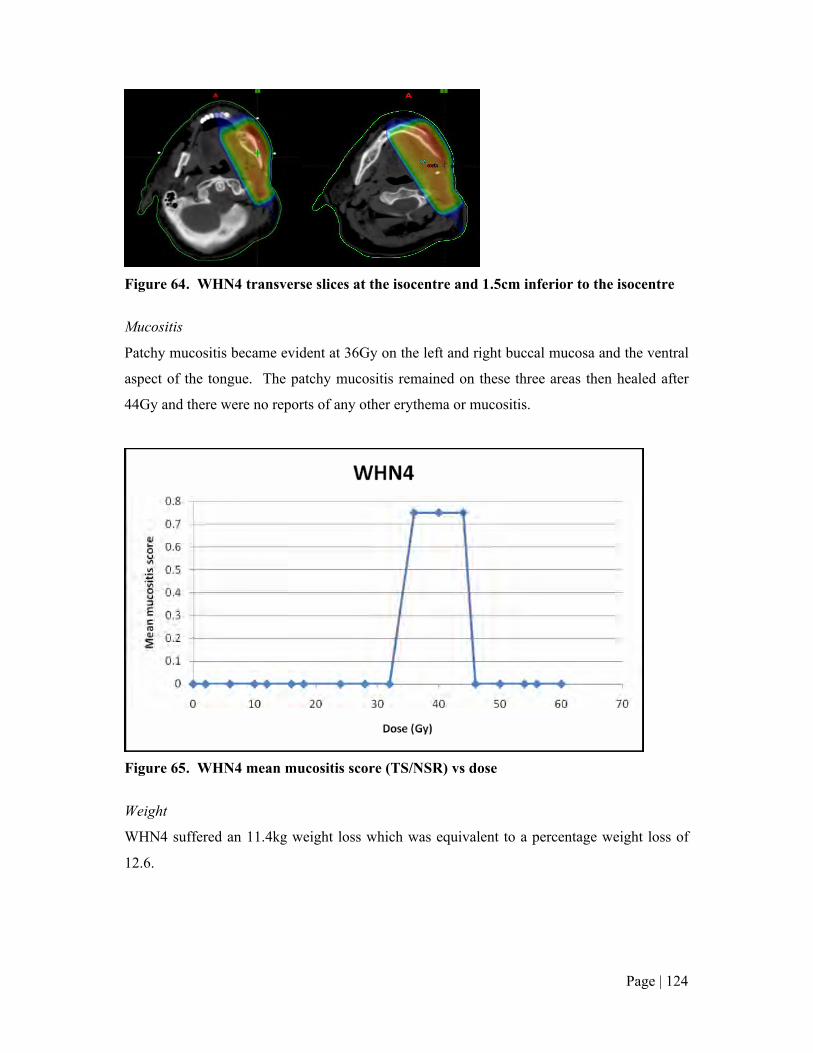
Page | 124
Figure 64. WHN4 transverse slices at the isocentre and 1.5cm inferior to the isocentre
Mucositis
Patchy mucositis became evident at 36Gy on the left and right buccal mucosa and the ventral
aspect of the tongue. The patchy mucositis remained on these three areas then healed after
44Gy and there were no reports of any other erythema or mucositis.
Figure 65. WHN4 mean mucositis score (TS/NSR) vs dose
Weight
WHN4 suffered an 11.4kg weight loss which was equivalent to a percentage weight loss of
12.6.
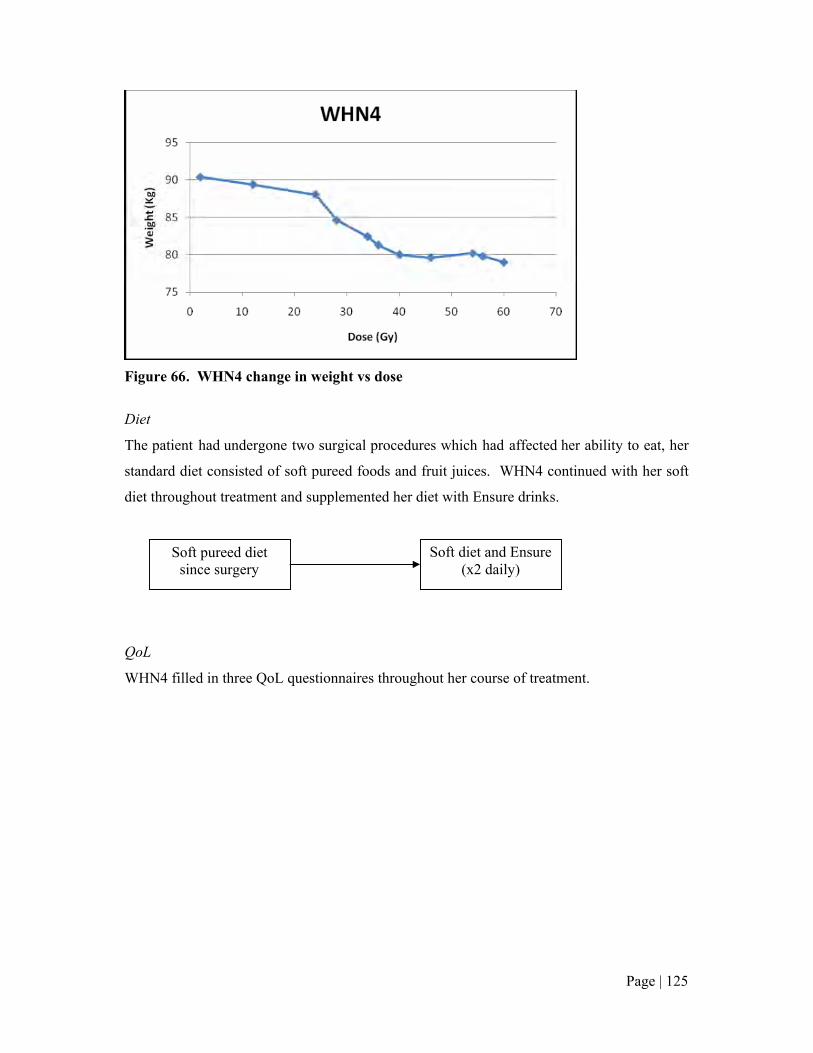
Page | 125
Figure 66. WHN4 change in weight vs dose
Diet
The patient had undergone two surgical procedures which had affected her ability to eat, her
standard diet consisted of soft pureed foods and fruit juices. WHN4 continued with her soft
diet throughout treatment and supplemented her diet with Ensure drinks.
QoL
WHN4 filled in three QoL questionnaires throughout her course of treatment.
Soft pureed diet since surgery
Soft diet and Ensure (x2 daily)
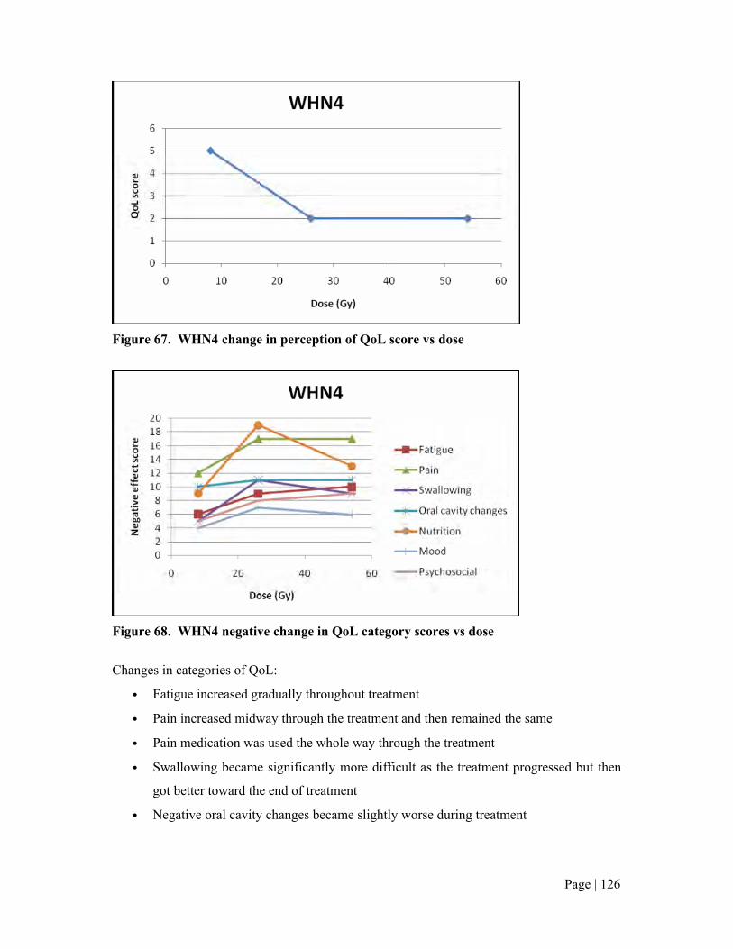
Page | 126
Figure 67. WHN4 change in perception of QoL score vs dose
Figure 68. WHN4 negative change in QoL category scores vs dose
Changes in categories of QoL:
• Fatigue increased gradually throughout treatment
• Pain increased midway through the treatment and then remained the same
• Pain medication was used the whole way through the treatment
• Swallowing became significantly more difficult as the treatment progressed but then
got better toward the end of treatment
• Negative oral cavity changes became slightly worse during treatment
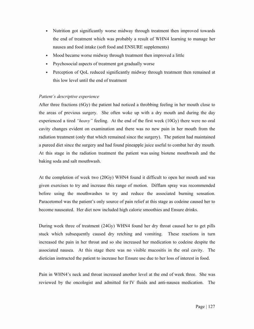
Page | 127
• Nutrition got significantly worse midway through treatment then improved towards
the end of treatment which was probably a result of WHN4 learning to manage her
nausea and food intake (soft food and ENSURE supplements)
• Mood became worse midway through treatment then improved a little
• Psychosocial aspects of treatment got gradually worse
• Perception of QoL reduced significantly midway through treatment then remained at
this low level until the end of treatment
Patient’s descriptive experience
After three fractions (6Gy) the patient had noticed a throbbing feeling in her mouth close to
the areas of previous surgery. She often woke up with a dry mouth and during the day
experienced a tired “heavy” feeling. At the end of the first week (10Gy) there were no oral
cavity changes evident on examination and there was no new pain in her mouth from the
radiation treatment (only that which remained since the surgery). The patient had maintained
a pureed diet since the surgery and had found pineapple juice useful to combat her dry mouth.
At this stage in the radiation treatment the patient was using biotene mouthwash and the
baking soda and salt mouthwash.
At the completion of week two (20Gy) WHN4 found it difficult to open her mouth and was
given exercises to try and increase this range of motion. Difflam spray was recommended
before using the mouthwashes to try and reduce the associated burning sensation.
Paracetomol was the patient’s only source of pain relief at this stage as codeine caused her to
become nauseated. Her diet now included high calorie smoothies and Ensure drinks.
During week three of treatment (24Gy) WHN4 found her dry throat caused her to get pills
stuck which subsequently caused dry retching and vomiting. These reactions in turn
increased the pain in her throat and so she increased her medication to codeine despite the
associated nausea. At this stage there was no visible mucositis in the oral cavity. The
dietician instructed the patient to increase her Ensure use due to her loss of interest in food.
Pain in WHN4’s neck and throat increased another level at the end of week three. She was
reviewed by the oncologist and admitted for IV fluids and anti-nausea medication. The
Page | 128
patient’s QoL form illustrates her poor condition with a large reduction in her perceived QoL
score. The patient reported all seven QoL categories had got worse.
Over the next couple of fractions the patient suffered vomiting even with water alone. She
was discharged from hospital but then did not eat for two days and only managed to drink
400mL of water a day. WHN4 was then readmitted to hospital through ED for further IV
management as she was severely dehydrated. The pain in her mouth was not well managed
so was increased to morphine (capsule and liquid) and paracetamol. WHN4 described a
strong metallic taste in her mouth.
The end of week four (38Gy) saw the patient experience a similar feeling like something
being stuck in her throat. She maintained her morphine medication and amitriptyline was
introduced for her left sided neuropathic pain. Ondansetron was also added at this stage to
combat her nausea after ongoing weight loss. Minimal mucosal reactions were evident.
At 46Gy (week five) oral thrush medication was prescribed for the patient and better pain
control allowed an increase in her appetite. WHN4 was able to gradually increase her oral
intake of soft foods.
After six weeks (60Gy) WHN4 was able to eat a lot better, her diet consisted of Ensure
supplements, yogurt, ice-cream and fluids. The oral thrush had cleared up and there was no
pain or mucositis evident although the thick saliva remained. The patient was extremely
relieved to finish treatment. She was advised to continue her Ensure supplemented diet and
to increase her intake of water.
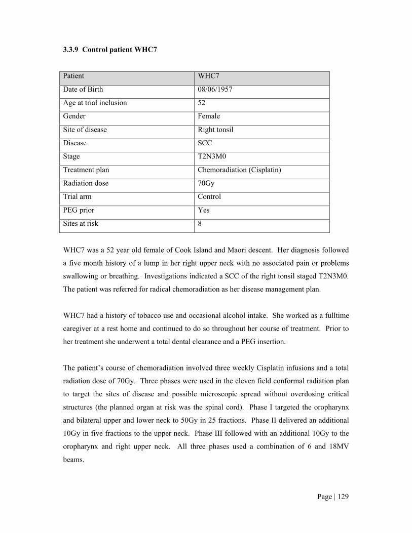
Page | 129
3.3.9 Control patient WHC7
Patient WHC7
Date of Birth 08/06/1957
Age at trial inclusion 52
Gender Female
Site of disease Right tonsil
Disease SCC
Stage T2N3M0
Treatment plan Chemoradiation (Cisplatin)
Radiation dose 70Gy
Trial arm Control
PEG prior Yes
Sites at risk 8
WHC7 was a 52 year old female of Cook Island and Maori descent. Her diagnosis followed
a five month history of a lump in her right upper neck with no associated pain or problems
swallowing or breathing. Investigations indicated a SCC of the right tonsil staged T2N3M0.
The patient was referred for radical chemoradiation as her disease management plan.
WHC7 had a history of tobacco use and occasional alcohol intake. She worked as a fulltime
caregiver at a rest home and continued to do so throughout her course of treatment. Prior to
her treatment she underwent a total dental clearance and a PEG insertion.
The patient’s course of chemoradiation involved three weekly Cisplatin infusions and a total
radiation dose of 70Gy. Three phases were used in the eleven field conformal radiation plan
to target the sites of disease and possible microscopic spread without overdosing critical
structures (the planned organ at risk was the spinal cord). Phase I targeted the oropharynx
and bilateral upper and lower neck to 50Gy in 25 fractions. Phase II delivered an additional
10Gy in five fractions to the upper neck. Phase III followed with an additional 10Gy to the
oropharynx and right upper neck. All three phases used a combination of 6 and 18MV
beams.

Page | 130
During the delivery of phase I the patient lost a lot of weight and had to be re-scanned and re-
planned with slight alterations in field sizes and beam weightings to account for the tissue
deficit. Due to the intensive treatment the patient had close involvement with the dietician,
social worker, district nurse, Pacific health centre and speech language therapist. She also
had weekly blood tests to meet the chemotherapy criteria. WHC7 was consented for trial
participation and was randomised to the control arm to receive the current standard of care at
the Wellington department.
Sites at risk
The eleven field radiation plan which put eight oral cavity sites at risk including the right and
left buccal mucosa, the ventral and dorsal aspect of the tongue, the hard and soft palate, the
floor of mouth and oropharynx.
Figure 69. WHC7 oral cavity sites at risk
The following Figures illustrate the distribution of dose above 40Gy delivered by the
conformal radiation plan.
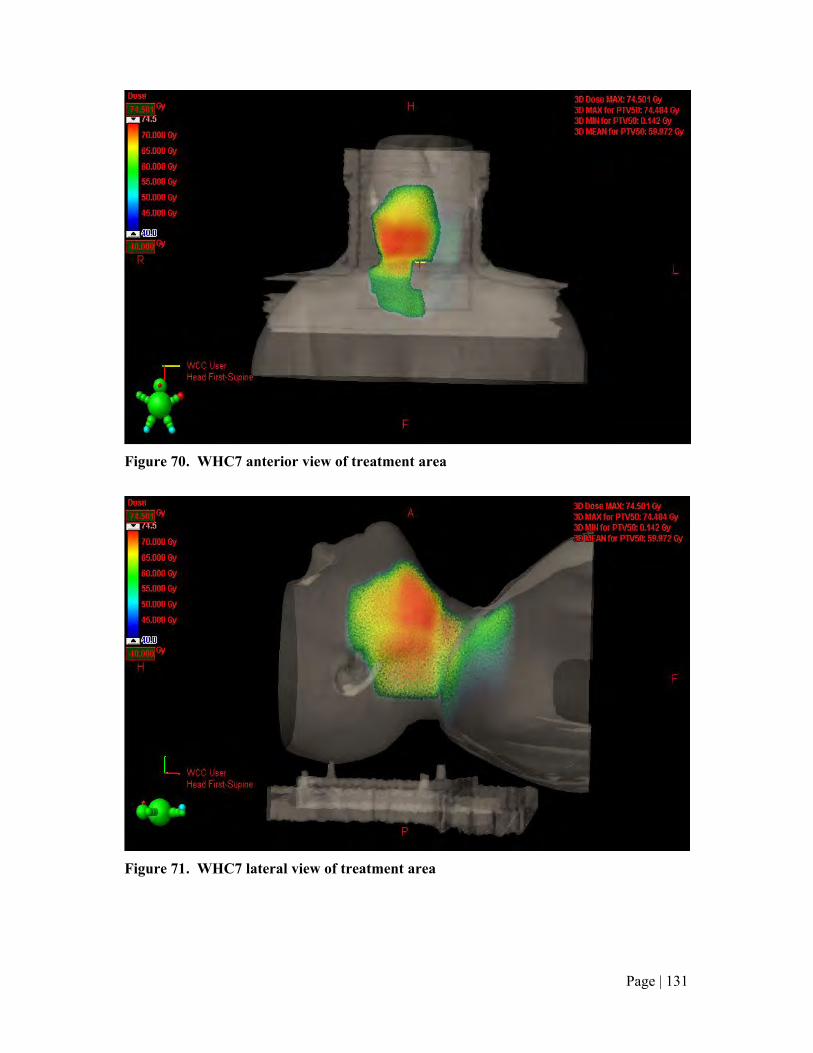
Page | 131
Figure 70. WHC7 anterior view of treatment area
Figure 71. WHC7 lateral view of treatment area
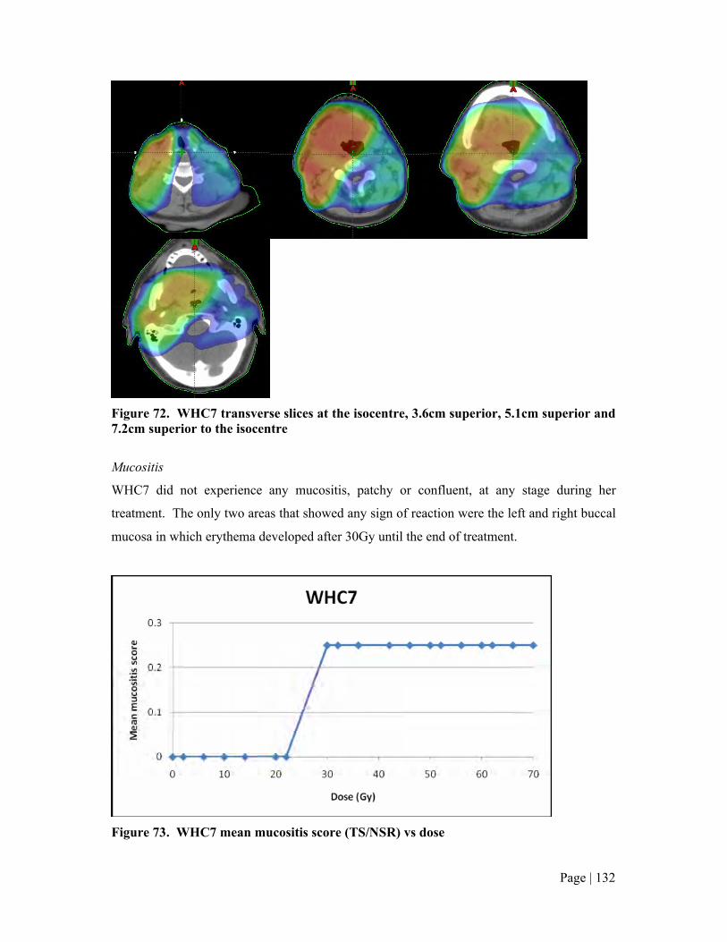
Page | 132
Figure 72. WHC7 transverse slices at the isocentre, 3.6cm superior, 5.1cm superior and 7.2cm superior to the isocentre
Mucositis
WHC7 did not experience any mucositis, patchy or confluent, at any stage during her
treatment. The only two areas that showed any sign of reaction were the left and right buccal
mucosa in which erythema developed after 30Gy until the end of treatment.
Figure 73. WHC7 mean mucositis score (TS/NSR) vs dose
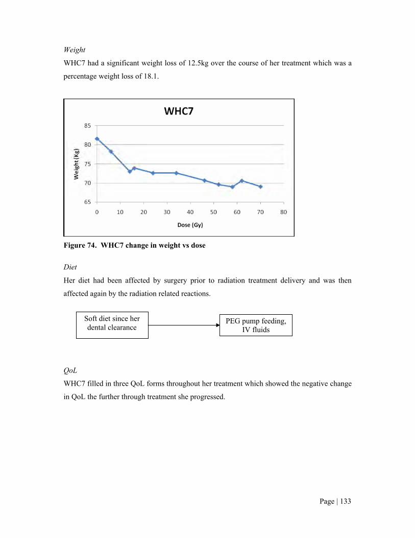
Page | 133
Weight
WHC7 had a significant weight loss of 12.5kg over the course of her treatment which was a
percentage weight loss of 18.1.
Figure 74. WHC7 change in weight vs dose
Diet
Her diet had been affected by surgery prior to radiation treatment delivery and was then
affected again by the radiation related reactions.
QoL
WHC7 filled in three QoL forms throughout her treatment which showed the negative change
in QoL the further through treatment she progressed.
Soft diet since her dental clearance
PEG pump feeding, IV fluids
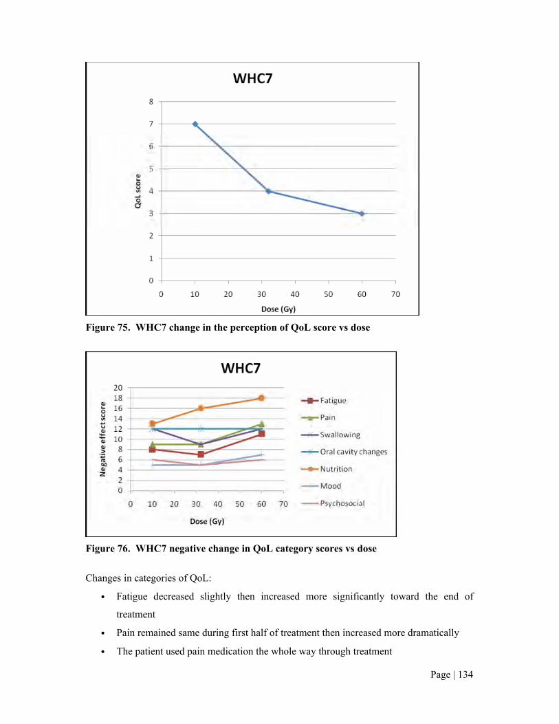
Page | 134
Figure 75. WHC7 change in the perception of QoL score vs dose
Figure 76. WHC7 negative change in QoL category scores vs dose
Changes in categories of QoL:
• Fatigue decreased slightly then increased more significantly toward the end of
treatment
• Pain remained same during first half of treatment then increased more dramatically
• The patient used pain medication the whole way through treatment
Page | 135
• Swallowing problems reportedly decreased midway through treatment (which was
unusual) then increased back to the initial level near the start of treatment
• Oral cavity changes stayed at a relatively low level the whole way through treatment
• Nutrition gradually got worse throughout treatment
• Mood became more negative towards the end of treatment
• Psychosocial effects remained fairly similar
• Perceived QoL gradually decreased over course of treatment
Patient’s subjective experience
The first reaction experienced by WHC7 was a dry mouth after three fractions (6Gy). At the
end of week one her oral intake was normal and she was using baking soda and salt
mouthwashes.
During week two (12G) there was no visible mucositis but the patient suffered from a loss of
taste and nausea. The patient’s diet was significantly reduced by these reactions as she found
it difficult to find foods she could tolerate without vomiting. Her diet consisted of ice cream,
yogurt and one Fortisip per day administered via her PEG. WHC7 found ginger ale better
tolerated than water alone. Later in week two of treatment the dietician advised WHC7 to
increase her Fortisip intake to three per day.
The patient reported no areas of pain but white areas were visible on the tongue and buccal
mucosa and were thought to be oral thrush. At the end of the week (20Gy) the tongue still
had thrush which was being treated with nystatin four times a day. The rest of the mouth was
clear with no pain. Midway through week three (24Gy) the patient had a further loss of
interest in food and was advised to increase her Fortisip intake to four per day.
After three weeks (30Gy) there was pain in the throat (primarily when swallowing), thick
saliva and slight erythema on the buccal mucosa. Three fractions later (36Gy) the patient was
nauseated from chemotherapy and had been vomiting for three days. The nausea, vomiting,
thick mucous and saliva in the mouth all contributed to a significant reduction in oral intake.
The patient was trained to use the PEG for overnight feeding and advised to take anti-nausea
medication prior to food.
Page | 136
Humidification was commenced after four weeks of treatment and difflam spray was
prescribed to help relieve pain. Overnight PEG feeding continuation was encouraged.
Despite increased pain medication (codeine) the throat pain increased after 44Gy.
Humidification was ceased during week five (46Gy) because the patient did not find it
helpful. At the end of five weeks of treatment the patient had a very low mood. Her pain
medications were increased to include xylocaine viscous and morphine.
The patient had a challenging time through to the end of the seven weeks of treatment (70Gy)
with dehydration being a major problem due to not being able to swallow water or other
liquids without gagging. The patient was admitted to hospital in the middle of week seven
(64Gy) when her blood levels indicated severe dehydration. IV fluids were administered on
the ward. If this patient had been randomised to the honey arm it is unlikely she would have
been able to adhere to the honey application protocol.

Page | 137
3.4 Mucositis trail: Comparison between the treatment and control arms
3.4.1 Oral mucositis
Mean average mucositis scores
Honey patients had higher mean average mucositis scores than control patients (Figure 77.)
even though in this small cohort the differences were not statistically significant.
0.0
0.5
1.0
1.5
0 10 20 30 40 50 60
Mean Average Mucositis Scores
Dose (Gy)
control patientshoney patients
Figure 77. A comparison of mean average oral mucositis scores of patients in control and honey arms
Percentage of patients developing oral mucositis
88.9% of all patients developed mucositis. 100% of honey patients developed mucositis
while only 75% of control patients did, the other 25% only developed erythema of the
mucosa. With respect to the extent of mucositis 44.4% of patients developed a maximum of
grade three mucositis, 44.4% of patients developed a maximum of grade two mucositis while
11.1% of patients only developed grade one mucositis (erythema). Of the four patients that
developed grade three mucositis 75% (n=3) were honey patients. Of the four patients that
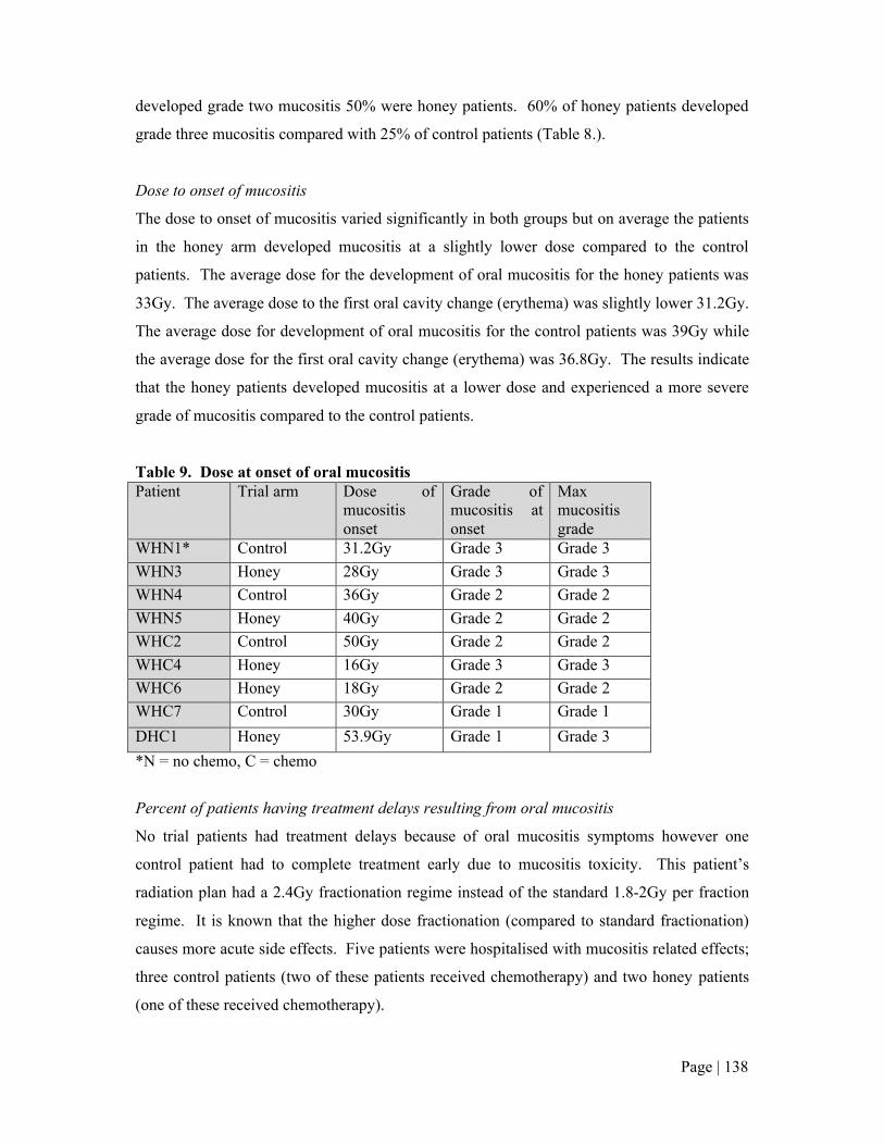
Page | 138
developed grade two mucositis 50% were honey patients. 60% of honey patients developed
grade three mucositis compared with 25% of control patients (Table 8.).
Dose to onset of mucositis
The dose to onset of mucositis varied significantly in both groups but on average the patients
in the honey arm developed mucositis at a slightly lower dose compared to the control
patients. The average dose for the development of oral mucositis for the honey patients was
33Gy. The average dose to the first oral cavity change (erythema) was slightly lower 31.2Gy.
The average dose for development of oral mucositis for the control patients was 39Gy while
the average dose for the first oral cavity change (erythema) was 36.8Gy. The results indicate
that the honey patients developed mucositis at a lower dose and experienced a more severe
grade of mucositis compared to the control patients.
Table 9. Dose at onset of oral mucositis Patient Trial arm Dose of
mucositis onset
Grade of mucositis at onset
Max mucositis grade
WHN1* Control 31.2Gy Grade 3 Grade 3 WHN3 Honey 28Gy Grade 3 Grade 3 WHN4 Control 36Gy Grade 2 Grade 2 WHN5 Honey 40Gy Grade 2 Grade 2 WHC2 Control 50Gy Grade 2 Grade 2 WHC4 Honey 16Gy Grade 3 Grade 3 WHC6 Honey 18Gy Grade 2 Grade 2 WHC7 Control 30Gy Grade 1 Grade 1 DHC1 Honey 53.9Gy Grade 1 Grade 3 *N = no chemo, C = chemo
Percent of patients having treatment delays resulting from oral mucositis
No trial patients had treatment delays because of oral mucositis symptoms however one
control patient had to complete treatment early due to mucositis toxicity. This patient’s
radiation plan had a 2.4Gy fractionation regime instead of the standard 1.8-2Gy per fraction
regime. It is known that the higher dose fractionation (compared to standard fractionation)
causes more acute side effects. Five patients were hospitalised with mucositis related effects;
three control patients (two of these patients received chemotherapy) and two honey patients
(one of these received chemotherapy).
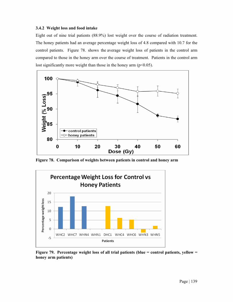
Page | 139
3.4.2 Weight loss and food intake
Eight out of nine trial patients (88.9%) lost weight over the course of radiation treatment.
The honey patients had an average percentage weight loss of 4.8 compared with 10.7 for the
control patients. Figure 78. shows the average weight loss of patients in the control arm
compared to those in the honey arm over the course of treatment. Patients in the control arm
lost significantly more weight than those in the honey arm (p<0.05).
Figure 78. Comparison of weights between patients in control and honey arm
Figure 79. Percentage weight loss of all trial patients (blue = control patients, yellow = honey arm patients)
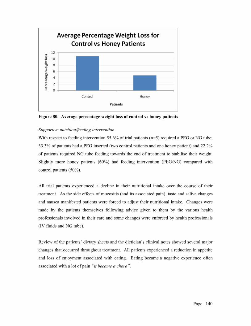
Page | 140
Figure 80. Average percentage weight loss of control vs honey patients
Supportive nutrition/feeding intervention
With respect to feeding intervention 55.6% of trial patients (n=5) required a PEG or NG tube;
33.3% of patients had a PEG inserted (two control patients and one honey patient) and 22.2%
of patients required NG tube feeding towards the end of treatment to stabilise their weight.
Slightly more honey patients (60%) had feeding intervention (PEG/NG) compared with
control patients (50%).
All trial patients experienced a decline in their nutritional intake over the course of their
treatment. As the side effects of mucositis (and its associated pain), taste and saliva changes
and nausea manifested patients were forced to adjust their nutritional intake. Changes were
made by the patients themselves following advice given to them by the various health
professionals involved in their care and some changes were enforced by health professionals
(IV fluids and NG tube).
Review of the patients’ dietary sheets and the dietician’s clinical notes showed several major
changes that occurred throughout treatment. All patients experienced a reduction in appetite
and loss of enjoyment associated with eating. Eating became a negative experience often
associated with a lot of pain “it became a chore”.
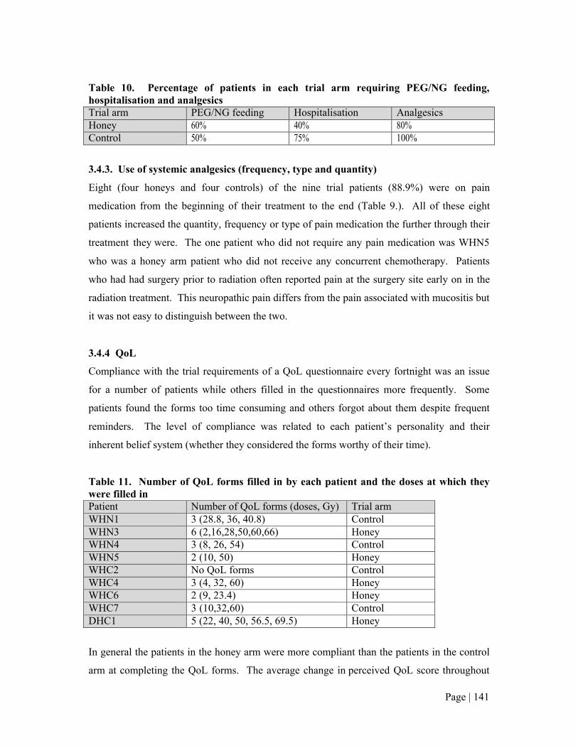
Page | 141
Table 10. Percentage of patients in each trial arm requiring PEG/NG feeding, hospitalisation and analgesics Trial arm PEG/NG feeding Hospitalisation Analgesics Honey 60% 40% 80% Control 50% 75% 100%
3.4.3. Use of systemic analgesics (frequency, type and quantity)
Eight (four honeys and four controls) of the nine trial patients (88.9%) were on pain
medication from the beginning of their treatment to the end (Table 9.). All of these eight
patients increased the quantity, frequency or type of pain medication the further through their
treatment they were. The one patient who did not require any pain medication was WHN5
who was a honey arm patient who did not receive any concurrent chemotherapy. Patients
who had had surgery prior to radiation often reported pain at the surgery site early on in the
radiation treatment. This neuropathic pain differs from the pain associated with mucositis but
it was not easy to distinguish between the two.
3.4.4 QoL
Compliance with the trial requirements of a QoL questionnaire every fortnight was an issue
for a number of patients while others filled in the questionnaires more frequently. Some
patients found the forms too time consuming and others forgot about them despite frequent
reminders. The level of compliance was related to each patient’s personality and their
inherent belief system (whether they considered the forms worthy of their time).
Table 11. Number of QoL forms filled in by each patient and the doses at which they were filled in Patient Number of QoL forms (doses, Gy) Trial arm WHN1 3 (28.8, 36, 40.8) Control WHN3 6 (2,16,28,50,60,66) Honey WHN4 3 (8, 26, 54) Control WHN5 2 (10, 50) Honey WHC2 No QoL forms Control WHC4 3 (4, 32, 60) Honey WHC6 2 (9, 23.4) Honey WHC7 3 (10,32,60) Control DHC1 5 (22, 40, 50, 56.5, 69.5) Honey
In general the patients in the honey arm were more compliant than the patients in the control
arm at completing the QoL forms. The average change in perceived QoL score throughout
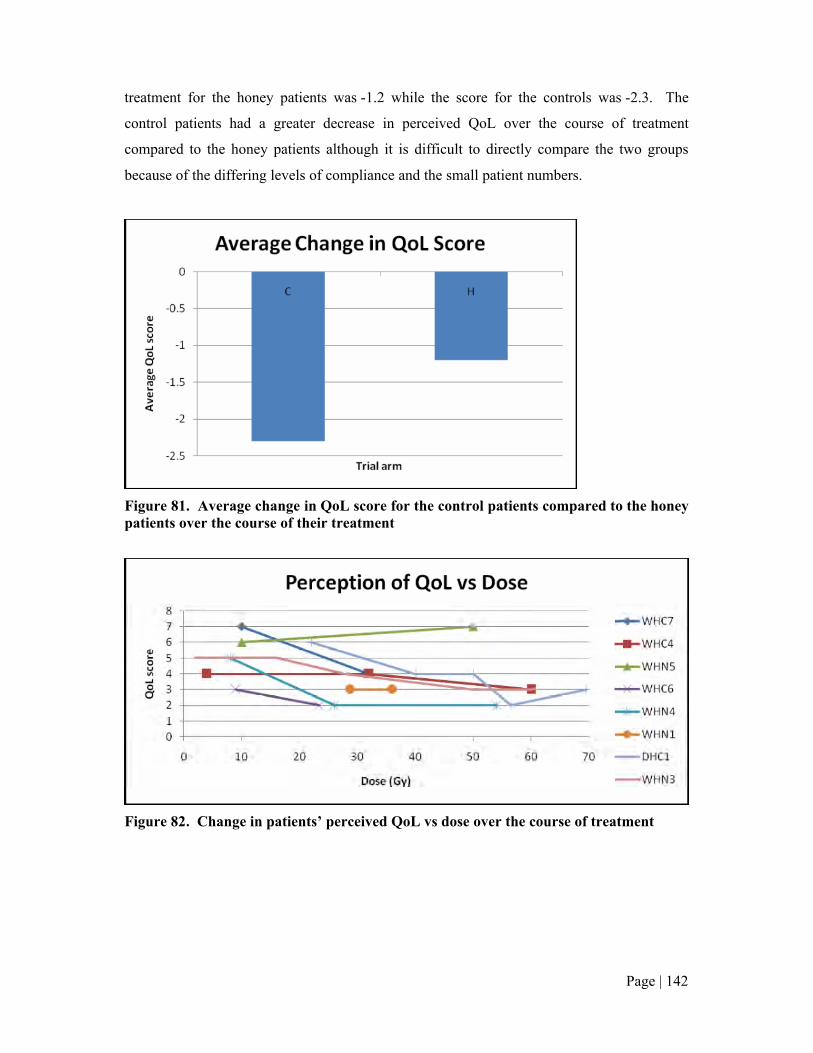
Page | 142
treatment for the honey patients was -1.2 while the score for the controls was -2.3. The
control patients had a greater decrease in perceived QoL over the course of treatment
compared to the honey patients although it is difficult to directly compare the two groups
because of the differing levels of compliance and the small patient numbers.
Figure 81. Average change in QoL score for the control patients compared to the honey patients over the course of their treatment
Figure 82. Change in patients’ perceived QoL vs dose over the course of treatment

Page | 143
Chapter 4 DISCUSSION
4.1 Interpretation of results
4.1.1 Severity of oral mucositis
The results of the New Zealand mucositis trial suggest that honey does not decrease the
severity of oral mucositis in the small cohort of patients. On the contrary, patients in the
honey arm had a higher mean mucositis score compared to the patients in the control arm.
All honey arm patients developed mucositis compared with only 75% of control patients. On
average patients in the honey arm also first developed mucositis at a slightly lower dose
compared to the control patients. These results suggest that the manuka honey is not superior
to the current standard of care for head and neck patients.
The results do not correspond with the results of the three previous honey trials by Biswal et
al. (2003), Rashad et al. (2008) and Motallebnejad et al. (2008) (described in Appendix B)
which showed that honey decreased the severity of oral mucositis significantly.
There were differences between the three previous trials and the New Zealand trial with
respect to the type of honey, the control group standard of care and the scoring system used.
The main difference in methodology between the overseas trials and the New Zealand trial
was the honey concentration. All three overseas trials used undiluted honey, whereas the
New Zealand trial used diluted honey. This was a direct result of significant issues
encountered with patient compliance. Biswal et al. (2003) and Rashad et al. (2008) did not
mention issues with patient compliance although Motallebnejad et al. (2008) did mention that
patients who experienced severe stinging of the mouth were removed from the trial.
Preliminary analysis of results from the three oncology departments involved in the New
Zealand trial (Wellington, Palmerston North and Dunedin) showed that manuka honey was
not well tolerated. Patients who experienced severe pain and nausea in response to the honey
pulled out of the trial because it was unethical to coerce them into continuing with the honey
application. Compliance with the honey application protocol also related to personality
factors:

Page | 144
• Each individual has a different pain tolerance and patients with lower pain thresholds
were more likely to stop taking honey when they found it caused a stinging sensation.
• The anticipatory nausea and vomiting phenomenon is a learned response that occurs
in patients who have had a negative experience with chemotherapy. One trial patient
who pulled had built a strong association between chemotherapy, manuka honey and
nausea and vomiting so was unable to continue with the honey application.
• The patients who persisted with the honey application all shared an intrinsic belief
that the honey was of benefit to them.
The intolerance of undiluted honey let to several amendments to the protocol, reducing the
overall amount of honey from 20mL to 10mL and diluting the honey 1:3 with liquid. The
lack of effect of the honey in the current study is most likely due to the diluting of the honey
beyond its effective concentration.
All three overseas trials had control groups but two did not receive the same standard of care
that is delivered in New Zealand. Most New Zealand head and neck patients are instructed
on the use of salt and baking soda mouthwashes, have access to topical and systemic
anasthetics and are cared for by a multidisciplinary team. The authors of the overseas trials
did not report poor application compliance or significant patient drop-out.
4.1.2 Weight, nutrition and QoL
Interestingly the effect of honey on weight, nutritional status and patient QoL was very
different to the effect on oral mucositis. The results of the weight loss comparison between
the honey arm and the control arm patients correspond to the results from the Biswal et al.
(2003) trial and the Motallebnejad et al. (2008) trial where on average the honey arm patients
lost less weight than the control patients. A higher percentage of control patients required
hospitalisation and analgesia over the course of treatment but a higher percentage of honey
arm patients required PEG/NG feeding. The manuka honey appeared to have a positive
effect on patients’ QoL with the average reduction in QoL score being less than the control
patients. Despite this result the descriptive accounts of the experiences of the honey arm
patients were often negative with regard to QoL.
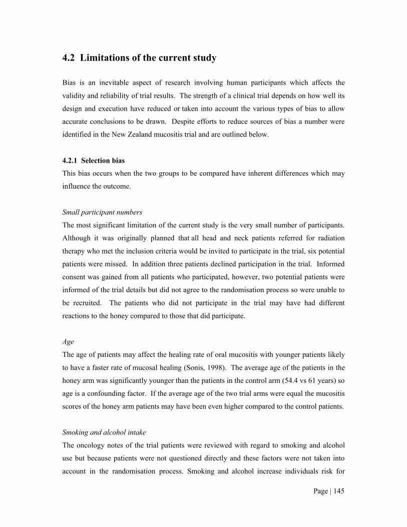
Page | 145
4.2 Limitations of the current study
Bias is an inevitable aspect of research involving human participants which affects the
validity and reliability of trial results. The strength of a clinical trial depends on how well its
design and execution have reduced or taken into account the various types of bias to allow
accurate conclusions to be drawn. Despite efforts to reduce sources of bias a number were
identified in the New Zealand mucositis trial and are outlined below.
4.2.1 Selection bias
This bias occurs when the two groups to be compared have inherent differences which may
influence the outcome.
Small participant numbers
The most significant limitation of the current study is the very small number of participants.
Although it was originally planned that all head and neck patients referred for radiation
therapy who met the inclusion criteria would be invited to participate in the trial, six potential
patients were missed. In addition three patients declined participation in the trial. Informed
consent was gained from all patients who participated, however, two potential patients were
informed of the trial details but did not agree to the randomisation process so were unable to
be recruited. The patients who did not participate in the trial may have had different
reactions to the honey compared to those that did participate.
Age
The age of patients may affect the healing rate of oral mucositis with younger patients likely
to have a faster rate of mucosal healing (Sonis, 1998). The average age of the patients in the
honey arm was significantly younger than the patients in the control arm (54.4 vs 61 years) so
age is a confounding factor. If the average age of the two trial arms were equal the mucositis
scores of the honey arm patients may have been even higher compared to the control patients.
Smoking and alcohol intake
The oncology notes of the trial patients were reviewed with regard to smoking and alcohol
use but because patients were not questioned directly and these factors were not taken into
account in the randomisation process. Smoking and alcohol increase individuals risk for
Page | 146
developing cancer because they contain carcinogens and cause inflammation (Evans et al.,
2003). For similar reasons these substances also increase the amount and severity of oral
mucositis so they should be taken into account in mucositis trials to minimize confounding.
Gender
There were more males in the honey arm than the control arm (80% vs 25%) and this may
have contributed to different mucositis scores between the two groups. The three overseas
honey trials also had a greater percentage of males recruited to the trial and there were more
males in the honey arm. Genetic differences may impact on rates of mucositis development
and the mucosal response to manuka honey.
Dose distribution
The tumour site and stage differed between all the trial patients which meant the dose
distribution in the oral cavity differed. The trial aimed to minimise the confounding effects
of dose distribution by dividing the total mucositis score by the number of sites at risk. The
two different types of mucosa in the oral cavity may have different sensitivities to radiation
damage so tumour site may impact on the degree of mucositis.
Planning software
Eclipse planning software was used to plan the Wellington patients’ treatment and doses were
modelled to achieve the best plan. The areas identified to be ‘at risk’ on the modelled plan
were not always the areas where reactions became evident. There is a small degree of
uncertainty in all modelling programmes. One specific factor that affects the accuracy of
radiation modelling is the presence of metallic fillings. These alter the dose distribution
within the oral cavity and may increase the severity of mucositis in the surrounding tissue. It
is standard that patients have a dental review prior to starting radiation treatment but some
patients proceed with fillings in place. The New Zealand trial patients’ teeth or fillings were
not examined. The presence of fillings may have accounted for some of the variation
between the number of sites identified to be at risk and those that did develop mucositis and
if the honey or control arm patients had fillings the rates of mucositis could have been higher.
Chemotherapy
Concurrent chemotherapy has been shown to increase the severity of radiation induced
reactions (Trotti, 2000). Two honey arm patients and two control arm patients received
Page | 147
concurrent Cisplatin chemotherapy but another honey patient had received neo-adjuvant R-
CHOP chemotherapy. The greater average mean mucositis scores of the honey patients could
have resulted from a higher percentage of the honey patients having received chemotherapy.
Surgery
Tissue that has undergone previous healing may have a different sensitivity to radiation and
could have contributed to the varying rates of mucositis. The majority of trial patients
(77.8%) had undergone surgery prior to radiation treatment referral. The two patients who
had not were in the control arm of the trial.
Humidifiers
Three public oncology departments in New Zealand are participating in the TROG
humidification trial to assess whether humidification can reduce oral mucositis in head and
neck cancer patients receiving radiation therapy. The trial proposes that heated
humidification of inspired air via a nasal interface may palliate symptoms of mucositis by
reducing discomfort associated with dry, sticky secretions. The (Fisher and Paykel
Healthcare) humidifiers used in the trial deliver gas which is heated and humidified to 37°C,
44 mgH20/L. Although the Wellington oncology department was not participating in the
humidification trial two humidifiers became available for patient use early in 2009. The
potential benefits (reduction in patient discomfort from oral dryness, reduction in pain
severity and the decrease in sticky secretions) meant that the humidifiers became part of the
standard of care for high risk head and neck patients. The use of humidification in three of
the manuka honey trial patients (two controls and one honey patient) could have reduced the
incidence or severity of mucositis independent of the manuka honey.
4.2.2 Randomisation bias
The purpose of randomisation is to avoid or reduce potentially confounding factors by
making the honey and control arms comparable. Although the original trial was designed to
randomize patients using computer-generated random numbers, after the protocol was
changed to diluted honey, additional patients were put on the honey arm without further
randomization. Due to the low patient numbers the two trial arms were unable to be matched
and so patient variables such as age, gender and ethnicity and treatment variables such as
PTV (planning target volume) location and size would have affected the rates and severity of
mucositis between the two trial arms.

Page | 148
4.2.3 Participant bias
There were a number of instances where participant bias could have been introduced to the
mucositis trial. Some patients may have agreed to be a part of the trial not because they
wanted to but because they felt obliged to.
Reporting bias
Because some of the trial results are subjective they are affected by patients’ personalities,
emotions and attitudes.
• It is possible that some patients do not act in their normal way when participating in a
trial and they may have altered their reports (verbal and written) to appease the
researcher.
• Pain is a subjective experience and is difficult to quantify; some people have lower
pain thresholds than others (one patient in the honey arm who despite having visible
mucositis did not notice any pain). The lack of an objective measure introduces a
degree of variability in the comparison of pain levels.
• It is possible that the degree of oral mucositis (inflammation) was affected by
patients’ psychology. (A lot of research has focussed on the effect of stress and
negative personality traits on the immune system and the incidence of disease. It may
be possible that personality traits also affect how much benefit patients derive from
certain treatments.)
• The placebo effect may have influenced the trial results. Patients in the honey arm
could have had a perceived increase in QoL when taking the honey (patients feel like
they are taking control and doing something to alleviate treatment side effects).
Recall bias
Recall or memory bias could have been introduced to the trial when patients were asked to
recall the events of the past few days (especially after weekends). Honey application and
tolerability, food and fluid consumption and medication requirements may not have been
accurately remembered. This bias probably applied to the QoL forms which were designed
to reflect the patients QoL the week preceding the filling in of the questionnaire, it is possible
the patient blocked out the true level of pain or fatigue they felt.
Page | 149
4.2.4 Compliance bias
Adherence to the honey protocol differed among patients and through discussion it was
obvious that adherence was due to the patient’s perceived benefit from the honey. The
patients who perceived a benefit were able to persevere through the pain and/or nausea.
Compliance with filling in QoL questionnaires also differed significantly.
Inconsistency of adherence to honey protocol
The descriptive experiences of the patients in the honey arm show that undiluted honey was
not well tolerated. Four patients randomised to the honey arm pulled out of the trial within
the first ten days of treatment. The five honey patients that persisted with honey application
frequently complained about the taste, texture and stinging nature of the honey. Only 40% of
the honey patients (n=2) finished treatment still applying the manuka honey and 60% of
honey patients found they had to mix the honey with a liquid other than water to reduce the
sweetness. The 60% of honey patients who ceased honey application may have done so at
the stage in treatment where the honey had the most benefit (patchy and confluent mucositis).
Sweetness
The sweetness of the honey was difficult to tolerate and associated with nausea. The sugar
content was also of concern to one patient randomised to the honey arm who was worried
about the increased risk of tooth decay. Tooth decay is a serious concern for head and neck
patients who, due to saliva composition changes are at an increased risk of cavities. It is
possible the high sugar content of honey makes it a cariogenic substance, however one report
stated that honey is useful to treat gum disease (Bardy et al., 2007) and another study reported
that honey is less cariogenic in dry mouth subjects (Sela et al., 1998).
Sticky saliva
Saliva changes are common in head and neck radiation therapy and the formation of thick
‘sticky’ saliva affects patients’ ability to swallow and causes patients considerable difficulty
(Funegard et al., 1994). Patients with xerostomia may be less able to tolerate manuka honey
due to thick saliva (one control patient developed ‘sticky’ saliva so severe she could not eat
so it is unlikely she could have tolerated the honey if randomised to that arm).

Page | 150
Exposure time
The trial originally proposed that all patients in the honey arm swallowed the honey to coat
the mucosal lining of the oropharynx after the oral cavity, this was however amended as
patient honey tolerance became an issue. Patients were then asked to swallow the honey
mixture if possible or to hold it in their mouth for as long as they could (ideally a minute) and
then spit it out. These different exposure times could have influenced the results. The
manuka honey may require a certain amount of time to have an effect (anti-inflammatory or
otherwise) on the mucosa.
Concentration
Amendments were made to the honey application instructions to make it more tolerable. The
honey was to be diluted by a ratio of 1:3 with water (or other liquids). Dilution decreases the
concentration of all the compounds found in honey, including those responsible for its anti-
inflammatory action. In addition it is possible that not all patients were equally precise in
diluting the honey. The process of diluting the honey added hassle for patients.
As the trial proceeded it became apparent some honey jars had small amounts of
‘crystallised’ honey. The crystals were more difficult to dissolve in liquid and so more
difficult to spread around the patient’s oral cavity. This would have affected the dilution
factor (some honey arm patients found ‘dregs’ of honey were left in the bottom of the vessel
used to mix the honey).
Timing of honey application
The timing of honey application differed between patients. It was originally proposed that
patients would apply the manuka honey before and after treatment and then again six hours
later. Some patients adhered to this protocol while others found this too difficult. Some had
to travel in from the greater Wellington region so applied the honey a long time before and
after treatment (travel times). Another patient experienced nausea in the morning so applied
the honey three times later in the day.
Honey Quality
The proposed method of action of the manuka honey on oral mucositis relates to the anti-
inflammatory properties of the honey constituents. Phenolic compounds may engage in
Page | 151
selective radical scavenging and methylglyoxal (MGO) has antibacterial activity (Stephens et
al., 2009). The bioactivity of manuka honey and its potential benefit depends on the
concentration of these compounds which may be affected by natural factors such as honey
age, monoflorality and geographical source (localised environmental factors or genetic
differences) (Stephens et al., 2009). In order to avoid these potentially confounding factors
all the honey used for the trial was from the same supplier (collected from the same beehives,
stored in the same conditions, tested for toxins and MGO content and gamma sterilized). The
honey was distributed to the different oncology departments and trial patients. At the
Wellington department the honey was stored in a refrigerator until given to patients who were
instructed to store the honey in a cool place out of direct light. Despite intentions and
instructions to keep the honey in stable conditions honey jars was likely exposed to different
conditions which may have affected the composition of the honey.
Similarly high temperature effects may have altered the honey composition during
microwaving of, or dilution of the honey with hot liquid prior to application (one patient
warmed the honey in the microwave several times before being told this was not ideal).
Compliance with filling in food diaries and QoL questionaires
The food diaries and QoL questionnaires were given to each patient with the same
instructions but the compliance differed. It was originally proposed that the food/drug diaries
would be filled out by the patients daily to gain insight into their diet and pain control and see
how this changed over the course of treatment. Several patients reported daily recording too
much of a hassle so moved to weekly recording or recording of significant changes in diet or
medication administration (for example the reduction in food intake, move to NG or PEG
feeding or the increase in amount or strength of pain relief). This change may not have
allowed enough insight into the diet differences between the two trial arms. One patient did
not complete any QoL forms.
4.2.5 Researcher bias
The attitudes and actions of the medical professionals (research assistant, nurse, dietician and
radiation oncologist) who were involved in the trial patients’ care could have influenced the
results and the level of compliance.
Page | 152
Personal interaction
The attitude of the oncologist who presented the trial to the patient would have influenced the
patient’s decision to participate; some oncologists may have ‘pushed’ the trial more than
others. It is important that patients are given all the necessary information without coercion
but it is inevitable that some opinions are conveyed. Other health professionals involved may
have encouraged patients to be more persistent with honey application. It became a bit of an
ethical dilemma when patients were obviously suffering pain and/or nausea from the honey
application; it was desirable patients continued with the application as long as possible for the
purpose of the trial but patients should not be expected to tolerate more pain or discomfort
than they would otherwise experience. All trials need to incorporate the values of medical
ethics (beneficence and nonmaleficence) (Miller & Rosenstein, 2003).
Mucositis scoring
The blinded nature of the proposed trial design was quickly amended because the research
assistant responsible for the mucositis scoring was the main point of contact for the patient.
The honey application instructions were given to the patient by the assistant so to ensure that
patients adhered to the protocol the researcher needed to know which arm the patients were
randomised to. The lack of blinding may have subconsciously affected the research
assistant’s mucositis scoring; the mucositis scores for the honey patients may have been
lower than scores from an assistant who had been blinded.
4.2.6 Measurement bias
Measurement biases introduce systematic error when data is collected.
Consistency in mucositis scoring was addressed in the trial design by the mucositis scoring
training session that the scorers attended prior to trial commencement. The scorer may have
become more confident with the scoring process further into the trial. However it is possible
that scoring inconsistencies were introduced the more time that passed since the training
session. Although the same person did the majority of the scoring for all patients at the
Wellington department there could have been inter-scorer variability between the
participating departments.

Page | 153
Lighting conditions in the rooms where oral assessments took place could have affected the
mucositis scores, especially the identification of oral erythema which is not as obvious as oral
mucositis. Head lamps, torches and well-lit clinic rooms were used in most scoring cases but
the light level could have varied.
Patient factors
Some patients found it difficult to open their mouths due to the effects of previous surgery or
mucositis-related pain. This factor made oral cavity assessments more difficult and may have
led to under scoring of some sites.
The mucosal lining of the oral cavity could have been affected by mechanical trauma not
radiation damage (for example a head and neck patient eating some rough food which results
in an area of perceived erythema). As outlined earlier the use of topical analgesics can
increase a patient’s risk of biting or burning their oral mucosa due to reduced sensitivity, this
could have affected mucositis scoring.
Infection
The presence of oral candidiasis may also have impacted on mucositis scoring. The layer of
white film that is often associated with the yeast infection is similar to the appearance of oral
mucositis. It is possible to confuse the two leading to potential under or over scoring of
mucositis.
Timing of mucositis assessment
Assessments were done three times a week (Monday, Wednesday, Friday). Due to a public
holiday falling on a Monday one patient had their oral cavity examined on a Tuesday and
Wednesday. It was noted that there was a degree of healing of the oral mucosa overnight.
One area of confluent mucositis was reported as patchy mucositis the next day. This example
showed that mucositis is dynamic and that even during treatment mucosal healing is
occurring. This highlights the issue of the timing of the mucositis assessments. Daily
assessment may give a more accurate record of the progression of mucositis but would be
more resource intensive and more difficult for patients to tolerate. The current study
minimized the effect of timing by plotting the average mucositis score over the entire course
of treatment.
Page | 154
4.3 Recommendations for future clinical trials
Patient accrual
Good lines of communication between all members of the patients’ multidisciplinary team
will ensure maximum patient accrual to future trials. Department software systems can also
help maximise patient accrual (for example the MOSAIQ software function that can identify
patients by treatment site so that potential trial patients can be easily identified prior to their
CT planning scan).
Dedicated research staff
Oncology departments involved in research should have dedicated research staff and
adequate resources to enable valuable research to be carried out. Large scale clinical trials
require time, money and commitment but they provide statistically significant data to
improve current standards of care.
Mucositis scoring
Specific lighting conditions should be reproduced for each mucositis assessment. The
mucositis scorers should have regular tests to ensure consistent correct mucositis scoring.
More oncology department staff members should be trained to score mucositis to cover
instances when the research assistant is unavailable. Continual monitoring of treatment
machine schedules is required (by the research assistant) to ensure that all trial patients have
their mucositis assessments conducted at the specified intervals.
Honey application
Further research is warranted to find the best way to apply manuka honey to the oral cavity so
a higher level of compliance can be achieved. Dilutions that do not reduce the potential
beneficial actions of honey should be investigated. The active constituents of manuka honey
may also be present in manuka essential oil which may be better tolerated by patients. A
study reported in the European Journal of Oncology Nursing investigated these oils and
found that they were able to delay the onset of mucositis and reduce oral pain (Maddocks-
Jennings et al., 2009). It may also be possible to remove the sugar content of manuka honey
to reduce the intense sweetness (make the honey more tolerable) and reduce the potential risk
of cavity formation.
Page | 155
Food diaries and QoL forms
Food diaries need to be presented in a more user-friendly format to encourage frequent
recording. They should also include a section where the patient can report on their honey use
and any problems they encounter. Patients should be clearly instructed on the QoL form
requirements prior to trial inclusion to ensure they will comply to increase the future rate of
return.
Patient information
More information about the patients recruited to trials would reduce the number of potential
confounding factors. Patients should be questioned prior to treatment with regard to smoking
(present or previous), alcohol intake, nutritional status, ethnicity and co-morbidities.
Page | 156
4.4 Conclusion
Oral mucositis causes considerable discomfort to patients and currently there is no definitive
treatment (Maddocks-Jennings et al., 2009). Manuka honey may have an immunomodulatory
effect and may be able to combat inflammatory processes such as mucositis. The New
Zealand manuka honey trial was originally designed as a stage II randomized single blinded
trial to determine whether Comvita manuka honey in addition to standard best practice is
superior to standard best practice alone in decreasing the extent of radiation-induced oral
mucositis.
In support of the hypothesis, three overseas trials showed that different honeys significantly
reduced the extent of radiation-induced oral mucositis in patient cohorts in Malaysia, Iran and
Egypt. The New Zealand trial, however, reported that undiluted manuka honey was not well
tolerated by the patient cohort. Patients complained of extreme nausea and stinging
sensations in the oral cavity and throat and the trial protocol had to be amended from 20mL
of undiluted honey to 10mL of honey diluted in 30mL of water (or other liquid). Preliminary
analysis showed that diluted manuka honey did not decrease the extent of oral mucositis in
the small cohort of New Zealand head and neck cancer patients when taken in addition to
current best practice.
Despite there being a lack of consistency in the care of head and neck patients in New
Zealand patients are well looked after with respect to oral cavity reactions; most patients
receive nutritional support, oral hygiene advice and pain relief as required along with
frequent contact with their oncologist and other members of the multi-disciplinary team. The
addition of manuka honey did not show any benefit and caused a large amount of discomfort
with respect to taste and stinging.
The trial did not yield statistically significant results due to the small sample size and
confounding factors but it did provide useful information about the tolerability of manuka
honey in head and neck patients. The reports from trial patients can be used in future trials
that involve honey application. Further research into ways to make manuka honey more
tolerable to patients and improve patient compliance is warranted.
Page | 157
References
Al-Waili, N., Saloom, K. (1999). Effects of topical Honey on post-operative Wound
Infections due to gram positive and gram negative Bacteria following Caesarean Sections and
Hysterectomies. European Journal of Medical Research, 4, 26-130.
Ang, K., Garden, A. (2002). Radiotherapy for Head and Neck Cancers. Indications
and Techniques, Second edition. Philadelphia PA: Lippincott Williams & Wilkins.
Antonadou, D., Pepelassi, M., Synodinou, M., Puglisi, M., Throuvalas, N. (2002).
Prophylactic use of Amifostine to prevent radiochemotherapy-induced Mucositis and
Xerostomia in Head-and-Neck Cancer. International Journal of Radiation Oncology Biology
Physics, 52, 739-747.
Barasch, A., Peterson, D. (2003). Risk factors for ulcerative oral Mucositis in Cancer
patients: unanswered questions. Oral Oncology, 39, 91-100.
Bardy, J., Slevin, N., Mais, K., Molassiotis, A. (2007). A systematic Review of
Honey uses and its potential Value within Oncology Care. Journal of Clinical Nursing, 17,
2604-2623.
Biron, P., Sebban, C., Gourmet, R., Chvetzoff, G., Philip, I., Blay, J. (2000).
Research Controversies in Management of oral Mucositis. Support Care Cancer, 8, 68-71.
Biswal, B., Zakaria, A., Ahmad, N. (2003). Topical Application of Honey in
Management of Radiation Mucositis. A Preliminary study. Support Care Cancer, 11, 242-
248.
Bomford, C., Kunkler, I. (2003). Textbook of Radiotherapy. Radiation Physics,
Therapy and Oncology: Sixth edition. Philadelphia, PA: Churchill Livingstone, Elsevier
Science Limited.

Page | 158
Cabral L., Carvalho, L., Salgado, J., Brandao A., Almeida J. (2010). Gingival
Squamous Cell Carcinoma: a Case Report. Journal of Oral & Maxillofacial Research, 1, 1-6.
Chambers, M., Garden, A., Kies, M., Martin, J. (2004). Radiation-induced
Xerostomia in Patients with Head and Neck Cancer: Pathogenesis, Impact on Quality of Life,
and Management. Head and Neck, 26, 796-807
Dodd, M., Miaskowski, G., Greenspan, D., MacPhail, L, Shih, A, Shiba, G., Facione,
N. Paul, S. (2003). Radiation-induced mucositis: a randomised clinical trial of micronized
sucralfate versus salt and soda mouthwashes. Cancer Investigation, 20, 21-33.
Duncan, W., MacDougall, R., Kerr, G., Downing, D. (1996). Adverse Effect of
Treatment Gaps in the outcome of Radiotherapy for Laryngeal Cancer. Radiotherapy and
Oncology, 41, 203-207.
Epstein, J., Silverman, S., Paggiarino, D., Crockett, S., Schubert, M., Senzer, N.,
Lockhart, P., Gallager, M., Peterson, D., Leveque, F. (2001). Benzydamine HCl for
Prophylaxis of Radiation-Induced Oral Mucositis. Cancer, 92, 876-885.
Evans, P., Montgomery, P., Gullane, P. (2003). Principles and Practice of Head and
Neck Oncology. London: Martin Dunitz, Taylor & Francis Group.
Funegard, U., Franzen, L., Ericson, T., Henriksson, R. (1994). Parotid Saliva
Composition During and After Irradiation of Head and Neck Cancer. Oral Oncology,
European Journal of Cancer, 30, 230-233.
Garden, A. (2003). Mucositis: Current Management and Investigations. Seminars in
Radiation Oncology, 13, 267-273.
Gibson, R., Bowen, J., Keefe, D. (2008). Technological advances in mucositis
research: New insights and new issues. Cancer Treatment Reviews, 34, 476-482.
Gil, Z., & Fliss, D. (2009). Contemporary management of head and neck cancers.
Israel Medical Association Journal, 11, 296-300.
Page | 159
Hosal, A., Carrau, L., Johnson, J., Myers, E. (2000). Selective Neck Dissection in the
Management of the Clinically Node-Negative Neck. The Laryngoscope, 110, 2037-2040.
Joiner, M., van der Kogel, A. (2009). Basic Clinical Radiobiology. Forth edition.
Hodder Arnold. MacMillan Publishing Group.
Leslie, M., Dische, S. (1991). Parotid Gland Function following accelerated and
conventionally fractionated Radiotherapy. Radiotherapy and Oncology, 22, 133-139.
Maddocks-Jennings, W., Wilkinson, J., Cavanagh, H., Shillington, D. (2009).
Evaluating the Effects of the Essential Oils Leptospermum scoparium (manuka) and Kunzea
ericoides (kanuka) on Radiotherapy induced Mucositis: A randomized, placebo controlled
feasibility Study. European Journal of Oncology Nursing, 13, 87-93.
Mavric, E., Wittmann, S., Barth, G., Henle, T. (2008). Identification and
Quantification of Methylglyoxal as the dominant Antibacterial Constituent of Manuka
(Leptospermum scoparium) Honeys from New Zealand. Molecular Nutrition and Food
Research, 52, 483-489.
Miller, F., Rosenstein, D. (2003). The Therapeutic Orientation to Clinical Trials.
New England Journal of Medicine, 348, 1383-1386.
Molan, P. (2001). Why Honey is Effective as a Medicine. The scientific explanation
of its effects. Bee World, 82, 22-40.
Molan, P. (2006). The evidence supporting the use of honey as a wound dressing.
International Journal of Lower Extremity Wounds, 5, 40-54.
Morton, R., Thomson, V., Macann, A., Gerard, C., Izzard, M., & Hay, K. (2008).
Home-based humidification for mucositis in patients undergoing radical radiotherapy:
preliminary report. Journal of Otolaryngology – Head & Neck Surgery, 37, 203-207.
Page | 160
Motallebnejad, M., Akram, S., Moghadamnia, A., Moulana, Z., Omidi, S. (2008).
The Effect of Topical Application of Pure Honey on Radiation-induced Mucositis: A
Randomised Clinical Trial. The Journal of Contemporary Dental Practice, 9, 1-9.
Mueller, B., Millheim, E., Farrington, E., Brusko, C., Wiser, T. (1995). Mucositis
management for hospitalised patients: national survey results. Journal of Pain and Symptom
Management, 10, 510-519.
Myers, E., Suen, J., Myers, J., Hanna, E. (2003). Cancer of the Head and Neck.
Fourth Edition. Philadelphia PA: Saunders an imprint of Elsevier.
Namias, N. (2003). Honey in the Management of Infections. Surgical Infections, 4,
219-226.
Pfreundschuh, M., Trumper, L., Osterborg, A., Pettengell, R., Trneny, M., Imrie, K.,
Ma, D., Gill, D., Walewski, J., Zinzani, P., Stahel, R., Kvaloy, S., Shpilberg, O., Jaeger, U.,
Hansen, M., Lehtinen, T., Lopez-Guillermo, A., Corrado, C., Scheliga, A., Milpied, N.,
Mendila, M., Rashford, M., Kuhnt, E., Loeffler, M. (2006). CHOP-like Chemotherapy plus
Rituximab versus CHOP-like Chemotherapy alone in young Patients with good-prognosis
Diffuse Large-B-cell Lymphoma: a randomised controlled trial by the MabThera
International Trial (MInT) Group. The Lancet, 7, 379-391.
Rashad, U., Al-Gezawy, S., El-Gezawy, E., Azzaz, A. (2008). Honey as topical
prophylaxis against Radiochemotherapy-induced Mucositis in Head and Neck Cancer. The
Journal of Laryngology & Otology, 123, 223-228.
Robson, V., Dodd, S., Thomas, S. (2009). Standardized Anti-bacterial Honey
(Medihoney) with Standard Therapy in Wound care: Randomized Clinical Trial. Journal of
Advanced Nursing, 65, 565-575.
Rosenthal, D., Mendoza T., Chambers, M. et al. (2007). Measuring head and neck
cancer symptom burden: The development and validation of the M. D. Anderson symptom
inventory, head and neck module. Head and Neck, 29, 923-931.
Page | 161
Rosenthal, D., Trotti, A., (2009). Strategies for Managing Radiation-Induced
Mucositis in Head and Neck Cancer. Seminars in Radiation Oncology, 19, 29-34.
Rusthoven, K., Raben, D., Ballonoff, A., Kane, M., Song, J., Chen, C. (2008). Effect
of Radiation Techniques in Treatment of Oropharynx Cancer. The Laryngoscope, 118, 635-
639.
Sela, M., Shapira, L., Grizim, I., Lewinstein, I., Steinberg, D. (1998). Effects of
Honey Consumption on Enamel Microhardness in normal versus xerostomic Patients.
Journal of Oral Rehabilitation, 25, 630-634.
Shih, A., Miaskowski, C., Dodd, M., Stotts, N., MacPhail, L. (2003). Mechanisms for
Radiation-induced Oral Mucositis and the Consequences. Cancer Nursing, 26, 222-229.
Silverman, S. (1999). Oral cancer: Complications of therapy. Oral Surgery Oral
Medicine Oral Pathology, 88, 122-126.
Sonis, S. (1998). Mucositis as a Biological Process: a new Hypothesis for the
Development of Chemotherapy-induced Stomatotoxicity. Oral Oncology, 34, 39-43.
Stephens, J., Schlothauer, R., Morris, B., Yang, D., Fernley, L., Greenwood, D.,
Loomes, K. (2009). Phenolic Compounds and Methylglyoxal in some New Zealand Manuka
and Kanuka Honeys. Food chemistry,120, 1-9.
Sutherland, S., Browman, G. (2001). Prophylaxis of oral Mucositis in irradiated
Head-and-Neck Cancer Patients: a proposed Classification Scheme of Interventions and
meta-analysis of Randomized Controlled Trials. International Journal of Radiation
Oncology Biology Physics, 49, 917-930.
Toner, M., O’Regan, E. (2009). Head and Neck Squamous Cell Carcinoma in the
Young: A Spectrum or a Distinct Group? Part 1. Head and Neck Pathology, 3, 246-248.
Tonks, A., Cooper, R., Jones, K., Blair, S., Parton, J., Tonks, A. (2003). Honey
stimulates Inflammatory Cytokine Production from Monocyte. Cytokine, 21, 242-247.
Page | 162
Trotti, A. (2000). Toxicity in Head and Neck Cancer: A Review of Trends and
Issues. International of Radiation Oncology Biology Physics, 47, 1-12.
Trotti, A., Garden, A., Warde, P., Symonds, P., Langer, C., Redman, R., Pajak, T.,
Fleming, T., Henke, M., Bourhis, J., Rosenthal, D., Junor, E., Cmelak, A., Sheehan, F.,
Pulliam, J., Devitt-Risse, P., Fuchs, H., Chambers, M., O’Sullivan, B., Kian Ang, K. (2004).
A Multinational, Randomised Phase III Trial of Iseganan HCl Oral Solution for Reducing the
Severity of Oral Mucositis in Patients Receiving Radiotherapy for Head-and-Neck
Malignancy. International Journal of Radiation Oncology Biology Physics, 58, 674-681.
Van den Berg, A., (2008). An in-vitro Examination of the Antioxidant and Anti-
inflammatory Properties of Buckwheat Honey. Journal of Wound Care, 17, 172-174, 176-
178.
Vokes, E., Weichselbaum, R., Lippman, S., Hong, W. (1993). Head and Neck
Cancer. New England Journal of Medicine, 328, 184-194.
Washington, C. M., Leaver, D. (1996). Principals and Practice of Radiation
Therapy. Second edition. St Louis, MO: Mosby, An Affiliate of Elsevier.
Worthington, H., Clarkson, J., Eden, T. (2009). Interventions for preventing oral
mucositis for patients with cancer receiving treatment. The Conchrane Library, 1, 1-166.
Yarbro, C., Frogge, M., Goodman, M. (2004). Cancer Symptom Management.
Sudbury, MA: Jones and Bartlett Publishers Inc.
Page | 163
Glossary Adjuvant refers to additional treatment; it describes the role of a therapy relative to other cancer treatments. Ageusia is the loss of taste functions of the tongue, particularly the inability to detect sweetness, sourness, bitterness and saltiness. Amifostine is used to reduce the incidence of neutropenia related fever and infection caused by DNA binding chemotherapeutic agents (chemo-protective drug). Amytriptyline is the most widely used tricyclic antidepressant (TCA). It is used to treat depression and pain associated with nerves (neuropathic). Apoptosis is the process of programmed cell death that occurs in multi-cellular organisms. Biochemical events lead to characteristic cell changes and death. Beneficence describes a medical ethics value; taking actions that promote the wellbeing of others (those actions that serve the best interests of patients). Cisplatin is a platinum based chemotherapy drug used to treat various types of cancers. The platinum complex reacts by binding to and causing cross-linking of DNA which ultimately triggers apoptosis. Comvita Company produces a number of healthcare products including manuka honey which is exported around the world as a food product as well as for medical use (Comvita supplied the honey for the New Zealand trial). Diazapam is a benzodiazepine derivative drug used to reduce tension and anxiety. Disseminated superficial actinic porokeratosis (DSAP) is an uncommon skin condition that causes red/brown scaly spots. It is due to an abnormal sun sensitivity leading to pre-cancerous skin cells. Dysgeusia is the distortion of taste commonly caused by chemotherapy and or radiation therapy. Dysphagia is the symptom of difficulty swallowing. Dysphasia describes a speech disorder where an individual has difficulty producing or comprehending spoken or written language. Erythroplakia is a flat red mucosal patch or lesion in the mouth (cannot be removed by scraping). Glossectomy is the surgical excision of part or all of the tongue. Haemoptysis is the coughing up (expectoration) of blood or blood-stained sputum from the bronchi, larynx or lungs. Immunoglobulin (antibody) is a protein produced by plasma cells and lymphocytes and has an essential role in the immune system. Isocentre is the point in space through which the central beam of radiation passes. Leukoplakia a white mucosal patch on the mucous membrane of oral cavity (cannot be removed by scraping). Monoclonal antibody is an antibody that binds to one cell type by recognising specific proteins on the surface of particular cancer cells. When used in cancer treatment they are designed to target a protein on the cancer cell surface and then trigger the body’s immune system to attack the cancer cell or cause the cancer cells to destroy themselves. Monoflorality describes honey that by international standards must come wholly or predominantly from the designated plant source. Morphine is an alkaloid narcotic drug extracted from opium, used to relieve pain (M -Elson and sevredol are forms of morphine). Myofibroblast is a type of fibroblast cell that have features similar to smooth muscle cells.
Page | 164
Nasopharyngoscope is an endoscope used to visually examine the nasal passages and nasopharynx. Neo-adjuvant (chemotherapy) describes the administration of therapeutic agents prior to the main treatment. This treatment aims to reduce the size or extent of the cancer before the main intervention. Neuropathy describes damage to the nerves of the peripheral nervous system. Neutropenia is a haematological condition characterised by abnormally low numbers of neutrophils in the blood. Patients with this condition are more susceptible to bacterial infections and are at risk of developing life-threatening sepsis. Non-maleficence describes a value of medical ethics; first do no harm to the patient. Ondansetron is a serotonin receptor antagonist mainly used as an antiemetic to treat nausea and vomiting. Oxycontin is a semi-synthetic opioid medication prescribed for relief of moderate to severe pain. Pilocarpine is an alkaloid medicine used to stimulate saliva production to aid mastication, tasting and swallowing. R-CHOP chemotherapy regimen consists of rituximab, cyclophosphamide, doxorubicin, vincristine and prednisone drugs. Rituximab is a monoclonal antibody which binds to the CD20 protein found on the surface of B cell lymphocytes and the surface of abnormal B cell lymphocytes. Studies have shown an improved effect when rituximab is given in conjunction with CHOP. Sialadenitis is inflammation of a salivary gland, most commonly the parotid gland. Trismus is the inability to normally open the mouth and can result from a many causes including inflammation in the oral cavity and medication use. UMF (unique manuka factor) is a factor used to describe the antibacterial effect of honey. It is determined by comparing the antibacterial activity of the honey with a standard reference antiseptic (phenol). An UMF value of 20+ would be equivalent in antiseptic potency to a 20% solution of phenol.
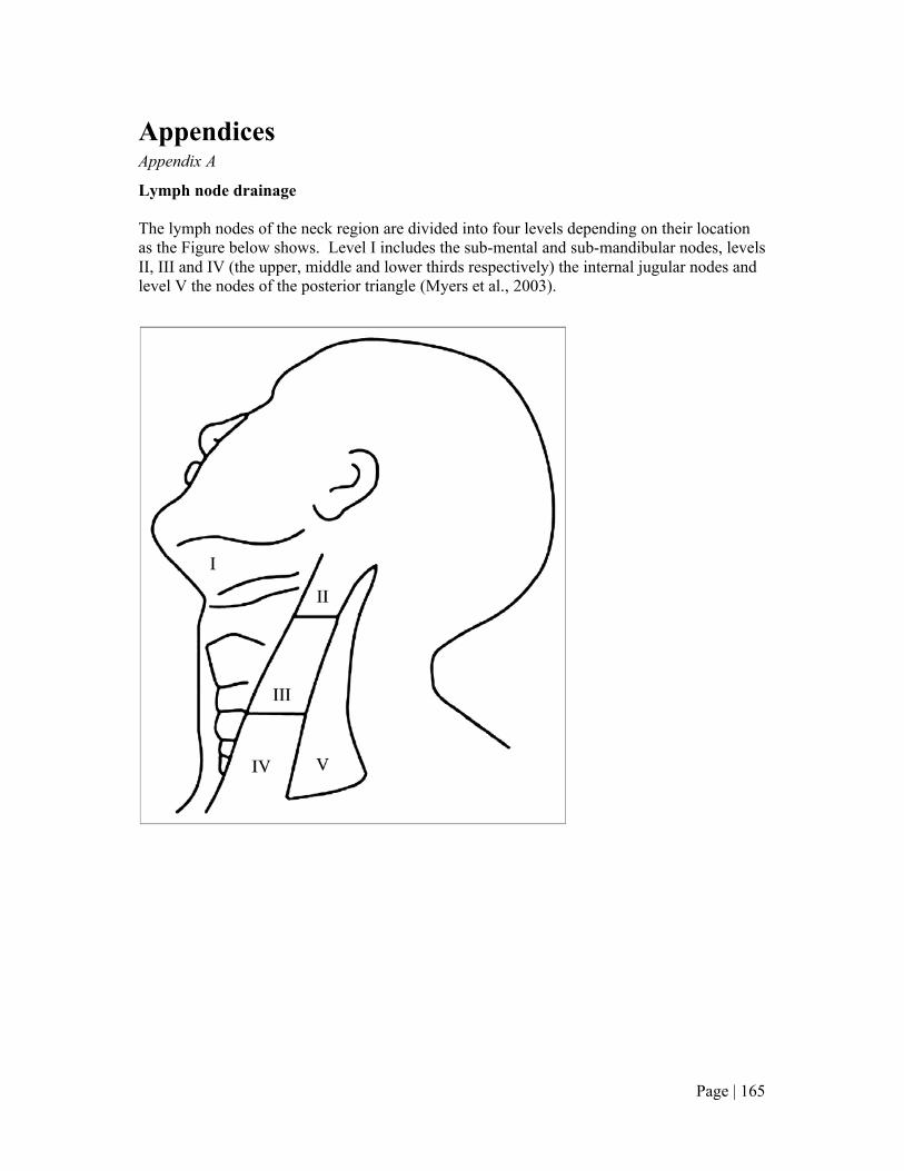
Page | 165
Appendices Appendix A
Lymph node drainage The lymph nodes of the neck region are divided into four levels depending on their location as the Figure below shows. Level I includes the sub-mental and sub-mandibular nodes, levels II, III and IV (the upper, middle and lower thirds respectively) the internal jugular nodes and level V the nodes of the posterior triangle (Myers et al., 2003).
Page | 166
Appendix B
Manuka honey clinical trials Topical application of honey in management of radiation mucositis. A Preliminary study Support Care Cancer 2003 Biswal, B., Zakaria, A., Ahmad, N. M The trial that was conducted in Malaysia in 2002 recruited 40 head and neck patients who received between 60 and 70Gy. The radiation treatment involved parallel opposed paired (POP) 6MV fields delivered by a linear accelerator. Patients receiving chemotherapy were excluded from the trial. Patients were randomised to the standard of care arm or the honey arm. Those patients in the honey arm were instructed to apply 20mL of honey three times a day. The application instructions were to rinse the whole of the oral mucosa and then swallow the honey to cover the pharyngeal mucosa. Mucositis was scored using the RTOG grading system. The results showed there were no changes in the grades one and two mucositis but there were fewer patients in the honey arm who developed severe mucositis (grade 3 or 4). Patients in both trial arms were advised on adequate fluid intake, supplementation of a high protein diet and oro-dental care. Honey as topical prophylaxis against radiochemotherapy-induced mucositis in head and neck cancer The Journal of Laryngology & Otology Rashad, U. M., Al-Gezawy, S. M., El-Gezawy, E., Azzaz, A. N. In 2008 a trial reported from Egypt found there were more incidences of grade three mucositis in the control arm compared to the honey arm. 40 patients were also recruited to this trial and they received between 60 and 66Gy using POP fields and anterior beams to treat the lower neck region. The honey application was similar to the Biswal et al. study and assessment was conducted weekly. Patients were excluded from the trial if they had received previous chemotherapy or radiation treatment (to the upper airways), previous radical surgery of the primary tumour and/or regional lymph nodes. All patients were instructed to use benzydamine HCl and supportive oral care measures. The Effect of Topical Application of Pure Honey on Radiation-induced Mucositis: A Randomised Clinical Trial The Journal of Contemporary Dental Practice Motallebnejad, M., Akram, S., Moghadamnia, A., Moulana, Z., Omidi, S. The trial conducted in Iran also recruited 40 head and neck patients who received a total dose between 50 and 60Gy. This trial maintained a blinded nature. Patients were excluded from the trial if they had had previous chemoradiation treatment. All patients were advised to avoid alcohol, spicy or acidic foods and smoking. Good oral hygiene and nutritional intake was also encouraged during the course of radiotherapy. Patients on the honey arm were instructed to use 20mLs of honey three times a day and apply it to the oral mucosa and to swallow. The control arm used 20mLs saline three times daily. Patients who experienced severe burning were excluded from the trial. The mucositis scores were taken weekly with the assessor not being aware of which arm each patient belonged to. The Oral Mucositis Assessing Scale (OMAS) was used in this trial to grade nine oral cavity areas. The results of this trial supported Biswal’s study.
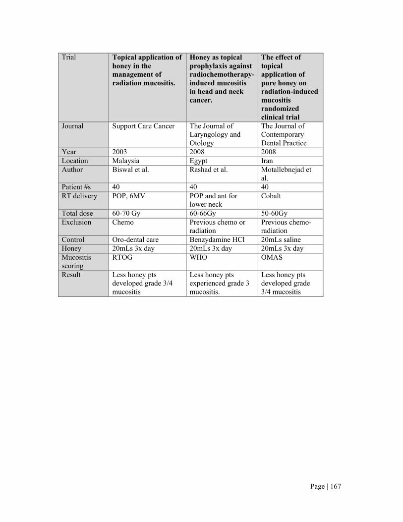
Page | 167
Trial Topical application of
honey in the management of radiation mucositis.
Honey as topical prophylaxis against radiochemotherapy-induced mucositis in head and neck cancer.
The effect of topical application of pure honey on radiation-induced mucositis randomized clinical trial
Journal Support Care Cancer The Journal of Laryngology and Otology
The Journal of Contemporary Dental Practice
Year 2003 2008 2008 Location Malaysia Egypt Iran Author Biswal et al. Rashad et al. Motallebnejad et
al. Patient #s 40 40 40 RT delivery POP, 6MV POP and ant for
lower neck Cobalt
Total dose 60-70 Gy 60-66Gy 50-60Gy Exclusion Chemo Previous chemo or
radiation Previous chemo-radiation
Control Oro-dental care Benzydamine HCl 20mLs saline Honey 20mLs 3x day 20mLs 3x day 20mLs 3x day Mucositis scoring
RTOG WHO OMAS
Result Less honey pts developed grade 3/4 mucositis
Less honey pts experienced grade 3 mucositis.
Less honey pts developed grade 3/4 mucositis
Page | 168
Appendix C
Patient information sheet
October, 2008
University of Otago, Wellington
Honey for Mouth Ulcers during Radiation Therapy PARTICIPANT INFORMATION SHEET You are invited to participate in a clinical trial which investigates the effect of honey on mouth ulcers experienced by patients who receive radiation therapy for head and neck cancers. All participation in this research is entirely voluntary and you are free to withdraw from the study or decline any particular question or test, at any time. Please discuss your participation in this trial with family and whanau and take the time to decide whether you wish to take part in this study. 1. Why should I participate in this study? Radiation therapy to the head and neck is given with the aim of killing cancer cells in this area. However, irradiation may cause the lining of the mouth to become red and sore and ulcers may form. Ulceration can be painful and may interfere with eating, drinking, talking and, in extreme cases, it may be necessary to delay your treatment. Currently, there is no ñbest treatmentò for radiation-induced mouth ulcers. However, a few small studies have recently shown that pure honey may be effective in decreasing the extent of ulceration in the mouth. This trial aims to test the effect of pure New Zealand manuka honey on mouth ulcers in 120 people, receiving radiation therapy for certain head and neck cancers. 2. What does my participation in the study involve? The trial starts as soon as your radiation therapy treatment begins and finishes 4 weeks after completion of treatment. A. Randomization Once you have been accepted into the trial you will be randomized to either the honey arm or the control arm. Randomization is done using a computer programme and cannot be influenced by the oncologist, the research assistant or yourself. It is important that you do not tell the research assistant whether you are in the honey or in the control arm.
Page | 169
B. Protocol All patients who enrol in this trial will be given a ñProtocol for Participants Sheet”. This sheet contains detailed information on how to take care of your mouth during the trial and which types of food and drink to avoid, to minimize discomfort.
• If you are randomized to the honey arm you will need to swirl pure Comvita Manuka honey (provided by the research assistant) around in your mouth, coating the entire inside of your mouth, three times a day.
• If you are randomized to the control arm, you will not need to do anything extra. You will simply get the standard treatment prescribed by your doctor.
C. Assessments 1. Physical examination by the radiation oncologist at the start of the trial. 2. Blood tests
A blood test at the start and finish of the trial will determine your blood chemistry (sugar levels), blood cells and liver functions. The tests will be done in a certified medical laboratory.
3. Assessment of Mouth Ulceration
The extent of oral mucositis will be monitored three times a week during radiation therapy treatment by the research assistant, who will look inside your mouth to record the extent of redness and ulceration in different parts of your mouth. Two more assessments will be done 2 and 4 weeks after completion of radiation therapy treatment.
4. Weight Measurements
Having a sore mouth may affect what and how much you eat and drink, and this will be reflected in your weight. Your weight will be measured once a week during radiation therapy treatment and 2 and 4 weeks after completion of treatment.
5. Quality of Life Assessment
Radiation therapy to the head and neck will affect the way in which you experience life from day to day. We would like to carefully monitor how you are affected by your treatment by asking you to fill in the same questionnaire every 2 weeks from start to finish of the trial. This Quality of Life questionnaire is relatively short and is designed specifically for patients that have head and neck cancers.
6. Food/Drink/Drug Diary
Because we are interested in how your treatment affects your health and quality of life, we want you to write down what (and how much) you eat and drink every day. We would also like you to write down how many pain killers you have used that day and what type(s) of pain killers these were. We also want you to write down whether you have had to use a feeding tube. Your protocol sheet will have an example of how to fill in such a diary.
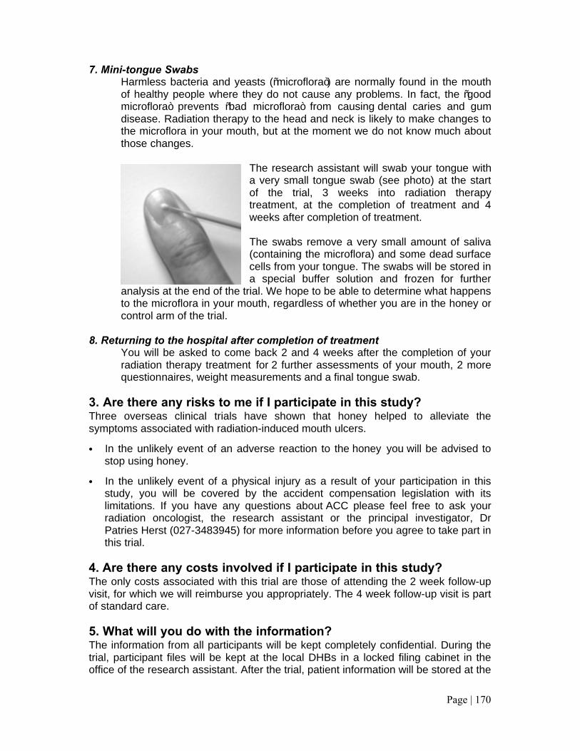
Page | 170
7. Mini-tongue Swabs Harmless bacteria and yeasts (ñmicrofloraò) are normally found in the mouth of healthy people where they do not cause any problems. In fact, the ñgood microfloraò prevents ñbad microfloraò from causing dental caries and gum disease. Radiation therapy to the head and neck is likely to make changes to the microflora in your mouth, but at the moment we do not know much about those changes.
The research assistant will swab your tongue with a very small tongue swab (see photo) at the start of the trial, 3 weeks into radiation therapy treatment, at the completion of treatment and 4 weeks after completion of treatment.
The swabs remove a very small amount of saliva (containing the microflora) and some dead surface cells from your tongue. The swabs will be stored in a special buffer solution and frozen for further
analysis at the end of the trial. We hope to be able to determine what happens to the microflora in your mouth, regardless of whether you are in the honey or control arm of the trial.
8. Returning to the hospital after completion of treatment You will be asked to come back 2 and 4 weeks after the completion of your radiation therapy treatment for 2 further assessments of your mouth, 2 more questionnaires, weight measurements and a final tongue swab.
3. Are there any risks to me if I participate in this study? Three overseas clinical trials have shown that honey helped to alleviate the symptoms associated with radiation-induced mouth ulcers.
• In the unlikely event of an adverse reaction to the honey you will be advised to stop using honey.
• In the unlikely event of a physical injury as a result of your participation in this study, you will be covered by the accident compensation legislation with its limitations. If you have any questions about ACC please feel free to ask your radiation oncologist, the research assistant or the principal investigator, Dr Patries Herst (027-3483945) for more information before you agree to take part in this trial.
4. Are there any costs involved if I participate in this study? The only costs associated with this trial are those of attending the 2 week follow-up visit, for which we will reimburse you appropriately. The 4 week follow-up visit is part of standard care. 5. What will you do with the information? The information from all participants will be kept completely confidential. During the trial, participant files will be kept at the local DHBs in a locked filing cabinet in the office of the research assistant. After the trial, patient information will be stored at the

Page | 171
University of Otago, Wellington, in a locked filing cabinet in the office of the Principal Investigator, Dr Patries Herst, for at least 10 years; after which time the files will be destroyed. Only the official investigators and the research assistants will have access to this information. After completion of the trial we will collate and analyse all the information from all participants of the study. This will tell us whether the manuka honey is better than bicarbonate soda mouth washes in treating radiation-induced mouth ulcers. If this is the case, we aim to conduct a larger multinational trial, and we would like to incorporate the data from this trial into this future study. We anticipate that this will lead to a better and more standardized treatment for radiation-induced mouth ulcers in New Zealand. Reporting: • We will report on the results of this study in scientific reports and publications. • You will be informed of the results of the study by a letter from the Principal
Investigator, Dr Patries Herst
NO material will be published which can identify you personally. 6. Does the study have ethical approval? Yes, the study has ethical approval from the Multi-Region Ethics Committee. 7. Do I have to participate in this study? No, there is absolutely no requirement to participate in the study. 8. Can I withdraw from the study if I change my mind? If you do agree to take part, you are free to withdraw from the study at anytime, without having to give a reason and this will in no way affect your future health care. If you wish to withdraw please contact your doctor, the research assistant or the principal investigator, Dr Patries Herst (027-3483945) and advise them that you have decided to withdraw so that all information and data that have been collected about you will be entirely deleted from the database. 9. What if I have more questions or concerns about this study? If you have any questions or concerns about your rights as a participant in this research study you can contact an independent health and disability advocate. This is a free NZ wide service provided under the Health and Disability Commissioner Act. Telephone 0800 555 050; Free Fax 0800 2787 7678 (0800 2 SUPPORT); Email: [email protected]. If there is a specific MǕori issue/concern please contact Linda Grennell at 0800 37 77 66I If you have any questions or concerns about any aspects of this study, at any time, please call the principal investigator, Dr Patries Herst (027-3483945).
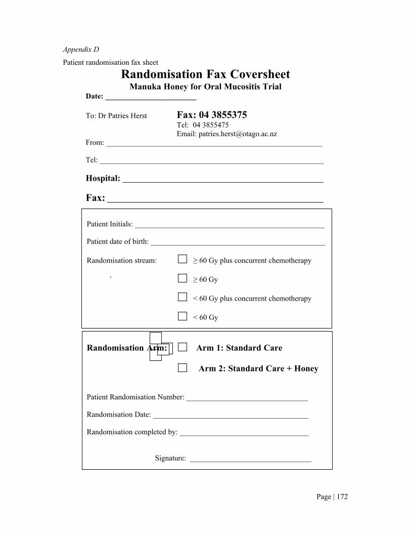
Page | 172
Appendix D
Patient randomisation fax sheet
Randomisation Fax Coversheet Manuka Honey for Oral Mucositis Trial
Date: ________________________ To: Dr Patries Herst Fax: 04 3855375 Tel: 04 3855475 Email: [email protected] From: _________________________________________________________ Tel: ___________________________________________________________ Hospital: _____________________________________________________ Fax: _________________________________________________________
Patient Initials: __________________________________________________ Patient date of birth: ______________________________________________ Randomisation stream: ≥ 60 Gy plus concurrent chemotherapy
` ≥ 60 Gy
< 60 Gy plus concurrent chemotherapy < 60 Gy
Randomisation Arm: Arm 1: Standard Care
Arm 2: Standard Care + Honey Patient Randomisation Number: ________________________________ Randomisation Date: _________________________________________ Randomisation completed by: __________________________________
Signature: ________________________________
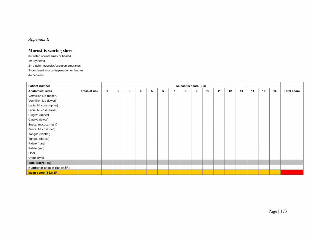
Page | 173
Appendix E Mucositis scoring sheet 0= within normal limits or healed 1= erythema 2= patchy mucositis/pseusomembranes 3=confluent mucositis/pseudomembranes 4= necrosis
Patient number Mucositis score (0-4)
Anatomical sites areas at risk 1 2 3 4 5 6 7 8 9 10 11 12 13 14 15 16 Total score Vermillion Lip (upper) Vermillion Lip (lower) Labial Mucosa (upper) Labial Mucosa (lower) Gingiva (upper) Gingiva (lower) Buccal mucosa (right) Buccal Mucosa (left) Tongue (ventral) Tongue (dorsal) Palate (hard) Palate (soft) Floor Oropharynx
Total Score (TS) Number of sites at risk (NSR) Mean score (TS/NSR)

Page | 174
Appendix F
Trial protocol
October, 2008
University of Otago, Wellington
Honey for Mouth Ulcers during Radiation Therapy PROTOCOL FOR PARTICIPANTS Please DO NOT tell the Research Assistant in which arm you are. All patients • Use good oral hygiene at all times, rinse with water after every meal. • Brush two times a day using soft tooth brushes and fluoride toothpaste. • Avoid very hot and/or spicy food, alcohol and tobacco. • Take regular sips of water. • Allowed: pain medication over the counter or prescription (note in diary). Other
oral mouthwashes or medication prescribed by your oncologist (note in diary) Honey Arm Use the measuring spoon to measure out 20 ml of honey and swirl the honey around in your mouth slowly. Take your time (one to several minutes) to ensure that all parts on the inside of the mouth are covered with honey, especially the parts that hurt. Then slowly swallow the honey; this will coat the sides of the swallowing tube (oesophagus) as the honey slides down.
Mondays to Fridays Do this 15-30 minutes before and 15-30 minutes after each radiation therapy treatment and again approximately 6 hours after treatment (3 times a day). Saturday and Sundays (and other non-treatment days) Take honey 3 times a day (morning, afternoon, evening)
Control Arm Standard practice of care; follow the advice by your oncologist.
Page | 175
Appendix G
Trial consent
October, 2008
University of Otago, Wellington
Honey for Mouth Ulcers during Radiation Therapy
INFORMED CONSENT
This form is to obtain your agreement to participate in our clinical trial which intends to find out whether New Zealand Manuka Honey decreases the extent of mouth ulcers during radiation therapy to the head and neck. REQUEST FOR INTERPRETER English I wish to have an interpreter
Yes No
Maori E hiahia ana ahau ki tetahi kaiwhakamaori/kaiwhaka pakeha korero
Ae Kao
Cook Island
Ka inangaro au i tetai tangata uri reo Ae Kare
Fijian Au gadreva me dua e vakadewa vosa vei au
Io Sega
Niuean Fia manako au ke fakaaoga e taha tagata fakahokohoko kupu
E Nakai
Samoan Ou te manaôo ia i ai se faôamatala upu
Ioe Leai
Tokelaun Ko au e fofou ki he tino ke fakaliliu te gagana Peletania kin a gagana o na motu o te Pahefika
Ioe Leai
Tongan Oku ou fiemaôu ha fakatonulea
Io Ikai
Informed Consent

Page | 176
• I have been given the opportunity to discuss my participation in this trial with family and whanau.
• I have had the opportunity to consider all the information presented and have had all my questions answered.
• I understand that my participation is completely voluntary (my choice) and that I may withdraw from the study at any time and this will in no way affect my future health care.
I would like to participate in this clinical trial and I give consent to participating in this study which includes: 1. Being randomized by computer to a honey arm and a control arm.
2. Following the guidelines of the honey arm and the control arm to the best of my abilities.
3. A physical examination by my doctor at the start of the trial.
4. Two blood tests at the start and after completion of the trial by a certified medical laboratory.
5. Weekly weight measurements.
6. Regular examinations of my mouth by the research assistant. These examinations will take place three times a week during radiation therapy treatment, 2 and 4 weeks after completion of treatment.
7. Filling in a quality of life questionnaire every two weeks until 4 weeks after completion of treatment.
8. Keeping a food/drink/drug diary in which I will record exactly what and how much food and fluids I have taken and how many and what type of pain killers or other drugs I have used.
9. Regular mini-tongue swabs, taken by the research assistant. These will be taken at the start of treatment, 3 weeks into treatment, at the end of treatment and 4 weeks after completion of treatment.
10. The use of my information as part of a future larger clinical trial.
Name:
Signature:
Date:
Researchers Dr Patries Herst (ph 04-3855475 ext 4753; mobile 027-3483945) The Radiation Oncologist who is the local principal investigator