The effect of mechanical and chemical polishing...
7
ORIGINAL ARTICLE The effect of mechanical and chemical polishing techniques on the surface roughness of heat-polymerized and visible light-polymerized acrylic denture base resins Abdul Aziz Abdullah Al-Kheraif * Dental Health Department, College of Applied Medical Sciences, King Saud University, Riyadh 11541, Saudi Arabia Received 21 August 2013; revised 26 November 2013; accepted 16 December 2013 Available online 3 February 2014 KEYWORDS Surface roughness; Mechanical polishing; Chemical polishing; Contact profilometer; Acrylic resin Abstract Objective: The purpose of this study was to compare the effects of mechanical polishing (MP) and chemical polishing (CP) on the average surface roughness (Ra) of heat-cured (HC) and light-cured (LC) denture base acrylic resins. Methods: A total of 120 specimens (30 · 15 · 3 mm) were prepared from one HC and one LC acrylic resin. To remove nodules and gross surface irregularities, all specimens were finished with a lathe-mounted small acrylic bur and 360-grit sandpaper. Ten finished specimens of each acrylic resin were randomly assigned to each of six polishing techniques: Resilit High-luster Polishing Liquid (RHPL), Universal Polishing Paste, Abraso-star K50, pumice, Jet Seal Liquid, or Acrypoint. MP was performed with an automatic polishing machine for 2 min, under 50 rpm and 500 g of load. CP was performed by immersing the HC and LC specimens in preheated methyl methacrylate at 75 ± 1 °C for 10 s. The surface roughness of the acrylic resin specimens was measured with a con- tact profilometer. The Ra values were analyzed by two-way analysis of variance, post hoc Scheffe’s test, and paired t-test (p 6 0.05). Polished and tested acrylic resin surfaces were evaluated by scan- ning electron microscopy. Results: MP was more effective than CP. The smoothest surface was obtained with the use of the RHPL on the LC (0.05 ± 0.01 lm) or HC (0.07 ± 0.01 lm) acrylic resin. Two-way ANOVA showed a statistically significant difference between MP and CP. Conclusions: MP produced the smoothest surface of denture base acrylic resin. The mean surface roughness values after MP and CP were not influenced by the type of acrylic resin. ª 2014 King Saud University. Production and hosting by Elsevier B.V. All rights reserved. 1. Introduction Acrylic resins and resin-based direct and indirect restorative materials have been used widely in dentistry, specifically in the field of prosthodontics, to fabricate different types of * Tel.: +966 554299995. E-mail address: [email protected]. Peer review under responsibility of King Saud University. Production and hosting by Elsevier The Saudi Dental Journal (2014) 26, 56–62 King Saud University The Saudi Dental Journal www.ksu.edu.sa www.sciencedirect.com 1013-9052 ª 2014 King Saud University. Production and hosting by Elsevier B.V. All rights reserved. http://dx.doi.org/10.1016/j.sdentj.2013.12.007
-
Upload
truongkhanh -
Category
Documents
-
view
227 -
download
5
Transcript of The effect of mechanical and chemical polishing...

The Saudi Dental Journal (2014) 26, 56–62
King Saud University
The Saudi Dental Journal
www.ksu.edu.sawww.sciencedirect.com
ORIGINAL ARTICLE
The effect of mechanical and chemical polishing
techniques on the surface roughness of
heat-polymerized and visible light-polymerized
acrylic denture base resins
* Tel.: +966 554299995.
E-mail address: [email protected].
Peer review under responsibility of King Saud University.
Production and hosting by Elsevier
1013-9052 ª 2014 King Saud University. Production and hosting by Elsevier B.V. All rights reserved.
http://dx.doi.org/10.1016/j.sdentj.2013.12.007
Abdul Aziz Abdullah Al-Kheraif *
Dental Health Department, College of Applied Medical Sciences, King Saud University, Riyadh 11541, Saudi Arabia
Received 21 August 2013; revised 26 November 2013; accepted 16 December 2013Available online 3 February 2014
KEYWORDS
Surface roughness;
Mechanical polishing;
Chemical polishing;
Contact profilometer;
Acrylic resin
Abstract Objective: The purpose of this study was to compare the effects of mechanical polishing
(MP) and chemical polishing (CP) on the average surface roughness (Ra) of heat-cured (HC) and
light-cured (LC) denture base acrylic resins.
Methods: A total of 120 specimens (30 · 15 · 3 mm) were prepared from one HC and one LC
acrylic resin. To remove nodules and gross surface irregularities, all specimens were finished with
a lathe-mounted small acrylic bur and 360-grit sandpaper. Ten finished specimens of each acrylic
resin were randomly assigned to each of six polishing techniques: Resilit High-luster Polishing
Liquid (RHPL), Universal Polishing Paste, Abraso-star K50, pumice, Jet Seal Liquid, or Acrypoint.
MP was performed with an automatic polishing machine for 2 min, under 50 rpm and 500 g of load.
CP was performed by immersing the HC and LC specimens in preheated methyl methacrylate at
75 ± 1 �C for 10 s. The surface roughness of the acrylic resin specimens was measured with a con-
tact profilometer. The Ra values were analyzed by two-way analysis of variance, post hoc Scheffe’s
test, and paired t-test (p 6 0.05). Polished and tested acrylic resin surfaces were evaluated by scan-
ning electron microscopy.
Results: MP was more effective than CP. The smoothest surface was obtained with the use of the
RHPL on the LC (0.05 ± 0.01 lm) or HC (0.07 ± 0.01 lm) acrylic resin. Two-way ANOVA
showed a statistically significant difference between MP and CP.
Conclusions: MP produced the smoothest surface of denture base acrylic resin. The mean surface
roughness values after MP and CP were not influenced by the type of acrylic resin.ª 2014 King Saud University. Production and hosting by Elsevier B.V. All rights reserved.
1. Introduction
Acrylic resins and resin-based direct and indirect restorativematerials have been used widely in dentistry, specifically inthe field of prosthodontics, to fabricate different types of

The effect of mechanical and chemical polishing techniques 57
prostheses, including complete and partial dentures, temporaryfixed partial dentures, implant-supported overdentures, andmaxillofacial prostheses (Kuhar et al., 2005). Acrylic resins
may be heat-cured (HC), autocured, or microwave-cured(Hong et al., 2009; Rached et al., 2004; Yunus et al., 1994).Conventional resins that are used in dentistry are based on
poly-methyl methacrylate (poly-MMA) (Danesh et al., 2012;Hong et al., 2009).
A major breakthrough in the application of acrylic resins in
the field of dentistry occurred with the introduction of visiblelight-cured (LC) acrylic resins, which are urethane dimethylac-rylate-based (Danesh et al., 2012; Jorge et al., 2003; Kedjaruneet al., 1999; Leggat and Kedjarune, 2003). LC acrylic resins are
activated by light in the wavelength range of 460–470 nm.They include larger molecular weight methacrylates anddimethacrylates (Haselden et al., 1998). LC resins have a lower
elution rate (0.06 wt%) than MMA-based acrylic resins (0.13–0.054 wt%) (Danesh et al., 2012; Ferracane, 1994). LC resinselicit less soft-tissue irritation, produce less heat during poly-
merization, and have a relatively pleasant odor compared toMMAs (Al Rifaiy, 2012; Haywood et al., 2003).
The surface finish of any dental prosthesis is an important
factor that determines patient’s comfort, prosthesis longevity,and esthetics (Rahal et al., 2004; Ulusoy et al., 1986). High val-ues for the free energy (hydrophilicity) (Busscher et al., 1986)and roughness of the prosthesis surface will increase the
chances of microbial adhesion and plaque retention, respec-tively, and reduce patient’s oral hygiene (Kagermeier-Callawayet al., 2000; Rahal et al., 2004; Ulusoy et al., 1986). Studies
have shown a direct correlation between surface roughnessand plaque retention, plaque maturation, Candida albicans col-onization, and associated denture stomatitis (Barbeau et al.,
2003; Berger et al., 2006; Radford et al., 1997).In vivo studies have suggested that, to be clinically accept-
able, prostheses and dental restorations should not have aver-
age (mean) surface roughness (Ra) values higher than 0.2 lm(Bollen et al., 1997; Quirynen et al., 1996; Seng-Kyun Kimet al., 2009). Below this value, no further reduction in plaqueaccumulation can be expected. Above this value, a propor-
tional increase in plaque accumulation occurs (Abuzar et al.,2010; Bollen et al., 1996; Kuhar et al., 2005; Quirynen et al.,1996).
Table 1 Polishing procedures and products used in the
Polishing
procedure
Polishing products Co
Mechanical Resilit High-luster
Polishing Liquid
Lo
oxi
Mechanical Universal Polishing
Paste
Lo
oxi
Mechanical Abraso-Star K50 Mi
abr
Mechanical Pumice Pu
am
Mechanical AcryPoint Bo
car
Chemical Jet Seal Liquid Me
Polishing can be performed through mechanical or chemi-cal methods (Goncalves et al., 2008). Mechanical polishing(MP) methods use abrasives to produce controlled wear of
the surface material to reduce surface roughness (Abuzaret al., 2010). Materials used for MP include polishing wheels,felt cones, prophylactic pastes, rubber polishers, abrasive
stones, aluminum oxide-based polishing pastes, silicone polish-ers, pumice, and lathe polishing (Braun et al., 2003; Sofouet al., 2001; Yamauchi et al., 1990).
As an alternative to the conventional method of MP, in1969, Gotusso introduced a method called superficial chemicalpolishing (CP) (Al-Rifaiy, 2010; Braun et al., 2003; Gotusso,1969; Rahal et al., 2004). In this technique, the finished acrylic
resin denture is placed in a chemical polisher containing heatedmonomer at 75 �C for 10 s (Al-Rifaiy, 2010; Rahal et al.,2004). Subsequent studies have proven the biocompatibility
of this technique (Nagem-Filho et al., 1973; Rahal et al., 2004).Although some studies have evaluated the effects of MP
and CP techniques on the surface roughness of HC, autocured,
and microwave-cured acrylic resins, no study has examined theeffects of polishing techniques on the surface roughness of LCdenture base resins. Therefore, the aim of this study was to
evaluate and compare the effects of MP and CP on the Raof visible LC and HC denture base resins.
2. Materials and methods
2.1. Preparation of test specimens
In this study, 120 specimens (30 · 15 · 3 mm) were preparedfrom HC acrylic resin (Lucitone, Dentsply International,Inc., York, PA, USA) and LC acrylic resin (Eclipse, Dentsply
International, Inc.). The HC acrylic resin specimens were pre-pared by investing the wax pattern (30 · 15 · 3 mm) in gypsumplaster by a conventional flasking procedure in dental flasks.
After dewaxing the plaster molds, the acrylic material waspacked and processed in accordance with manufacturers’instructions.
A Perspex mold with a glass lid was designed to prepare theLC specimens. After applying petroleum jelly, the mold waspreheated at 55 �C for 2 min in a special oven (ConditioningOven, Dentsply Trubyte). The LC acrylic resins were
study.
mposition Manufacturer
ose abrasives (aluminum
de-Al2O3) in liquid
Renfert, GmbH
ose abrasives (aluminum
de-Al2O3) in paste
Ivoclar Vivadent,
Schaan, Liechtenstein
xture of waxes and
asives
Bredent GmbH & Co.
KG
mice (coarse CL-60),
orphous silica and quartz
WhipMix
Corporation,
Kentucky, USA.
nded abrasives (silicon
bide in silicon matrix)
Shofu Inc., Kyoto,
Japan
thyl methacrylate Lang Dental Mfg. Co.,
USA.

58 A.A.A. Al-Kheraif
compacted into the mold cavity by finger pressure. A glass slabwas pressed on top to remove excess material and to obtainspecimens of uniform thickness. An air-barrier coating
(Eclipse ABC, Dentsply Trubyte, USA) was applied to theresin surface to prevent the inhibition of polymerization byoxygen. Polymerization was performed in an LC unit (Eclipse
Processing Unit, Dentsply Trubyte) containing six Eclipsehalogen lamps (41 V each; Dentsply Trubyte) by exposingthe sample to visible light at 400 to 500 nm for 10 min.
Polymerized specimens were retrieved from the flask andfinished to remove nodules and gross irregularities. Finishingwas performed by using a small acrylic trimming bur mountedon a laboratory lathe. Final finishing was done with 360-grit
sandpaper mounted on a lathe. After final finishing of all thespecimens, 10 specimens of each acrylic resin were randomlyassigned to one of the six polishing techniques shown in
Table 1. MP was performed with an automatic polishingmachine (The Wirtz, Jean Wirtz, Dusheldory W, Germany)for 2 min, under 50 rpm and 500 g of load with Resilit
High-luster Polishing Liquid (RHPL), Universal PolishingPaste (UPP), Abraso-Star K50 (K50), pumice, Jet Seal Liquid(JSL), or AcryPoint, as recommended by the manufacturer.
CP was performed by immersing the HC and LC specimensin preheated MMA (JSL) at 75 ± 1 �C for 10 s (Al-Rifaiy,2010).
2.2. Surface roughness measurements
The surface roughness of the acrylic resin specimens was deter-mined with a contact profilometer (Tylor Hobson Ltd., series
No. 339, Leicester, UK). The instrument’s diamond styluswas moved across the specimen surface under constant pres-sure. Three measurements were made for each specimen, with
a cutoff value of 5 mm. The average of three readings for eachspecimen was considered as the final Ra value of the particularspecimen. To test for significant differences in Ra of the two
acrylic resins, six polishing techniques and their interactionswere compared. Paired and unpaired Student’s t-tests and
Table 2 Two-way analysis of variance.
Effect Sum of squares df
Material 0.005 1
Technique 0.514 5
Material*Technique 0.112 5
Error 0.141 48
Table 3 Mean and standard deviations of roughness
systems (lm). The value with the same superscript letter
Comparison Light-cured acrylic res
Resilit High-luster Polishing Liquid 0.05a ± 0.01
Universal Polishing Paste 0.06a ± 0.01
Abraso-Star K50 0.06a ± 0.00
Pumice 0.11a ± 0.04
Jet Seal Liquid 0.29b ± 0.04
AcryPoint 0.23b ± 0.06
two-way ANOVA followed by a post hoc Scheffe’s test formultiple comparisons were performed (p < 0.05).
2.3. Scanning electron microscopy (SEM)
Polished samples were subjected to SEM analysis (JSM 6360,JEOL Ltd., Japan) at a magnification of 50· under a high-vac-
uum condition. Specimens were air dried in desiccators,cleaned with 70% alcohol in an ultrasonic cleaner, and sput-ter-coated with gold for up to 200 A (Polaron E-5200 Energy
Beam Sciences, Agawan, MA, USA).
3. Results
Table 2 and Table 3 shows the Ra values and standard deviations
(SDs). The Ra values varied depending on the polishing technique
(p < 0.000 by two-way ANOVA). There was no significant difference
in the Ra between the HC and LC acrylic resins (p< 0.2) (Table 2).
There was a significant interaction between the material and the polish-
ing technique, indicating that the effect of one factor was dependent on
the other. Paired Student’s t-test revealed no significant difference be-
tween the materials after each polishing technique.
The post hoc Scheffe’s test was used to compare the six pol-
ishing techniques for each type of acrylic resin (Table 3). The
smoothest surface was obtained with the RHPL on the LC
(0.05 ± 0.01 lm) or HC (0.07 ± 0.01 lm) acrylic resin (Figs. 1
and 6), with these values being well below the clinically accepted
threshold value of 0.2 lm. The highest Ra value was obtained with
AcryPoint on the HC (0.25 ± 0.05 lm) or LC (0.23 ± 0.06 lm) acrylic
resin (Figs. 5 and 11), with these values being above the threshold
value of 0.2 lm. After CP with JSL, the same Ra value was obtained
for the HC and LC specimens (0.29 ± 0.04 and ± 0.05 lm, respec-
tively), which significantly exceeded the threshold value of 0.2 lm(Figs. 6 and 12).
The surface irregularities of the acrylic resins by SEM corre-
sponded with the respective Ra values of the particular specimens
(Figs. 1–12). SEM micrographs confirmed that highest roughness
was found in with AcryPoint and Jet Seal Liquid polishing systems
(Figs. 5, 6, 11 and 12).
Mean-squared F p
0.005 1.690 0.200
0.103 34.963 0.000
0.022 7.599 0.000
0.003
values for acrylic specimens polished with different
indicates no significance.
in (mean ± SD) Heat-cured acrylic resin (mean ± SD)
0.07a ± 0.01
0.08a ± 0.02
0.10a ± 0.05
0.10a ± 0.01
0.29b ± 0.05
0.25b ± 0.05
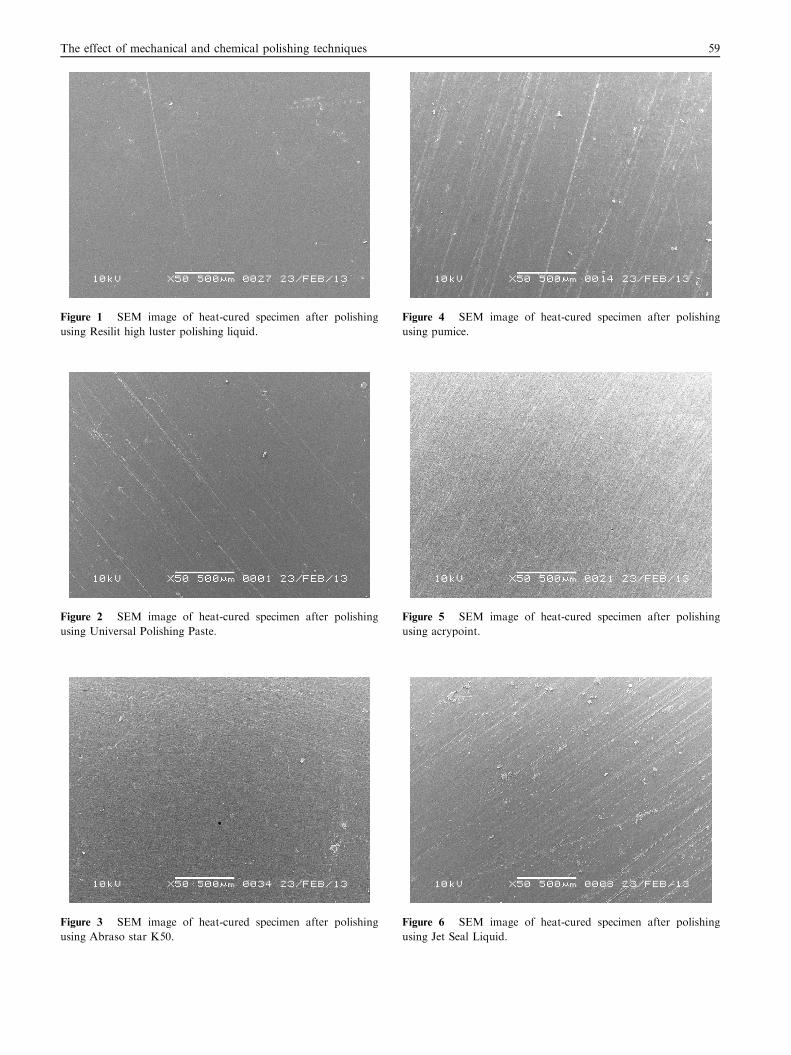
Figure 1 SEM image of heat-cured specimen after polishing
using Resilit high luster polishing liquid.
Figure 3 SEM image of heat-cured specimen after polishing
using Abraso star K50.
Figure 4 SEM image of heat-cured specimen after polishing
using pumice.
Figure 2 SEM image of heat-cured specimen after polishing
using Universal Polishing Paste.
Figure 5 SEM image of heat-cured specimen after polishing
using acrypoint.
Figure 6 SEM image of heat-cured specimen after polishing
using Jet Seal Liquid.
The effect of mechanical and chemical polishing techniques 59

Figure 7 SEM image of visible light cured specimen after
polishing using Resilit high luster polishing liquid.
Figure 8 SEM image of visible light cured specimen after
polishing using Universal Polishing Paste.
Figure 9 SEM image of visible light cured specimen after
polishing using Abraso star K50.
Figure 10 SEM image of visible light cured specimen after
polishing using pumice.
Figure 12 SEM image of visible light cured specimen after
polishing using Jet Seal Liquid.
Figure 11 SEM image of visible light cured specimen after
polishing using acrypoint.
60 A.A.A. Al-Kheraif

The effect of mechanical and chemical polishing techniques 61
4. Discussion
Dentures and artificial acrylic teeth are hard surfaces that tendto attract food debris and to form plaque and calculus if not
polished (Morgan and Wilson, 2001). A smooth surface is aprerequisite for denture base acrylics, to prevent plaque accu-mulation and further denture-induced stomatitis (Machado
et al., 2011). To reduce the surface roughness, an acrylic pros-thesis is finished and polished by various finishing and polish-ing techniques in a sequential manner. Sequential proceduresare applied to remove gross irregularities and nodules from
the rough superficial surface by using various lathe-mountedacrylic burs, felt cones, rubber wheels, and disks, followed byfinal finishing under light pressure. The ultimate smooth and
glossy finish is achieved by polishing with different commer-cially available polishing agents.
In the present study, before the specimens were subjected to
polishing, they were finished with a lathe-mounted acrylic burto remove gross irregularities and surface nodules. Final finish-ing was performed with lathe-mounted 360-grit sandpaper
with light pressure. After final finishing, the specimens weresubjected to six polishing techniques.
Although manufacturers’ instructions and standard labora-tory protocols were employed throughout the study, the study
still has some limitations. First, the specimens were not fabri-cated to resemble dentures, because dentures do not have com-pletely flat surfaces, as was the case for the specimens used in
this study. Second, MP systems will have various degrees ofabrasiveness. The abrasiveness of a polishing system and theresulting surface smoothness depend on the size of abrasive
particles present in the polishing system.In the present study, MP techniques were more effective
than CP, producing surface roughness values that were less
than the threshold value of 0.2 lm. This fact was especiallytrue when the samples were polished using RHPL, UPP,K50, or pumice, which have been used routinely in the labora-tory for quite some time. AcryPoint showed an Ra value that
was very close to the threshold value. This finding could be dueto the size of abrasive particles contained in that particularpolishing system (Seng-Kyun Kim et al., 2009). The Ra values
of the CP specimens were above the acceptable threshold (Ta-ble 3). Al-Rifaiy (2010); Rahal et al. (2004), and Alves et al.(2007) also found that CP produced a higher Ra in acrylic res-
ins than MP.A statistically significant difference in Ra was achieved in
specimens treated by MP compared to CP. This differencecould be attributed to the MMA molecules present in the pol-
ishing fluid, which penetrate the superficial polymeric chains ofthe acrylic resin, breaking the secondary bonds that join themand promoting a final plasticizing effect of the acrylic resin sur-
face (Rahal et al., 2004). This effect may increase the surfaceroughness. Another reason for this difference could be relatedto the abrasive mechanical action, which decreases the surface
roughness during polishing (Fionnuala O’Donnell et al., 2003).The MP agent may contain finer abrasive particles, which helpto achieve a smooth surface (Phillips, 1982).
Interestingly, there were significant differences in the Ravalues between MP and CP, except for polishing with Acry-Point, in both types of acrylic resin. The type of acrylic resindid not influence the Ra after MP or CP. MP rather than
CP is indicated because it yields lower Ra values (Rahalet al., 2004).
The visual comparison of SEM images with Ra valuesshowed that after polishing, the LC and HC acrylic resin spec-imens polished using AcryPoint (Figs. 5 and 11) and JSL
(Figs. 6 and 12) contained large pores compared to the surfacetextures of specimens polished by other polishing systems.These specimens showed a similar pattern, with very minute
debris at the surface. Kuhar et al. (2005) reported that pores,similar to surface roughness, enhance the accumulation of den-ture plaque and staining on some parts of dentures.
5. Conclusion
Within the limitations of this study, the following conclusions
can be made:
(1) The RHPL, UPP, and K50 agents produced superior
surface smoothness for all acrylic resin specimens anda mean Ra significantly below the threshold Ra of0.2 lm (p < 0.0017).
(2) Ra values after MP and CP were not influenced by the
type of acrylic resin.(3) MP was the most effective polishing technique.
Ethical statement
This research did not involve human subjects or where human
subjects were involved was conducted in full accordance withethics principles, including the World Medical AssociationDeclaration of Helsinki.
Conflict of interest
The author has no conflicts of interest to declare.
Acknowledgement
The authors would like to extend their appreciation to the Re-search Centre, College of Applied Medical Sciences and the
Deanship of Scientific Research at King Saud University forfunding this research.
References
Abuzar, M.A., Bellur, S., Duong, N., Kim, B.B., Lu, P., Palfreyman,
N., et al, 2010. Evaluating surface roughness of a polyamide
denture base material in comparison with poly (methyl methacry-
late). J. Oral Sci. 52, 577–581.
Al Rifaiy, M.Q., 2012. Shear bond strength between light polymerized
hard reline resin and denture base resin subjected to long term
water immersion. Saudi Dental J. 24, 23–27.
Al-Rifaiy, M.Q., 2010. The effect of mechanical and chemical
polishing techniques on the surface roughness of denture base
acrylic resins. Saudi Dental J. 22, 13–17.
Alves, P.V.M., Lima Filho Roberto, M.A., Telles, E., 2007. Surface
roughness of acrylic resins after different curing and polishing
techniques. Angle Orthodontist 77 (3).
Barbeau, J., Seguin, J., Goulet, J.P., de Koninck, L., Avon, S.L.,
Lalonde, B., et al, 2003. Reassessing the presence of Candida
albicans in denture-related stomatitis. Oral Surg. Oral Med. Oral
Pathol. Oral Radiol. Endod. 95, 51–59.

62 A.A.A. Al-Kheraif
Berger, J.C., Driscoll, C.F., Romberg, E., Luo, Q., Thompson, G.,
2006. Surface roughness of denture base acrylic resins after
processing and after polishing. J. Prosthodont. 15, 180–186.
Bollen, C.M., Papaioanno, W., Van Eldere, J., Schepers, E., Quirynen,
M., van Steenberghe, D., 1996. The influence of abutment surface
roughness on plaque accumulation and peri-implant mucositis.
Clin. Oral Implants Res. 7, 201–211.
Bollen, C.M., Lambrechts, P., Quirynen, M., 1997. Comparison of
surface roughness of oral hard materials to the threshold surface
roughness for bacterial plaque retention: a review of the literature.
Dent. Mater. 13, 258–269.
Braun, K.O., Mello, J.A.N., Rached, R.N., Del Bel Cury, A.A., 2003.
Surface texture and some properties of acrylic resins submitted to
chemical polishing. J. Oral Rehabil. 30, 91–98.
Busscher, H.J., Uyen, M.H.M.J.C., Weerkamp, A.H., Postma, W.J.,
Arends, J., 1986. Reversibility of adhesion of oral streptococci to
solids. FEMS Microbiol. Lett. 35, 303–306.
Danesh, G., Hellak, T., Reinhardt, K.J., Vegh, A., Schafer, E.,
Lippold, C., 2012. Elution characteristics of residual monomers in
different light- and auto-curing resins. Exp. Toxicol. Pathol. 64,
867–872.
Ferracane, J.L., 1994. Elution of leachable components from compos-
ites. J. Oral Rehabil. 21, 441–452.
Fionnuala O’Donnell, E., Radford, David R., Sinclair, Gary F., Clark,
Robert K.F., 2003. Chairside polishing of heat-cured acrylic resin:
an SEM and EDA study. Intern J. Prosth. 16 (3), 233–238.
Goncalves, T.S., Spohr, A.M., de Souza, R.M., Macedo de Menezes,
L., 2008. Surface roughness of auto polymerized acrylic resin
according to different manipulation and polishing methods: an
in situ evaluation. Angle Orthod. 78, 931–934.
Gotusso, M.J., 1969. Chemical surface treatment of acrylic resins. Rev.
Asoc. Odonto.l Argent 57, 359–361.
Haselden, C.A., Hobkirk, J.A., Pearson, G.J., Davies, E.H., 1998. A
comparison between the wear resistance of three types of denture
resin to three different dentifrices. J. Oral Rehabil. 25, 335–339.
Haywood, J., Basker, R.M., Watson, C.J., Wood, D.J., 2003. A
comparison of three hard chairside denture reline materials. Part I.
clinical evaluation. Eur. J. Prosthodont Restor. Dent. 11, 157–163.
Hong, G., Murata, H., Li, Y., Sadamori, S., Hamada, T., 2009.
Influence of denture cleansers on the color stability of three types of
denture base acrylic resin. J. Prosthet. Dent. 101, 205–213.
Jorge, J.H., Giampaolo, E.T., Machado, A.L., Vergani, C.E., 2003.
Cytotoxicity of denture base acrylic resins: a literature review. J.
Prosthet. Dent. 90, 190–193.
Kagermeier-Callaway, A.S., Willershausen, B., Frank, T., Stender, E.,
2000. In vitro colonisation of acrylic resin denture base materials by
Streptococcus oralis and Actinomyces viscosus. Int. Dent. J. 50, 79–85.
Kedjarune, U., Charoenworaluk, N., Koontongkaew, S., 1999.
Release of methyl methacrylate from heat-curved and autopoly-
merized resins: cytotoxicity testing related to residual monomer.
Aust. Dent. J. 44, 25–30.
Kuhar, M., Funduk, N., 2005. Effects of polishing techniques on the
surface roughness of acrylic denture base resins. J. Prosthet. Dent.
93, 76–85.
Leggat, P.A., Kedjarune, U., 2003. Toxicity of methyl methacrylate in
dentistry. Int. Dent. J. 53, 126–131.
Machado, A.L., Giampaolo, E.T., Vergani, C.E., Souza, J.F., Jorge,
J.H., 2011. Changes in roughness of denture base and reline
materials by chemical disinfection or microwave irradiation:
surface roughness of denture base and reline materials. J. Appl.
Oral Sci. 19 (5), 521–528.
Morgan, T.D., Wilson, M., 2001. The effects of surface roughness
and type of denture acrylic on biofilm formation by Streptococ-
cus oralis in a constant depth film fermentor. J. Appl. Microbiol.
19, 47–53.
Nagem-Filho, H., Chiodi-Netto, J., De Araujo, P.A., 1973. Biocom-
patibility of acrylic resins implants in connective tissue. Estomatol.
Cultura 7, 120–123.
Ralph W. Phillips., 1982 science of dental materials, eighth ed,
Philadelphia, PA.
Quirynen, M., Bollen, C.M., Papaioannou, W., Van Eldere, J., van
Steenberghe, D., 1996. The influence of titanium abutment surface
roughness on plaque accumulation and gingivitis: short-term
observations. Int. J. Oral Maxillofac. Implants 11, 169–178.
Rached, R.N., Powers, J.M., Del Bel Cury, A.A., 2004. Repair
strength of autopolymerizing, microwave, and conventional heat-
polymerized acrylic resins. J. Prosthet. Dent. 92, 79–82.
Radford, D.R., Watson, T.F., Walter, J.D., Challacombe, S.J.,
1997. The effects of surface machining on heat cured acrylic resin
and two soft denture base materials: a scanning electron
microscope and confocal microscope evaluation. J. Prosthet.
Dent. 78, 200–208.
Rahal, J.S., Mesquita, M.F., Henriques, G.E., Nobilo, M.A., 2004.
Surface roughness of acrylic resins submitted to mechanical and
chemical polishing. J. Oral Rehabil. 31, 1075–1079.
Seng-Kyun Kim, Ju-Mi Park, Min-Ho Lee, Jae-Youn Jung, Shipu Li,
Xinyu Wang. Effects of Chairside Polishing and brushing on
Surface Roughness of Acrylic Denture Base Resins. J. Wuhan
Univ. Tech-Mater. Sci. Ed. 2009;24:1:100–105.
Sofou, A., Emmanouil, J., Peutzfeldt, A., Owall, B., 2001. The effect
of different polishing techniques on the surface roughness of
acrylic resin materials. Eur. J. Prosthodont. Restor. Dent. 9,
117–122.
Ulusoy, M., Ulusoy, N., Aydin, A.K., 1986. An evaluation of
polishing techniques on surface roughness of acrylic resins. J.
Prosthet. Dent. 56, 107–112.
Yamauchi, M., Yamamoto, K., Wakabayashi, M., Kawano, J., 1990.
In vitro adherence of microorganisms to denture base resin with
different surface texture. Dent. Mater. J. 9, 19–24.
Yunus, N., Harrison, A., Huggett, R., 1994. Effect of microwave
irradiation on the flexural strength and residual monomer levels of
an acrylic resin repair material. J. Oral Rehabil. 21, 641–648.