The Effect of Implants Loaded with Autologous Mesenchymal Stem Cells on the Healing of Canine...
9
The Effect of Implants Loaded with Autologous Mesenchymal Stem Cells on the Healing of Canine Segmental Bone Defects* by SCOTT P. BRUDER, KARL H. KRAUS, VICTOR M. GOLDBERG, and SUDHA KADIYALA J Bone Joint Surg Am Volume 80(7):985-96 July 1, 1998 ©1998 by The Journal of Bone and Joint Surgery, Inc.
-
Upload
emma-watkins -
Category
Documents
-
view
214 -
download
1
Transcript of The Effect of Implants Loaded with Autologous Mesenchymal Stem Cells on the Healing of Canine...

The Effect of Implants Loaded with Autologous Mesenchymal Stem Cells on the Healing of Canine
Segmental Bone Defects*
by SCOTT P. BRUDER, KARL H. KRAUS, VICTOR M. GOLDBERG, and SUDHA KADIYALA
J Bone Joint Surg AmVolume 80(7):985-96
July 1, 1998
©1998 by The Journal of Bone and Joint Surgery, Inc.
Fig. 1 Drawing showing the location and orientation of the longitudinal section (A) and the cross sections (B and C) that were obtained for histological and histomorphometric studies.
SCOTT P. BRUDER et al. J Bone Joint Surg Am 1998;80:985-96
©1998 by The Journal of Bone and Joint Surgery, Inc.

Fig. 2 Radiographs of a segmental defect that had been left untreated (no ceramic cylinder was implanted).
SCOTT P. BRUDER et al. J Bone Joint Surg Am 1998;80:985-96
©1998 by The Journal of Bone and Joint Surgery, Inc.

Fig. 3 Radiographs of a segmental defect that had been treated with a ceramic cylinder that had not been loaded with mesenchymal stem cells.
SCOTT P. BRUDER et al. J Bone Joint Surg Am 1998;80:985-96
©1998 by The Journal of Bone and Joint Surgery, Inc.

Fig. 4 Radiographs of two segmental defects that had been treated with a ceramic cylinder that had been loaded with mesenchymal stem cells.
SCOTT P. BRUDER et al. J Bone Joint Surg Am 1998;80:985-96
©1998 by The Journal of Bone and Joint Surgery, Inc.

Fig. 5 Graph illustrating the mean thickness of the callus around the implants that had been loaded with mesenchymal stem cells.
SCOTT P. BRUDER et al. J Bone Joint Surg Am 1998;80:985-96
©1998 by The Journal of Bone and Joint Surgery, Inc.

Fig. 6 Photomicrograph, made at sixteen weeks, of a representative histological section from an untreated defect (no ceramic cylinder implanted).
SCOTT P. BRUDER et al. J Bone Joint Surg Am 1998;80:985-96
©1998 by The Journal of Bone and Joint Surgery, Inc.
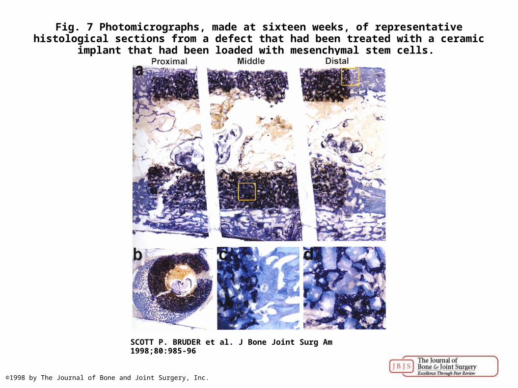
Fig. 7 Photomicrographs, made at sixteen weeks, of representative histological sections from a defect that had been treated with a ceramic implant that had been loaded with mesenchymal
stem cells.
SCOTT P. BRUDER et al. J Bone Joint Surg Am 1998;80:985-96
©1998 by The Journal of Bone and Joint Surgery, Inc.

Fig. 8 Photomicrographs, made at sixteen weeks, of representative histological sections from a defect that had been treated with a ceramic implant that had not been loaded with mesenchymal
stem cells.
SCOTT P. BRUDER et al. J Bone Joint Surg Am 1998;80:985-96
©1998 by The Journal of Bone and Joint Surgery, Inc.