THE EFFECT OF DEXAMETHASONE ON IL-33-MEDIATED MAST CELL ...
80
Virginia Commonwealth University Virginia Commonwealth University VCU Scholars Compass VCU Scholars Compass Theses and Dissertations Graduate School 2015 THE EFFECT OF DEXAMETHASONE ON IL-33-MEDIATED MAST THE EFFECT OF DEXAMETHASONE ON IL-33-MEDIATED MAST CELL ACTIVATION CELL ACTIVATION Oksana I. Chernushevich Follow this and additional works at: https://scholarscompass.vcu.edu/etd Part of the Immunology and Infectious Disease Commons © The Author Downloaded from Downloaded from https://scholarscompass.vcu.edu/etd/3772 This Thesis is brought to you for free and open access by the Graduate School at VCU Scholars Compass. It has been accepted for inclusion in Theses and Dissertations by an authorized administrator of VCU Scholars Compass. For more information, please contact [email protected].
Transcript of THE EFFECT OF DEXAMETHASONE ON IL-33-MEDIATED MAST CELL ...

Virginia Commonwealth University Virginia Commonwealth University
VCU Scholars Compass VCU Scholars Compass
Theses and Dissertations Graduate School
2015
THE EFFECT OF DEXAMETHASONE ON IL-33-MEDIATED MAST THE EFFECT OF DEXAMETHASONE ON IL-33-MEDIATED MAST
CELL ACTIVATION CELL ACTIVATION
Oksana I. Chernushevich
Follow this and additional works at: https://scholarscompass.vcu.edu/etd
Part of the Immunology and Infectious Disease Commons
© The Author
Downloaded from Downloaded from https://scholarscompass.vcu.edu/etd/3772
This Thesis is brought to you for free and open access by the Graduate School at VCU Scholars Compass. It has been accepted for inclusion in Theses and Dissertations by an authorized administrator of VCU Scholars Compass. For more information, please contact [email protected].

i
© Oksana Igorevna Chernushevich April 2015
All Rights Reserved

THE EFFECT OF DEXAMETHASONE ON IL-33-MEDIATED
MAST CELL ACTIVATION
A Thesis submitted in partial fulfillment of the requirements for the degree of Master of
Science at Virginia Commonwealth University.
by
OKSANA IGOREVNA CHERNUSHEVICH
BACHELOR OF SCIENCE, UNIVERSITY OF VIRGINIA, 2011
MASTER OF SCIENCE, VIRGINIA COMMONWEALTH UNIVERSITY, 2015
Director: JOHN J. RYAN, PH.D.
PROFESSOR OF BIOLOGY, DEPARTMENT OF BIOLOGY
Virginia Commonwealth University
Richmond, Virginia
April, 2015

iii
Acknowledgement
I would like to thank Dr. John Ryan for his mentoring. His enthusiasm, positive
attitude, and guidance have made this experience unforgettable. I would also like to thank
my committee members, Dr. Daniel Conrad, Dr. Jennifer Stewart, and Dr. David Straus for
their help with my thesis. I especially would like to thank Anuya Paranjape for all her help
and support, without which I would have been lost. I would like to thank my
coworkers/labmates in Dr. Ryan’s lab for all their support, assistance, and friendship. I
greatly appreciate all of their help. Finally, I would like to thank my family: my parents,
Igor Chernushevich and Natalia Chernushevich, and my brother, Dmitry Chernushevich.
Their love, support and confidence in me means so much.

iv
Table of Contents
Page
Acknowledgements ............................................................................................................ iii
List of Tables....................................................................................................................... v
List of Figures .................................................................................................................... vi
List of Abbreviations........................................................................................................ viii
Abstract ............................................................................................................................... x
Introduction ......................................................................................................................... 1
Materials and Methods ........................................................................................................ 6
Results ............................................................................................................................... 13
Discussion ......................................................................................................................... 20
Table .................................................................................................................................. 26
Figures ............................................................................................................................... 27
References ......................................................................................................................... 62
Vita .................................................................................................................................... 68

v
List of Tables Page
Table 1: IC50 values (μM) for Dexamethasone-mediated suppression of IL-33-induced
cytokine production on two genetic backgrounds ............................................................. 26

vi
List of Figures Page
Figure 1: Dexamethasone blocks IL-33-mediated cytokine production with 24 pretreatment
.............................................................................................................................................27
Figure 2: Time course of Dexamethasone-mediated suppression of IL-33 stimulation ... 29
Figure 3: Dexamethasone blocks IL-33-mediated cytokine production without pretreatment
........................................................................................................................................... 32
Figure 4: Dexamethasone decreases IL-33-induced pro-inflammatory cytokine production
ex vivo... ............................................................................................................................ 34
Figure 5: Dexamethasone suppresses the enhancing effects of IL-33 on FcεRI-mediated
activation. .......................................................................................................................... 36
Figure 6: Dexamethasone blocks IL-33-mediated enhancement of Ag-induced migration..
........................................................................................................................................... 38
Figure 7: Dexamethasone suppresses IL-33 stimulated cytokine production (in-cell
staining) ............................................................................................................................. 40
Figure 8: 24-hour Dexamethasone treatment downregulates ST2 surface expression ..... 43
Figure 9: Short term Dexamethasone treatment does not downregulate ST2 or c-Kit surface
expression .......................................................................................................................... 45
Figure 10: Receptor downregulation does not correlate with Dexamethasone –mediated
cytokine suppression ......................................................................................................... 47
Figure 11: Dexamethasone decreases IL-33-mediated ERK phosphorylation ................. 49
vii
Figure 12: ERK Inhibitor treatment mimics Dexamethasone effects on IL-33-induced
cytokine production ........................................................................................................... 52
Figure 13: VDR-KO mast cells are less responsive to Dexamethasone, and vit D enhances
responsiveness to Dex in WT cell ..................................................................................... 54
Figure 14: miR-155 KO mast cells are less responsive to Dexamethasone than WT....... 56
Figure 15: Atorvastatin enhances Dexamethasone responsiveness in IL-33-activated mast
cells. .................................................................................................................................. 58
Figure 16: Dexamethasone blocks Il-33-mediated neutrophil recruitment....................... 60

viii
List of Abbreviations
ANOVA: Analysis of Variance
BMMC: Bone marrow-derived mast cells
BHK: Supernatant from SCF-producing Baby Hamster Kidney fibroblast cell line
cDNA: Complementary Deoxyribonucleic acid
c-Kit: SCF receptor
DNP-HSA: Dinitrophenylated human serum albumin
FcεRI: High-affinity IgE receptor
IC50: Half maximal inhibitory concentration
IgE: Immunoglobulin E
IgG: Immunoglobulin G
IL-3: Interleukin 3, vital for mast cell survival
IL-6: Interleukin 6, a pro-inflammatory cytokine
IL-13: Interleukin 13, mimics some IL-4 properties, promotes mucus production
IL-33: Interleukin 33, a DAMP produced by stressed or injured cells, binds the ST2
receptor
JNK: c-Jun N-terminal kinases

ix
MAPK: Mitogen-activated protein kinases
MCP-1: Monocyte chemotactic protein 1
NFκB: Nuclear factor κB
p38: p38 mitogen-associated protein kinases
p65: Transcription factor 65, also known as nuclear factor NFκB p65 subunit
PBS: Phosphate buffered saline
RT-PCR: Reverse Transcription Polymerase Chain Reaction
SCF: Stem cell factor, vital for mast cell survival
TAK1-Transforming growth factor β activated kinase 1
TBS: Tris Buffered Saline
TBST: TBS + 0.1% Tween-20
TNF: Tumor necrosis factor α, a pro-inflammatory cytokine
WEHI: Supernatant from IL-3-producing WEHI-3B mouse myelomonocytic cell line

ix
Abstract
THE EFFECT OF DEXAMETHASONE ON IL-33-MEDIATED MAST CELL
ACTIVATION
By Oksana Igorevna Chernushevich, B.S.
A thesis submitted in partial fulfillment of the requirements for the degree of Master of
Science in Biology at Virginia Commonwealth University.
University of Virginia, 2011
Major Director: John J. Ryan, Ph.D.
Professor of Biology, Department of Biology
Dexamethasone has been shown to inhibit IgE-mediated mast cell activation, and the
present research investigated its role in suppressing IL-33-mediated mast cell activation. We have
found that micromolar concentrations of Dexamethasone are capable of suppressing IL-33-
mediated mast cell cytokine production, on several genetic backgrounds, and in not only bone
marrow derived mast cells, but also peritoneal mast cells. Intracellular staining demonstrated that
Dexamethasone significantly reduces expression of the IL-33 receptor, T1/ST2, in mast cells;
however, the cytokine suppression is independent of T1/ST2 downregulation. At the same time,
Dexamethasone pretreatment significantly reduced ERK phosphorylation, but our data suggests
that inhibition occurs even prior to ERK blockade. Finally, Dexamethasone treatment in vivo
reduced IL-33-mediated cytokine production and neutrophil infiltration in the murine peritoneum.
Thus, Dexamethasone, a well-established therapy for inflammatory disease, can suppress IL-33-

x
mediated mast cell activation, and may therefore be effective for treating diseases now being
attributed to IL-33 effects.

1
Introduction
Mast cells
Mast cells have many functions in the innate and adaptive immune response. They
belong to the myeloid lineage of hematopoietic cells, and circulate in blood as committed
precursors until they move to vascularized tissues for final maturation. Although they are
found in most tissues, they are often located in areas that are in close proximity to the
environment, such as epithelial tissues of skin, lungs, and intestine, due to their role in
defense against parasites (Urb and Sheppard, 2012). However, they are also essential in
IgE-mediated allergic reactions. Mast cells express high-affinity IgE receptors (FcεRI), and
are usually activated when FcεRI binds antigen-specific IgE antibodies, leading to cross-
linkage by antigen. As a result of mast cell activation, various pre-formed and newly
synthesized pro-inflammatory mediators are released, including histamine, bioactive lipids,
and cytokines and chemokines (Kalesnikoff and Galli, 2008). Thus, mast cells are
important effectors of allergic disorders, such as anaphylaxis, hay fever, allergic rhinitis,
atopic dermatitis, eczema, and asthma (Oskeritzian et al., 2010). At the same time, mast
cells have also been implicated in a range of other disorders, from autoimmune diseases to
cancer, since they can elicit chemotaxis of other immune effector cells, altering
inflammation in a complex way that leads to either protective or pathologic reactions,
depending on the molecules secreted (Ryan et al., 2009). Thus, mast cell activation can

2
both enhance and inhibit inflammation, but the specific processes and mechanisms are not
fully understood.
IL-33-mediated mast cell activation
Although IgE-mediated activation has been most extensively studied, many other
molecules can activate or amplify mast cell activation, such as complement, IgG, specific
pathogen-associated molecular patterns, and various cytokines, such as IL-33 (Moulin et
al., 2007).
IL-33, a member of IL-1 cytokine family, is constitutively expressed in the nucleus
of several cell types, including epithelial cells, endothelial cells and innate immune cells
such as macrophages and dendritic cells (Moussion et al., 2008; Prefontaine et al., 2010;
Kurowska-Stolarska et al., 2009; Schmitz et al., 2005). It can also be induced under
inflammatory conditions as an alarmin. Studies have shown that it is likely that IL-33 is
released through cell necrosis or injury, as well as a response to allergens (Luthi et al.,
2009; Yoshimoto and Matsushika, 2014). IL-33 can also bind to NFκB and chromatin,
inducing pro-inflammatory gene expression (Ali et al., 2011; Choi et al., 2012; Carriere et
al., 2007; Saluja et al., 2015). Thus, IL-33 has a dual function as a cytokine and a
transcription factor.
Mature, full length IL-33, a 30-kDa protein, is a ligand for IL-33Rα (T1/ST2, also
termed IL-1RL1), which induces signal transduction after forming a heterodimeric receptor
complex containing IL-1RAcP. IL-1RAcP contains an intracellular TIR domain with
adapter proteins, including MyD88, TRAF6, and IRAK4, leading to activation of the

3
NFκB and MAPK/AP-1 pathways (Luthi et al., 2009; Cayrol and Girard, 2009; Yoshimoto
and Matsushika, 2014; Pecaric-Petkovic, 2009; Chow et al., 2010; Saluja et al., 2015).
T1/ST2 receptor is similar to other IL-1 receptors, and is expressed on mast cells,
basophils, eosinophils, neutrophils, T cell subsets, B-1 cells, macrophages, dendritic cells,
and ILC2. All respond to IL-33 (Bergers et al., 1994; Moulin et al., 2007; Liew et al.,
2010; Matsushita and Yoshimoto, 2014; Yoshimoto and Matsushika, 2014). At the same
time, the T1/ST2 receptor exists as soluble isoform (sST2) that serves as a decoy receptor
for IL-33. It binds IL-33 and reduces its level in the serum (Saluja et al., 2015). Elevated
serum concentrations of sST2 have been shown to be associated with abnormal Th2
responses and inflammatory conditions (Oshikawa et al., 2001; Kuroiwa et al., 2001;
Moulin et al., 2007).
T1/ST2 is highly expressed on mast cells, which respond to IL-33 by increasing
production of Th2 cytokines, such as IL-6 and IL-13, with or without IgE-activation
(Moulin et al., 2007; Allakhverdi et al., 2007; Iikura et al., 2007; Silver et al., 2010; Saluja
et al., 2015; Kondo et al., 2008; Ho et al., 2007; Smithgall et al., 2008; Yoshimoto and
Matsushika, 2014). The resulting chemokines can recruit eosinophils, basophils, and
neutrophils to the site of inflammation (Kondo et al., 2008; Schmitz et al., 2005;
Kurowska-Stolarska et al., 2009; Haenuki et al., 2012; Yasuda et al., 2012; Yoshimoto and
Matsushika, 2014). Furthermore, IL-33 can stimulate both innate and adaptive immune
cells to increase or decrease Th2 cytokine production, leading to eosinophilic
inflammation. Thus, IL-33 could be an attractive therapeutic target for potential treatment
of allergic and inflammatory diseases (Yoshimoto and Matsushika, 2014).

4
Glucocorticoids
Glucocorticoids are steroid hormones that are widely used to treat autoimmune and
inflammatory conditions due to their anti-inflammatory and immune-suppressive
properties (de Haij et al., 2004). Dexamethasone is a highly effective synthetic
glucocorticoid used as a therapeutic agent for conditions such as arthritis, colitis, severe
allergies and asthma, and in cancer therapy. Glucocorticoids act on a variety of cell types,
including lymphocytes, eosinophils, basophils, and mast cells, but the mechanism of action
can vary in each cell type (Ohta and Yamashita, 1999; Schleimer et al., 1981; Schleimer et
al., 1982; Yamaguchi et al., 1994; Yamamoto and Gaynor, 2001).
The main mechanism of action of glucocorticoids is through binding to the
cytoplasmic glucocorticoid receptor (GR). GR dissociates from chaperone proteins, such
as heat shock protein (hsp90) and translocates into the nucleus, where it binds DNA at
glucocorticoid response elements in the promoter region of corticosteroid-responsive
genes, altering their transcription (Tsai and O'Malley, 1994; Truss and Beato, 1993; Barnes
and Adcock, 1998; Bassam and Mayank, 2012). Glucorticoids bound to their receptors can
also interact and inhibit activities of DNA-binding transcription factors, such as AP-1 and
NFκB, and, therefore, downregulate transcription of pro-inflammatory genes, or they can
recruit co-repressors of certain inflammatory proteins (Karin, 1998; Ito et al., 2000;
Bassam and Mayank, 2012; Heck et al., 1994; Kassel et al., 2004; Luecke and Yamamoto,
2005; Oppong et al., 2013; Almawi et al., 1998; de Haij et al., 2004).
This canonical signaling cascade cannot account for the effects of glucocorticoids
that occur within seconds or minutes (Croxtall at al., 2000; Oppong et al., 2013), which are

5
too fast to involve transcriptional or translational inhibition. Thus, it has been proposed
that the rapid action mechanism may involve plasma membrane and cytoplasmic activities
of the glucocorticoid receptors (Zhou et al., 2008; Liu et al., 2007; Oppong et al., 2013).
Certain effects of glucocorticoids in mast cells, such as decreased number of mast cells in
tissue and decreased IgE/antigen-induced cytokine production and other responses, have
been reported (Benhamou et al., 1986; Robin et al., 1985; Daëron et al., 1982; Wershil et
al., 1995; Eklund et al., 1997; Finotto et al., 1997; Yamaguchi et al., 1994). However, the
mechanism of glucocorticoid-mediated rapid effects in mast cells is lacking, as are the
effects of glucocorticoids on IL-33-mediated activation. Our data show that
Dexamethasone is a rapid and potent suppressor of IL-33-induced mast cell functions.

6
Materials and Methods
Animals
C57BL/6J and 129/SvJ mice were purchased from The Jackson Laboratory (Bar
Harbor, ME) and used at a minimum of 6 weeks old, with approval from the Virginia
Commonwealth University institutional animal care and use committee (IACUC).
Mouse Mast Cell Cultures
Mouse bone marrow-derived mast cells (BMMCs) were derived by harvesting bone
marrow aspirates from the femur of mice, followed by culture in complete RPMI (cRPMI)
1640 medium (Invitrogen Life Technologies, Carlsbad, CA) containing 10% FBS, 2mM L-
glutamine, 100 U/ml penicillin, 100 μg/ml streptomycin, 1mM sodium pyruvate, and 1mM
HEPES (all from Corning, Corning, NY), supplemented with IL-3-containing supernatant
from WEHI-3B cells and SCF-containing supernatant from BHK-MKL cells. The final
concentration of IL-3 and SCF were adjusted to 1ng/ml and 10ng/ml, respectively, as
measured by ELISA. BMMC were used after 3 weeks of culture, at which point these
primary populations are >90% mast cells, based on staining for c-Kit and FcεRI
expression.
Cytokines and reagents
All cytokines, including IL-33, IL-3 and SCF, were purchased from Biolegend (San
Diego, CA). APC-coupled anti-mouse IL-6, APC-coupled anti-mouse TNF, FITC-coupled
anti-mouse CD117, and FITC-coupled rat IgG1 isotype control were purchased from

7
Biolegend (San Diego, CA). PE-coupled rat Anti-mouse TNF, FITC-coupled rat anti-
mouse T1/ST2, mouse IgE, and purified rat anti-mouse CD16/CD32 were purchased from
BD Biosciences (San Jose, CA). Propidium Iodide and DNP-HSA were purchased from
Sigma-Aldrich (St. Louis, MO). Dexamethasone was purchased from Tocris Bioscience
(Bristol, UK). ERK inhibitor (Cat#328006) was purchased from Calbiochem (EMD
Biosciences, La Jolla, CA).
Cytokine measurement
BMMC were cultured in cRPMI 1640 with 10 ng/mL IL-3 and SCF overnight at a
concentration of 1x10⁶ cells/mL. BMMC were washed twice in PBS, then resuspended at
1x10⁶
cells/mL in cRPMI 1640 with 10 ng/mL IL-3 and SCF. The cells were activated with
50 ng/mL IL-33 simultaneously with the addition of Dexamethasone or other drugs, and
incubated for 6-18 hours at 37°C, after which supernatants were collected. Alternatively,
cells were kept overnight at 37°C with 0.5ug/ml of IgE, washed twice with PBS and
activated with 50 ng/ml of dinitrophenyl-conjugated human serum albumin (DNP-HSA).
IL-6, TNF, MCP-1, and IL-13 supernatant levels were measured using ELISA kits from
Biolegend (San Diego, CA) for IL-6, TNF, and MCP-1, and eBioscience (San Diego, CA)
and PeproTech (Rocky Hill, NJ) for IL-13. μQuant (Bio-Tek Instruments, Inc) was used as
ELISA plate reader.

8
Peritoneal mast cell culture
Peritoneal lavage was performed on C57BL/6J mice. Cells were harvested and
cultured in WEHI/BHK at 37°C for 10 days to allow for mast cell expansion. Mast cells
were positively selected and separated using the EasySep Magnet from StemCell
Technologies (Vancouver, BC) using c-Kit as a positive marker of mast cells. Flow
cytometry was used as confirmation of mast cells. Cells were then washed twice with PBS
and resuspended at 5x10⁵/ml in cRPMI with IL-3 and SCF at 10ng/ml at 37°C overnight.
Cells were treated with either 5 M Dexamethasone or vehicle control, activated
simultaneously with 50 ng/ml IL-33, and incubated for 16 hours at 37°C, after which
supernatants were collected. Levels of IL-6, TNF, MCP-1, and IL-13 were measured by
ELISA.
Measurement of cytokine production due to synergy between Antigen and IL-33
BMMC were cultured in cRPMI 1640 with 10 ng/mL of IL-3 and SCF overnight at
a concentration of 1x10⁶ cells/mL with or without 0.5ug/ml of IgE. BMMC were washed
twice in PBS, then resuspended at 1x10⁶ cells/mL in cRPMI 1640 with 10 ng/mL IL-3 and
SCF and with vehicle control or 1M or 5M Dexamethasone +/-50 ng/ml DNP-HSA and
+/- 50 ng/ml IL-33 added to appropriate wells. The cells were incubated for 18 hours at
37°C, after which supernatants were collected. Levels of IL-6, TNF, MCP-1, and IL-13
were measured by ELISA.

9
Migration assay
BMMC were cultured in cRPMI 1640 with 10 ng/mL IL-3 and SCF overnight at a
concentration of 1x10⁶ cells/mL with 0.5 ug/ml IgE. BMMC were washed twice in PBS,
then resuspended at 2x10⁶ cells/mL in FBS-free cRPMI supplemented with 10 mg/ml BSA
and 2 M Dexamethasone for 1 hour. Eight m polycarbonate 24-well transwell inserts
from Corning were coated in FBS-free cRPMI/BSA, and plates were incubated for 1 hour
at 37° C. Bottom chambers contained 900 μL of FBS-free cRPMI with IL-3 at 1 ng/mL
and with 50 ng/ml SCF, +/- 50 ng/ml DNP-HSA, and +/- 200 ng/ml IL-33. 200 μL of the
previously starved BMMC supplemented with 1 ng/mL IL-3 were placed in the upper well.
Sample were incubated for 16 hours at 37° C, after which cells migrated to the bottom
chamber were counted using flow cytometry with propidium-iodide exclusion staining.
Fold of media control was calculated for all groups.
Flow cytometric analysis
Cells were cultured at 1x10⁶/ml in cRPMI with IL-3 and SCF (10ng/ml each)
overnight prior to staining. Afterwards, cells were washed in PBS twice, centrifuged and
resuspended in PBS containing the indicated antibodies (FITC-labeled IgG, anti-c-Kit, and
anti-T1/ST2) to assess expression of cell surface molecules. Samples were incubated at 4°
C for 30-45 minutes, washed twice with PBS, and resuspended in 200 μl of PBS. Samples
were then analyzed using a on a BD FACSCalibur (BD Biosciences).
To assess the expression of intracellular IL-6 and TNF, cells were resuspended in
cRPMI containing IL-3 and SCF at 10 ng/ml and 1 M Dex or vehicle control and

10
incubated overnight at 37°C. Cells were then activated with 50 ng/ml IL-33 for 90 min at
37°C, treated with 5 uM monensin for 5-6 hours at 37°C, washed twice with PBS and
stained with anti-T1/ST2 or IgG for 40 min at 4°C.The cells were then washed twice with
PBS and fixed in PBS containing 4% paraformaldehyde for 20 min at room temperature,
washed with PBS, and stored overnight at 4°C. The cells were then pelleted and
resuspended in saponin buffer (PBS, 0.1% BSA, 0.01M HEPES, 0.5% saponin) for 20
minutes at room temperature. Cell pellets were then incubated for 40 min at 4°C with
indicated antibodies (PE-TNF, APC-TNF, APC-IL-6, or PE-cKit) diluted in saponin
buffer. Finally, cells were washed with staining buffer and resuspended in 200 L PBS.
Western Blotting
Cells were cultured at 2x10⁶/ml in cRPMI with IL-3 and SCF at 10ng/ml with 2
M Dexamethasone or vehicle control for 18 hours. Cells were then starved for 4 hours in
cRPMI lacking growth factors prior to activation with 100 ng/ml of IL-33. Cells were then
lysed in Lysis Buffer (Cell Signaling Technology) supplemented with 1.5 ProteaseArrest
(G-Biosciences, Maryland Heights, MO). Protein concentration was determined using the
Pierce BCA protein assay kit (Thermo Scientific). Proteins were separated on 4-20% Mini-
Protean TGX Gels (Bio-Rad, Hercules, CA) using 25 μg of total protein per sample.
Transfer was done onto nitrocellulose membranes, which were then blocked for 1 hour at
room temperature with 2% BSA in PBS. Membranes were rinsed in PBS and then
incubated overnight at 4°C in 0.1% TWEEN in PBS containing 2% BSA and primary
antibody diluted 1:1000. Membranes were washed the next day with 0.1% TWEEN in PBS

11
every 5 minutes for a total of 30 minutes, then incubated with a 1:10,000 dilution of either
goat anti-rabbit or goat anti-mouse IgG (Jackson ImmunoResearch, West Grove, PA).
Membranes were rinsed a final time before being read on an Odyssey CLx infrared scanner
(Li-Cor, Lincoln, Nebraska).
ERK Inhibitor
ERK inhibitor, Cat#328006 from Calbiochem (EMD Biosciences, La Jolla, CA),
was solubilized in dimethyl sulfoxide (DMSO), and used at working concentrations of 50
M. It was added to cultures one hour prior to activation with 50 ng/mL of IL-33.
Supernatants were collected 6 hours later and ELISAs were run to determine cytokine
production.
Effect of Dexamethasone on VDR-KO or miR-155KO mast cells
BMMC from VDR-KO, miR-155 KO and control C57BL/6J mice were cultured in
cRPMI 1640 with IL-3 and SCF, 10 ng/ml each, overnight at 37°C at a concentration of
1x10⁶ cells/mL, and then treated with Dexamethasone. Where indicated, 1,25(OH)2
vitamin D3 (hence referred to as Vitamin D) at 1x10⁻⁸
M was added just prior to activation
with 50 ng/ml IL-33. The cells were incubated for 18 hours at 37°C. Supernatants were
collected, and levels of IL-6, TNF, MCP-1, and IL-13 were measured by ELISA.

12
Measurement of neutrophil recruitment in vivo
C57BL/6J mice, 12 weeks or older, were injected i.p. with 2 ml PBS or
Dexamethasone (2mg/kg) one hour prior to i.p. injection with 1 g IL-33. Six hours later,
peritoneal lavage and cardiac puncture were performed. Cells were stained with antibodies
against Gr1, Mac1, Ly6G and c-Kit prior to analysis by flow cytometry. Plasma was
analyzed by ELISA.
Statistics
Data presented are the mean ± SEM of at least 3 independent experiments. P-values
were calculated with GraphPad Prism software by paired or unpaired, two-tailed Student’s
t Test as appropriate. The IC50 values were calculated from the dose–response curves. P-
values of <0.05 were considered statistically significant.

13
Results
The effect of Dexamethasone on IL-33 activated BMMC
There have been many studies of Dexamethasone effects on IgE-activated mast
cells, but no data on Dexamethasone effects on IL-33-mediated mast cell function have
been published (Oppong et al., 2013). To determine if Dexamethasone affects IL-33-
induced cytokine production, we conducted a dose response (Figure 1) and time course
(Figure 2), with assays for IL-6, TNF, MCP-1, and IL-13. We pretreated C57BL/6 BMMC
for 24 hours with Dexamethasone, then activated with 50 ng/ml IL-33. We also added
Dexamethasone after activation with IL-33 at several time points (Figure 3). The data show
that the effect of Dexamethasone on mast cell cytokine production is dose-dependent with
24 hour pretreatment with Dexamethasone, and the effect of Dexamethasone decreases
when it is administered for longer periods or after IL-33 activation. Interestingly, the
suppressive effects were even stronger when Dexamethasone was added simultaneously
with IL-33.
Genetic background does not alter responsiveness to Dexamethasone
Our lab has recently found that genetic background can alter responsiveness to
statin drugs, TGF1, and IL-10 (Fernando et al., 2013; Speiran et al., 2009, and
unpublished data). C57BL/6 mice, the strain used to this point, are Th1-prone. We

14
conducted a dose response on 129/SvJ mice, which are Th2-prone, and found that the IC50
values for Dexamethasone-mediated IL-6, TNF, MCP-1, and IL-13 suppression were not
significantly different from C57BL/6J BMMC, using 24-hour pretreatment with
Dexamethasone prior to IL-33 activation (Table 1).
Dexamethasone decreases pro-inflammatory cytokines ex vivo
In the experiments so far, we demonstrated that Dexamethasone affects BMMC
cultured in vitro. To determine if these effects are similar among mast cells differentiated
in vivo, we used peritoneal mast cells. Mast cells were treated with 5 M Dexamethasone
and activated with 50 ng/ml of IL-33. Our data demonstrated that Dexamethasone
suppresses production of IL-6, TNF, MCP-1, and IL-13 from IL-33-activated peritoneal
mast cells similarly to BMMC (Figure 4).
Dexamethasone suppresses IL-33-mediated amplification of IgE-induced cytokine
production
It has been shown in Andrade’s article that IL-33 can synergize with IgE/antigen to
increase mast cell cytokine production (Andrade et al., 2011). Since IL-33 could therefore
worsen the symptoms and severity of allergic and autoimmune conditions, we determined
if Dexamethasone suppresses this synergy. As shown in Figure 5, Dexamethasone
suppressed IgE/antigen- and IL-33-induced cytokine production, and greatly reduced the
cooperativity of these two stimuli.

15
Dexamethasone blocks IL-33 enhancement of Ag-induced migration
Increased migration of mast cells toward inflammation sites is associated with
various inflammatory conditions (Ishizuka et al., 2001). Because IgE-coated mast cells
migrate toward antigen, and IL-33 enhances antigen-mediated cytokine production, we
determined if IL-33 also increases antigen-induced migration, and if Dexamethasone is
capable of suppressing it. We show in Figure 6 that IL-33 alone did not elicit BMMC
migration, but enhanced IgE/antigen-mediated mast cell migration. Moreover,
Dexamethasone did not reduce antigen-mediated migration, but reversed the effects of IL-
33.
Dexamethasone suppresses IL-33 stimulated cytokine protein production
We have shown that Dexamethasone suppresses IL-33-mediated cytokine
secretion. To determine if this coincided with a reduction in total cytokine protein
production, we employed intracellular staining and flow cytometry. Our data showed that
Dexamethasone suppressed intracellular IL-6 and TNF production (Figure 7).
Dexamethasone downregulates surface expression of IL-33R subunit T1/ST2, depending
on time of Dexamethasone exposure
To determine if the suppressive effects of Dexamethasone are due to altered
T1/ST2 receptor expression, we conducted a dose response of Dexamethasone for 24 hours

16
and assessed T1/ST2 expression by flow cytomentry. Dexamethasone dose-dependently
reduced T1/ST2 surface expression with 24 hour pretreatment (Figure 8).
To determine a cause for the differential cytokine production caused by shorter
exposure to Dexamethasone, we assessed changes in surface receptor levels of T1/ST2 and
c-Kit using flow cytometry. We investigated the effect of Dexamethasone on c-Kit
receptor expression since it has been shown that BMMC cultured with SCF (the c-Kit
ligand) increase cytokine production, and one paper suggested that IL-33 may signal partly
through c-Kit (Drube et al., 2010). Cells were cultured in Dexamethasone for either 6, 4, or
0 hours and then stained for T1/ST2 and c-Kit surface expression (Figure 9). The results
showed a modest decrease in T1/ST2 and c-Kit surface expression at these shorter time
points.
Receptor downregulation does not correlate with cytokine suppression due to
Dexamethasone
Next we assessed if the cytokine suppression observed with intracellular staining
corresponded with T1/ST2 downregulation. As shown in Figure 10, cells expressing
different levels of T1/ST2 do not have differences in cytokine production within the
Dexamethasone-treated population, similar to the vehicle-treated population. Therefore,
there is no correlation between T1/ST2 suppression and reduced cytokine production,
suggesting that cytokine suppression is independent of T1/ST2 downregulation.
Dexamethasone decreases IL-33-mediated ERK phosphorylation

17
To begin determining the effects of Dexamethasone on IL-33 signaling, we
investigated the MAPK cascade. Western blot analysis was performed on cells receiving
Dexamethasone treatment simultaneously with IL-33 activation and on those receiving 18
hour Dexamethasone pretreatment before IL-33 activation. After normalization to vehicle
controls, these experiments showed that 18 hour Dexamethasone pretreatment compared to
vehicle significantly reduced phosphorylated ERK (Figure 11). JNK, p38, and NFB p65
were not significantly altered by 18 hour Dexamethasone pretreatment. Further, the
simultaneous addition of Dexamethasone with IL-33 activation resulted in no changes in
phosphorylation of ERK, JNK, or p65.
ERK2 inhibitor mimics Dexamethasone effect on IL-33 activation
The decrease in ERK phosphorylation in western blot analysis correlated with the
ability of Dexamethasone pretreatment to reduce IL-33-mediated cytokine secretion. To
determine if blocking this pathway alone was sufficient to mimic the effects of
Dexamethasone, C57BL/6J BMMC were treated with 50 M ERK2 inhibitor, which acts
by binding to ERK2 and preventing its interaction with protein substrates. We found that
IL-33-induced secretion of IL-6, TNF, IL-13 and MCP-1 was significantly reduced,
regardless of whether the cells were treated simultaneously with IL-33 activation or
pretreated with the ERK2 inhibitor for 1 hour prior to activation (Figure 12). Thus,
Dexamethasone effects on ERK phosphorylation could be functionally important.

18
VDR-KO mast cells are less responsive to Dexamethasone, and Vitamin D enhances
responsiveness to Dexamethasone
It has been shown that Vitamin D (1,25-dihydroxyvitamin D3) can affect immune
responses. For example, vitamin D can inhibit IgE-mediated mast cell activation, while
VDR KO BMMC are hyperresponsive to IgE (Yip et al., 2014). No data on the effects of
Vitamin D in IL-33 signaling have been published. We compared the effect of
Dexamethasone on VDR-KO mice and control BMMC activated with IL-33, and how
vitamin D alters these effects. Our studies showed that VDR-KO BMMC were not more
responsive to IL-33 than WT control BMMC. VDR KO BMMC were slightly but
significantly less responsive to Dexamethasone. Vitamin D alone, given simultaneously
with IL-33, did not suppress IL-6 secretion from WT or VDR KO BMMC. Vitamin D did
enhance Dexamethasone-mediated suppression of IL-33. This effect was lost in VDR KO
BMMC as judged by IC50 values (Figure 13). These data suggest that Vitamin D can act
through VDR to enhance the effects of Dexamethasone.
miR-155 KO mast cells are less responsive to Dexamethasone than WT
MicroRNAs (miRNAs) are small regulatory molecules that can control the
translation of target mRNAs and, thus, regulate various biological processes at a
posttranscriptional level. miR-155 has been shown to possess anti-inflammatory properties
in mast cells activated with IgE (Biethahn et al., 2014). We assessed the effects of
Dexamethasone on wild type and miR-155-KO BMMC. Our data showed that miR-155-
19
KO BMMC were significantly less responsive to Dexamethasone, based on IC50 values
(Figure 14). These data suggest that miR-155 expression is partly required for
Dexamethasone effects.
Atorvastatin enhances Dexamethasone responsiveness in IL-33-activated mast cells
Atorvastatin is a medication that blocks the production of cholesterol, and is often
used in combination with other drugs. Our lab recently found that Atorvastatin suppresses
IL-33-mediated cytokine production (unpublished data). Thus, we determined if addition
of Atorvastatin would increase Dexamethasone effects on mast cells. BMMC were
pretreated with 10 M Atorvastatin for 24 hours and then activated with 50 ng/ml of IL-33
+/- simultaneous Dexamethasone treatment. Our studies showed that Atorvastatin
significantly enhanced mast cell responsiveness to Dexamethasone (Figure 15).
Dexamethasone blocks Il-33-mediated neutrophil recruitment
In order to demonstrate that Dexamethasone effects are consistent in vivo, we
measured IL-33-mediated cytokine production and neutrophil recruitment. This effect was
recently published to be mast cell-dependent (Enoksson et al., 2013). Our data showed that
Dexamethasone significantly suppressed neutrophil recruitment by mast cells in C57BL/6J
mice after 1 hour activation with 1 g of IL-33, injected intraperitoneally. Dexamethasone
also significantly suppressed serum IL-6 levels in the same mice (Figure 16).

20
Discussion
Dexamethasone and other synthetic glucocorticoids have been effectively used in
the treatment of inflammatory disorders, particularly allergy, since they suppress the
effector functions of many different types of immune cells (Oppong et al., 2013;
Yamaguchi et al., 1994). This leads to alleviation of the severity of allergic inflammation
in various conditions, such as asthma and allergic rhinitis (Rumsaeng et al., 1997)
At the same time, IL-33 has been shown to stimulate mast cells and promote the
Th2 immune response (Moulin et al., 2007). Numerous studies demonstrated IL-33’s role
in inflammatory diseases, including asthma (Jung et al., 2013). Thus, IL-33 is a potential
therapeutic target in chronic inflammation (Jung et al., 2013; Ciccia et al., 2013). Because
mast cells play such an important role in many disorders, including asthma, it is imperative
to not only find the appropriate inhibitor of mast cell activation, but also understand its
mechanism of action.
This study provides new information on Dexamethasone effects on IL-33-activated
mast cells. We established that Dexamethasone suppresses pro-inflammatory cytokine
production with 24 hour pretreatment prior to IL-33 activation, and that Dexamethasone
effects are even more pronounced when added simultaneously with IL-33. We find IC50
values ranging from 9 nM for IL-6 suppression to 32 nM for MCP-1. We recently found
that genetic background can alter mast cell responsiveness to statin drugs, with C57BL/6
and 129/SvJ BMMC showing widely varying sensitivity (unpublished findings). However,
Dexamethasone responses were similar among BMMC from these backgrounds, when

21
assessing IC50 values for suppression of IL-33-induced IL-6, TNF, MCP-1, and IL-13
production.
Because bone marrow-derived mast cells are not considered fully mature, and are
differentiated in vitro, we assessed Dexamethasone effects on peritoneal mast cells
expanded in culture. These cells are known to retain most of the morphological,
phenotypic, and functional features of peritoneal mast cells (Malbec et al., 2007).
Dexamethasone significantly suppressed IL-33 mediated cytokine production in peritoneal
mast cells, arguing that these effects are physiologically relevant.
IL-33 has been shown to synergize with FcRI to increase mast cell cytokine
production (Andrade et al., 2011), and thus worsen allergic inflammation. In this study we
confirmed that IL-33 synergizes with antigen and demonstrated that Dexamethasone not
only inhibited antigen- and IL-33-induced cytokine production, but also the cooperativity
of these two signals. This is important because it suggests that Dexamethasone therapy
may be efficacious in a setting where both IL-33 and IgE are activating mast cells, which is
likely the case in vivo.
Increased migration and accumulation of mast cells at sites of inflammation are
common for many inflammatory conditions (Oppong et al., 2013). It has previously been
shown that antigen induces mast cell migration (Ishizuka et al., 2001), but no one has yet
reported that IL-33 effects on this process. Our data demonstrated that IL-33 significantly
increases antigen-induced migration and that Dexamethasone suppresses it. Further studies
revealing the mechanism by which migration is reduced are needed.

22
We also investigated the mechanism by which Dexamethasone affects IL-33
signaling. Our studies showed that with 24 hour pretreatment, Dexamethasone suppresses
intracellular levels of IL-6 and TNF, but this does not markedly correlate with T1/ST2
receptor downregulation, which was moderately suppressed by 24 hour pretreatment. It has
recently been published that IL-33-induced signaling via T1/ST2 cross-activates c-Kit,
(Drube et al., 2010). We demonstrated that c-Kit receptor expression is slightly diminished
by Dexamethasone, but this required at least 4 hours of pretreatment. Thus, receptor
downregulation cannot account for the immediate effects of Dexamethasone, and also do
not correlate with its longer term effects.
IL-33 is known to activate MAP3K signaling (Moulin et al., 2007), that leads to
JNK, ERK, and p38 phosphorylation in mouse and human mast cells (Kunisch et al., 2012;
Saluja et al., 2015). The MAP3K TAK1 has been postulated to lie upstream of NFB in
IL-33 signaling as well (Andrade et al., 2011). We performed western blot analysis, and
found that Dexamethasone significantly reduced ERK phosphorylation with 18 hour
pretreatment. Correspondingly, ERK inhibitor treatment mimicked Dexamethasone effects
on IL-33-mediated IL-6, TNF, MCP-1, and IL-13 production. However, none of these
molecules were altered when Dexamethasone was added simultaneously with IL-33.
Collectively these data suggest that ERK blockade may be a means by which
Dexamethasone exerts its longer term effects, but that inhibition can occur by more than
one mechanism.
Vitamin D is emerging as an important player in the pathogenesis of allergic
diseases, particularly in asthma, due to its ability to suppress Th1 and Th2 responses. Many

23
tissues express vitamin D receptor (VDR), including brain, colon, breast and immune cells,
such as mast cells (Aranow, 2011; Bikle, 2008). Studies have shown that vitamin D (1,25-
dihydroxyvitamin D3) can affect the immune responses, such as inhibiting IgE-mediated
mast cell activation, and that VDR KO BMMC are hyperresponsive to IgE (Yip et al.,
2014; Aranow 2011). Furthermore, low serum vitamin D levels correlate with higher
steroid use among patients, and higher expression of vitamin D leads to better suppression
of TNF-alpha production by Dexamethasone (Searing and Leung, 2010). However, there
have also been some studies showing that Vitamin D receptor (VDR)-deficient mice fail to
develop experimental allergic asthma (Wittke et al., 2004). Thus, more work is needed to
elucidate such a complex mechanism. In our work, by comparing suppression of cytokine
production in BMMC derived from VDR-KO and WT mice, we concluded that VDR-KO
mast cells are less responsive to Dexamethasone, implying that its effects are partially
through VDR in IL-33-activated mast cells. Also, vitamin D addition improved
Dexamethasone responsiveness in wild type but not VDR-KO BMMC, suggesting that
VDR is the main receptor by which vitamin D affects IL-33-activated mast cells.
Mast cells have been shown to be affected by miRNAs, particularly miR-155,
which was recently shown to suppress PI3K-mediated signals downstream of FcRI
(Biethahn et al., 2014). Dexamethasone has been shown to upregulate miR-155 in MDSCs,
but no studies have been done in mast cells activated with IL-33 (Biethahn et al., 2014; Li
et al., 2014). We found that miR-155 KO BMMC were less responsive to Dexamethasone
than WT BMMC, implying that miR-155 might play a role in Dexamethasone effects on
mast cell activated with IL-33. Further investigation is warranted. Our current studies show
24
that miR-155 is constitutively expressed in BMMC, but that its levels are unchanged by
Dexamethasone under our conditions (not shown). This suggests that baseline miR-155
may serve a suppressive role that is required for full responsiveness to Dexamethasone.
Combination drug therapy is common, so it is clinically important to understand
how various drugs may enhance or suppress Dexamethasone effects. Atorvastatin is a
widely used statin that blocks cholesterol production. Our lab has recently found that
Atorvastatin inhibits IL-33-mediated mast cell activation. Thus, we treated BMMC with
Atorvastatin in addition to Dexamethasone and found that the addition of Atorvastatin
enhanced mast cell suppression, as assessed by IL-33-induced pro-inflammatory cytokine
production. Since statins are already being used as an add-on therapy for allergic disease,
understanding how Dexamethasone efficacy can be improved by statin therapy could be
immediately relevant to patient therapy.
Finally, located in various vascularized tissues close to the external environment,
mast cells are known for increasing the vascular permeability and attracting leukocytes, to
sites of inflammation (Brown and Hatfield, 2012). Although it has been shown that
glucocorticoids are capable of inhibiting IgE-mediated neutrophil recruitment by mast cells
(Schramm and Thorlacius, 2004), no one reported the same for IL-33-activated mast cells.
We showed that Dexamethasone treatment inhibited IL-33-mediated neutrophil
recruitment and IL-6 production in vivo. These data support our in vitro findings,
suggesting that IL-33 effects can be mitigated by Dexamethasone vivo. Collectively, our
data show that Dexamethasone is an effective means of antagonizing IL-33-mediated mast

25
cell activation, supporting its use in allergic disorders in which IL-33 is now proposed as
an exacerbating factor.
Collectively our data show that Dexamethasone is an effective means of
suppressing IL-33-induced mast cell activation in vitro and in vivo. Its effects may be
mediated in part by suppressing T1/ST2 signaling, specifically ERK activation. How
Dexamethasone effects are altered by vitamin D levels and other drugs such as statins
could be clinically relevant and fundamentally instructive. These data support the use of
steroids for mast cell-driven diseases in which IL-33 may be an inflammatory mediator.

26
Table 1: IC50 values (M) for Dexamethasone-mediated suppression of
IL-33-induced cytokine production on two genetic backgrounds.
BL/6 129
IL-6 0.009 0.0063
TNF- α 0.017 0.024
MCP-1 0.032 0.047
IL-13 0.005 0.004

26
Figures
Figure 1A Figure 1B
Figure 1C Figure 1D
0.0001 0.001 0.01 0.1 10
10000
20000
30000
40000
Conc. of DEX mM
IL-6
(p
g/1
mill cells)
0.0001 0.001 0.01 0.1 10
200
400
600
Conc. of DEX mM
TN
F-a
(p
g/1
mill cells)
0.0001 0.001 0.01 0.1 10
500
1000
1500
Conc. of DEX mM
MC
P-1
(p
g/1
mill cells)
0.0001 0.001 0.01 0.1 10
2000
4000
6000
Conc. of DEX mM
IL-1
3 (p
g/1
mill cells)
27

35
Figure 1. Dexamethasone blocks IL-33-mediated cytokine production with 24 hour
pretreatment. C57BL/6J BMMC were cultured in IL-3 and SCF overnight, with 24 hours
pretreatment of Dexamethasone at concentrations indicated, then activated with IL-33 for
16 hours. Supernatants were collected and assessed by ELISA to determine cytokine
concentrations. The results are expressed as the mean ± SEM of at least 3 independent
experiments conducted in triplicate.
28

35
Figure 2A Figure 2B
Figure 2C Figure 2D
0 5 10 15 200
5000
10000
15000
Dex
Vehicle
Hours of Dex pretreatment
IL-6
(p
g/m
l)
0 5 10 15 200
50
100
150
200
Dex
Vehicle
Hours of Dex pretreatment
TN
F-a
(p
g/m
L)
0 5 10 15 200
100
200
300
400
500
DexVehicle
Hours of Dex pretreatment
MC
P - 1
(p
g/m
L)
0 5 10 15 200
500
1000
1500
2000
Dex
Vehicle
Hours of Dex pretreatment
IL-1
3 (p
g/m
L)
29

26
Figure 2E Figure 2F
Figure 2G Figure 2H
0 2 4 6 80
1000
2000
3000
4000
Dex
Vehicle
Hours after Dex treatment
IL-6
(p
g/m
L)
0 2 4 6 80
10
20
30
40
50
Dex
Vehicle
Hours after Dex treatment
TN
F-a
(p
g/m
L)
0 2 4 6 80
100
200
300
400
DexVehicle
Hours after Dex treatment
MC
P - 1
(p
g/m
L)
0 2 4 6 80
500
1000
1500
DexVehicle
Hours after Dex treatment
IL-1
3 (p
g/m
L)
30

35
Figure 2. Time course of Dexamethasone-mediated suppression of IL-33 stimulation.
BMMC’s were cultured in IL-3 and SCF, with or without Dexamethasone or vehicle for
the stipulated time periods, then activated with IL-33 for 8-18 hours. For the post-
treatment, Dexamethasone was added at indicated time periods after IL-33 activation.
Supernatants were assessed by ELISA to determine cytokine concentrations. The results
are representative of 3 independent experiments conducted in triplicate.
31

35
Figure 3A Figure 3B
Figure 3C Figure 3D
0.0001 0.001 0.01 0.1 10
2000
4000
6000
8000
Conc. of DEX mM
IL-6
(p
g/m
l)
0.0001 0.001 0.01 0.1 10
100
200
300
Conc. of DEX mM
TN
F-a
(p
g/m
L)
0.0001 0.001 0.01 0.1 10
500
1000
1500
2000
Conc. of DEX mM
MC
P - 1
(p
g/m
L)
0.0001 0.001 0.01 0.1 10
1000
2000
3000
Conc. of DEX mM
IL-1
3 (p
g/m
L)
32

35
Figure 3. Dexamethasone blocks IL-33-mediated cytokine production without
pretreatment. C57BL/6J BMMC were cultured in IL-3 and SCF overnight followed by
Dexamethasone at the indicated concentrations, added simultaneously with IL-33.
Supernatants were collected after 16 hours, and assessed by ELISA to determine cytokine
concentrations. The results are expressed as the mean ± SEM of at least 3 independent
experiments conducted in triplicate.
33

33
Figure 4A Figure 4B
Figure 4C Figure 4D
None Vehicle Dex0
200
400
600
800
1000 ***
IL-6
(p
g/m
l)
None Vehicle Dex0
10
20
30
40***
TN
F-a
(p
g/m
L)
None Vehicle Dex0
200
400
600
800
**
MC
P - 1
(p
g/m
L)
None Vehicle Dex0
200
400
600
800***
IL-1
3 (p
g/m
L)
34
35
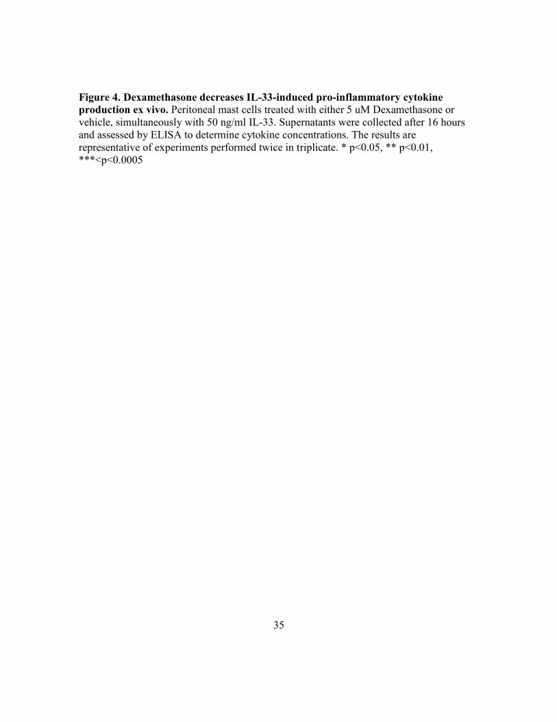
Figure 4. Dexamethasone decreases IL-33-induced pro-inflammatory cytokine
production ex vivo. Peritoneal mast cells treated with either 5 uM Dexamethasone or
vehicle, simultaneously with 50 ng/ml IL-33. Supernatants were collected after 16 hours
and assessed by ELISA to determine cytokine concentrations. The results are
representative of experiments performed twice in triplicate. * p<0.05, ** p<0.01,
***<p<0.0005

35
Figure 5A Figure 5B
Figure 5C
None Vehicle Dex Vehicle Dex Vehicle Dex0
10000
20000
30000
60000
70000
80000
IL-33 IgE XL Both
***
***
***
***
IL-6
(p
g/m
l)
None Vehicle Dex Vehicle Dex Vehicle Dex0
200
400
600
800
1000
3000
3500
4000
IL-33 IgE XL Both
***
***
***
***
TN
F-a
(p
g/m
L)
None Vehicle Dex Vehicle Dex Vehicle Dex0
200
400
600
800
10002100
2200
2300
2400
2500
IL-33 IgE XL Both
******
***
***
MC
P -
1 (
pg
/mL
)
36

35
Figure 5. Dexamethasone suppresses the enhancing effects of IL-33 on FcεRI-
mediated activation. BMMC were cultured in cRPMI IL-3 and SCF with or without
0.5ug/ml of IgE overnight prior to the addition of 1uM of Dexamethasone +/-50 ng/ml
DNP-HSA and 50 ng/ml IL-33. The cells were incubated for 18 hours, after which
supernatants were assessed by ELISA. The results are representative of experiments
performed twice in triplicate. * p<0.05, ** p<0.01, ***<p<0.0005
37

36
Figure 6
Media IL-33 Ag50 Ag50+Dex Ag50+IL-33 Ag50+IL33+Dex0
5
10
15 *
ns
*F
old
of m
ed
ia c
on
tro
l
38

37
Figure 6. Dexamethasone blocks IL-33-mediated enhancement of Ag-induced
migration. C57BL/6J BMMC were assessed for migration in response to Antigen as
described in Materials and Methods. Fold migration is based on comparison to media alone
samples. The results are expressed as the mean ± SEM of 2 independent experiments.
* p<0.05, ** p<0.01, ***<p<0.0005
39
38
Figure 7A
Isotype
TNF-α
IL-6
98.9 1.1
78.5 21.5
74.9 25.1
99.2 0.7
96.6 3.4
3.5 96.5
Control Dex
40

39
Figure 7B
Figure 7C
Vehicle Dex Vehicle Dex 0
5
10
15TNF-a IL-6
******
gM
FI
Vehicle Dex Vehicle Dex 0
5
10
15
20
TNF-a IL-6
% c
yto
kin
e p
ositiv
e c
ells
***
***
41

40
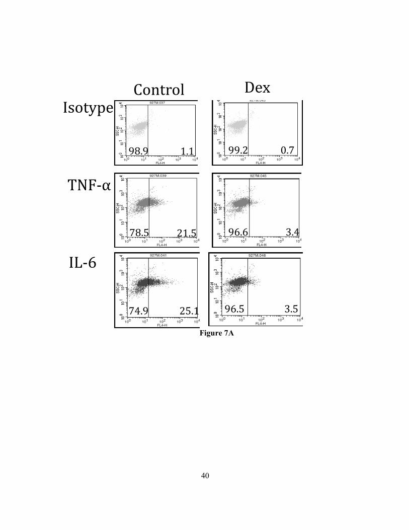
Figure 7. Dexamethasone suppresses IL-33 stimulated cytokine production (in-cell
staining). C57BL/6J BMMC were cultured in IL-3 and SCF +/- Dexamethasone or vehicle
for 24 hours. The cells were then activated with IL-33 for 90 minutes prior to the addition
of monensin for 6 hours and fixation. Cells were then permeabilized stained to detect the
indicated cytokines. Example dot plots are shown on the left. Summary of changes in
geometric mean fluorescent intensity (gMFI) and percent positive is shown on the right.
The results are representative of experiments performed twice in triplicate. * p<0.05,
** p<0.01, ***<p<0.0005
42
41
Figure 8A
Figure 8B
Figure 8C
Vehicle 1 0.1 0.010
20
40
60
80
gM
FI T
1/S
T2
***
***
**
Vehicle 1 0.1 0.010
5
10
15
20
25
***
***
% T
1/S
T2 n
eg
ative c
ells
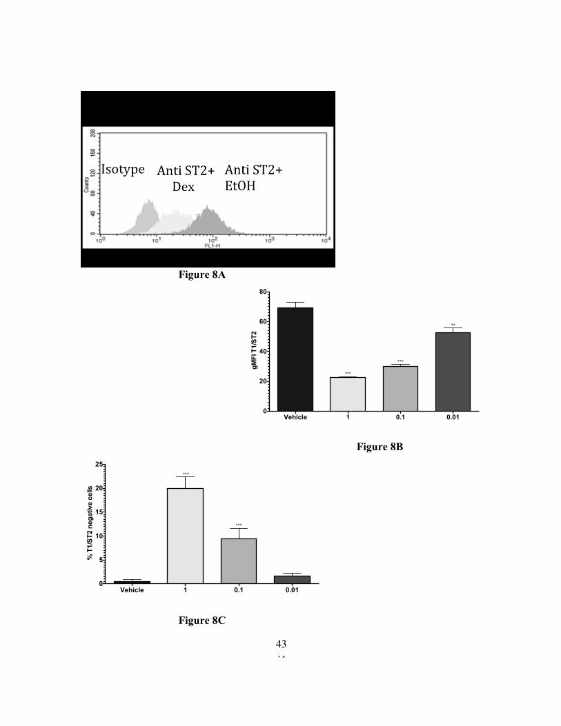
43

42
Figure 8. 24-hour Dexamethasone treatment downregulates ST2 surface expression.
C57BL/6J BMMC were treated with vehicle or Dex at the indicated concentrations for 24
hours, then stained to detect ST2 surface expression was measured by flow cytometry. The
results are representative of experiments performed twice in triplicate. * p<0.05,
** p<0.01, ***<p<0.0005
44

43
Figure 9A
Figure 9B
IgG controlVehicle Dex Vehicle Dex Vehicle Dex0
20
40
60
80
0 hours 2 hours 6 hours
****
gM
FI T
1/S
T2
IgG controlVehicle Dex Vehicle Dex Vehicle Dex0
10
20
30
40
50
0 hours 4 hours 6 hours
** **
gM
FI c-k
it
45

44
Figure 9. Short term Dexamethasone treatment does not downregulate ST2 or c-Kit
surface expression. C57BL/6 BMMC were treated with Vehicle or Dexamethasone at 5
μM for 0-6 hours. Surface expression of A) T1/ST2 and B) c-Kit was measured by flow
cytometry. The data are representative of experiments performed twice in quadruplicate.
* p<0.05, ** p<0.01, ***<p<0.0005
46

45
Figure 10A
Figure 10B
0 20 40 60 80 10020
25
30
35
40
45 Vehicle
Dex
gMFI T1/ST2
gM
FI IL
-6
0 20 40 60 80 10020
25
30
35
40 VehicleDex
gMFI T1/ST2
gM
FI T
NF
-a
47

46
Figure 10. Receptor downregulation does not correlate with Dexamethasone–
mediated cytokine suppression. C57BL/6J BMMC were cultured in IL-3 and SCF +/-
Dexamethasone or vehicle for 24 hours. The cells were then activated with IL-33 for 90
minutes prior to the addition of monensin for 6 hours, then fixed in 4% paraformaldehyde.
Cells were permeabilized in saponin buffer stained to detect ST2, TNF- α and IL-6. The
results are representative of 3 independent experiments conducted in triplicate.
48

35
Figure 11A
Figure 11B
0 min 5 min 30 min 0 min 5 min 30 min0
20
40
60
80
***
ns
Vehicle Dex
Fo
ld in
du
ctio
n (p
hE
RK
/tota
l ER
K)
ph ERK
Total ERK
0 min 0 min 5 min 5 min 15 min 15 min 30 min 30 min
ph p65
Total p65
ph JNK
Total JNK
ph p38
Total p38
Dex
Vehicle
+ + + +
+ + + + -
-
- - -
- - -
49

48
Figure 11C
ph ERK
Total ERK
0 min 0 min 5 min 5 min 15 min 15 min 30 min 30 min
ph p65
Total p65
ph JNK
Total JNK
Dex
Vehicle + + +
+
+
+ + + - - -
- -
-
- -
50

49
Figure 11. Dexamethasone decreases IL-33-mediated ERK phosphorylation. A) 18
hour pretreatment with Dex. B) ph ERK/total ERK normalized to the loading control, with
18 hour Dex pretreatment. The results are expressed as the mean ± SEM of 2 independent
experiments. C) Simultaneous addition of Dex and IL-33 to cells. * p<0.05, ** p<0.01,
***<p<0.0005
51

35
Figure 12A Figure 12B
Figure 12C Figure 12D
None Vehicle Dex Vehicle Inh0
2000
4000
6000
8000
10000
******
IL-6
(p
g/m
l)
None Vehicle Dex Vehicle Inh0
100
200
300
400***
***
TN
F-a
(p
g/m
L)
None Vehicle Dex Vehicle Inh0
500
1000
1500
2000
2500
***
***
MC
P - 1
(p
g/m
L)
None Vehicle Dex Vehicle Inh0
1000
2000
3000
4000
***
***
IL-1
3 (p
g/m
L)
52

52
Figure 12. ERK Inhibitor treatment mimics Dexamethasone effects on IL-33-induced
cytokine production. C57BL/6J BMMC were cultured in IL-3 and SCF +/- ERK inhibitor
for 1 hour prior to IL-33 activation. The cells were incubated for 18 hours at 37°C, after
which supernatants were analyzed by ELISA to detect cytokine secretion kits. The results
are representative of experiments done twice in triplicate. * p<0.05, ** p<0.01,
***<p<0.0005
53

35
Figure 13A Figure 13B
Figure 13C
None IL-33 0.01 0.005 0.001 0.0001 vitD IL-33 0.01 0.005 0.001 0.00010
5000
10000
15000
vit D
WT
Vehicle
IL-6
(p
g/m
l)
None IL-33 0.01 0.005 0.001 0.0001 vitD IL-33 0.01 0.005 0.001 0.00010
5000
10000
15000
IL-6
(p
g/m
l)
VDR-KO
Vehicle vit D
Dex Dex+vitD Dex Dex+vitD0.000
0.002
0.004
0.006 WT VDR-KO
ns
*
*
ns
IC-5
0 (IL
-6)
54

35
Figure 13. VDR-KO mast cells are less responsive to Dexamethasone, and vit D
enhances responsiveness to Dex in WT cell. BMMC from VDR-KO and WT C57BL/6J
mice were cultured in cRPMI with IL-3 and SCF and then treated with Dexamethasone
(0.01, 0.005, 0.001, 0.0001uM) +/- Vitamin D, and simultaneously activated with IL-33.
The cells were incubated for 18 hours at 37°C, after which supernatants were collected.
Levels of IL-6 were measured by Elisa kits. The results are representative. * p<0.05,
** p<0.01, ***<p<0.0005
55

35
Figure 14A Figure 14B
Figure 14C
None Vehicle 0.1 0.01 0.001 None Vehicle 0.1 0.01 0.0010
2000
4000
6000
8000
10000
***
***
***
miR-155 - KOWT
IL-6
(p
g/m
l)
None Vehicle 0.1 0.01 0.001 None Vehicle 0.1 0.01 0.0010
500
1000
1500
IL-1
3 (p
g/m
L)
ns ***
***
WT miR-155 - KO
WT KO WT KO0.0000
0.0005
0.0010
0.0015
0.0020
0.0025
***
*
IL-6 IL-13
IC-5
0
56
35
Figure 14. miR-155 KO mast cells are less responsive to Dexamethasone. BMMC from
miR155-KO and WT C57BL/6J mice were cultured in cRPMI with IL-3 and SCF and then
treated with Dexamethasone (0.1, 0.01, 0.001uM) and activated with IL-33 6 hours later.
The cells were incubated for 18 hours at 37C, and then the supernatants were collected,
and levels of IL-6, TNF-α, MCP-1, and IL-13 were measured by Elisa kits (as described
above). The cells were incubated for 18 hours at 37°C, after which supernatants were
collected. Levels of IL-6, TNF-α, are MCP-1 were measured by Elisa kits. The results are
representative. * p<0.05, ** p<0.01, ***<p<0.0005
57

35
Figure 15A Figure 15B
Figure 15C
None IL-33 0.1 0.01 0.001 Atorv IL-33 0.1 0.01 0.0010
1000
2000
3000AtorvastatinVehicle
IL-6
(p
g/m
l)
None IL-33 0.1 0.01 0.001 Atorv IL-33 0.1 0.01 0.0010
200
400
600
800 Vehicle Atorvastatin
MC
P - 1
(p
g/m
L)
Dex Dex+Atorv Dex Dex+Atorv0.00
0.02
0.04
0.06
0.08
0.10IL-6 MCP-1
**
IC-5
0
58

26
Figure 15. Atorvastatin enhances Dexamethasone responsiveness in IL-33-activated
mast cells. C57BL/6J BMMC were cultured in cRPMI with IL-3, SCF and 10 uM
Atorvastatin for 24 hours and then treated with Dexamethasone (0.1, 0.01, 0.001uM) and
simultaneously activated with IL-33. The cells were incubated for 18 hours at 37°C, after
which supernatants were collected. Levels of IL-6 and MCP-1 were measured by Elisa
kits. The results are representative. * p<0.05, ** p<0.01, ***<p<0.0005
59

4
Figure 16A
Figure 16B
PBS+PBS PBS+IL-33 Dex+PBS Dex+IL-330.0
0.5
1.0
1.5
2.0
2.5**** ****
ns
% N
eu
tro
ph
ils (
% G
r1+
Ma
c1
+)
PBS+PBS PBS+IL-33 Dex+PBS Dex+IL-330
50
100
150
200* **
ns ns
Seru
m IL
-6 (p
g/m
L)
60

5
Figure 16. Dexamethasone blocks Il-33-mediated neutrophil recruitment. C57BL/6J
mice, 12 weeks or older, were injected i.p. with 2 ml PBS or Dexamethasone (2mg/kg)
with or without 1 ug of IL-33 that was injected in 1 hour, following by peritoneal lavage
and Flow cytometry. Cells were stained with Gr1, Mac1, LysG and c-kit antibodies. A) %
Neutrophil recruitment (Gr1+Mac1+) B) SerumIL-6. The results are representative of
experiments done twice in triplicate. * p<0.05, ** p<0.01, ***<p<0.0005
61

6
Reference List
1. Ali S, Mohs A, Thomas M, Klare J, Ross R, Schmitz ML, Martin MU 2011 The
dual function cytokine IL-33 interacts with the transcription factor NFκB to
dampen NFκB-stimulated gene transcription. J Immunol 187:1609–1616
2. Allakhverdi Z, Smith DE, Comeau MR, Delespesse G 2007 Cutting edge:
TheST2 ligand IL-33 potently activates and drives maturation of human mast cells.
J Immunol 179:2051–2054
3. Almawi WY, Hess DA, Rieder MJ 1998 Multiplicity of glucocorticoid action in
inhibiting allograft rejection. Cell Transplant 7:511−523
4. Andrade MV, Iwaki S, Ropert C, Gazzinelli RT, Cunha-Melo JR, Beaven MA 2011 Amplification of cytokine production through synergistic activation of NFAT
and AP-1 following stimulation of mast cells with antigen and IL-33. Eur J
Immunol 41:760–7210
5. Aranow C 2011 Vitamin D and the immune system. J Investig Med 59(6):881-6
6. Barnes PJ, Adcock IM 1998 Transcription factors and asthma. Eur Respir J
12(1):221-34
7. Bassam M, Mayank V 2012 Steroids in Asthma: Friend or Foe, Glucocorticoids -
New Recognition of Our Familiar Friend, Dr. Xiaoxiao Qian (Ed.), ISBN: 978-953-
51-0872-6, InTech, DOI: 10.5772/50536.
8. Benhamou M, Ninio E, Salem P, Hieblot C, Bessou G, Pitton C, Liu FT,
Mencia-Huerta JM 1986 Decrease in IgE Fc receptor expression on mouse bone
marrow-derived mast cells and inhibition of PAF-acether formation and of β-
hexosaminidase release by dexamethasone. J Immunol 136:1385
9. Bergers G, Reikerstorfer A, Braselmann S, Graninger P, Busslinger M 1994
Alternative promoter usage of the Fos-responsive gene Fit-1 generates mRNA
isoforms coding for either secreted or membrane-bound proteins related to the IL-1
receptor. EMBO J 13:1176–1188
10. Biethahn K, Orinska Z, Vigorito E, Goyeneche-Patino DA, Mirghomizadeh F,
Föger N, Bulfone-Paus S 2014 miRNA-155 controls mast cell activation by
regulating the PI3Kγ pathway and anaphylaxis in a mouse model. Allergy
69(6):752-62
11. Bikle D 2008 Nonclassic Actions of Vitamin D. J Clin Endocrinol Metab 94(1):
26–34
12. Brown MA, Hatfield JK 2012 Mast Cells are Important Modifiers of
Autoimmune Disease: With so Much Evidence, Why is There Still Controversy?
Front Immunol 3:147
13. Carriere V, Roussel L, Ortega N, Lacorre DA, Americh L, Aguilar L, Bouche
G, Girard JP 2007 IL-33, the IL-1-like cytokine ligand for ST2 receptor, is a
chromatin-associated nuclear factor in vivo. Proc Natl Acad Sci USA 104:282–287
14. Cayrol C, Girard JP 2009 The IL-1-like cytokine IL-33 is inactivated after
maturation by caspase-1. Proc Nat Acad Sci U S A 106:9021-6
62

7
15. Choi YS, Park JA, Kim J, Rho SS, Park H, Kim YM, Kwon YG 2012
NuclearIL-33 is a transcriptional regulator of NF-κB p65 and induces endothelial
cell activation. Biochem Biophys Res Commun 421:305–311
16. Chow JY, Wong CK, Cheung PF, Lam CW 2010 Intracellular signaling
mechanisms regulating the activation of human eosinophils by the novel Th2
cytokineIL-33: implications for allergic inflammation. Cell Mol Immunol 7:26–34.
17. Ciccia F, Alessandro R, Rizzo A, Raimondo S, Giardina A, Raiata F, Boiardi
L, Cavazza A, Guggino G, De Leo G, Salvarani C, Triolo G 2013 IL-33 is
overexpressed in the inflamed arteries of patients with giant cell arthritis. Ann
Rheum Dis72(2):258-64
18. Croxtall JD, Choudhury Q, Flower RJ 2000 Glucocorticoids act within minutes
to inhibit recruitment of signaling factors to activated EGF receptors through a
receptor-dependent, transcription-independent mechanism. Br J Pharmacol
130:289–298
19. Daëron M, Sterk AR, Hirata F, Ishizaka T 1982 Biochemical analysis of
glucocorticoid-induced inhibition of IgE-mediated histamine release from mouse
mast cells. J Immunol 129(3):1212-8
20. de Haij S, Daha MR, van Kooten C 2004 Mechanism of steroid action in renal
epithelial cells. Kidney Int 65:1577–1588
21. Drube S, Heink S, Walter S, Löhn T, Grusser M, Gerbaulet A, Berod L,
Schons J, Dudeck A, Freitag J, Grotha S, Reich D, Rudeschko O, Norgauer J,
Hartmann K, Roers A, Kamradt T 2010 The receptor tyrosine kinase c-Kit
controls IL-33 receptor signaling in mast cells. Blood 115(19):3899-906
22. Eklund KK, Humphries DE, Xia Z, Ghildyal N, Friend DS, Gross V, Stevens
RL 1997 Glucocorticoids inhibit the cytokine-induced proliferation of mast cells,
the high affinity IgE receptor-mediated expression of TNF-alpha, and the IL-10-
induced expression of chymases. J Immunol 158(9):4373-80
23. Enoksson M, Moller-Westerberg C, Wicher G, Fallon P, Forsberg-Nilsson K,
Lunderius-Andersson C, Nilsson G 2013 Intraperitoneal influx of neutrophils in
response to IL-33 is mast cell-dependent. Blood 121(3):530-6
24. Fernando J1, Faber TW, Pullen NA, Falanga YT, Kolawole EM, Oskeritzian
CA, Barnstein BO, Bandara G, Li G, Schwartz LB, Spiegel S, Straus DB,
Conrad DH, Bunting KD, Ryan JJ 2013 Genotype-dependent effects of TGF-β1
on mast cell function: targeting the Stat5 pathway. J Immunol 191(9):4505-13
25. Finotto S, Mekori YA, Metcalfe DD 1997 Glucocorticoids decrease tissue mast
cell number by reducing the production of the c-kit ligand, stem cell factor, by
resident cells: in vitro and in vivo evidence in murine systems. J Clin Invest
99(7):1721-8
26. Haenuki Y, Matsushita K, Futatsugi-Yumikura S, Ishii KJ, Kawagoe T, Imoto
Y, Fujieda S, Yasuda M, Hisa Y, Akira S, Nakanishi K, Yoshimoto T 2012 A
critical role of IL-33 in experimental allergic rhinitis. J Allergy Clin Immunol
130:184-94
63

8
27. Heck S, Kullmann M, Gast A, Ponta H, Rahmsdorf HJ, Herrlich P, Cato AC 1994 A distinct modulating domain in glucocorticoid receptor monomers in the
repression of activity of the transcription factor AP-1. EMBO J 13:4087–4095
28. Ho LH, Ohno T, Oboki K, Kajiwara N, Suto H, Iikura M, Okayama Y. Akira
S, Saito H, Galli SJ, Nakae S 2007 IL-33 induces IL-13 production by mouse
mastcells independently of IgE-FcepsilonRI signals. J Leukoc Biol 82:1481–1490
29. Iikura M, Suto H, Kajiwara N, Oboki K, Ohno T, Okayama Y., Saito H, Galli
SJ, Nakae S 2007 IL-33 can promote survival, adhesion and cytokine production
in human mast cells. Lab Invest 87:971–978
30. Ishizuka T, Okajima F, Ishiwara M, Iizuka K, Ichimonji I, Kawata T,
Tsukagoshi H, Dobashi K, Nakazawa T, Mori M 2001 Sensitized mast cells
migrate toward the antigen: a response regulated by p38 mitogen-activated protein
kinase and Rho-associated coiled-coil-forming protein kinase. J Immunol
167(4):2298-304
31. Ito K, Barnes PJ, Adcock IM 2000 Glucocorticoid receptor recruitment of histone
deacetylase 2 inhibits interleukin-1beta-induced histone H4 acetylation on lysines 8
and 12. Mol Cell Biol 20(18):6891-903
32. Jung MY, Smrž D, Desai A, Bandara G, Ito T, Iwaki S, Kang JH, Andrade
MV, Hilderbrand SC, Brown JM, Beaven MA, Metcalfe DD, Gilfillan AM 2013 IL-33 induces a hyporesponsive phenotype in human and mouse mast cells. J
Immunol 190(2):531-8
33. Kalesnikoff J, Galli SJ 2008 New developments in mast cell biology. Nat
Immunol 9:1215–1223
34. Karin M 1998 New twists in gene regulation by glucocorticoid receptor: is DNA
binding dispensable? Cell 93(4):487-90
35. Kassel O, Schneider S, Heilbock C, Litfin M, Gottlicher M, Herrlich P 2004 A
nuclear isoform of the focal adhesion LIM-domain protein Trip6 integrates
activating and repressing signals at AP-1- and NF-κB-regulated promoters. Genes
Dev 18:2518–2528
36. Kondo Y, Yoshimoto T, Yasuda K, Futatsugi-Yumikura S, Morimoto M,
Hayashi N, Hoshino T, Fujimoto J, Nakanishi K 2008 Administration of IL-33
induces airway hyperresponsiveness and goblet cell hyperplasia in the lungs in the
absence of adaptive immune system. Int Immunol 20:791-800
37. Kunisch E, Chakilam S, Gandesiri M, Kinne RW 2012 IL-33 regulates TNF-
alpha dependent effects in synovial fibroblasts. Int J Mol Med 29:530–540
38. Kuroiwa K, Arai T, Okazaki H, Minota S, Tominaga S 2001 Identification of
human ST2 protein in the sera of patients with autoimmune diseases. Biochem
Biophys Res Commun 284:1104–1108
39. Kurowska-Stolarska M, Stolarski B, Kewin P, Murphy G, Corrigan CJ, Ying
S, Pitman N, Mirchandani A, Rana B, van Rooijen N, Shepherd M, McSharry
C, McInnes IB, Xu D, Liew FY 2009 IL-33 amplifies the polarization of
alternatively activated macrophages that contribute to airway inflammation. J
Immunol 183:6469–77
64

9
40. Li L, Zhang J, Diao W, Wang D, Wei Y, Zhang CY, Zen K 2014 MicroRNA-
155 and MicroRNA-21 promote the expansion of functional myeloid-derived
suppressor cells. J Immunol 192(3):1034-43
41. Liew FY, Pitman NI, McInnes IB 2010 Disease-associated functions of IL-33: the
new kid in the IL-1 family. Nat Rev Immunol 10:103-10
42. Liu C, Zhou J, Zhang LD, Wang YX, Kang ZM, Chen YZ, Jiang CL 2007
Rapid inhibitory effect of corticosterone on histamine release from rat peritoneal
mast cells. Horm Metab Res. 39:273–277
43. Luecke HF, Yamamoto KR 2005 The glucocorticoid receptor blocks P-TEFb
recruitment by NF-κB to effect promoter-specific transcriptional repression. Genes
Dev 19:1116–1127
44. Luthi AU, Cullen SP, McNeela EA, Duriez PJ, Afonina IS, Sheridan C,
Brumatti G, Taylor RC, Kersse K, Vandenabeele P, Lavelle EC, Martin SJ.
2009 Suppression of interleukin-33 bioactivity through proteolysis by apoptotic
caspases. Immunity 31:84-98
45. Malbec O, Roget K, Schiffer C, Iannascoli B, Dumas AR, Arock M, Daëron M 2007 Peritoneal cell-derived mast cells: an in vitro model of mature serosal-type
mouse mast cells. J Immunol 178(10):6465-75
46. Matsushita K, Yoshimoto T 2014 Interleukin-33: A Multifunctional alarmin that
promotes both health and disease. In: Yoshimoto T, Yoshimoto T (eds). Cytokine
Frontiers: Regulation of Immune Responses in Health and Disease. New York:
Springer. 267-99.
47. Moulin D, Donzé O, Talabot-Ayer D, Mézin F, Palmer G, Gabay C 2007
Interleukin (IL)-33 induces the release of pro-inflammatory mediators by mast
cells. Cytokine 40:216–225
48. Moussion C, Ortega N, Girard J-P 2008 The IL-1-like cytokine IL-33 is
constitutively expressed in the nucleus of endothelial cells and epithelial cells in
vivo: A novel ‘‘alarmin’’? PLoS One 3(10):e3331
49. Ohta K, Yamashita N 1999 Apoptosis of eosinophils and lymphocytes in allergic
inflammation. J Allergy Clin Immunol 104(1):14-21
50. Oppong E, Flink N, Cato AC 2013 Molecular mechanisms of glucocorticoid
action in mast cells. Mol Cell Endocrinol 380(1-2):119-26
51. Oshikawa K, Kuroiwa K, Tago K, Iwahana H, Yanagisawa K, Ohno S,
Tominaga SI, Sugiyama Y 2001 Elevated soluble ST2 protein levels in sera of
patients with asthma with an acute exacerbation. Am J Respir Crit Care Med
164:277–281
52. Oskeritzian CA, Price MM, Hait NC, Kapitonov D, Falanga YT, Morales JK,
Ryan JJ, Milstien S, Spiegel S 2010 Essential roles of sphingosine-1-phosphate
receptor 2 in human mast cell activation, anaphylaxis, and pulmonary edema. J Exp
Med 207:465–474
53. Pecaric-Petkovic T, Didichenko SA, Kaempfer S, Spiegl N, Dahinden CA 2009
Human basophils and eosinophils are the direct target leukocytes of the novel IL-1
family member IL-33. Blood 113:1526–1534
65

10
54. Pre´fontaine D, Nadigel J, Chouiali F, Audusseau S, Semlali A, Chakir J,
Martin JG, Hamid Q 2010 Increased IL-33 expression by epithelial cells in
bronchial asthma. J Allergy Clin Immunol 125(3):752-4
55. Robin JL, Seldin, DC, Austen KF, Lewis RA 1985 Regulation of mediator
release from mouse bone marrow-derived mast cells by glucocorticoids. J Immunol
135(4):2719-26
56. Rumsaeng V, Vliagoftis H, Oh CK, Metcalfe DD 1997 Lymphotactin gene
expression in mast cells following Fcεreceptor I aggregation: modulation by TGF-
beta, IL-4, dexamethasone, and cyclosporin A. J Immunol 158(3):1353-60
57. Ryan JJ, Morales JK, Falanga YT, Fernando JFA, Macey MR 2009 Mast cell
regulation of the immune response. World Allergy Organization Journal 2:224-232
58. Saluja R, Ketelaar ME, Hawro T, Church MK, Maurer M, Nawijn MC 2015
The role of the IL-33/IL-1RL1 axis in mast cell and basophil activation in allergic
disorders. Mol Immunol 63(1):80-5
59. Schleimer RP, Lichtenstein LM, Gillespie E 1981 Inhibition of basophil
histamine release by anti-inflammatory steroids. Nature 292(5822):454-5
60. Schleimer RP, MacGlashan DW, Jr, Gillespie E, Lichtenstein LM 1982
Inhibition of basophil histamine release by anti-inflammatory steroids. II. Studies
on the mechanism of action. J. Immunol 129(4):1632-6
61. Schmitz J, Owyang A, Oldham E, Song Y, Murphy E, McClanahan TK,
Zurawski G, Moshrefi M, Qin J, Li X, Gorman DM, Bazan JF, Kastelein RA
2005 IL-33, an interleukin-1-like cytokine that signals via the IL-1 receptor-related
protein ST2 and induces T helper type 2- associated cytokines. Immunity 23:479-
90
62. Schramm R, Thorlacius H 2004 Neutrophil recruitment in mast cell-dependent
inflammation: inhibitory mechanisms of glucocorticoids. Inflamm. Res. 53, 644-
652. 1023-3830/04/120644-09. Doi 10.1007/s00011-004-1307-8.
63. Searing DA, Leung DYM 2010 Vitamin D in Atopic Dermatitis, Asthma and
Allergic Disease. Immunol Allergy Clin North Am 30(3):397-409
64. Silver MR, Margulis A, Wood N, Goldman SJ, Kasaian M, Chaudhary D 2010
IL-33 synergizes with IgE-dependent and IgE-independent agents to promote mast
cell and basophil activation. Inflamm Res 59:207–218
65. Smithgall MD, Comeau MR, Yoon BR, Kaufman D, Armitage R, Smith DE
2008 IL-33 amplifies both Th1- and Th2-type responses through its activity on
human basophils, allergen-reactive Th2 cells, iNKT and NK cells. Int Immunol
20:1019-30
66. Speiran K, Bailey DP, Fernando J, Macey M, Barnstein B, Kolawole M,
Curley D, Watowich SS, Murray PJ, Oskeritzian C, Ryan JJ 2009 Endogenous
suppression of mast cell development and survival by IL-4 and IL-10. J Leukoc
Biol 85(5):826-36
67. Truss M, Beato M 1993 Steroid hormone receptors: Interaction with
deoxyribonucleic acid and transcription factors. Endocr Rev 14:459−479
66

11
68. Tsai MJ, O'Malley BW 1994 Molecular mechanisms of action of steroid/thyroid
receptor superfamily members. Annu Rev Biochem 63:451−486
69. Urb M, Sheppard DC 2012 The role of mast cells in the defense against
pathogens. PLoS Pathog 8(4):e1002619
70. Wershil BK, Furuta GT, Lavigne JA, Choudhury AR, Wang Z-S, Galli SJ 1995 Dexamethasone or cyclosporin A suppress mast cell–leukocyte cytokine
cascades. Multiple mechanisms of inhibition of IgE- and mast cell-dependent
cutaneous inflammation in the mouse. J Immunol 154(3):1391-8
71. Wittke A, Weaver V, Mahon BD, August A, Cantorna MT 2004 Vitamin D
receptor-deficient mice fail to develop experimental allergic asthma. J Immunol
173(5):3432-6
72. Yamaguchi M, Hirai K, Komiya A, Miyamasu M, Furumoto Y, Teshima R,
Ohta K, Morita Y, Galli SJ, Ra C, Yamamoto K 2001 Regulation of mouse mast
cell surface FcεRI expression by dexamethasone. Int Immunol 13:843–851
73. Yamaguchi M, Hirai K, Nakajima K, Ohtoshi T, Takaishi T, Ohta K, Morita
Y, Ito K 1994 Dexamethasone inhibits basophil migration. Allergy 49(5):371-5
74. Yamamoto Y, Gaynor RB 2001 Therapeutic potential of inhibition of the NF-κB
pathway in the treatment of inflammation and cancer. J Clin Invest 107:135−142.
75. Yasuda K, Muto T, Kawagoe T, Matsumoto M, Sasaki Y, Matsushita K, Taki
Y, Futatsugi-Yumikura S, Tsutsui H, Ishii KJ, Yoshimoto T, Akira S,
Nakanishi K 2012 Contribution of IL-33- activated type II innate lymphoid cells to
pulmonary eosinophilia in intestinal nematode-infected mice. Proc Natl Acad Sci
USA 109:3451-6
76. Yip KH, Kolesnikoff N, Yu C, Hauschild N, Taing H, Biggs L, Goltzman D,
Gregory PA, Anderson PH, Samuel MS, Galli SJ, Lopez AF, Grimbaldeston
MA 2014 Mechanisms of vitamin D3 metabolite repression of IgE-dependent mast
cell activation. J Allergy Clin Immunol 133(5):1356-64, 1364.e1-14
77. Yoshimoto T, Matsushika K 2014 Innate-type and acquired-type allergy regulated
by IL-33. Allergol Int 63 Suppl 1:3-11
78. Zhou J, Liu DF, Liu C, Kang ZM, Shen XH, Chen YZ, Xu T, Jiang CL 2008
Glucocorticoids inhibit degranulation of mast cells in allergic asthma via
nongenomic mechanism. Allergy 63:1177–1185
67

12
VITA
Oksana Igorevna Chernushevich was born in Konkovichy, Belarus on January 25,
1989. She graduated from Falls Church High School, VA in 2007. She then attended
University of Virginia, Charlottesville, VA, and graduated with a Bachelor of Science in
Biochemistry in 2011. She worked as a Research Associate in the lab of the Department of
Research of biochemical markers of the risk of chronic noninfectious diseases, National
Research Centre for Preventive Medicine in Moscow, Russia, before joining the Master of
Science in Biology program and Dr. Ryan’s Immunology Lab at Virginia Commonwealth
University, Richmond, Virginia in 2013.
68