The Effect of Cyclosporin A on the Proliferating Chick ... · The Effect of Cyclosporin A on the...
6
Physiol. Res. 43:223-228, 1994 The Effect of Cyclosporin A on the Proliferating Chick Embryo Erythroblasts M. PETERKA, Z. LIKOVSKY, R. PETERKOVA Institute of Experimental Medicine, Academy of Sciences of the Czech Republic, Prague, Czech Republic Received September 17, 1993 Accepted April 20, 1994 Summary The proportion of proliferating erythroblasts, i.e. proerythroblasts, basophilic erythroblasts and polychromatophilic erythroblasts in blood islands of the chick embryo yolk sac, were counted during embryonic days 2—10. From day 2 when high amounts of erythroblasts signalized the onset of embryonic erythropoiesis, the percentage of less mature erythroid cells gradually decreased. Intraamniotic injection of cyclosporin A in doses 1.5 or 15.0 /rg per embryo on day 5 led to significant changes in the proportion of proliferating erythroblasts in the yolk sac blood islands. We speculate that these changes were caused initially by the release of the more mature cells into the circulation and later by a dose-dependent decrease in the number of stem cells. The estimation of proerythroblast percentage from all proliferating erythroblasts in the yolk sac blood islands may serve as a valuable indication of toxic damage in the late avian embryo. Key words Chick embryo - Cyclosporin A - Erythroblasts - Proliferation Introduction During the first days of avian embryo development, blood islands of the yolk sac represent a major and the only erythropoietic organ. Here, the primitive erythroid cells are formed and they became replaced in the course of embryonic development by the first generation of definitive erythroid cells (Lemez 1964, Dieterlen-Lievre 1975, 1988, Dieterlen-Lievre et al 1976). Numerous studies were published about the development of red cell line in the avian embryo. However, none has been devoted to the differential counts of the maturation stages of red cell precursors in early hematopoietic organs. During the first five days of avian embryo development it is relatively easy to study all manifestations of embryotoxicity (death, developmental defects or growth retardation). It is not possible to develop structural defects after day five. For this reason we searched for other markers of toxic damage to the embryo. We found the morphogenetic system of proliferating erythroid cells in blood islands of the chick embryo yolk sac very convenient. Our study provides basic information on the quantitative changes in proliferating maturation stages of the erythroid line, i. e. of proerythroblasts (erythroblasts), basophilic erythroblasts (early polychromatophilic erythroblasts) and polychromatophilic erythroblasts (mid- polychromatophilic erythroblasts) during the first ten days of embryonic development. Our study does not involve the subsequent, nonproliferating maturation stages of the red cell line, i. e. orthochromatophilic erythroblasts (late polychromatophilic erythroblasts), reticulocytes (immature erythrocytes) and mature erythrocytes. Cyclosporin A (CsA) in embryotoxic doses was used for influencing the erythroid cell development. Haematopoiesis is only slightly affected by therapeutic doses of CsA (Ryffel 1981, 1986, Ryffel et al. 1983). Actually, bone marrow in man is successfully transplanted due to the administration of CsA 10 -12 mg/kg/day (Gratwohl and Speck 1986). On the other hand, in man and experimental mammals, CsA inhibits the proliferation of not only lymphoid but also of various non-lymphoid cells (Sharpe and Fischer 1990, McCauley et al. 1991, Ono et al. 1991, Rush 1991).
Transcript of The Effect of Cyclosporin A on the Proliferating Chick ... · The Effect of Cyclosporin A on the...

Physiol. Res. 43: 223-228, 1994
The Effect of Cyclosporin A on the Proliferating Chick Embryo Erythroblasts
M. PETERKA, Z. LIKOVSKY, R. PETERKOVA
Institute o f Experimental Medicine, Academy o f Sciences o f the Czech Republic, Prague, Czech Republic
Received September 17, 1993 Accepted April 20, 1994
SummaryThe proportion of proliferating erythroblasts, i.e. proerythroblasts, basophilic erythroblasts and polychromatophilic erythroblasts in blood islands of the chick embryo yolk sac, were counted during embryonic days 2 —10. From day 2 when high amounts of erythroblasts signalized the onset of embryonic erythropoiesis, the percentage of less mature erythroid cells gradually decreased. Intraamniotic injection of cyclosporin A in doses 1.5 or 15.0 /rg per embryo on day 5 led to significant changes in the proportion of proliferating erythroblasts in the yolk sac blood islands. We speculate that these changes were caused initially by the release of the more mature cells into the circulation and later by a dose-dependent decrease in the number of stem cells. The estimation of proerythroblast percentage from all proliferating erythroblasts in the yolk sac blood islands may serve as a valuable indication of toxic damage in the late avian embryo.
Key wordsChick embryo - Cyclosporin A - Erythroblasts - Proliferation
Introduction
During the first days of avian embryo development, blood islands of the yolk sac represent a major and the only erythropoietic organ. Here, the primitive erythroid cells are formed and they became replaced in the course of embryonic development by the first generation of definitive erythroid cells (Lemez 1964, Dieterlen-Lievre 1975, 1988, Dieterlen-Lievre et al 1976).
Numerous studies were published about the development of red cell line in the avian embryo. However, none has been devoted to the differential counts of the maturation stages of red cell precursors in early hematopoietic organs.
During the first five days of avian embryo development it is relatively easy to study all manifestations of embryotoxicity (death, developmental defects or growth retardation). It is not possible to develop structural defects after day five. For this reason we searched for other markers of toxic damage to the embryo. We found the morphogenetic system of proliferating erythroid cells in blood islands of the chick embryo yolk sac very convenient. Our study provides basic information on the quantitative changes
in proliferating maturation stages of the erythroid line,i. e. of proerythroblasts (erythroblasts), basophilic erythroblasts (early polychromatophilic erythroblasts) and polychromatophilic erythroblasts (mid- polychromatophilic erythroblasts) during the first ten days of embryonic development. Our study does not involve the subsequent, nonproliferating maturation stages of the red cell line, i. e. orthochromatophilic erythroblasts (late polychromatophilic erythroblasts), reticulocytes (immature erythrocytes) and mature erythrocytes.
Cyclosporin A (CsA) in embryotoxic doses was used for influencing the erythroid cell development. Haematopoiesis is only slightly affected by therapeutic doses of CsA (Ryffel 1981, 1986, Ryffel et al. 1983). Actually, bone marrow in man is successfully transplanted due to the administration of CsA 10 -12 mg/kg/day (Gratwohl and Speck 1986). On the other hand, in man and experimental mammals, CsA inhibits the proliferation of not only lymphoid but also of various non-lymphoid cells (Sharpe and Fischer 1990, McCauley et al. 1991, Ono et al. 1991, Rush 1991).
224 Peterka et al. Vol. 43
Our results have shown that high doses of CsA affected the chick embryo proliferating erythroid cell precursors.
Material and Methods
Fertilized eggs of the domestic fowl (Gallus gallus Linnaeus, 1758 f. domesticus) White Leghorn random-bred population, Velaz strain, were used in our experiments. Eggs were incubated for five days in the horizontal position at 37.5 °C and relative air humidity 40 - 60 % in a dark thermostatic oven. The window technique was used for opening the egg shell (Jelinek 1977, Peterka et al. 1992).
For basic information about the proportion of proliferating erythroblasts present in the blood islands during embryonic development, samples of area vasculosa were taken from 6 embryos on each incubation day 2-10.
On day 5, cyclosporin A (Sandimmun, Sandoz) diluted with redistilled water or redistilled water only were administered intraamniotically in 3.0 //I volumes
using a special glass micropipette. There were three groups of treated embryos, each of them consisting of 66 specimens. Two experimental groups received single doses of 1.5 pg CsA (corresponding approximately to the therapeutic doses in man) and 15.0 fig CsA, respectively. The third, control group, received redistilled water only. The samples of area vasculosa (2x2 mm) were taken at time intervals 1, 2, 3, 4, 5, 6, 8, 10, 12, 24, 48 and 120 h after CsA administration.
Contact preparations (Kirschbaum and Downey 1937) were stained by the Pappenheim method (Wolf 1954). From all blood cells, present in the blood islands, only the percentage of proliferating erythroblasts, i. e. proerythroblasts, basophilic erythroblasts and polychromatophilic erythroblasts (Schermer 1958) were determined in each sample of 100 cells of these maturation stages.
Data were expressed as sample means and standard errors of the mean. The differences between experimental and control groups were tested by the t-test using the 0.05 and 0.01 significance levels.
C(DÜ
<DCl
— O — pro
embryonic day
“* • - baso •” a- poly
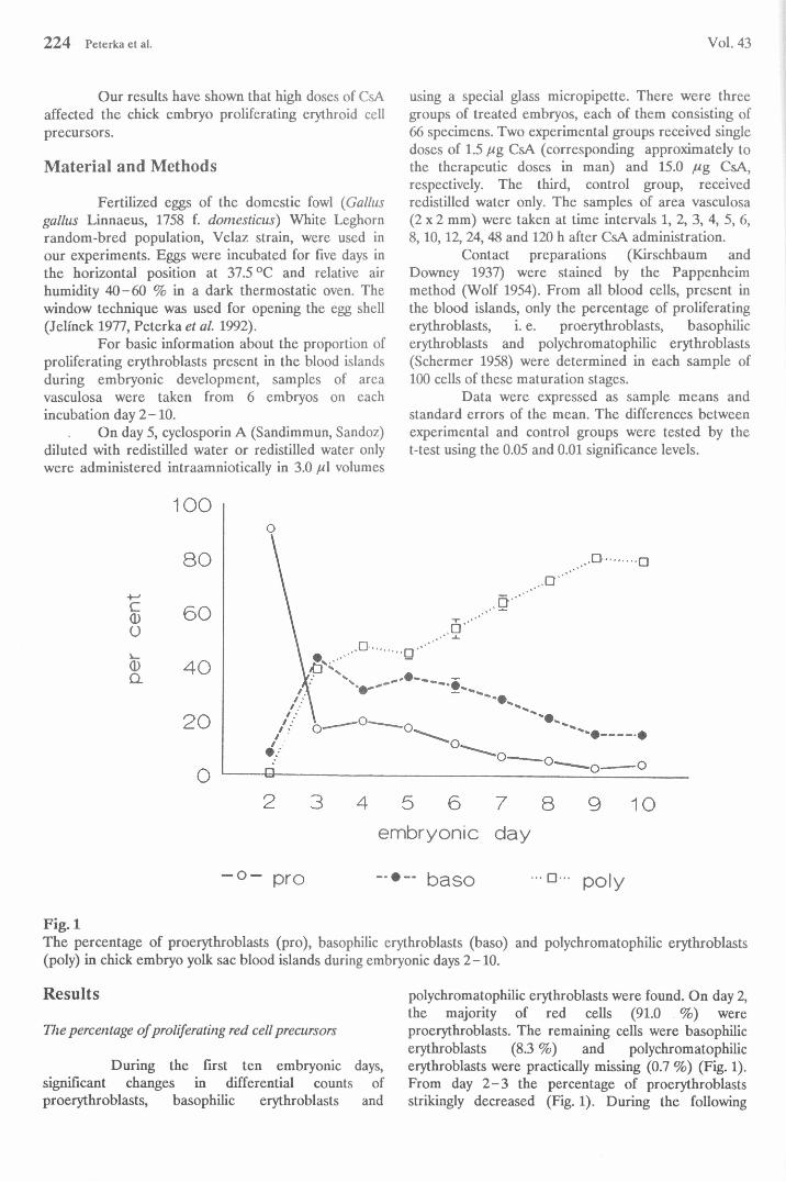
Fig. 1The percentage of proerythroblasts (pro), basophilic erythroblasts (baso) and polychromatophilic erythroblasts (poly) in chick embryo yolk sac blood islands during embryonic days 2-10.
Results
The percentage o f proliferating red cell precursors
During the first ten embryonic days, significant changes in differential counts of proerythroblasts, basophilic erythroblasts and
polychromatophilic erythroblasts were found. On day 2, the majority of red cells (91.0 %) wereproerythroblasts. The remaining cells were basophilic erythroblasts (8.3 %) and polychromatophilicerythroblasts were practically missing (0.7 %) (Fig. 1). From day 2 -3 the percentage of proerythroblasts strikingly decreased (Fig. 1). During the following

per
cent
pe
r ce
nt
Per
cent
1994 Cyclosporin A Effect on Chick Embryo Erythroblasts 225
embryonic days, they declined gradually from 20.3 % on day 4 to 3.2 % on day 9. The percentage of basophilic erythroblasts was highest on day 3 (43.5 %); thereafter they gradually decreased to 15.7 % on day 10
(Fig. 1). The percentage of polychromatophilic erythroblasts suddenly rose on day 3 to 39.3 %. During the following embryonic days they gradually increased to 81.0 % on the 9th embryonic day (Fig. 1).
— CsA 1.5 ug - o - control — CsA 15.0 ug - o - control
Fig. 2The percentage of proerythroblasts (pro), basophilic erythroblasts (baso) and polychromatophilic erythroblasts (poly) after intraamniotic administration of 1.5 [i g CsA. Asterisks denotes significant differences from controls: * P < 0.05, ** P < 0.01.
Fig. 3The percentage of proerythroblasts (pro), basophilic erythroblasts (baso) and polychromatophilic erythroblasts (poly) after intraamniotic administration of 15.0 ¿Mg CsA. Asterisks denotes significant differences from controls: * P < 0.05, ** P < 0.01.

226 Peterka et al. Vol. 43
■••O--- co n tro l — • “ * C sA 1.5 — C sA 15.0
Fig. 4The percentage of proerythroblasts in the controls and after intraamniotic administration of 1.5 //g and 15.0 /tg of CsA.
- o - pro baso poly
Fig. 5The percentage of proliferating erythroblasts, i. e. proerythroblasts (pro), basophilic erythroblasts (baso) and polychromatophilic erythroblasts (poly) after intraamniotic administration of 1.5 ¡ug CsA.

1994 Cyclosporin A Effect on Chick Embryo Erythroblasts 227
- ° - pro baso poly
Fig. 6The percentage of proliferating erythroblasts, i. e. proerythroblasts (pro), basophilic erythroblasts (baso) and polychromatophilic erythroblasts (poly) after intraamniotic administration of 15.0 //g CsA.
Changes after CsA administration
One hour after the administration of 1.5 //g CsA a significant increase of proerythroblasts was observed. Later their percentage significantly fell; at time intervals of 2 -8 h, the values were not different from the control ones at time intervals 10 -120 h (Figs 2a, 4, 5). The values of basophilic erythroblasts was lower than in the controls after 2 and 6 h, and - on the contrary - higher 24, 48 and 120 h after administration (Figs 2b, 5). The percentage of polychromatophilic erythroblasts was higher compared to controls at time intervals 2 -6 h, and lower 12-120 h after CsA administration (Figs 2c, 5).
Furthermore, the administration of 15.0 /<g of CsA led to significant changes in the proportion of proliferating erythroblasts. An increase ofproerythroblasts was observed one hour after CsA administration. After this, their percentage fell at time interval 3 -48 h after CsA administration (Figs 3a, 4,6). The values of basophilic erythroblasts were variable in comparison with the controls, being higher 3 and 12 h, and lower 8 and 48 h after CsA administration (Figs 3b, 6). The percentage of polychromatophilic erythroblasts was higher compared to controls at time intervals 6 -8 and 48 h after CsA administration (Fig. 3c, 6).
Discussion
It is difficult to assess our results at present. Similar data are completely missing in the available literature on avian or sauropsid haematology.
The changes in the percentage of all proliferating maturation stages in the yolk sac blood islands during the first days have been related to the incubation day, i. e. chronological stage of embryonic development. On day 2 the percentage ofproerythroblasts reflected the outburst oferythropoietic activity in the blood forming tissue, the more mature cells were not yet present. From day 3-10, the values of all three following maturation stages changed gradually with a decrease of the proerythroblast and basophilic erythroblast percentage.
We speculate that, shortly after theadministration of both CsA doses, the more mature stages of proliferating erythroblasts, particularlypolychromatophilic erythroblasts, were released from yolk sac blood islands into the circulation.Consequently, the percentage of less mature erythroblasts, namely proerythroblasts, was increased (Figs 4, 5, 6). The presence of immature stages of the erythroid line in the circulation during early chick embryonic life is usual - these cells mature and
228 Peterka et al. Vol. 43
proliferate in the circulation (Dieterlen ; rv The antiproliferative effect of high dose ■» metabolically active proerythroblasts vas a . est d later and its duration was dose-dependent. The percentage of proerythroblasts did not differ from the controls 10 h after the lower (1.5/eg) dose of CsA (Figs 4, 5). After a high (15.0 /eg) dose of CsA, the percentages of proerythroblasts were lower during the time interval of 2 -48 h; the "normal" v alues were only found 120 h after administration (Figs 4, 6).
The basophilic erythroblasts arise from proerythroblasts by proliferation and maturation. The observed changes in the percentage of the basophilic erythroblasts after CsA treatment reflect not only alterations of the proliferative activity of stem calls, but also the transfer of the next maturation stage, i.e. polychromatophilic erythroblasts, into the circulation (Figs 5 and 6).
We assume that the percentage ofproerythroblasts from the total number of proliferating erythroblasts may serve as a sufficient marker of toxic effects of drugs upon the avian embryo erythropoiesis and, consequently, of toxic damage to the late embryo in general.
AcknowledgementThe authors are indebted to Dr. L. Mrklas (Institute of Stomatology, Ministry of Health of the Czech Republic, Prague) and Ing. J. Hondlik (Research Institute of Pharmacology and Biochemistry, Prague) valuable help in the statistical procedures, and Mrs. A. Jelínková for technical assistance.This work was supported by the Ministry of Health of Czech Republic (Grant number 1054-3) and by Grant Agency of the Czech Republic (Grant number 304/93/0594).
References\
DIETERLEN-LIEVRE F.: On the origin of haemopoietic stem cells in the avian embryo: an experimental approach./. Embryol. exp. Morph. 33:607-619,1975.
DIETERLEN-LIEVRE F.: Avian blood cells. In: Vertebrate Blood Cells. A.F. ROWLEY, NA. RATCLIFFE (eds), Cambridge University Press, Cambridge, 1988, pp. 257-336.
DIETERLEN-LIEVRE F., BEAUPAIN D., MARTIN C.: Origin of erythropoietic stem cells in avian development: shift from the yolk sac to an intraembryonic site. Ann. Immunol. (Inst. Pasteur) 127C: 857-863,1976.
GRATWOHL A., SPECK B.: Bone marrow transplantation with ciclosporin. Prog. Allergy 38: 404-431, 1986.JELÍNEK R.: The chick embryotoxicity screening test (CHEST). In: Methods in Prenatal Toxicology. D.
NEUBERT, H.J. MERKER, G. KWASIGROCH (eds), Thieme, Stuttgart, 1977, pp. 381-386.KIRSCHBAUM A., DOWNEY H.: A comparison of some of the methods used in studies of hemopoietic tissues.
Anat. Rec. 68: 227-235, 1937.LEMEZ L.: The blood of chick embryos quantitative embryology at a cellular level. In: Advances in
Morphogenesis. M. ABERCROMBFI ?. 8RACHET (eds), Academic Press, New York, 1964, pp. 197-245.
MCCAULEY J., FARKUS Z., PRASAD SJ , PLUMMER HA., MURRAY SA.: Cyclosporine and FK-506 induced inhibition of renal epithelial cell proliferation. Transplant. Proc. 23: 2829 - 2830, 1991.
ONO M., HATAMOCHI A., ARAKAWA M., UEKI H.: Effects of cyclosporin A on cell proliferation and collagen production by human skin fibroblasts./. Dermatol. Sci. 2: 274 - 280, 1991.
PETERKA M., JELÍNEK R., PAVLIK A. Embryotoxicity of 25 psychotropic drugs: a study using CHEST. Reprod. Toxicol. 6: 367 - 374, 1992.
RUSH D.N.: Cyclosporine toxicity to organs other than the kidney. Clin. Biochem. 24:101-105, 1991.RYFFEL B.: Experimental toxicological studies with cyclosporin A. In: Cyclosporin A. D.G.J. WHITE (ed.),
Elsevier, Amsterdam, 1981, pp. 45-75.RYFFEL B.: Toxicology - experimental studies. Prog. Allergy 38:181-197, 1986.RYFFEL B., DONATSCH P., MADÓRIN M., MATTER B.E., RÚTTIMANN G., SCHÓN H., STOLL R.,
WILSON J.: Toxicological evaluation ct cyclosporin A. Arch. Toxicol. 53:107-141,1983.SCHERMER S.: Die Blutmorphologie der Laboratoriumstiere, J. A. Barth, Leipzig, 1958.SHARPE G.R., FISCHER C.: Time dependent inhibition of growth of human keratinocytes and fibroblasts by
cyclosporin A: effect on keratinocytes at therapeutic blood levels. Br. J. Dermatol. 123: 207-213,1990.WOLF J.: Microscopical Technique (in Czech). Prague, Avicenum, 1954.
Reprint RequestsDr. M. Peterka, Institute of Experimental Medicine, Academy of Sciences of the Czech Republic, Vídeňská 1083, 142 20 Prague 4, Czech Republic.