The Distance Between Your Dreams And Reality Is …ksumsc.com/download_center/2nd/2) GNT Block/Teams...
44
1 Lecture No.2 “The Distance Between Your Dreams And Reality Is Action” Text Only in Females’ slide Only in Males’ slides Important Numbers Doctor notes Notes and explanation
-
Upload
nguyencong -
Category
Documents
-
view
216 -
download
1
Transcript of The Distance Between Your Dreams And Reality Is …ksumsc.com/download_center/2nd/2) GNT Block/Teams...
1
Lecture
No.2
“The Distance Between Your Dreams
And Reality Is Action”
Text
Only in Females’ slide
Only in Males’ slides
Important
Numbers
Doctor notes
Notes and explanation

Esophageal motility and pathophysiology of
reflux disease
Objectives:
1. Mastication & chewing.
2. Salivary glands.
3. Secretion of saliva.
4. Contents of saliva.
5. Functions of saliva.
6. Control of salivary secretion.
7. Swallowing.
8. Types of esophageal peristalsis.
9. Function of lower esophageal sphincter.
2

3
1. Salivary
secretion

Structures of the GI Tract
4
Structures arranged linearly in the following sequence:
1. Mouth
2. Pharynx
3. Esophagus
4. Stomach
5. Small intestine
6. Large intestine, and anus
Other structures of GI tract (glands)
- Salivary glands, pancreas, liver, and gallbladder.
Only in Females’ Slides
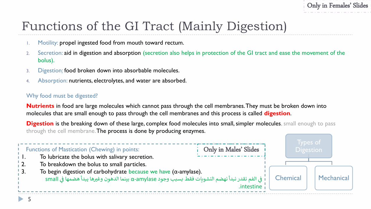
Functions of the GI Tract (Mainly Digestion)
5
1. Motility: propel ingested food from mouth toward rectum.
2. Secretion: aid in digestion and absorption (secretion also helps in protection of the GI tract and ease the movement of the
bolus).
3. Digestion: food broken down into absorbable molecules.
4. Absorption: nutrients, electrolytes, and water are absorbed.
Why food must be digested?
Nutrients in food are large molecules which cannot pass through the cell membranes. They must be broken down into
molecules that are small enough to pass through the cell membranes and this process is called digestion.
Digestion is the breaking down of these large, complex food molecules into small, simpler molecules, small enough to pass
through the cell membrane. The process is done by producing enzymes.
Types of Digestion
Chemical Mechanical
Functions of Mastication (Chewing) in points:
1. To lubricate the bolus with salivary secretion.
2. To breakdown the bolus to small particles.
3. To begin digestion of carbohydrate because we have (α-amylase).
ي الفم نقدر نبدأ نهضم النشويات فقط بسبب وجود ي α-amylaseف
ها يبدأ هضمها ف smallبينما الدهون وغير
intestine .
Only in Females’ Slides
Only in Males’ Slides

Structures of the GI Tract
6
Oral cavity: mechanical, chemical digestion.
Salivary glands: saliva lubricates food.
Saliva = mucus,
salivary amylase (starch breakdown)
Mastication: teeth chew food.
Only in Females’ Slides
Digestion in Mouth
Mechanical Digestion (Chewing, Mastication) Chemical Digestion
Teeth are designed for mastication.
Chewing muscles are innervated by 5th CN.
Chewing process is caused by a reflex (The food bolus in the mouth initiates the reflex by inhibiting muscles of mastication -drop of lower jaw-, stretch of jaw muscles leads to contraction-followed by inhibition of muscles of mastication again.
(more information in the next slide)
Done by Salivary Amylase.
Starch digestion at ph of 6.5 or 7.0.
Continues to digest for another one hour in stomach.
Stomach acid inactivates it.
Substrate > Starch.
Product > Maltose.
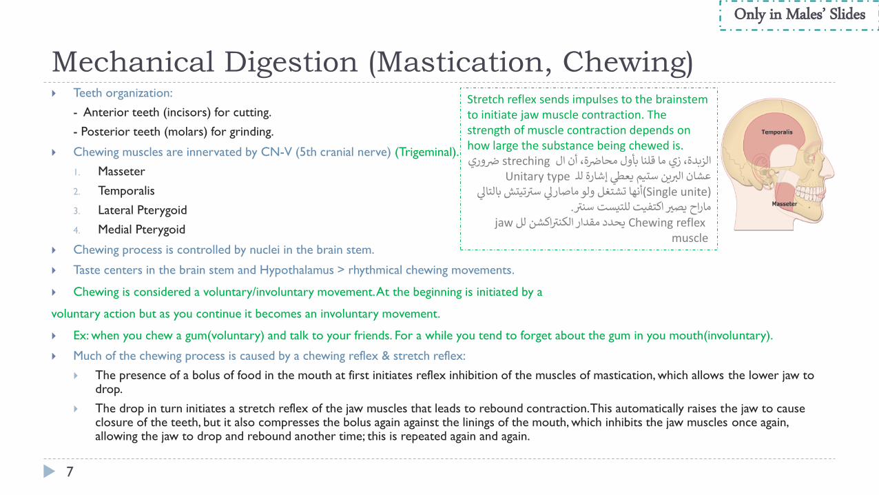
Mechanical Digestion (Mastication, Chewing)
7
Teeth organization:
- Anterior teeth (incisors) for cutting.
- Posterior teeth (molars) for grinding.
Chewing muscles are innervated by CN-V (5th cranial nerve) (Trigeminal).
1. Masseter
2. Temporalis
3. Lateral Pterygoid
4. Medial Pterygoid
Chewing process is controlled by nuclei in the brain stem.
Taste centers in the brain stem and Hypothalamus > rhythmical chewing movements.
Chewing is considered a voluntary/involuntary movement. At the beginning is initiated by a
voluntary action but as you continue it becomes an involuntary movement.
Ex: when you chew a gum(voluntary) and talk to your friends. For a while you tend to forget about the gum in you mouth(involuntary).
Much of the chewing process is caused by a chewing reflex & stretch reflex:
The presence of a bolus of food in the mouth at first initiates reflex inhibition of the muscles of mastication, which allows the lower jaw to drop.
The drop in turn initiates a stretch reflex of the jaw muscles that leads to rebound contraction. This automatically raises the jaw to cause closure of the teeth, but it also compresses the bolus again against the linings of the mouth, which inhibits the jaw muscles once again, allowing the jaw to drop and rebound another time; this is repeated again and again.
Stretch reflex sends impulses to the brainstem to initiate jaw muscle contraction. The strength of muscle contraction depends on how large the substance being chewed is.
ة، أن الالزبدة، زي وري strechingما قلنا بأول محاض ض ين عشان Unitary typeــ لليعطي إشارة ستيمالير
(Single unite) تيتشماصارلي ولو أنها تشتغل بالتالي سير.سنير للتيستيصير اكتفيت ماراح
Chewing reflex اكشنيحدد مقدار jawللالكنيرmuscle
Only in Males’ Slides

Secretory Functions of Alimentary Tract
8
Functions of the Secretory Glands:
1. Secretion of digestive enzymes.
2. Provide mucus for lubrication and protection.
Most digestive secretions are formed only in response to the presence of food in the alimentary
tract, and the quantity secreted in each segment of the tract is almost exactly the amount needed for
proper digestion. Types of Glands
Single-cell mucous glands
(goblet cells)Crypts of lieberkühn Tubular glands
Salivary glands, pancreas,
and liver
(Accessory glands)
They produce mucus.
- At the mucosal pits
Intestine (they represent
invaginations of the epithelium
into the submucosa)
They release several
digestive enzymes.
- Especially in small intestines.
- Crypts release digestive
enzymes that digest
carbohydrates and to some
extent digest fat as well.
- They include an acid and
pepsinogen-secreting
gland (In the stomach).
- Parietal cells release
hydrochloric acid.
They are located outside
the walls of GI tract. They
contain millions of acini
lined with secreting
glandular cells; these acini
feed into a system of
ducts that finally empty
into the alimentary tract
itself.
Only in Males’ Slides

Effect of Contact of Food with the Epithelium
9
Effect of Contact of Food with the Epithelium:
Function of Enteric Nervous Stimuli:
The mechanical presence of food in a particular segment of the GI tract usually causes the glands to secrete
moderate to large quantities of juices.
The types of stimuli that do this are:
1. Tactile stimulation (Tactile stimulation by touching with fingers or food).
2. Chemical irritation.
3. Distention of the gut wall.
Only in Males’ Slides
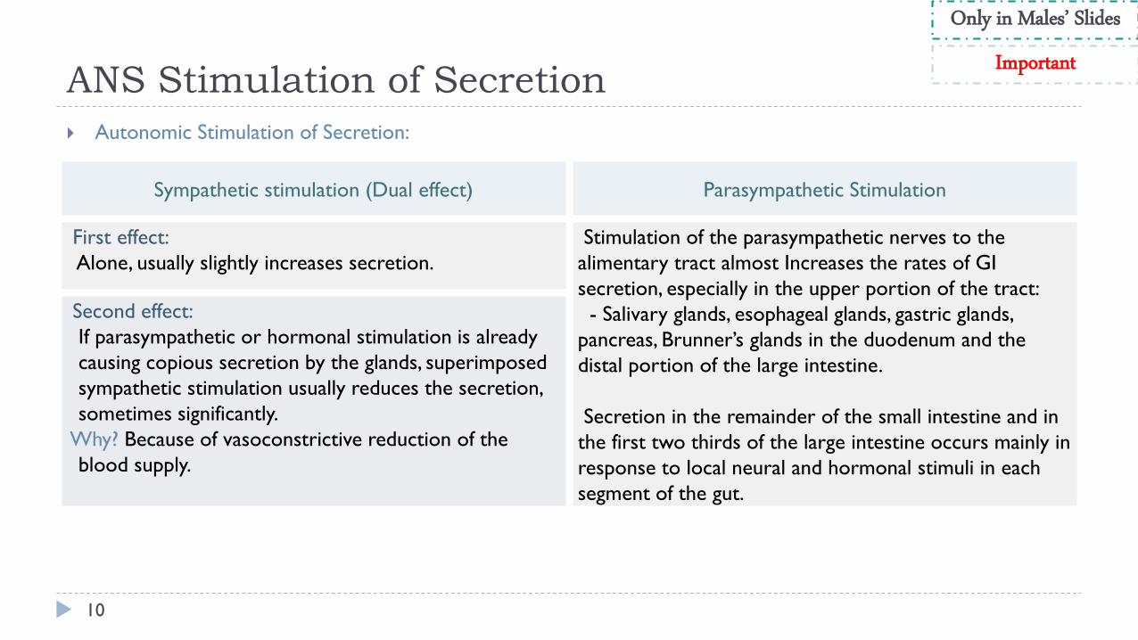
ANS Stimulation of Secretion
10
Autonomic Stimulation of Secretion:
Sympathetic stimulation (Dual effect) Parasympathetic Stimulation
First effect:
Alone, usually slightly increases secretion.
Stimulation of the parasympathetic nerves to the
alimentary tract almost Increases the rates of GI
secretion, especially in the upper portion of the tract:
- Salivary glands, esophageal glands, gastric glands,
pancreas, Brunner’s glands in the duodenum and the
distal portion of the large intestine.
Secretion in the remainder of the small intestine and in
the first two thirds of the large intestine occurs mainly in
response to local neural and hormonal stimuli in each
segment of the gut.
Second effect:
If parasympathetic or hormonal stimulation is already
causing copious secretion by the glands, superimposed
sympathetic stimulation usually reduces the secretion,
sometimes significantly.
Why? Because of vasoconstrictive reduction of the
blood supply.
Only in Males’ Slides
Important

Regulation of Glandular Secretion by Hormones
11
Regulation of Glandular Secretion by Hormones:
In the stomach and intestines
several GI hormones help regulate the volume
and character of the secretions
They are liberated from the GI mucosa in
response to the presence of food in the
lumen of the gut.
Then, the hormones are absorbed into the blood and carried to
the glands, where they stimulate secretion.
Only in Males’ Slides
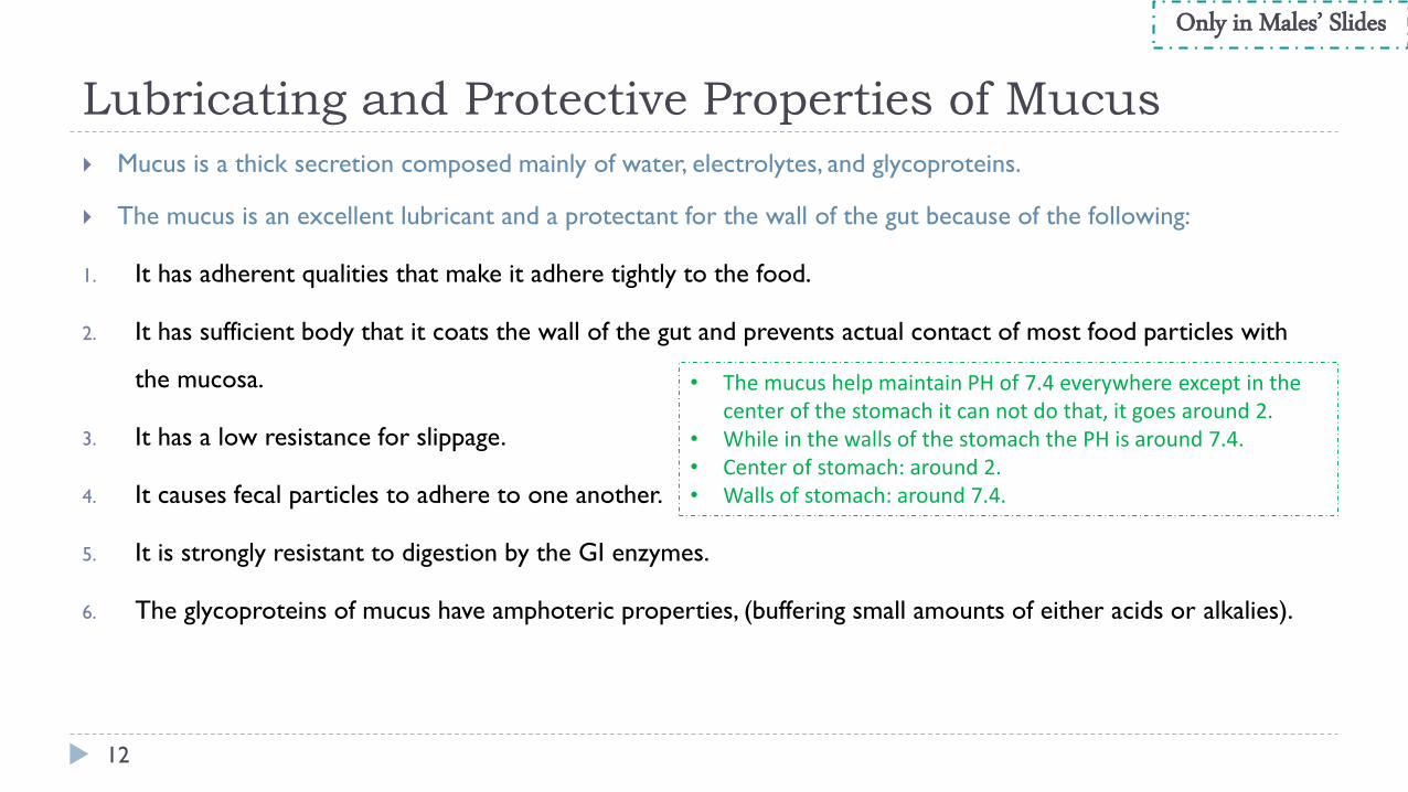
Lubricating and Protective Properties of Mucus
12
Mucus is a thick secretion composed mainly of water, electrolytes, and glycoproteins.
The mucus is an excellent lubricant and a protectant for the wall of the gut because of the following:
1. It has adherent qualities that make it adhere tightly to the food.
2. It has sufficient body that it coats the wall of the gut and prevents actual contact of most food particles with
the mucosa.
3. It has a low resistance for slippage.
4. It causes fecal particles to adhere to one another.
5. It is strongly resistant to digestion by the GI enzymes.
6. The glycoproteins of mucus have amphoteric properties, (buffering small amounts of either acids or alkalies).
Only in Males’ Slides
• The mucus help maintain PH of 7.4 everywhere except in the center of the stomach it can not do that, it goes around 2.
• While in the walls of the stomach the PH is around 7.4.• Center of stomach: around 2.• Walls of stomach: around 7.4.
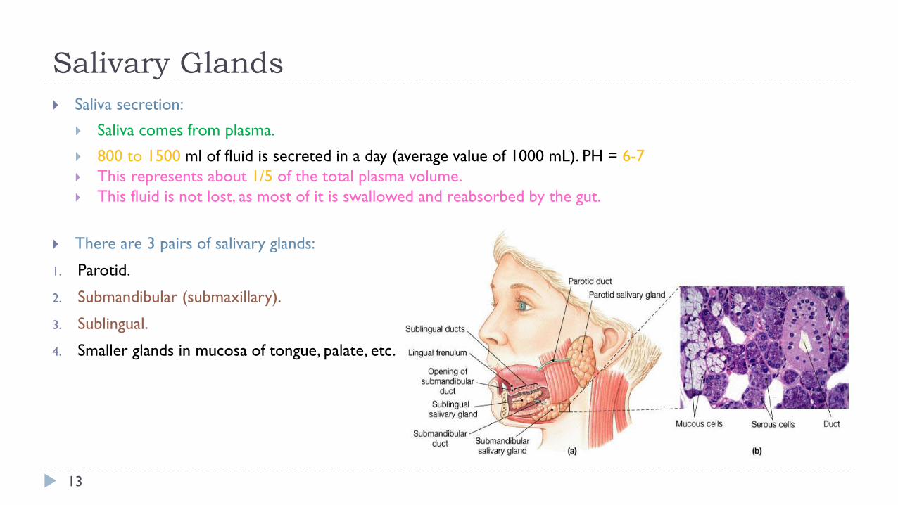
Salivary Glands
13
Saliva secretion:
Saliva comes from plasma.
800 to 1500 ml of fluid is secreted in a day (average value of 1000 mL). PH = 6-7
This represents about 1/5 of the total plasma volume.
This fluid is not lost, as most of it is swallowed and reabsorbed by the gut.
There are 3 pairs of salivary glands:
1. Parotid.
2. Submandibular (submaxillary).
3. Sublingual.
4. Smaller glands in mucosa of tongue, palate, etc.
Secretion of Saliva and its Characteristics
14
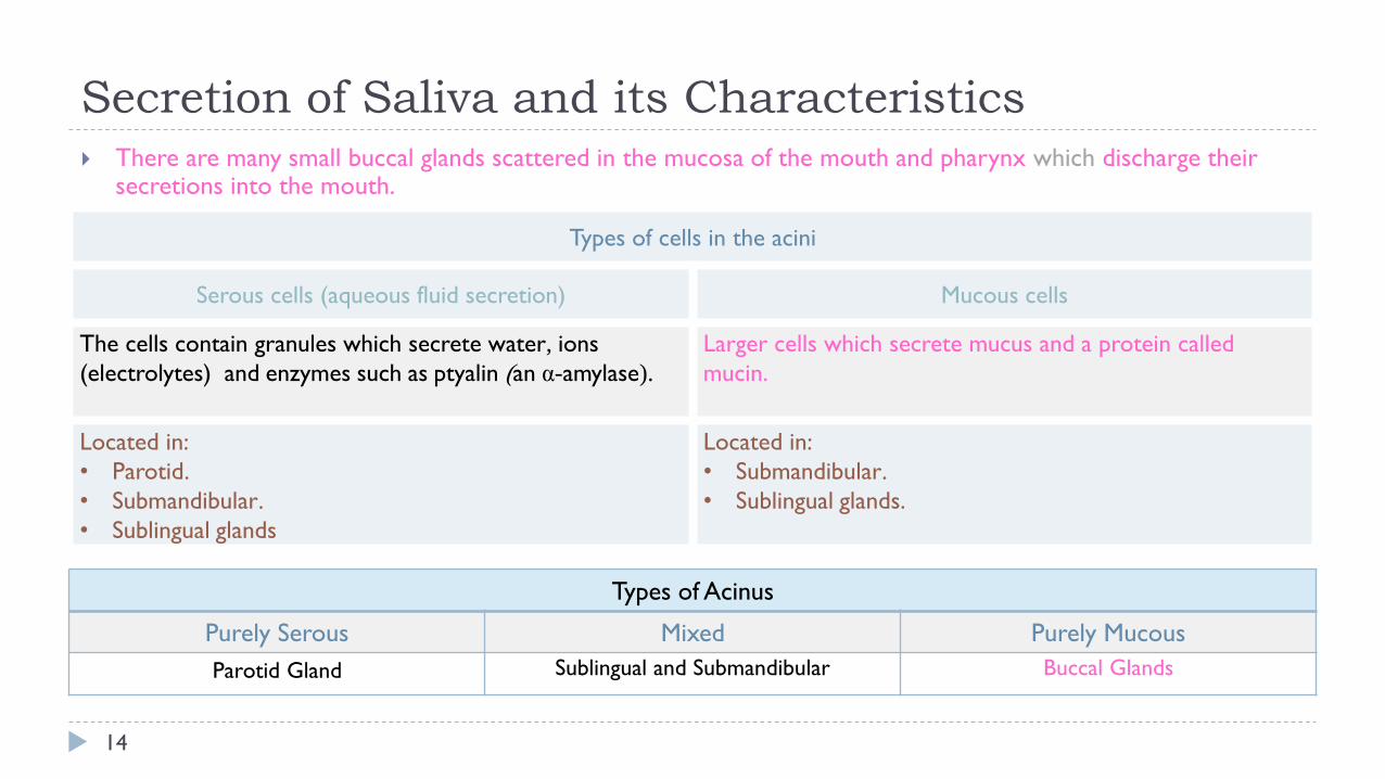
There are many small buccal glands scattered in the mucosa of the mouth and pharynx which discharge their secretions into the mouth.
Types of cells in the acini
Serous cells (aqueous fluid secretion) Mucous cells
The cells contain granules which secrete water, ions
(electrolytes) and enzymes such as ptyalin (an α-amylase).
Larger cells which secrete mucus and a protein called
mucin.
Located in:
• Parotid.
• Submandibular.
• Sublingual glands
Located in:
• Submandibular.
• Sublingual glands.
Types of Acinus
Purely Serous Mixed Purely Mucous
Parotid Gland Sublingual and Submandibular Buccal Glands

Composition of Saliva
15
Aqueous Fluids: H2O, K, HCO3, Na, Cl, -amylase, Lingual lipase, IgA, Kallikrein, Muramidase (lyses muramic acid of Staphylococcus), Lactoferrin (antimicrobial activity ) and epithelial growth factor (EGF) (Normal saliva has HIGH potassium and Bicarbonate, but low NaCl).
Only in Males’ Slides
Hypotonic Solution
1. Ions 2.Water 3. Enzymes
Na, K, CI, HCO3: the concentrations of these ions are altered with altered flow rates), e.g., at low flow rate (under resting condition).
0.5
L saliva/day
-amylase
(from parotid glands) Lingual lipase Kallikrein
Low Na+ and Cl-
(1/7 or 1/10 their
concentrations in
plasma)
High K
(7 times as great as in plasma)
And HCO3
(2-3 times that of plasma)
-
• Cleaves -1 ,4-glycosidic bonds.
• The optimal pH for this enzyme to work properly is 7.
• Inactivated at pH 4 but continues to work forsometime in unmixed food in Orad portion of stomach.
• Hydrolyzes lipids.• Continues working
in the duodenum.• Lingual lipase (to
digest fat but its not used in the mouth b/c of the atmosphere and time).
Kallikrein (a protease from acinar cells, which is not secreted into the salivary secretion):• Catalyzes production of
bradykinin (good vasodilator) from 2-globulin.
• Bradykinin increases local blood flow.
Very Important
Composition of Saliva
Composition Functions
99.5% water and 0.5% solutes
• Bicarbonate ions buffer acidic foods (pH 6.35-6.85) in mouth & esophagus.
• Chemical digestion of starch begins with enzyme (salivary amylase).
• Mucus lubricates food & facilitate swallowing.
Only in
Females’ Slides

Secretory unit (Salivon)
16
The secretory unit: The basic building block of all salivary glands.
The basic unit “salivon” consists of:
1. Acinus -initial secretory process.
2. Intercalated duct -initial portion of duct.
3. Striated duct -modification of secretory product.
The epithelial cells lining the intralobular ducts are metabolically very active and responsible for active transport of electrolytes.
Myoepithelial cells: (To modify the secretions)
Are found between the basement membrane and the cells lining the lumen of acini and intralobular ducts, they contract and increase salivary flow.
Surround acinus and intercalated duct.
Their contraction moves saliva, and prevents development of back pressure.
Only in Females’ Slides
• In striated duct-modification of secretory product.• Modification depends on time so when there is enough time
modification happens and when there is no enough time it doesn’t happen.

Secretory unit (Salivon)
17
The basic unit “salivon” consists of:
1. Acinus -initial secretory process.
2. Intercalated duct -initial portion of duct.
3. Striated duct -modification of secretory product.
4. Myoepithelial cells:
Surround acinus and intercalated duct.
Their contraction moves saliva, and prevents development of back pressure.
The secretory unit: The basic building block of all salivary glands.
The epithelial cells lining the intralobular ducts are metabolically
very active and responsible for active transport of electrolytes.
Myoepithelial cells are found between the basement membrane and
the cells lining the lumen of acini and intralobular ducts, they
contract and increase salivary flow.
Only in Females’ SlidesOnly in Males’ Slides
Only in Females’ Slides

Two Stage Hypothesis of Saliva Formation
18
Only in Females’ Slides
• Amount of Na and Cl resorbed is more than the amount of K and HCO3.
• Secreted which leads to hypotonic saliva.
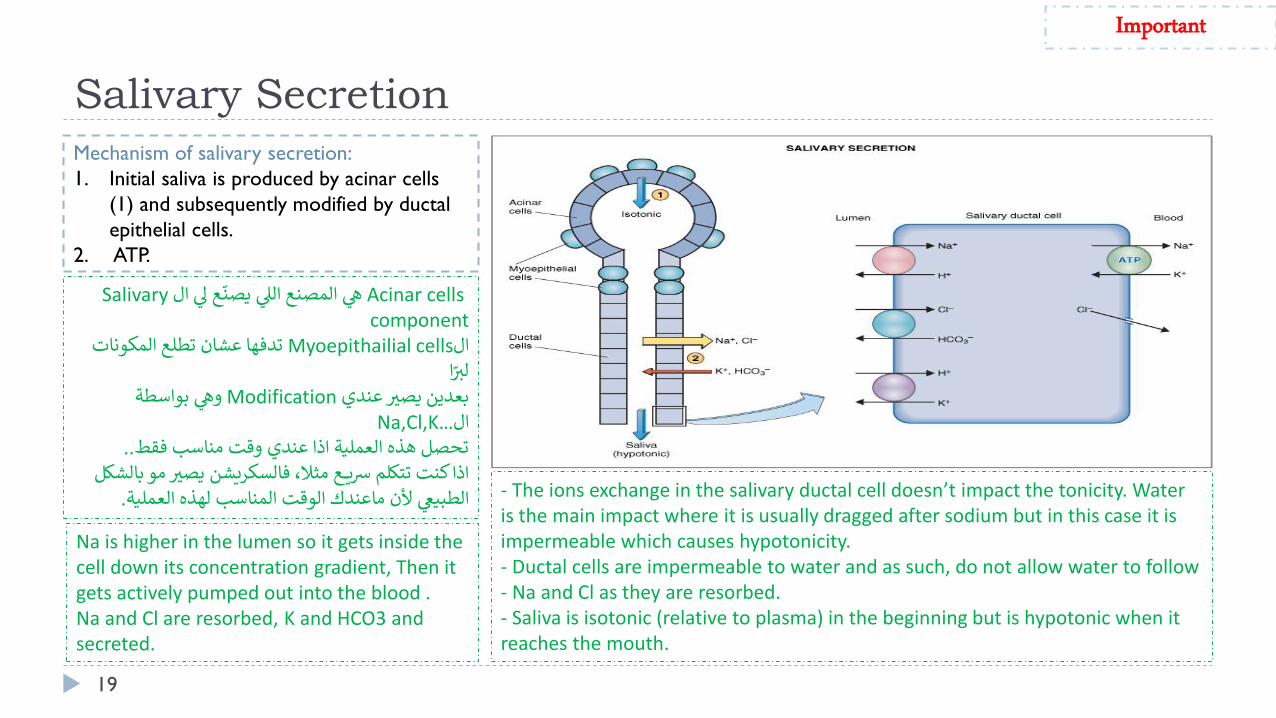
Salivary Secretion
19
Mechanism of salivary secretion:
1. Initial saliva is produced by acinar cells
(1) and subsequently modified by ductal
epithelial cells.
2. ATP.
Acinar cells ع لي الّ Salivaryهي المصنع اللي يصن
componentMyoepithailialال cellsتدفها عشان تطلع المكوناتا ّ لير
وهي بواسطة Modificationبعدين يصير عندي …Na,Cl,Kال
..تحصل هذه العملية اذا عندي وقت مناسب فقطبالشكل و ميصير فالسكريشناذا كنت تتكلم رسيــــع مثال،
.ةالوقت المناسب لهذه العمليماعندكألن الطبيعي
Important
Na is higher in the lumen so it gets inside the cell down its concentration gradient, Then it gets actively pumped out into the blood .Na and Cl are resorbed, K and HCO3 and secreted.
- The ions exchange in the salivary ductal cell doesn’t impact the tonicity. Water is the main impact where it is usually dragged after sodium but in this case it is impermeable which causes hypotonicity.- Ductal cells are impermeable to water and as such, do not allow water to follow- Na and Cl as they are resorbed.- Saliva is isotonic (relative to plasma) in the beginning but is hypotonic when it reaches the mouth.

Characteristics of Saliva and Flow Rate
Only in Males’ Slides
• When flow rate is decreased (normal saliva secretion) low concentration of Na and Cl in the mouth (since there enough time for modification).
• When flow rate is increased(increased saliva), Na and Cl are highly concentrated in the mouth (there is no modification by ductal cells-no time for Na and Cl to go back to blood stream OR Increase in speed of secretion will cause the saliva to have more Na and Cl in its composition and it will be more isotonic.
• The bicarbonate is in a plateau condition b/c it is not affected by flow rate. It gets activated by parasympathetic pathway and increase its influx.
20
Functions of Saliva
21
Saliva helps prevent the deteriorative processes in the mouth in
several ways:
It moistens food.
It begins digestion only.
It adjusts salt appetite.
The flow of saliva helps wash away pathogenic bacteria.
Saliva contains several factors that destroy bacteria such as
thiocyanate ions, antibodies, lactoferrin which chelates iron
necessary for bacterial growth and proteolytic enzymes such as
lysozyme which is:
1. Active against bacterial walls.
2. Helps thiocyanate ions in entering bacterial wall where they
become bactericidal.
Only in Females’ SlidesOnly in Males’ Slides
Facilitates speech.
By acting as a solvent, saliva is important for the sense of
taste.
Enzyme (lysozyme) helps destroy bacteria.
Epidermal growth factor is responsible for healing of ulcers
in the mucous membrane of oral cavity.
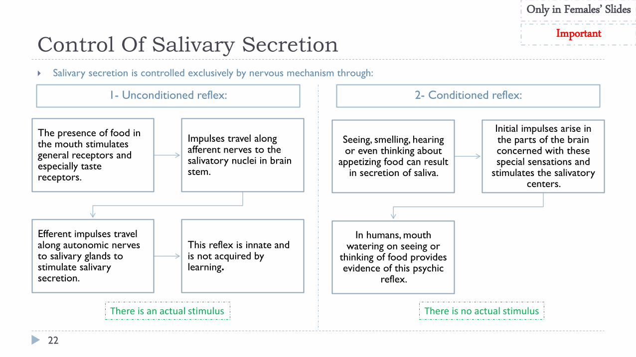
Control Of Salivary Secretion
22
Salivary secretion is controlled exclusively by nervous mechanism through:
The presence of food in the mouth stimulates general receptors and especially taste receptors.
Impulses travel along afferent nerves to the salivatory nuclei in brain stem.
Efferent impulses travel along autonomic nerves to salivary glands to stimulate salivary secretion.
This reflex is innate and is not acquired by learning.
Seeing, smelling, hearing or even thinking about
appetizing food can result in secretion of saliva.
Initial impulses arise in the parts of the brain concerned with these special sensations and
stimulates the salivatorycenters.
In humans, mouth watering on seeing or
thinking of food provides evidence of this psychic
reflex.
1- Unconditioned reflex: 2- Conditioned reflex:
Only in Females’ Slides
Important
There is no actual stimulusThere is an actual stimulus

Unique aspect of control of salivary secretion.
Secretion rate depends entirely on neural control
autonomic nervous system (ANS).
Both Parasympathetic and Sympathetic lead to
increased secretion.
Composition modified by Aldosterone:
1. Increases Na+ and Cl- reabsorption.
2. Increases K+ secretion.
Control Of Salivary SecretionOnly in Females’ SlidesOnly in Males’ Slides

Parasympathetic Supply
1. Origin of Parasympathetic Neurons/Nerves:
Salivary nuclei in medulla and pons (brain stem).
2. Outflow: VII (7) & IX (9) Cranial Nerves.
3. Transmitter: Acetylcholine
4. It is stimulated in response to:
Conditioned reflexes (taste, smell, and tactile stimuli)
3. Its stimulation is reduced due to:
Sleep, fear, dehydration
3. Stimulates:
- Secretion of aqueous fluids (protein poor, high k and HCO3).
- Contraction of myoepithelial cells.
- Metabolic rate.
- Blood flow.
- Direct innervation of blood vessels.
- Growth and development of different cells.
7. Transecting (cutting) of parasympathetic markedly decreases flow &
leads to atrophy
وع وتكون ومتوتر او اثناء النومخايفلما يكون عندك مقابلة او عرض مشرThe Parasympathetic will be inhibit it, so we will have dry mouth.
The Parasympathetic and the sympathetic all of them increase the
secretion, but the parasympathetic is more dominant.
1. Origin of Parasympathetic Neurons/Nerves:
Superior & inferior salivary nuclei in brain stem.
2. Fibers from the superior salivary nucleus:
Leave in VII (7) cranial nerve to supply both submandibular and sublingual
glands.
3. Fibers from the inferior salivary nucleus:
Leave the medulla in IX (9) cranial nerve to supply the parotid gland.
Parasympathetic Supply
Only in Females’
Slides
Only in Males’
Slides
Very Important

Sympathetic Supply
1. The origin of sympathetic nerves are:
Intermediolateral gray T1-T3
2. Transmitter: norepinephrine
3. Stimulates:
• Secretion (mostly enzymes).
• Contraction of myoepithelial cell.
• Metabolic rate.
• Growth and development of different cells.
4. Transecting (cutting): of sympathetic nerves has minimal
impact on secretion.
1. The origin of sympathetic nerves are:
Superior cervical ganglion, and reach the 3 pairs of
salivary glands through blood vessels.
2. Functions:
• Act on mucous cells and produce small amount of viscous
secretion.
• Cause vasoconstriction.
• Reduced blood flow > Reduced plasma > Thick saliva.
Sympathetic Supply
Only in Females’ SlidesOnly in Males’ Slides
Very Important

Increase the synthesis and secretion of salivary amylase and mucin producing watery secretion (they act on serous cells).
Enhances the transport activities of ductal epithelium.
Increases blood flow due to marked vasodilatation (via release of kallikrin enzyme from active gland tissues) which cause conversion of α2 globulin into bradykinine, a potent vasodilator.
Stimulates glandular growth and metabolism.
Functions
1
2
3
4
Only in Females’ Slides

Control Of Saliva Secretion by Autonomic Nervous
System
27
Stimulation of both sympathetic
and parasympathetic nerves cause
contraction of myoepithelial cells
that empty the acinar contents
into the ducts, thus augments the
salivary secretion.
Acetylcholine’s action can be
blocked by atropine.
Regulation of salivary secretion by the autonomic nervous system.
ACh, Acetylcholine; β, β receptor; cAMP, cyclic adenosine
monophosphate; CN, cranial nerve; M, muscarinic receptor; NE,
norepinephrine; T1-T3, thoracic segments.
Only in Females’
Slides
Conditioning like when
thinking about food
Dehydration like in fasting
28
2. Deglutition

Esophagus
29
Collapsible muscular tube that conveys food from pharynx to stomach (10 inches long ).
Structure:
Inner circular muscle.
Outer longitudinal muscle.
Food passes through quickly because of peristalsis1.
Physiologically the esophagus is divided into three Functionally distinct
regions:
Upper esophageal sphincter
Esophageal body
Lower esophageal sphincter
29 1 a series of wave-like muscle contractions that moves food to different processing stations in the digestive tract.
Only in Females’ Slides
In an upright position faster passage of food due to gravity
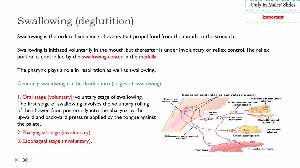
Swallowing (deglutition)
30
Swallowing is the ordered sequence of events that propel food from the mouth to the stomach.
Swallowing is initiated voluntarily in the mouth, but thereafter is under involuntary or reflex control. The reflex
portion is controlled by the swallowing center in the medulla.
The pharynx plays a role in respiration as well as swallowing.
Generally swallowing can be divided into (stages of swallowing):
1. Oral stage (voluntary): voluntary stage of swallowing.
The first stage of swallowing involves the voluntary rolling
of the chewed food posteriorly into the pharynx by the
upward and backward pressure applied by the tongue against
the palate.
2. Pharyngeal stage (involuntary).
3. Esophageal stage (involuntary).
Only in Males’ Slides
Important

Stages of swallowing (Deglutition)
31 2 chewed food at the moment of swallowing
Oral stage:
The first stage of swallowing initiated voluntarily when the
tongue forces a bolus2 of food (upward and backward pressure
against the palate) posteriorly toward the pharynx which
contains high density of somatosensory receptors. The
activation of these receptors initiates the involuntary
swallowing reflex in the medulla. From here on, swallowing
becomes entirely automatic and can not be stopped.
Pharyngeal stage:
Four stages:
1. Soft palate is pulled upward.
2. the epiglottis moves to cover opening of larynx.
3. the upper esophageal sphincter relaxes allowing food to
move from pharynx to esophagus.
4. peristalsis wave of contraction initiated in the pharynx
moves food from pharynx through the upper esophageal
sphincter.
Breathing is inhibited during the pharyngeal stage of
swallowing.
Only in Males’ Slides
Once the somatosensory receptors are activated, the involuntary pharyngeal stage begins.

Ingestion of food
32
Soft palate is pulled upward to close the
posterior nares which prevents the food from
entering the nasal cavities.
The palatopharyngeal folds on each side of the pharynx are
pulled medially to approximate each other. These folds form a sagittal slit through which food must pass into the posterior
pharynx.
• The vocal cords of the larynx are strongly approximated and the larynx is pulled upward and anteriorly by the neck muscles.
• These action and the ligaments that prevent the epiglottis from moving upward, cause the epiglottis to swing backward over the opening of the larynx. These effects prevent food from going into the nose and trachea. Destruction of the vocal cords or the muscle that approximate them can cause strangulation.
The upward movement of the larynx pulls up and enlarges the opening to the esophagus. The
upper esophageal sphincter (or the pharyngoesophageal sphincter)
relaxes and allows food to move freely from the posterior pharynx
into the upper esophagus.
Once the larynx is raised and the pharyngoesophageal
sphincter relaxes, the entire muscular wall of the pharynx
contracts (superior, middle, then inferior parts) propelling the food by peristalsis into the
esophagus.
Pharyngeal stage:
At the pharynx, the bolus of food stimulates epithelial swallowing receptor areas all around the pharynx opening and impulses from this area pass to
the brain stem (swallowing center) and accordingly initiate a series of autonomic pharyngeal muscle contractions as follows: (the time of process is
less that two seconds).
Only in Males’ Slides

Ingestion of food
33
Summary of pharyngeal stage of swallowing: The trachea is closed, the esophagus is opened,
and a fast peristaltic wave initiated by the nervous system of the pharynx forces the bolus of food into the upper
esophagus. (time of process is < 2 seconds).
Muscles of the pharynx are almost striated muscles so they are considered skeletal muscles rather than smooth
muscles. When they contract, the contraction that happens is not considered peristalsis.
Only in Males’ Slides
Only in Females’ Slides

Nervous initiation of the
pharyngeal stage of swallowing
34
Impulses transmitted from pharyngeal opening (greatest sensitivity at tonsillar pillars)
Sensory impulses from the mouth are received by the nucleus tractus solitarius (NTS) via the medulla oblongata through the trigeminal and glossopharyngeal nerves.
The most sensitive areas of the posterior mouth and pharynx for initiating the pharyngeal stage of swallowing are located in a ring around the pharyngeal opening including the tonsillarpillars.
The successive stages of swallowing are then automatically initiated by neuronal areas of the reticular substance (5th and 9th
CN) of the medulla and lower portion of the pons (collectively called the deglutition or swallowing center).
The motor impulses to the pharynx and upper esophagus are transmitted from the swallowing center by the 5th, 9th, 10th, and12th cranial nerves and few of the superior cervical nerves.
Effect of the pharyngeal stage of
swallowing on respiration
The entire pharyngeal stage of swallowing occurs in
less than 6 sec, during which time the swallowing
center inhibits the respiratory center in the medulla
which stops respiration during the swallowing cycle.
In summary, the pharyngeal stage of swallowing is a
reflex act initiated by the voluntary movement of
food into the back of the mouth which stimulates
involuntary pharyngeal sensory receptors to elicit
the swallowing reflex.
Only in Males’ Slides
Very Important
Activation of swallowing center leads to inhibition of respiratory center.

Stages of swallowing (Deglutition)
35 3 is a surgical procedure that involves removing part of the vagus nerve
Esophageal stage:
The esophagus is a conduit to move food rapidly from the pharynx to the stomach.
The esophageal stage is controlled:
1. partly by the swallowing reflex and
2. partly by the enteric nervous system (ENS).
When bolus of food passes through the upper esophageal sphincter, the swallowing reflex closes the sphincter so food cannot
reflux into the pharynx.
In case of vagotomy3, enteric nervous system takes over
Location of muscle Type of muscle Innervation
The musculature of pharyngeal wall
and upper 1/3 of esophagus
Striated muscle Vagus (10th cranial) &
glossopharyngeal nerves (9th cranial)
The musculature of lower two thirds
of esophagus
Smooth muscle Vagus (10th cranial) through
connections with esophageal
myenteric nervous system
Only in Males’ Slides
• The pharyngeal and esophageal stages are managed by swallowing reflex.
• In esophageal stage another reflex is initiated called the “receptive relaxation”. As the name suggests, it relaxes the lower esophageal sphincter.

Cont.
36
Esophageal stage:
It exhibits two types of peristaltic movements:
Primary peristalsis: It is simply a continuation of the peristaltic wave that begins in the pharynx and spreads into
the esophagus during the pharyngeal stage of swallowing. This wave passes from the pharynx to the stomach in
8-10 sec.
Secondary peristalsis: If this primary peristaltic wave fails to move the food to the stomach, then the distention in
the esophagus caused by the food will initiate secondary peristaltic wave which will continue until all the food is
emptied into the stomach (Second peristalsis occurs behind the area of occlusion).
Only in Males’ Slides

Summary of Stages of swallowing
37
Only in Females’ Slides

Ingestion of food
38
Summary of the whole process:
Only in Females’ Slides

Gastro-esophageal (lower esophageal) sphincter
39
The esophageal sphincter is formed by the esophageal circular muscle located in an area of ~ 3 cm upward of the junction with
the stomach.
This sphincter remains tonically constricted (protects the esophagus from the stomach acidic juices) until the peristaltic
swallowing wave passes down the esophagus and causes a “receptive relaxation” of the sphincter and the emptying of the
propelled food into the stomach.
Failure of the sphincter to relax will result in achalasia.
Only in Males’ Slides
• It is important to keep the gastro-esophageal (lower esophageal) sphincter contracted unless a bolus is moving down the esophagus.
• Three mechanisms for contraction:• Normal tonic contraction: found nearly in all sphincters of the GI
system.• The diaphragm particularly during inhalation; When you inhale
the diaphragm contracts and goes down, pushing the abdominal cavity and squeezing the last portion of the esophagus which prevents the reflux of stomach materials .
• Valve like mechanism: the last part of the esophagus gets inserted into the lumen of the stomach (not mouth-to-mouth attachment). When you increase the pressure of the stomach by inhalation, it will close the lower esophagus sphincter and prevent reflux.
Tonic contraction of LES is most important in preventing stomach acid from moving up to the esophagus.

Function of the lower esophageal sphincter
40
When the esophageal peristaltic waves reaches
the stomach
The stomach relaxes through inhibition of myenteric neurons
Which prepares the stomach to receive the
food that is propelled into the esophagus during
swallowing.
Function of the lower esophageal sphincter (gastro-esophageal sphincter):
1. Receptive relaxation of stomach:
2. Additional Prevention of Esophageal Reflux by Valve-like Closure of the Distal End of the Esophagus.
This is another protective mechanism (safety factor) that prevents reflux of gastric secretions into the lower portion
of the esophagus. This mechanism involves a short portion of the esophagus that extends slightly into the stomach
and that caves the esophagus inward in response to increased intra-abdominal pressure.
If this mechanism was absent the gastric secretions will cause damage to the epithelial layer of the esophagus.
Only in Males’ Slides
Receptive relaxation reflex is also called vagovagal reflex: Inhibitory motor neuron is activated releasing vasoactive intestinal peptides and nitric oxide allowing relaxation and easier passage of the bolus.
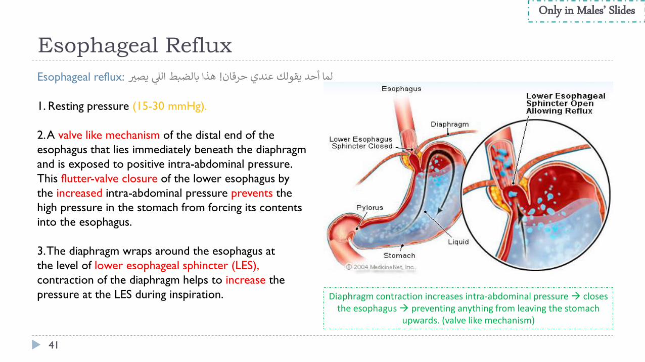
Esophageal Reflux
41
Esophageal reflux: هذا بالضبط اللي يصير ! لما أحد يقولك عندي حرقان
1. Resting pressure (15-30 mmHg).
2. A valve like mechanism of the distal end of the
esophagus that lies immediately beneath the diaphragm
and is exposed to positive intra-abdominal pressure.
This flutter-valve closure of the lower esophagus by
the increased intra-abdominal pressure prevents the
high pressure in the stomach from forcing its contents
into the esophagus.
3. The diaphragm wraps around the esophagus at
the level of lower esophageal sphincter (LES),
contraction of the diaphragm helps to increase the
pressure at the LES during inspiration.
Only in Males’ Slides
Diaphragm contraction increases intra-abdominal pressure closes the esophagus preventing anything from leaving the stomach
upwards. (valve like mechanism)
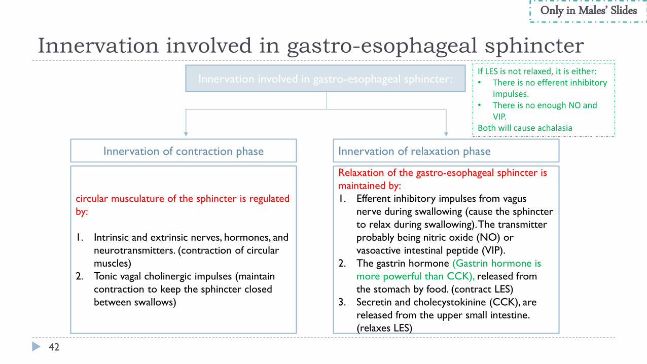
Innervation involved in gastro-esophageal sphincter
42
Innervation involved in gastro-esophageal sphincter:
Innervation of contraction phase Innervation of relaxation phase
circular musculature of the sphincter is regulated
by:
1. Intrinsic and extrinsic nerves, hormones, and
neurotransmitters. (contraction of circular
muscles)
2. Tonic vagal cholinergic impulses (maintain
contraction to keep the sphincter closed
between swallows)
Relaxation of the gastro-esophageal sphincter is
maintained by:
1. Efferent inhibitory impulses from vagus
nerve during swallowing (cause the sphincter
to relax during swallowing). The transmitter
probably being nitric oxide (NO) or
vasoactive intestinal peptide (VIP).
2. The gastrin hormone (Gastrin hormone is
more powerful than CCK), released from
the stomach by food. (contract LES)
3. Secretin and cholecystokinine (CCK), are
released from the upper small intestine.
(relaxes LES)
Only in Males’ Slides
If LES is not relaxed, it is either:• There is no efferent inhibitory
impulses. • There is no enough NO and
VIP.Both will cause achalasia

Achalasia
43 (VIP: Vasoactive intestinal peptide) (NO: Nitric oxide)
1. A condition due to high resting pressure at the LES that fails to relax during swallowing. As a result, food transmission from the esophagus into the stomach is prevented.
2. Physiological basis of this condition is either pathology of or absence of the myenteric plexus containing VIP & NO in the lower third of esophagus.
3. The musculature of the lower esophagus instead remains contracted and the myenteric plexus has lost the ability to transmit a signal to cause relaxation of the LES.
Only in Males’ Slides

Thank you!
The Physiology 436 Team:
44
.اعمل لترسم بسمة، اعمل لتمسح دمعة، اعمل و أنت تعلم أن هللا ال يضيع أجر من أحسن عمال
Females Members:
Rana Barasain
Reema AlShayie
Allulu Alsulayhim
Ghada Alskait
Reema Alshayie
Males Members:
Faisal Alfawaz
Fouad Bahgat
Team Leaders: Laila Mathkour
Mohammad Alayed
Contact us:
References: • 2017-2018 Dr. Hana Alzamel’s Lecture.
• 2017-2018 Dr. Mohammed Al Zoghaibi’s Lecture.
• Guyton and Hall Textbook of Medical Physiology (Thirteenth Edition.)
ي إليه ياي استودعتك ما حفظت وما قرأت وما فهمت، فرده لي وقت حاجتر
.من ال تضيع عنده الودائعاللهم ان