Webinar Presentation: New Generation Diagnostic Tools of KPIT and Use Cases

RESEARCH ARTICLE Open Access
The diagnostic value of metagenomicnext⁃generation sequencing in infectiousdiseasesHongxia Duan 1†, Xuan Li 1†, Aihong Mei 1†, Ping Li1, Yang Liu1, Xiaofeng Li2, Weiwei Li3, Changhui Wang1* andShuanshuan Xie1*
Abstract
Background: Although traditional diagnostic techniques of infection are mature and price favorable at present,most of them are time-consuming and with a low positivity. Metagenomic next⁃generation sequencing (mNGS)was studied widely because of identification and typing of all pathogens not rely on culture and retrieving all DNAwithout bias. Based on this background, we aim to detect the difference between mNGS and traditional culturemethod, and to explore the relationship between mNGS results and the severity, prognosis of infectious patients.
Methods: 109 adult patients were enrolled in our study in Shanghai Tenth People’s Hospital from October 2018 toDecember 2019. The diagnostic results, negative predictive values, positive predictive values, false positive rate, falsenegative rate, pathogen and sample types were analyzed by using both traditional culture and mNGS methods.Then, the samples and clinical information of 93 patients in the infected group (ID) were collected. According towhether mNGS detected pathogens, the patients in ID group were divided into the positive group of 67 cases andthe negative group of 26 cases. Peripheral blood leukocytes, C-reactive protein (CRP), procalcitonin (PCT) andneutrophil counts were measured, and the concentrations of IL-2, IL-4, IL-6, TNF-α, IL-17A, IL-10 and INF-γ in theserum were determined by ELISA. The correlation between the positive detection of pathogens by mNGS and theseverity of illness, hospitalization days, and mortality were analyzed.
(Continued on next page)
© The Author(s). 2021 Open Access This article is licensed under a Creative Commons Attribution 4.0 International License,which permits use, sharing, adaptation, distribution and reproduction in any medium or format, as long as you giveappropriate credit to the original author(s) and the source, provide a link to the Creative Commons licence, and indicate ifchanges were made. The images or other third party material in this article are included in the article's Creative Commonslicence, unless indicated otherwise in a credit line to the material. If material is not included in the article's Creative Commonslicence and your intended use is not permitted by statutory regulation or exceeds the permitted use, you will need to obtainpermission directly from the copyright holder. To view a copy of this licence, visit http://creativecommons.org/licenses/by/4.0/.The Creative Commons Public Domain Dedication waiver (http://creativecommons.org/publicdomain/zero/1.0/) applies to thedata made available in this article, unless otherwise stated in a credit line to the data.
* Correspondence: [email protected];[email protected]†Hongxia Duan, Xuan Li and Aihong Mei contributed equally to this work.1Department of Respiratory Medicine, Shanghai 10th People’s Hospital,Tongji University School of Medicine, #301, Mid Yanchang Rd, Shanghai200072, ChinaFull list of author information is available at the end of the article
Duan et al. BMC Infectious Diseases (2021) 21:62 https://doi.org/10.1186/s12879-020-05746-5

(Continued from previous page)
Results: 109 samples were assigned into infected group (ID, 92/109, 84.4%), non-infected group (NID, 16/109,14.7%), and unknown group (1/109, 0.9%). Blood was the most abundant type of samples with 37 cases, followedby bronchoalveolar lavage fluid in 36 cases, tissue, sputum, pleural effusion, cerebrospinal fluid (CSF), pus, bonemarrow and nasal swab. In the ID group, the majority of patients were diagnosed with lower respiratory systeminfections (73/109, 67%), followed by bloodstream infections, pleural effusion and central nervous system infections.The sensitivity of mNGS was significantly higher than that of culture method (67.4% vs 23.6%; P < 0.001), especiallyin sample types of bronchoalveolar lavage fluid (P = 0.002), blood (P < 0.001) and sputum (P = 0.037), while thespecificity of mNGS was not significantly different from culture method (68.8% vs 81.3%; P = 0.41). The number ofhospitals stays and 28-day-motality in the positive mNGS group were significantly higher than those in the negativegroup, and the difference was statistically significant (P < 0.05). Age was significant in multivariate logistic analysesof positive results of mNGS.
Conclusions: The study found that mNGS had a higher sensitivity than the traditional method, especially in blood,bronchoalveolar lavage fluid and sputum samples. And positive mNGS group had a higher hospital stay, 28-day-mortality, which means the positive of pathogen nucleic acid sequences detection may be a potential high-riskfactor for poor prognosis of adult patients and has significant clinical value. MNGS should be used more in earlypathogen diagnosis in the future.
Keywords: Next-generation sequencing, Sensitivity, Diagnostic, Infection, Survival
BackgroundInfectious diseases are a leading cause of morbidity andmortality worldwide and spread quickly. As the first-line ofpathogen detection, microbiology laboratory plays an im-portant role in infection control by means of microscopicexamination, culture, identification, drug sensitivity and soon [1]. However, the limitation of molecular diagnosis andgenotyping methods remain that pathogens are undetectedin up to 60% of cases [2–4]. Failure to identify pathogens intime may delay the precise treatment of antibiotics, leadingto unnecessary use of broad-spectrum antibiotics, inducingresistance, and increasing medical costs [5].With the completion of the human genome project in
the early twenty-first century and the rapid developmentof sequencing technology, high-throughput and low-costsecond-generation sequencing technology emerged [6].It had been used in whole genome sequencing, wholeexome sequencing, macro gene sequencing and so on,among which metagenomic next⁃generation sequencing(mNGS) was studied most widely. The advantage ofmNGS lies in the single run to obtain the sequence in-formation of microbial nucleic acid fragments, throughanalysis and comparison of which to detect all microbialspecies and sequence [7]. Besides, mNGS can be usedfor the identification and typing of all pathogens becausemNGS does not rely on culture and retrieve all DNAwithout bias [8]. Based on mNGS results, antimicrobialresistance, virulence, typing and other information canbe used for epidemic investigation. It lays a theoreticalfoundation for the investigation of infectious diseasesoutbreak in hospital. Therefore, this technology may playa huge role in infection prevention and medical micro-biology laboratory.
Thus, based on microbiome sequencing technology,we compared the sensitivity and specificity of mNGSmethod and traditional culture method to detect patho-gens, and discussed the influence of mNGS detection re-sults on the severity and prognosis of patients withinfection in our study.
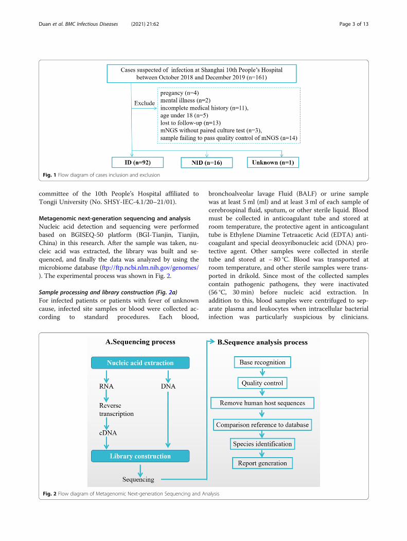
MethodsStudy patientsWe retrospectively reviewed 161 cases suspected ofacute or chronic infection from respiratory and criticalcare medicine department, geriatric department, emer-gency intensive care unit and emergency department atShanghai 10th People’s Hospital in Shanghai, China, be-tween October 2018 and December 2019. Excluding pa-tients with pregnancy, mental illness and under the ageof 18, 109 samples were included in our study and foranalysis and then they were categorized into 3 groups,infectious disease (ID) group, noninfectious disease(NID) group, and unknown group according to finaldiagnosis. Specimens were subjected to mNGS testing(BGI, Intertek, Biotecan, China) and regular clinicalmicrobiological assay in a pairwise manner and finaldiagnosis was determined by clinicians based on both ofthem and imaging, clinical feature of patients. Mean-while, clinical data of all enrolled patients, includingcomplete blood count, C-reactive protein (CRP), procal-citonin (PCT), neutrophil count, interleukin (IL)-2, IL-4,IL-6, Tumor Necrosis Factor-α (TNF-α), IL-17A, IL-10and Interferon-γ (INF-γ) were collected. The flow dia-gram of cases inclusion and exclusion was shown inFig. 1. This research had been approved by the ethics
Duan et al. BMC Infectious Diseases (2021) 21:62 Page 2 of 13
committee of the 10th People’s Hospital affiliated toTongji University (No. SHSY-IEC-4.1/20–21/01).
Metagenomic next-generation sequencing and analysisNucleic acid detection and sequencing were performedbased on BGISEQ-50 platform (BGI-Tianjin, Tianjin,China) in this research. After the sample was taken, nu-cleic acid was extracted, the library was built and se-quenced, and finally the data was analyzed by using themicrobiome database (ftp://ftp.ncbi.nlm.nih.gov/genomes/). The experimental process was shown in Fig. 2.
Sample processing and library construction (Fig. 2a)For infected patients or patients with fever of unknowncause, infected site samples or blood were collected ac-cording to standard procedures. Each blood,
bronchoalveolar lavage Fluid (BALF) or urine samplewas at least 5 ml (ml) and at least 3 ml of each sample ofcerebrospinal fluid, sputum, or other sterile liquid. Bloodmust be collected in anticoagulant tube and stored atroom temperature, the protective agent in anticoagulanttube is Ethylene Diamine Tetraacetic Acid (EDTA) anti-coagulant and special deoxyribonucleic acid (DNA) pro-tective agent. Other samples were collected in steriletube and stored at − 80 °C. Blood was transported atroom temperature, and other sterile samples were trans-ported in drikold. Since most of the collected samplescontain pathogenic pathogens, they were inactivated(56 °C, 30 min) before nucleic acid extraction. Inaddition to this, blood samples were centrifuged to sep-arate plasma and leukocytes when intracellular bacterialinfection was particularly suspicious by clinicians.
Fig. 1 Flow diagram of cases inclusion and exclusion
Fig. 2 Flow diagram of Metagenomic Next-generation Sequencing and Analysis
Duan et al. BMC Infectious Diseases (2021) 21:62 Page 3 of 13

Sputum samples were liquefied by using 0.1% dithiothre-itol (DTT) for 30 min at room temperature after inacti-vation [5]. After that, DNA were extracted by TIANampMicro DNA Kit (DP316, Tiangen Biotech) according tothe manufacturer’s recommendation. DNA libraries wereconstructed in steps of DNA fragmentation by enzymedigestion, DNA supplementation terminal, dA tail andsequencing common connector connection. The con-structed DNA library was used to obtain the sequencedata of DNA fragments by gene sequencing instrument,and the results were analyzed by biological informationsoftware. Each trial included internal, negative and posi-tive controls. Internal parameters is a specific moleculartag that is placed in the sample before nucleic acid ex-traction to track the entire process and to control thequality of DNA. The detection results of negative controlproducts should be no pathogens detected. If there arerelevant pathogens detected, it indicates that there maybe DNA pollution sources in the environment. Positivecontained specific microbic DNA.
Bioinformatic analysis (Fig. 2b)
Quality control A. Sequencing subtracted of humanhost sequences need to be above 90%; B. Reads of micro-bial detection sequences need to be longer than 50 bpand the effective sequencing data volume should not beless than 20M without removing the human genomecomponent.
Data filtering In order to obtain high quality sequencedata, the qualified data was further filtered by bioinfor-matics analysis to remove low quality sequences. FASTQformat was used for analysis. The initial pretreatmentsteps include low quality read filtering, low- complexityread filtering and adapter trimming. Host subtractionwas performed by mapping to host genome and/or tran-scriptome. The remaining unmapped reads are aligneddirectly with large reference databases, such as the Na-tional Center for Biotechnology Information (NCBI)GenBank database.
Sequences alignment The filtered sequences were com-pared with the reference sequences in the pathogendatabase, which covers bacteria, fungi, viruses, protozoaand other pathogenic microorganisms. According to thefinal results of pathogen comparison, all parameters ofdetected pathogens were calculated, including sequencenumber, relative abundance, genome coverage anddepth, etc.
Report generation The species listed in the report wereall the microorganisms detected in this test. They wereclassified by bacteria, viruses, fungi, parasites,
mycoplasma, chlamydia and rickettsia. They were rankedfrom high to low according to their reads and the rela-tive content of the former is higher. When the reportgoes to the clinic, whether the suspected pathogen de-tected is related to infection from the clinical dimensionwas judged, and the final diagnosis was determine bycombining the detection parameters.
Determination of cytokinesDetection of TNF-a, IL-2, IL-4, IL-6, IL-8, IL-10, IL-17Aand INF-r in serum was by solid phase, sandwich andchemiluminescence using the IMMULITE/IMMULIE1000 analyzer. The analyzer and chemiluminescence kitwere both from SIEMENS, Germany. The processedspecimens were sent to the analyzer for testing accord-ing to the manufacturer’s instructions, and the corre-sponding cytokine concentrations were recorded.
Cell classification and count detectionCells were classified using the automatic flow cytometer(Thermo Fisher SCIENTIFIC, American) and dividedinto total white blood cells, neutrophil count, CD4+ Tcell count, CD8+ T cell count, B cells, and NK, T cellcount.
Statistical analysisComparative analysis was conducted by Pearson χ2 testand t test. Data analysis was performed by using SPSS22.0 software. P values < 0.05 were considered signifi-cant, and all tests were 2-tailed. Logistic regression ana-lysis explored the risk factors associated with positivedetection of mNGS.
ResultsSample and patient characteristicsDemographic features of the patients were provided inTable 1. 87 males and 22 females participated in ourstudy, whose average age was 61 years old, averagelength of stay was 17.5 days and the case fatality ratewere 11.9%. Most (37/109, 33.9%) of our samples werefrom blood, 36 of 109 (33.0%) were from BALF, 12 of109 (11.0%) were from tissue and 9 (8.3%) of 109 werefrom sputum, followed by pleural fluid (7, 6.4%), CSF (4,3.7%), pus (2, 1.8%), bone marrow (1, 0.9%) and nasalswab (1, 0.9%) (Fig. 3a). In the study cohort, 92 (84.4%)patients diagnosed with confirmed pathogens by clini-cians were assigned to ID group. The remaining speci-mens were subdivided into the NID (16/109, 14.7%) andunknown (1/109, 0.9%) groups (Fig. 3b). There were nostatistical differences between ID group and NID groupin age, gender, length of stay and case fatality rate (p >0.05 in all). Most patients were diagnosed with respira-tory system infections (73/109, 67.0%), followed bybloodstream infections (10/109, 9.17%), pleural effusion
Duan et al. BMC Infectious Diseases (2021) 21:62 Page 4 of 13

(6/109, 5.50%) and central nervous system infections (6/109, 5.50%) as shown in Fig. 3c.
Diagnostic performance comparison of mNGS and cultureComparison of diagnostic performance for differentiating IDfrom NIDThe cases of mNGS and culture tests in this study wereillustrated in Fig. 4a. In the chi-square test of positiverate, there were statistical differences between mNGSand culture of all and of ID group, but no differences in
NID and unknown group for the limited amounts. 105samples were included for further study to compare thediagnostic efficiency for differentiating ID from NID.The positive predictive values and negative predictivevalues of diagnosing infectious disease by mNGS were92.3 and 27.5%, respectively. The positive likelihood ra-tio and negative likelihood ratio being 2.16 and 0.47.The results showed that mNGS increased the sensitivityrate (positive number in ID/ID number) by approxi-mately 44% compared with that of culture (67.4% vs
Table 1 Demographic characteristic of samples
Total ID NID Unknown P value between ID & NID
Samples amount, n (%) 109 (100%) 92 (84.40) 16 (14.68) 1 (0.92) /
Age, average years (range) 61.02 (25–95) 60.26 (25–95) 66 (40–90) 61(/) 0.43
Gender,male,n (%) 87 (79.82) 74 (80.43) 12 (75.00) 1 (100%) 0.62
Length of stay, average days (range) 17.53 (1–70) 16.88 (1–70) 20.87 (6–61) 22(/) 0.31
case fatality rate, % 11.93 13.04 6.25 0 0.39
Abbreviations: ID infectious disease, NID noninfectious disease
Fig. 3 Patients composition and samples types. a. In samples of this study, 33.9% were from blood which was the most, 33.0% from BALF, 11.0%from tissue and the others were from sputum (8.3%), pleural fluid (6.4%), CSF (3.7%), pus (1.8%), bone marrow (0.9%) and nasal swab (0.9%). b.Patients were subdivided into ID (92/109, 84.4%), NID (16/109, 14.7%) and unknown (1/109, 0.9%) groups according to their diagnosis byconventional technique. c. Infection sites of patients in ID group. Most were respiratory system infections (73/109, 67.0%) and followed bybloodstream infections (10/109, 9.17%), pleural effusion (6/109, 5.50%), central nervous system infections (6/109, 5.50%), cardiovascular systeminfection (2/109,1.83%), eye, ear, nose, throat, or mouth infection (2/109,1.83%), skin and soft tissue infection (1/109, 0.92%), multifocal infection (1/109, 0.92%), urinary system infection (1/109, 0.92%). Abbreviations: CSF, cerebrospinal fluid; BALF, bronchoalveolar lavage fluid
Duan et al. BMC Infectious Diseases (2021) 21:62 Page 5 of 13

23.6%; P < 0.001) and decreased the specificity rate(negative number in NID /NID number) by 12.5%compared with that of culture (68.8% vs 81.3%; P =0.41) (Fig. 4b).
Concordance between mNGS and culture for pathogendetectionIn this study, mNGS and culture were both positive in21 of 109 (19.3%) cases and were both negative in 25 of109 (22.9%) cases. There were 58 cases (53.2%) werepositive by mNGS only and 5 (4.6%) were positive onlyby culture. The 2 results in double-positive cases werecompletely matched (overlapped of all pathogens) in 3of 21 and totally mismatched (overlapped of no patho-gen) in 3 of 21 (Fig. 4c). The remaining 15 cases werefound to at least one but not all overlapped of pathogensin polymicrobial results, which defined as “partlymatched”.
“False positives” and “false negatives” of mNGSIn the ID group, three culturable pathogens were missedby mNGS. Among the three “mNGS false-negative” sam-ples, there were 2 culture results paradoxical with clin-ical diagnosis, the other 1 was completely unidentifiedby mNGS. At the same time, the possible reasons for the7 cases of “mNGS false-positive” in the NID group in-cluded potential concomitant infection with NIDs (3/7),overinterpretation (3/7) and unknown (1/7) (Table 2).
Comparison of mNGS and culture testing by pathogensand samplesComparison analysis at the pathogen-type levelKlebsiella (10/69) was the most commonly detectedpathogen among the 69 microbes isolated in mNGS andculture testing, followed by bacteria without MTB/NTM(9/69), Aspergillus (6/69), Pseudomonas (6/69) and EBV(6/69) (Fig. 5a). The percentage of mNGS-positive
Fig. 4 Diagnostic Performance Comparison of mNGS and Culture. a. Positive and negative cases in all, ID, NID and unknown group of mNGS andthe culture, respectively. There were statistical differences between mNGS and culture of all (P < 0.01) and of ID group (P < 0.01), but nodifferences in NID and unknown group for the limited amounts(P > 0.05). b. Contingency tables showed the sensitivity and specificity of mNGSwere 67.4 and 68.8%, while those of culture were 23.6 and 81.3%. mNGS increased the sensitivity in comparison with that of culture (P < 0.001)while there were no differences in specificity between them (P = 0.41). c. Pie chart demonstrated the positivity distribution of mNGS and culturefor all samples from 3 groups. 53.21% were positive by mNGS, 4.59% by culture, 19.27% by both and 22.94% were both negative. Abbreviations:NPV, negative predictive values; PPV, positive predictive values
Duan et al. BMC Infectious Diseases (2021) 21:62 Page 6 of 13

samples observed to have a higher yield rate than that of culture,but the differences were not significant (P>0.05) in terms ofKlebsiella, bacteria without MTB/NTM, EBV, CMV due to thesmall sample size. In Acinetobacter baumannii (n=2) and MTB(n=3), the number of mNGS-positive samples was equally withthat of culture-positive samples. While only mNGS indicatedpositive results in NTM (n=4), Anaerobes (n= 4), Saccharomy-ces cerevisiae (n= 2), Proteus (n= 1), Pneumocystis carinii (n=2), Abiotrophia (n= 1), Nocardia (n= 3), Staphylococcus aureus(n= 2), Enterococcu (n= 2) and Escherichia coli (n= 1).
Comparison analysis at the sample-type levelIn the types of BALF, tissue, blood and sputum samples,mNGS detection had significantly higher sensitivity than theculture method (P= 0.002 for BALF, P= 0.025 for tissue, P <0.001 for blood, P= 0.018 for sputum), and the overall sensi-tivity of mNGS in the sample types was significantly different(P= 0.03). In the types of pleural fluid, CSF, pus, bone mar-row and nasal swab, there were no significant differences insensitivity between two methods (P > 0.05). In addition, inthe culture method, the positive rate in BALF was higherthan that in the whole blood (P= 0.019), and there was nodifference in the overall sensitivity of the culture method inthe sample type, as shown in Fig. 5b.
Comparison of infection indexes in positive and negativegroup by mNGS in IDClassification and counting of leukocyte and lymphocyte inpositive and negative group by mNGSIn this study, complete blood count, CRP and PCT testswere examined on the day of examination of pathogenicmicroorganisms to determine the differences in the total
number of white blood cells, lymphocytes and neutrophilsbetween the positive group and the negative group bymNGS. The results showed (Table 3) that there were nostatistically differences in leukocyte and lymphocyte be-tween positive and negative groups by mNGS (P > 0.05).
Comparison of cytokine concentrations in positive andnegative group by mNGSIn order to explore the correlation between the statusof immune function in patients and the positive re-sults of pathogen examination, this study detectedand analyzed the peripheral blood (TNF-a, IL-2, IL-4,IL-6, IL-8, IL-10, IL-17A and INF-r) in infected pa-tients. The results indicated that the peripheral bloodconcentrations of IL-10 in the positive group washigher than that in the negative group, and the differ-ences were statistically significant (P = 0.044), whileother cytokine showed no difference between groupsas shown in Table 3.
Analysis of correlative factors for positive result ofpathogen extraction by mNGSIn order to further explore the related risk factors ofpositive mNGS test in infected patients, this studyused Logistic multivariate regression analysis toanalyze the patients’ information and whether thepathogen was detected in the patients. After the con-founding factors were removed, the variables thatwere significant for detection was age (P = 0.037, OR:1.076, 95% CI:1.005–1.152), which promoted the de-tection of pathogens (Table 4).
Table 2 “False Positives” and “False Negatives” of mNGS
Pathogens Detected Only by mNGS in the NID Group
SampleNo.
Specimensource
Diagnosis mNGS result Possible explanation
2 BALF Hematencephalon Acinetobacter baumannii, Klebsiella, Enterococcu Unknown
33 Blood Lymphoma Pseudomonas, CMV Potential cause oflymphoma
62 Blood Aplastic anemia Acinetobacter baumannii, Enterococcu Overinterpretation
67 Blood myelofibrosis Phycomyces blakesleeanus Overinterpretation
74 Pleural Fluid Pleural effusion Fusobacterium nucleatum, Streptococcus constellatus,Porphyromonas gingivalis
Potential cause ofinflammation
86 Blood Ulcerative Colitis Porphyromonas gingivalis, HSV Potential cause ofinflammation
88 Blood Lung cancer Saccharomyces cerevisiae Overinterpretation
Culturable Pathogens Missed by mNGS in the ID Group
Microbe Count Possible explanation
MTB 2 Positive Not Detected
Pseudomonas 1 Microbes “Weak”
Abbreviations: mNGS metagenomic next-generation sequencing, ID infectious disease, NID noninfectious disease, CMV metagenomic next-generationsequencing, HSV herpes simplex virus, MTB Mycobacterium tuberculosis
Duan et al. BMC Infectious Diseases (2021) 21:62 Page 7 of 13

Potential implications of clinical mNGS testPotential inappropriate antibiotic usage for patients withvirus isolatesThere were 4 viruses identified by mNGS from 23 pa-tients in this study, the majority of the identified viruseswere herpes simplex virus (n = 15), followed by Epstein-Barr virus/ herpes simplex virus (n = 5), Epstein-Barrvirus (n = 1), Hepatitis A virus (n = 1) and torque tenovirus (n = 1). Nearly 50% of patients were diagnosed witha hospital-acquired infection (12/23) and 17 of 23 pa-tients were given broad-spectrum antibiotics based onsymptoms, imaging. 10 of 23 patients were suspected ofinappropriate antibiotic usage, which means after broad-spectrum antibiotic treatment, patients’ symptoms didnot improve or even worsened and after identifying the
real pathogen through mNGS and adjusting the anti-biotic use based on that, patients’ condition improved. 7of 23 were considered immunocompromised hosts char-acterized by deficiency of the immune system or im-mune response caused by infectious factors, mycotoxins,drugs and nutritional deficiencies. (Table 5).
The influence of positive by mNGS on the hospital days andsurvival of patientsAs Table 6 showed, there were 67 samples in positivegroup with 57 males and 26 in negative group with 20males. There was no significant difference in mean agebetween the two groups (59.70 yrs. vs 60.50 yrs., P =0.84). Positive group had a longer hospital day (HOD,176.63 days vs 150.96 days, P = 0.047) and a higher 28-
Fig. 5 The overlap of positivity between mNGS and culture in pathogen and sample types. a. 19 pathogens detected in ID group with theircorresponding frequencies were showed in histograms. Klebsiella, bacteria without MTB/NTM, EBV, CMV, NTM, Anaerobes, Saccharomycescerevisiae, Proteus, Pneumocystis carinii, Abiotrophia, Nocardia, Staphylococcus aureus, Enterococcu and Escherichia coli demonstrated a trend ofhigher positivity rate in mNGS than that in culture with no statistical differences (P > 0.05). Acinetobacter baumannii and MTB were found equallyin two groups. b. The overall sensitivity of mNGS in the different sample types were significantly different (P = 0.03) while sample types did notaffect the sensitivity of pathogens in culture. Interestingly, especially in the types of BALF, blood and sputum samples, mNGS had significantlyhigher sensitivity than the culture (P = 0.002 for BALF, P < 0.001 for blood, P = 0.037 for sputum). Abbreviations: BALF, bronchoalveolar lavage fluid;CSF, cerebrospinal fluid; mNGS, metagenomic next-generation sequencing; HSV, herpes simplex virus; CMV, cytomegalovirus; EBV, Epstein-Barrvirus; MTB, Mycobacterium tuberculosis; NTM, nontuberculous mycobacteria; ns, no significant difference
Duan et al. BMC Infectious Diseases (2021) 21:62 Page 8 of 13

day mortality (9.0% vs 0%, P = 0.049) than those of nega-tive group, but there were no statistical differences in14-day mortality (4.5% vs 0%, P = 0.278) and 90-daymortality (13.4% vs 3.9%, P = 0.180) between groups.The average survival time of two groups were 176.64days and 150.96 days, respectively, but P value for t testbetween groups was 0.425, no statistical differences. Thesurvival curves of the two groups were shown in Fig. 6.At the meantime, we analyzed the relationship betweenpathogens read number and HOD, 14-day-mortality, 28-day-mortality and 90-day-mortality, which showed thatthe higher pathogens read number, the higher 90-day-mortality and the longer HOD (Table 7).
DiscussionThe traditional clinical model for diagnosing infectiousdiseases is for doctors to make a differential diagnosisand then conduct a series of tests to try to identify thepathogen [9–12]. Traditional diagnostic techniques ofmicrobiology laboratory ranges from smear microscopy,microorganisms’ culture, antigen antibody detection andPCR mainly. Whereas most traditional methods wereoften time-consuming and has a lower positive rate than
mNGS [2–4]. Although molecular diagnostic assays area quick way to diagnose the most common infections, al-most all conventional microbial trials in use today onlytarget a limited number of pathogens at a time or re-quire successful culture of microorganisms from clinicalsamples [13]. While mNGS analyze the entire micro-biome in patients’ samples [8] so it has been used to dis-cover novel viral pathogens and diagnose viral infectionsin people widely [14–16]. Therefore, we explored the ap-plication and differences between traditional culturemethod and mNGS in clinical infectious diseases inadults. BALF, blood, sputum, tissue, CSF, pleural fluid,pus, bone marrow or nasal swab from 109 patients sus-pected of infection were collected and specimens weresubjected to regular clinical microbiological assay andmNGS testing in a pairwise manner in our study. Wethen systematically compared the clinical features andtest results of mNGS and traditional culture.
Table 3 The counts of WBC, Cytokines and lymphocytes inpositive and negative groups by mNGS
Positive Negative P
Cytokines pg/ml
IL-2 100.35 + 68.21 1.31 + 0.94 0.511
IL-4 2.74 + 0.41 1.52 + 0.94 0.206
IL-6 70.8 + 18.27 68.96 + 33.18 0.964
TNF-α 2.48 + 0.42 2.26 + 1.32 0.842
IL-17a 13.77 + 2.35 10.45 + 8.01 0.592
IL-8 1154 + 0 – –
IL-10 26.14 + 7.75 8.29 + 3.33 0.044
IFN-γ 8.91 + 1.89 13.59 + 6.92 0.361
Cellular Immunity %
CD4/CD8 1.42 + 0.23 2.06 + 0.44 0.185
Th cell 35 + 3.36 43.83 + 5.75 0.201
Ts cell 68.06 + 3.07 66.67 + 3.64 0.18
NK cell 15.71 + 2.17 15.5 + 1.89 0.958
B cell 13.53 + 2.94 12.83 + 3.73 0.899
T cell 68.06 + 3.07 66.67 + 3.64 0.958
WBC ×109 8.32 + 0.52 7.36 + 0.48 0.283
Neu× 109 6.99 + 0.58 5.38 + 0.48 0.109
PCT ng/ml 0.34 + 0.17 3.42 + 3.32 0.112
CRP mg/l 87.63 + 8.32 63.61 + 13.47 0.129
Abbreviations: mNGS metagenomic next-generation sequencing, WBC whiteblood cells, IL- interleukin-, IFN-γ Interferon-γ, TNF-α Tumor Necrosis Factor-α,CD4 Cluster of Differentiation 4 receptors, CD8 Cluster of Differentiation 8receptors, Th helper T cell, Ts suppressor T cell, NK natural killer cell, Neuneutrophil, PCT procalcitonin, CRP C-reactive protein
Table 4 The analysis of the relevant factors of pathogens DNApositive in patients
Values B SE (B) Wald X2 P OR 95% CI
Age 0.073 0.035 4.367 0.037 1.076 1.005–1.152
Sex −0.545 1.157 0.222 0.637 0.58 0.06–5.601
Read Number −2.371 0.599 15.677 0.000 0.093 0.029–0.302
HOD −0.028 0.061 0.216 0.642 0.972 0.863–1.095
Survival Time −0.007 0.005 1.888 0.169 0.993 0.983–1.003
Cytokines pg/ml
IL-2 0.171 1.115 0.023 0.878 1.186 0.133–10.553
IL-4 −1.299 0.893 2.116 0.146 0.273 0.047–1.57
IL-6 −0.005 0.019 0.077 0.781 0.995 0.957–1.033
TNF-α −0.374 0.373 1.003 0.316 0.688 0.331–1.430
IL-17a 0.202 0.137 2.165 0.141 1.223 0.935–1.6
IL-10 −2.64 0.206 1.639 0.201 0.768 0.513–1.151
IFN-γ 0.09 0.071 1.606 0.205 1.095 0.952–1.259
Cellular Immunity %
CD4/CD8 −0.488 0.965 0.256 0.613 0.614 0.093–4.067
Th cell 0.318 0.296 1.151 0.283 1.374 0.769–2.454
Ts cell 0.244 0.317 0.589 0.443 1.276 0.685–2.377
NK cell −0.223 0.211 1.121 0.29 0.800 0.529–1.209
B cell −0.26- 0.245 1.172 0.279 0.767 0.227–1.475
T cell 0.5485 0.478 1.315 0.252 0.578 0.475–1.239
WBC ×109 −0.123 1.228 0.01 0.92 0.884 0.08–9.819
Neu×109 0.141 1.39 0.01 0.919 1.151 0.076–17.535
PCT ng/ml −0.681 1.514 0.202 0.653 0.506 0.026–9.844
CRP mg/l −0.004 0.015 0.073 0.788 0.996 0.968–1.025
Abbreviations: HOD hospital day, WBC white blood cells, IL- interleukin-, IFN-γInterferon-γ, TNF-α Tumor Necrosis Factor-α, CD4 Cluster of Differentiation 4receptors, CD8 Cluster of Differentiation 8 receptors, Th helper T cell, Tssuppressor T cell, NK natural killer cell, Neu neutrophil, PCT procalcitonin, CRPC-reactive protein
Duan et al. BMC Infectious Diseases (2021) 21:62 Page 9 of 13

The results suggested that there were no significantdifferences in age, gender, length of stay and fatality ratebetween two groups and mNGS had an advantage insensitivity rate compared with traditional culturemethod. A team of researchers also found that mNGSdetected potential pathogenic bacteria, which had advan-tages in speed and sensitivity compared with culture andpathology [17], Miao’s team [5] showed that mNGS hada sensitivity of 50.7% for the diagnosis of infectious dis-eases, higher than traditional culture (50.7% vs 35.2%).In particular, the diagnosis of MTB, virus, anaerobicbacteria, nocardia and fungi has obvious advantages. Theresults were similar to our results, which showed thatthe sensitivity of mNGS was 67.4%, significantly higherthan that of culture method (23.6%). High sensitivity ofmNGS may because pathogen DNA has a long survivaltime in plasma, the use of antibiotics has a small impacton mNGS results, while traditional cultures are greatlyaffected by the use of antibiotics. Because of the smallsample size, mNGS showed no statistical differencecompared with culture method in pathogen types al-though there was a trend of superiority in Klebsiella,bacteria without MTB/NTM, EBV, CMV, NTM,
Anaerobes, Saccharomyces cerevisiae, Proteus, Pneumo-cystis carinii, Abiotrophia, Nocardia, Staphylococcus aur-eus, Enterococcus and Escherichia coli. However, mNGSdetection had a significantly higher sensitivity than theculture method in BALF (P = 0.002), tissue (P = 0.025),blood (P < 0.001) and sputum (P = 0.018) samples.Based on the advantages shown by mNGS, we then in-
vestigated the influence of positive mNGS detection re-sults on the severity and prognosis of patients withinfection. By comparing the classification and countingof leukocyte, lymphocyte and cytokine concentrations inpositive and negative groups, we found that IL-10 con-centration in peripheral blood in the positive group washigher than that in the negative group and there wereno statistically differences in other cytokine concentra-tions, leukocyte and lymphocyte. According to the re-sults of correlative factors analysis for positive test ofmNGS, patients’ age may promote the detection of path-ogens. In the survival analysis, positive group had ahigher 28-day mortality (9.0% vs 0%, P = 0.049) than that
Table 5 Clinical Characteristics of Patients with Virus Isolates (n = 23)
Type of Virus HAI ImmunosuppressedPatients
Broad-spectrumAntibioticsa
Suspected Inappropriate Antibiotic Usage Treatment Responsive
Yes No Yes No Yes No Yes No Yes No
HSV (n = 15) 8 7 7 8 10 5 5 10 8 7
HAV(n = 1) 1 0 0 1 1 0 1 0 1 0
HSV/EBV (n = 5) 3 2 0 5 4 1 2 3 3 2
TTV(n = 1) 0 1 0 1 1 0 1 0 0 1
EBV (n = 1) 0 1 0 1 1 0 1 0 0 1
Total (N = 23) 12 11 7 16 17 6 10 13 12 11
Abbreviations: EBV Epstein-Barr virus, HAI hospital-acquired infection, HSV herpes simplex virus, HAV herpes simplex virus, TTV torqueteno virus
Table 6 The basic demographic and clinical characteristics ofinitial and outcome patient variables
Positive Negative P
Sex
Female 10 6 0.355
Male 57 20
Age 59.70 + 2.16 60.50 + 3.06 0.84
HOD 176.63 + 17.70 150.96 + 103.14 0.047
14 days of death 4.5% 0 0.278
28 days of death 9.0% 0 0.049
90 days of death 13.4% 3.9% 0.180
Read Number 5295.62 + 2507.26 16.67+ 4.79 0.039
Survival time 176.64 + 17.70 150.96+ 21.05 0.425
Abbreviation: HOD hospital day
Fig. 6 The survival curves of positive and negative group of mNGSin ID. The survival curves suggested that the overall survival ratedeclined faster in the positive group, however, there was nostatistically differences between the two groups
Duan et al. BMC Infectious Diseases (2021) 21:62 Page 10 of 13

of negative group, but there were no statistical differ-ences in average survival time. The pathogens read num-ber by mNGS was positive related to the HOD and 90-day-mortality of patients with infectious diseases. All ofthat indicated older people were more likely to havepositive results and positive results of mNGS detectionmay represent a worse outcome.Fortunately, mNGS has moved from scientific applica-
tion to clinical practice and is changing the way diseasediagnosed and treated [18–20]. In addition to what wementioned above, mNGS also has merits in many otheraspects. Firstly, mNGS does not need prior clinical infor-mation to detect infectious pathogens, and the resultscan be reported quickly and accurately, greatly shorten-ing the diagnosis time of infectious pathogens. Early andrapid reporting of the results by mNGS provides clinicalclues to the next step in diagnosis and treatment, espe-cially avoiding overuse of antibiotics for viral infections[21, 22]. Rapid results reported by mNGS also can pro-mote timely adjustment of treatment in clinical practice.As our data showed, almost one-half of patients withvirus infection were suspected of inappropriate antibioticusage. Secondly, mNGS was used in some rare infectiouspathogens. It detected Naegleria fowleri [23], brucellosis[24], cysticercosis, taenia bocinea [25], gondii [26] inCSF, Hepatic tuberculosis in blood [27] in previous re-ports. Thirdly, studies have shown that mNGS can beused not only for pathogen identification, but also formicrobiome characterization, parallel analyses of humanhost responses, drug resistance gene and virulence factordetection. All of these led to the rapid development ofmNGS in immunodeficiency difficult-to-diagnose cases andimmunocompromised patients [13]. Thirdly, antibiotic usagehad little influence on mNGS results due to the long survivaltime of pathogen DNA in plasma, but traditional cultureswere affected by antibiotic use [21, 22]. Higher sensitivity ofmNGS than culture in this study may because that mNGS isless affected by prior antibiotic usage. However, mNGS stillhas some limitations at present, such as human background,background bacteria contamination, no uniform standardsfor detailed experimental procedures [2, 28–31], inability todistinguish infection and colonization, standardization of
bioinformatics analysis process, and problem of report inter-pretation. The results must be interpreted in the context ofthe clinical situation. It’s worth noting that background mi-crobial contamination is a common problem faced bymNGS technology, which can be partially eliminatedthrough negative quality control, but it requires clinical fa-miliarity with common background bacteria and better inter-pretation results combined with clinical practice [24].In this study, we systematically compared mNGS and
traditional culture method in sensitivity, specificity,pathogen type and sample type. On this basis, we alsocompared and analyzed the differences between thepositive and negative groups of mNGS which was few atpresent. Patients of positive group found to have a trendof worse prognosis suggested need more attention clinic-ally. Small sample size was the biggest deficiency of ourstudy, so that there were many results indicated a certaintrend without reaching statistical significance unfortu-nately. Therefore, more patients need to be included inthe study in the future. Not randomized controled wasalso the limitation of study. As a retrospective study, thisstudy has some limitations like limited data and data ac-cumulation not controlled by the researcher. Besides,limit generalizability caused by single-center study, lackof a gold standard comparator for diagnostics, lack ofantibiotic usage detail and classification bias were alsothe limitations of this study.
ConclusionsIn summary, mNGS had a higher sensitivity than culture, es-pecially in the types of BALF, blood and sputum samples,and there was a trend of higher sensitivity of Klebsiella,CMV and EBV detection. The worse trend of outcome inpatients with positive mNGS results than negative groupprompted more clinical attention to patients with positivemNGS results is required. Therefore, based on what wefound above and other advantages of mNGS like quick re-sults, less affected by prior antibiotic exposure and so on, wesuggest that mNGS should be used more in early pathogendiagnosis in the future. Nonetheless, interpreting data ofmNGS will be a challenge for doctors in guiding clinicaltreatment of infectious diseases.
Table 7 The analysis between the pathogens read number and HOD,14, 28 and 90-day-mortality
Read Number
0 1–9 10–99 100–999 1000- F P
No 20 15 20 24 14
HOD 14.84 + 8.58 13.07 + 5.18 15.80 + 9.12 20.70 + 16.5 27.92 + 24.06 2.685 0.037
14-mortality 0 0 0 0.04 0.14 1.898 0.118
28-mortality 0 0.13 0.05 0.083 0.29 2.253 0.07
90-mortality 0.05 0.2 0.05 0.083 0.36 2.598 0.042
Survival Time 169.74 + 102.68 138.40 + 100.27 158.70 + 125.83 185.45 + 124.82 194.71 + 216.79 0.424 0.791
Abbreviation: HOD hospital day
Duan et al. BMC Infectious Diseases (2021) 21:62 Page 11 of 13

AbbreviationsmNGS: Metagenomic next-generation sequencing; BALF: Bronchoalveolarlavage fluid; CSF: Cerebrospinal fluid; Dx: Diagnosis; ID: Infectious disease;NID: Noninfectious disease; DNA: Deoxyribonucleic acid; EDTA: EthyleneDiamine Tetraacetic Acid; EBV: Epstein-Barr virus; HAI: Hospital-acquiredinfection; HboV: Human bocavirus; HSV-1: Herpes simplex virus 1;TTV: Torqueteno virus; HAV: Hepatitis A virus; HOD: Hospital day; IL-2: Interleukin-2; IL-4: Interleukin-4; IL-6: Interleukin-6; IL-10: Interleukin-10; IL-8: Interleukin-10; CD4: Cluster of Differentiation 4 receptors; CD8: Cluster ofDifferentiation 8 receptors; WBC: White blood cell; Th: Helper T; NK: Naturalkiller cell; PCT: Procalcitonin; CRP: C-reactive protein; IFN-γ: Interferon-γ; IL-17a: Interleukin-17a; TNF-α: Tumor Necrosis Factor-α; PPV: Positive predictivevalue; NPV: Negative predictive value; CMV: Cytomegalovirus;MTB: Mycobacterium tuberculosis; ns: No signicant difference;NTM: Nontuberculous mycobacteria; G + : Gram Positive; G-: Gram Negative
AcknowledgmentsWe would like to thank all the patients who donate their biological samples.
Authors’ contributionsConception and design: H.X.D, S.S.X, X. L, A.H.M. Acquisition, statisticalanalysis or interpretation of the data: P. L, Y. L, X.F.L, W.W.L. Drafting of themanuscript: C.H.W. All authors reviewed and approved the final version ofthe manuscript. All authors had read and approved the manuscript.
FundingThis work was supported by the National Natural Science Foundation ofChina (No. 81802262), the Fundamental Research Funds for the CentralUniversities (No. 22120180584), Shanghai Tenth Hospital’s Improvement Planfor NSFC (No. 04.03.17.032, 04.01.18.048, SYGZRPY2017014). Changhui Wangis the recipient of the foundations and was responsible for editingmanuscript in our study.
Availability of data and materialsAll data generated or analysed during this study are included in thispublished article. The data that support the findings of this study areavailable from the corresponding author upon reasonable request.
Ethics approval and consent to participateAll experiments performed in this study are in accordance with theapproved ethical standards by the ethics committee of the 10th People’sHospital affiliated to Tongji University (No. SHSY-IEC-4.1/20–21/01). Formalconsent is waived for this type of study.
Consent for publicationNot Applicable.
Competing interestsThe authors declare that they have no competing interests.
Author details1Department of Respiratory Medicine, Shanghai 10th People’s Hospital,Tongji University School of Medicine, #301, Mid Yanchang Rd, Shanghai200072, China. 2Department of Emergency, Shanghai 10th People’s Hospital,Tongji University School of Medicine, Shanghai 200072, China. 3Departmentof Geriatrics, Shanghai 10th People’s Hospital, Tongji University School ofMedicine, Shanghai 200072, China.
Received: 21 March 2020 Accepted: 27 December 2020
References1. Zhou K, Lokate M, Deurenberg RH, Tepper M, Arends JP, Raangs EG, Lo-Ten-
Foe J, Grundmann H, Rossen JW, Friedrich AW. Use of whole-genomesequencing to trace, control and characterize the regional expansion ofextended-spectrum beta-lactamase producing ST15 Klebsiella pneumoniae.Sci Rep. 2016;6:20840.
2. Schlaberg R, Chiu CY, Miller S, Procop GW, Weinstock G, ProfessionalPractice C, Committee on Laboratory Practices of the American Society forM, Microbiology Resource Committee of the College of American P.
Validation of metagenomic next-generation sequencing tests for universalpathogen detection. Arch Pathol Lab Med 2017; 141:776–786.
3. Ewig S, Torres A, Angeles Marcos M, Angrill J, Rano A, de Roux A, Mensa J,Martinez JA, de la Bellacasa JP, Bauer T. Factors associated with unknownaetiology in patients with community-acquired pneumonia. Eur Respir J.2002;20:1254–62.
4. van Gageldonk-Lafeber AB, Heijnen ML, Bartelds AI, Peters MF, van der PlasSM, Wilbrink B. A case-control study of acute respiratory tract infection ingeneral practice patients in the Netherlands. Clin Infect Dis. 2005;41:490–7.
5. Miao Q, Ma Y, Wang Q, Pan J, Zhang Y, Jin W, Yao Y, Su Y, Huang Y, WangM, Li B, Li H, Zhou C, Li C, Ye M, Xu X, Li Y, Hu B. Microbiological diagnosticperformance of metagenomic next-generation sequencing when applied toclinical Practice. Clin Infect Dis. 2018;67:S231–40.
6. Grumaz S, Stevens P, Grumaz C, Decker SO, Weigand MA, Hofer S, BrennerT, von Haeseler A, Sohn K. Next-generation sequencing diagnostics ofbacteremia in septic patients. Genome Med. 2016;8:73.
7. Lecuit M, Eloit M. The diagnosis of infectious diseases by whole genomenext generation sequencing: a new era is opening. Front Cell InfectMicrobiol. 2014;4:25.
8. Lefterova MI, Suarez CJ, Banaei N, Pinsky BA. Next-generation sequencingfor infectious disease diagnosis and management: a report of theAssociation for Molecular Pathology. J Mol Diagn. 2015;17:623–34.
9. Khare R, Espy MJ, Cebelinski E, Boxrud D, Sloan LM, Cunningham SA, PrittBS, Patel R, Binnicker MJ. Comparative evaluation of two commercialmultiplex panels for detection of gastrointestinal pathogens by use ofclinical stool specimens. J Clin Microbiol. 2014;52:3667–73.
10. Leber AL, Everhart K, Balada-Llasat JM, Cullison J, Daly J, Holt S, Lephart P,Salimnia H, Schreckenberger PC, DesJarlais S, Reed SL, Chapin KC, LeBlanc L,Johnson JK, Soliven NL, Carroll KC, Miller JA, Dien Bard J, Mestas J,Bankowski M, Enomoto T, Hemmert AC, Bourzac KM. Multicenter evaluationof BioFire FilmArray meningitis/encephalitis panel for detection of Bacteria,viruses, and yeast in cerebrospinal fluid specimens. J Clin Microbiol. 2016;54:2251–61.
11. Ruggiero P, McMillen T, Tang YW, Babady NE. Evaluation of the BioFireFilmArray respiratory panel and the GenMark eSensor respiratory viral panelon lower respiratory tract specimens. J Clin Microbiol. 2014;52:288–90.
12. Tang YW, Gonsalves S, Sun JY, Stiles J, Gilhuley KA, Mikhlina A, Dunbar SA,Babady NE, Zhang H. Clinical evaluation of the Luminex NxTAG respiratorypathogen panel. J Clin Microbiol. 2016;54:1912–4.
13. Chiu CY, Miller SA. Clinical metagenomics. Nat Rev Genet. 2019;20:341–55.14. Moustafa A, Xie C, Kirkness E, Biggs W, Wong E, Turpaz Y, Bloom K, Delwart
E, Nelson KE, Venter JC, Telenti A. The blood DNA virome in 8,000 humans.PLoS Pathog. 2017;13:e1006292.
15. Rascovan N, Duraisamy R, Desnues C. Metagenomics and the humanVirome in asymptomatic individuals. Annu Rev Microbiol. 2016;70:125–41.
16. Somasekar S, Lee D, Rule J, Naccache SN, Stone M, Busch MP, Sanders C,Lee WM, Chiu CY. Viral surveillance in serum samples from patients withacute liver failure by metagenomic next-generation sequencing. Clin InfectDis. 2017;65:1477–85.
17. Li H, Gao H, Meng H, Wang Q, Li S, Chen H, Li Y, Wang H. Detection ofpulmonary infectious pathogens from lung biopsy tissues by metagenomicnext-generation sequencing. Front Cell Infect Microbiol. 2018;8:205.
18. Houldcroft CJ, Beale MA, Breuer J. Clinical and biological insights from viralgenome sequencing. Nat Rev Microbiol. 2017;15:183–92.
19. Schlaberg R, Queen K, Simmon K, Tardif K, Stockmann C, Flygare S, KennedyB, Voelkerding K, Bramley A, Zhang J, Eilbeck K, Yandell M, Jain S, Pavia AT,Tong S, Ampofo K. Viral pathogen detection by Metagenomics and Pan-viral group polymerase chain reaction in children with pneumonia lackingidentifiable etiology. J Infect Dis. 2017;215:1407–15.
20. Wilson MR, Naccache SN, Samayoa E, Biagtan M, Bashir H, Yu G, Salamat SM,Somasekar S, Federman S, Miller S, Sokolic R, Garabedian E, Candotti F,Buckley RH, Reed KD, Meyer TL, Seroogy CM, Galloway R, Henderson SL,Gern JE, DeRisi JL, Chiu CY. Actionable diagnosis of neuroleptospirosis bynext-generation sequencing. N Engl J Med. 2014;370:2408–17.
21. Rhodes J, Hyder JA, Peruski LF, Fisher C, Jorakate P, Kaewpan A, Dejsirilert S,Thamthitiwat S, Olsen SJ, Dowell SF, Chantra S, Tanwisaid K, Maloney SA,Baggett HC. Antibiotic use in Thailand: quantifying impact on blood cultureyield and estimates of pneumococcal bacteremia incidence. Am J Trop MedHyg. 2010;83:301–6.
22. Gosiewski T, Ludwig-Galezowska AH, Huminska K, Sroka-Oleksiak A,Radkowski P, Salamon D, Wojciechowicz J, Kus-Slowinska M, Bulanda M,
Duan et al. BMC Infectious Diseases (2021) 21:62 Page 12 of 13

Wolkow PP. Comprehensive detection and identification of bacterial DNA inthe blood of patients with sepsis and healthy volunteers using next-generation sequencing method - the observation of DNAemia. Eur J ClinMicrobiol Infect Dis. 2017;36:329–36.
23. Guo LY, Feng WY, Guo X, Liu B, Liu G, Dong J. The advantages of next-generation sequencing technology in the detection of different sources ofabscess. J Inf Secur. 2019;78:75–86.
24. Fan S, Ren H, Wei Y, Mao C, Ma Z, Zhang L, Wang L, Ge Y, Li T, Cui L, Wu H,Guan H. Next-generation sequencing of the cerebrospinal fluid in thediagnosis of neurobrucellosis. Int J Infect Dis. 2018;67:20–4.
25. Hu Z, Weng X, Xu C, Lin Y, Cheng C, Wei H, Chen W. Metagenomic next-generation sequencing as a diagnostic tool for toxoplasmic encephalitis.Ann Clin Microbiol Antimicrob. 2018;17:45.
26. Ai JW, Li Y, Cheng Q, Cui P, Wu HL, Xu B, Zhang WH. Diagnosis of localhepatic tuberculosis through next-generation sequencing: smarter, fasterand better. Clin Res Hepatol Gastroenterol. 2018;42:178–81.
27. Du B, Tao Y, Ma J, Weng X, Gong Y, Lin Y, Shen N, Mo X, Cao Q.Identification of sparganosis based on next-generation sequencing. InfectGenet Evol. 2018;66:256–61.
28. Blauwkamp TA, Thair S, Rosen MJ, Blair L, Lindner MS, Vilfan ID, Kawli T,Christians FC, Venkatasubrahmanyam S, Wall GD, Cheung A, Rogers ZN,Meshulam-Simon G, Huijse L, Balakrishnan S, Quinn JV, Hollemon D, HongDK, Vaughn ML, Kertesz M, Bercovici S, Wilber JC, Yang S. Analytical andclinical validation of a microbial cell-free DNA sequencing test for infectiousdisease. Nat Microbiol. 2019;4:663–74.
29. Deurenberg RH, Bathoorn E, Chlebowicz MA, Couto N, Ferdous M, Garcia-Cobos S, Kooistra-Smid AM, Raangs EC, Rosema S, Veloo AC, Zhou K,Friedrich AW, Rossen JW. Application of next generation sequencing inclinical microbiology and infection prevention. J Biotechnol. 2017;243:16–24.
30. Gargis AS, Kalman L, Lubin IM. Assuring the quality of next-generationsequencing in clinical microbiology and public health laboratories. J ClinMicrobiol. 2016;54:2857–65.
31. Miller S, Naccache SN, Samayoa E, Messacar K, Arevalo S, Federman S, StrykeD, Pham E, Fung B, Bolosky WJ, Ingebrigtsen D, Lorizio W, Paff SM, Leake JA,Pesano R, DeBiasi R, Dominguez S, Chiu CY. Laboratory validation of aclinical metagenomic sequencing assay for pathogen detection incerebrospinal fluid. Genome Res. 2019;29:831–42.
Publisher’s NoteSpringer Nature remains neutral with regard to jurisdictional claims inpublished maps and institutional affiliations.
Duan et al. BMC Infectious Diseases (2021) 21:62 Page 13 of 13