The Diagnostic Value of Color Doppler Ultrasound in...
6
International Journal of Medical Imaging 2018; 6(2): 12-17 http://www.sciencepublishinggroup.com/j/ijmi doi: 10.11648/j.ijmi.20180602.11 ISSN: 2330-8303 (Print); ISSN: 2330-832X (Online) The Diagnostic Value of Color Doppler Ultrasound in Ureteral Calculi Fan Weibo Department of Ultrasound, Jingzhou Central Hospital, The Second Clinical Medical College, Yangtze University, Jingzhou, China Email address: To cite this article: Fan Weibo. The Diagnostic Value of Color Doppler Ultrasound in Ureteral Calculi. International Journal of Medical Imaging. Vol. 6, No. 2, 2018, pp. 12-17. doi: 10.11648/j.ijmi.20180602.11 Received: July 21, 2018; Accepted: August 1, 2018; Published: August 30, 2018 Abstract: Ureteral calculi are one of the most common acute abdomen, most of which are from renal calculi. Color Doppler ultrasound plays a very important role in the diagnosis of ureteral calculi, and has the advantages of simplicity, economy, non-invasive, no radiation, and repeatable examination. The purpose of this study is to investigate the clinical value of color Doppler ultrasound in the diagnosis of ureteral calculi. To achieve this goal, a retrospective analysis was performed to analyze the clinical symptoms, ultrasonographic appearances and results of 112 patients with ureteral calculi who were treated in author’s hospital from March 2016 to March 2018. It showed that there were 5 cases of bilateral ureteral calculi, 59 cases of left ureteral calculi, 48 cases of right ureteral calculi, 42 cases of upper ureteral calculi, 11 cases of middle ureteral calculi and 59 cases of lower ureteral calculi in the results of the 112 patients with ureteral calculi. As ultrasound examination can show the location, number and size of the calculi, which can help clinical treatment and can be used as the first choice for ureteral calculi. Therefore, it is concluded that ultrasound diagnosis of ureteral calculi has the advantages of painless, non-invasive, convenient and easy to operate repeatedly, and can be used as the first choice for checking ureteral calculi. Keywords: Color Doppler Ultrasound, Ureteral Calculi, Diagnostic Value 1. Introduction In recent years, with the continuous development of ultrasound equipment and the improvement of the diagnostic level of technicians, the detection rate of ureteral calculi by ultrasound has been relatively improved. At the same time, CT, KUB and IVU are not suitable for repeated inspections because of the high price and radiation. After all, ultrasound is non-invasive, non-radiative, and can be repeatedly tested. The advantage of ultrasonography is that it can clearly show the X-ray negative calculi in the ureter, which compensates for the deficiency of X-ray examination to varying degrees. In the course of ultrasound imaging, the degree of hydro-nephrosis due to ureteral obstruction can be known, so that renal function can be estimated generally, and other diseases of the urinary system coexisting with ureteral calculi can also be found at the same time. Despite this, during the ultrasound imaging process, it is easy to cause missed diagnosis of ureteral calculi due to the physical characteristics of ultrasound and other reasons. The reasons are as follows: 1. The patient's obesity and the interference of intestinal contents can make the sonogram blurred, leading to misdiagnosis. The patients’ huge body and old age may result in poorer penetrating ultrasound and an unclear strong echo structure of the calculi. The patients’ poor intestinal condition may influence the penetration of the ultrasound in the ureteral area, which can be solved by allowing the patient to be re-examined after bowel preparation. 2. Due to the small ureteral calculi or short-term obstruction, there is no obvious accumulation of water in the renal pelvis and ureter, ultrasound is not found and easily leads to misdiagnosis. 3. The patients often have an acute onset, poor bladder-filling, and difficulty in cooperating with pain, which is also one of the reasons for misdiagnosis. Therefore, for patients with high suspicion of ureteral calculi and undetectable ultrasound examinations, abdominal plain films, CT or retrograde pyelography can be performed to avoid misdiagnosis of ureteral calculi [1]. Ureteral calculi are one of the most common acute abdomen, most of which are from renal calculi. The stimulation to the ureteral wall from calculi can cause local visceral mucosal
-
Upload
vuongnguyet -
Category
Documents
-
view
214 -
download
0
Transcript of The Diagnostic Value of Color Doppler Ultrasound in...
International Journal of Medical Imaging 2018; 6(2): 12-17
http://www.sciencepublishinggroup.com/j/ijmi
doi: 10.11648/j.ijmi.20180602.11
ISSN: 2330-8303 (Print); ISSN: 2330-832X (Online)
The Diagnostic Value of Color Doppler Ultrasound in Ureteral Calculi
Fan Weibo
Department of Ultrasound, Jingzhou Central Hospital, The Second Clinical Medical College, Yangtze University, Jingzhou, China
Email address:
To cite this article: Fan Weibo. The Diagnostic Value of Color Doppler Ultrasound in Ureteral Calculi. International Journal of Medical Imaging.
Vol. 6, No. 2, 2018, pp. 12-17. doi: 10.11648/j.ijmi.20180602.11
Received: July 21, 2018; Accepted: August 1, 2018; Published: August 30, 2018
Abstract: Ureteral calculi are one of the most common acute abdomen, most of which are from renal calculi. Color Doppler
ultrasound plays a very important role in the diagnosis of ureteral calculi, and has the advantages of simplicity, economy,
non-invasive, no radiation, and repeatable examination. The purpose of this study is to investigate the clinical value of color
Doppler ultrasound in the diagnosis of ureteral calculi. To achieve this goal, a retrospective analysis was performed to analyze the
clinical symptoms, ultrasonographic appearances and results of 112 patients with ureteral calculi who were treated in author’s
hospital from March 2016 to March 2018. It showed that there were 5 cases of bilateral ureteral calculi, 59 cases of left ureteral
calculi, 48 cases of right ureteral calculi, 42 cases of upper ureteral calculi, 11 cases of middle ureteral calculi and 59 cases of
lower ureteral calculi in the results of the 112 patients with ureteral calculi. As ultrasound examination can show the location,
number and size of the calculi, which can help clinical treatment and can be used as the first choice for ureteral calculi. Therefore,
it is concluded that ultrasound diagnosis of ureteral calculi has the advantages of painless, non-invasive, convenient and easy to
operate repeatedly, and can be used as the first choice for checking ureteral calculi.
Keywords: Color Doppler Ultrasound, Ureteral Calculi, Diagnostic Value
1. Introduction
In recent years, with the continuous development of
ultrasound equipment and the improvement of the diagnostic
level of technicians, the detection rate of ureteral calculi by
ultrasound has been relatively improved. At the same time, CT,
KUB and IVU are not suitable for repeated inspections
because of the high price and radiation. After all, ultrasound is
non-invasive, non-radiative, and can be repeatedly tested. The
advantage of ultrasonography is that it can clearly show the
X-ray negative calculi in the ureter, which compensates for the
deficiency of X-ray examination to varying degrees. In the
course of ultrasound imaging, the degree of hydro-nephrosis
due to ureteral obstruction can be known, so that renal
function can be estimated generally, and other diseases of the
urinary system coexisting with ureteral calculi can also be
found at the same time.
Despite this, during the ultrasound imaging process, it is
easy to cause missed diagnosis of ureteral calculi due to the
physical characteristics of ultrasound and other reasons. The
reasons are as follows: 1. The patient's obesity and the
interference of intestinal contents can make the sonogram
blurred, leading to misdiagnosis. The patients’ huge body and
old age may result in poorer penetrating ultrasound and an
unclear strong echo structure of the calculi. The patients’ poor
intestinal condition may influence the penetration of the
ultrasound in the ureteral area, which can be solved by
allowing the patient to be re-examined after bowel preparation.
2. Due to the small ureteral calculi or short-term obstruction,
there is no obvious accumulation of water in the renal pelvis
and ureter, ultrasound is not found and easily leads to
misdiagnosis. 3. The patients often have an acute onset, poor
bladder-filling, and difficulty in cooperating with pain, which
is also one of the reasons for misdiagnosis. Therefore, for
patients with high suspicion of ureteral calculi and
undetectable ultrasound examinations, abdominal plain films,
CT or retrograde pyelography can be performed to avoid
misdiagnosis of ureteral calculi [1].
Ureteral calculi are one of the most common acute abdomen,
most of which are from renal calculi. The stimulation to the
ureteral wall from calculi can cause local visceral mucosal
13 Fan Weibo: The Diagnostic Value of Color Doppler Ultrasound in Ureteral Calculi
damage, edema and concurrent inflammation, causing ureteral
obstruction and spasmodic contraction, leading to dull or
paroxysmal abdominal pain, accompanied with gross or
microscopic hematuria. Ureteral calculi can cause urinary
tract obstruction and different degrees of hydrocele in the
affected kidney. In severe cases, the renal cortex becomes
thinner, its function is lost or even get uremia. The clinical
data of 112 patients who were treated in author’s hospital from
March 2016 to March 2018 were analyzed and reported as
follows.
2. Materials and Methods
2.1. Clinical Data
This clinical study selected 112 patients from March 2016
to March 2018 in author’s hospital, which include 70 males
and 42 females aged 16-72 years, mean age (38.2). ±11.3)
years old. Those patients are accompanied by sudden,
intermittent lumbar pain, radiating along the ipsilateral ureter
to the lower abdomen, perineum, external genitalia and inner
thighs, and often accompanied by nausea, vomiting,
microscopic hematuria or even gross hematuria. They were
confirmed by surgery, extracorporeal shock wave lithotripsy
and self-extracting calculi.
2.2. Instruments and Methods
The PHILIPS IU22 ultrasound diagnostic instrument is
used, and the 3.5-5MHz convex array probe is routinely used.
Patients with upper ureteral calculi were placed in the lateral
or prone position, and the probe was placed in the lateral
lumbar region with the renal hilum or renal pelvis as the sign
showing the ureteropelvic junction. Patients with the second
stenosis calculi were placed in the supine position. The probe
was obliquely cut in the lower abdomen and the iliac artery
was found by compression. The dilated ureter was found in
front of the iliac artery, and the probe was used to display the
long axis of the ureter. Patients with lower ureteral calculi
were placed in supine position, whose bladder was moderately
filled and the probe was pressurized. Then the lower ureteral
opening was scanned in the trigone of the bladder, and the
scanning was performed upward. By following a certain
examination sequence, combined with a variety of
examination methods, multi-position and multi-angle scan,
and abdominal ultrasound can be done to patients with ureteral
calculi. It can also improve the detection rate of ureteral
urinary calculi by some scanning methods like moderate
bladder-filling, diuretic pressurization and adding
high-frequency probes [2].
3. Results
In this group of 112 patients, 36 cases of upper ureteral
calculi, 5 cases of middle calculi, and 45 cases of lower
ureteral calculi were detected by multi-position and
multi-angle scanning of abdominal ultrasound. A total of 86
cases were detected, and the detection rate was 76.8%. Seven
cases of ureteral calculi were detected by diuretic
pressurization method, and 15 cases of pelvic and
intra-ureteral calculi were detected by moderate
bladder-filling method. There were 108 cases of ureteral
calculi were detected by multiple checking methods in this
group, and the detection rate was 96.4%. There were 4 cases
of misdiagnosis couldn’t be detected because of the patient's
obesity and flatulence interference. The diagnosis only
indicated the separation of renal sinus echo, but the upper limit
of upper ureteral diameter was normal. There were 101
patients (90.2%) who had different degrees of hydronephrosis
in this group of patients with ureteral calculi. Eleven patients
had normal renal function and had no obvious separation in
the collecting system. The diameter of the calculi is 4-14mm,
which is more commonly seen in 5-8mm.
4. Discussion
Starting from the renal pelvis to the bladder triangle, the
ureter is about 20-34cm long that located in the posterior
peritoneum. There are three physiological stenosis parts in the
ureter, which are separately located at the junction of the renal
pelvis and ureter, the site that ureter spans the iliac vessels and
the ureteral bladder wall. Calculi can often stay in these
stenotic sites. Most ureteral calculi are derived from renal
calculi, and primary calculi are rare. The ureteral calculi can
cause urinary tract obstruction, and lead to different degrees of
hydrocele in the affected renal. The higher the position of
ureteral calculi, the more severe the degree of obstruction and
hydro-nephrosis. When the sonogram shows the
hydro-nephrosis, checking along with the dilated ureter, it can
be seen that the dilated ureter is suddenly interrupted, and the
strong echo and the wall boundary is clearly defined, followed
by an acoustic shadow. Because of the different size, shape,
location and composition of ureteral calculi, their sonograms
are also displaying differently. For example, the uric acid
calculi has a loose texture and rough surface, which presents a
round or elliptical high echogenic area and the acoustic
shadow behind it is weak or not obvious. The calcium oxalate
calculi, possessing hard texture and smooth surface, the
sonogram of which can only show that its surface profile
presents like an arc-shaped high echogenic area and there is an
acoustic shadow at the rear. The small stone with a rough
surface has a strong echo of point, and there is no obvious
acoustic shadow.
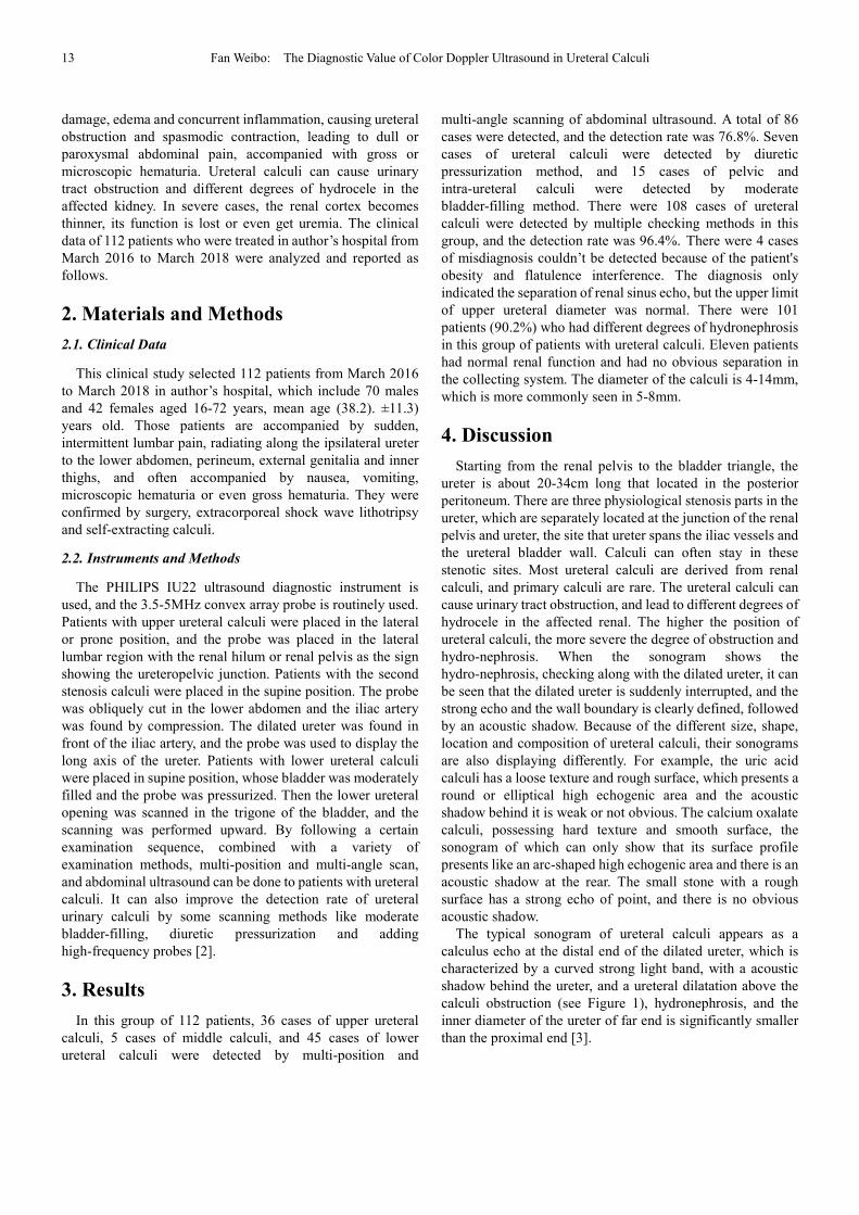
The typical sonogram of ureteral calculi appears as a
calculus echo at the distal end of the dilated ureter, which is
characterized by a curved strong light band, with a acoustic
shadow behind the ureter, and a ureteral dilatation above the
calculi obstruction (see Figure 1), hydronephrosis, and the
inner diameter of the ureter of far end is significantly smaller
than the proximal end [3].
International Journal of Medical Imaging 2018; 6(2): 12-17 14
Figure 1. Upper ureteral calculi on the right side.
To detect ureteral calculi quickly and accurately, you must
be familiar with the anatomy and characteristics of the ureter,
and use reasonable timing and examination sequence to save
time and diagnose quickly. The ureteral calculi are best
examined during an acute attack of renal colic, because at this
time the ureter is mostly accompanied by dilatation, and it is
easier to find the obstruction site when the bladder is not full.
According to the literature cited [4]: the inner segment of
the bladder wall is the narrowest part of the ureter. With its
unique dynamic characteristics, 70% of the ureteral stones are
located at this site [5]. During the examination, the inner
segment of the bladder wall is first found to achieve a rapid
diagnosis. If no stone echoes is found in the inner segment of
the bladder wall, then the hydronephrosis should be checked.
If there is, a hydroureterosis should be confirmed from the
hydronephrosis to the pelvic ureter junction, then the
multi-faceted scan of the abdomen, the back and the lateral
waist will allow for the examination of most of the calculi. If
no stones are found in the first and the third stenosis, then the
second stenosis should be scanned, during which the supine
position is usually taken. The common iliac artery is first
found, and the long axis of the ureter is found in front of the
common iliac artery. Scanning below and following the
hydroureterosis can assure the detection of calculi. The calculi
can also be found by firstly being examined by the bladder,
which shows the opening of bilateral ureters in the bladder
wall, and then scan up.
During the examination of ureteral calculi, if the
sonographer can master a variety of ultrasound diagnostic
techniques for ureteral calculi, the display rate of ureteral
calculi can be more effectively improved: 1. The locating
scan can be done based on the most painful location of the
patient, which can reduce the blind scanning time, especially
for patients with no hydronephrosis and no obvious ureteral
dilatation, and it is not easy to track each segment of the
ureter. 2. For some patients who has no obvious pain
symptoms, let them to fill the bladder moderately and
increase the degree of dilatation of the ureter as well as
appropriately reduce the gain, pressurization, multi-position
and multi-section, trying to make the sound beam
perpendicular to the ureter that makes it easy to find smaller
stones. 3. It has been reported that intracavity ultrasound has
obvious advantages in the examination of lower ureteral
calculi, especially for patients with acute abdomen and less
bladder filling [6].
Because of the obvious bladder irritation, not easy to urinate,
small calculi and ureter without dilatation, obesity, intestinal
contents and gas interference, etc., patients who have lower
ureteral calculi can affect the diagnosis of lower ureteral
calculi by abdominal ultrasound, while intracavity ultrasound
requires less filling of the bladder, and the diagnosis of calculi
is more sensitive than the abdomen. However, patients with
renal calculi often appear in the emergency department at
night, so it is not convenient to carry out intracavity ultrasound.
Therefore, intracavity ultrasound has not been used in author’s
hospital to diagnose ureteral calculi. 4. Ultrasonic diagnosis of
ureteral calculi with diuretic compression method is
convenient and quick to check. The patient has no noticable
discomfort and adverse reactions and is easy to accept. Check
again by using the diuretic abdominal compression method
(use the sandbag to compress the second stenosis of the ureter
closest to the abdominal wall), cleaning the enema, using the
diuretic and filling the bladder, forming a good urine-stone
acoustic interface, which helps to improve the calculi
detection rate. 5. On the basis of two-dimensional ultrasound
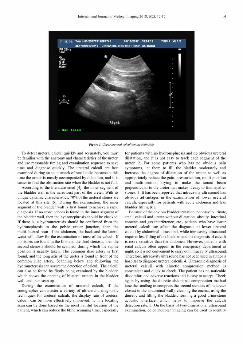
examination, color Doppler imaging can be used to identify
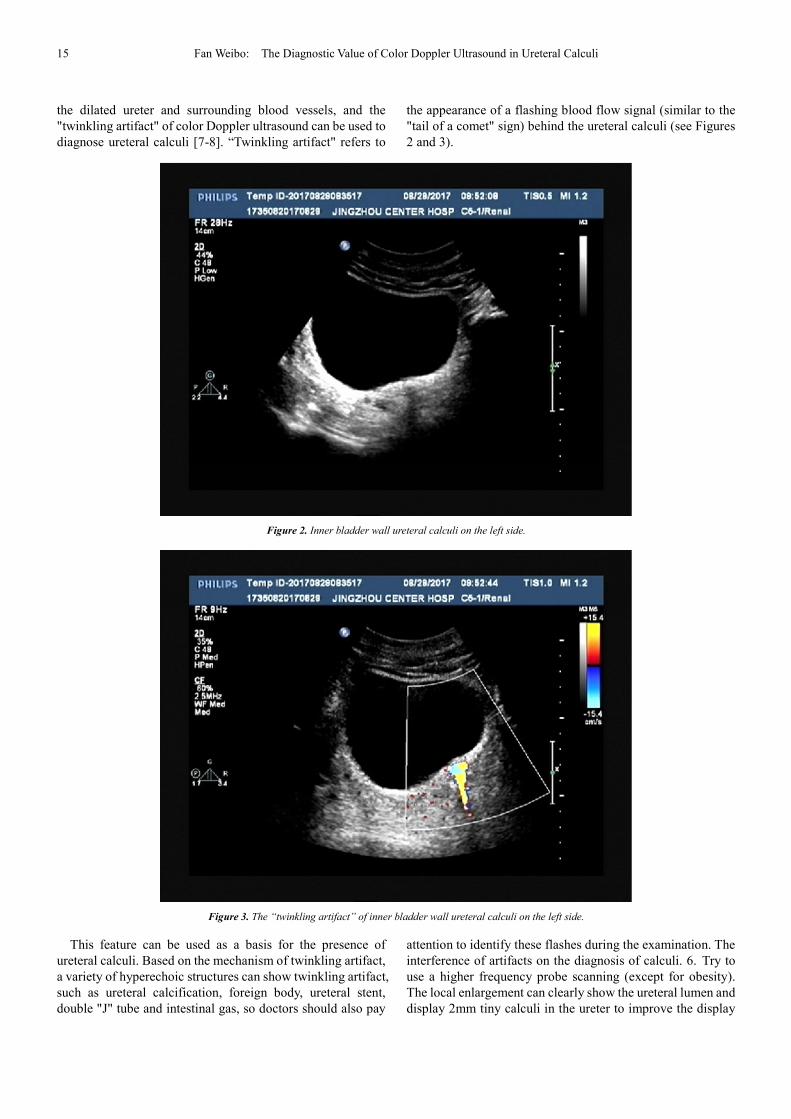
15 Fan Weibo: The Diagnostic Value of Color Doppler Ultrasound in Ureteral Calculi
the dilated ureter and surrounding blood vessels, and the
"twinkling artifact" of color Doppler ultrasound can be used to
diagnose ureteral calculi [7-8]. “Twinkling artifact" refers to
the appearance of a flashing blood flow signal (similar to the
"tail of a comet" sign) behind the ureteral calculi (see Figures
2 and 3).
Figure 2. Inner bladder wall ureteral calculi on the left side.
Figure 3. The “twinkling artifact” of inner bladder wall ureteral calculi on the left side.
This feature can be used as a basis for the presence of
ureteral calculi. Based on the mechanism of twinkling artifact,
a variety of hyperechoic structures can show twinkling artifact,
such as ureteral calcification, foreign body, ureteral stent,
double "J" tube and intestinal gas, so doctors should also pay
attention to identify these flashes during the examination. The
interference of artifacts on the diagnosis of calculi. 6. Try to
use a higher frequency probe scanning (except for obesity).
The local enlargement can clearly show the ureteral lumen and
display 2mm tiny calculi in the ureter to improve the display
International Journal of Medical Imaging 2018; 6(2): 12-17 16
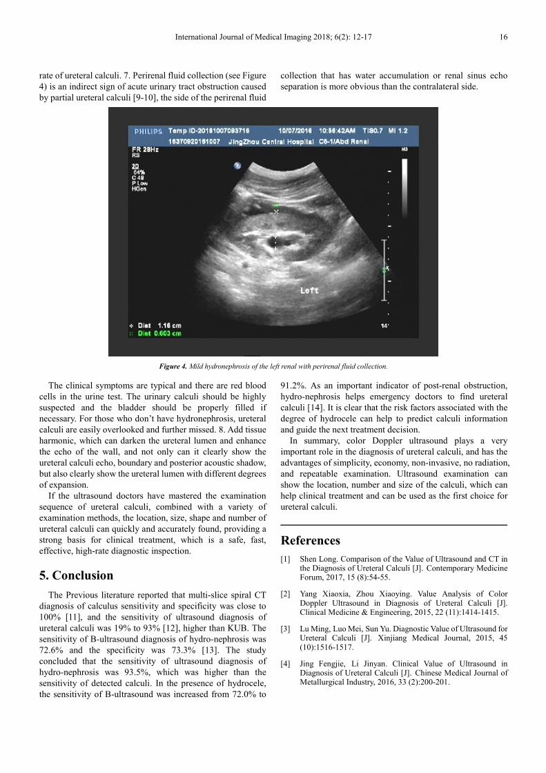
rate of ureteral calculi. 7. Perirenal fluid collection (see Figure
4) is an indirect sign of acute urinary tract obstruction caused
by partial ureteral calculi [9-10], the side of the perirenal fluid
collection that has water accumulation or renal sinus echo
separation is more obvious than the contralateral side.
Figure 4. Mild hydronephrosis of the left renal with perirenal fluid collection.
The clinical symptoms are typical and there are red blood
cells in the urine test. The urinary calculi should be highly
suspected and the bladder should be properly filled if
necessary. For those who don’t have hydronephrosis, ureteral
calculi are easily overlooked and further missed. 8. Add tissue
harmonic, which can darken the ureteral lumen and enhance
the echo of the wall, and not only can it clearly show the
ureteral calculi echo, boundary and posterior acoustic shadow,
but also clearly show the ureteral lumen with different degrees
of expansion.
If the ultrasound doctors have mastered the examination
sequence of ureteral calculi, combined with a variety of
examination methods, the location, size, shape and number of
ureteral calculi can quickly and accurately found, providing a
strong basis for clinical treatment, which is a safe, fast,
effective, high-rate diagnostic inspection.
5. Conclusion
The Previous literature reported that multi-slice spiral CT
diagnosis of calculus sensitivity and specificity was close to
100% [11], and the sensitivity of ultrasound diagnosis of
ureteral calculi was 19% to 93% [12], higher than KUB. The
sensitivity of B-ultrasound diagnosis of hydro-nephrosis was
72.6% and the specificity was 73.3% [13]. The study
concluded that the sensitivity of ultrasound diagnosis of
hydro-nephrosis was 93.5%, which was higher than the
sensitivity of detected calculi. In the presence of hydrocele,
the sensitivity of B-ultrasound was increased from 72.0% to
91.2%. As an important indicator of post-renal obstruction,
hydro-nephrosis helps emergency doctors to find ureteral
calculi [14]. It is clear that the risk factors associated with the
degree of hydrocele can help to predict calculi information
and guide the next treatment decision.
In summary, color Doppler ultrasound plays a very
important role in the diagnosis of ureteral calculi, and has the
advantages of simplicity, economy, non-invasive, no radiation,
and repeatable examination. Ultrasound examination can
show the location, number and size of the calculi, which can
help clinical treatment and can be used as the first choice for
ureteral calculi.
References
[1] Shen Long. Comparison of the Value of Ultrasound and CT in the Diagnosis of Ureteral Calculi [J]. Contemporary Medicine Forum, 2017, 15 (8):54-55.
[2] Yang Xiaoxia, Zhou Xiaoying. Value Analysis of Color Doppler Ultrasound in Diagnosis of Ureteral Calculi [J]. Clinical Medicine & Engineering, 2015, 22 (11):1414-1415.
[3] Lu Ming, Luo Mei, Sun Yu. Diagnostic Value of Ultrasound for Ureteral Calculi [J]. Xinjiang Medical Journal, 2015, 45 (10):1516-1517.
[4] Jing Fengjie, Li Jinyan. Clinical Value of Ultrasound in Diagnosis of Ureteral Calculi [J]. Chinese Medical Journal of Metallurgical Industry, 2016, 33 (2):200-201.
17 Fan Weibo: The Diagnostic Value of Color Doppler Ultrasound in Ureteral Calculi
[5] WOLF J S Jr. Treatment selection and outcomes:ureteral calculi [J]. Urol Clin North Am, 2007, 34:421-430.
[6] Jiang Ju, Gao Jicheng, Zhou Chunyan, et. al. Comparative Analysis of Intracavitary Ultrasound and Transabdominal Ultrasound in the diagnosis of Ureteral Calculi [J]. Journal of Hebei Medical University, 2014, 35 (3):294-296.
[7] Cui Chuanxiang. The Application Value of Color Doppler Flash Artifact in the Diagnosis of Ureteral Calculi [J]. Huaihai Medicine, 2015, 33 (6):554-555.
[8] Wu Jianhui, Meng Hongzhou, He Yi. The Value of Color Doppler Ultrasound Scintillation Artifacts in Ultrasonic Localization of Extracorporeal Shock Wave Lithotripsy [J]. China Modern Doctro, 2016, 54 (25):15-18.
[9] Wen Xingyue. Ultrasound Observation and Analysis of Ureteral Calculi with Acute Perirenal Effusion. Journal of Clinical Medical Literature, 2014, 1 (7):1197-1201.
[10] Yang Yanzhen. Value of Ultrasound in the Diagnosis of Acute Ureteral Calculi Associated with Perirenal Effusion [J]. Clinical Journal of Chinese Medicine, 2017, 9 (8):69-72.
[11] Na Yanqun, Ye Zhangqun, Sun Yinghao, Sun Guang. Guidelines for the Diagnosis and Treatment of Urological Diseases in China. People’s Medical Publishing House (PMPH), 2014.
[12] D P Viprakasit, M D Sawyer, S D Herrell, et a1. Limitations of ultrasonography in the evaluation of urolithiasis:a correlation with computed tomography. JEndourol, 2012, 26 (3):209~213.
[13] M K Herbst, G Rosenberg, B Daniels, et a1. Effect of provider experience on clinician—performed ultrasonography for hydronephrosis in patients with suspected renal colic. Ann Emerg Med, 2014, 64 (3):269-276.
[14] B Daniels, CP Gross, A Molinaro, et a1. STONE PLUS:Evaluation of Emergency Department Patients With Suspected Renal Colic, Using a Clinical Prediction Tool Combined With Point-of- Care Limited Ultrasonography. Ann Emerg Med, 2015.