The Diagnosis of Esophageal Varices in Portal Hypertension
5
THE DIAGNOSIS OF ESOPHAGEAL VARICES IN PORTAL HYPERTENSION C. R. HUGHES, M.D., and J. M. FALOMIR, M.D. Department of Roentgenology The roentgen demonstration of esophageal varices is important in establishing a diagnosis of portal hypertension. In atypical cases varices may be the first demonstrable evidence of this disease. The roentgen demonstration of varices may be difficult and will frequently be missed if a thorough examination of the esophagus is not made. Recent ad- vances in the clinical management of portal hypertension and in the control of hemorrhage from esophageal varices by surgical procedures render an early diagnosis essential in the treatment of this condition. We have reviewed a series of 25 cases with an established diagnosis of portal hypertension. These cases re-emphasize the clinical and diag- nostic features that have been described by Schatzki, 1,2 Templeton, 3 and other writers. 4,5,6 Two case reports which stress the importance of the roentgen demonstration of varices are presented. The physiologic changes that take place in the development of esophageal varices have been demonstrated by Mclndoe 7 and Kegaries. 8 In the presence of obstruction to the portal system a collateral circula- tion returns the venous blood to the systemic circulation. The most common route of this venous return is through the communications between the coronary vein of the stomach, the splenic vein, and the esophageal venous plexus. Obstruction to the portal system, such as occurs in cirrhosis or in thrombosis of the splenic vein, results in an increase in pressure and in a reverse of the flow of blood through the coronary vein and the esophageal venous plexus. As a result of this, varices develop. The coronary veins are less susceptible to dilatation because of the supportive connective tissue and consequently are in- frequently demonstrated. On the other hand, the loose connective tissue of the esophagus permits the veins to dilate. Esophageal varices fre- quently bleed because the veins may lie immediately beneath the mucosa and are subjected to the trauma associated with esophageal function. Roentgen Diagnosis of Esophageal Varices ' The roentgen demonstration of varices requires considerable care and time. Attention should be directed to the lower third of the esopha- gus immediately above the cardia. Varices can occur throughout the 58 other uses require permission. on October 4, 2021. For personal use only. All www.ccjm.org Downloaded from
Transcript of The Diagnosis of Esophageal Varices in Portal Hypertension
THE DIAGNOSIS OF ESOPHAGEAL VARICES IN PORTAL HYPERTENSION
C. R. HUGHES, M.D., and J. M. FALOMIR, M.D. Department of Roentgenology
The roentgen demonstration of esophageal varices is important in establishing a diagnosis of portal hypertension. In atypical cases varices may be the first demonstrable evidence of this disease. The roentgen demonstration of varices may be difficult and will frequently be missed if a thorough examination of the esophagus is not made. Recent ad-vances in the clinical management of portal hypertension and in the control of hemorrhage from esophageal varices by surgical procedures render an early diagnosis essential in the treatment of this condition.
We have reviewed a series of 25 cases with an established diagnosis of portal hypertension. These cases re-emphasize the clinical and diag-nostic features that have been described by Schatzki, 1,2 Templeton,3
and other writers. 4,5,6 Two case reports which stress the importance of the roentgen demonstration of varices are presented.
The physiologic changes that take place in the development of esophageal varices have been demonstrated by Mclndoe7 and Kegaries.8
In the presence of obstruction to the portal system a collateral circula-tion returns the venous blood to the systemic circulation. The most common route of this venous return is through the communications between the coronary vein of the stomach, the splenic vein, and the esophageal venous plexus. Obstruction to the portal system, such as occurs in cirrhosis or in thrombosis of the splenic vein, results in an increase in pressure and in a reverse of the flow of blood through the coronary vein and the esophageal venous plexus. As a result of this, varices develop. The coronary veins are less susceptible to dilatation because of the supportive connective tissue and consequently are in-frequently demonstrated. On the other hand, the loose connective tissue of the esophagus permits the veins to dilate. Esophageal varices fre-quently bleed because the veins may lie immediately beneath the mucosa and are subjected to the trauma associated with esophageal function.
Roentgen Diagnosis of Esophageal Varices '
The roentgen demonstration of varices requires considerable care and time. Attention should be directed to the lower third of the esopha-gus immediately above the cardia. Varices can occur throughout the
58
other uses require permission. on October 4, 2021. For personal use only. Allwww.ccjm.orgDownloaded from
ESOPHAGEAL VARICES
esophagus but appear first in this region. We have not observed varices in that portion of the esophagus below the cardia.
X-ray examination must be carried out in both the erect and re-cumbent position. We have found that the horizontal position is essential and that both regular and medium-thick barium should be used. The barium-filled esophagus will obscure early and moderate-sized varices. The diagnosis is dependent on obtaining a thin coating of barium on the mucosal folds of the relaxed esophagus, in which condition the tortuous and irregular polypoid-like changes are best demonstrated. Occasionally barium regurgitating from the stomach, with or without the Valsalva test, provides this thin coating. The barium-filled esophagus permits diagnosis of only large varices, which produce an inconstant, serrated appearance of the walls.
It is not uncommon to note slight dilation of the lower esophagus and some hesitancy in the emptying of barium through the cardia. On the other hand, active peristalsis may completely empty the esophagus as well as contract the dilated veins. Under such conditions the varices may be obscured.
Fluoroscopic observation cannot be relied upon for the diagnosis of varices. It is necessary to supplement fluoroscopy with serial spot films or multiple survey films. Films should be made in both the AP and oblique positions following a swallow of medium-thick barium. Because of the technical difficulties a second roentgen examination or esophagos-copy may be necessary to establish a diagnosis.
Esophageal varices can usually be differentiated from other lesions of the esophagus by the inconstant character of the defects, the oblitera-tion of the defects by peristalsis, and the presence of flexible walls.
Some indication of the incidence of varices is given by Schatzki.1 He found that esophageal varices are demonstrable by x-ray in 50 per cent of all cases of cirrhosis and that the percentage is higher in advanced cases with splenomegaly, ascites, or hematemesis.
Clinical Importance of Demonstration of Varices Case Reports
Two case histories from our series of 25 are briefly reviewed to illus-trate the importance of roentgen demonstration of esophageal varices.
Case 1. A man, aged 41, entered Cleveland Clinic Hospital in August, 1945. He gave a history of tarry stools occurring intermittendy over a period of one and a half years. He had experienced the first attack in 1944 and because of associated weakness and loss of blood had required several transfusions. X-ray examination of the esophagus,
5 9
other uses require permission. on October 4, 2021. For personal use only. Allwww.ccjm.orgDownloaded from

C . R . H U G H E S AND J . M . FALOMIR
stomach, and duodenum following the first attack was reported negative except for a "spastic duodenal cap". In June, 1945, he had had recurrent attacks of bleeding, trans-fusions were required, and an exploratory operation was performed. The stomach and duodenum were normal, but changes in the liver suggested early cirrhosis. A biopsy of the liver was reported as showing evidence of parenchymatous degeneration, but evi-dence was not conclusive for cirrhosis. Following operation he had experienced recurring tarry stools.
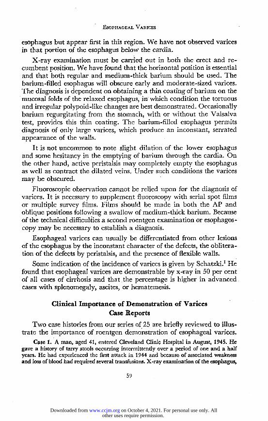
At the time of admission, the clinical examination was negative except for evidence of anemia and very questionable evidence of enlarged spleen. The liver was not palpable. There was no history of abdominal pain or vomiting. The laboratory tests showed a hypochromic anemia (red blood cell count 3,810,000; hemoglobin 9.9 Gm.), increased bromsulphalein retention (36 per cent in thirty minutes), and decreased total serum protein (6.4 Gm.). X-ray examination demonstrated moderately large varices in the esophagus, confirming the evidence of portal hypertension (fig. 1, 2). During the next year the patient had repeated esophagoscopies with injection of the varices, the last in-jection being done on November 25, 1945, at which time he appeared in moderately good condition.
FIG. 1. Varices are obscured by retained barium in the esophagus.
This case illustrates that the diagnosis of portal hypertension must be considered despite the absence of an enlarged liver or the absence of ascites. It emphasizes the importance of checking the x-ray findings in cases of unexplained bleeding. Adequate x-ray examination might
6 0
other uses require permission. on October 4, 2021. For personal use only. Allwww.ccjm.orgDownloaded from
E S O P H A G E A L V A R I C E S
a b FIG. 2. (a) Polypoid appearance of esophageal varices with a thin coating of barium,
(b) Same, showing obliteration of varices by barium.
have established the diagnosis of varices at the original examination and avoided an exploratory operation.
Case 2. A man, aged 40, was admitted to Cleveland Clinic Hospital on August 14, 1944, complaining of recurrent attacks of nausea, vomiting, and weakness during a two-year period. Intermittent fever (102° and 103°) and vague abdominal pain had begun to occur at the end of this time. The patient had lost 60 pounds in weight, and repeated blood examinations .had shown a progressive anemia. Therapy had consisted of three courses of sulfa drugs and five injections of penicillin as well as supportive therapy.
Upon admission, physical examination was negative except for evidence of anemia, fever (101°), and an enlarged spleen. Laboratory studies showed a hypochromic anemia (3,060,000; 6.8 Gm. hemoglobin), a leukocytosis of 16,550 (90 per cent neutrophils, 6 per cent lymphocytes; 4 per cent monocytes). The icteric index was 4, and a bone marrow puncture was not diagnostic. Stool examination for occult blood was positive, and x-ray examination of the gastrointestinal tract demonstrated esophageal varices. A nonfunctioning gall bladder was also found. Following blood transfusions the patient was discharged.
He returned to the Clinic two months later (October 18, 1944), stating that he had had recurrent attacks of fever (104°) and, for the first time, hematemesis. The bromsul-phalein test showed a retention of 32 per cent in thirty minutes. The spleen was large, the liver not palpable. A final diagnosis of portal hypertension, possibly due to throm-bosis of the splenic vessels, was made. Splenectomy was advised and performed else-where. Subsequently the patient had the varices injected by esophagoscopy on three different occasions between August 16 and November 29, 1945. At the last injection it was noted that the varices were much less prominent. In a follow-up letter on December 7, 1946, the patient reported that his general condition had remained good and that there had been no subsequent bleeding.
6 1 «
other uses require permission. on October 4, 2021. For personal use only. Allwww.ccjm.orgDownloaded from

C . R . HUGHES AND J . M . FALOMIR
The clinical picture of this patient complicated the diagnosis. Roentgenologic demonstration of varices was the prominent feature in establishing a diagnosis, emphasizing the importance of an adequate x-ray examination of the esophagus in all patients.
Summary
1. It has been shown that portal hypertension may be first suspected by the roentgen demonstration of esophageal varices.
2. The roentgen technic necessary to demonstrate varices is empha-sized.
3. Two case histories from this series of 25 have been reported.
References 1. Schatzki, R.: Roentgen demonstration of esophageal varices; its clinical importance.
Arch. Surg. 41:1084-1100 (Nov.) 1940. 2. Holmes, G. W., and Schatzki, R.: Examination of mucosal relief as diagnostic aid in
diseases of gastrointestinal tract. Am. J. Roentgenol. 34:145-157 (Aug.) 1935. 3. Templeton, F. E.: X-Ray Examination of the Stomach. (Chicago: University of
Chicago Press, 1944). 4. Wolf, G.: Die Erkennung von Oesophagus-Varzin in Röntgenbilde. Fortschr. a. d.
Geb. d. Röntgenstrahlen 37:890-893 (June) 1928. 5. Kirklin, B. R., and Moersch, H. J.: Report of a case of roentgenological^ demonstrable
esophageal varices complicating splenomegaly. Radiology 17:573-575 (Sept.) 1931. 6. Oppenheimer, A.: Esophageal varices. Am. J. Roentgenol. 38: 403-414 (Sept.) 1937. 7. Mclndoe, A. H.: Vascular lesions of portal cirrhosis. Arch. Path. & Lab. Med. 5:23-42
(Jan.) 1928. . . . , 8. Kegaries, D. L.: Venous plexus of esophagus* Surg., Gynec. & Obst. 58:46-51 (Jan.)
1934.
62
other uses require permission. on October 4, 2021. For personal use only. Allwww.ccjm.orgDownloaded from
![Small Esophageal Varices in Patients with Cirrhosis—Should ... · Varices of medium/large size (>5-mm diameter), or small varices with red spot signs [3†]. According to the](https://static.fdocuments.net/doc/165x107/5fa0286b8b7f711ce374a04d/small-esophageal-varices-in-patients-with-cirrhosisashould-varices-of-mediumlarge.jpg)