
The Developmental Mechanics and Potencies of the...
31
The Developmental Mechanics and Potencies of the Undifferentiated Mesenchyme of the Mandible. (Strangeways Research Laboratory, Cambridge) By W. Jaeobson (Sir Halley Stewart Research Feflow) and H. B. Fell (Royal Society Messel Research Fellow) With 4 Text-figures and Plates 27 to 31. INTRODUCTION. IN accounts of the embryonic development of cartilage and membrane bone, the first stage is usually described as the accumulation of undifferentiated mesenchyme ceils in certain areas where cartilaginous or osseous ground substance is later deposited. Important developmental processes must, however, precede the appearance of these mesenchymal condensations, and various problems arise regarding the antecedents of the cells composing the condensations. In the first place it would be interesting to know whether these cells are predetermined to form cartilage or bone, or whether they are non-specific and differentiate in response to local environmental factors. In the second place the origin of the cells is obscure. A condensation might arise in one of the following ways: (a) By local proliferation to form a small thickening which might then expand, forcing its way through the surrounding mesenchyme until it filled the site of the future rudiment. (b) The original small thickening might be enlarged, not solely by its own growth and by invasion of the adjacent tissue,
Transcript of The Developmental Mechanics and Potencies of the...
The Developmental Mechanics and Potencies ofthe Undifferentiated Mesenchyme
of the Mandible.(Strangeways Research Laboratory, Cambridge)
By
W. Jaeobson(Sir Halley Stewart Research Feflow)
and
H. B. Fell(Royal Society Messel Research Fellow)
With 4 Text-figures and Plates 27 to 31.
INTRODUCTION.
IN accounts of the embryonic development of cartilage andmembrane bone, the first stage is usually described as theaccumulation of undifferentiated mesenchyme ceils in certainareas where cartilaginous or osseous ground substance is laterdeposited.
Important developmental processes must, however, precedethe appearance of these mesenchymal condensations, andvarious problems arise regarding the antecedents of the cellscomposing the condensations.
In the first place it would be interesting to know whetherthese cells are predetermined to form cartilage or bone, orwhether they are non-specific and differentiate in response tolocal environmental factors. In the second place the origin ofthe cells is obscure. A condensation might arise in one of thefollowing ways:
(a) By local proliferation to form a small thickening whichmight then expand, forcing its way through the surroundingmesenchyme until it filled the site of the future rudiment.
(b) The original small thickening might be enlarged, notsolely by its own growth and by invasion of the adjacent tissue,

564 W. JACOBSON AND H. B. FELL
but as the result of a wave of proliferation spreading graduallyover the whole area finally occupied, by the rudiment. .
(c) The cells might not. originate at the site of the condensa-tion at all; they might arise elsewhere, in some restricted areaof proliferation, then migrate to the site of the future condensa-tion and there accumulate, multiply, and differentiate.
In general more is known about the very early developmentof muscle than about that of bone and cartilage, but in someparts of the body the origin of the myogenic cells has neverbeen established with certainty.
It was decided to investigate these problems relating to theorigin of cartilage, bone, and muscle in the mandible of theembryonic fowl. This provides very favourable material forsuch a study, because in the early stages of development theundifferentiated mesenehyme shows localized condensationswhich form Meckel's cartilage, the mandibular bones, and themandibular muscles respectively. A further advantage of themandibular rudiment is the absence of perichondral ossification(except at the articulation), so that only the simpler membranousossification need be considered.
A combined experimental and histological study of the jawfrom the third to the seventh day of embryonic life was made.By excising different regions of the mandible, and observingtheir development in v i t r o , it was possible to make a fairlycomplete analysis of the potencies of the mesenehyme at suc-cessive stages of development. These experimental results werecorrelated with histological findings obtained by the usualmorphological methods.
MATERIAL AND METHODS.
In all, 260 embryonic fowl mandibles were used in these experi-ments. Usually one small area only was explanted from each jaw,but occasionally two explants were taken. After the tissue frag-ment had been excised, the rest of the jaw was fixed and sectioned,so that the exact boundaries of the explant could be accuratelydetermined. Two hundred and seventy-four explants were madeand were cultivated in v i t ro for periods varying from 2 to 16days. They were distributed over the various ages as follows:

DEVELOPMENT OF MANDIBLE 565
from 3-day embryos 67 explants4 „ 615
67
j ?
j j
J ?
5850326
The tissue fragments which were excised from the jaws byspecially sharpened cataract knives and sewing needles mountedin thin glass rods were usually explanted by the watch-glassmethod. Each explanfc was laid on the surface of a clot com-posed of equal parts of plasma and embryo extract, with theepithelial surface upwards. After 2 days' growth, the epi-thelium was removed and the tissue transferred to an ordinaryhanging drop preparation. It was then sub-cultivated every48 hours.
It was important to remove the epithelium, which otherwisetended to envelope the entire explant and prevent its furthergrowth and differentiation. The extremely small size of thetissue fragment made this impossible when the explant wasfirst removed from the jaw, but after it had been growing in awatch-glass for 2 days, most of the epithelium could bestripped off fairly easily.
Some of the explants were grown by the watch-glass methodthroughout the culture period, whilst others were grownentirely by the hanging drop technique. On the whole muchbetter results were obtained when both methods were used inthe manner described above.
All the explants were serially sectioned. Most of the culturesand control mandibles were fixed in Carnoy's mixture for 10-15minutes in the case of the cultures, and for 30 minutes in the caseof the mandibles. The tissue was then transferred to absolute al-cohol, cleared in methylbenzoate containing 2 per cent, celloidinor in cedar wood oil, and embedded in paraffin. The explantswere not removed from the coverslips until after infiltrationwith paraffin. Fixation of the mandibles in Zenker's fluid for30 minutes also gave good results. The serial sections wereusually stained with haemalum and either erythrosin or chroma-

566 W. JACOBSON AND H. B. FELL
trop 2B; iron haematoxylin followed by differentiation in5 per cent, phosphotungstic acid was occasionally used todemonstrate ossification centres, as the bone fibrils stain reddishorange by this method. Sections were also stained with safraninand picro-indigo-carmine, a method which stains cartilage pinkand bone and osteoid tissue blue.
GROSS ANATOMY OF THE MANDIBULAB AECH.
At the 3-day stage (fig. 1, PI. 27) the first branchial arch—thefuture mandible—is slightly bow-shaped with a well-markedsymphysis in the mid-line. The junction with the maxilla ison the lateral side and the main part of the arch runs in alateral-medial direction. At this stage the medio-lateral dimen-sion is larger than the proximo-distal, but the ratio of these two'diameters' (the proximo-distal and the medio-lateral) is soonchanged as the first branchial arch begins to form the lower jaw(figs. 2, 4, and 5, PL 27). The mechanism of the transformationfrom a bow-shaped branchial arch into a jaw will be discussedin the following chapters as the tissue movements inside themandible are closely connected with the general changes inshape of the growing mandible.
In regard to the nomenclature it may be mentioned that the' anterior' or ' mouth surface' is that covered by the epitheliumof the buccal cavity, and the 'posterior' or 'skin surface' isthat which faces the hyoid and borders the first branchial cleft.
THE MYOGENIC CELLS.
The first tissue to be distinguished histologically in the jawis the myogenic tissue. The myogenic cells do not originatein the mandibular arch but migrate into it between the forty-eighth and sixtieth hour of development as a compact groupsurrounding and preceding the ramus mandibularis of thetrigeminus nerve. They form a large cap over the distal endof the nerve and enter the mandibular arch at its lateral endwhere it is fused to the maxilla, growing rapidly through thescanty mesenchyme of the mandibular arch in a medial direc-tion. They have almost reached the mid-line of the mandible

DEVELOPMENT OF MANDIBLE 567
by the third day, closely attached to the anterior side of thewall of the first branchial artery (fig. 6, PL 27).
In the living mandible the myogenic tissue can be distin-guished from the surrounding mesenchyme by its slightlylighter appearance. In the 3-day embryo it appears as a broadband extending from the lateral side of the arch in a medialdirection almost to the mid-line (fig. 1, PL 27).
The myogenic cells, which are closely packed together, arerounded in shape and are larger and lack the irregular processesof the surrounding mesenchyme cells. When the myogenic cellsfirst enter the mandible they occupy most of the space betweenthe two layers of epithelium, as the mesenchyme of the mandibleis very sparse at this stage. Very numerous degenerate mesen-chyme cells appear in the mid-line near the medial and anteriormargin of the mass of myogenic cells. This local degenerationof the mesenchyme provides the space required for the ingrowingmyoblasts (figs. 8 and 9, PL 28).
After the third day the further growth of the myogenie tissuecan no longer continue in a medial direction. Instead, the broadband of myogenic cells extends in a medio-distal direction,thereby causing converging forward movements from eitherside which stretch the branchial arch in a distal direction.Twenty-four hours later the well defined mass of myogenictissue breaks up into smaller groups of cells which eventuallyform the individual muscles innervated by the third ramus ofthe trigeminus nerve (figs. 19, PI. 29, and 31, PL 31).
The precise origin of the mandibular myoblasts was notascertained, and it would be interesting to know from whichpart of the non-segmented head mesoderm they are derived.
The site of the myogenie tissue in the mandibular arch wasalso demonstrated experimentally by means of tissue culture.Of the explants of the mandibular mesenchyme sixty-nineformed muscle-fibres, and the controls showed that all thesecultures contained part of the myogenic tissue described above.Myogem'c cells removed from the mandible as early as thethird day of incubation were able to differentiate into muscle-fibres with cross-striated myofibrils (fig. 7, PL 27). There wasno evidence that the differentiation of these cells depends in any

566 W. JACOBSON AND H. B. FELL
trop 2E; iron haematoxylin followed by differentiation in5 per cent, phosphotungstie acid was occasionally used todemonstrate ossification centres, as the bone fibrils stain reddishorange by this method. Sections were also stained with safraninand picro-indigo-carmine, a method which stains cartilage pinkand bone and osteoid tissue blue.
GROSS ANATOMY OF THE MANDIBULAR ARCH.
At the 3-day stage (fig. 1, PI. 27) the first branchial arch—thefuture mandible—is slightly bow-shaped with a well-markedsymphysis in the mid-line. The junction with the maxilla ison the lateral side and the main part of the arch runs in alateral-medial direction. At this stage the medio-lateral dimen-sion is larger than the proximo-distal, but the ratio of these two'diameters' (the proximo-distal and the medio-lateral) is soonchanged as the first branchial arch begins to form the lower jaw(figs. 2, 4, and 5, PL 27). The mechanism of the transformationfrom a bow-shaped branchial arch into a jaw will be discussedin the following chapters as the tissue movements inside themandible are closely connected with the general changes inshape of the growing mandible.
In regard to the nomenclature it may be mentioned that the' anterior' or ' mouth surface' is that covered by the epitheliumof the buccal cavity, and the 'posterior' or 'skin surface' isthat which faces the hyoid and borders the first branchial cleft.
THE MYOGENIC CELLS.
The first tissue to be distinguished histologically in the jawis the myogenic tissue. The myogenic cells do not originatein the mandibular arch but migrate into it between the forty-eighth and sixtieth hour of development as a compact groupsurrounding and preceding the ramus mandibularis of thetrigeminus nerve. They form a large cap over the distal endof the nerve and enter the mandibular arch at its lateral endwhere it is fused to the maxilla, growing rapidly through thescanty mesenchyme of the mandibular arch in a medial direc-tion. They have almost reached the mid-line of the mandible
DEVELOPMENT OF MANDIBLE 567
by the third day, closely attached to the anterior side of thewall of the first branchial artery (fig. 6, PI. 27).
In the living mandible the myogenic tissue can be distin-guished from the surrounding mesenchyme by its slightlylighter appearance. In the 3-day embryo it appears as a broadband extending from the lateral side of the arch in a medialdirection almost to the mid-line (fig. 1, PL 27).
The myogenic cells, which are closely packed together, arerounded in shape and are larger and lack the irregular processesof the surrounding mesenchyme cells. When the myogenic cellsfirst enter the mandible they occupy most of the space betweenthe two layers of epithelium, as the mesenchyme of the mandibleis very sparse at this stage. Very numerous degenerate mesen-chyme cells appear in the mid-line near the medial and anteriormargin of the mass of myogenic cells. This local degenerationof the mesenchyme provides the space required for the ingrowingmyoblasts (figs. 8 and 9, PI. 28).
After the third day the further growth of the myogenic tissuecan no longer continue in a medial direction. Instead, the broadband of myogenic cells extends in a medio-distal direction,thereby causing converging forward movements from eitherside which stretch the branchial arch in a distal direction.Twenty-four hours later the well defined mass of myogenictissue breaks up into smaller groups of cells which eventuallyform the individual muscles innervated by the third ramus ofthe trigeminus nerve (figs. 19, PL 29, and 31, PL 31).
The precise origin of the mandibular myoblasts was notascertained, and it would be interesting to know from whichpart of the non-segmented head mesoderm they are derived.
The site of the myogenic tissue in the mandibular arch wasalso demonstrated experimentally by means of tissue culture.Of the explants of the mandibular mesenehyme sixty-nineformed muscle-fibres, and the controls showed that all thesecultures contained part of the myogenic tissue described above.Myogenic cells removed from the mandible as early as thethird day of incubation were able to differentiate into muscle-fibres with cross-striated myofibrils (fig. 7, PL 27). There wasno evidence that the differentiation of these cells depends in any

568 W. JACOBSON AND H. B. FELL
way on contact with the nerve-fibres or cells of the trigeminus,and even after 16 days in v i t r o the cross-striated fibresshowed spontaneous contractions. Similar contractions werealso observed in muscle-fibres in which the myofibrils remainednon-striated.
Since the development of the myogenic cells does not dependon contact with nervous tissue or on the presence of cartilageor bone, it would seem that their future differentiation is strictlydetermined when they first enter the mandibular arch. Themesenchyme surrounding the myogenic cells is incapable ofdifferentiating into contractile tissue. This was shown by thefact that in more than 100 explants, from which the myogenictissue had been completely omitted, no muscle appeared duringcultivation.
THE CHONDROGENIC CELLS.
The chondrogenie cells arise in the mandible itself in a smallarea, about 200/* in diameter, immediately beneath the epi-thelium of the buccal cavity. This region, which is an activegrowth centre, is shifted in a distal direction by the elongationof the mandibular arch to form the jaw. As it moves this pro-liferation centre leaves behind it a trail of chondrogenie cellswhich eventually develop into Meckel's cartilage. The detailsof this process are as follows.
The Chondrogenie Area at 3-3J Days.—Betweenthe third and fourth day of incubation the scanty mesenchymecells occupying the narrow space between the epithelium of thebuccal cavity and the myogenic tissue begin to proliferate(fig. 10, PI. 28). Explantation experiments showed that part ofthis tissue is chondrogenie.
Histologically the chondrogenie area is distinguishable fromthe surrounding mesenchyme by its more closely packed cellsand higher mitotic index. Explants containing all or part ofthis small area (eighteen explants) formed cartilage in v i t r ousually in the form of a long rod, e.g. in explant 257, anexplant only 120 x 120/*, after 8 days' cultivation had developeda cartilage rod 1-3 mm. long (figs. 11 and 12, PI. 28).
The boundaries of the chondrogenie area were determined

DEVELOPMENT OF MANDIBLE 569
by isolating fragments of the peripheral parts of the mandibulararch and studying their development in v i t r o (Text-fig. 1).
Medial explants (nine) extending laterally from the mid-linefor a distance of 120-150^ formed no cartilage during cultivation,indicating that the medial boundary of the chondrogenic areamust lie about 150/x from the mid-line.
The position of the l a t e r a l boundary varies considerably•with age. In the early 3-day mandible it appears to lie about300fi from the mid-line, since explants (five) taken more than
TEXT-FIG. 1.
The site of the chondrogenic proliferation centre beneath themouth epithelium in a 3-day mandible (cf. figs. 10-12, PI. 28).The arrow indicates the direction of growth.
300-50 [i. laterally from the mid-line failed to form cartilagein v i t ro .
The extreme distal end of the mandible is non-chondrogeniefor a distance of about 100 yu. (four explants), showing that thed i s t a l boundary lies about 100//. from the tip of the mandible.
Explants of small strips of tissue removed from the mouthsurface near the pharynx always developed cartilage (six ex-plants), so that the p rox ima l boundary of the chondrogenicarea must lie close to the pharyngeal border of the mandible.
The Chondrogenic Area at 4 Days.—Two processesare involved in the further development of the chondrogenicarea (fig. 13, PL 28).
(a) Cells continually migrate from the sub-epithelial pro-liferation centre into the deeper parts of the mandible wherethey later differentiate (arrow marked A on fig. 13, PL 28).
(b) The proliferation centre is shifted bodily in a distaldirection by the active elongation of the mandibular arch, withthe result that the mass of chondrogenic tissue formed by theproliferation centre assumes the elongated form characteristicof Meckel's cartilage (arrow marked B on fig. 13, PL 28).

570 W. JACOBSON AND H. B. FELL
These two processes were analysed both experimentally andhistologically.
Cell multiplication in the sub-epithelial proliferation centreis so active that by the end of the fourth day a small, roundedswelling is produced on the mouth surface on either side of themid-line at the apex of the primitive jaw (fig. 3, PI. 27). Thatthese apical swellings, which persist up to the beginning of thesixth day, arechondrogenic has been demonstrated by explanta-tion experiments (thirty-one explants).
In sagittal sections of the 4-day mandible (fig. 14, PI. 29)each swelling is seen to consist of closely packed cells with largeround nuclei. Mitotic figures are numerous, especially near theepithelium, and are frequently orientated with their long axesvertical to the base of the epithelium, showing that the directionof growth is towards the deeper part of the mandible.
Some of the mitotic figures in the proliferation centre areasymmetrical. During prophase and metaphase these asym-metrical cells become oval instead of rounded and fine shortprocesses resembling pseudopodia protrude from the polefarthest from the epithelium, suggesting that the cell is activelymigrating in a proximal direction. In metaphase the chromo-some plate is not equatorial, but lies either obliquely in thecell (see fig. 26, PL 30) or nearer the pole which points towardsthe epithelium. This asymmetrical arrangement is retained bythe two groups of daughter chromosomes at anaphase, so thatone group is nearer the surface of the cytoplasm than the other.A similar asymmetry of the chromosome plates, correlated withthe presence of pseudopodia projecting in the direction ofmigration, has been observed in the dividing cells of thesclerotomes as they grow towards the mid-line to form thecartilaginous bodies of the vertebrae.
All the cells of the two proliferation centres contain verysmall osmic acid reducing granules which are soluble in alcohol.
The chondrogenic cells are therefore distinguishable histo-logically from the surrounding mesenchyme by the followingcharacteristics:
1. Their dense aggregation.2. Their very active cell division.

DEVELOPMENT OF MANDIBLE 571
3. The frequent orientation of the dividing cells in a directionvertical to the epithelium.
4. The frequent asymmetry of the position of the chromo-some plates.
5. The presence of fine osmic acid reducing granules (fat) inthe cytoplasm.
In the more proximal parts of the mandible the chondrogenictissue is further differentiated than in the apex of the jaw(fig. 13, PI. 28). It lies close to the mass of myoblasts in theinterior and is separated from the epithelium of the buccalcavity by a layer of loose mesenchyme which, when isolated andexplanted in v i t r o , formed no cartilage.
That the proximal part of the chondrogenic rudiment growsless actively than the distal part which contains the sub-epithelial proliferation centre was demonstrated by the followingexperiments.
1. When the proximal part of the chondrogenic region wasexplanted in v i t r o it formed only a short rod or oval nodule,while explants of the distal end formed long cartilaginous rods.For example, explant 87, which contained the greater part ofthe chondrogenic rudiment except the distal 400/*, i.e. theproliferation centre (Text-fig. 2), formed an oval nodule 300ju.in length (fig. 15, PI. 29). Explant 85, on the other hand, con-tained a further 200/x of the distal tissue, i.e. it included most ofthe proliferation centre (Text-fig. 2) and developed a carti-laginous rod just over 1 mm. long (fig. 16, PL 29).
2. Carbon patches were placed at different points on thesurface of fourteen explants of whole 3-day mandibles. Therelative distances between two marks at the time of explanationand during subsequent growth were then measured by meansof camera lucida drawings. These measurements showed thatthe elongation of the branchial arch to form the primitive jawis chiefly due to the growth of the distal part of the arch con-taining the two apical proliferation centres. While the raroiare elongating in this way they also converge, so that the anglebetween them becomes acute (figs. 17 and 18, PI. 29). Thiscombined elongation and convergence of the rami causes themedian proximo-distal axis of the jaw to lengthen. Thus

572 W. JACOBSON AND H. B. FELL
the average increase in this dimension in the 3-day explantsduring 24 hours' growth was 53 per cent., and in seven explantsmaintained for 48 hours, the average increase was 88 per cent.
TEXT-JIG. 2.
The site of explant 87 and 85. Both were taken from the mesen-chyme beneath the mouth epithelium. The latter includedmost of the chondrogenic proliferation centre (cf. figs. 15 and 16,PI. 29).
The Chondrogenic Area at 5 Days.—The two distalproliferation centres are still present as large round swellings(fig. 4, PL 27), and have the same histological structure asin the 4-day mandible. That these areas are still activelyforming chondrogenic cells is demonstrated by the followingtwo experiments:
1. Two explants, containing about 350^ of the distal partof the chondrogenic condensation (explants 1 and 9) werecultivated in v i t r o for 8 days, and each formed a rod ofcartilage 1 mm. long. On the other hand, an explant (103)which excluded the anterior 350^. of the chondrogenic region,i.e. the proliferation centre, but contained 600p of the proximalpart of the ehondrogenie tissue, after 8 days' cultivation formeda rod 750ju. in length. Thus an explant which contained onlythe proliferation centre of the chondrogenic tissue nearlytrebled its length during cultivation, whilst one lacking theproliferation centre increased by only one-quarter of its originallength.
2. Pour explants (94-7) of the whole chondrogenic regionwere marked at five or six points with carbon particles and werecultivated for 5 days. Meckel's cartilage developed as a longrod. Measurements of the distance between the marks showed
DEVELOPMENT OP MANDIBLE 573
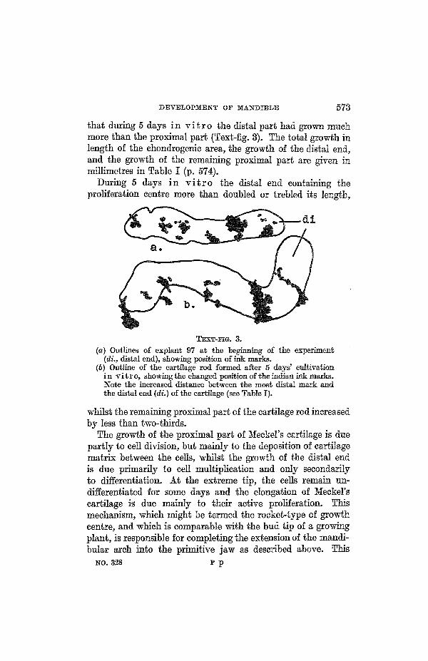
that during 5 days in v i t r o the distal part had grown muchmore than the proximal part (Text-fig. 3). The total growth inlength of the chondrogenic area, the growth of the distal end,and the growth of the remaining proximal part are given inmillimetres in Table I (p. 574).
During 5 days in v i t r o the distal end containing theproliferation centre more than doubled or trebled its length,
TEXT-ITG. 3.
(a) Outlines of explant 97 at the beginning of the experiment(di., distal end), showing position of ink marks.
(6) Outline of the cartilage rod formed after 5 days' cultivationin vitro, showing the changed position of the Indian ink marks-Note the increased distance between the most distal mark andthe distal end (di.) of the cartilage (see Table I).
whilst the remaining proximal part of the cartilage rod increasedby less than two-thirds.
The growth of the proximal part of Meckel's cartilage is duepartly to cell division, but mainly to the deposition of cartilagematrix between the cells, whilst the growth of the distal endis due primarily to cell multiplieation and only secondarilyto differentiation. At the extreme tip, the cells remain un-differentiated for some days and the elongation of Meckel'scartilage is due mainly to their active proliferation. Thismechanism, which might be termed the rocket-type of growthcentre, and which is comparable with the bud tip of a growingplant, is responsible for completing the extension of the mandi-bular arch into the primitive jaw as described above. This
NO. 328 P p

574 W. JACOBSON AND H. B. FELL
TABLE I. Shoiving Measurements made on 4 Explantsmarked with Indian Ink.
Length of explant
Distance between mostdistal mark and dis-tal end of explant,i.e. area includingthe chondrogenie pro-liferation centre
Length of the remain-ing proximal part
Initial measure-ment
Final measure-ment
Percentageincrease
Initial measure-ment
Final measure-ment
Percentageincrease
Initial measure-ment
Final measure-ment
Percentageincrease
94(after
4 days)
1-40
1-97
41
0-05
0-14
ISO
1-35
1-83
36
95
1-47
2-53
71
0-08
0-27
237
1-39
2-26
63
96
1-92
3-50
82
0-40
0-97
142
1-52
2-53
66
97
2-00
2-85
43
0-17
0-60
25?
1-83
2-25
23
stretching process was begun in the earliest stages of develop-ment by the elongation of the ingrowing mass of myogenictissue.
Meckel ' s Car t i l age at 6-7 Days.—Since those cellswhich have left the proliferation centre first, i.e. the mostproximal cells, are the first to differentiate, chondrificationspreads from the proximal to the distal part of the jaw (fig. 5,PI. 27).
Procartilage appears in the proximal end of Meckel's rod atabout the fifth day (see fig. 13, PI. 28). By the sixth day theproliferation centre has left its position immediately beneaththe epithelium of the buccal cavity from which it is nowseparated by loose, undifferentiated mesenchyme. Explantationexperiments demonstrated that the layer of mesenchymeseparating the proliferation centre from the epithelium has nofurther developmental potencies and forms connective tissueonly during cultivation in v i t r o .

DEVELOPMENT OF MANDIBLE 575
By the end of the seventh day chondrification has spread tothe distal end of Meckel's rod. The extreme tip, however,remains unchondrified. It consists of a cap of nndifferentiatedcells which may be regarded as a periehondral thickening, andindicates that the cartilage is still growing in length. In differ-entiating explants of the chondrogenic area from earlier stagesthe proliferation centre became reduced to a similar peri-ehondral cap when chondrification had reached the distal endof Meckel's rod, and it was noted that when this cap wasexhausted, growth in length ceased.
In the 7-day mandible the cartilage is completely coveredby a thin perichondrium of flattened cells and is embedded inloose mesenchyme with no chondrogenic potencies in v i t r o .At the proximal end of Meckel's rod a small area on its lower(caudal) surface is covered by a slightly thickened perichondriumfrom which the cartilaginous processus angularis later develops(fig. 19, PL 29). The part played by this process in the develop-ment of the mandibular bones will be discussed in the nextsection.
THE OSTEOGENIC CELLS.
Like the chondrogenic tissue, the osteogenic cells alsooriginate in the mandibular arch itself. The results describedbelow showed that the jaw-bones develop from two primarycentres. The larger centre occupies an area about 150/Lt x 100/xbeneath the epithelium of the first branchial cleft (fig. 22, PL 30)and from it cells migrate in a distal direction giving rise to theossa angulare, supra-angulare, and spleniale (operculare). Thesmaller centre, which forms the os dentale only, appears beneaththe epithelium of the buccal cavity on the latero-distal aspectof the mandible. The detailed development of the two centreswill now be described.
The Osteogenic Area a t 3 Days.—No osteogenictissue could be detected in the 3-day mandible by the explanta-tion method, although numerous cultures were made of theregion which, when removed from a 4-day mandible, readilyformed bone in v i t r o . In the 4-day jaw, as described below,it was shown experimentally that the osteogenic area is situated

576 W. JACOBSON AND H. B. PELL
at the proximal end of the mandibular arch, immediately underthe skin surface and facing the hyoid arch. In the 3-dayembryo this region consists of a very small, narrow strip ofloose mesenchyme sandwiched between the presumptive epi-dermis and the myogenic cells. Its failure to form bone whencultivated in v i t r o may be due to technical factors. Thatosteogenic mesenchyme explanted at this stage is able to formbone in culture is indicated by the fact that in one culture theossification centre of the os dentale, the histogenesis of whichwill be considered later, developed a minute island of osteoidtissue (60X40x30yw.) during cultivation in v i t r o .
TEXT-ITG. 4.
The site of the osteogenic proliferation centre beneath the epi-thelium lining the first branchial cleft in a 4-day mandible(cf. figs. 20 and 21, PI. 29). The arrow indicates the directionof growth.
The Osteogenic Area at 4 Days.—Bone developedin thirteen explants from 4-day mandibles. The region commonto all these explants was a proximal, posterior area beneath theectoderm lining the deepest part of the first branchial pouch(Text-fig. 4, and figs. 20 and 21, PI. 29). The ectoderm of thisarea is slightly higher than the neighbouring epithelium andforms a small, shallow groove (see fig. 23, PI. 30), the osteogenicmesenchyme cells which lie immediately beneath this grooveare more closely packed than the surrounding, indifferent cellsand are actively dividing (see fig. 22, PI. 30).
The osteogenie tissue was found to be restricted to an areaextending 200-50fi in a proximo-distal direction, and culturesfrom the distal two-thirds of the mandible formed no bone(explants 74, 88, 90). That the osteogenic tissue is confinedto the sub-epidermal mesenchyme at this stage is shown by

DEVELOPMENT OF MANDIBLE 577
the following experiment. One-half of the mandible (eightcases) was split into three layers: an upper, including the epi-thelium of the buccal cavity, a middle and a lower, includingthe presumptive epidermis and underlying mesenchyme. Theupper layer formed cartilage, the middle layer formed muscleand contained also a few nerve-cells, and the lower layerdeveloped bone only.
The young proliferation centre for the osteogenic cells wasclearly demonstrated in the mandibles of some 3§- and 4-dayembryos which had been injected with colchicine.1 Colchicinearrests mitosis at metaphase without preventing cells fromentering division and the proliferation centre was thereforerendered conspicuous by the large number of arrested mitoseswhich it contained (fig. 22, PL 30). The number of cells in theosteogemc centre which extended through five 10/x sections wascounted. The total number of cells in this proliferation areawas 1,729, of which 563 were in mitosis, i.e. the mitotic indexwas 32-6 per cent. Elsewhere in the mandible in the samesections 6,257 cells were counted, of which 1,035 were in mitosis,i.e. the mitotic index was only 16-6 per cent. Thus the propor-tion of cells in mitosis in the proliferative zone was nearlydouble the proportion in other parts of the mandible.
The difference in mitotic activity indicated by these figuresis certainly significant, as was shown by a more detailed statisti-cal analysis of the mitotic counts in two of the sections.2 Inthese sections the proportion of cells was determined in a largenumber of 30 x 30 ju. squares, the proportions averaged, and thestandard deviations determined. The results were:
Section.
12
Proportions of cells in mitosis inproliferative osteogenic area.
34-4+.2-033-7+2-1
Elsewhere inmandible.
I9-4±l-520-9+1-6
The Osteogenic Area between the Fifth and1 The authors are much indebted to Dr. A. F. W. Hughes for injecting
the embryos with colehicine.3 The authors are much indebted to Dr. D. E. Lea for the statistical
analysis.

578 W. JAOOBSON AND H. B. FELL
Six th Day.—Experiments showed that the osteogenic cellsgrow from their proximal proliferation centre in a distal direc-tion, and by the fifth day have spread about half-way alongthe proximo-distal axis of the mandible. Explants of the distalhalf (about 500//. long) of the posterior part of the mandibleformed either no bone or, if the most distal osteogenic cells areincluded (explant 119), a minute trabecula only. The proximalhalf of this region, on the other hand, always formed bone(explants G, 180,169 (figs. 28 and 24, PI. 30)). Between the fifthand sixth day the osteogenic cells enter the distal half of thejaw as demonstrated by the fact that at this stage bonedeveloped when the distal region was cultivated in v i t r o(explant 17 b).
These experimental findings are confirmed by the histologicalappearance of the jaw at this stage. The osteogenic cells areclosely packed together, unlike those of the surrounding loosemesenchyme, and in horizontal sections the long axis of boththeir cytoplasm and nuclei is seen to be orientated in a proximo-distal direction (figs. 25 and 26, PI. 30). The dividing cells donot become spherical and frequently show asymmetry similarto that already described in the migrating chondrogenic tissue.The osteogenic cells derived from the proximal proliferationcentre eventually form three membrane bones, viz. the osangulare, supra-angulare, and spleniale (operculare). Themethod by which the original single centre becomes sub-divided into three will be described later. The fourth mandi-bular bone, the os dentale, originates independently of the othermembrane bones. A small proliferation centre appears at aboutthe fifth day in the disto-lateral region immediately beneaththe epithelium of the buccal cavity, which is here slightlythickened (fig. 27, PI. 31). This epithelial thickening is therudimentary enamel ridge and corresponds to the first stage ofdevelopment of the enamel epithelium in reptiles and mammals.
If this distal proliferation centre is removed and explantedin v i t ro it forms a small bone (explants 113 (figs. 28 and 29,PI. 81) and 114 from 5-day mandibles, explant 148 from 5|-daymandible). With the one exception mentioned on p. 576 it wasimpossible to obtain bone-formation in explants of this region

DEVELOPMENT OF MANDIBLE 579
removed from 3|-4-day mandibles, i.e. before the accumulationof mesenchyme cells had appeared and before the coveringepithelium had begun to thicken.
The Osteogenic Area at 6 Days.—Bone developedin fifteen explants of the osteogenic tissue from 6-day mandibles.Sections show that at this stage the proximal osteogenic con-densation has passed the level of the 'corner' of the mouth.It has also become subdivided into two distinct centres ofclosely packed cells.
This partition is brought about by two processes: intensecellular degeneration in the interior of the original condensation(fig. 30, PL 31) and a high mitotic rate on either side of thedegenerate area. As a result of this subdivision further out-growth towards the distal end proceeds in two streams in theposterior part of the jaw (fig. 25, PI. 30), one lateral and theother medial to Meckel's cartilage. The lateral stream formsthe os supra-angulare and the medial the os angulare and theos operculare.
The cause of the local degeneration in the original condensa-tion is not clear. It occurs beneath the perichondral thickeningon the lower surface of Meckel's rod where the cartilaginousprocessus angularis later develops and the processus eventuallygrows between the os angulare and os supra-angulare. Since,however, the separation of the two ossification centres takesplace before the ingrowth of the processus angularis it is difficultto understand how the cartilage could cause cellular degenera-tion. It is also unlikely to be due to insufficient vascularizationsince the capillaries in this area seem as plentiful and as normalas in other parts of the osteogenic region.
That the splitting of the original centre into two parts maybe due to some extrinsic factor is, however, suggested by theexperiments in v i t r o . Thus, with one exception, eleven ex-plants of the proximal centre from 4-day mandibles, in whichsplitting had not yet appeared, formed a single bone duringcultivation, whilst similar explants from 6-day mandibles(explants 190, 196), in which subdivision had already begun,formed two separate bones (figs. 31 and 32, PI. 31).
Explants of the osteogenic region of 4- and 5-day embryos

580 W. JACOBSON AND H. B. FELL
formed oval bones in v i t r o , while similar explants from6-day embryos developed elongated bones of comparativelynormal shape (fig. 34, PI. 31). This suggests that the bonescannot assume their characteristic elongated form without thestretching effect of the growing cartilage. In normal develop-ment this happens mainly between the fifth and sixth day.
As development proceeds, the medial stream of osteogenictissue also becomes subdivided. Horizontal sections throughthe mandible indicate that this subdivision is caused by themedial branches of the mandibular nerve which cut through themedial stream of osteogenic cells (fig. 25, PL 30). The osteogenictissue in the path of the nerve-fibres degenerates and theossification centre becomes divided transversely into a distalpart, which forms the os spleniale, and a larger, proximal part,which develops into the os angulare.
Experiments on the origin of the os dentale were made atthis stage of development. Explants of one-half of the mandible,from which the tip was excluded (explants 199, 201) (figs. 33and 34, PI. 31), formed three bones only, viz. the os angulare,os spleniale, and os supra-angulare in addition to muscle andMeckel's cartilage. On the other hand, an intact half developedall four ossification centres in v i t r o (explant 200). Explantsof the isolated disto-lateral area (explants 192, 204) formed asingle bone, the os dentale. These results provided additionalevidence of the independent origin of the os dentale.
DISCUSSION.
The work of Fell and Eobison (1930) has already demon-strated that chondrification and ossification occur in explantsfrom 5J-6-day old mandibles.
The results of the present study have shown that the threetypes of mesenchyme cells in the mandible—myogenic, chondro-genic, and osteogenic—are independent in origin, distinct intime of appearance, and already determined while being formed.They originate in some restricted area of proliferation, andthence migrate to the site of the future condensation where theyaccumulate, multiply, and differentiate. The mesenchyme cells

DEVELOPMENT OF MANDIBLE 581
which are not derived from these centres have no potency toform muscle, cartilage, or bone.
Both the chondrogenic and the osteogenic centres originateimmediately beneath the epithelium which, in the case of theosteogenic centres, is thickened in those areas. A similar relation-ship was observed by one of us (W. J.) in the development of theos dentale in mammalian embryos in which the thickened areaof epithelium differentiates into the enamel organ and thedentale cells provide the only source of osteogenic tissue in themandible. Whether the thickened epithelium acts as anorganizer and induces the underlying mesenchyme to formbone, or whether the two tissues develop independently of eachother is not known.
The separate origin of the os dentale demonstrated experi-mentally in the present investigation is also well illustrated bythe fact that in some birds a suture persists between the dentaleand the bones formed by the angulare group, as in Puf f inusK u h l i . In other species a fontanelle persists throughout lifebetween the dentale and the other bones, as in Pygosca l i spapua and A n t h r o p o i d e s v i r g o , or a foramen remainsopen at this place, as in Te t r ao u r o g a l l u s . In Capri-mulgus a joint is even formed between the dentale and theangulare group.
The results have shown that the chondrogenic proliferationcentre fulfils not only a histogenie but also a morphogenicfunction. Thus the mechanism of its growth produces a longrod of cartilage which stretches the branchial arch into the formof a primitive jaw and at the same time exerts tension on theossification centres, causing the bones to assume their charac-teristic, elongated shape.
Certain stages in the development of the mandible arecharacterized by the appearance of morphogenetic cell degenera-tions in the mesenchyme. As described above, such degenera-tions appear immediately in front of the ingrowing myogeniccells for which space is thus provided. They are found also inthe mid-line of the branchial arch, where their occurrence iscorrelated with the convergence of the two halves of the archas they elongate to form the jaw. Finally, they occur locally

582 W. JACOBSON AND H. B. FELL
in the proximal osteogenic centres, where they subdivide eachcentre into three parts.
Similar morphogenetie cell degenerations have been describedby Gliicksmann (1930) in the development of epithelial struc-tures, e.g. neural tube, eye and lens, and by Fell (1939) in themesenchyme of the ventral body-wall during the developmentof the sternum. The cause remains obscure. The cell degenera-tion cannot be due to local vascular deficiencies as the blood-vessels in these areas of the mandible are plentiful and normal.Nor are mechanical forces responsible, since the cells show nosign of being subjected to pressure; they retain their fine cyto-plasmic processes until they die, and then round up and dis-integrate. Moreover, when the median region of the arch wasexcised and explanted in v i t r o at the third day of develop-ment, i.e. before the two halves of the arch had begun to con-verge, degeneration continued in v i t r o (eight explants),although any possible mechanical effect of the convergence ofthe rami had been eliminated.
SUMMARY.
The origin and early development of the muscles, cartilage,and bone of the embryonic avian mandible were investigatedby histological and tissue culture methods applied to theembryonic domestic fowl.
1. Muscle.—The muscles of the mandible do not arise inthe mandibular arch.
The myogenic cells migrate into the arch between the secondand third day as a histologically distinct group. Explantationexperiments in v i t r o showed that the myogenic cells aredetermined to form cross-striated muscles as early as the thirdday.
2. Cartilage.—The chondrogenic cells of either sideoriginate from a small proliferation centre immediately beneaththe buccal epithelium of the branchial arch. Proliferation of thechondrogenic cells continues from the third to the sixth day,forming a pronounced, rounded swelling near the distal endof the mandible. It was demonstrated experimentally thatelongation of the mandibular arch to form the jaw is mainly

DEVELOPMENT OF MANDIBLE 583
due to apical growth caused by proliferation from the pairedchondrogenic centres. By the seventh day the chondrogenicproliferation centre is exhausted.
Experiments showed that the cells of the chondrogenicproliferation centre in the 3-day jaw are already determinedto form cartilage.
3. Bone.—The osteogenic cells destined to form the osangulare, spleniale, and supra-angulare arise from a singleproliferation centre which appears at about the fourth dayimmediately beneath a slightly thickened patch of presumptiveepidermis at the proximo-lateral end of the mandible.
This centre becomes subdivided into a medial and a lateralpart by cell degeneration in the interior. Both parts continueto grow in a distal direction. The lateral division forms theos supra-angulare, while the medial division is cut transverselyinto two sections by branches of the mandibular nerve. Thedistal section forms the os spleniale and the larger, proximalsection the os angulare.
It was demonstrated by explantation experiments that thecells of the osteogenic proliferation centre in the 4-day mandibleare already determined to form bone.
The osteogenic cells destined to form the os dentale originateat about the fifth day from a separate centre in the disto-lateralpart of the mandible, beneath a thickened patch of mouthepithelium corresponding to the rudiment of the enamelepithelium in mammals.
The cells of this disto-lateral osteogenic centre were shownto be self-differentiating in v i t r o when explanted at thefifth day.
The authors are greatly indebted to the Eockefeller Founda-tion for a grant in aid of this research; they also wish to thankMr. V. C. Norfield, senior assistant of the Strangeways EesearcbLaboratory, for his help in preparing the figures.
LIST OF KEFEBENCES.
Fell, H.B., 1939.—"Origin and developmental mechanics of avian ster-num", 'Phil. Trans. Roy. Soc. London', Ser. B, ^ 9 .
Fell, H. B., and Robison, R., 1930.—"Development and phoapliatase

584 W. JACOBSON AND H. B. FELL
activity . . . of mandibular skeletal tissue of embryonic fowl", 'Biochem.Jburn.', 24.
Glucksmann, A., 1930.—"Bedeutung von Zellvorgangen fiir die Form-bildung epithelialer Organe, &c", 'Z. Anat.', 93.
Jacobson, W., 1932.—"Zellvorgiinge in den ersten Entwicklungsstadiendes knorpel- und knochenbildenden Gewebes", 'Anat. Anz.', 75 Suppl.
EXPLANATION OF FIGURES.
ABBBEVATIONS.
i.e., first branchial cleft; c , chondrogenic tissue; d., degenerating cells;di., distal; m., myogenic cells; ma., maxilla; 31.c, Meckel's cartilage;md., mandible; me., mesenchyme; mi., mitosis; n., trigeminal nerve;o., osteogenic cells; o.a., os angulare; o.d., os dentale; o.o., os spleniale(operculare); o.s., os supra-angulare; p., proximal; pe., perichondriinn;p.o., proliferation centre of os dentale; s., site of explant; t., thyroid.
PLATE 27.
Figs. 1-5 are photographs of unfixed mandibles as used for experiments.X25.
Fig. I.—Three-day mandible, seen from the mouth surface: note thelight band of myogenic tissue on either side.
Fig. 2.—Four-day mandible, seen from the mouth surface: note the twoswellings on either side of the mid-line which contain the chondrogenicproliferation centres.
Fig. 3.—The same in frontal view.Fig. 4.—Five-day mandible, seen from the mouth surface.Fig. 5.—Six-day mandibles, seen from the mouth surface. Meckel's
cartilage is already in a procartilaginous stage.Fig. 6.—Frontal section of 3-day mandible, showing the closely packed
myogenic tissue. Iron-haematoxylin-erythrosin. X175.Fig. 7.—Part of a culture of myogenic tissue from a 3-day mandible,
fixed after 16 days in v i t r o . Cross-striated muscle-fibres differentiatedin v i t r o . Safranin-picro-indigo-carmine. x 1,200.
PLATE 28.
Fig. 8.—Frontal section of a 4-day mandible, showing the myogenictissue still forming a compact group of cells. Haem.-eryth. X 65. Themarked area is shown in fig. 9.
Fig. 9.—Same section as in fig. 8, showing the border between the in-growing myogenic tissue and the indifferent mesenchyme. Note the largenumber of degenerating cells in the latter, x 1,200.
Fig. 10.—Left half of frontal section through a 3-day mandible, showingthe chondrogenic area. Haem.-eryth. X 85.
Fig. 11.—Frontal section through a 3-day mandible, showing the site

DEVELOPMENT OF MANDIBLE 585
of an explant which included the whole chondrogenic area. Haem.-eryth.X90.
Fig. 12.—The explant removed from the mandible shown in fig. 11 after8 days' cultivation in v i t r o . A cartilage rod over 1 mm. in lengthdeveloped in culture. Haem.-eryth. x 90.
Fig. 13.—Horizontal section through, the right half of a 4J-day mandible.Arrow A indicates the movements of the cells in the chondrogenic pro-liferation centre, to form the distal parts of Meckel's cartilage. Arrow Bindicates the forward movement of the distal part of the mandible.Haem.-eryth. x 88.
PLATE 29.
Fig. 14.—Part of a chondrogenic proliferation centre in a 4-day mandible.Note the numerous mitotic figures. Haem.-eryth. x 330.
Fig. 15.—Culture of an explant, 200/x long, of the proximal region of thechondrogenic tissue of a 4-day mandible. After 9 days in v i t r o acartilage 300/i long was formed. Haem.-eryth. X100.
Fig. 16.—Culture of an explant, 400ft long, from a 4-day mandible,comprising the same proximal part as that shown in fig. 15 plus 200/Aof the chondrogenic proliferation centre. The cartilage rod formed after9 days in v i t r o is over 1 mm. long. Haem.-eryth. x 100.
Fig. 17.—Three-day mandible, explanted and marked with indian ink,photographed at the beginning of the experiment, x 25.
Fig. 18.—The same mandible after 24 hours' cultivation in v i t r o .X 25. By the convergent movement of the two halves of the mandible,the left mark has been moved towards the mid-line and the two marksnear the middle have been moved forward in a distal direction.
Fig. 19.—Frontal section through the proximal part of a 6-day mandible.Note the thickened periehondrium at the lower end of Meckel's cartilagewhere the processus angularis will be formed and also the osteogeniccondensations on either side beneath Meckel's cartilage. Haem.-eryth.X50.
Fig. 20.—Frontal section through the proximal region of the right halfof a 4-day mandible, showing the site of an explant containing the osteo-genic tissue. Haem.-eryth. X115.
Fig. 21.—Bone formed by the osteogenie tissue taken from the mandibleshown in fig. 20. Fixed after 6 days' cultivation in v i t r o . Safranin-picro-indigo-earmine. X175.
PLATE 30.
Fig. 22.—Lateral part of the left mandible of a 3§-day embryo treatedwith colchicine. The cells above the first branchial eleft form the osteogenicproliferation centre. Note the large number of arrested mitotic divisionsin the osteogenie area. Feulgen light-green, x 300.
Fig. 23.—Frontal section through a 5-day mandible (about 600ft fromthe distal end). On the right the osteogenic tissue has been excised forexplantation. Haem.-eryth. X50.

586 W. JACOBSON AND H. B. FELL
Fig. 24.—Bone formed by the explant removed from the mandibleshown in fig. 23. Haem.-eryth. x300.
Fig. 25.—Horizontal section through the left half of a 5-day mandible,showing the arrangement of the osteogenic tissue. Haem.-eryth. X50
Fig. 26.—Part of fig. 25 under higher magnification, showing the orienta-tion of the osteogenic cells in a proximo-distal direction. Note the stretchedmetaphase (*). Haem.-eryth. x525.
PLATE 31.
Fig. 27.—Sagittal section through a 5-day mandible, showing the pro-liferation centre for the os dentale under the mouth epithelium. Haem.-eryth. x 100.
Fig. 28.—Frontal section through a 5-day mandible (about 500/z. fromthe distal end), showing the site of an explant including the centre for theos dentale. Haem.-eryth. X 50.
Fig. 29.—The explant indicated in fig. 28 formed, after 11 days' cultiva-tion in v i t r o , a small os dentale and an oval shaped cartilage derivedfrom the underlying chondrogenic tissue. Haem.-eryth. x300.
Fig. 30.—The interior of the osteogenic tissue of the mandible shownin fig. 19. Note the degenerating mesenchyme cells by which the osteogeniccentre is being divided into two groups of cells. Haem.-eryth. X 600.
Fig. 31.—Frontal section through the proximal part of a 6-day mandible,showing the site of an explant containing 'the two groups of osteogeniccells (median and lateral). Haem.-eryth. x 35.
Fig. 32.—The explant indicated in fig. 31. After 6 days in v i t r o twoseparate bones developed during cultivation. Haem.-eryth. X100.
Fig. 33.—Frontal section through a 6-day mandible at the border ofthe distal and middle third, showing the site of an explant which removedthe whole tissue of the mandible on the right-hand side with the excep-tion of the proliferation centre for the os dentale. Haem.-eryth. X 50.
Fig. 34.—The explant, indicated in fig. 33, after 4 days' cultivation inv i t r o . A long cartilage rod (not in the plane of the section) and threeelongated bones developed in culture. On the left the os angulare andsupra-angulare, and on the right the thin and folded os spleniale are seen;no os dentale was formed. Haem.-eryth. x 100.

m.-
P-
Quart. Journ. Micr. Sci. Vol 82 N. 8., PL 27
— m.
1
i
\ :
• m .
•Jacobson & Fell

Quart. Journ. Micr. Sci. Vol. 82 N. S., PL 28
v : ' - • • ' •
•*.?•-".••" "':••• • ? • • - . - ' " • - ' - • • • '
1 '/
• . '• ' I
Jacobscm & Fdl

Quart. Journ. Micr. Sci. Vol. 82 N. 8., PI. 29
m 19
Jacobson dh Fell

na.
V-' :-
Quart. Journ. Micr. Sci. Vol. 82 N. 8., PL SO
e.\,
. • • • • • • . ' ' • : • • / . -
Jacobson & Fell

Quart. Journ. Micr. Sci. Vol. 82 N. 8., PL 31
-P .O.
- acobson & Fell