The Development of the Vestibular Apparatus Under Conditions of ...
28
General Disclaimer One or more of the Following Statements may affect this Document This document has been reproduced from the best copy furnished by the organizational source. It is being released in the interest of making available as much information as possible. This document may contain data, which exceeds the sheet parameters. It was furnished in this condition by the organizational source and is the best copy available. This document may contain tone-on-tone or color graphs, charts and/or pictures, which have been reproduced in black and white. This document is paginated as submitted by the original source. Portions of this document are not fully legible due to the historical nature of some of the material. However, it is the best reproduction available from the original submission. Produced by the NASA Center for Aerospace Information (CASI) https://ntrs.nasa.gov/search.jsp?R=19850008113 2018-03-26T16:40:08+00:00Z
-
Upload
trinhthuan -
Category
Documents
-
view
220 -
download
1
Transcript of The Development of the Vestibular Apparatus Under Conditions of ...
General Disclaimer
One or more of the Following Statements may affect this Document
This document has been reproduced from the best copy furnished by the
organizational source. It is being released in the interest of making available as
much information as possible.
This document may contain data, which exceeds the sheet parameters. It was
furnished in this condition by the organizational source and is the best copy
available.
This document may contain tone-on-tone or color graphs, charts and/or pictures,
which have been reproduced in black and white.
This document is paginated as submitted by the original source.
Portions of this document are not fully legible due to the historical nature of some
of the material. However, it is the best reproduction available from the original
submission.
Produced by the NASA Center for Aerospace Information (CASI)
https://ntrs.nasa.gov/search.jsp?R=19850008113 2018-03-26T16:40:08+00:00Z
NASA TECHNICAL MEMORANDUM NASA TM-77517
THE DEVELOPMENT OF THE VFSTIBULAk APPARATUS UNDER CONDITIONSOF WEIGHTLESSNESS
Ya.A. Vinnikov, O.G. Gazenko, D.V. Lychakcv, L.R. Pal1mbakhr
(NASA-TM-77517) !HE LEVELCFMENT CF THE N85-16422TESTIBULAE IPcABATUS UNLEL CCNDITIONS OFWEIGHTLESSNESS gNational Aeronautics andSpace Adainistration) 27 F HC A03/MX A01 Unclas
CSC.. 06C G3/51 13674
Translation of "Razvitiye vestibulyarnogo apparatav usloviyakh nevesomosti", Zhurnal obshchey biologiiVol. 44, No. 2, 1983, pp 147-163.
^O
NATIONAL AERONAUTICS AND SPACE ADMINISTRATION
WASHINGTON, D.C. 20546 AUGUST 1984
i I
STANDARD T!I LE oAGE
1. Roper$ No.
NASA TM-77517 7 2. Government Accession Me. 1. Red^laM'i Cateleg fie.
Title and S"t„i'(e THE DEVELOPMENT OF THE S. Report Dote
VESTIBULAR APPARATUS UNDE ". CONDITIONS 846. Per fervaing Organisation CedaOF WEIGHTLESSNESS
7. Audket (s) Ya.A. Vinnikov, O.G. Gazenko, L Pa,(a,WnipOrgenixetionReoe,tHe.D.V. Lychakov and L.R. Pal'mbakh
10. Rory Unit He.
9. Perlarm:ng Crgo+isorron Nome end AddressLeo Kanner Associates
11. Contract at Grant Ne.NASW-'541
Redwood City CA 94063 12. Type of Report cni Period co.ered I
T-anslation17. ;pensor,ng Agency Nome and Addreee
National Aeronautics and Space Adminis-tration, Washington, D.C. 20546 1^• ^'""`'"'A:' ne y coe.
is. Sc;;Isr..sntcrr Noyes
Translation of "Razvitiye vestibulyarnogo apparata vusloviyakh nevesomosti," Zhurnal obshchey biologii,Vol. 44, No. 2, 1983, pp 147-163.
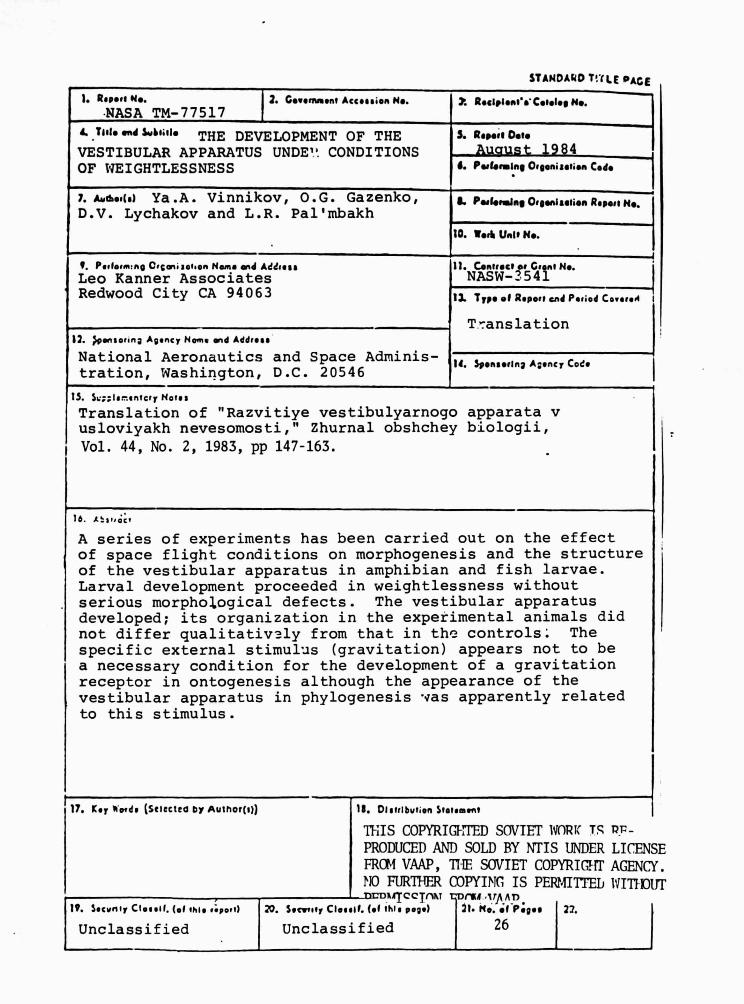
A series of experiments has been carried out on the effectof space flight conditions on morphogenesis and the structureof the vestibular apparatus in amphibian and fish larvae.Larval development proceeded in weightlessness withoutserious morphological defects. The vestibular apparatusdeveloped; its organization in the experimental animals didnot differ qualitativ:ly from that in the controls. Thespecific external stimulus (gravitation) appears not to bea necessary condition for the development of a gravitationreceptor in ontogenesis although the appearance of thevestibular apparatus in phylogenesis was apparently relatedto this stimulus.
17. Key *'orris (Sclecteo Or Author(%)) 18. Dletrlbur,on Stotemant
IMS COPYRIMITED SOVIET 1VORiC TS Pr-
PRODUCED AND SOLD BY NTIS UNDER LICEMFROM VAAP, ME SOVIET COPYRIGTrr AGENC)
NO FURTHER COPYING IS PEP24ITTEL IVIT))OirDx'TccTnXT raf ,trn nn
19. Secvniy Clotelt. (of ale a;porA 20. Secarrtr Claself. (of this page) 71. No. of P;goe 22.
Unclassified Unclassified 26
>E
TI'
THE DEVELOPMENT OF THE VESTIBULAR APPARATUS UNDER CONDITIONSOF WEIGHTLESSNESS
Ya.A. Vinnikov, O.G. Gazenko, D.V. Lycbakov, L.R. Pal'mbakh
As is well known, the development of the structure and function /147*
of the vestibular apparatus in vertebrates precedes the appearance of
other sensory organs. We suggest that this is related to the fact
that the embryo may very early occupy a determined place with respect
to the earth's gravitational field. This phenomenon is demonstrated
very clearly when we observe the disturbance of chick embryos at
various stages of incubation. The question arises of whether the
development of the vestibular apparatus is subject to the effect of
the earth's gravitational field or whether it is genetically pro-
grammed and its morphogenic development unaffected by a specific
stimulus. We should note, however, that in phy l ogenesis the organ
may appear only in connection w'th the effect of a gravitational
force. We obtained an answer to this question during an experimental
study of the development of fish and amphibian embryos under the con-
ditions of weightlessness on space vehicles. If we set up the fish
and amphibian embryos at the stage of the gastrula, neurula or even
tail bud (amphibians), then undF.r conditions of weightlessness,
according to our data, normal development of the embryos as a whole,
and of their tissues and organs, including the vestibular apparatus,
will occur. The experiments which we have conducted [Vinnikov et al., 44
1972, 1976, 1979, 1980; Vinnikov, 1974; Vinnikov et al., 1976), the
results of which are shown in the table, allowed us to affirm that the
development of this organ is not related to the effect of the earth's
gravitational field. Stimulation with gravity is possible only when
fish are returned to earth. However, in order to reach this cc,nclu-
sion, it was necessary for us to run a set of complex experiments and
investigations, to the results of which this article is devoted.
* Numbers in the margin indicate pagination in the foreign text.
1
.'
\J
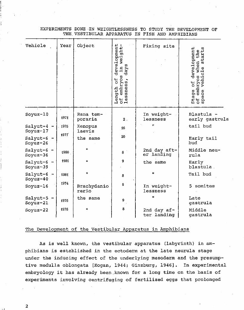
EXPERIMENTS DONE ·IN WEIGHTLESSNESS TO STUDY THE DEVELOPMENT OF THE. VESTIBULAR APPARATUS IN FISH AND AMPHIBIANS
Vehicle Year Object
Soyuz-10 1971
Salyut...,.4 - . 1975 Soyuz-17
Salyut-6 -Soyuz-26
Salyut-6 -Soyuz-36
Salyut-6 -Soyuz-39
Salyut-6 -Soyuz-40
Soyuz-16
Salyut-5 -Soyuz-2l
Soyuz-22
1977'
1980
1981
1981
1974
1976
1976
Rana temporaria
Xenopus laevis
the same
II
II
II
Brachydanio rerlo
the same
II
2,
16
20
8
9
8
6
9
8
Fixing site
In weightlessness
II
2nd day af'ter landing
the same
"
In weightlessness
II
2nd day after landing
Blastula -early gastrula tail bud
Early tail bud
Middle neurula Early blastula,
Tail bud
5 somites
Late gastrula
Middle gastrula
The Development of the Vestibular Apparatus in Amphibians
As is well known, the vestibular apparatus (labyrinth) in am
phibians is established in the ectoderm at the late neurula stage
under the inducing effect of the underlying mesoderm and the presump
tive medulla oblongata [Kogan, 1944; Ginzburg, 1946]. In experimental
embryology it has already been.known for a long time on the basis of
experiments involving centrifuging of fertilized eggs that prolonged
2

and considerable acceleration exerts a negative effect on their de-
velopment. Short and weak accelerations usually do not affect fur-
ther embryogenesis. This in particular follows also from the experi-
ments of Young and Tremor (Young, Tremor, 1968] in which-the eggs of
frog Rana pipiens were placed 12 hours after fertilization in the
flight apparatus Bios-2. After brief acceleration during the ascent /148
of the apparatus any 44 hours under conditions of weightlessness,
as in vivo observations and microscopic investigations showed, the
eggs reached the neurula stage, the leaves primordium of which had a
normal structure. From the embryos left alive mobile tadpoles and
then frogs developed. However, since in Lhese experiments the em-
bryos were kept under the influence of weightlessness prior to organo-
genesis, the gaestion of the effect of weightlessness on the forma-
tion of the presumptive foundations of the organs and their develop-
ment as well as on further embryogenesis remains essentially open.
In order to study the development of the vestibular apparatus
under conditions of weigti,t'.-LLsness without the influence of any other
additional .factors, our first experiment on Soyuz-10 [Vinnikov et al.,
197x1 wa- planned in such a way that the accelerations during the as-
cent of t'Je flight apparatus into space occurred at the stage of em-
bryogenesis at which the foundation of the vestibular apparatus as
well as of the other organs i ,as still absent. As the object fer-
tilized eggs of the frog Rana temporaria at the blastula and early
gastrula stages were selected. In order to create the physiological
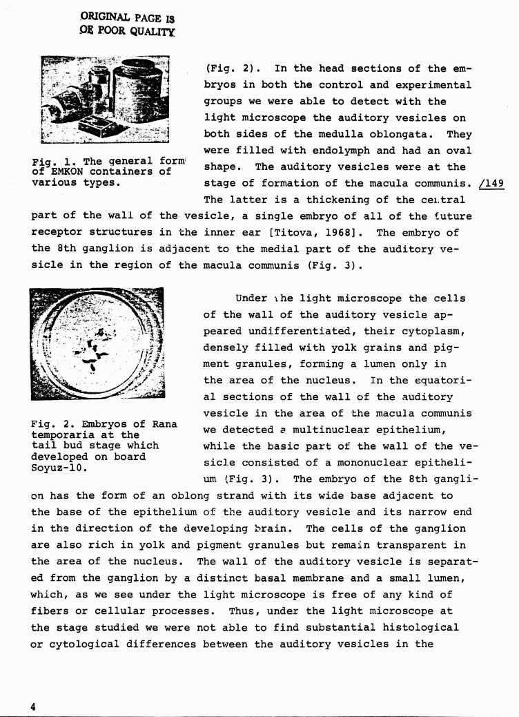
conditions for the development Df the eggs we constructed a special
device, an EMKON container (Fig. 1) in which the eggs were kept
throughout the experiment. At the same time this device i%ade it pos-
sible to fix th(a developing eggs under conditions of weightlessness
at any stage of their development. The embryos which developed under
conditions of weightlessness were fixed at the end of the 2nd day of
flight, on the 4th day after fertilization.
Examination of the material obtained showed that the embryos
which developed under conditions of weightlessness as well as the
controls were at the early tail bud stage at the time of fixation
3
ORIGINAL PAGE I3M POOR QUALITY
(Fig. 2). In the head sections of the em-
_} bryos in both the control and experimental
" groups we were able to detect with the
., light microscope the auditory vesicles on
both sides of the medulla oblongata. They
were filled with endolymph and had an ovalFig. 1.of EMKON
The qeneralcontainers
formof shape. The auditory vesicles were at the
various types. stage of formation of the macula communis. /149
The latter is a thickening of the cei.tral
part of the wall of the vesicle, a single embryo of all of the future
receptor structures in Che inner ear [Titova, 1968]. The embryo of
the 8th ganglion is adjacent to the medial part of the auditory ve-
sicle in the region of the macula communis (Fig. 3).
Q'J Y^ Under the light microscope the cells
C Z `''-- of the wall of the auditory vesicle ap-peared undifferentiated their cytoplasm,densely filled with yolk grains and pig-ment granules, forming a lumen only in
^.l... the area of the nucleus. In the equatori-
yr 'y al sections of the wall of the auditory
vesicle in the area of the macula communisFig. 2. Embryos of Ranatemporaria at the we detected a multinuclear epithelium,
tail bud stage which while the basic part of the wall of the ve-developed on board ,
sicl.e consistec. of a mononuclear epitheli-Soyuz-10.um (Fig. 3). The embryo of the 8th gangli-
on has the form of an oblong strand with its wide base adjacent to
the base of the epithelium of the auditory vesicle and its narrow end
in the direction of the developing :rain. The cells of the ganglion
are also rich in yolk and pigment granules but remain transparent in
the area of the nucleus. The wall of the auditory vesicle is separat-
ed from the ganglion by a distinct basal membrane and a small lumen,
which, as we see under the light microscope is free of any kind of
fibers or cellular processes. Thus, under the light microscope at
the stage studied we were not able to find substantial histological
or cytological differences between the auditory vesicles in the
4
L
i
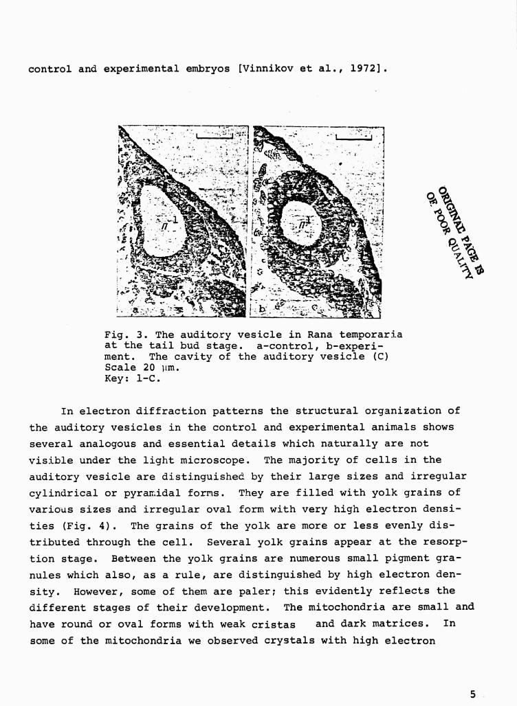
control and experimental embryos [Vinnikov et al., 19721.
0
Fig. 3. The auditory vesicle in Rana temporari_aat the tail bud stage. a-control, b-experi-ment. The cavity of the auditory vesicle (C)Scale 20 lim.Key: 1-C.
In electron diffraction patterns the structural organization of
the auditory vesicles in the control and experimental animals shows
several analogous and essential details which naturally are not
visible under the light microscope. The majority of cells in the
auditory vesicle are distinguished by their large sizes and irregular
cylindrical or pyramidal forms. They are filled with yolk grains of
various sizes and irregular oval form with very high electron densi-
ties (Fig. 4). The grains of the yolk are more or less evenly dis-
tributed through the cell. Several yolk grains appear at the resorp-
tion stage. Between the yolk grains are numerous small pigment gra-
nules which also, as a rule, are distinguished by high electron den-
sity. However, some of them are paler; this evidently reflects the
different stages of their development. The mitochondria are small and
have round or oval forms with weak cristas and dark matrices. In
some of the mitochondria we observed crystals with high electron
P' •ir;.JAL
s r
r ^^
9C
5
rx
mssx.7^^
`Ain i
^A,
oT4•o
^^ ^,ID
SV
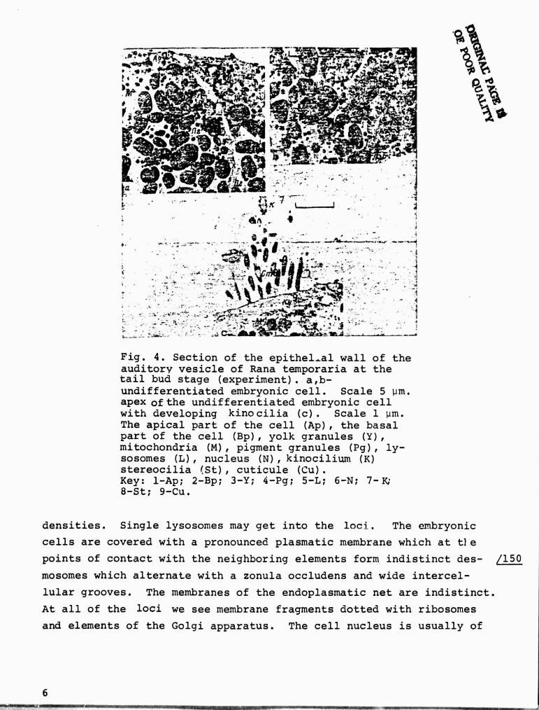
Fig. 4. Section of the epithelial wall of theauditory vesicle of Rana temporaria at thetail bud stage (experiment). a,b-undifferentiated embryonic cell. Scale 5 Um.apex ofthe undifferentiated embryonic cellwith developing kino cilia (c). Scale 1 um.The apical part of the cell (Ap), the basalpart of the cell (Bp), yolk granules (Y),mitochondria (M), pigment granules (Pg), ly-sosomes (L), nucleus (N), kinocilium (K)stereocilia (St), cuticule (Cu).Key: 1-Ap; 2-Bp; 3-Y; 4-Pg; 5-L; 6-N; 7-K;8-St; 9-Cu.
densities. Single lysosomes may get into the loci. The embryonic
cells are covered with a pronounced plasmatic membrane which at t}e
points of contact with the neighboring elements form indistinct des- 150
mosomes which alternate with a zonula occludens and wide intercel-
lular grooves. The membranes of the endoplasmatic net are indistinct.
At all of the loci we see membrane fragments dotted with ribosomes
and elements of the Golgi apparatus. The cell nucleus is usually of
6

moderate dimensions anc. has an irregular rounded form. It is covered
by a double porous membrane, while the outer membrane may form a
series of storages, converting t'.e loci into cisterns of the endo-
plasmatic network. The karyoplesm is distinguished by a fine grained
structure. In it we can see nu.erous large grains of chrom;,tin
which have a tendency to occupy the boundary position on the interior
surface of the nuclear membrane. The large nucleolus occupies the
central position and in its form recalls a mulberry with uneven elec-
tron density. Along with such essentially undifferentiated embryonic
cells in the wall of ti.^ auditory vesicle in both the control and ex-
perimental animals in the area of the macula communis, we observe
individual elements with clear signs of the beginning of cyto-
logical differentiation. Amnng these are evidently embryonic dif-
ferentiating receptor cells (Fig. 4). Differentiation is expressed
in the liberation of the apex of such a cell from the yolk grains and
pigment granules. Instead of them, long, slightly coiled mitochondria
with numerous cristas and a light matrix fill the cytoplasm. In this
apical part of the cell is a centrosome, near to which we find a basal
corpuscle with a stem. From the basal corpuscle to the interior of
the cytoplasm may run a radicle with a clear period of striation ora
individual microtubules. The basal corpuscle, equipped with a stem
with a radicle or microtubules, forms the beginning of the developing
kinocilium which contains nine pairs of peripheral and two central
fibrils. The central fibrils begin from the high basal membrane.
The kinocilium is covered with the extension of the plasmatic membrane.
A.lo^ig with the embryonic cells in which one kinocilium has developed,
we observe cells which are also distinguished by the beginning of the
development of stereocilia. They develop from microvilli which are
somewhat recessed from the kinocilium. Thus, the polar distribution
of the beam of the stereocilia with respect to the kinocilium occurs
at the moment of their differentiation. Til° stereocilia are covered
by the extension of the plasmatic membrane of the cell. Inside the /151
stereocilia we observe thin fibrils which penetrate into the apical
cytoplasm of the cell in the form of a constricted radicle. These
fibrils, as it turned out, are actinic fibers which are connected in
the cuticle to the myosin ring [Flock et al., 1981). The apical
7
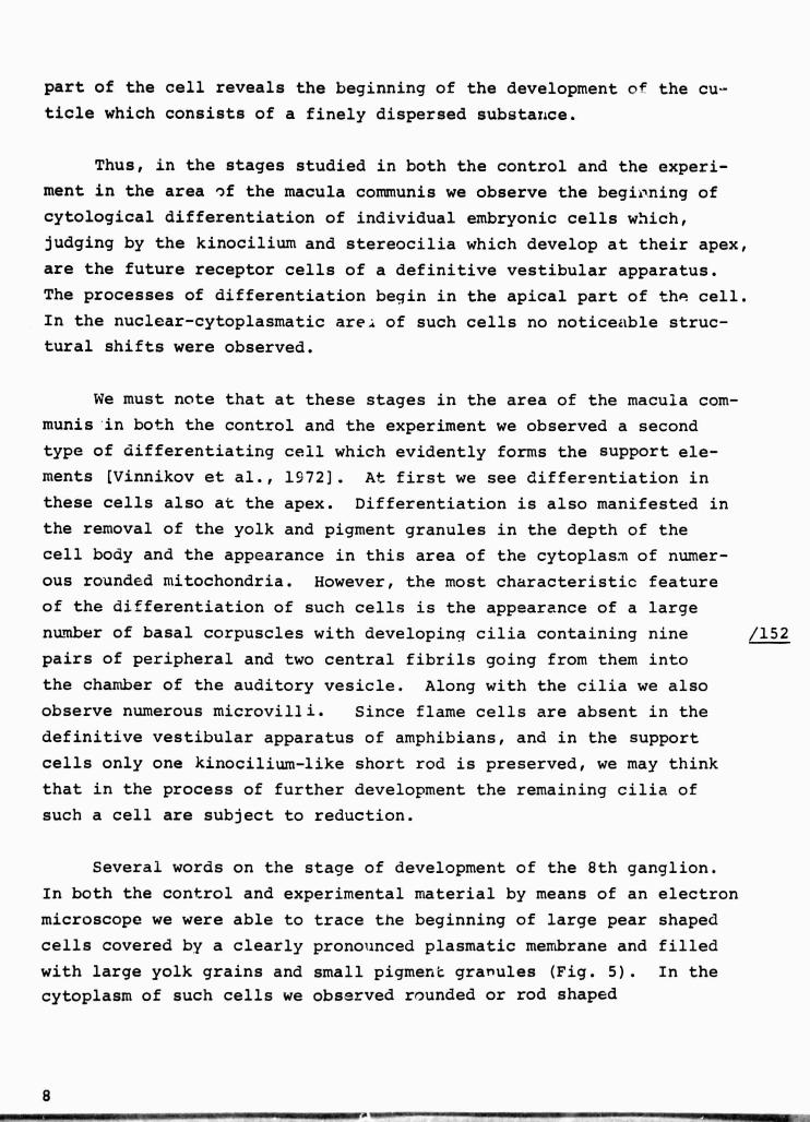
part of the cell reveals the beginning of the development of the cu-ticle which consists of a finely dispersed substance.
Thus, in the stages studied in both the control and the experi-
ment in the area of the macula communis we observe the begioning of
cytological differentiation of individual embryonic cells which,
judging by the kinocilium and stereocilia which develop at their apex,
are the future receptor cells of a definitive vestibular apparatus.
The processes of differentiation begin in the apical part of th? cell.
In the nuclear-cytoplasmatic are: of such cells no noticeable struc-
tural shifts were observed.
We must note that at these stages in the area of the macula com-
munis in both the control and the experiment we observed a second
type of differentiating cell which evidently forms the support ele-
ments (Vinnikov et al., 1972). At first we see differentiation in
these cells also at the apex. Differentiation is also manifested in
the removal of the yolk and pigment granules in the depth of the
cell body and the appearance in this area of the cytoplasm of numer-
ous rounded mitochondria. However, the most characteristic feature
of the differentiation of such cells is the appearance of a large
number of basal corpuscles with developing cilia containing nine /152
pairs of peripheral and two central fibrils going from them into
the chamber of the auditory vesicle. Along with the cilia we also
observe numerous microvilli. Since flame cells are absent in the
definitive vestibular apparatus of amphibians, and in the support
cells only one kinocilium-like short rod is preserved, we may think
that in the process of further development the remaining cilia of
such a cell are subject to reduction.
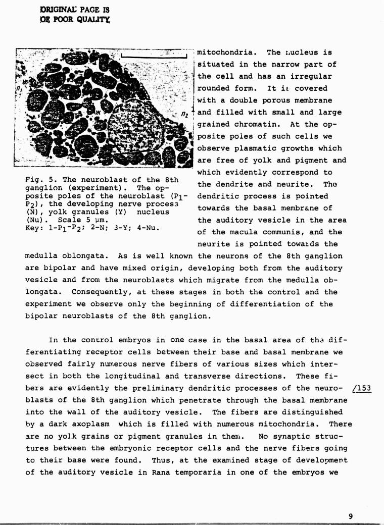
Several words on the stage of development of the 8th ganglion.
In both the control and experimental material by means of an electron
microscope we were able to trace the beginning of large pear shaped
cells covered by a clearly pronounced plasmatic membrane and filled
with large yolk grains and small pigment granules (Fig. 5). In the
cytoplasm of such cells we observed rounded or rod shaped
8
ORIGINAL: PAGE I3
DOE POOR QUALITY
The nucleus is'" 4 }situated in the narrow part of
•. ^.' the cell and has an irregularbt
rounded form. It if coveredwith a double porous membrane
kbW ._. and filled with small and largegrained chromatin. At the op-
posite poles of such cells we
observe plasmatic growths which
` ^• are free of yolk and pigment and
y ^^ which evidently correspond toFig. 5. The neuroblast of the 8th
the dendrite and neurite. Theganglion, (experiment). The op-posite poles of the neuroblast (P l - dendritic process is pointedP 2 ), the developing nerve proces_,(N), yolk granules (Y) nucleus towards the basal membrane of
(Nu). Scale 5 um. the auditory vesicle in the areaKey: 1-P 1 -P 2 ; 2-N; 3-Y; 4-Nu.
of the macula communis, and the
neurite is pointed towards the
medulla oblongata. As is well known the neurons of the 8th ganglion
are bipolar and have mixed origin, developing both from the auditory
vesicle and from the neuroblasts which migrate from the medulla ob-
longata. Consequently, at these stages in both the control and the
experiment we observe only the beginning of differentiation of the
bipolar neuroblasts of the 8th ganglion.
In the control embryos in one case in the basal area of th; dif-
ferentiating receptor cells between their base and basal membrane we
observed fairly numerous nerve fibers of various sizes which inter-
sect in both the longitudinal and transverse directions. These fi-
bers are evidently the preliminary dendritic processes of the neuro- / 153
blasts of the 8th ganglion which penetrate through the basal membrane
into the wall of the auditory vesicle. The fibers are distinguished
by a dark axoplasm which is filled with numerous mitochondria. There
are no yolk grains or pigment granules in ther<<. No synaptic struc-
tures between the embryonic receptor cells and the nerve fibers going
to their base were found. Thus, at the examined stage of development
of the auditory vesicle in Rana temporaria in one of the embryos we
9
were able to see the beginning of the approach of the afferent fibers
to the future receptor cells. We were not able to see this at the
same stages in the experimental embryos. We should note that the
approach of the nerve fibers to the basal part of the cell does; not
accelerate the process of its differentiation. This is demonstrated
by the similarity of the description of the beginning of differentia-
tion in the apical region of the embryonic receptor cells in the con-
trol and experimental animals.
The factual material obtained showed that under conditions in
S
which a fertilized egg was kept for 2 days and then the embryo of the
frog Rana temporaria was kept for 2 days under terrestrial conditions
and then for two days in weightlessness, the development of the em-
bryo in the EMKON container was not delayed. In its external appear-
ance such an embryo practically is ir.distinguishahle from the 4-day
old control which remained for the entire time under conditions of
the unchanging gravitational field of the earth. The embryo (controlF
and experimental) reached. the tail bud stage of development. The
embryo passed through the first stages of division and the subsequent
stages of the blastula and early gastrula under terrestrial conditions.
n Then, at the gastrula stage the embryo, which had undergone the ef-
fect of acceleration during the ascent, continued to develop for 40
hours under conditions of weightlessness, where it passed through the
neurula stage and then into the tail bud stage, at which point it
was fixed. Judging from the results obtained, the last two stages oc-
curred under conditions of weightlessness as th , did under conditions
of the earth's gravitational field. If there were any deviations as-
sociated with the effect of short term acceleration and vibration,
they were corrected in subsequent development under conditions of
weightlessness. As is well known, at there stages amphibian embryos
are distinguished by a broad range of regulation. Judging from the
structure of the auditory vesicles which developed under conditions
of weightlessness, they did not differ in any way from the controls;
there is no doubt that their foundation, induction and formation,
which are related to immersion and subsequent shrinkage from the ec-
toderm under conditions of weightlessness, occurred as in the control.
10AM. - •

In the set up of the experiment is was envisioned that the embryo be
subjected to accelerations at the moment of launch of the space vehi-
cle at the early gastrula stage when the foundation of the future
vestibular apparatus is still lacking. This made it possible to
pre^.rent the acceleration from affecting the development of the audi-
tory vesicle which, as our experiments on the definitive vestibular
apparatus have shown, may affect the s..ructural, cytochemical and
functional organization of the receptor cells (Vi.nnikov, 1974).
Under conditions of weightlessness, not only the formation of the
auditory vesicle, but also the beginning of the process of organ dif-
ferentiation (the macula communis is establish^d and the embryo of
the 8th ganglion is formed) occurs. At t:_e same time, in indivicualsections of the macula ccmmunis the processes of cytological differen-
tiation begin; these are manifested in the beginning of the f :.,-%tion
of individual receptor and support cells. It is basically ::h, ;:i.ces
of these cells which different;ate; t'-.Ls precedes, as was alre
noted, the freeing or the apical part of the cell from yolk and pig-
ment granules and the concentration of mitochondria at this locus.
This indicates an increase in the energy processes in the differen-
tiating parts of the cell. E\_aently, at the later stages of embryo-
genesis, during division of the macula communis, these cells get into
the loci of thc. future utricular, saccular and lagenal maculas and
cristas of the ampoules of the semicircular canals and are converted /154
into the definitive receptor cells.
In the embryonic 8th ganglion in both the control and the experi-
ment cytological differentiation is manifested at the beginnin gs of
the formation of pear shaped b'polar nouroblasts with two cellular pro-
cesses in a polar arrangement. In. one of the control embryos we noted
the penetration of these processes, i.e. the future nerve fibers from
the 8th ganglion, into the epithelium of the auditory vesicle. This
fact may have several explanations. In the first place, in the other
embryos we were not able to find them, which is highly possible when
working with ultrathin sections. In the second place, which is more
probable, this may be explained by the circumstance that, as i^, deli
i
11
known, at the early stages of embryogenesis in embryos in a single
clutch which develop under constant conditions we always observe
some asynchronicity in development. Finally, in the third place,
this may be explained by the fact that because of circumstances out
of our control the control material was fixed 4 hours later than the
experimental. In all it seems to us that this single deviation is
not of fundamental importance. It does not hinder the basic conclusion
of our investigation, that when frog embryos are kept beginning with
the gastrula stage for 2 days in an EM70M con-
tainer in weightlessness on a flight appa-atus their development
practically does not differ from that of control embryos [Vinnikov
et al., 19721.
Thus, conditions of weightlessness practically do not affect the
development of frog embryos at the neurula or tail bud stages, or
organ formation or the beginning of cytological development, includ-
ing that of the future vestibular apparatus, which accompany these
stages.
In order to answer the question of the effect of conditions of
weightlessness on later stages of embryonic development in amphibians
and their transformation into tadpoles, we did a series of experi-
ments on the orbital space stations Salyut-4 a::a Salyut-6 with eggs
of the frog Xenopus laevis which had been fertilized on
earth [Vinnikov et al., 1976, 1980]. Until the launc`i of the space
vehicle the embryos were at the early blastula, middle neurula,
early tail bud and tail bu stages (Table). Development oc-
curred in EMKON biocontainers (Fig. 1) in a special BIOTERNI-4 thermo-
stat F.t 15°C. In some of the experiments fixation was done on board.
the space station during space flight while in other experiments the
larvae were returned alive to earth and processed in the ]-aboratorv.
We found that the development of the embryos and hatching of the lar-
-%: ^_ occurred on the whole normally under conditions of weightlessness
ana that the developing tadpoles moved actively in the EMKON containers
both dur..aq the flight and after the space vehicle landed on earth.
We should note, however, that the nature of their movement underr
12
conditions of weightlessness differs somewhat from their usual be-
havior: the animals reveal an unusual "spinning" movement.
The vestibular apparatus of amphibians, as of other animals, has
a complex spatial configuration. In order to decipher its structure,
in an experiment on the orbital complexes Salyut-6 - Soyuz-26 and
Salyut-6 - Soyuz-36 we prepared a series of frcntal sections of the
heads of the animals for li ght microscopy (the sections were 10 or 15
um thick). From these ultrathin sections were cut for electron micro-
scope analysis [Vinnikov et al., 1980]. When they were examined under
the binocular microscope the experimental animals did not differ
noticeably from the controls, no anomalies in general development were
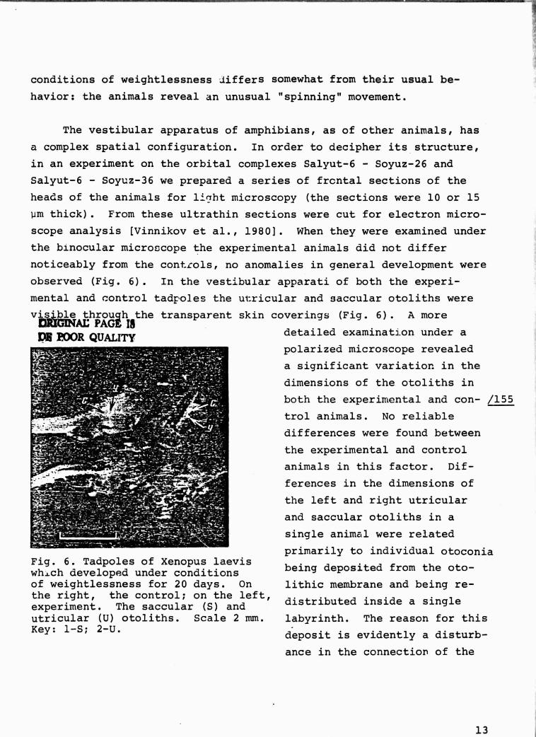
observed (Fig. 6). In the vestibular apparati of both the experi-
mental and control tadpoles the utricular and saccular otoliths were
visible through the transparent skin coverings (Fig. 6). A moreCjRYGIIVAL PAGE 16
DR PWR QUALITY
detailed examination under a
polarized microscope revealed
a significant variation in the
dimensions of the otoliths in
both the experimental and con- /155
trol animals. No reliable
differences were found between
the experimental and control
animals in this factor. Dif-
ferences in the dimensions of
the left and right utricular
and saccular otoliths in a
single animal were related
primarily to individual otoconiaFig. 6. Tadpoles of Xenopus laeviswhich developed under conditions being deposited from the oto-
of weightlessness for 20 days. On lithic membrane and being re-the right, the control; on the left,experiment. The saccular (S) and distributed inside a single
utricular (U) otoliths. Scale 2 mm. labyrinth. The reason for thisKey: 1-S; 2-U.
deposit is evidently a disturb-
ance in the connection of the
13
otoconia with the yolk layer in the otolithic membrane which is caused
by fixation of the material. The study of semi-thin sections also
did not reveal substantial differences between the experimental and
control material. The vestibular apparatus in Xenopus tadpoles
showed utricular and saccular maculas with otolithic complexes con-
sisting of numerous otoconia and cristas of the semicircular canals.
The lagenal macula was not observed at these stages of development.
The maculas were pronounced and differentiated according to the
developmental stage of the animal (Fig. 7). The sac-
• ^ fi+, ''=-^ ' s `:; '^ -- ,̀ •^'•' a 'a :;.^, = ^^' ^^^ - ^
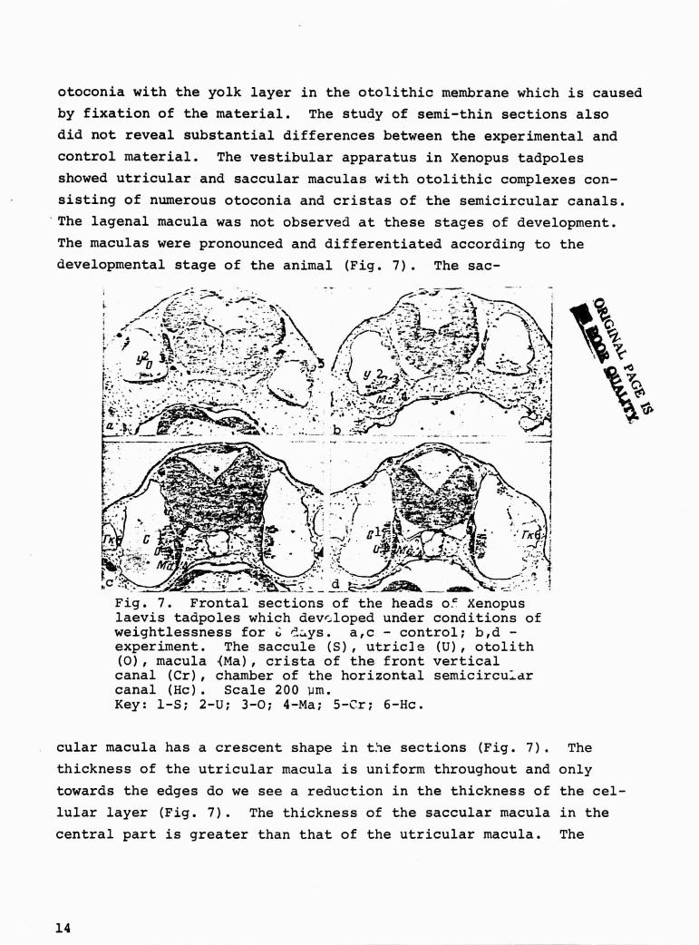
Fig. 7. Frontal sections of the heads o` Xenopuslaevis tadpoles which developed under conditions ofweightlessness for (,- '.-ys. a,c - control; b,d -experiment. The saccule (S), utricle (U), otolith(0), macula (Ma), crista of the front verticalcanal (Cr), chamber of the horizontal semicircu--drcanal (Hc). Scale 200 um.Key: 1-S; 2-U; 3-0; 4-Ma; 5-Cr; 6-Hc.
cular macula has a crescent shape in t.ie sections (Fig. 7). The
thickness of the utricular macula is uniform throughout and only
towards the edges do we see a reduction in the thickness of the cel-
lular layer (Fig. 7). The thickness of the saccular macula in the
central part is greater than that of the utricular macula. The
t
N
14
Ai
nuclei of the support and receptor cells in the utricular macula are
arranged in a single row, while in the saccular macula the cell nuclei
lie at various levels (Fig. 7). In the cellular nuclei ene or more
rarely two smal.L dense nucleoli are clearly visible (Iig. 7). The
cells within a single macula, particularly the saccular macula, vary
in optical density. In the basal section we can see light, vacuol-
ized formations. Similar vacuoles are found in the experimental and
control animals; they are clearest in the saccular macula. The oto-
conia differ in dimensions, have polygonal forms arid double refrac-
tion. In the utricle the otolithic membrane has fewer otoconia than
in the saccule.
An examination of ultrathin sections showed that the receptor
and support cells of the maculas of the otolithic organs are highly
differentiated. On the surface of the receptor cells is a well de-
veloped cuticle from which comes a cluster of sensory hairs consist-
ing of a large number of stereocilia and one kinocilium in a polar
arrangement. The cytoplasm of the receptor cell is rich in ribo- /156
somes and polysomes and has a large number of mitochondria with
matrices with high electron density. The Golgi apparatus and the en-
doplasmatic network are moderately well developed. The nerve endings
go towards the base of the cell and they for:* synaptic contacts with
the cell. In the area of contact with the afferent endings in the
receptor cell is a spherical synaptic corpuscle with high electron
density which is surrounded by synaptic vesicles (Fig. 8). In the
basal sections of the receptor cel.i the synaptic vesicles are indis-
tinct. An electron microscope analysis of the otoconia in the oto- /157
lithic organs showed that they consist of a thin fibrillar matrix
with material of high electron density. Within the otolithic complex
of a single receptor organ the type of inclusions of material of high
electron density and the clarity of the fibrillar matrix differ in
the otoconia.
Thus, a comparison of light and electron microscopic examinations
of the otolithic organs and a light microscope examination of the
crista of the semicircular canals in Xenopus which developed in
15

DISMAL; PAGjt fS
Dl!''OOR QUALITY
Fig. 8. The basal region of the cell of the utricularmacula in Xenopus (20 day experiment). Receptor cell(Rc), support cell (Sc), afferent nerve ending (N).Scale 1 um.Key: 1-Rc; 2-Sc; 3-N.
weightlessness and on earth showed that there are no significant dif-
ferences in the structure of these organs or anomalies in the develop-
ment of the vestibular apparatus in either group of animals.
The Development of the Vestibular Apparatus in Bony Fish
The experiments which we did to examine the development of the
vestibular apparatus in embryos of the fish Brachydanio rerio at the
gastrula and five somite stage on space vehicles Soyuz-16, -21 and
-22 (Tal-)le) showed that the general development under conditions of
space flight proceeds normally. The embryos developed at 23.5°C.
In the experimental larvae as well as in the control larvae which de-
veloped in weightlessness the vestibular apparatus was comprised of
the utricle, saccule and embryos of the semicircular canals. The
otolithic apparatus consisted of utricular and saccular otoliths (Fig.
9). After preparation the material was made into a semi-thin sec-
tion of 10 mk in order to study the formation of the utricular oto-
lith under the light microscope. The same sections were used for
ultrastructural examinations. Under the light microscope we were not
16
V^ Ar
PACE rsR
QUALITY
able to detect any deviations in the
general structure of the vestibular
apparatus of the experimental and
control larvae (Fig. 10). During an
examination under the electron micro-
scope special attention was paid to
the structure of the receptor lin-
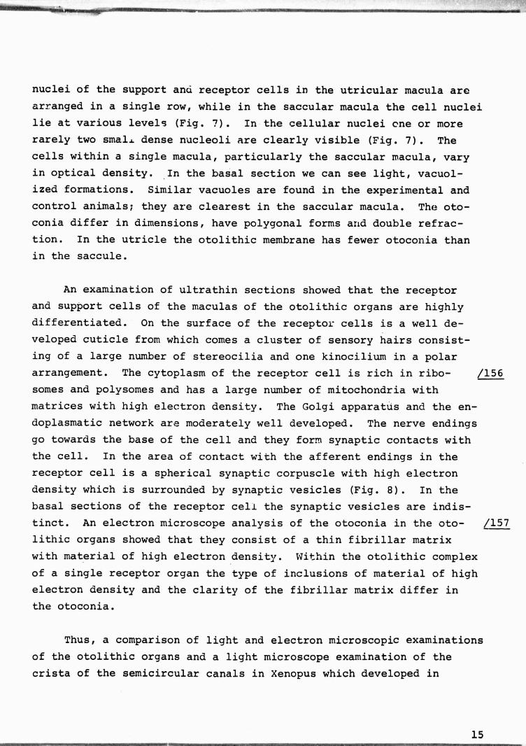
.ig. 9. Larvae of Brachydanio ings of the utricle and sacculererio which developed underconditions of weightlessness and their otolithic complex. An exam-
for 9 days (experiment). Sac- ination of the receptor epitheliumcular (S) and utricular (U) showed that in both the experimentalotoliths. Scale 0.5 mm.Key: 1-S; 2-U. and control larvae its central part
consists of clearly differentiated
receptor and support cells (Fig. 11).
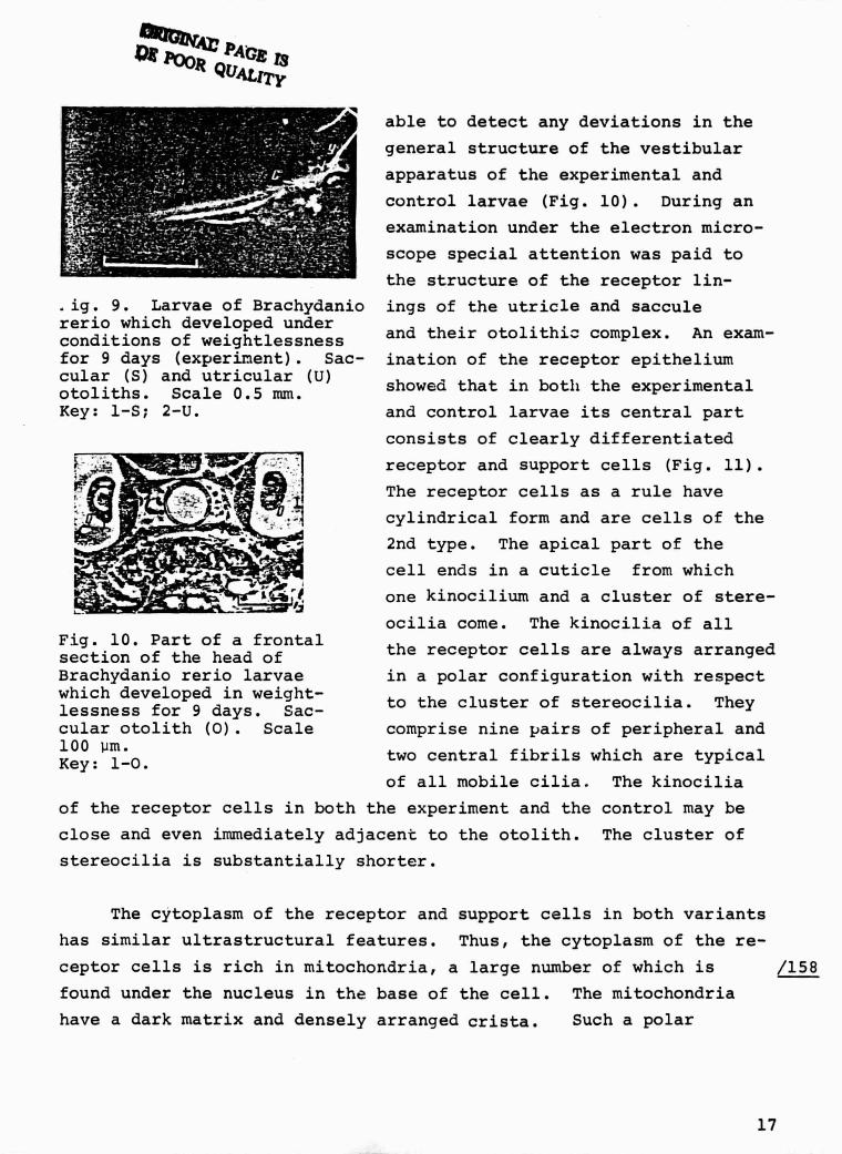
The receptor cells as a rule havecylindrical form and are cells of the
2nd type. The apical part of the
cell ends in a cuticle from which
ra yone kinocilium and a cluster of stere-
ocilia come. The kinocilia of allFig. 10. Part of a frontal the receptor cells are always arrangedsection of the head ofBrachydanio rerio larvae in a polar configuration with respectwhich developed in weight- to the cluster of stereocilia. Theylessness for 9 days. Sac-cular otolith (0). Scale comprise nine ,airs of peripheral and100 um. two central fibrils which are typicalKey: 1-0.
of all mobile cilia. The kinocilia
of the receptor cells in both the experiment and the control may be
close and even immediately adjacent to the otolith. The cluster of
stereocilia is substantially shorter.
The cytoplasm of the receptor and support cells in both variants
has similar ultrastructural features. Thus, the cytoplasm of the re-
ceptor cells is rich in mitochondria, a large number of which is /158
found under the nucleus in the base of the cell. The mitochondriar.
have a dark matrix and densely arranged crista. Such a polar
17

OP Poo' Q at J$
ALlrx
arrangement of the mitochondria is ex-
plained by the fact that the nerve end-
ings go towards the base of the receptor
cell and form synapses with the receptor
cells.
Pro At this stage of development the
"'^,r* .= nerve fibers are amyelinic and go to-
,.c^; wards the receptor cells from the basal
membrane. The bud shaped nerve endings
Fig. 11. A section of the which are in contact with the receptorutricular macula and oto-
cells are light, having clearly definedlith of Brachydanio reriowhich developed in weight- neurofibrils and small mitochondria.lessness for 9 days. Oto-lith (0), nucleus of the Evidently these are afferent nerve end-
otolith (No), receptor ings. In the region of the synapsescell (Rc), cluster ofstereocilia (St). Scale in both the experiment and the control,
10 Um. so-called synaptic corpuscles are oftenKey: 1 -0; 2 -No; 3-Rc;4-St. visible. This indicates that the re-
ceptor cells are in an active functional.
state. As distinct from the receptor cells, the cytoplasm of the sup-
port cells has a more highly developed endoplasmati.c reticulum, se-
cretory vesicles and vacuoles. The support cells are arranged with
their bases on the basal membrane. On their apical ends they may
have microvilli.
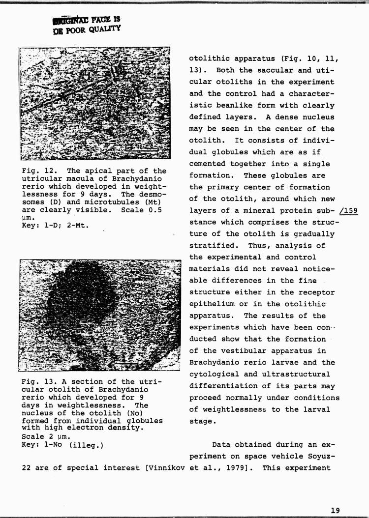
We should note the well developed system of specialized cellular
contacts in the receptor linings which are represented by a large
number of desmosomes which are arranged both on the apical surface of
the receptor epithelium zone and through all its levels (Fig. 12).
Such contacts may be formed both between the receptor cells and be-
tween the receptor and support cells. Thick clusters and long threads
of tonofibrils which consist, in turn, of clusters of protofibrils,
come from the desmosomes. Such a mechanical support system is seen
to equal degree in the experiment and the control.
There were also no clear differences in the structure of the
18
^GIIPA^ gA^'E IS01[ POOR QUALITY
Fig. 12. The apical part of theutricular macula of Brachydaniorerio which developed in weight-lessness for 9 days. The desmo-somes (D) and microtubules (Mt)are clearly visible. Scale 0.5Pm.Key: 1-D; 2-Mt.
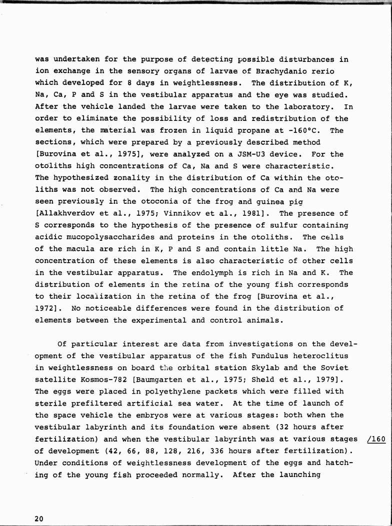
Fig. 13. A section of the utri-cular otolith of Brachydaniorerio which developed for 9days in weightlessness. Thenucleus of the otolith (No)formed from individual globuleswith high electron density.Scale 2 um.
otolithic apparatus (Fig. 10, 11,
13). Both the saccular and uti-
cular otoliths in the experiment
and the control had a character-
istic beanlike form with clearly
defined layers. A dense nucleus
may be seen in the center of the
otolith. It consists of indivi-
dual globules which are as if
cemented together into a single
formation. These globules are
the primary center of formation
of the otolith, around which new
layers of a mineral protein sub- /159
stance which comprises the struc-
ture of the otolith is gradually
stratified. Thus, analysis of
the experimental and control
materials did not reveal notice-
able differences in the fine
structure either in the receptor
epithelium or in the otolithic
apparatus. The results of the
experiments which have been con-
ducted show that the formation
of the vestibular apparatus in
Brachydanio rerio larvae and the
cytological and ultrastructural
differentiation of its parts may
proceed normally under conditions
of weightlessness to the larval
stage.
Key: 1-No (illeg.)
Data obtained during an ex-
periment on space vehicle Soyuz-
22 are of special interest (Vinnikov et al., 1979]. This experiment
19
was undertaken for the purpose of detecting possible disturbances in
ion exchange in the sensory organs of larvae of Brachydanio rerio
which developed for 8 days in weightlessness. The distribution of K,
Na, Ca, P and S in the vestibular apparatus and the eye was studied.
After the vehicle landed the larvae were taken to the laboratory. In
order to eliminate the possibility of loss and redistribution of the
elements, the material was frozen in liquid propane at -160°C. The
sections, which were prepared by a previously described method
[Burovina et al., 19751, were analyzed on a JSM-U3 device. For the
otoliths high concentrations of Ca, Na and S were characteristic.
The hypothesized zonality in the distribution of Ca within the oto-
liths was not observed. The high concentrations of Ca and Na were
seen previously in the otoconia of the frog and guinea pia
[Allakhver.dov et al., 1975; Vinnikov et al., 1981]. The presence of
S corresponds to the hypothesis of the presence of sulfur containingacidic mucopolysaccharides and proteins in the otoliths. The cells
of the macula are rich in K, P and S and contain little Na. The high
concentration of these elements is also characteristic of other cellsin the vestibular apparatus. The endolymph is rich in Na and K. The
distribution of elements in the retina of the young fish corresponds
to their localization in the retina of the frog [Burovina et al.,
1972]. No noticeable differences were found in the distribution of
elements between the experimental and control animals.
of particular interest are data from investigations on the devel-opment of the vestibular apparatus of the fish Fundulus heteroclitus
»- in weightlessness on board t:Le orbital station Skylab and the Soviet
satellite Kosmos-782 [Baumgarten et al., 1975; Sheld et al., 1979].
The eggs were placed in polyethylene packets which were filled with
sterile prefiltered artificial sea water. At the time of launch of
the space vehicle the embryos were at various stages: both when the
vestibular labyrinth and its foundation were absent (32 hours afterfertilization) and when the vestibular labyrinth was at various stages /160
of development (42, 66, 88, 128, 216, 336 hours after fertilization).
Under conditions of weightlessness development of the eggs and hatch-ing of the young fish proceeded normally. After the launching
20
apparatus had splashed down part of the material was fixed for micro-
scopic examinatio 6 : light, transmission and scanning electron micro-
scopy. The authors were not able to detect perceptible differences
in the dimensions, form or surface relief in the otoliths in the ex-
perimental and control fish [Sheld et al., 1979]. The ultrastructure
of the otoliths and macular cells in the flight animals was normal.
In addition to the vestibular apparatus the development of the central
nervous system, the eye and the cardiovascular system was studied.
No significant structural disturbances were observed here either. In
addition to the morphological studies, behavioral studies were under-
taken both on board Skylab and on earth after landing. It was found
that under conditions of space flight there may be unusual movement
of the young fish which is of cyclic nature. Furthermore, deviations
in the geotaxial reaction were observed after 6 month adaptations on
earth in fish which had been at the 32 hour stage at the time of the
launch [Shell et al., 1979). On the whole, from the evidence of
these authors we may say that fish which were in space are more sen-
sitive to environmental conditions than are the control animals.
t-___1 „O; ^1
The question of the development of the vestibular apparatus under
conditions of a constant gravitational field is of fundamental impor-
tance and was examined by us in the context of the general problem of
differentiation of the receptor apparatus under the effect of on ap-
propriate stimulus. It is well known, for example, that the phctc-
receptors of amphibians can undergo final differentiation either when
the animals are in light or in complete darkness [Eakin, 1965].
In our laboratory L. K. Titova [1968] studied the embryology of
the vestibular apparatus in vertebrates as well as in fish and amphi-
bians. The basic ultrastructural, cytochemical and functional char-
acteristics of differentiation of the receptor structures and their
relation to the brain and the developing otolithic apparatus were
traced. From these data we may suggest that differentiation of the
receptor structures in the vestibular apparatus during embryogenesis
i
21
'rte,
is related to the pressure of the developing otoliths on the surface 161
of the utricular and saccular maculas. If this is actually so, then
under conditions of weightlessness we may expect disturbances in the
normal differentiation of the receptor structures of the vestibular
apparatus. The possibility cannot be excluded that disturbances in
the normal formation of the otoliths themselves occur in weightless-
ness. For this reason, the establishment of the presence or absence
of a causal link between the effect of the gravitational force and
differentiation of the gravitation receptor was of considerable prac-
tical interest.
However, experiments which study the development of the vesti-
bular system are significantly hindered by the fact that it is prac-
impossible to create conditions of complete deprivation - in all
transferrals of the animal, whether on earth or under conditions cf
weightlessness, inertial forces arise which are adequate stimuli for
one or another receptor organ in the inner ear. Nevertheless, the
use of methods which simulate changes in the gravitational field such
as klinostating, centrifuging and the creation of weightlessness,
contribute substantially to the solution of this problem. However,
we must note that we must proceed very carefully in approaching experi-
ments which study the effect of klinostating as a model of weightless-
ness on morphogenesis of the vestibular apparatus, and in particular
with respect to data on the appearance under "zero gravity" of vacu-
oles filled with necrobiotic mitochondria and bits of the endoplas-
matic network in the basal part of the maculas. We have in mind the
experiments of Neubert (Neubert, 1979) and Neubert and Briegleb [Neu-
bert, Briegleb, 19801 and experiments which studied the effect of
klinostating on the development of the vestibular apparatus in embry-
os of Rana temporaria. We must recall that from the moment that a
highly sensitive specialized receptor appears, the gravitational
forces arising during rotation of the klinostating chamber and the
constantly acting force of gravity are adequate stimuli for the ves-
tibular system and, consequently, that klinostating may be used as
a model of weightlessness only at the very early stages of develop-
ment of the labyrinth. It was precisely at these stages that Neubert
22
a
and Briegleb [Neubert, Briegleb, 19801 did not find any structural±
changes in the developing vestibular appar.ati of embryos in the kline-
stating chamber in comparison with the control.
The factual data presented al)ove show that in fish and amphibian
embryos which develop in weightlessness beginning at the stages at
which the vestibular apparatus is still absent or is at various stages
of morpnogenesis, there is normal formation of the maculas, cristas
and otolithic apparati [Vinnikov.et al., 1972, 1979, 1980; Sheld et
al., 19791. No significant qualitative (and we emphasize qualitative)
disturbances or delays in the formation of the ultrastructural organi-
zation of the receptor and support cells were observed. Calcification
of the otoliths proceeds on the whole normally; this is shown by the
presence of double refraction, high hardness of the otoliths and their
high Ca concentration. Thus, a specific stimulus - the force of gra-
vity - is evidently not a necessary condition for the formation of the
structural organization of the vestibular apparatus at the early
stages of ontogenesis. At the same time, we are alert to the fact
that the unusual movement of the young animals (cyclic movement in
the fish and spinning movement in the tadpoles) is maintained to one
degree or another practically throughout the flight [Baumgarten et
al., 1975; Sheld et al., 1979]. However, after landing in conditions
of normal gravitation, the larvae very quickly acquire the horizontal
position and begin to move as the controls do. This indicates that
the adaptation of the larve to the unusual environmental conditions
should not be seen as fixed. This does not preclude the fact that
the prolonged effect of weightlessness may exert a certain effect on
the development of the structural functional organization of the ves-
tibular apparatus. However, in experiments which studi o-' the effect
of prolonged exposure to hypergravitation (or centrifuging) on the /162
formation and development of the vestibular apparatus in rats, no
important structural changes were observed (Lim et al., 1974). we
may only hope that future studies on long periods of flight, in
which it will be possible to trace the development of the labyrinth
as the larvae are transformed into adult animals, will answer this
question.
23
REFERENCES
1. Allakhverdov, B.L., Burovina, I.V., Govardo ,.skiy, I.I. andKoychev, K.A., "The ion composition of the :ells in the Laby-rinth and its surroundings", Dokl. AN SSSR 220/3, 746 (1975).
2. Baumgarten, R.J. von, Simmonds, R.C., Boyd, J.F. and Garriott,O.K., "Effects of prolonged weightlessness on the swimmingpattern of fish aboard Skylab 3," Aviat. Space and Environ.Med. 46, 902 (1975).
3. Burovina,I.V., Chmykhova, N.M. and Shapovalov, A.I., "A cuanti-tative determination of the potassium concentration in themotor neurons of the frog spine by the method of roentgenspectral microanalysis," Zh. evolyuts. biokhim. i fiziol. 11/3,282 (1975).
4. Burovina, I.V., Govardovskiy, V.I. and Sidorov, A.F., ":'pecificfeatures of the distribution of K, Ca, P and Na in the net-work of the frog during light and dark adaptation," Dokl. ANSSSR 206/1, 222 (1972).
5. Eakin, R.M., "Differentiation of rods and cones in the darkness,"J. Cell. Eiol. 25, 162 (1965).
6. Flock, A., Cheung, H.C., Flock, B. and Utter, G., "Three sets ofactin filaments in sensory cells of the inner ear," J. Neuro-cytol. 10, 133 (1981).
7. Ginzburg, A.S., "Species features of the determination of thelabyrinth in the triton," Dokl. AN SSSR 54, 377 (1946).
8. Kogan, R.Ye., "The chordomesoderm as an inductor of the auditoryvesicles," Dokl. AN SSSR 45, 42 (1944).
9. Lim, D.J., Stith, J.A., Stockwell, C.W. and Oyama, J., "Observa-tions on saccules of rats exposed to long-term hypergravity,"Aerospace Med. 45, 705 (1974).
10. Neubert, J., "UltraFtructural development of the vestibularsystem under conditions of simulated weightlessness," Aviat.Space Environ. Med. 50, 1058 (1979).
il. Neubert, J. and Briegleb, W., Changes in the microstructure ofthe gravity sensory organ of tadpoles (Rana temporaria) devel-oped in zero-g simulation conditions, Cospar F. 2.3.2., Bumpest, 1980.
12. Sheld, G.V., Boyd, J., Fuller, R.M., Hoffman, R.B., Kif, G.R.,Oppenheimer, G.M. and Salinas, G.A., "The development of Pun-dulus heteroclitus in weightlessness" In: Biologicheskiye is-sledo ,,aniya na biosputnikakh "Kosmos" (Biological studies on
24
AL

board the biosatellite " Kosmos", Moscow, Nauka, 1979, p. 54.
13. Titova, L.K., Razvitiye retseptornykh strukt , ir vnutrenriego ukhapozvcnochnykh ( The development of receptor structures i n theinner ear of vc -tebrates), Leningrad, Nauka, 1968, p. 192.
14. Vinnikov, Ya.A., "Evolution of the grarity receptor," MinervaOtolaryngol. 24, 1 (1974).
15. Vinnikov, Ya.A,,Gazenko,O.G., Titova, L.K., Bronshtevn, A.A.,Govardovskiy, V.T., Palmba'ch, L.R., Pevzner, R.A., Gribakin,F.G., Aronova, M.Z., Kharke yevich, T.A., Tsurulis, T.P.,Pyatk`'r.a, G.A., and Semak, T.V., "Formation n,' vestibular ap-paratus in the weightless condition," Minerve o:t rinolaryngol.26, 69 (1976).
16. Vinnikov, Ya.A., Gazenko, O.G., Titova, K.K., Bronshteyn, A.A.,Govardovskiy, V.I., Pevzner, R.A., Gribakin, F.V., Aronova,M.Z., Kharkeyevich, T.A., Tsirulis, T.F., Pyatkina, G.A.,Semak, T.V. and Pal'mbakh, L.R., "The development of the ves-tibular apparatus under conditions of weightlessness," Arkhivanat., gistol. i embriol. 70/1, 11 (1976).
17. Vinnikov, Ya.A., Gazenko, O.G., Titova, L.K., Govardovskiy, V.I.,Gribakin, F.G., Brons'.lteyn, A.A., Pevzner, R.A., Aronova, M.Z.,Mashanskiy, A.L., Pal'mbakh, L.R., Ivanov, V.P., Tsurulis, T.P.,Kharkeyevich, T.A. a-id Pyatkina, G.A., "The development of thevestibular apparatus (labyrinth) in the frog Rana temporariaunder conditions of weightlessness," Zh. evolyuts. biokhim. i_fiziol. 6/3, 343 (1972).
^rt
18. Vinnikov, Ya.A., Gribakin, F.G., Burovina, I.V., Lychakov, D.V.,and Adanina, V.O., A study of-the sensory or(-ans of fishwhich develop under conditions of weightlessness, In: Tez. IIVseso uzn, konf. po elektronnoy mikroskopii ;Summary of theII All-Union conference on electron microscopy], Moscow, Nauka,1979, vol. 2, p. 174.
19. Vinnikov, Ya.A., Lychakov, D.V., Koychev, K.A., bcyadzhieva-Mikhaylova, A., Khristov, I. and Lavrova, Ye.A., "A study ofthe otolithic membrane of the utricle of the guinea pig," Zh.evolyuts. biokhim. i fiziol. 17/5, 474 (1981).
20. Vinnikov, Ya., Lychakov, D.V., Pal'mbakh, L.R., Govardovskiy, V.I., Adanina, V.O., Allakhverdov, B.L. and Pogorelov, A.G., "Astudy of the vestibular apparatus of the frog Xenopus laevisand rats under conditions of prolonged weightlessness," Zh_.evolyuts. biokhim. i fiziol. 16/6, 574 (1980).
21. Young, R . S.. Tremor, J.W., "The effect of weightlessness on thedividing egg of Rana pipiens," Bioscience 18, 609 ( 1968).
25