
the development of the cerebrospinal fluid spaces and choroid ...
33
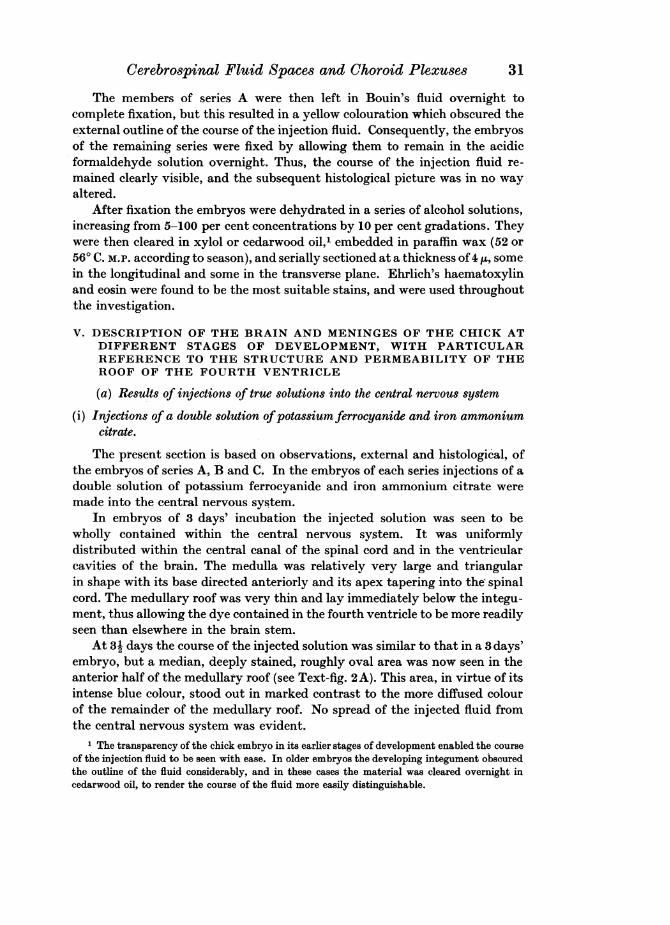
THE DEVELOPMENT OF THE CEREBROSPINAL FLUID SPACES AND CHOROID PLEXUSES IN THE CHICK BY HENRY COHEN AND SARAH DAVIES From the Department of Medicine, Universzty of Liverpool CONTENTS PAGE I. Introduction .23 II. Survey of literature (a) The development of the cerebrospinal fluid spaces in the Mammalia 23 (b) The avian meninges ..26 III. Material .. .27 IV. Methods of investigation .. .27 V. Description of the brain and meninges of the chick at different stages of development, with particular reference to the structure and permeability of the roof of the fourth ventricle (a) Results of id4ections of true solutions into the central nervous system (i) Injections of a double solution of potassium ferrocyanide and iron ammonium citrate .. .31 (ii) Injections of a solution of iron ammonium citrate alone 48 (b) Results of injections of true solutions into the vascular system 49 VI. General Summary and Discussion .. .. .50 VII. Conclusions .52 References .52 Explanation of Plates I and II .53 I. INTRODUCTION THE work described in this paper is part of a study comprising the com- parative morphology and development of the choroid plexuses, the meninges and the meningeal spaces. Chick embryos of accurately determined age were investigated, utilizing a similar method to that adopted by L. H. Weed in his classical studies on pig and human embryos. II. SURVEY OF LITERATURE (a) The development of the cerebrospinal fluid spaces in the Mammralia Two workers, L. H. Weed (1917) and J. J. Keegan (1917), have investi- gated the development of the cerebrospinal fluid spaces by the method of injecting a true solution into the central nervous system of the living embryo. Weed's comprehensive monograph appeared in 1917, and very shortly after Keegan published a very brief report of his own investigations. In a personal communication to us, Dr Keegan stated that in view of Weed's earlier publica-
-
Upload
truongtruc -
Category
Documents
-
view
220 -
download
0
Transcript of the development of the cerebrospinal fluid spaces and choroid ...
THE DEVELOPMENT OF THE CEREBROSPINAL FLUIDSPACES AND CHOROID PLEXUSES IN THE CHICK
BY HENRY COHEN AND SARAH DAVIES
From the Department of Medicine, Universzty of Liverpool
CONTENTSPAGE
I. Introduction .23II. Survey of literature
(a) The development of the cerebrospinal fluid spaces in the Mammalia 23(b) The avian meninges ..26
III. Material .. .27IV. Methods of investigation .. .27V. Description of the brain and meninges of the chick at different stages of development,
with particular reference to the structure and permeability of the roof of the fourthventricle(a) Results of id4ections of true solutions into the central nervous system
(i) Injections of a double solution of potassium ferrocyanide and ironammonium citrate .. .31
(ii) Injections of a solution of iron ammonium citrate alone 48(b) Results of injections of true solutions into the vascular system 49
VI. General Summary and Discussion .. . . .50VII. Conclusions .52
References .52Explanation of Plates I and II .53
I. INTRODUCTION
THE work described in this paper is part of a study comprising the com-parative morphology and development of the choroid plexuses, the meningesand the meningeal spaces. Chick embryos of accurately determined age wereinvestigated, utilizing a similar method to that adopted by L. H. Weed in hisclassical studies on pig and human embryos.
II. SURVEY OF LITERATURE
(a) The development of the cerebrospinal fluid spaces in the Mammralia
Two workers, L. H. Weed (1917) and J. J. Keegan (1917), have investi-gated the development of the cerebrospinal fluid spaces by the method ofinjecting a true solution into the central nervous system of the living embryo.Weed's comprehensive monograph appeared in 1917, and very shortly afterKeegan published a very brief report of his own investigations. In a personalcommunication to us, Dr Keegan stated that in view of Weed's earlier publica-
Henry Cohen and Sarah Davies
tion of almost identical work, he felt it was unnecessary to make a furtherdetailed report of his own findings. In the light of the present investigation,however, Dr Keegan's paper, brief though it was, proved to be of great interest.
Weed (1917) investigated the condition of the cerebrospinal fluid spaces inpig embryos at different stages of development. He replaced the fluid withinthe central nervous system by a 1 per cent double solution of potassium ferro-cyanide and iron ammonium citrate. Fixation of the embryo in an acidmedium secured precipitation of Prussian blue (ferric ferrocyanide) in situand thus revealed the path taken by the injection solution. He records that thefirst extra-ventricular spread of the injected solution occurred in pig embryosof 14 mm. length. Further, he found that the injected fluid made its escapeinto the surrounding mesenchymal tissue through two differentiated areas, oneanterior and the other posterior, in the roof of the fourth ventricle. Weed termsthese areas the area nzembranacea superior and the area membranacea inferiorrespectively, and states that both " differentiate at a slightly earlier period thanthat at which they function actively " (1917, p. 107). The cells of these areas aredistinguishable by their low stature and narrow, oval or slightly elongatednuclei.
The area membranacea superior is first differentiated in embryos of 11 mm.and reaches its maximum development at the 18 mm. stage. The fully differ-entiated form is maintained until about the 20 mm. stage, when the areacommences a gradual regression, and disappears completely in embryos of33 mm. Its disappearance is brought about by the encroachment of the de-veloping cerebellum on the roof of the fourth ventricle, and by the formation ofthe choroid plexus.
The inferior area -shows the first signs of differentiation in embryos of15 mm.; it develops but slowly until the 18 mm. stage is reached, then differ-entiates extremely rapidly. Unlike the superior area, it persists and " graduallyoccupies the major portion of the velum choroidea inferior" (Weed, 1917,p. 107).
The first escape of fluid from the spaces of the central nervous system in the14 mm. embryos was found to be coincident with the first indication oftufting of the developing choroid plexus of the fourth ventricle. A furthermarked extra-ventricular extension of the injected solution occurred inembryos of 19 mm., and this was associated with the development of thechoroid plexuses of the third and lateral ventricles at this stage. Furthermore,these plexuses were only definitely differentiated in embryos of 23 mm., andat this stage a complete periaxial spread occurred. From these observationsWeed concludes that "there seems to be a very definite relationship betweenthe developing choroid plexuses and the periaxial spread of the embryoniccerebrospinal fluid" (1917, p. 93)..
Weed has also identified and described similar areas of differentiation in theroof of the fourth ventricle of human foetuses at different stages of develop-ment, and the sequence of events resembles that found in pig embryos.
24
Cerebrospinal Fluid Spaces and Choroid Plexmese 2
The presence of an area membranacea superior is also recorded in the em-bryos of chick, rabbit, sheep and cat, but no mention is made of an inferior areain these types.
Keegan (1917) injected a 1 per cent double solution of potassium ferro-cyanide and iron ammonium citrate into the brain cavities of living rabbit andchick embryos at various stages of development, and secured precipitation ofPrussian blue granules by fixation in an acid medium.
He records in both types the presence of two thin oval areas, one anteriorand the other posterior to the choroid plexus, in the roof of the fourth ventricle.Each area was associated with a dense collection of protein coagula on itsventricular surface. In all early embryos injected with the double solution, acondensation of precipitated Prussian blue granules was also found below theseareas.
It is further reported that "injection with the double solution in rabbitembryos up to the age of 17 days showed no extra-ventricular spread of thefluid, which was rather surprising in view of the fact that the choroid plexus isquite well developed at this age" (Keegan, 1917, p. 379). On the other hand,when a solution of ferric ammonium citrate alone was injected, extra-ventri-cular spread of the fluid occurred in the region of the fourth ventricle before thedevelopment of the choroid plexus, and there was only a slight condensation ofblue granules below the thin areas of this roof. "After the development of thechoroid plexus there was a very rapid absorption into the circulation "' (Keegan,1917, p. 380).
Similarly, in chick embryos injected with the double solution, no extra-ventricular spread of the fluid had occurred, even in 9 days' embryos, althoughthe choroid plexus was present at this stage. Again, when the citrate solutionalone was injected, extra-ventricular spread of the fluid invariably occurred,"and after the development of the choroid plexus (sixth to seventh day) veryrapidly entered the circulation. At the seven day stage, a considerable portionremained within the ventricle and as a very evident blue coloration in themesenchymal tissue over the rhombencephalon and mesencephalon.... In theeight and nine day chick the escape of the citrate solution was even more rapid,practically all leaving the ventricle within 10-20 min." (Keegan, 1917, p. 380).
Keegan infers therefore that "this membranous area of the roof of thefourth ventricle is non-permeable to the double solution in the early embryostages while it is permeable to the citrate solution; that a slight escape of thecerebrospinal fluid occurs before the development of the choroid plexus; andthat the collection of protein coagula and Prussian blue granules in contactwith the inner surface of the membranous area represents a dialysis pheno-menon of this semi-permeable membrane" (Keegan, 1917, p. 380).
No details are given of the histological features of the membranous areas inthe roof of the fourth ventricle, nor of the exact stages at which these areasmake their appearance; neither is any mention made of their ultimate fate.Moreover, Keegan in his use of the term "choroid plexuses" does not clearly
25
Henry Cohen and Sarah Davies
distinguish between those of the lateral and third ventricles and that of thefourth ventricle-a distinction which is shown to be important in the light ofthe present investigation.
(b) The avian meninges
Cuvier (1809) and Owen (1868J stated that three meninges are present inbirds, a pia mater, arachnoid and dura mater, essentially similar to thosefound in the Mammalia and bearing the same relation to each other.
Sterzi (1902), on the other hand, states that only two meninges are presentin birds, an outer, thin, non-vascular -dra mater, and an inner, secondarymeninx, these being comparable to the two meninges found in the reptilia. Thesecondary meninx further consists of thiee layers, a thin, outermost endo-thelial layer, a middle vascular layer of loose texture, and an inner layerimmediately adjacent to the cord. This structure of the secondary meninxshows an advance on the reptilian condition, where it is a thin single layer, andforeshadows the pia and arachnoid of the Mammalia.
Streeter (1904), investigating the condition of the meninges in the ostrich,asserts that three membranes are present, similar to those found in theMammalia.
The first detailed description of the development of the avian meningesappears to be that given by Farrar in 1906. In his description of the develop-ment of the pia-arachnoid membrane in the chick, Farrar designates thegeneral mesenchymal tissue surrounding the embryonic nervous system as thearachnoid sheath or mesh, and states that the pia-arachnoid sheath arises fromthe cells of the arachnoid mesh immediately adjacent to the nervous system.The pia-arachnoid develops as a "single membrane consisting of a loosereticulum, the trabeculae of which are formed by the branching and anastomos-ing processes of connective tissue cells. At the outer and inner borders thesecells tend to arrange themselves horizontally to form limiting membranes"(Farrar, 1906, p. 297). Blood channels make their appearance within the de-veloping pia-arachnoid and increase in number to such an extent as to form analmost uninterrupted chain of vascular spaces within this zone. These bloodchannels are essentially similar to those found in the general arachnoid mesh.While the pia-arachnoid is developing from the inner zone of the arachnoidmesh, a second outer non-vascular zone of condensation makes its appearance,separated from the pia-arachnoid by an intermediate region of undifferentiatedmesenchyme. In this outer zone, " the connective tissue elements are assumingelongated forms and crowding together with long axes parallel, giving rise to avery close mesh with long but extremely narrow spaces, in contradistinctionto the loose, irregular reticulum of the pia-arachnoid" (Farrar, 1906, p. 297).This outer zone is the primitive dura and is clearly demarcated from the pia-arachnoid. According to Farrar, therefore, two meninges are present in thechick, an outer dura and an inner pia-arachnoid; " in the thickness of the latter,
26
Cerebrospinal Fluid Spaces and Choroid Plexuses
however, no separating line can be drawn subdividing it into two membranes"(1906, p. 297).
No details are given of the stages at which these developmental changesoccur.
Lillie (1908), in his description of the developing brain of the chick,records that up to the eighth day the roof of the fourth ventricle retains itsthin epithelial character, whilst the floor and sides thicken considerably, but nostatement is made regarding the existence of any thin differentiated roof areas.
Hansen-Pruss (1923) maintains that three distinct meninges are present inbirds. Moreover, he demonstrated the presence of cerebrospinal fluid in theloosely trabeculated subarachnoid space and succeeded in injecting into thisspace India ink, and also a double solution of potassium ferrocyanide and ironammonium citrate. Further, he found that the injected fluids "extendedcaudally, surrounding the spinal cord, to the upper pole of the sinus rhom-boidalis" (p. 2061. Cephalad, the injection fluids were traced "over bothhemispheres and optic lobes as far forward as the sinus frontalis and down onthe median and lateral sides" (p. 206).
Ariens-Kappers (1929), and Ariens-Kappers et al. (1936) state that in birdstwo meninges are present, an outer dura and an inner meninx, also termed thesecondary meninx, and in which small arachnoid spaces make their appearance.
Thus, according to Cuvier (1809), Owen (1868), Streeter (1904) and Hansen-Pruss (1923), three meninges exist in birds. Sterzi (1902), Farrar (1906),Ariens-Kappers (1929), and Ariens-Kappers et al. (1936) on the other hand,maintain that only two such membranes are present.
III. MATERIAL
The present account is based on observations of six series of chick embryos,which for purposes of convenience are referred to as series A, B, C, D, E and Frespectively.
The eggs of each series were incubated at a temperature of 38-400 C., carebeing taken that these limits were not exceeded.
IV. METHODS OF INVESTIGATION
Immediately prior to injection the embryo was removed from the eggtogether with a portion of the extra-embryonic yolk-sac, and placed inPannett and Compton's saline solution, warmed to a temperature of 38-40° C.This solution was prepared in the following way from two stock solutions:
Stock solution A:Sodium chloride (NaCl) ... ... 12411 g.Potassium chloride (KCl) ... ... 1V55 g.Calcium chloride (CaC12) ... ... 0-77 g.Magnesium chloride (MgCl2.6H2O) ... 1'27 g.Distilled water to make 100 c.c. of solution.
27
Henry Cohen and Sarah Davies
Stock solution B:Prepared from the following solutions:
(1) Sodium dihydrogen phosphate (NaH2PO4.12H20): distilled waterto make 100 c.c. of solution ... 0*2 g.
(2) Disodium hydrogen phosphate (Na2HP04.12H20): distilled waterto make 100 c.c. of solution ... 0-52 g.
These solutions are mixed in the following proportions:Solution (1) ... 5 c.c.Solution (2) ... 55 c.c.
and are then autoclaved.The final solution consists of 1 c.c. of stock solution A and 15 c.c. of stock
solution B in 22-5 c.c. of distilled water.The embryo was exposed by careful removal of the chorion and amnion,
and was kept in the warm saline solution throughout the injection.For the purpose of injection two types of solutions were used, a double
solution of potassium ferrocyanide and iron ammonium citrate, and a solutionof iron ammonium citrate alone. It was found by experiment on the red bloodcorpuscles of the hen that a solution of 0*8 per cent concentration' was forpractical purposes isotonic with the body fluids of the embryo, and this con-centration was used throughout the investigation.
During the present investigation injections were made either into thecentral nervous system or into the vascular system.
Two methods of injecting the fluid were employed. The first method was bymeans of a short glass tube of narrow bore drawn out into a fine capillary pointat one end, and provided with a small rubber bulb at the other. In the case ofinjection into the central nervous system, a small quantity of fluid was injectedunder very slight pressure into the caudal end of the central canal of the spinalcord. Undue increase of intra-cerebral pressure was avoided by simultaneouswithdrawal of any fluid contained within the central nervous system. This waseffected by inserting a similar capillary needle into the lateral ventricle ofeither cerebral hemisphere. In the case of injection into the vascular system, aminute quantity of fluid was injected into the left ventricle of the heart undervery slight pressure. After the dye had passed from the ventricle into theaorta, the injection was repeated and this procedure was continued until theblood vessels of the head region were seen to contain the injected dye.
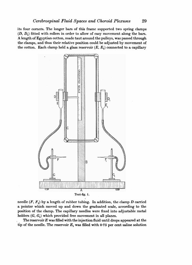
The second method of injection was replacement of the fluid within thecentral nervous system by the injection solution, using the apparatus shown inText-fig. 1. This consisted of a flat wooden base (A) supporting an upright rod(B), to which was affixed a graduated scale indicating the pressure in milli-metres of water under which the injection was made. The upright rod alsocarried a rectangular brass frame (C), provided with a small pulley at each of
1 In the case of the double solution, 04 g. of each salt were dissolved in a 100 c.c. of solution;in the case of the single solution 0-8 g. of iron ammonium citrate were dissolved in a 100 c.c. ofsolution.
28
Cerebrospinal Fluid Spaces and Choroid Plexuses
its four comers. The longer bars of this frame supported two spring clamps(D, D1) fitted with rollers in order to allow of easy movement along the bars.A length of Egyptian cotton, made taut around the pulleys, was passed throughthe clamps, and thus their relative position could be adjusted by movement ofthe cotton. Each clamp held a glass reservoir (E, E1) connected to a capillary
Text-fig. 1.
needle (F, F1) by a length of rubber tubing. In addition, the clamp D carrieda pointer which moved up and down the graduated scale, according to theposition of the clamp. The capillary needles were fixed into adjustable metalholders (G, G1) which provided free movement in all planes.
The reservoirE was filled with the injection fluid until drops appeared at thetip of the needle. The reservoir E1 was filled with 0-75 per cent saline solution
29
Henry Cohen and Sarah Davies
in the same manner. The flow of the fluid from each needle could be completelyarrested by means of a clip placed on the rubber tubing at a short distancebehind the needles.
The advantage of this apparatus is twofold-the reservoirs are firmlysupported but can still be moved freely up and down, and the pressure underwhich the injection is made is automatically recorded.
The embryo to be injected was supported on a block placed on the stand A.The reservoirs were filled with the injection fluid and saline solution to thesame level. The needles were adjusted to a suitable position and then insertedinto the embryo, needle F into the caudal end of the central canal of the spinalcord and needle F1 into the lateral ventricle of either cerebral hemisphere.When both needles were in position the clips were removed from the rubbertubing and the reservoir E was slowly raised, while the reservoir E1 was loweredsimultaneously. As fluid was withdrawn from the central nervous system intothe needle F1, it was automatically replaced by the injection solution from theneedle F. It was found that complete replacement was effected at a pressureequivalent to a column of water 70-75 mm. in height.
The methods of treatment of the different series of embryos are tabulatedbelow.
Age limitof series
Series days System injected Injection fluid usedA 3-9 Central nervous system Double solution of potassium ferro-
cyanide and iron ammonium citrateB 3-9 Central nervous system Double solution of potassium ferro-
cyanide and iron ammonium citrateC 4-9 Central nervous system Double solution of potassium ferro-
cyanide and iron ammonium citrateD 3j-9 Central nervous system Solution of iron ammonium citrateE 5-9 Vascular system Double solution of potassium ferro-
cyanide and iron ammonium citrateF 5-9 Vascular system Solution of iron ammonium citrate
The embryos were injected between the age limits stated at intervals ofapproximately 12 hours. In each series at least two embryos were injected ateach stage of development. After each injection the embryo, still in the salinesolution, was kept alive in an incubator at 38-40° C. to allow of the spread ofthe injection fluid, the embryonic heart-beat serving as the criterion of survival.It was found that At hour in the case of the smaller embryos, and 1-1k hoursin the larger, were needed for complete spread.
Embryos injected with the double solution were then placed in a solutioncontaining 10 per cent formaldehyde and 1 per cent hydrochloric acid. Thiscaused precipitation of Prussian blue (ferric-ferrocyanide) and partial fixationof the embryo. Specimens injected with iron ammonium citrate alone wereplaced in a similar acid formaldehyde solution, to which some potassiumferrocyanide solution had been added. This also caused precipitation ofPrussian blue and partial fixation. The time required for precipitation was.2-1 hour, according to the size of the embryo.
30
Cerebrospinal Fluid Spaces andr Choroid Plexuses 31
The members of series A were then left in Bouin's fluid overnight tocomplete fixation, but this resulted in a yellow colouration which obscured theexternal outline of the course of the injection fluid. Consequently, the embryosof the remaining series were fixed by allowing them to remain in the acidicformaldehyde solution overnight. Thus, the course of the injection fluid re-mained clearly visible, and the subsequent histological picture was in no wayaltered.
After fixation the embryos were dehydrated in a series of alcohol solutions,increasing from 5-100 per cent concentrations by 10 per cent gradations. Theywere then cleared in xylol or cedarwood oil,' embedded in paraffin wax (52 or56° C. M.P. according to season), and serially sectioned at a thickness of4 ,u, somein the longitudinal and some in the transverse plane. Ehrlich's haematoxylinand eosin were found to be the most suitable stains, and were used throughoutthe investigation.
V. DESCRIPTION OF THE BRAIN AND MENINGES OF THE CHICK ATDIFFERENT STAGES OF DEVELOPMENT, WITH PARTICULARREFERENCE TO THE STRUCTURE AND PERMEABILITY OF THEROOF OF THE FOURTH VENTRICLE
(a) Results of injections of true solutions into the central nervous system
(i) Injections of a double solution ofpotassium ferrocyanide and iron ammoniumcitrate.
The present section is based on observations, external and histological, ofthe embryos of series A, B and C. In the embryos of each series injections of adouble solution of potassium ferrocyanide and iron ammonium citrate weremade into the central nervous system.
In embryos of 3 days' incubation the injected solution was seen to bewholly contained within the central nervous system. It was uniformlydistributed within the central canal of the spinal cord and in the ventricularcavities of the brain. The medulla was relatively very large and triangularin shape with its base directed anteriorly and its apex tapering into the spinalcord. The medullary roof was very thin and lay immediately below the integu-ment, thus allowing the dye contained in the fourth ventricle to be more readilyseen than elsewhere in the brain stem.
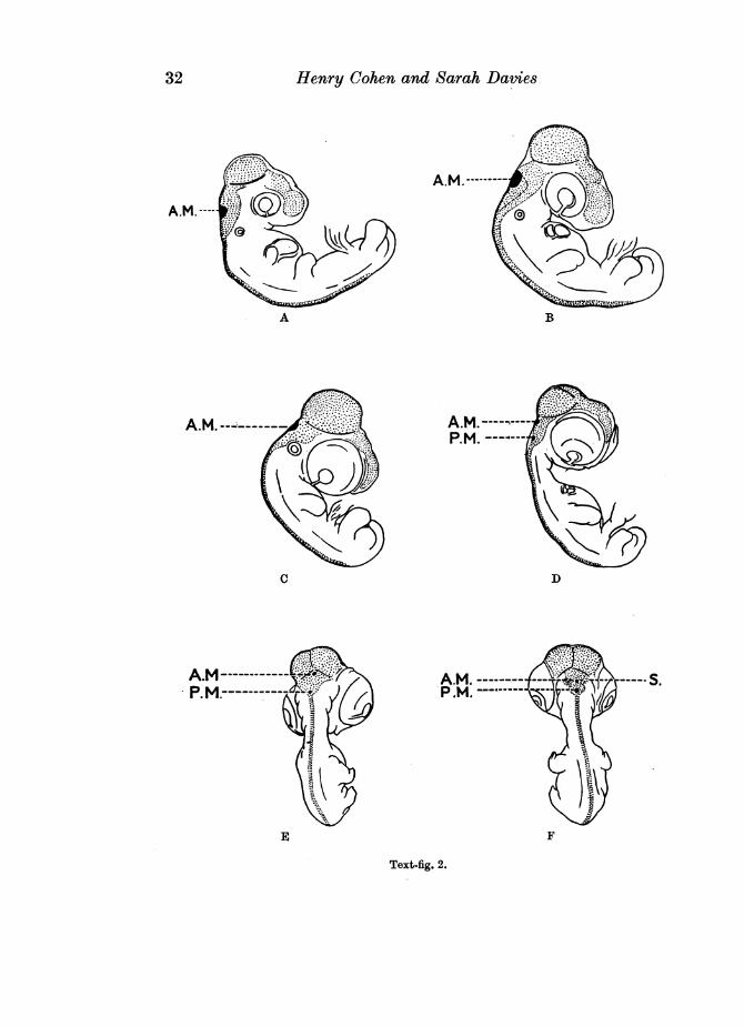
At 3A days the course of the injected solution was similar to that in a 3 days'embryo, but a median, deeply stained, roughly oval area was now seen in theanterior half of the medullary roof (see Text-fig. 2A). This area, in virtue of itsintense blue colour, stood out in marked contrast to the more diffused colourof the remainder of the medullary roof. No spread of the injected fluid fromthe central nervous system was evident.
1 The transparency of the chick embryo in its earlier stages of development enabled the courseof the injection fluid to be seen with ease. In older embryos the developing integument obscuredthe outline of the fluid considerably, and in these cases the material was cleared overnight incedarwood oil, to render the course of the fluid more easily distinguishable.
Henry Cohen and Sarah Davies
A.M.-- -
A B
A.M. .---
C D
E F
Text-fig. 2.
32
Cerebrospinal Fluid Spaces and Choroid Plexuses 33
Embryos of 31 and 4 days' incubation showed a distribution of the injectedfluid similar to that in the preceding stage, but the deeply staining oval areain the anterior portion of the medullary roof was relatively larger.
In 42 days' embryos, again, the injected dye still lay wholly within thecentral canal of the spinal cord and ventricular system of the brain, but themedian, deeply stained area was relatively much larger than previously,occupying the major portion of the anterior part of the medullary roof (seeText-fig. 2B).
At 5 days the distribution of the injected fluid was similar to that inembryos of 42 days' incubation, but the deeply staining oval area in themedullary roof showed a slight reduction in extent when compared with thatof the preceding stages (see Text-fig. 2C).
Embryos of 51 days showed that the fluid was still confined within thecentral nervous system, but the median oval area showed even further re-duction in extent when compared with that of the preceding stage.
At 6 days the distribution of the injected fluid was similar to that of theprevious stage, but the reduction in size of the deeply staining oval area in themedullary roof was even more marked.
In 61 and 61 days' embryos the dye was still confined to the spaces of thecentral nervous system. The median, deeply staining area in the anterior halfof the medullary roof was at this stage very small indeed, and a second similararea was now visible in the extreme apical region of the posterior half of theroof (see Text-fig. 2D and E).
Cleared specimens of 7 days' incubation showed that a very slight spreadof the dye had occurred from the roof of the fourth ventricle, but the outline ofthis roof was still clearly visible. The deeply stained area in the anterior partof the roof showed even greater reduction in extent than previously, whilstthat in the posterior half was relatively larger (see Text-fig. 2F).
In embryos of 71 days' incubation the outline of the medullary roof was nolonger clearly visible, even in specimens cleared in cedarwood oil. This sug-gested tentatively that an even more extensive escape of dye had occurredfrom the fourth ventricle into the overlying spaces. The deeply coloured area inthe anterior half of the medullary roof was now so small as to be hardly dis-tinguishable, but that in the posterior half was relatively larger than in thepreceding stage.
In cleared specimens of 8, 8- and 9 days' incubation the outline of themedullary roof was obscured completely; the deeply coloured area in theanterior half of this roof was no longer visible, but that in the posterior halfwas very large and, in the 9 days' embryos, occupied almost the whole of theapical half of the triangular medullary roof.
The embryos described above were examined histologically with a view todetermining the exact nature of the deeply staining oval areas in the roof ofthe fourth ventricle. At the same time, an attempt was made to discover anypossible correlation between the time of appearance of these areas, the spread
Anatomy LXX[II 3
Henry Cohen and Sarah Davies
of the injected dye from the central nervous system, and the development ofthe choroid plexuses and meningeal spaces.
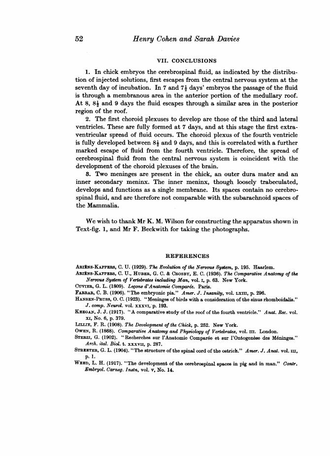
In embryos of 3 days' incubation the wall of the brain stem consisted chieflyof ependyma only, or of ependyma associated with a narrow stratum of over-lying nerve tissue. The roof of the third and fourth ventricles was in each casecontinuous and unfolded throughout and showed no indication of a choroidformation. The roof of the third ventricle consisted of an ependymal layer,3-4 cells deep. The outline of the individual cells was difficult to distinguishowing to the dense granulation of the deeply staining cell cytoplasm. Thenuclei were round or slightly elongated in a plane at right angles to thelongitudinal axis of the body, and possessed a deeply staining nuclear mem-brane and 2-3 nucleoli in the chromatin mesh. The roof of the fourth ventriclewas triangular in shape, the wide base being directed anteriorly, and the narrowapex posteriorly. The greater part of this roof was extremely thin, consisting ofa single layer of low cuboidal cells with granular cytoplasm and round well-defined nuclei touching both the inner and outer cell walls. The nucleoli andnuclear membrane were intensely stained, in contradistinction to the palenucleoplasm. In the anterior half of this roof a small median oval area couldbe distinguished, where the cells showed a slight tendency to flattening. Thisarea was not sharply delimited from the surrounding epithelium but graduallymerged into it (see Text-fig. 7).
At this stage the general mesenchymal tissue consisted of loosely arrangedstellate cells enclosing large, irregular intercellular spaces. The cytoplasm ofthese cells assumed the form of delicate, pale, granular strands radiating from acentrally disposed nucleus. The nuclei were round or slightly elongated andpossessed a deeply staining nuclear membrane, and 2-3 nucleoli in theirchromatin mesh. Where the mesenchymal tissue abutted on to the embryonicbrain, its cells showed a slight tendency to elongation, the cytoplasmic strandslying parallel to the brain surface.
The precipitated Prussian blue granules were seen wholly within thecentral nervous system, no extension of the dye into the surrounding mesen-chyme being evident. The granules were distributed in a uniform manner,chiefly within the central canal of the spinal cord and in the fourth ventricle ofthe brain, and showed no condensation associated with any particular area inthe brain stem. The distribution of the granules in the cavities of the opticlobes, thalamencephalon, and cerebral hemispheres was very sparse.
In embryos of 31 days' incubation the slightly differentiated median areain the roof of the fourth ventricle was now abruptly demarcated from thesurrounding roof epithelium. The cells of this area were extremely flat, thegranular cytoplasm forming a thin investment around the elongated nucleus.Apart from a marked difference in shape, these cells resembled those of thesurrounding epithelium, showing a similar degree of granulation and richnessof nuclear chromatin material.
Where the mesenchyme abutted on to the brain surface its cells were not
34
Cerebrospinal Fluid Spaces and(Choroid Plexuses
only elongated as in the previous stage, but also showed a slight degree ofcondensation, and were more compactly arranged than elsewhere. The narrowill-defined zone thus formed adhered closely to the brain surface and intimatelyfollowed its outline.
The injected fluid was again strictly confined within the central nervoussystem but no longer exhibited the same uniformity'of distribution within thecerebral ventricles as in the preceding stage. There was a dense accumulation ofblue granules intermingled with coagulum, adhering to, and co-extensive with,the ventricular surface of the thin membranous area in the roof of the fourthventricle. The individual cells of this area, however, were entirely free ofthe dye.
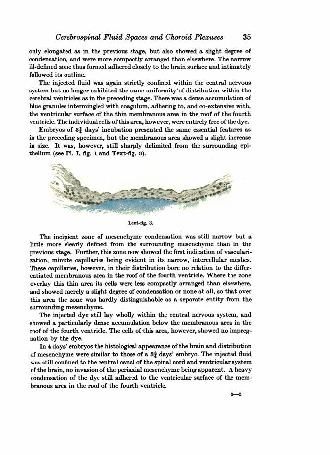
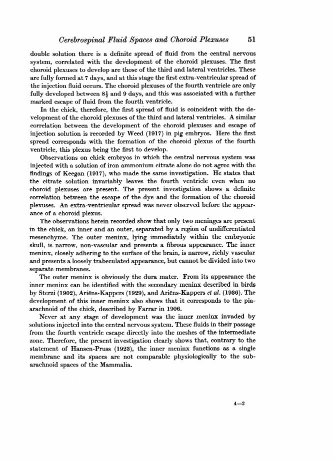
Embryos of 38 days' incubation presented the same essential features asin the preceding specimen, but the membranous area showed a slight increasein size. It was, however, still sharply delimited from the surrounding epi-thelium (see PI. I, fig. 1 and Text-fig. 8).
.'~~~~~~~~~f...~ ~ -. -- ..
Text-fig. 3.
The incipient zone of mesenchyme condensation was still narrow but alittle more clearly defined from the surrounding mesenchyme than in theprevious stage. Further, this zone now showed the first indication of vasculari-zation, minute capillaries being evident in its narrow,' intercellular meshes.These capillaries, however, in their distribution bore no relation to the differ-entiated membranous area in the roof of the fourth ventricle. Where the zoneoverlay this thin area its cells were less compactly arranged than elsewhere,and showed merely a slight degree of condensation or none at all, so that overthis area the zone was hardly distinguishable as a separate entity from thesurrounding mesenchyme.
The injected dye still lay wholly within the central nervous system, andshowed a particularly dense accumulation below the membranous area in theroof of the fourth ventricle. The cells of this area, however, showed no impreg-nation by the dye.
In 4 days' embryos the histological appearance of the brain and distributionof mesenchyme were similar to those of a 8t days' embryo. The injected fluidwas still confined to the central canal of the spinal cord and ventricular systemof the brain, no invasion of the periaxial mesenchyme being apparent. A heavycondensation of the dye still adhered to the ventricular surface of the mem-branous area in the roof of the fourth ventricle.
3-2
35
Henry Cohen and Sarah Davies
At 41 days the wall of the brain was thicker than in preceding stages, thegreater part consisting throughout of ependyma, associated with a stratum ofnerve tissue of variable width. The dorso-mesial walls of the cerebral hemi-spheres and the roof of the third ventricle, however, were narrower than else-where, but still showed no indication of a choroid formation. The myelence-phalic roof was still smooth and unfolded. The median membranous area in itsanterior portion was slightly larger than in the preceding stages, but was stillabruptly demarcated from the surrounding epithelium.
The zone of mesenchyme condensation surrounding the brain was stillnarrow but more clearly defined than hitherto, and showed a higher degree ofvascularization. The general mesenchyme overlying the roof of the fourthventricle was more loose-meshed than elsewhere and still showed little or nocondensation where it actually abutted on to the membranous area in theanterior portion of this roof.
As in earlier embryos, the dye was still confined within the central nervoussystem, but had now extended anteriorly in uniform distribution to thefurthermost recesses of the lateral ventricles. A particularly dense accumula-tion of blue granules was still apparent below the membranous area in the roofof the fourth ventricle.
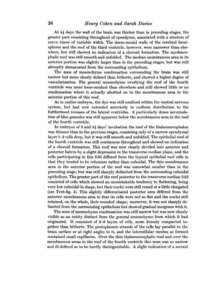
In embryos of 5 and 51 days' incubation the roof of the thalamencephalonwas thinner than in the previous stages, consisting only of a narrow ependymallayer 1-2 cells deep, but it was still smooth and unfolded. The epithelial roof ofthe fourth ventricle was still continuous throughout and showed no indicationof a choroid formation. This roof was now clearly divided into anterior andposterior halves by a slight depression in the transverse median plane, and thecells participating in this fold differed from the typical epithelial roof cells inthat they tended to be columnar rather than cuboidal. The thin membranousarea in the anterior portion of the roof was somewhat smaller than in thepreceding stage, but was still sharply delimited from the surrounding cuboidalepithelium. The greater part of the roof posterior to the transverse median foldconsisted of cells which showed an unmistakable tendency to flattening, beingvery low cuboidal in shape, but their nuclei were still round or a little elongated(see Text-fig. 4). This slightly differentiated posterior area differed from theanterior membranous area in that its cells were not so flat and the nuclei stillretained, on the whole, their rounded shape; moreover, it was not sharply de-limited from the surrounding epithelium but showed gradual mergence with it.
The zone of mesenchyme condensation was still narrow but was now clearlyvisible as an entity distinct from the general mesenchyme from which it hadoriginated. It consisted of 2-3 layers of cells, more densely compacted to-gether than hitherto. The protoplasmic strands of the cells lay parallel to thebrain surface or at right angles to it, and the intercellular meshes so formedcontained small capillaries. Over the thin thalamencephalic roof and over themembranous areas in the roof of the fourth ventricle this zone was so narrowand ill-defined as to be hardly distinguishable. A slight indication of a second
36
Cerebrospinal Fluid Spaces and (Choroid Plexu.,es 37
outer zone of condensation was apparent in the general mesenchyme in certainlocalized areas, chiefly in front of the cerebral hemispheres and on the dorsaland ventral surfaces of the thalamencephalon. Where this zone was in processof formation it was very irregular and ill-defined, merging imperceptibly intothe general mesenchyme. It was separated from the narrow inner zone by anintermediate region of mesenchymal tissue of variable width. In this newlyforming outer zone the typical mesenchyme cells were more compactlyarranged than elsewhere and showed a tendency to elongation in a planeparallel to the longitudinal axis of the body. This zone showed no indication ofvascularization, its narrow and elongated intercellular meshes being entirelyfree of blood capillaries. The intermediate zone consisted of typical loose-meshed stellate mesenchyme cells and contained minute capillaries. These
iA.19,/S,>^#..* .9*\
Text-fig. 4.
were uniformly distributed throughout this zone and showed no special con-centration over the differentiated areas in the roof of the fourth ventricle.
The injected dye was still confined within the central nervous system, andwas particularly condensed below the membranous area in the anterior regionof the roof of the fourth ventricle. No such condensation was evident below theslightly flattened area in the posterior region of this roof, but merely a finedistribution of blue granules closely adhering to its ventricular surface.
In embryos of 6 days' incubation the thin roof of the thalamencephalonpresented a slightly undulating appearance, forming 2-3 shallow irregularfolds, invaded by the overlying mesenchyme. The transverse median foldseparating the myelencephalic roof into anterior and posterior regions wasmore emphasized than previously. Further, the anterior membranous area ofthis same roof had diminished considerably in extent, but was still sharply
Henry Cohen and Sarah Davies
delimited from the surrounding cuboidal epithelium. The posterior slightlyflattened area was more extensive than in the preceding stage and, on the whole,exceeded the anterior area both in length and width. It still showed no cleardemarcation from the surrounding epithelium. Apart from the transversemedian depression, the roof of the fourth ventricle showed no indication offolding and was continuous throughout.
The inner zone of condensation was still narrow, yet clearly defined, exceptover the thin thalamencephalic roof and over the membranous areas in theroof of the fourth ventricle. The mesenchyme abutting on to these surfacesstill showed little or no condensation. The outer zone was more clearly markedthan previously but still merged gradually into the surrounding mesenchyme(see PI. II, fig. 6). This zone had now extended posteriorly over the optic lobesand to the lateral and ventral surfaces of the medulla. In the dorsal medullaryregion its identity was completely lost in the general mesenchyme. Localizedcentres of cartilage formation were evident within the substance of this outerzone, particularly in that part of it flanking the medulla. The cells at thesepoints of chondrification tended to a regular disposition, and were isolated fromeach other by a clear homogeneous matrix. Their nuclei were rounded and welldefined, and possessed only a thin investment of pale granular cytoplasm.Where the nervous system approximated closely to the integument, i.e. over theanterior surfaces of the cerebral hemispheres and the dorsal surfaces of the opticlobes, the inner and outer zones lay in such close juxtaposition that the inter-mediate zone of loose-meshed mesenchyme was almost completely obliterated.
The injected fluid was still strictly confined within the spaces of the centralnervous system, and showed the same degree of concentration below themembranous areas of the myelencephalic roof as in the previous stage, but theaccumulation of blue granules below the posterior area was slightly denser.
In embryos of 6j days the incipient folding of the thalamencephalic roofwas even more pronounced than previously. The transverse median fold in theroof of the fourth ventricle was deeper than in the preceding stage and its cellswere more columnar in shape. The anterior membranous area in this roofshowed even further reduction in extent, but was still sharply delimited fromthe surrounding epithelium. The posterior area, on the other hand, was slightlylarger than in the previous stage, but again merged gradually into the generalroof epithelium. Its cells were still very low cuboidal in shape with round oronly slightly elongated nuclei, but those at the extreme posterior region of thisarea were extremely flat and resembled the pavement cells of the anterior area.
At this stage the narrow inner zone of mesenchyme condensation showedwell-marked intrazonalI spaces. Its cells were extremely flat and elongated, andarranged in two ill-defined layers parallel to the brain surface. The very narrowspaces between the cells were traversed by delicate protoplasmic trabeculaearising from the cells of both the inner and outer layers. The numerousintercellular meshes so formed contained small capillaries and consequentlythis zone, particularly around the cerebral hemispheres, had the appearance of
38
Cerebrospinal Fluid Spaces and Choroid Plexuses
a chain of minute vascular lumina. Where the mesenchyme cells of this zoneabutted on to the thin thalamencephalic roof and membranous areas in theroof of the fourth ventricle they showed little or no condensation, so that thezone was virtually absent in these regions. The outer zone of mesenchymecondensation was more clearly defined than in previous stages and had nowextended round the entire brain stem. It was still irregular, however, and wasseparated from the inner zone by an intermediate layer of typical loose-meshed mesenchyme of variable width. Occasionally the two condensationzones were separated by large irregular spaces containing isolated mesenchymecells, indicating that these spaces had originated by the breaking down of smallportions of the mesenchymal tissue and subsequent confluence of the inter-cellular spaces.
The injected dye was still retained within the spaces of the central nervoussystem. A particularly dense accumulation of blue granules and coagulum wasstill seen adhering to, and coextensive with, the ventricular surface of theanterior membranous area. Further, a heavy condensation was now evidentbelow the extremely flat cells at the apex of the slightly differentiated posteriormembranous area.
In embryos of 61 days the invaginations of the thalamencephalic roof weremore pronounced than previously, and extended forward for a very shortdistance into the lateral ventricles via the foramina of Munro. The anteriormembranous area of the myelencephalic roof was smaller than in the precedingstage, and its border no longer showed an abrupt transition from the surround-ing epithelium but gradually merged into it. The posterior membranous area,on the other hand, was now sharply delimited from the general roof epi-thelium and showed considerable increase in extent. Its cells were extremelyflat and possessed narrow elongated nuclei with a thin investment of cytoplasm(see P1. I, fig. 2).
The outer zone of mesenchyme condensation was a little more clearlydefined than previously. It showed a slight increase in width and a higherdegree of cartilage formation within its substance.
The injected fluid was still confined within the central nervous system, andwas particularly condensed below the membranous areas in the roof of thefourth ventricle.
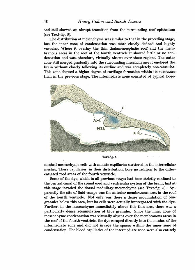
In 7 days' embryos the folds of the thalamencephalic roof were morepronounced than in the preceding stage and, as they extended forward into thelateral ventricles, showed a slight degree of branching, forming incipientchoroid villi. In transverse section each villus consisted of an axial core ofloose-meshed mesenchyme containing minute capillaries in its intercellularspaces and surrounded by an epithelium 1-2 cells deep. The cells were columnarin shape with granular cytoplasm and round well-defined nuclei. The mediantransverse fold in the roof of the fourth ventricle was deeper than hitherto andits cells were more markedly columnar. The anterior membranous area wassmaller than previously but the posterior area, on the other hand, was larger
39
40 Henry Cohen and Sarah Datie8
and still showed an abrupt transition from the surrounding roof epithelium(see Text-fig. 5).
The distribution of mesenchyme was similar to that in the preceding stage,but the inner zone of condensation was more clearly defined and highlyvascular. Where it overlay the thin thalamencephalic roof and the mem-branous areas in the roof of the fourth ventricle it showed little or no con-densation and was, therefore, virtually absent over these regions. The outerzone still merged gradually into the surrounding mesenchyme; it enclosed thebrain without closely following its outline and was completely non-vascular.This zone showed a higher degree of cartilage formation within its substancethan in the previous stage. The intermediate zone consisted of typical loose-
Text-fig. 5.
meshed mesenchyme cells with minute capillaries scattered in the intercellularmeshes. These capillaries, in their distribution, bore no relation to the differ-entiated roof areas of the fourth ventricle.
Some of the dye, which in all previous stages had been strictly confined tothe central canal of the spinal cord and ventricular system of the brain, had atthis stage invaded the dorsal medullary mesenchyme (see Text-fig. 5). Ap-parently the site of fluid escape was the anterior membranous area in the roofof the fourth ventricle. Not only was there a dense accumulation of bluegranules below this area, but its cells were actually impregnated with the dye.Further, in the mesenchyme immediately above this thin area there was aparticularly dense accumulation of blue granules. Since the inner zone ofmesenchyme condensation was virtually absent over the membranous areas inthe roof of the fourth ventricle, the dye escaped directly into the meshes of theintermediate zone and did not invade the spaces within the inner zone ofcondensation. The blood capillaries of the intermediate zone were also entirely
Cerebrospinal Fluid Spaces and Choroid Plexuses
free of the dye, although here and there a slight accumulation of blue granuleswas evident, adhering to the outer surface of the capillary wall. A heavyprecipitation of the injected fluid was also seen below the posterior mem-branous area. There was, however, no evidence of escape from this region, itscells showing no invasion by the dye. Neither was there evidence of fluidescape from any other point in the ependymal roof of the fourth ventricle. Thecuboidal cells of this layer were entirely free of the dye, and exhibited only afine granular distribution on their ventricular surface.
In embryos of 7k days' incubation the folding of the thalamencephalic roofwas even more marked than in the preceding stage, the folds being slendererand exhibiting a higher degree of branching. The median transverse invagina-tion in the roof of the fourth ventricle was deeper and the columnar appearanceof its constituent cells more pronounced. The ventro-lateral walls of this foldshowed a slight degree of undulation, but no true choroid villi were apparent.The anterior membranous area of this roof was even smaller than in thepreceding stage, but the posterior area was relatively larger.
The outer zone of mesenchyme condensation was wider and more clearlydefined than in the previous stage. The mesenchyme of the intermediate zoneimmediately above the differentiated roof areas of the fourth ventricle wasmore loose-meshed than elsewhere and contained large irregular spaces. Thecapillaries in this zone now tended to be more concentrated in the dorsalmedullary region.
The extra-ventricular spread of the injected fluid was slightly more markedthan previously. Anteriorly it had extended as far as the cerebellar anlage,and posteriorly as far as the median transverse fold in the medullary roof. Theblue granules lay in the meshes of the intermediate zone and did not invade theinner zone of condensation.
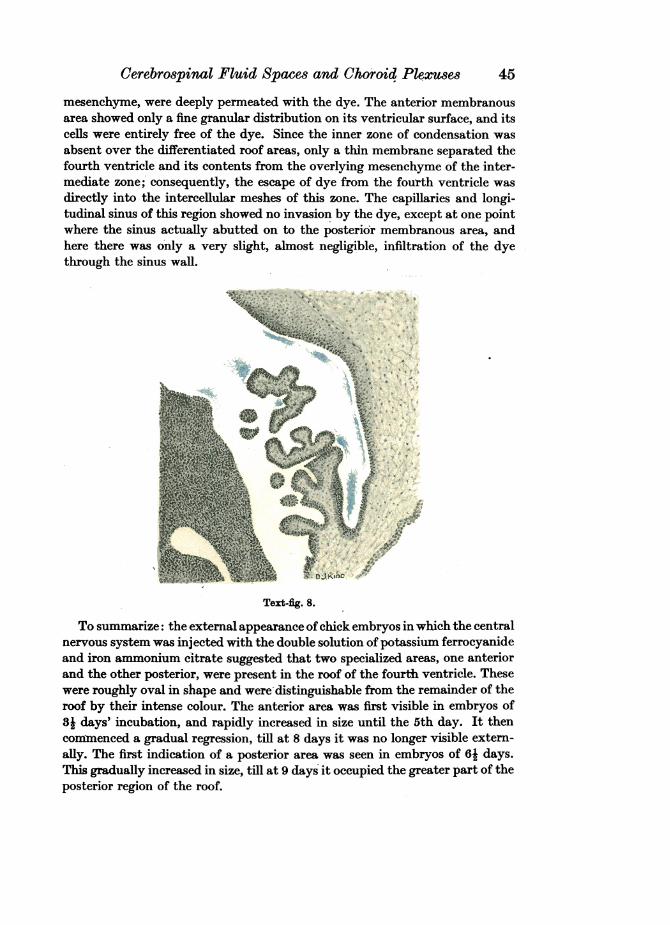
In 8 days' embryos, as in preceding stages, the thalamencephalic roofconsisted of an ependymal layer only 2-3 cells deep. The degree of folding ofthis roof, however, was much more marked than hitherto, and the long narrowbranching choroid villi so formed extended forward into the lateral ventriclesvia the foramina of Munro (see Text-fig. 8). The roof of the fourth ventricleshowed striking morphological changes. The median transverse fold was ex-tremely well developed and extended almost to the floor of the ventricle. Thisfold was lined by the inner zone of condensation, and was supported by mesen-chymal tissue continuous with that of the intermediate zone. Its walls con-sisted of columnar cells with granular cytoplasm and round well-defined nuclei.The undulations in the floor of this fold, first recorded in 7- days' embryos,were here much more pronounced, forming short unbranched incipient villi.These were concentrated in two ill-defined clusters, one at each side of thefloor. The anterior portion of the roof of the fourth ventricle was much shorterthan in the preceding stage, owing apparently to the encroachment of thedeveloping cerebellar anlage. A median membranous area was still evident inthis anterior region but was much smaller in extent than previously. The
41
Henry Cohen and Sarah Daviem
posterior membranous area, on the other hand, was relatively much larger andoccupied practically the whole of the roof region posterior to the median fold(see Text-fig. 9).
The outer zone of mesenchyme condensation was wider and a little moreclearly defined than in the preceding stage. Where the intermediate zoneabutted on to the areas of chondrification in the outer zone its cells showed aslight condensation. They were slightly elongated in a plane parallel to theinner surface of the developing bands of cartilage, thus forming an extremelynarrow ill-defined zone of fibrous appearance.
The extra-ventricular distribution of the injected dye was more extensivethan in the previous stage. It extended for a short distance over the posteriorlip of the cerebellar anlage, and even invaded the mesenchyme lying in theupper region of the transverse median fold in the roof of the fourth ventricle.The inner zone of condensation still showed no invasion by the dye. Apparentlythe posterior membranous area in the medullary roof was now functioning asthe site of fluid escape. There was a particularly dense accumulation of bluegranules adhering to its ventricular surface. Its cells were deeply impregnatedwith the dye, and the mesenchyme immediately above this area was moreintensely stained than elsewhere. Below the anterior area there was littlecondensation of blue granules, its cells showed scarcely any impregnation withthe dye and the overlying mesenchyme was only slightly coloured.
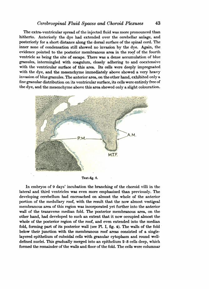
In embryos of 81 days' incubation the choroid villi of the lateral ventriclesexhibited a higher degree of branching than previously. The incipient villi inthe floor of the median fold of the medullary roof were more pronounced thanin the preceding specimen, and were slightly branched (see PI. I, fig. 3). Theanterior portion of the medullary roof was even shorter in longitudinal section,bringing the posterior lip of the cerebellum near to the transverse median fold.The membranous area in the anterior region of the roof was very small indeedand was now relegated to the upper part of the anterior wall of the median fold.This membranous area was no longer sharply delimited from the surroundingepithelium but gradually merged into it. Its constituent cells had lost theirflat appearance and were now low cuboidal in shape. The posterior mem-branous area was very extensive, occupying practically the whole of the roofbehind the transverse fold. It remained sharply delimited from the surround-ing epithelium, and its cells, in contrast to those of the anterior area, were stillextremely flat (see Text-fig. 6).
As in previous stages, the inner zone of mesenchyme condensation was stillnarrow, but nevertheless very clearly defined. Over the membranous areas inthe roof of the fourth ventricle it was absent altogether, so that in these regionsonly a very thin membrane separated the fourth ventricle and its contentsfrom the intercellular meshes of the overlying intermediate zone. The outerzone was wider than previously and showed an even higher degree of cartilageformation. Further, the fibrous layer immediately within this zone was a littlemore clearly defined.
42
Cerebrospinal Fluid Spaces and Choroid Plexuse8 43
The extra-ventricular spread of the injected fluid was more pronounced thanhitherto. Anteriorly the dye had extended over the cerebellar anlage, andposteriorly for a short distance along the dorsal surface of the spinal cord. Theinner zone of condensation still showed no invasion by the dye. Again, theevidence pointed to the posterior membranous area in the roof of the fourthventricle as being the site of escape. There was a dense accumulation of bluegranules, intermingled with coagulum, closely adhering to and coextensivewith the ventricular surface of this area. Its cells were deeply impregnatedwith the dye, and the mesenchyme immediately above showed a very heavyinvasion of blue granules. The anterior area, on the other hand, exhibited only afine granular distribution on its ventricular surface, its cells were entirely free ofthe dye, and the mesenchyme above this area showed only a slight colouration.
IS' I o/
M.T.F.
Text-fig. 6.
In embryos of 9 days' incubation the branching of the choroid villi in thelateral and third ventricles was even more emphasized than previously. Thedeveloping cerebellum had encroached on almost the whole of the anteriorportion of the medullary roof, with the result that the now almost vestigealmembranous area of this region was incorporated yet further into the anteriorwall of the transverse median fold. The posterior membranous area, on theother hand, had developed to such an extent that it now occupied almost thewhole of the posterior region of the roof, and even extended into the medianfold, forming part of its posterior wall (see PI. I, fig. 4). The walls of the foldbelow their junction with the membranous roof areas consisted of a single-layered epithelium of cuboidal cells with granular cytoplasm and round well-defined nuclei. This gradually merged into an epithelium 2-3 cells deep, whichformed the remainder of the walls and floor of the fold. The cells were columnar
Henry Cohen and Sarah Davies
and their cytoplasm was so densely granular that the individual cell boundarieswere difficult to distinguish. Their nuclei were round or slightly elongated andpossessed deeply staining nucleoli and nuclear membrane. The floor of the foldexhibited a higher degree of villus formation than in the preceding stage, andthe branching of the villi was even more pronounced (see PI. I, fig. 5).
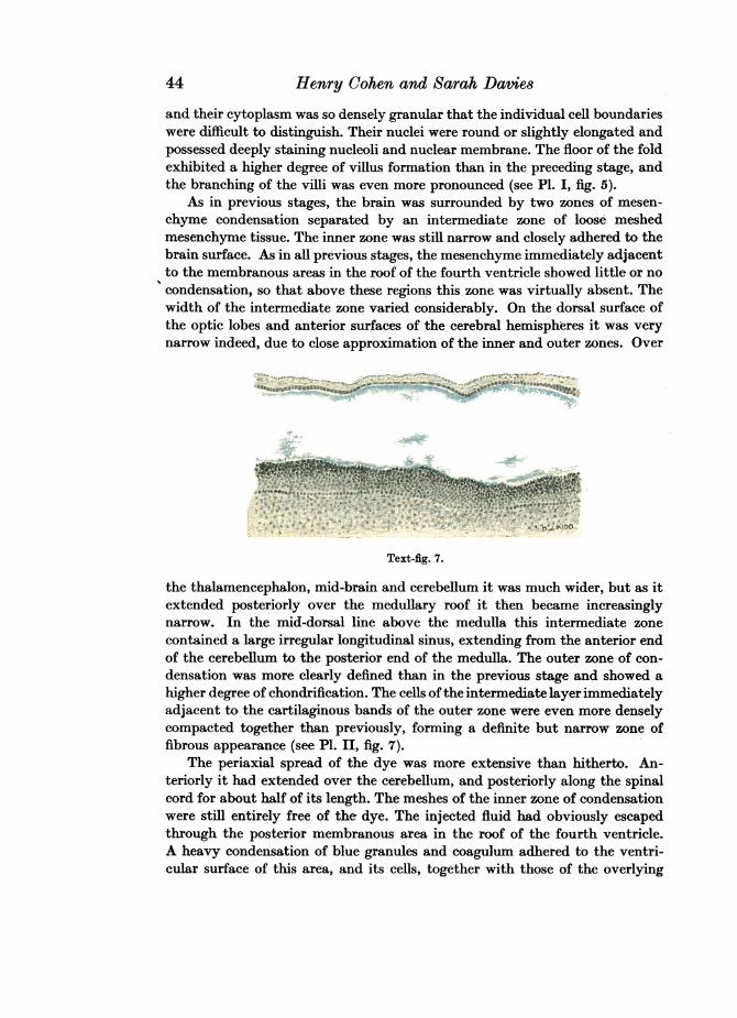
As in previous stages, the brain was surrounded by two zones of mesen-chyme condensation separated by an intermediate zone of loose meshedmesenchyme tissue. The inner zone was still narrow and closely adhered to thebrain surface. As in all previous stages, the mesenchyme immediately adjacentto the membranous areas in the roof of the fourth ventricle showed little or nocondensation, so that above these regions this zone was virtually absent. Thewidth of the intermediate zone varied considerably. On the dorsal surface ofthe optic lobes and anterior surfaces of the cerebral hemispheres it was verynarrow indeed, due to close approximation of the inner and outer zones. Over
- - .~~~~~~00. 1,;
Text-fig. 7.
the thalamencephalon, mid-brain and cerebellum it was much wider, but as itextended posteriorly over the medullary roof it then became increasinglynarrow. In the mid-dorsal line above the medulla this intermediate zonecontained a large irregular longitudinal sinus, extending from the anterior endof the cerebellum to the posterior end of the medulla. The outer zone of con-densation was more clearly defined than in the previous stage and showed ahigher degree of chondrification. The cells ofthe intermediate layer immediatelyadjacent to the cartilaginous bands of the outer- zone were even more denselycompacted together than previously, forming a definite but narrow zone offibrous appearance (see PI. II, fig. 7).
The periaxial spread of the dye was more extensive than hitherto. An-teriorly it had extended over the cerebellum, and posteriorly along the spinalcord for about half of its length. The meshes of the inner zone of condensationwere still entirely free of the dye. The injected fluid had obviously escapedthrough the posterior membranous area in the roof of the fourth ventricle.A heavy condensation of blue granules and coagulum adhered to the ventri-cular surface of this area, and its cells, together with those of the overlying
44
(Jerebrospinal Fluid Spaces and (Choroid Plexuses 45
mesenchyme, were deeply permeated with the dye. The anterior membranousarea showed only a fine granular distribution on its ventricular surface, and itscells were entirely free of the dye. Since the inner zone of condensation wasabsent over the differentiated roof areas, only a thin membrane separated thefourth ventricle and its contents from the overlying mesenchyme of the inter-mediate zone; consequently, the escape of dye from the fourth ventricle wasdirectly into the intercellular meshes of this zone. The capillaries and longi-tudinal sinus of this region showed no invasion by the dye, except at one pointwhere the sinus actually abutted on to the posterior membranous area, andhere there was only a very slight, almost negligible, infiltration of the dyethrough the sinus wall.
"I '"'Text-fig. 8.
To summarize: the external appearance ofchick embryos in which the centralnervous system was injected with the double solution of potassium ferrocyanideand iron ammonium citrate suggested that two specialized areas, one anteriorand the other posterior, were present in the roof of the fourth ventricle. Thesewere roughly oval in shape and were'distinguishable from the remainder of theroof by their intense colour. The anterior area was first visible in embryos of34 days' incubation, and rapidly increased in size until the 5th day. It thencoimnenced a gradual regression, till at 8 days it was no longer visible extern-ally. The first indication of a posterior area was seen in embryos of 64 days.This gradually increased in size, till at 9 days it occupied the greater part of theposterior region of the roof.
46 Henry Cohen and Sarah DaviesDuring the earlier stages of development there appeared to be no escape
of the injected dye from the central nervous system. At the seventh day ofincubation, however, a slight spread was evident from the roof of the fourthventricle, and this became increasingly marked from the seventh day onward.
These observations of the injected embryos were verified by a detailedhistological examination.
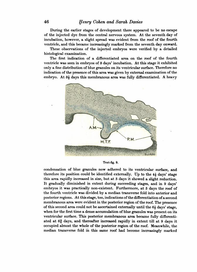
The first indication of a differentiated area on the roof of the fourthventricle was seen in embryos of 3 days' incubation. At this stage it exhibitedonly a fine distribution of blue granules on its ventricular surface. Therefore noindication of the presence of this area was given by external examination of theembryo. At 3j days this membranous area was fully differentiated. A heavy
m.TF M
v J, . f. *>A 42 ~~~~~~~~~~~~~- D..(O
condensaton of blu granules Text-fig. 9.
condensation of blue granules now adhered to its ventricular surface, andtherefore its position could be identified externally. Up to the 4J days' stagethis area rapidly increased in-size, but at 5 days it showed a slight reduction.It gradually diminished in extent during succeeding stages, and in 9 days'embryos it was practically non-existent. Furthermore, at 5 days the roof ofthe fourth ventricle was divided by a median transverse fold into anterior andposterior regions. At this stage, too, indications ofthe differentiation ofa secondmembranous area were evident in the posterior region of the roof. The presenceof this second area could not be ascertained externally until the 6J days' stage,when for the first time a dense accumulation of blue granules was present on itsventricular surface. This posterior membranous area became fully differenti-ated at 61 days, and thereafter increased rapidly in extent till at 9 days itoccupied almost the whole of the posterior region of the roof. Meanwhile, themedian transverse fold in this same roof had become increasingly marked
Cerebrospinal Fluid Spaces and Choroid Plexuses
throughout successive stages, till at 9 days it almost reached the floor of theventricle. At the same time the cells participating in this fold gradually losttheir original cuboidal appearance and became columnar.
In embryos up to and including the age of 61 days the injected dye remainedentirely within the central canal of the spinal cord and ventricular system ofthe brain. At 7 days, however, there was a slight extension of the dye from theanterior membranous area in the roof of the fourth ventricle into the overlyingmesenchyme of the intermediate zone. At 71 days this spread was slightly moremarked, the injected dye still escaping through the anterior membranous area.In embryos of 8 days' incubation the passage of the dye from the fourthventricle was even more extensive than previously. At this stage, the injectedfluid made its escape through the posterior membranous area in the medullaryroof, and no longer through the anterior membranous area. At 81 days therewas a very marked periaxial spread of the dye, even along the dorsal surface ofthe spinal cord, its passage again being through the posterior membranousarea. At 9 days the spread of the dye from this same area was even morepronounced. Anteriorly it had invaded the mesenchyme overlying the wholecerebellar anlage, while posteriorly it extended along the cord for a considerabledistance.
The first choroid plexuses to develop were those of the third and lateralventricles. In embryos of 6 days' incubation the thin thalamencephalic roofpresented a slightly undulating appearance owing to the formation of 2-3broad irregular folds. These undulations were more marked at 6j days, while inembryos of 61 days they extended anteriorly for a short distance into thelateral ventricles via the foramina of Munro, still in the form of simple un-branched villi. At 7 days, however, the folds exhibited a slight degree ofbranching, forming an embryonic choroid plexus. From this stage onward theformation of branching choroid villi increased rapidly, till at 9 days theyoccupied almost the whole of the lateral ventricles. The first indication of thedevelopment of the choroid plexus of the fourth ventricle was seen in embryosof 7J days' incubation. At this stage the ventro-lateral walls of the transversemedian fold in the roof of this ventricle showed a slight degree of undulation,but no true villi were apparent. At 8 days the undulations were much moremarked, resulting in the formation of short unbranched villi concentrated intwo ill-defined clusters in the floor of the fold. In 81 days' embryos the forma-tion of these villi was more pronounced, and they exhibited a slight tendencyto branching, which became even more marked at 9 days.
In 9 days' embryos the brain was enclosed by the developing cranium andtwo meninges, an inner and an outer, which were separated by an intermediatelayer of undifferentiated mesenchyme. The inner meninx was narrow, closelyadhered to the surface of the brain, was loosely trabeculated and containedminute capillaries in its intercellular meshes. Where this layer abutted on tothe membranous areas in the roof of the fourth ventricle it was so thin as to bealmost absent. The outer meninx, on the other hand, lay close to the inner
47
48 Henry Cohen and Sarah Davies
surface of the embryonic skull; it was wider than the inner membrane, wasnon-vascular and presented a fibrous appearance.- The intermediate zoneseparating the inner and outer meninges was of variable width and possessed afairly rich blood supply. Where this zone overlay the roof of the fourth ventricleit contained large irregular spaces.
Both meninges and the cranial anlage originated in situ by a process ofcellular condensation in the general mesenchyme surrounding the brain. Theinner meninx arose as a single membrane from a zone of condensation im-mediately adjacent to the brain surface. It was first distinguishable in embryosof 31 days' incubation, but at this stage was narrow and ill-defined. At 41 daysit was still narrow but more clearly visible as a distinct layer, and now showed aslight degree of vascularization. During successive stages of development thiszone became increasingly vascular and more clearly defined. The cranial anlagewas first visible in embryos of 5 days' incubation where it was represented byan extremely irregular and ill-defined zone of mesenchyme condensationsituated at some distance from the inner meninx. At 6 days this zone was moreclearly marked, and localized centres of cartilage formation were evident withinits substance. In later stages this zone became increasingly well defined andshowed a progressive degree of chondrification. Meanwhile, at 8 days the cellsof the intermediate zone immediately adjacent to the developing craniumshowed a slight degree of condensation, forming a very narrow ill-defined layerof fibrous appearance. At 8- days this zone was more marked, and at 9 days itwas clearly visible as a definite but narrow fibrous membrane immediatelywithin the embryonic cranium.
Owing to the absence of the inner meninx over the membranous areas in theroof of the fourth ventricle, the injected dye, in its passage from this ventricle,escaped directly into the meshes of the intermediate zone. Never at any stagewas it seen to invade either the inner or the outer meninx.
(ii) Injections of a solution of iron ammonium citrate alone
The present section is based on external and histological observations of theembryos of series D. These were injected with a solution of iron ammoniumcitrate into the central nervous system. Since a detailed account of the de-velopment of the roof of the fourth ventricle, choroid plexuses and meningeshas been given in the previous section, a description of the distribution of theinjected fluid alone is given below.
In embryos up to and including the age of 61 days the injected solution wasseen to be contained within the central nervous system. In cleared specimensof 7 days' incubation a slight escape of fluid was evident from the roof of thefourth ventricle. At 71 days this spread was more marked and partly obscuredthe outline of the medullary roof. In embryos of 8, 81 and 9 days' incubationthe outline of this roof was no longer visible externally, suggesting that aneven greater extra-ventricular spread of the injected fluid had occurred.
Cerebrospinal Fluid Spaces and Choroid Plexuse8 49
These observations on the external appearance of the injected embryoswere confirmed by histological examination.
In embryos of this series the development of the membranous areas in theroof of the fourth ventricle was similar to that described in embryos of seriesA, B and C. When these areas were present a heavy condensation of dye wasseen adhering to their ventricular surface (see PI. II, fig. 8).
Until the age of 7 days the injected solution was confined within the centralnervous system. At 7 days there was a slight escape of the dye through theanterior membranous area into the overlying mesenchyme between the twozones of condensation (see PI. II, fig. 9). At 7J days this spread had increased,but was still through the anterior area. In 8 days' embryos the extension of thedye from the fourth ventricle was even more pronounced than previously. Atthis stage the posterior area alone functioned as the site of fluid escape. At81 days the injected fluid had spread to an even greater extent, its passage stillbeing through the posterior membranous area. In 9 days' embryos the escapeof the dye through this same area was even more marked. Anteriorly it hadinvaded the mesenchyme overlying the optic lobes, while posteriorly it ex-tended along the spinal cord for about half of its length.
The meninges of the brain in embryos of series D showed no invasion by thedye. The injected fluid in its passage from the fourth ventricle escaped directlyinto the meshes of the intermediate zone.
Thus, embryos in which the central nervous system is injected with asolution of iron ammonium citrate alone yield the same results as those in-jected with the double solution of potassium ferrocyanide and iron ammoniumcitrate.
(b) Results of injections of true solutions into the vascular systemThe observations recorded in sections Va (i) and Va (ii) showed that the
first escape of injected fluids from the central nervous system into the sur-rounding tissue occurred in embryos of 7 days' incubation. At this stage thepassage of the dye was through the anterior membranous area in the roof of thefourth ventricle, and from 8 days onwards through the posterior area.
Injections were made into the vascular system in order to ascertain whetherthe anterior and posterior areas at 7 and 8 days respectively allowed the passageof dye into the fourth ventricle from the surrounding tissue. For this purposetwo series of embryos were injected, series E and F. In both cases the age limitsof the series were 5 and 9 days, thus allowing an ample margin of time beforeand after the critical ages.
In the embryos of series E, injections of the double solution of potassiumferrocyanide and iron ammonium citrate were made into the left ventricle ofthe heart. Embryos of series F were similarly injected, but with a solution ofiron ammonium citrate alone.
In embryos of both series Prussian blue granules were seen in the capillaries-of the head region and even in those of the inner meninx. There was, however,
Anatomy LXXII 4
Henry Cohen and Sarah Davies
no evidence of any passage of the dye through the capillary walls into thesurrounding tissue. Therefore, it cannot be said whether the membranous areasallow the passage of dye into the fourth ventricle or not. But one fact emergedclearly-there was no passage of fluid from the vascular system into the ven-tricles of the brain.
VI. GENERAL SUMMARY AND DISCUSSION
The present investigation shows that at certain stages in the developmentof chick embryos two differentiated areas, one anterior and the other posterior,are present in the roof of the fourth ventricle. The two areas are sharplydelimited from the general roof epithelium, and consist of very flat cells withnarrow elongated nuclei. These areas can be identified with the area mem-branacea superior and the area membranacea inferior described by Weed(1917), in the roof of the fourth ventricle of pig and human embryos.
In the chick the anterior area is first differentiated in embryos of 31 days.It then increases rapidly, till at 4I days it occupies the greater part of theanterior region of the medullary roof. At 5 days this area shows a slight re-duction in extent, and gradually diminishes during successive stages, till at9 days it is almost non-existent. As in pig and in human embryos, the disap-pearance of this area is brought about by the encroachment of the developingcerebellum on the anterior portion of the medullary roof. The posterior areafirst appears in embryos of 5 days' incubation. At 63 days it is fully differenti-ated, and thereafter increases rapidly, till at 9 days it occupies the greater partof the medullary roof behind the choroid plexus.
Thus the development of these membranous areas in the roof of the fourthventricle of the chick shows the same sequence of events as that described byWeed (1917) in pig and in human embryos.
The passage of injected fluids from the central nervous system into thesurrounding tissue is first seen in chick embryos of 7 days. At this stage thespread of the fluid is very slight and occurs through the anterior membranous-area alone. At 71 days the escape of the solution is more marked and stilltakes place through the anterior area. This extra-ventricular spread is muchmore extensive at 8 days, but the dye now passes through the posterior areaonly. In embryos of 81 and 9 days the fluid escapes even more extensivelythrough this same area, spreading over the cerebellum, optic lobes and anteriorpart of the spinal cord.
From the work of Weed (1917), it appears that the membranous areas in theroof of the fourth ventricle function almost as soon as they are formed. In thechick, however, these areas are fully differentiated several days before theyallow of any fluid escape.
According to Keegan (1917), in chick embryos injected with the doublesolution of potassium ferrocyanide and iron ammonium citrate, no escape offluid occurs from the central nervous system, even when the choroid plexusesare well developed. We -found, however, that in embryos injected with the
50
Cerebrospinal Fluid Spaces and Choroid Plexuses
double solution there is a definite spread of fluid from the central nervoussystem, correlated with the development of the choroid plexuses. The firstchoroid plexuses to develop are those of the third and lateral ventricles. Theseare fully formed at 7 days, and at this stage the first extra-ventricular spread ofthe injection fluid occurs. The choroid plexuses of the fourth ventricle are onlyfully developed between 81 and 9 days, and this was associated with a furthermarked escape of fluid from the fourth ventricle.
In the chick, therefore, the first spread of fluid is coincident with the de-velopment of the choroid plexuses of the third and lateral ventricles. A similarcorrelation between the development of the choroid plexuses and escape ofinjection solution is recorded by Weed (1917) in pig embryos. Here the firstspread corresponds with the formation of the choroid plexus of the fourthventricle, this plexus being the first to develop.
Observations on chick embryos in which the central nervous system wasinjected with a solution of iron ammonium citrate alone do not agree with thefindings of Keegan (1917), who made the same investigation. He states thatthe citrate solution invariably leaves the fourth ventricle even when nochoroid plexuses are present. The present investigation shows a definitecorrelation between the escape of the dye and the formation of the choroidplexuses. An extra-ventricular spread was never observed before the appear-ance of a choroid plexus.
The observations herein recorded show that only two meninges are presentin the chick, an inner and an outer, separated by a region of undifferentiatedmesenchyme. The outer meninx, lying immediately within the embryonicskull, is narrow, non-vascular and presents a fibrous appearance. The innermeninx, closely adhering to the surface of the brain, is narrow, richly vascularand presents a loosely trabeculated appearance, but cannot be divided into twoseparate membranes.
The outer meninx is obviously the dura mater. From its appearance theinner meninx can be identified with the secondary meninx described in birdsby Sterzi (1902), Ariens-Kappers (1929), and Arimns-Kappers et al. (1936). Thedevelopment of this inner meninx also shows that it corresponds to the pia-arachnoid of the chick, described by Farrar in 1906.
Never at any stage of development was the inner meninx invaded bysolutions injected into the central nervous system. These fluids in their passagefrom the fourth ventricle escape directly into the meshes of the intermediatezone. Therefore, the present investigation clearly shows that, contrary to thestatement of Hansen-Pruss (1923), the inner meninx functions as a singlemembrane and its spaces are not comparable physiologically to the sub-arachnoid spaces of the Mammalia.
4-2
51
52 Henry Cohen and Sarah Davies
VII. CONCLUSIONS
1. In chick embryos the cerebrospinal fluid, as indicated by the distribu-tion of injected solutions, first escapes from the central nervous system at theseventh day of incubation. In 7 and 7I days' embryos the passage of the fluidis through a membranous area in the anterior portion of the medullary roof.At 8, 81 and 9 days the fluid escapes through a similar area in the posteriorregion of the roof.
2. The first choroid plexuses to develop are those of the third and lateralventricles. These are fully formed at 7 days, and at this stage the first extra-ventricular spread of fluid occurs. The choroid plexus of the fourth ventricleis fully developed between 81 and 9 days, and this is correlated with a furthermarked escape of fluid from the fourth ventricle. Therefore, the spread ofcerebrospinal fluid from the central nervous system is coincident with thedevelopment of the choroid plexuses of the brain.
8. Two meninges are present in the chick, an outer dura mater and aninner secondary meninx. The inner meninx, though loosely trabeculated,develops and functions as a single membrane. Its spaces contain no cerebro-spinal fluid, and are therefore not comparable with the subarachnoid spaces ofthe Mammalia.
We wish to thank Mr K. M. Wilson for constructing the apparatus shown inText-fig. 1, and Mr F. Beckwith for taking the photographs.
REFERENCES
AEiiNS-KAPPESm, C. U. (1929). The Evolution of the Nervous System, p. 195. Haarlem.AxiiqEs-KArrPiPs, C. U., HUBER, G. C. & CROSBY, E. C. (1936). The Comparative Anatomy of the
Nervous System of Vertebrates induding Man, vol. i, p. 63. New York.CuVIER, G. L. (1809). Lefons d'Anatomie Comparfe. Paris.FARRAR, C. B. (1906). "The embryonic pia." Amer. J. Insanity, vol. GExIi, p. 296.HANSEN-PRUSS, 0. C. (1923). "Meninges of birds with a consideration of the sinus rhomboidalis."
J. comp. Neurol. vol. xxxvi, p. 193.KEEGAN, J. J. (1917). "A comparative study of the roof of the fourth ventricle." Anat. Rec. vol.
xiI, No. 6, p. 379.LILLIE, F. R. (1908). The Development of the Chick, p. 252. New York.OWEN, R. (1868). Comparative Anatomy and Physiology of Vertebrates, vol. m. London.STERZI, G. (1902). "Recherches our l'Anatomie Comparee et sur l'Ontogenbse des MWninges."
Arch. ital. Biol. t. xxxvII, p. 287.STREETER, G. L. (1904). "The structure of the spinal cord of the ostrich." Amer. J. Anat. vol. iiI,
p. 1.WEED, L. H. (1917). "The development of the cerebrospinal spaces in pig and in man." Contr.
Embryol. Carneg. Instn, vol. v, No. 14.
Journal of Anatomy, Vol. LXXII, Part 1 Plate I
A.M.j<R M A AM
pM.~~~~~~¼APm.
D.A.G.
I~.::'.,<74.
Fig. 3 Fig. 4
N.B.
'. .- . - - i _. _ . . A.M
_ P M .-.-
Fig. 3 Fig. 4
e v _ _vvw~~~~~~~
45F :O
Fig. 5
COHEN AND DAVIES-CEREBROSPINAL FLUID SPACES AND CHOROID PLEXUSES
I..:1%
Journal of Anatomy, Vol. LXXII, Part 1
Fig. 6
D.A.G.J1/
A.M.
.) .dS7
CR.
Fig 7
a..
Fig. 8 Fig. 9
COHEN AND DAVIES-CEREBROSPINAL FLUID SPACES AND CHOROID PLEXUSES
Plate II
,._-. -W ,, ._Rr-._O ..... I- .. wS,
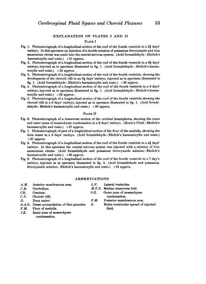
Cerebrospinal Fluid Spaces and Choroid Plexuses 53
EXPLANATION OF PLATES I AND II
PLATE I
Fig. 1. Photomicrograph of a longitudinal section of the roof of the fourth ventricle in a 31 days'embryo. In this specimen an injection of a double solution of potassium ferrocyanide and ironammonium citrate was made into the central nervous system. (Acid formaldehyde: Ehrlich'shaematoxylin and eosin.) x 31 approx.
Fig. 2. Photomicrograph of a longitudinal section of the roof of the fourth ventricle in a 61 days'embryo, injected as in specimen illustrated in fig. 1. (Acid formaldehyde: Ehrlich's haema-toxylin and eosin.) x 24 approx.
Fig. 3. Photomicrograph of a longitudinal section of the roof of the fourth ventricle, showing thedevelopment of the choroid villi in an 8j days' embryo, injected as in specimen illustrated infig. 1. (Acid formaldehyde: Ehrlich's haematoxylin and eosin.) x 38 approx.
Fig. 4. Photomicrograph of a longitudinal section of the roof of the fourth ventricle in a 9 days'embryo, injected as in specimen illustrated in fig. 1. (Acid formaldehyde: Ehrlich's haema-toxylin and eosin.) x 28 approx.
Fig. 5. Photomicrograph of a longitudinal section of the roof of the fourth ventricle showing thechoroid villi in a 9 days' embryo, injected as in specimen illustrated in fig. 1. (Acid formal-dehyde: Ehrlich's haematoxylin and eosin.) x 36 approx.
PLATE II
Fig. 6. Photomicrograph of a transverse section of the cerebral hemispheres, showing the innerand outer zones of mesenchyme condensation in a 6 days' embryo. (Bouin's Fluid: Ehrlich'shaematoxylin and eosin.) x 37 approx.
Fig. 7. Photomicrograph of part of a longitudinal section of the floor of the medulla, showing thedura mater in a 9 days' embryo. (Acid formaldehyde: Ehrlich's haematoxylin and eosin.)x 37 approx.
Fig. 8. Photomicrograph of a longitudinal section of the roof of the fourth ventricle in a 41 days'embryo. In this specimen the central nervous system was injected with a solution of ironammonium citrate. (Acid formaldehyde and potassium ferrocyanide solution: Ehrlich'shaematoxylin and eosin.) x 30 approx.
Fig. 9. Photomicrograph of a longitudinal section of the roof of the fourth ventricle in a 7 day'sembryo, injected as in specimen illustrated in fig. 8. (Acid formaldehyde and potassiumferrocyanide solution: Ehrlich's haematoxylin and eosin.) x 24 approx.
ABBREVIATIONS
A.M. Anterior membranous area. L. V. Lateral ventricles.C.B. Cerebellum. M.T.F. Median transverse fold.CR. Cranium. O.Z. Outer zone of mesenchymeC. V. Choroid villi. condensation.D. Dura mater. P.M. Posterior membranous area.D.A.G. Dense accumulation of blue granules. S. Extra-ventricular spread of injectedF.M. Floor of medulla. fluid.I.Z. Inner zone of mesenchyme
condensation.