The development of RNA interference-based biotechnologies ...
129
The development of RNA interference-based biotechnologies for the sustainable management of Sclerotinia sclerotiorum by Austein G McLoughlin A Thesis submitted to the Faculty of Graduate Studies of The University of Manitoba In partial fulfilment of the requirements of the degree of MASTER OF SCIENCE Department of Biological Sciences University of Manitoba Winnipeg, Manitoba, Canada Copyright © 2018 by Austein G McLoughlin
Transcript of The development of RNA interference-based biotechnologies ...

The development of RNA interference-based biotechnologies for the
sustainable management of Sclerotinia sclerotiorum
by
Austein G McLoughlin
A Thesis submitted to the Faculty of Graduate Studies of
The University of Manitoba
In partial fulfilment of the requirements of the degree of
MASTER OF SCIENCE
Department of Biological Sciences
University of Manitoba
Winnipeg, Manitoba, Canada
Copyright © 2018 by Austein G McLoughlin

ii
ABSTRACT
Necrotrophic pathogens, such as Sclerotinia sclerotiorum and Botrytis cinerea, threaten
global agricultural production. Traditional agrochemicals may negatively impact the
environment and chemical resistance is rising. Therefore, novel strategies are needed to
overcome these obstacles. RNA interference (RNAi), a cellular defence pathway for targeted
mRNA degradation using double stranded RNA (dsRNA), could be a next-generation fungicide.
RNA sequencing of the host-pathogen interface identified hundreds of genes for RNAi-based
control development. I first demonstrated the dsRNA molecules elicited gene silencing in vitro.
Next, I formulated foliar applications to protect both Brassica napus and Arabidopsis thaliana
from S. sclerotiorum and B. cinerea using designer molecules targeting homologous transcripts.
Finally, I engineered host induced gene silencing (HIGS) to suppress fungal colonization on A.
thaliana and explored host defense and death responses during HIGS. Together, these findings
provide compelling evidence for an RNAi-based biotechnological management strategy and will
be a valuable resource for future research endeavours.

iii
ACKNOWLEDGEMENTS
I would like to thank Dr. Mark Belmonte and Dr. Steve Whyard for the opportunity to do
my masters research within your labs. I am greatly appreciative of the guidance and support over
the course of my Masters research. I feel that I have grown as a researcher and learned many
skills. I would also like to thank the other members of my committee, Dr. Dilantha Fernando and
Dr. Steven Harris, as well as Dr. Khalid Rashid for the continual donation of fungal spores. I also
thank the generous financial support by the National Science and Engineering Research Council,
the Manitoba government, the University of Manitoba, and the Department of Biological
Sciences. I thank Manitoba Agricultural Rural Development Initiative, the Canola Council of
Canada, and the Western Grains Foundation for kindly supporting my research.
Next, I would like to thank all members of the Belmonte (Deirdre Khan, Nick Wytinck,
Phil Walker, Michael Becker, Ian Girard, Jenna Kalichuk, Joey Wan, Rachel Robinson, Daniel
Sullivan, Nina Huynh, Nadège Pulgar-Vidal, Kirsten Biggar Vanessa Hoy, Dylan Ziegler, and
Jazzmin Cameron) and Whyard (Aditi Singh, Cass Erdelyan, David Giesbrecht, Alison Tayler,
Dave Boguski) labs for the support and assistance over the past few years. I am truly thankful for
the hard work, help, and friendship you all have given me over the past 2 years. I would also like
to thank Stephanie Tkachuck and Arfa Khan for the coffee, friendship, and support.
Finalmente, muito obrigado às minhas professoras de Português, Fabia Rodrigues e
Barbara Borges pelos apoios de sempre. Muito obrigado pelo café, pelo chá, pelas conversas e
pela paciência comigo durante meu mestrado. To my family, Belmir de Jesus Júnior, Diana
Panting, Daryl McLoughlin, Frances Panting, Tristan McLoughlin, Christina McLoughlin,
Austein Odebust, and Jake McLoughlin, thank you so much for the love, support, and the
strength to accomplish my goals. Vocês contribuiram para meu sucesso!

iv
TABLE OF CONTENTS
TITLE .................................................................................................................................. i
ABSTRACT ................................................................................................................................. ii
ACKNOWLEDGEMENTS ........................................................................................................ iii
LIST OF TABLES ..................................................................................................................... viii
LIST OF SUPPLEMENTARY TABLES ................................................................................ viii
LIST OF FIGURES ..................................................................................................................... ix
LIST OF SUPPLEMENTARY FIGURES ................................................................................. x
NON-COMMON ABBREVIATIONS USED ........................................................................... xi
CONTRIBUTION OF AUTHORS ........................................................................................... xii
CHAPTER 1 : AN INTRODUCTION TO DEVELOPING NEW RNA INTERFERENCE
TECHNOLOGIES TO CONTROL FUNGAL PATHOGENS ................................................ 1
1.1 ABSTRACT ................................................................................................................... 1
1.2 INTRODUCTION ........................................................................................................ 2
1.3 THE MECHANISM OF RNA INTERFERENCE .................................................... 4
1.4 THE SAFETY OF RNA INTERFERENCE TECHNOLOGY ................................ 8
1.5 DEVELOPMENT OF NOVEL TRAITS THOUGH HOST-INDUCED GENE
SILENCING ............................................................................................................................ 11
1.6 IN PLANTA EXPRESSION OF HPRNA OVERCOMES FUNGAL
PRESSURE .............................................................................................................................. 13
v
1.7 FOLIAR APPLICATIONS OF DSRNA REDUCED FUNGAL DISEASE
SYMPTOMS ........................................................................................................................... 14
1.8 RNA SEQUENCING AS AN INFORMATIVE GUIDE FOR RNA
INTERFERENCE ................................................................................................................... 16
1.9 OUTLOOK .................................................................................................................. 17
1.10 RESEARCH OBJECTIVES ...................................................................................... 18
1.11 BIOLOGICAL QUESTIONS AND HYPOTHESES .............................................. 19
1.11.1 Target Identification and development of topical RNAi-based technology for
fungal control ...................................................................................................................... 19
1.11.2 Host-induced gene silencing (HIGS) of S. sclerotiorum ................................... 20
1.12 REFERENCES ............................................................................................................ 20
PREFACE TO EXPERIMENTAL CHAPTERS: AN INTRODUCTION TO
SCLEROTINIA SCLEROTIORUM ........................................................................................... 34
CHAPTER 2 : IDENTIFICATION AND APPLICATION OF RNA INTERFERENCE-
BASED FOLIAR CONTROL OF SCLEROTINIA SCLEROTIORUM AND BOTRYTIS
CINEREA IN PLANTS .............................................................................................................. 42
2.1 ABSTRACT ................................................................................................................. 42
2.2 INTRODUCTION ...................................................................................................... 43
2.3 METHODS .................................................................................................................. 46
2.3.1 Brassica napus growth conditions for RNA sequencing ...................................... 46
2.3.2 Leaf inoculation and S. sclerotiorum tissue collection for RNA sequencing ...... 46
2.3.3 RNA extraction and sequencing ............................................................................ 47
2.3.4 Bioinformatics Pipeline .......................................................................................... 48

vi
2.3.5 Selection of gene targets and Target Identification Pipeline (TIP) .................... 49
2.3.6 RNA extraction, cDNA synthesis and in vitro production of dsRNAs ............... 50
2.3.7 Quantification of relative transcript abundance following dsRNA application in
vitro 51
2.3.8 Foliar applications of dsRNAs to the leaf surface ................................................ 52
2.4 RESULTS .................................................................................................................... 53
2.4.1 Gene expression of S. sclerotiorum grown in vitro and on susceptible and
tolerant hosts of B. napus ................................................................................................... 53
2.4.2 Differential expression and GO term enrichment analysis in target gene
identification ........................................................................................................................ 53
2.4.3 QRT-PCR assessment of RNAi in S. sclerotiorum ............................................... 57
2.4.4 Foliar applications of dsRNA reduce S. sclerotiorum infection in B. napus ...... 60
2.4.5 Topical dsRNAs mitigate S. sclerotiorum infection on A. thaliana ..................... 64
2.4.6 DsRNAs targeting homologues in Botrytis cinerea attenuate fungal infection .. 66
2.5 DISCUSSION .............................................................................................................. 66
2.6 ACKNOWLEDGEMENTS ....................................................................................... 73
2.7 REFERENCES ............................................................................................................ 74
CHAPTER 3 HOST-INDUCED GENE SILENCING AS AN RNA INTERFERENCE
BIOTECHNOLOGY FOR SCLEROTINIA SCLEROTIORUM MANAGEMENT ............. 86
3.1 ABSTRACT ................................................................................................................. 86
3.2 INTRODUCTION ...................................................................................................... 87
3.3 METHODS .................................................................................................................. 90
3.3.1 Growth conditions for Arabidopsis thaliana ......................................................... 90

vii
3.3.2 Transformation and Identification of A. thaliana transformants ...................... 90
3.3.3 Arabidopsis Infection Assays, fungal load quantification, and target gene
knockdown ........................................................................................................................... 92
3.4 RESULTS .................................................................................................................... 95
3.4.1 Host-induced gene silencing technology lessens lesion progression ................... 95
3.4.2 Infection under host–induced gene silencing decreases the presence of fungal
DNA 97
3.4.3 Specific fungal transcripts were reduced during infection on A. thaliana
SS1G01703RNAi and A. thaliana SS1G05899RNAi ............................................................... 97
3.4.4 Defence markers are elevated and cell death markers are repressed
differentially during infection of hpRNA producing and non-hpRNA producing A.
thaliana ................................................................................................................................. 99
3.5 DISCUSSION ............................................................................................................ 101
3.6 REFERENCES .......................................................................................................... 107
CHAPTER 4 : CONCLUSIONS AND FUTURE DIRECTIONS ....................................... 116

viii
LIST OF TABLES
Table 2.1 Selected list of Sclerotinia sclerotiorum target genes for RNA interference testing in
liquid culture, and infection assays on Brassica napus and Arabidopsis thaliana...............61
Table 3.1 Primer sequences for cloning Sclerotinia sclerotiorum target genes and confirmation
of genetic insertion within Arabidopsis thaliana lines.........................................................93
Table 3.2 Quantitative PCR primer sequences and efficiencies for Sclerotinia sclerotiorum and
Arabidopsis thaliana target transcripts and S. sclerotiorum 18S rDNA...............................93
LIST OF SUPPLEMENTARY TABLES
Supplementary Table 2.1 . Mapping summary for all biological replicates of in-vitro and in-
planta samples......................................................................................................................83
Supplementary Table 2.2 Target identification pipeline guidelines for selecting targets to
control phytopathogenic fungi using RNAi..........................................................................84

ix
LIST OF FIGURES
Figure 1.1. Overview of the mechanism of RNAi within fungal hyphae......................................6
Figure 2.1 Hierarchical clustering of gene expression of the growth and penetration of
Sclerotinia sclerotiorum in-vitro (PDA) and in-planta (Brassica napus cv. Westar
(Susceptible) and ZhongYou821 (Tolerant))........................................................................54
Figure 2.2 Identification of Sclerotinia sclerotiorum genes and biological processes significantly
up and down regulated during infection (in-planta).............................................................56
Figure 2.3 Timing of RNAi silencing in Sclerotinia sclerotiorum for the select targets
SS1G_01703, SS1G_05899, and SS1G_06487....................................................................58
Figure 2.4 Effect of dsRNA dose on Sclerotinia sclerotiorum grown in liquid cultures for the
select targets SS1G_01703, SS1G_05899, SS1G_06487, and SS1G_11912.......................59
Figure 2.5 DsRNA targeting Sclerotinia sclerotiorum suppresses lesion size on Brassica napus
susceptible cultivar Westar...................................................................................................63
Figure 2.6 DsRNA treatment on Arabidopsis thaliana leaves reduces Sclerotinia sclerotiorum
lesion area.............................................................................................................................65
Figure 2.7 Botrytis cinerea homologues targeted with foliar dsRNA controls fungal infection.67
Figure 3.1 Sclerotinia sclerotiorum infection was limited on hpRNA-producing Arabidopsis
thaliana................................................................................................................................96
Figure 3.2 Relative presence of Sclerotinia sclerotiorum 18S rDNA was reduced on Arabidopsis
thaliana SS1G01703RNAi and A. thaliana SS1G05899RNAi compared to wild-type A.
thaliana................................................................................................................................98
x
Figure 3.3 Relative transcript levels of SS1G_01703 and SS1G_05899 during infection on
Arabidopsis thaliana SS1G01703RNAi and A. thaliana SS1G05899RNAi compared to wild-type
A. thaliana..........................................................................................................................100
Figure 3.4 Relative Arabidopsis thaliana transcript levels of cell death (ATG8B, BI1, SEN1) and
defense markers (PDF1.2, PR1) during Sclerotinia sclerotiorum colonization.................102
LIST OF SUPPLEMENTARY FIGURES
Supplementary Figure 2.1 Sclerotinia sclerotiorum infection on Brassica napus leaves after
treatment of water or GFP-dsRNA.......................................................................................85

xi
NON-COMMON ABBREVIATIONS USED
AGO ARGONAUTE DCL DICER-LIKE dsRNA double stranded RNA FPKM fragments per kilobase of million mapped reads GO Gene ontology HIGS Host induced gene silencing hpRNA hairpin RNA JA Jasmonic acid milRNA micro-like RNA miRNA micro RNA mRNA messenger RNA nt nucleotides OA Oxalic acid QIP QUELLING DEFICIENT-2 INTERACTING PROTEIN qPCR Quantitative polymerase chain reaction qRT-PCR Quantitative real-time polymerase chain reaction RdRP RNA dependent RNA Polymerase RISC RNA induced silencing complex RNAi RNAi interference RNA-Seq RNA sequencing ROS Reactive oxygen species SA Salicylic acid siRNA small interfering RNA sRNA small RNA
xii
CONTRIBUTION OF AUTHORS
Chapter 1
The authors for Chapter 1, submitted (with modifications) to the Canadian Journal of
Plant Pathology in 2017 are in order: Austein G McLoughlin, Phil L Walker, Nick Wytinck,
Daniel Sullivan, Steve Whyard, and Mark F Belmonte.
AGM, SW, and MFB conceptualized, wrote, and edited the document. PLW wrote the HIGS
section and contributed to the editing process. NW described the uptake of dsRNA. DS
illustrated the mechanism of RNAi with contributions from AGM, SW, and MFB.
Chapter 2
The authors for Chapter 2, submitted (with modifications) to Scientific Reports in 2017
are in order: Austein G McLoughlin, Nick Wytinck, Phil L Walker, Ian J Girard, Khalid Y
Rashid, Teresa de Kievit, WG Dilantha Fernando, Steve Whyard, and Mark F Belmonte.
AM performed bioinformatics analyses, designed experiments, primer design, target
identification, RNA extractions, qPCR, infection assays, and manuscript preparation. PLW
performed bioinformatics analyses, RNA extraction, manuscript preparation. NW performed
RNA extractions, infection assays, and manuscript preparation. IJG performed library
preparation for RNA Sequencing and RNA extractions. KYR collected S. sclerotiorum spores
from agriculture fields. TdK and WGDF contributed as part of a collaborative grant. MFB and
SW conceived the ideas and prepared the manuscript.
Preface, Chapter 3, and Chapter 4
I performed all the experiments stated in Chapter 3 and prepared thesis. Mark F Belmonte
and Steve Whyard contributed to the final document preparation.

1
CHAPTER 1 : AN INTRODUCTION TO DEVELOPING NEW RNA INTERFERENCE
TECHNOLOGIES TO CONTROL FUNGAL PATHOGENS
Austein G Mcloughlin, Phil L Walker, Nick Wytinck, Daniel Sullivan, Steve Whyard, and Mark
Belmonte
This chapter has been submitted to Canadian Journal of Plant Pathology (with modifications)
for publication
1.1 ABSTRACT
Current agricultural output is challenged by increasing fungal pressure that can lead to
considerable losses in crop yield and post-harvest storage. Traditional chemical fungicides used
to treat pathogenic fungi can be ineffective and harmful to the environment if not managed
properly. With fungicide resistance increasing, new environmentally-friendly and sustainable
technologies are required to protect some of the world’s most important crops. RNA interference
(RNAi) is an intrinsic cellular mechanism, mediated by double-stranded RNA (dsRNA), which
can suppress protein expression through targeted destruction of mRNAs, and with recent
advances in dsRNA delivery or expression in plants, this mechanism has the potential to
revolutionize crop management strategies. Examples of RNAi-based control to manage
pathogenic fungal species are steadily increasing, as the technology offers new options for the
development of new fungicides with improved species-specificity and/or potency against fungi
for which existing fungicides have been ineffective. RNAi technology can be adapted to provide
either robust and multi-crop plant protection using topical sprays or can provide more durable
resistance through transgene expression of dsRNAs within susceptible plant tissues. Using RNA

2
sequencing to identify fungal gene targets, RNAi-based control technology will continue to
develop as a potent and environmentally-conscious alternative to traditional agrochemicals for
crop protection
1.2 INTRODUCTION
Access to safe, healthy, and sustainable food sources is one of the defining challenges of
our time. Given the rapid increase of the human population, it is estimated that we will require an
additional production of 200 billion calories, equating to a 100-110% increase in crop demand to
meet nutritional needs by the year 2050 (Bebber and Gurr, 2015; Tilman et al., 2011; United
Nations, 2017). Currently, 40% of ice-free land is used to grow crops; however, global climatic
changes challenge our current production systems by decreasing yield potentials and shrinking
arable land resources (Myers et al., 2017; Pugh et al., 2016). Moreover, biotic agents, such as
necrotrophic and biotrophic fungi, further complicate agronomic production, causing global
losses of up to 30-40% in crop yield in field and post-harvest (Bebber and Gurr, 2015; Myers et
al., 2017). With rapid globalization and migration across the globe, fungal pathogens are
predicted to spread rapidly to virgin lands, presenting new challenges for crop production
globally (Bebber et al., 2014).
Traditional chemical treatments used to combat fungal disease epidemics and control
necrotrophic pathogens, like Sclerotinia sclerotiorum (Lib.) de Bary, achieve mixed success in
field (Huzar-Novakowiski et al., 2017). For example, chemical applications at sub-lethal levels
were shown to promote fungicide resistance (Amaradasa and Everhart, 2016). Common
fungicides like boscalid, iprodine, thiophanate methyl, azoxystrobin, and pyracostrobin, when
diluted to sub-lethal levels resulted in increased mutation rates of up to 60-fold due to cellular

3
stress in S. sclerotiorum (Amaradasa and Everhart, 2016). Additional treatments of fungicides
resulted in reduced fungal sensitivity due to genetic mutations suggesting that common
agricultural techniques could promote fungicide resistance (Amaradasa and Everhart, 2016).
Development of resistance may lead to a positive feedback loop, where chemical use selects for
further resistance, attributing to the rise of fungicide resistant S. sclerotiorum, Botrytis cinerea
Pers., and Magnaporthe oryzae Couch (Castroagudín et al., 2015; Penaud and Walker, 2015;
Rupp et al., 2017). For these reasons, there is a clear need to develop new methods to control
disease outbreaks using sustainable technologies.
Despite the suggestion that precise chemical applications could reduce environmental
impacts, fungicide use has deleterious effects on the surrounding agro-ecological landscape due
to its general biocidal nature, as well as the dispersal and persistence within the environment (Le
Cointe et al., 2016; Sabatier et al., 2014; Smalling et al., 2013). The use of fungicidal
compounds was noted to alter the structure and function of aquatic communities, culminating in
severe physiological pathologies, such as increased mortality, reduced reproductive rates, and
decreased enzyme activity, for zooplankton, gastropods, amphibians, and earthworms
(McMahon et al., 2012; Rico et al., 2016; Zubrod et al., 2011). Furthermore, fungicidal
compounds have been implicated in reduced bee health and abnormal behaviors, such as reduced
nest recognition, decreased colony initiation, and uncoupled mitochondrial respiration, all of
which may contribute to the decline of bee populations (Elston et al., 2013; Simon-Delso et al.,
2014; Syromyatnikov et al., 2017). The development of any new species-specific fungicides
should provide both environmentally-safer control strategies, but also provide effective control
of the fungus to ensure improved crop yields.
4
Recently, a new generation of species-specific control methods, taking advantage of a
cellular defence mechanism called RNA interference (RNAi), demonstrated successful control of
insects, nematodes, viruses, and parasitic plants (Alakonya et al., 2012; Papolu et al., 2013;
Schmitt-Engel et al., 2015; Whyard et al., 2009). The first commercially approved, transgenic
plants carrying RNAi constructs against corn rootworm (Diabrotica virgifera virgifera LeConte)
and bean golden mosaic virus were approved for cultivation in the US and Brazil, respectively
(Tollefson, 2011; United States Environmental Protection Agency, 2017). Despite the successes
of RNAi approaches, few studies have applied this revolutionary technique for the management
of fungal phytopathogens, likely due to the lack of reasonable target identification tools and poor
fungal genomic annotation. Here, we describe how RNAi technology exploits intrinsic cellular
pathways in eukaryotes for the development of novel fungal control strategies. A summary of
aspects relating to safety of this new technology will be explored, as well as how integrating
modern genomics techniques could help guide the development of next-generation RNAi-based
control of fungal pathogens.
1.3 THE MECHANISM OF RNA INTERFERENCE
RNAi is a conserved pathway in eukaryotes that protects cells from viruses and controls
transposon activity. The mechanism utilizes short interfering RNAs (siRNAs) to guide the
targeted degradation of transcripts using sequence homology (Torres-Martínez and Ruiz-
Vázquez, 2017). The description of RNAi in Caenorhabditis elegans (Maupas) Dougherty by
Andrew Fire and Craig Mello earned them the Nobel Prize for Medicine in 2006 (Fire et al.,
1998; Nobel Media AB, 2017). However, similar phenomena had been noted in other
organisms. Romano and Macino (1992) (Romano and Macino, 1992) earlier had described a
5
post-transcriptional silencing phenomenon, which they termed quelling, in the ascomycete
Neurospora crassa Shear & B.O. Dodge. The core proteins in the quelling pathway are the same
proteins implicated in the RNAi pathway: ARGONAUTE (AGO), QUELLING DEPENDENT-2
(QDE-2), DICER-like (DCL), and RNA-dependent RNA Polymerase (RdRP) (Torres-Martínez
and Ruiz-Vázquez, 2017).
Since RNAi is an intrinsic cellular defence process against invading double stranded
RNA (dsRNA) viruses, introducing in vitro synthesized dsRNAs or producing the molecules in
planta exploits this cellular reaction as a crop management strategy naturally (Wang and Jin,
2017) (Fig. 1.1a). Transport of long dsRNA and shorter, small interfering RNA (siRNA) into B.
cinerea spores was observed using fluorescein-labeled nucleotides (nt) (Wang et al., 2016a);
however, the mechanism of transport remains undefined in fungi. In both humans and C.
elegans, dsRNA diffuses passively through a dsRNA-specific channel, SID1 (Duxbury et al.,
2005; Whangbo et al., 2017). Homologues of the SID1 channel do not exist in fungi, and thus,
dsRNA transport must occur via an alternative mechanism(Wang et al., 2016a). In various
insects, exogenous dsRNA is transported using receptor-mediated clathrin endocytosis (reviewed
in Huvenne and Smagghe, 2010) (Huvenne and Smagghe, 2010). An “RNAi of RNAi” approach
provided evidence for endocytic involvement in dsRNA transport. Initially, cells were treated
with dsRNA targeting a specific component implicated in clathrin-mediated endocytosis, before
a following treatment of GFP-dsRNA was applied. Using a GFP-reporter system, components
involved in uptake of the molecules could be elucidated by observing a diminished fluorescent
signal. Thus, the components of receptor mediated endocytosis were inferred to be clathrin heavy
chain, clathrin adaptor protein 50, vacuolar H+ ATPase, and ADP ribosylation factor ARF72A
(Saleh et al., 2006). Similarly, application of chemical inhibitors confirmed endocytosis as a

6
Figure 1.1. Overview of the mechanism of RNAi within fungal hyphae. Upon encountering double stranded RNA (dsRNA), the molecules are transported into the cytoplasm through an undefined mechanism (a). Once in the cytoplasm, the molecules are recognized by DICER-LIKE (DCL) (b), which cleaves the molecules into small interfering RNA (siRNA) 21-25 nucleotides (nt) in length (c). The siRNA molecules then complex with ARGONAUTE (AGO) (d), which nicks the siRNA and recruits QUELLING DEFICIENT-2 INTERACTING PROTEIN (QIP) to degrade the passenger strand (e). With the removal of the passenger strand, RISC becomes activated to seek messenger RNA (mRNA) transcripts with complementary sequences to the internal siRNA (f) for degradation (g). The degraded mRNA and the siRNA can function as primers in secondary dsRNA synthesis using RNA-dependent RNA Polymerase (RdRP) (h) to further amplify gene silencing.

7
secondary pathway of dsRNA uptake in C. elegans (Xiao et al., 2015). Without SID1, clathrin-
mediated endocytic pathways may play an integral function in the transport of dsRNA in fungi.
Once in the cytoplasm, the presence of a dsRNA molecule is recognized by the dsRNA
binding domain of DCL1 or DCL2 (Lee et al., 2010; Li et al., 2010) (Fig. 1.1b). Upon
recognition of dsRNA, DCL recruits the SAGA complex with histone acetyltransferase activity
to increase transcription from the dcl and ago promoters, and mobilize RNAi machinery within
the fungus (Andika et al., 2017). To cleave dsRNA molecules, the 5’ end of the dsRNA anchors
in the PAZ (Piwi-Argonaute-Zwille/Pinhead) domain within DCL, allowing two consecutive
RNase III domains to cleave the ribose-phosphate backbone, resulting in siRNAs of 21-25 nt in
length with a 5’ monophosphate and a 3’ 2 nt overhang (Kandasamy and Fukunaga, 2016)
(Figure 1.1c). Once the double stranded siRNA is generated, AGO complexes with the siRNA to
recruit QIP (QDE-2-INTERACTING PROTEIN) and form the RNA-induced silencing complex
(RISC) (Dang et al., 2011) (Fig. 1.1d). Once bound, AGO nicks the siRNA duplex; QIP
recognizes and degrades the nicked passenger strand with exonuclease activity (Cheng et al.,
2016; Maiti et al., 2007) (Fig. 1.1e). The RISC complex then becomes activated to seek
transcripts with complementary sequences to the remaining siRNA strand, termed the guide
strand (Fig. 1.1f). When a messenger RNA (mRNA) base pairs to the guide strand in RISC,
exonuclease activity is activated to degrade complementary RNA, resulting in a reduction of
mRNA accumulation within the fungal hyphae (Dang et al., 2011) (Fig. 1.1g).
Some of the resulting siRNAs produced during long dsRNA processing and the degraded
target mRNA could act as a primer to initiate RdRP activity and temporarily sustain silencing
(Fig. 1.1h). siRNAs anneal to complementary mRNA transcripts in the absence of DCL to act as
a primer for second strand synthesis through recruited RdRPs. The secondary dsRNAs produced

8
by RdRPs invoke further RISC complex formation to amplify RNAi-mediated silencing
(Ghildiyal and Zamore, 2009; Villalobos-Escobedo et al., 2016). The existence of RdRPs vary
greatly amongst fungi, with species like Fusarium graminearum (Schwienitz) Petch having five,
while others, like N. crassa, have two (Chen et al., 2015; Zong et al., 2009). The presence of
RdRP may be required for potent silencing to occur, as is the case for Mucor circinelloides.
However, for most fungi, RdRPs are likely nonessential, but can serve a role in RNAi signal
amplification as well as being involved in miRNA (microRNA) and transposon silencing (Calo
et al., 2012; Dang et al., 2011).
1.4 THE SAFETY OF RNA INTERFERENCE TECHNOLOGY
The sequence-specific mechanism of RNAi provides unparalleled evidence that RNAi-
based technologies offer safer and more environmentally-conscious alternatives to more
traditional agrochemicals. DsRNA can be designed to avoid sequences of other organisms within
the environment. Thorough ecological testing of the recently approved transgenic corn
expressing RNAi constructs targeting corn rootworm best demonstrates the specificity of RNAi
technology (United States Environmental Protection Agency, 2017). For example, both
implementations of RNAi technology, transgenic plant material and topical in vitro synthesized
dsRNA molecules, were used to assess cross-reactivity amongst a variety of invertebrates (e.g.
Apis mellifera L., Eisenia Andrei Bouché, Coleomegilla maculate De Geer), vertebrates (e.g.
Gallus domesticus L., Ictalurus punctatus Rafinesque), and soil microorganisms. No observable
change in physiology, nutrient assimilation, or reproduction in the tested organisms was
observed, regardless of the dsRNA source (Bachman et al., 2016). The expression of interfering
RNA in the pollen did not adversely affect honeybees (A. mellifera) during the experiment, and
previously, diet-derived miRNA from pollen demonstrated negligible transfer to and
9
dissemination within honeybees (Bachman et al., 2016; Masood et al., 2016). Furthermore,
honeybee feeding assays of long dsRNA not specific for any honeybee gene also failed to
demonstrate an effect on the bees (Tan et al., 2016). The findings from these studies suggest that
honeybees exposed to exogenously delivered dsRNAs, whether derived from either transgenic
plants or foliar applications, may not pose serious threats to these important pollinators. Further
studies will likely be required to satisfy government regulators that any dsRNA applied to a crop
does not affect honeybees or any other species within the surrounding environment.
One asset of RNAi-based control is the stability of the dsRNA molecule within the
phyllosphere (Miguel and Scott, 2015). Synthetic molecules can be readily made in the
laboratory and are more thermodynamically stable than single stranded RNA (Nicholson, 2014;
Wang and Jin, 2017). Due to the double stranded structure of the molecules, dsRNAs are also
more resilient to nuclease degradation than mRNA (Aryani and Denecke, 2015; Hoerter et al.,
2011). As a topical application, dsRNA molecules were bioactive against Colorado potato beetle
(Leptinotarsa decemlineata Say) on the surface of potato (Solanum tuberosum L.) leaves for over
28 days under greenhouse conditions (Miguel and Scott, 2015). Using natural chemistries, such
as clay nanosheets (BioClay)(Mitter et al., 2017), dsRNA efficacy was improved against
cucumber mosaic virus and pepper mild mottle virus under adverse environmental conditions.
The clay nanosheets shielded the dsRNA molecules from environmental RNase III degradation
and improved adhesion to the leaf surface (Ladewig et al., 2009; Mitter et al., 2017). Thus,
dsRNA induced gene silencing could be a suitable solution as foliar application against
phyllophillic pathogens.
Another advantage of RNAi management is the lack of persistence within the pedosphere
(Dubelman et al., 2014). Regardless of dsRNA molecule size or the composition of the

10
agricultural soil, the majority of dsRNA rapidly degrades within 24 hours (Dubelman et al.,
2014; Fischer et al., 2016). Similarly, dsRNAs were demonstrated to dilute and degrade greatly
within 96 hours upon entering water systems (Albright et al., 2017). Likely, bacterial RNA
metabolic processes and RNase III enzymes cause the dsRNA degradation in the soil and aquatic
environments (Cho, 2017; Urich et al., 2008). Therefore, dsRNA sourced from plant material or
foliar sprays will not likely disseminate far from the point of application through soil leaching.
Coupled with its sequence specificity, RNAi-based fungicides are unlikely to have
environmentally adverse effects compared to traditional chemical methods of fungal control and
therefore, they represent an attractive alternative to the broad-spectrum fungicides currently in
use.
For consumer food production, RNAi technology would not differ from the current
consumption of small RNAs (sRNAs) that naturally populate grains, fruits, vegetables, and
meats. Large pools of sRNAs, which include siRNAs, miRNAs, piRNA, and endogenous
dsRNA, are produced from a variety of sources, such as viruses, transposons, or the host cell
itself (Frizzi et al., 2014; Ivashuta et al., 2009). In silico analyses predict siRNAs with 100%
complementarity to human mRNA transcripts, yet no pathological effects have been associated
with daily consumption of sRNA (Jensen et al., 2013). Humans inherently have evolved many
barriers that would prevent or limit the transport of sRNA, such as nucleases in the saliva and
gastrointestinal track, acid in the stomach, and the unfavourable transport of large, polar
molecules across a hydrophobic membrane (Juliano et al., 2009; O’Neill et al., 2011). The lack
of dietary siRNA efficacy was supported in mouse model research. Mice were dosed daily with
either siRNA or long dsRNA targeting an essential vacuolar ATPase over a 28-day period and no
adverse cellular pathologies were noted (Petrick et al., 2015). The lack of bioavailability could
11
be attributed to instability passing through the gastrointestinal track or rapid metabolism in the
bloodstream (Christensen et al., 2013; Dickinson et al., 2013). Therefore, any dsRNA or siRNA
molecules present should not elicit adverse effects from dietary sources rich in nucleic acid.
Furthermore, many food products undergo many processing techniques before consumer
consumption (Chemat et al., 2017; Misra et al., 2017). The majority of processing techniques
(baking, microwaving, solvent extraction, thermal treatment, fermentation, acidification,
alkalization, and bleaching) result in effective nucleic acid destabilization and degradation prior
to the final food product (Gryson, 2010; Vijayakumar et al., 2009). For example, edible oils
undergo multiple steps involving heat, pressure, and solvent treatments, which exclude polar
molecules, such as nucleic acid, and/or result in molecule fragmentation (Belur et al., 2017; Mba
et al., 2015). Similarly, heating and purification in sugar production eliminates DNA by a factor
of 1014 (Klein et al., 1998). Based on the chemical and physical similarities, dsRNA would likely
have a similar fate to DNA during food processing (Forbes and Peppas, 2012; Lipfert et al.,
2014). Thus, any nucleic acid introduced from RNAi technology would not survive food
processing. Taken together, RNAi technology, due to both the chemistry of the RNA molecules
and the sequence-specificity of the molecule, could be considered a safe, green technology,
which can be expressed as a novel trait (transgene) or through topical formulations.
1.5 DEVELOPMENT OF NOVEL TRAITS THOUGH HOST-INDUCED GENE
SILENCING
Host-induced gene silencing (HIGS) is an emerging biotechnology in which plants are
engineered to produce sRNAs capable of silencing target genes of a target organism. HIGS
utilizes the RNAi pathway by equipping the host plant with hairpin RNAs (hpRNAs) containing
12
sequence homology to target genes. Upon transcription, these hpRNA molecules mimic dsRNA
and initiate the inherent cellular RNAi pathway. Export of hpRNA from the plant nucleus to the
cytoplasm is hypothesized to be facilitated by the binding of an exportin protein HASTY to
guide successful nucleocytoplasmic transport (Bollman, 2003; Tuan Le and Wang, 2011). Once
in the cytoplasm, hpRNA initiates the host RNAi pathway leading to the generation of
approximately 21 nt siRNA molecules, as in the case for fungi. For siRNAs to function in target
gene silencing, they must undergo successful transfer from host plant cell to the pest or
pathogen. While evidence suggests target gene silencing in pathogenic fungal species may
operate through host derived siRNAs (Panwar et al., 2013), the mechanism of siRNA transfer
from host to pathogen remains unclear. Studies in animal systems show that secreted miRNA
may be associated with host-derived exosomes and lead to successful transfer between organisms
(Valadi et al., 2007). Exosomal uptake by the receiving cell is hypothesized to utilize exosome-
mediated endocytosis, where the vesicular membrane of the sRNA containing exosome fuses
with the receiving plasma membrane leading to the release of sRNA into the pathogen’s
cytoplasm (Valadi et al., 2007). Another hypothesis for potential sRNA transfer involves
transmembrane transporter-mediated uptake without utilization of host derived vesicles.
Previously, membrane-free sRNAs were found within the extracellular space and associated with
high density lipoproteins. These lipoproteins may facilitate successful transfer of extracellular
sRNA to recipient cells (Vickers et al., 2011). While there is still much to discover about the
mechanisms of HIGS, this technology represents a promising strategy to protect plants against
difficult to control fungal pathogens.
The safety and efficacy of HIGS technology led to the approval of two RNAi crops for
commercial production in the United States and Brazil. The Brazilian National Technical

13
Commission approved RNAi pinto beans (Phaseolus vulgaris) for commercial production in
2011 (Tollefson, 2011). Pinto beans were engineered to disrupt early viral replication of the Bean
Golden Mosaic Virus by targeting the viral gene AC1 (Bonfim et al., 2007). Similarly, the
Environmental Protection Agency approved maize plants expressing dsRNA targeting DvSnf4, a
component of the ESCRT-III complex involved in endosomal sorting and lysosomal degradation
in corn rootworms, D. virgifera virgifera, for production in the US (Bolognesi et al., 2012;
United States Environmental Protection Agency, 2017). DvSnf4 dsRNA was potent at low doses,
leading to accumulation of ubiquinated proteins in the midgut cells of larvae. Since autophagy
was impaired, the cells malfunctioned, the gut’s digestive processes ceased, and the insects failed
to grow and eventually died (Baum et al., 2007; Ramaseshadri et al., 2013). Given the flexibility
and utility of the technology, when applied to fungal pathogens affecting crops, RNAi-HIGS
technology has the potential for regulatory approval and full scale agronomic production.
1.6 IN PLANTA EXPRESSION OF HPRNA OVERCOMES FUNGAL PRESSURE
The creation of stably transformed plants expressing dsRNA molecules could impart full
plant protection. HIGS has been implemented to reduce fungal pressure and toxin biosynthesis
for a variety of phytopathogenic fungi (Koch et al., 2013; Thakare et al., 2017; Zhang et al.,
2016). Engineering novel traits like fungal resistance using genetic techniques is an efficient
strategy to build resistance in crop plants. While traditional germplasm screening and breeding
strategies have achieved limited success in providing resistance against S. sclerotiorum (Disi et
al., 2014), the use of HIGS in Nicotinia tobacum L. expressing hpRNA targeting fungal chitin
synthase (Sschs) proved effective in controlling S. sclerotiorum. Expression of hpRNA reduced
the level of Sschs mRNA within the fungus, indicating S. sclerotiorum could take up dsRNA

14
from the host through yet to be determined cellular mechanisms (Andrade et al., 2016). Since
phytopathogens are likely capable of taking up environmental dsRNA from host cells, HIGS
could provide a functional strategy to reduce fungal pressure on the plant.
The root-pathogen interface could also be protected from many fungal pathogens that
initiate root infections from the soil, such as Verticillium sp., Fusarium sp., and Rhizoctonia
solani J.G. Kühn (De Coninck et al., 2015; Tedersoo et al., 2014). While topical formulations of
dsRNA would likely degrade rapidly within the soil (Dubelman et al., 2014), GM plants could
deliver siRNA to the pathogen at the root-pathogen interface, as exemplified by transgenic RNAi
cotton (Gossypium sp.). Roots of cotton expressing hpRNA targeting Verticillium dahliae Kleb
hygrophobins1 gene were able to resist severe root infection sufficiently compared to the wilted
non-transformed control (Zhang et al., 2016). Additionally, plants engineered with RNAi
constructs expressing hpRNA could convey resistance throughout the plant lifecycle. Transgenic
banana plants (Musa spp.) expressing hpRNA directed at either F. oxysporum f. sp. cubense
genes velvet or Fusarium transcription factor 1, resisted infection for 8 months post-inoculation
(Ghag et al., 2014). Thus, engineered plants expressing novel RNAi traits conferring resistance
against economically important plant pathogens represents an additional level of durability that
would benefit growers interested in sustainable crop protection technologies.
1.7 FOLIAR APPLICATIONS OF DSRNA REDUCED FUNGAL DISEASE
SYMPTOMS
Foliar dsRNA applications offer shorter term protection from fungal infections, relative
to transgene mediated resistance, but nevertheless, they could be particularly useful to protect
agri-food products during post-harvest storage and in protecting plants without defined or

15
efficient transformation protocols for HIGS (Wang and Jin, 2017). Despite recent advances to
control insect pests and viral pathogens, few studies have implemented in vitro synthesized
molecules for fungal control. In a pioneering study by Mumbanza et al. (2013), 14 different
genes involved in processes such as transcription, RNA modification, DNA replication, and
intracellular transport were tested in vitro, using nutrient plates rather than plants, for spore
germination inhibition in F. oxysporum f. sp. cubense and Mycosphaerella fijiensis Morelet.
Treatment of fungal spores with dsRNA molecules inhibited germination of the two banana
pathogens by up to 95.9% and 65.8% respectively. In vitro testing provided compelling evidence
for the potential for fungal suppression, despite not having tested these molecules on their plant
hosts.
Recently, topical application of dsRNA to the surface of treated barley (Hordeum vulgare
L.) leaves reduced F. graminearum growth. By targeting three fungal cytochrome P450
lanosterol C-14α demethylases, required for fungal ergosterol synthesis and a common fungicide
target, reductions over 50% of both target transcript and fungal DNA accumulation were
achieved (Koch et al., 2016). Similarly, using long dsRNA targeting Dcl1 and Dcl2
simultaneously in B. cinerea, Wang et al. (2016)(Wang et al., 2016a) demonstrated remarkable
levels of protection in Arabidopsis thaliana (L.) Heynh, tomato, grape, strawberry, onion, and
rose petals from infection, with average lesion sizes, transcript levels, and fungal DNA all being
reduced by over 80%. The observed protection in a wide-range of host tissues suggests broad
utility of the technology in the protection of food and ornamental species. The protection of
barley leaves and tomato leaves demonstrates the utility for field management of crops, while
reduced fungal presence on various fruits and vegetables could be useful after harvest, protecting
food in transport, storage, or on store shelves (Koch et al., 2016; Wang et al., 2016a). Taken

16
together, these studies provide solid evidence of the flexibility of RNAi-based molecular sprays
that confer protection along the food production pipeline.
1.8 RNA SEQUENCING AS AN INFORMATIVE GUIDE FOR RNA INTERFERENCE
Despite the advances in the development of RNAi-based phytopathogenic control, the
selection of target genes still represents a significant challenge in the design and effectiveness of
fungal suppression. Previously, RNAi targets were selected using gene deletions or chemical
inhibition. For example, Koch et al. (2016) chose a common fungicidal target, while Wang et al.
(2016) developed foliar applications based on previous genetic deletions of B. cinerea strains
(Weiberg et al., 2013). Fungal transformation protocols are lengthy and at times inefficient,
thereby limiting the effective size of target gene identification screens (Meyer, 2008). However,
dual RNA-sequencing of both fungal pathogen and host plant now provides unprecedented
opportunities to identify novel RNAi targets based on the transcriptome atlas of the pathogen
lifecycle, infection state, tissue type, host defense response, or treatment conditions (Westermann
and Barquist, 2017). Genes implicated in plant resistance response provide useful information for
plant breeding and manipulation, while specific fungal responses essential for pathogenesis could
eventually be used as targets for RNAi-based fungal control (Girard et al., 2016). For example,
transcriptomic sequencing across major developmental stages of S. sclerotiorum uncovered
genes important for pathogenicity and development. In particular, two small cysteine-rich
effector proteins, SsCVMH and SsSSVP1, as well as a microbial opsin homologue were
upregulated during infection (Lyu et al., 2015). S. sclerotiorum transformed with RNAi silencing
vectors targeting these effector proteins significantly inhibited the ability of the fungus to
colonize Brassica napus L.. (Lyu et al., 2015; Lyu et al., 2016a; Lyu et al., 2016b). Clearly,
17
RNA-seq identification of transcripts essential for pathogenesis could be very useful in the
development of future RNAi target genes for fungal control.
The lack of development of RNAi-based fungal strategies is likely due to poor genomic
annotation of many fungi and a lack of accessible bio-computational platforms. User-friendly
programs, such as FungiFun2 (elbe.hki-jena.de/fungifun/fungifun.php; Priebe et al., 2015) and
SeqEnrich (belmontelab.com; Becker et al., 2017), have overcome many of the problems
associated with both a lack of functional annotation and complicated bioinformatics analyses.
Once gene ontology (GO) is resolved using FungiFun2, researchers could use SeqEnrich, to
identify significantly upregulated GO terms and genes. Melding both programs together would
provide a solid basis for the execution of RNAi-based technologies. Upregulated transcripts and
processes could then be selected for the development of either foliar implementations or HIGS
technology. RNA-seq provides greater depth and efficiency for the rapid identification of
hundreds of critical, upregulated transcripts and processes within infecting pathogens. Moreover,
RNA-seq offers a marked improvement over previous searches reliant on labor-intensive fungal
transformation studies. The use of RNA sequencing technology has the potential to revolutionize
and expedite the next-generation of species-specific fungal management strategies.
1.9 OUTLOOK
RNAi technology provides a flexible and environmentally-conscious solution to combat
an array of devastating pathogens. Both HIGS and foliar dsRNAs represent potent and
sustainable next-generation strategies capable of controlling pathogens that affect global food
production. Although further research exploring the interaction of dsRNA within food should be
explored, RNAi strategies have proven to be safe through its species-specificity and molecule
18
degradation during food processing. Fundamental aspects of small molecule transport have yet to
uncover the underlying molecular mechanism of RNAi-based control of fungal species.
Additionally, the identification of new targets for pathogen control may provide additional clues
into conserved pathogenesis genes or common targets for broad levels of efficacious fungal
control. Furthermore, RNA sequencing could provide an effective and efficient method for
recognizing targets for RNAi, accelerating the further establishment of novel, species-specific
fungicides. The field of RNAi technology is developing for fungi, RNAi is a sustainable solution
to dealing with fungal resistance and food security challenges.
1.10 RESEARCH OBJECTIVES
Canada’s canola (B.napus ) fields are greatly threatened by the necrotrophic pathogen, S.
sclerotiorum. Traditional strategies to control fungi have relied on chemical fungicides, which
cause deleterious effects on the agro-ecological landscape and promote fungicide resistance.
Given the economic importance of the crop and the need for green management strategies, there
is an opportunity to develop modern RNAi tools to ameliorate fungal pressure. RNAi-based
controls have been implemented previously to control the necrotrophic phytopathogens F.
graminearum and B. cinerea. Most previous S. sclerotiorum research focused on fungal
transformations, which do not represent a pragmatic approach for target identification or control.
Therefore, the objective of my research was to develop a higher throughput approach to RNAi-
based control against S. sclerotiorum. Fusing the modern technologies, RNAi and RNA-seq, I
will elucidate the criteria that define potent RNAi control and provide a demonstration of the
utility of both foliar dsRNA molecules and the protection imparted by in planta production of
hpRNA using host-induced gene silencing (HIGS).

19
1.11 BIOLOGICAL QUESTIONS AND HYPOTHESES
1.11.1 Target Identification and development of topical RNAi-based technology for fungal
control
Question: Can foliar applications of in vitro synthesized dsRNA control fungal pathogens
effectively? Are there patterns between well- and poor- performing targets? Can
homologous targets in related species offer protection?
Hypothesis:
I hypothesize that RNAi targets can be identified from an RNA-seq dataset, which
compared S. sclerotiorum infection on susceptible and tolerant B. napus cultivars.
Additional targets could also be determined from the Database of Essential Genes. Using
qRT-PCR, I will demonstrate capability of synthesized molecules to reduce transcript
levels in vitro. Using B. napus and Arabidopsis thaliana infection screens, I will
demonstrate the functional utility of foliar dsRNA molecules to reduce lesion size,
uncovering trends for target identification for RNAi. Finally, I expect to show board-level
control when targeting homologous targets using dsRNA in the close-related fungus, B.
cinerea.
Relevance:
RNAi technology is more specific and environmentally-conscious than traditional
agrochemicals. However, there is a lack of RNAi-based control for fungal
phytopathogens, despite the characterization of functional RNAi pathways. The major
issue of target identification represents a significant gap for RNAi fungal control. RNA-
seq could provide the pragmatic link between transcripts implicated in pathogenesis and
20
target identification for the development of RNAi management. A similar method
informed by transcriptomics could be deployed in a variety of other economically
important species.
1.11.2 Host-induced gene silencing (HIGS) of S. sclerotiorum
Question: Can plants engineered with silencing constructs convey potent protection to
resist fungal infection pressure?
Hypothesis:
I hypothesize that engineering plants with RNAi silencing vectors will repress
fungal proliferation through the transcription and subsequent processing of hpRNA. The
siRNA molecules produced in planta will be sufficient to diminish lesion size, fungal
DNA presence, and transcript abundance in the leaf tissue.
Relevance:
Current germplasm screening and breeding approaches to develop host resistance
have achieved limited success. Using HIGS, resistance could be engineered against S.
sclerotiorum in a more targeted and efficient manner. The in-planta application of RNAi
could protect plants throughout the duration of the lifecycle. Together, my body of work
will provide a platform for additional applications of informed RNAi biotechnologies to
control fungal pathogens.
1.12 REFERENCES
Alakonya, A., Kumar, R., Koenig, D., et al. (2012) Interspecific RNA Interference of SHOOT
MERISTEMLESS-Like Disrupts Cuscuta pentagona Plant Parasitism. Plant Cell 24, 3153–
3166.

21
Albright, V.C., Wong, C.R., Hellmich, R.L. and Coats, J.R. (2017) Dissipation of double-
stranded RNA in aquatic microcosms. Environ. Toxicol. Chem. 36, 1249–1253.
Amaradasa, B.S. and Everhart, S.E. (2016) Effects of Sublethal Fungicides on Mutation Rates
and Genomic Variation in Fungal Plant Pathogen, Sclerotinia sclerotiorum. PLoS One 11,
e0168079.
Andika, I.B., Jamal, A., Kondo, H. and Suzuki, N. (2017) SAGA complex mediates the
transcriptional up-regulation of antiviral RNA silencing. Proc. Natl. Acad. Sci. 114, E3499–
E3506.
Andrade, C.M., Tinoco, M.L.P., Rieth, A.F., Maia, F.C.O. and Aragão, F.J.L. (2016) Host-
induced gene silencing in the necrotrophic fungal pathogen Sclerotinia sclerotiorum. Plant
Pathol. 65, 626–632.
Aryani, A. and Denecke, B. (2015) In vitro application of ribonucleases: comparison of the
effects on mRNA and miRNA stability. BMC Res. Notes 8, 164.
Bachman, P.M., Huizinga, K.M., Jensen, P.D., Mueller, G., Tan, J., Uffman, J.P. and
Levine, S.L. (2016) Ecological risk assessment for DvSnf7 RNA: A plant-incorporated
protectant with targeted activity against western corn rootworm. Regul. Toxicol. Pharmacol.
81, 77–88.
Baum, J. a, Bogaert, T., Clinton, W., et al. (2007) Control of coleopteran insect pests through
RNA interference. Nat. Biotechnol. 25, 1322–6.
Bebber, D.P. and Gurr, S.J. (2015) Crop-destroying fungal and oomycete pathogens challenge
food security. Fungal Genet. Biol. 74, 62–64.
Bebber, D.P., Holmes, T. and Gurr, S.J. (2014) The global spread of crop pests and pathogens.
Glob. Ecol. Biogeogr. 23, 1398–1407.
22
Becker, M.G., Walker, P.L., Pulgar-Vidal, N.C. and Belmonte, M.F. (2017) SeqEnrich: A
tool to predict transcription factor networks from co-expressed Arabidopsis and Brassica
napus gene sets. PLoS One 12, 1–13.
Belur, P.D., Iyyasami, R., Sampath, C. and Chandrasekhar, V. (2017) Refining
Technologies for Edible Oils. In Edible Oils Estraction, Processing, and Applications., pp.
99–128. Boca Raton, FL, USA: Taylor & Francis Group.
Bollman, K.M. (2003) HASTY, the Arabidopsis ortholog of exportin 5/MSN5, regulates phase
change and morphogenesis. Development.
Bolognesi, R., Ramaseshadri, P., Anderson, J., et al. (2012) Characterizing the Mechanism of
Action of Double- Stranded RNA Activity against Western Corn Rootworm ( Diabrotica
virgifera virgifera LeConte ). 7.
Bonfim, K., Faria, J.C., Nogueira, E.O.P.L., Mendes, É.A., Aragão, F.J.L., Recursos, E.,
Norte, P.W. and Brasília, U. De (2007) RNAi-Mediated Resistance to Bean golden mosaic
virus in Genetically Engineered Common Bean ( Phaseolus vulgaris ). 20, 717–726.
Calo, S., Nicolás, F.E., Vila, A., Torres-Martínez, S. and Ruiz-Vázquez, R.M. (2012) Two
distinct RNA-dependent RNA polymerases are required for initiation and amplification of
RNA silencing in the basal fungus Mucor circinelloides. Mol. Microbiol. 83, 379–394.
Castroagudín, V.L., Ceresini, P.C., Oliveira, S.C. de, Reges, J.T.A., Maciel, J.L.N., Bonato,
A.L. V, Dorigan, A.F. and McDonald, B.A. (2015) Resistance to QoI Fungicides Is
Widespread in Brazilian Populations of the Wheat Blast Pathogen Magnaporthe oryzae.
Phytopathology 105, 284–94.
Chemat, F., Rombaut, N., Meullemiestre, A., Turk, M., Perino, S., Fabiano-Tixier, A.S. and
Abert-Vian, M. (2017) Review of Green Food Processing techniques. Preservation,

23
transformation, and extraction. Innov. Food Sci. Emerg. Technol. 41, 357–377.
Chen, Y., Gao, Q., Huang, M., Liu, Y., Liu, Z., Liu, X. and Ma, Z. (2015) Characterization of
RNA silencing components in the plant pathogenic fungus Fusarium graminearum. Sci.
Rep. 5, 12500.
Cheng, L., Ling, J., Liang, L., Luo, Z., Zhang, J. and Xie, B. (2016) Qip gene in Fusarium
oxysporum is required for normal hyphae morphology and virulence. 1203.
Cho, K.H. (2017) The structure and function of the gram-positive bacterial RNA degradosome.
Front. Microbiol. 8, 1–10.
Christensen, J., Litherland, K., Faller, T., Kerkhof, E. Van De, Natt, F., Hunziker, J.,
Krauser, J. and Swart, P. (2013) Metabolism Studies of Unformulated Internally [ 3 H ] -
Labeled Short Interfering RNAs in Mice s.
Cointe, R. Le, Simon, T.E., Delarue, P., Hervé, M., Leclerc, M. and Poggi, S. (2016)
Reducing the use of pesticides with site-specific application: The chemical control of
Rhizoctonia solani as a case of study for the management of soil-borne diseases. PLoS One
11, 1–18.
Coninck, B. De, Timmermans, P., Vos, C., Cammue, B.P.A. and Kazan, K. (2015) What lies
beneath: Belowground defense strategies in plants. Trends Plant Sci. 20, 91–101.
Dang, Y., Yang, Q., Xue, Z. and Liu, Y. (2011) RNA Interference in Fungi: Pathways,
Functions, and Applications. Eukaryot. Cell 10, 1148–1155.
Dickinson, B., Zhang, Y., Petrick, J., Heck, G. and Ivashuta, SergeyMarshall, W.S. (2013)
Lack of detectable oral bioavailability of plant microRNAs after feeding in mice. Nat.
Biotechnol. 31, 965–967.
Disi, J.O., Mei, J., Wei, D., Ding, Y. and Qian, W. (2014) Inheritance of leaf and stem

24
resistance to Sclerotinia sclerotiorum in a cross between Brassica incana and Brassica
oleracea var. alboglabra. J. Agric. Sci. 152, 146–152.
Dubelman, S., Fischer, J., Zapata, F., Huizinga, K., Jiang, C., Uffman, J., Levine, S. and
Carson, D. (2014) Environmental fate of double-stranded RNA in agricultural soils. PLoS
One 9, 1–7.
Duxbury, M.S., Ashley, S.W. and Whang, E.E. (2005) RNA interference: A mammalian SID-
1 homologue enhances siRNA uptake and gene silencing efficacy in human cells. Biochem.
Biophys. Res. Commun. 331, 459–463.
Elston, C., Thompson, H.M. and Walters, K.F.A. (2013) Sub-lethal effects of thiamethoxam,
a neonicotinoid pesticide, and propiconazole, a DMI fungicide, on colony initiation in
bumblebee (Bombus terrestris) micro-colonies. Apidologie 44, 563–574.
Fire, A., Xu, S., Montgomery, M.K., Kostas, S.A., Driver, S.E. and Mello, C.C. (1998)
Potent and specific genetic interference by double-stranded RNA in Caenorhabditis
elegans. Nature 391, 806–811.
Fischer, J.R., Zapata, F., Dubelman, S., Mueller, G.M., Jensen, P.D. and Levine, S.L.
(2016) Characterizing a novel and sensitive method to measure dsRNA in soil.
Chemosphere 161, 319–324.
Forbes, D.C. and Peppas, N.A. (2012) Oral delivery of small RNA and DNA. J. Control.
Release 162, 438–445.
Frizzi, A., Zhang, Y., Kao, J., Hagen, C. and Huang, S. (2014) Small RNA profiles from
virus-infected fresh market vegetables. J. Agric. Food Chem. 62, 12067–12074.
Ghag, S.B., Shekhawat, U.K.S. and Ganapathi, T.R. (2014) Host-induced post-transcriptional
hairpin RNA-mediated gene silencing of vital fungal genes confers efficient resistance

25
against Fusarium wilt in banana. Plant Biotechnol. J. 12, 541–53.
Ghildiyal, M. and Zamore, P.D. (2009) Small silencing RNAs: an expanding universe. Nat.
Rev. Genet. 10, 94–108.
Girard, I.J., McLoughlin, A.G., Kievit, T.R. De, Fernando, D.W.G. and Belmonte, M.F.
(2016) Integrating large-scale data and RNA technology to protect crops from fungal
pathogens. Front. Plant Sci. 7.
Gryson, N. (2010) Effect of food processing on plant DNA degradation and PCR-based GMO
analysis : a review. , 2003–2022.
Hoerter, J.A.H., Krishnan, V., Lionberger, T.A. and Walter, N.G. (2011) SiRNA-like
double-stranded RNAs are specifically protected against degradation in human cell extract.
PLoS One 6, 1–9.
Huvenne, H. and Smagghe, G. (2010) Mechanisms of dsRNA uptake in insects and potential of
RNAi for pest control: A review. J. Insect Physiol. 56, 227–235.
Huzar-Novakowiski, J., Paul, P.A. and Dorrance, A.E. (2017) Host resistance and chemical
control for management of Sclerotinia stem rot of soybean in Ohio. Phytopathology 107,
937–949.
Ivashuta, S.I., Petrick, J.S., Heisel, S.E., Zhang, Y., Guo, L., Reynolds, T.L., Rice, J.F.,
Allen, E. and Roberts, J.K. (2009) Endogenous small RNAs in grain: Semi-quantification
and sequence homology to human and animal genes. Food Chem. Toxicol. 47, 353–360.
Jensen, P.D., Zhang, Y., Wiggins, B.E., Petrick, J.S., Zhu, J., Kerstetter, R. a, Heck, G.R.
and Ivashuta, S.I. (2013) Computational sequence analysis of predicted long dsRNA
transcriptomes of major crops reveals sequence complementarity with human genes. GM
Crops Food 4, 90–7.
26
Juliano, R., Bauman, J., Kang, H. and Ming, X. (2009) Biological Barriers to Therapy with
Antisense and siRNA. Mol. Pharm. 6, 686–695.
Kandasamy, S.K. and Fukunaga, R. (2016) Phosphate-binding pocket in Dicer-2 PAZ domain
for high-fidelity siRNA production. Proc. Natl. Acad. Sci. 113, 14031–14036.
Klein, J., Altenbuchner, J. and Mattes, R. (1998) Nucleic acid and protein elimination during
the sugar manufacturing process of conventional and transgenic sugar beets. 60, 145–153.
Koch, A., Biedenkopf, D., Furch, A., et al. (2016) An RNAi-Based Control of Fusarium
graminearum Infections Through Spraying of Long dsRNAs Involves a Plant Passage and
Is Controlled by the Fungal Silencing Machinery. PLoS Pathog. 12, 1–22.
Koch, A., Kumar, N., Weber, L., Keller, H., Imani, J. and Kogel, K. (2013) Host-induced
gene silencing of cytochrome P450 lanosterol C14 α -demethylase – encoding genes confers
strong resistance to Fusarium species. 110, 19324–19329.
Ladewig, K., Xu, Z.P. and Lu, G.Q. (Max) (2009) Layered double hydroxide nanoparticles in
gene and drug delivery. Expert Opin. Drug Deliv. 6, 907–922.
Lee, H.C., Li, L., Gu, W., et al. (2010) Diverse Pathways Generate MicroRNA-like RNAs and
Dicer-Independent Small Interfering RNAs in Fungi. Mol. Cell 38, 803–814.
Li, L., Chang, S.S. and Liu, Y. (2010) RNA interference pathways in filamentous fungi. Cell.
Mol. Life Sci. 67, 3849–3863.
Lipfert, J., Skinner, G.M., Keegstra, J.M., et al. (2014) Double-stranded RNA under force and
torque: Similarities to and striking differences from double-stranded DNA. Proc. Natl.
Acad. Sci. 111, 15408–15413.
Lyu, X., Shen, C., Fu, Y., Xie, J., Jiang, D., Li, G. and Cheng, J. (2016a) A Small Secreted
Virulence-Related Protein Is Essential for the Necrotrophic Interactions of Sclerotinia

27
sclerotiorum with Its Host Plants Ma, W., ed. PLOS Pathog. 12, e1005435.
Lyu, X., Shen, C., Fu, Y., Xie, J., Jiang, D., Li, G. and Cheng, J. (2015) Comparative
genomic and transcriptional analyses of the carbohydrate-active enzymes and secretomes of
phytopathogenic fungi reveal their significant roles during infection and development. Sci.
Rep. 5, 15565.
Lyu, X., Shen, C., Fu, Y., Xie, J., Jiang, D., Li, G. and Cheng, J. (2016b) The microbial opsin
homolog sop1 is involved in Sclerotinia sclerotiorum development and environmental stress
response. Front. Microbiol. 6, 1–14.
Maiti, M., Lee, H.-C. and Liu, Y. (2007) QIP, a putative exonuclease, interacts with the
Neurospora Argonaute protein and facilitates conversion of duplex siRNA into single
strands. Genes Dev. 21, 590–600.
Masood, M., Everett, C.P., Chan, S.Y. and Snow, J.W. (2016) Negligible uptake and transfer
of diet-derived pollen microRNAs in adult honey bees. RNA Biol. 13, 109–118.
Mba, O.I., Dumont, M.-J. and Ngadi, M. (2015) Palm oil: Processing, characterization and
utilization in the food industry – A review. Food Biosci. 10, 26–41.
McMahon, T.A., Halstead, N.T., Johnson, S., Raffel, T.R., Romansic, J.M., Crumrine, P.W.
and Rohr, J.R. (2012) Fungicide-induced declines of freshwater biodiversity modify
ecosystem functions and services. Ecol. Lett. 15, 714–722.
Meyer, V. (2008) Genetic engineering of filamentous fungi - Progress, obstacles and future
trends. Biotechnol. Adv. 26, 177–185.
Miguel, K.S. and Scott, J.G. (2015) The next generation of insecticides : dsRNA is stable as a
foliar-applied insecticide. Pest Manag. Sci.
Misra, N.N., Koubaa, M., Roohinejad, S., Juliano, P., Alpas, H., Inácio, R.S., Saraiva, J.A.
28
and Barba, F.J. (2017) Landmarks in the historical development of twenty first century
food processing technologies. Food Res. Int. 97, 318–339.
Mitter, N., Worrall, E.A., Robinson, K.E., et al. (2017) Clay nanosheets for topical delivery of
RNAi for sustained protection against plant viruses. Nat. Plants 3, 16207.
Mumbanza, F.M., Kiggundu, A., Tusiime, G., Tushemereirwe, W.K., Niblett, C. and
Bailey, A. (2013) In vitro antifungal activity of synthetic dsRNA molecules against two
pathogens of banana, Fusarium oxysporum f. sp. cubense and Mycosphaerella fijiensis. Pest
Manag. Sci. 69, 1155–1162.
Myers, S.S., Smith, M.R., Guth, S., Golden, C.D., Vaitla, B., Mueller, N.D., Dangour, A.D.
and Huybers, P. (2017) Climate Change and Global Food Systems: Potential Impacts on
Food Security and Undernutrition. Annu. Rev. Public Health 38, 259–277.
Nicholson, A.W. (2014) Ribonuclease III mechanisms of double-stranded RNA cleavage. Wiley
Interdiscip. Rev. RNA 5, 31–48.
Nobel Media AB (2017) The Nobel Prize in Physiology or Medicine 2006.
O’Neill, M.J., Bourre, L., Melgar, S. and Driscoll, C.M.O. (2011) Intestinal delivery of non-
viral gene therapeutics : physiological barriers and preclinical models. Drug Discov. Today
16, 203–218.
Panwar, V., McCallum, B. and Bakkeren, G. (2013) Host-induced gene silencing of wheat
leaf rust fungus Puccinia triticina pathogenicity genes mediated by the Barley stripe mosaic
virus. Plant Mol. Biol. 81, 595–608.
Papolu, P.K., Gantasala, N.P., Kamaraju, D., Banakar, P., Sreevathsa, R. and Rao, U.
(2013) Utility of host delivered RNAi of two FMRF amide like peptides, flp-14 and flp-18,
for the management of root knot nematode, Meloidogyne incognita. PLoS One 8, 1–16.
29
Penaud, A. and Walker, A.-S. (2015) Oilseed Rape Pathogens in France. In Fungicide
Resistance in Plant Pathogens: Principles and a Guide to Practical Management. (Ishii, H.
and Hollomon, D.W., eds), pp. 389–399. Tokyo: Springer Japan.
Petrick, J.S., Moore, W.M., Heydens, W.F., Koch, M.S., Sherman, J.H. and Lemke, S.L.
(2015) A 28-day oral toxicity evaluation of small interfering RNAs and a long double-
stranded RNA targeting vacuolar ATPase in mice. Regul. Toxicol. Pharmacol. 71, 8–23.
Priebe, S., Kreisel, C., Horn, F. and Guthke, R. (2015) Databases and ontologies FungiFun2 :
a comprehensive online resource for systematic analysis of gene lists from fungal species.
31, 445–446.
Pugh, T.A.M., Müller, C., Elliott, J., Deryng, D., Folberth, C., Olin, S., Schmid, E. and
Arneth, A. (2016) Climate analogues suggest limited potential for intensification of
production on current croplands under climate change. Nat. Commun. 7, 12608.
Ramaseshadri, P., Segers, G., Flannagan, R., Wiggins, E., Clinton, W., Ilagan, O., McNulty,
B., Clark, T. and Bolognesi, R. (2013) Physiological and Cellular Responses Caused by
RNAi- Mediated Suppression of Snf7 Orthologue in Western Corn Rootworm (Diabrotica
virgifera virgifera) Larvae. PLoS One 8, 1–10.
Rico, A., Sabater, C. and Castillo, M.-A. (2016) Lethal and sub-lethal effects of five pesticides
used in rice farming on the earthworm Eisenia fetida. Ecotoxicol. Environ. Saf. 127, 222–
229.
Romano, N. and Macino, G. (1992) Quelling: transient inactivation of gene expression in
Neurospora crassa by transformation with homologous sequences. Mol. Microbiol. 6,
3343–3353.
Rupp, S., Weber, R.W.S., Rieger, D., Detzel, P. and Hahn, M. (2017) Spread of botrytis

30
cinerea strains with multiple fungicide resistance in german horticulture. Front. Microbiol.
7, 1–12.
Sabatier, P., Poulenard, J., Fanget, B., et al. (2014) Long-term relationships among pesticide
applications, mobility, and soil erosion in a vineyard watershed. Proc. Natl. Acad. Sci. 111,
15647–15652.
Saleh, M.-C., Rij, R.P. van, Hekele, A., Gillis, A., Foley, E., O’Farrell, P.H. and Andino, R.
(2006) The endocytic pathway mediates cell entry of dsRNA to induce RNAi silencing.
Nat. Cell Biol. 8, 793–802.
Schmitt-Engel, C., Schultheis, D., Schwirz, J., et al. (2015) The iBeetle large-scale RNAi
screen reveals gene functions for insect development and physiology. Nat. Commun. 6,
7822.
Simon-Delso, N., Martin, G.S., Bruneau, E., Minsart, L.A., Mouret, C. and Hautier, L.
(2014) Honeybee colony disorder in crop areas: The role of pesticides and viruses. PLoS
One 9, 1–16.
Smalling, K.L., Reilly, T.J., Sandstrom, M.W. and Kuivila, K.M. (2013) Occurrence and
persistence of fungicides in bed sediments and suspended solids from three targeted use
areas in the United States. Sci. Total Environ. 447, 179–185.
Syromyatnikov, M.Y., Kokina, A. V., Lopatin, A. V., Starkov, A.A. and Popov, V.N. (2017)
Evaluation of the toxicity of fungicides to flight muscle mitochondria of bumblebee
(Bombus terrestris L.). Pestic. Biochem. Physiol. 135, 41–46.
Tan, J., Levine, S.L., Bachman, P.M., et al. (2016) No impact of DvSnf7 RNA on honey bee
(Apis mellifera L.) adults and larvae in dietary feeding tests. Environ. Toxicol. Chem. 35,
287–294.

31
Tedersoo, L., Bahram, M., Põlme, S., et al. (2014) Global diversity and geography of soil
fungi. Science (80-. ). 346, 1052–1053.
Thakare, D., Zhang, J., Wing, R.A., Cotty, P.J. and Schmidt, M.A. (2017) Aflatoxin-free
transgenic maize using host-induced gene silencing. , 1–9.
Tilman, D., Balzer, C., Hill, J. and Befort, B.L. (2011) Global food demand and the
sustainable intensi fi cation of agriculture. 108.
Tollefson, J. (2011) Brazil cooks up transgenic bean. Nature 478, 168–168.
Torres-Martínez, S. and Ruiz-Vázquez, R.M. (2017) The RNAi Universe in Fungi : A Varied
Landscape of Small RNAs and Biological Functions. Annu. Rev. Microbiol. 71, 371–391.
Tuan Le, N. and Wang, M.-B. (2011) The Role of RNA Silencing in Plant Stress Responses.
Omi. Plant Abiotic Stress Toler., 151–162.
United Nations (2017) World Population Prospects: The 2017 Revision, Key Findings and
Advance Tables,.
United States Environmental Protection Agency (2017) EPA Registers Innovative Tool to
Control Corn Rootworm.
Urich, T., Lanzén, A., Qi, J., Huson, D.H., Schleper, C. and Schuster, S.C. (2008)
Simultaneous assessment of soil microbial community structure and function through
analysis of the meta-transcriptome. PLoS One 3.
Valadi, H., Ekström, K., Bossios, A., Sjöstrand, M., Lee, J.J. and Lötvall, J.O. (2007)
Exosome-mediated transfer of mRNAs and microRNAs is a novel mechanism of genetic
exchange between cells. Nat. Cell Biol. 9.
Vickers, K.C., Palmisano, B.T., Shoucri, B.M., Shamburek, R.D. and Remaley, A.T. (2011)
MicroRNAs are Transported in Plasma and Delivered to Recipient Cells by High-Density

32
Lipoproteins. Nat. Cell Biol. 13, 423–433.
Vijayakumar, K.R., Martin, A., Gowda, L.R. and Prakash, V. (2009) Detection of
genetically modified soya and maize : Impact of heat processing. Food Chem. 117, 514–
521.
Villalobos-Escobedo, J.M., Carreras-Villaseñor, N. and Herrera-Estrella, A. (2016) The
interaction of fungi with the environment orchestrated by RNAi. Mycologia 108, 15-246-.
Wang, M. and Jin, H. (2017) Spray-Induced Gene Silencing: a Powerful Innovative Strategy
for Crop Protection. Trends Microbiol. 25, 4–6.
Wang, M., Weiberg, A., Lin, F.-M., Thomma, B.P.H.J., Huang, H.-D. and Jin, H. (2016a)
Bidirectional cross-kingdom RNAi and fungal uptake of external RNAs confer plant
protection. Nat. Plants 2, 16151.
Weiberg, A., Wang, M., Lin, F.-M., et al. (2013) Fungal small RNAs suppress plant immunity
by hijacking host RNA interference pathways. Science 342, 118–23.
Westermann, A.J. and Barquist, L. (2017) Resolving host – pathogen interactions by dual. , 1–
19.
Whangbo, J.S., Weisman, A.S., Lu, J., Chae, J. and Hunter, C.P. (2017) SID-1 domains
important for dsRNA import in C. elegans. bioRxiv 2.
Whyard, S., Singh, A.D. and Wong, S. (2009) Ingested double-stranded RNAs can act as
species-specific insecticides. Insect Biochem. Mol. Biol. 39, 824–832.
Xiao, D., Gao, X., Xu, J., Liang, X., Li, Q., Yao, J. and Zhu, K.Y. (2015) Clathrin-dependent
endocytosis plays a predominant role in cellular uptake of double-stranded RNA in the red
flour beetle. Insect Biochem. Mol. Biol. 60, 68–77.
Zhang, T., Jin, Y., Zhao, J.-H., Gao, F., Zhou, B.-J., Fang, Y.Y. and Guo, H.-S. (2016) Host-
33
Induced Gene Silencing of the Target Gene in Fungal Cells Confers Effective Resistance to
the Cotton Wilt Disease Pathogen Verticillium dahliae. Mol. Plant, 1–4.
Zong, J., Yao, X., Yin, J., Zhang, D. and Ma, H. (2009) Evolution of the RNA-dependent
RNA polymerase (RdRP) genes: Duplications and possible losses before and after the
divergence of major eukaryotic groups. Gene 447, 29–39.
Zubrod, J.P., Bundschuh, M., Feckler, A., Englert, D. and Schulz, R. (2011)
Ecotoxicological impact of the fungicide tebuconazole on an aquatic decomposer-
detritivore system. Environ. Toxicol. Chem. 30, 2718–2724.
34
PREFACE TO EXPERIMENTAL CHAPTERS: AN INTRODUCTION TO
SCLEROTINIA SCLEROTIORUM
Brassica napus L., commonly referred to as canola or rapeseed, contributes $26.7 billion
and 250,000 jobs to the Canadian economy annually (Canola Council of Canada, 2017). The
high oil content of the seed and the relatively low degree of saturation of its fatty acids make
canola a valued crop and its oil a healthy cooking product. Consumption of canola oil has been
linked to improved cardiovascular health through the lowering of the glycemic index, low
density lipids, and serum cholesterol (Jenkins et al., 2014; Kruse et al., 2015; Lin et al., 2013).
Following lipid extraction, a nutritious, high protein animal feed for the dairy and porcine
industries can also be produced from the seed (Broderick et al., 2015; Zhou et al., 2013).
Additionally, a variety of industrial bio-products have also been manufactured from the seeds,
such as bio-lubricant, bio-diesel, and new bio-degradable packaging (Madankar et al., 2013;
McVetty and Duncan, 2015; Zhang et al., 2017). Due to its variety of applications, canola has
been and will undoubtedly continue to be one of Canada’s most economically significant crops.
Despite the growing demand for canola’s products, canola fields are threatened by
necrotrophic fungal pathogens, such as Sclerotinia sclerotiorum (Lib.) de Bary, which can also
infect an additional 500 plant species around the globe (Kamal et al., 2016). The infection cycle
initiates in the early spring under damp conditions. Hyphal congregates, termed sclerotia,
germinate within the soil. The lifecycle then progresses through one of two ways: spore or
infective mycelial production. Root infections, which are common attack strategies for crops
such as sunflowers and carrot, are uncommon in canola (Derbyshire and Denton-Giles, 2016;
Foster et al., 2008; Peluffo et al., 2010). Once germinated, the fungal hyphae form spore-bearing
structures called apothecia, which release millions of ascospores. The ascospores are carried in
35
wind currents up to 10 km away from release, and land on senescing tissues of the canola plant
(Kamal et al., 2016; Sharma et al., 2015). The ascospores then germinate, using the freely
available nutrients to launch an infection against the host plant. The fungus forms an infection
peg to penetrate the leaf cuticle, entering into the host plant (Davidson et al., 2016). Due to the
necrotrophic nature of the fungus, the subsequent infection is characterized by pathogen-induced
host cell death to procure nutrients, appearing as necrotic tissue on the leaf. The lesion spreads,
as the fungus enters into the vasculature, and the infection becomes systemic (Kabbage et al.,
2015; Silva et al., 2009). Once in the stem, new sclerotia form and the host plant becomes
susceptible to lodging and shattering, which deposit sclerotia into the soil, where they will
overwinter and infect crops in the subsequent years (Bolton et al., 2006).
Since S. sclerotiorum does not exhibit a gene-for-gene response during interactions with
the host, a plant cannot simply develop resistance by altering a single target protein; often, many
gene products would need to mutate, such that the small contributions of many genes
cumulatively confer a more effective host defence. Pathogens that require this so-called
quantitative resistance are inherently more difficult to develop control (Corwin and Kliebenstein,
2017). Secreted fungal proteins protect the pathogen against host defences (e.g. peptidase
inhibitors, superoxide dismutases), induce apoptosis (e.g. necrosis-inducing peptides), and
digesting host cell walls (e.g. pectinesterases) during infection (Guyon et al., 2014; Heard et al.,
2015; Veluchamy et al., 2012). Additionally, oxalic acid (OA), a key contributor to fungal
pathogenicity, has three main roles in modulating the host environment: acidification, chelation
of ions, and host cell death. The secretion of OA acidifies host tissues to pH 4, providing an
optimal pH for the activity of secreted fungal enzymes and depletes the pectin of plant cell walls
of calcium ions simultaneously. Therefore, the host cells essentially unglue, facilitating access of

36
digestive enzymes for nutrient acquisition and allowing hyphae to grow intercellularly (Uloth et
al., 2015b; Williams et al., 2011). Lastly, OA modulates the host environment by initially
dampening host respiratory burst response to allow for fungal establishment, before initiating an
apoptotic programmed cell death during the colonization process to procure nutrients (Kabbage
et al., 2013; Kabbage et al., 2015). Using this multitude of pathogenicity factors, S. sclerotiorum
is a successful and aggressive phytopathogen.
The plethora of factors utilized by S. sclerotiorum has thus far defeated our attempts to
breed robust host resistance. Tolerant cultivars that demonstrated reduced infection symptoms
have been bred, but true resistance has yet to be attained (Disi et al., 2014; Garg et al., 2010).
Commercial cultivars labelled as resistant show increased cell wall deposition and intense
lignification to restrain fungal infection within host tissues (Garg et al., 2010; Uloth et al.,
2015a). While some of these cultivars show improved resistance against the pathogen, yield and
oil quality were negatively affected (Bradley et al., 2006; Derbyshire and Denton-Giles, 2016).
In general, the discovery of tolerant cultivars involves lengthy germplasm screening protocols
(Taylor et al., 2015). Resistance could be more rapidly developed within more agronomically
favoured lines using genetic engineering techniques.
Other traditional management strategies of S. sclerotiorum involve crop rotations and
chemical field treatments. However, crop rotations may fail due to the presence of sclerotia in
the soil or the wind dispersion of ascospores from adjacent fields. Due to the large host range of
S. sclerotiorum, many weed species can also propagate the sclerotial load within the soil, despite
the use of non-host crops (Derbyshire and Denton-Giles, 2016; Kamal et al., 2016). The sclerotia
can persist for 7 – 10 years, surviving periods of anoxia and temperatures above 107°C (Hind-
Lanoiselet et al., 2005; Wu et al., 2008). Additionally, common agrochemicals have achieved
37
variable success suppressing S. sclerotiorum disease incidence (Bradley et al., 2006). Due to the
expense of applying chemical treatments, diluted doses used by some producers have lead to the
rise of fungicide resistant strains (Amaradasa and Everhart, 2016; Penaud and Walker, 2015).
Furthermore, due to the biocidal nature of fungicidal compounds and persistence in the
environment, chemical applications are not a conscious choice for fungal management (Maltby
et al., 2009; Smalling et al., 2013). Taken together, there is a need to develop new methodologies
and novel strategies to tip the scales against this devastating pathogen.
REFERENCES
Amaradasa, B.S. and Everhart, S.E. (2016) Effects of Sublethal Fungicides on Mutation Rates
and Genomic Variation in Fungal Plant Pathogen, Sclerotinia sclerotiorum. PLoS One 11,
e0168079.
Bolton, M.D., Thomma, B.P.H.J. and Nelson, B.D. (2006) Sclerotinia sclerotiorum (Lib.) de
Bary: Biology and molecular traits of a cosmopolitan pathogen. Mol. Plant Pathol. 7, 1–16.
Bradley, C.A., Lamey, H.A., Endres, G.J., Henson, R.A., Hanson, B.K., McKay, K.R.,
Halvorson, M., LeGare, D.G. and Porter, P.M. (2006) Efficacy of Fungicides for Control
of Sclerotinia Stem Rot of Canola. Plant Dis. 90, 1129–1134.
Broderick, G.A., Faciola, A.P. and Armentano, L.E. (2015) Replacing dietary soybean meal
with canola meal improves production and efficiency of lactating dairy cows1. J. Dairy Sci.
98, 5672–5687.
Canola Council of Canada (2017) Economic Impact of the Canola Industry.
Corwin, J.A. and Kliebenstein, D.J. (2017) Quantitative Resistance : More Than Just
Perception of a Pathogen.
38
Davidson, A.L., Blahut-Beatty, L., Itaya, A., Zhang, Y., Zheng, S. and Simmonds, D. (2016)
Histopathology of Sclerotinia sclerotiorum infection and oxalic acid function in susceptible
and resistant soybean. Plant Pathol. 65, 878–887.
Derbyshire, M.C. and Denton-Giles, M. (2016) The control of Sclerotinia stem rot on oilseed
rape ( Brassica napus ): Current practices and future opportunities. Plant Pathol. 65, 859–
877.
Derbyshire, M.C. and Denton-Giles, M. (2016) The control of sclerotinia stem rot on oilseed
rape (Brassica napus): current practices and future opportunities. Plant Pathol. 65, 859–
877.
Disi, J.O., Mei, J., Wei, D., Ding, Y. and Qian, W. (2014) Inheritance of leaf and stem
resistance to Sclerotinia sclerotiorum in a cross between Brassica incana and Brassica
oleracea var. alboglabra. J. Agric. Sci. 152, 146–152.
Foster, A.J., McDonald, M.R. and Boland, G.J. (2008) Disease progression of Sclerotinia rot
of carrot, caused by Sclerotinia sclerotiorum, from shoot to root before and after harvest.
Can. J. Plant Pathol. 30, 206–213.
Garg, H., Li, H., Sivasithamparam, K., Kuo, J. and Barbetti, M.J. (2010) The infection
processes of Sclerotinia sclerotiorum in cotyledon tissue of a resistant and a susceptible
genotype of Brassica napus. Annu. Bot., 897–908.
Guyon, K., Balagué, C., Roby, D. and Raffaele, S. (2014) Secretome analysis reveals effector
candidates associated with broad host range necrotrophy in the fungal plant pathogen
Sclerotinia sclerotiorum. BMC Genomics 15, 336.
Heard, S., Brown, N.A. and Hammond-kosack, K. (2015) An Interspecies Comparative
Analysis of the Predicted Secretomes of the Necrotrophic Plant Pathogens Sclerotinia

39
sclerotiorum and Botrytis cinerea. PLoS One, 1–27.
Hind-Lanoiselet, T.L., Lanoiselet, V.M., Lewington, F.K., Ash, G.J. and Murray, G.M.
(2005) Survival of Sclerotinia sclerotia under fire. Australas. Plant Pathol. 34, 311.
Jenkins, D.J.A., Kendall, C.W.C., Vuksan, V., et al. (2014) Effect of lowering the glycemic
load with canola oil on glycemic control and cardiovascular risk factors: A randomized
controlled trial. Diabetes Care 37, 1806–1814.
Kabbage, M., Williams, B. and Dickman, M.B. (2013) Cell Death Control: The Interplay of
Apoptosis and Autophagy in the Pathogenicity of Sclerotinia sclerotiorum. PLoS Pathog. 9.
Kabbage, M., Yarden, O. and Dickman, M.B. (2015) Pathogenic attributes of Sclerotinia
sclerotiorum: Switching from a biotrophic to necrotrophic lifestyle. Plant Sci. 233, 53–60.
Kamal, M.M., Savocchia, S., Lindbeck, K.D. and Ash, G.J. (2016) Biology and biocontrol of
Sclerotinia sclerotiorum (Lib.) de Bary in oilseed Brassicas. Australas. Plant Pathol. 45, 1–
14.
Kruse, M., Loeffelholz, C. von, Hoffmann, D., et al. (2015) Dietary rapeseed/canola-oil
supplementation reduces serum lipids and liver enzymes and alters postprandial
inflammatory responses in adipose tissue compared to olive-oil supplementation in obese
men. Mol. Nutr. Food Res. 59, 507–519.
Lin, L., Allemekinders, H., Dansby, A., Campbell, L., Durance-Tod, S., Berger, A. and
Jones, P.J. (2013) Evidence of health benefits of canola oil. Nutr. Rev. 71, 370–385.
Madankar, C.S., Dalai, A.K. and Naik, S.N. (2013) Green synthesis of biolubricant base stock
from canola oil. Ind. Crops Prod. 44, 139–144.
Maltby, L., Brock, T.C.M. and Brink, P.J. Van Den (2009) Fungicide risk assessment for
aquatic ecosystems: Importance of interspecific variation, toxic mode of action, and
40
exposure regime. Environ. Sci. Technol. 43, 7556–7563.
McVetty, P.B.E. and Duncan, R.W. (2015) Canola, Rapeseed, and Mustard: For Biofuels and
Bioproducts. In Industrial Crops. Handbook of Plant Breeding. (Cruz, V.M.V. and Dierig,
D.A., eds), pp. 133–156. New York, NY, US: Springer, New York, NY.
Peluffo, L., Lia, V., Troglia, C., et al. (2010) Metabolic profiles of sunflower genotypes with
contrasting response to Sclerotinia sclerotiorum infection. Phytochemistry 71, 70–80.
Penaud, A. and Walker, A.-S. (2015) Oilseed Rape Pathogens in France. In Fungicide
Resistance in Plant Pathogens: Principles and a Guide to Practical Management. (Ishii, H.
and Hollomon, D.W., eds), pp. 389–399. Tokyo: Springer Japan.
Sharma, P., Meena, P.D., Verma, P.R., Saharan, G.S., Mehta, N., Singh, D. and Kumar, A.
(2015) Sclerotinia sclerotiorum ( Lib .) de Bary causing Sclerotinia rot in oilseed Brassicas :
A review. J. Oilseed Brassica 6, 1–44.
Silva, A.P. De, Bolton, M.D. and Nelson, B.D. (2009) Transformation of Sclerotinia
sclerotiorum with the green fluorescent protein gene and fluorescence of hyphae in four. ,
487–496.
Smalling, K.L., Reilly, T.J., Sandstrom, M.W. and Kuivila, K.M. (2013) Occurrence and
persistence of fungicides in bed sediments and suspended solids from three targeted use
areas in the United States. Sci. Total Environ. 447, 179–185.
Taylor, A., Coventry, E., Jones, J.E. and Clarkson, J.P. (2015) Resistance to a highly
aggressive isolate of Sclerotinia sclerotiorum in a Brassica napus diversity set. , 932–940.
Uloth, M.B., Clode, P.L., You, M.P. and Barbetti, M.J. (2015a) Attack modes and defence
reactions in pathosystems involving Sclerotinia sclerotiorum , Brassica carinata , B. juncea
and B. napus. Ann. Bot., mcv150.
41
Uloth, M.B., Clode, P.L., You, M.P. and Barbetti, M.J. (2015b) Calcium Oxalate Crystals: An
Integral Component of the Sclerotinia sclerotiorum/Brassica carinata Pathosystem. PLoS
One 10, e0122362.
Veluchamy, S., Williams, B., Kim, K. and Dickman, M.B. (2012) The CuZn superoxide
dismutase from Sclerotinia sclerotiorum is involved with oxidative stress tolerance,
virulence, and oxalate production. Physiol. Mol. Plant Pathol. 78, 14–23.
Williams, B., Kabbage, M., Kim, H.J., Britt, R. and Dickman, M.B. (2011) Tipping the
balance: Sclerotinia sclerotiorum secreted oxalic acid suppresses host defenses by
manipulating the host redox environment. PLoS Pathog. 7.
Wu, B.M., Subbarao, K. V, Liu, Y., Bary, L. De and Jagger, S. (2008) Comparative Survival
of Sclerotia of Sclerotinia minor and S . sclerotiorum. 98, 659–665.
Zhang, Y., Liu, Q. and Rempel, C. (2017) Processing and characteristics of canola protein-
based biodegradable packaging: A review. Crit. Rev. Food Sci. Nutr. 0, 1–11.
Zhou, X., Oryschak, M.A., Zijlstra, R.T. and Beltranena, E. (2013) Effects of feeding high-
and low-fibre fractions of air-classified, solvent-extracted canola meal on diet nutrient
digestibility and growth performance of weaned pigs. Anim. Feed Sci. Technol. 179, 112–
120.
42
CHAPTER 2 : IDENTIFICATION AND APPLICATION OF RNA INTERFERENCE-
BASED FOLIAR CONTROL OF SCLEROTINIA SCLEROTIORUM AND BOTRYTIS
CINEREA IN PLANTS
Austein G Mcloughlin, Nick Wytinck, Philip L Walker, Ian J Girard, Khalid Y. Rashid, Teresa
de Kievit, WG Dilantha Fernando, Steve Whyard, and Mark F Belmonte
This chapter has been submitted to Scientific Reports (with modifications) for publication
2.1 ABSTRACT
Sclerotinia sclerotiorum, the causal agent of white stem rot, is responsible for significant
losses in crop yield around the globe. While our understanding of S. sclerotiorum pathogenicity is
becoming clearer, genetic control of the pathogen has been elusive and effective control of
pathogen colonization using traditional broad-spectrum agro-chemical protocols are less effective
than desired. In the current study, we developed species-specific RNA interference-based
biocontrol treatments capable of reducing fungal infection. Development of a target identification
pipeline using global RNA sequencing data for selection and application of double stranded RNA
(dsRNA) molecules identified single gene targets of the fungus. Using this approach, we
demonstrate the utility of this technology through foliar applications of dsRNAs to the leaf surface
that significantly reduce fungal infection and S. sclerotiorum disease symptoms. Select target gene
homologs were also tested in the closely related species, Botrytis cinerea, reducing lesion size and
providing compelling evidence of the adaptability and flexibility of this technology in protecting
plants against devastating fungal pathogens.

43
2.2 INTRODUCTION
Sclerotinia sclerotiorum (Lib.) de Bary is a necrotrophic fungal pathogen and the causal
agent of white stem rot in canola (Brassica napus L.). This ascomycete infects over 500 different
plant species and causes major economic losses around the globe (Bolton et al., 2006; Kamal et
al., 2016). Current methods to control S. sclerotiorum infection through the application of broad-
spectrum fungicides remains ineffective due to poor penetration and may have deleterious or
unwanted effects on the surrounding agro-ecological landscape, if not properly managed. Other
management practices, such as crop rotation, have proven ineffective due to the formation of
overwintering sclerotia structures that can remain in the soil for several years (Bradley et al.,
2006; Derbyshire and Denton-Giles, 2016; Schwartz and Singh, 2013). In addition, few plant
cultivars are considered completely genetically resistant to S. sclerotiorum and further
complicate effective disease management (Sharma et al., 2015). For these reasons, new,
environmentally-safe, species-specific fungicides capable of suppressing S. sclerotiorum are
eagerly being sought.
Species-specific molecular insecticides, using RNA interference (RNAi) approaches,
have been shown to control insect pests in the laboratory (Killiny et al., 2014; Price and
Gatehouse, 2008; San Miguel and Scott, 2016; Whyard et al., 2009), and recently, the first
transgenic plants with RNAi-based genetic constructs have been approved for field use (United
States Environmental Protection Agency, 2017). RNAi-based control technologies are dependent
on double stranded RNA (dsRNA) molecules, which are designed with complementary
sequences to a given mRNA within the target organism. Once the dsRNA molecules enter the
cell they complex with DICER-LIKE and the molecule is fragmented into small interfering
RNAs (siRNAs) of 21-24 nucleotides in length. These siRNAs then associate with

44
ARGONAUTE within the RNA-induced silencing complex (RISC), which acts as an
endonuclease, cleaving the target mRNA molecules that share complementarity with the siRNA
sequences (Billmyre et al., 2013; Majumdar et al., 2017; Nicolás and Garre, 2016). While the
number of studies that describe the application of RNAi to control insect pests is increasing
steadily, there are considerably fewer studies that describe the potential of RNAi to control
fungal plant pathogens, despite the characterization of fungal RNAi machinery in different
species (Campo et al., 2016; Chen et al., 2015; Koch et al., 2016; Wang et al., 2016).
Recently, S. sclerotiorum engineered to express an RNAi construct targeting SsITL
(integrin-like immune suppressor), SsMADS (MADS transcription factor), SsSl2 (a cell wall
protein), and SsBi1 (Bax-inhibitor protein) showed compromised pathogenicity and altered
cellular development (Qu et al., 2014; Yu et al., 2012; Yu et al., 2015; Zhu et al., 2013). Another
study demonstrated limited fungus-induced lesion formation in tobacco plants (Nicotiana
tabacum) expressing hairpin RNA (hpRNA) molecules targeting S. sclerotiorum Sschs (chitin
synthase) (Andrade et al., 2016). Despite these advances, the ability to control S. sclerotiorum is
still limited and no study has shown effective control of this fungus on the leaf surface using
topical applications of dsRNA molecules.
A pioneering study used Fusarium oxysporum f. sp. cubense (E.F. Smith) Snyder &
Hansen and Mycosphaerella fijiensis Morelet to demonstrate the potency of synthesized dsRNA
molecules under in vitro conditions to target essential genes of the fungi (Mumbanza et al.,
2013). dsRNAs have also been used to control Botrytis cinerea Pers. and Fusarium graminearum
Schwabe infections in planta. For example, dsRNAs that targeted Dicer-like 1 and 2 transcripts
in B. cinerea, reduced disease symptoms in a range of plant tissues (Wang et al., 2016).
Similarly, dsRNA targeting three F. graminearum cytochrome P450 lanosterol C-14-α

45
demethylases, a common fungicide target, protected barley (Hordeum vulgare L.) against fungal
colonization (Koch et al., 2016). These studies established the feasibility of topical applications
of dsRNA to control pathogenic fungi. However, in both studies, the researchers demonstrated
that targeting more than one protein-encoding mRNA was more effective than targeting a single
mRNA, although in each case, they targeted genes encoding proteins involved in shared
biological processes. To date, no study has demonstrated effective fungal foliar suppression
targeting only a single transcript or has identified multiple unique targets for RNAi-based
control.
In this study, RNA sequencing (RNA-seq) was used to identify genes associated with
fungal pathogenicity in the B. napus – S. sclerotiorum pathosystem to identify novel targets for
fungal control using RNAi. Bioinformatics analysis identified global changes of gene expression
of S. sclerotiorum on both susceptible (B. napus cv. Westar) and moderately tolerant (B. napus
cv. ZhongYou821) cultivars of canola. Global changes in gene expression revealed biological
processes associated with cell growth, cellular homeostasis, and infection. DsRNA molecules
were designed to target genes associated with reactive oxygen species (ROS) responses,
transcription, and host colonization, in addition to those identified as essential in Aspergillus
fumatigus (Hu et al., 2007). Target transcripts were successfully knocked down in vitro and
topical applications of dsRNA reduced lesion progression on B. napus leaves. Knockdown of
many of these target mRNAs also proved effective in suppressing S. sclerotiorum growth on
leaves of Arabidopsis thaliana. The versatility of the RNAi targets was demonstrated by
similarly reducing the growth of another phytopathogenic fungus, B. cinerea. Taken together, the
results clearly demonstrate the utility of RNA-seq technology to guide selection of multiple

46
target genes for RNAi and to extend the utility of large scale datasets to protect agronomically-
important plants against phytopathogenic fungi.
2.3 METHODS
2.3.1 Brassica napus growth conditions for RNA sequencing
Seeds of susceptible cv. Westar and tolerant cv. Zhongyou821 (ZY821) B. napus were
grown in Sunshine Mix No.1 at 22˚C, 50-70% humidity under long day conditions (16 hours
light, 8 hours dark 150-200 µE/m2/s). ZY821 plants were subjected to a 1-month vernalization
treatment after planting (8 hours light, 16 hours dark, 8-10˚C, 40% humidity and 100 µE/m2/s),
before being transferred back to long day conditions (Girard et al., 2017).
2.3.2 Leaf inoculation and S. sclerotiorum tissue collection for RNA sequencing
Sclerotinia. sclerotiorum ascospores were collected at the Morden Research and
Development Centre, Agriculture and Agri-Food Canada, Morden, MB, Canada and stored at
4˚C in desiccant in the dark. S. sclerotiorum ascospores (8 x10⁴ mL-1) were suspended in a
0.02% Tween80 (Sigma-Aldrich, St. Louis, MO, USA) solution. 25µL of the ascospore solution
was transferred onto senescing B. napus petals in a petri plate and sealed with Parafilm.
Ascsospore-inoculated petals were stored at room temperature (21˚C) for 72 hours and allowed
to germinate prior to being inoculated on the leaf surface.
Brassica napus petals were then transferred to Westar and ZY821 leaves between 1-3 PM
at the 30% bloom stage and covered with a clear plastic bag to maintain high relative humidity.
After 24 hours, at least ten lesions (1 cm2) were collected from the site of inoculation for each of
three biological replicates to identify early infection stage pathogenicity factors. To identify
genes associated with S. sclerotiorum grown in-vitro, sclerotia were cut into halves and placed

47
open side down on PDA media (Kamesh Krishnamoorthy et al., 2016). Mycelium tissue was
collected from the leading hyphal edge after 3 days and flash frozen in liquid nitrogen prior to
RNA isolation.
2.3.3 RNA extraction and sequencing
RNA was isolated using Invitrogen Plant RNA Purification Reagent and treated with
Ambion Turbo DNA-free DNase according to the manufacturer’s protocol (Thermo Fisher,
Waltham, MA, USA). Quantity and purity were assessed spectrophotometrically and the quality
of RNA samples verified using electropherogram profiles and RNA Integrity Numbers (RIN)
with an Agilent 2100 Bioanalyzer and RNA Nano Chip (Aligent, Santa Clara, CA, USA). RNA-
sequencing cDNA libraries were prepared from 5µg of total RNA according to the methods of
Kumar et al. (2012) with some modifications. Isolation of mRNA from was performed using the
NEBNext® Poly(A) mRNA Magnetic Isolation Module (New England Biolabs, Ipswich, MA,
US) according to manufacturer’s instructions with the following modifications: all reaction
volumes were 7.5µL of Oligo d(T)25 beads per sample. The remaining preparation steps were
performed according to the HTR protocol starting with the first strand cDNA synthesis.
NEXTflex™ ChIP-Seq Barcodes (Bio Scientific, Austin, TX, USA) were used as adaptors for
the adapter ligations and NEXTflex™ PCR Primer Mix for the library enrichment PCR step.
Library quality was assessed using a High Sensitivity DNA chip on an Agilent 2100 Bioanalyzer
and size selected using E-Gel® SizeSelect™ 2% agarose gel (Life Technologies, Carlsbad, CA,
USA) to target fragments from 250-500 bp in length. 100 bp single-end RNA sequencing was
carried out using the Illumina HiSeq2000 platform (Génome Québec Innovation Centre, McGill
University, Montreal, Canada).

48
2.3.4 Bioinformatics Pipeline
FastQ files were trimmed using Trimmomatic 0.33 (Bolger et al., 2014): adapter sequences,
initial 12 bases of raw reads, low quality reads with a quality score under 20 over a sliding
window of 4 bases, and reads with an average quality score under 30 removed during the
trimming process. Remaining reads shorter than 50 nucleotides were also removed. The splice
junction mapping software TopHat (v2.1.0, http://ccb.jhu.edu/software/tophat/index.shtml;
Trapnell et al., 2012) was then used to align trimmed reads to the S. sclerotiorum genome
(Amselem et al., 2011). Gene expression was quantified using Cuffquant (v2.2.1,
https://github.com/cole-trapnell- lab/cufflinks) and expression values normalized to FPKM
(fragments per kilobase per million mapped reads) using Cuffnorm (v2.2.1,
https://github.com/cole-trapnell-lab/cufflinks). Differential expression analysis was performed
using Cuffdiff (v2.2.1, https://github.com/cole-trapnell-lab/cufflinks) and resulting significantly
differentially expressed genes used as input for GO term enrichment using SeqEnrich
(https://github.com/nagreme/SeqEnrich; Becker et al., 2017). GO terms were collected from
UniProt KB (http://www.uniprot.org/; Bateman et al., 2015) and kindly made available by
Nicolas Lapalu (L’Institut national de la recherché argonomique, Versailles, France; Amselem et
al., 2011). Clustering analyses were performed using the pvclust package in R studio
(https://cran.r-project.org/web/packages/pvclust/index.html) for hierarchical clustering and the
DESeq package in R studio for principle component analysis clustering, in both analyses the
Cuffnorm outputted FPKM transcript expression values with a value > 1 were used as input
values for clustering (http://www-huber.embl.de/users/anders/DESeq/; Anders and Huber, 2010).
GO term heat map visualization was carried out using the gplots package in R studio
(https://cran.r-project.org/web/packages/gplots/index.html), terms were considered enriched at

49
P < 0.001 with a blue-red color scale representing levels of statistical enrichment. Venn diagram
visualization was performed using Venny 2.1(http://bioinfogp.cnb.csic.es/tools/venny/; Oliveros,
2015).
2.3.5 Selection of gene targets and Target Identification Pipeline (TIP)
Targets for RNAi were identified from a list of differentially upregulated genes shared
between S. sclerotiorum grown on Westar and ZY821 compared to in vitro control. Essential
genes from close relatives were also identified using the Database of Essential Genes
(www.essentialgene.org; Luo et al., 2014) and known regulators of infection (Liang et al., 2014).
Genes were then selected based on enrichment of biological processes (GO terms)
associated with cell wall modification, mitochondria, ROS response, protein modification,
pathogenicity factors, transcription, splicing, protein modification, and translation while those
associated with growth, transport, transcription factors, electron carriers, signal transduction,
pigment synthesis, and carbohydrate metabolism were avoided due to the complex nature,
functional redundancy, and non-compromising roles the biological process at play. Putative
functions and accessions were determined and confirmed using NCBI (National Center for
Biotechnology Information; http://www.ncbi.nlm.nih.gov/) and KEGG (Kyoto Encyclopedia of
Genea and Genomes; http://www.genome.jp/kegg/) (Kanehisa et al., 2017). Only targets of at
least 200 nucleotides were selected to avoid natural sequence variations that could impair RNAi
silencing (Joga et al., 2016).
The list of target genes was reduced further based on FPKM values of 1-500 and log2-
fold change thresholds of -0.5 and 4. Highly regulated targets, targets with functional
redundancy and genes with multiple homologues were also avoided. Genes encoded within
50
organelles, such as mitochondria were also avoided since mitochondria cannot process or import
the dsRNA from the cytoplasm. The Target Identification Pipeline is summarized in Table S2.
2.3.6 RNA extraction, cDNA synthesis and in vitro production of dsRNAs
Actively growing fungal hyphae grown in vitro were ground in liquid nitrogen and RNA
extracted using Invitrogen Plant RNA Reagent (Invitrogen, Carlsbad, CA, US) and treated with
Turbo DNase (Ambion, Carlsbad, CA, US). cDNA was synthesized with the Maxima First
Strand reverse transcriptase (Thermo Scientific, Waltham, MA, US) using 500 ng of RNA in a
10 µL reaction.
Sclerotinia. sclerotiorum and B. cinera gene sequences (Genbank;
http://www.ncbi.nlm.nih.gov/) and primers were designed using Primer BLAST
(www.ncbi.nlm.nih.gov/tools/primer-blast) to PCR amplify gene fragments ranging between 180
and 480 bp in length and quality assessed using Primer3 (http://bioinfo.ut.ee/primer3/)
(Untergasser et al., 2012). Primer sets were designed to limit regions of homology (>20 bases) to
other Eukaryotes by searching BLASTN (http://blast.ncbi.nlm.nih.gov/Blast) RefSeq accessions
to discover sequence homologies in putative dsRNA. A BLASTN query using Sclerotinia
sclerotiorum UF-80 RefSeq entries was performed to ensure each dsRNA molecule reacted with
a single transcript within the fungus (Mocellin and Provenzano, 2004). A complete list of
primers used in the paper are found in Additional file 3.
Target gene sequences were amplified using Phusion Taq (Thermo Scientific, Waltham,
MA, US) under the following conditions: 98°C for 30 s; 35 cycles of: 98°C for 10s, 57°C for
20s, and 72°C for 20s; and a final extension of 72 °C for 7 min. Amplicons were gel purified
(New England Biolabs, Ipswich, MA, US) and digested using FastDigest KpnI and XbaI or XhoI
(Thermo Scientific, Waltham, MA, US) according to the manufacturer’s protocols. The products

51
were ligated into the similary digested pL4440 vector (kindly donated by Andrew Fire, Stanford
University) using T4 ligase (Invritogen, Carlsbad, CA, US) according to the manufacturer’s
protocol. Prepared plasmids were used to transformed E. coli MachI cells (Thermo Scientific,
Waltham, MA, US) and sequence inserts were confirmed using Sanger sequencing (The Centre
for Applied Genomics. Toronto, ON, Canada).
Primers (F: CAACCTGGCTTATCGAA; R: TAAAACGACGGCCAGTGA) designed to
amplify T7 promoters flanking each insert were used in a Phusion PCR: 98°C for 3 min, 35
cycles of: 98°C for 15s, 57°C for 15s, and 72°C for 40s; and a final extension of 72 °C for 10
min. The PCR reaction was purified using a PCR cleanup kit (New England Biolabs, Ipswich,
MA, US) and dsRNA synthesized using the MEGAScriptTM RNAi kit (Invitrogen, Carlsbad, CA,
US) according to manufacturer’s instructions.
2.3.7 Quantification of relative transcript abundance following dsRNA application in
vitro
A 1 mm plug was taken from the leading edge of freshly cultured 3-day old fungal colony
and placed in stationary 7 mL of potato dextrose broth (PDB) containing ampicillin (50 µg/mL)
in a 60mm x 15mm petri dish for 48 hours. DsRNAs were applied at a dose of 500 ng mL-1 and
tissue collected at 0, 24, 48, 72, and 96 hours post-inoculation (hpi). To examine the effect of
RNA concentration on target gene knockdown, 100 ng ml-1, 200 ng ml-1 , 500 ng ml-1 , and 1000
ng ml-1 of dsRNA were added to a 3 mL liquid medium, shaking at 200 rpm, and tissue collected
3 dpi.
MRNA transcripts for the target genes were determined using qPCR on the Bio-Rad
CFX96 Connect Real-Time system using SsoFast EvaGreen Supermix (Bio-Rad Laboratories,
Hercules, CA, US) in 10 µL reactions according to the manufacturer’s protocol under the

52
following conditions: 95°C for 30s, and 45 cycles of: 95°C for 2s and 60°C for 5s. Melt curves
with a range of 65 – 95°C with 0.5°C increments were used to assess nonspecific amplification,
primer dimers, and aberrant amplifications. Primers and corresponding efficiencies are given in
Additional file 3. Relative accumulation was calculated using the ΔΔCq method, relative to Sac7
(SS1G_12350) and GFP-dsRNA control with the corresponding dose (Livak and Schmittgen,
2001; Llanos et al., 2015).
2.3.8 Foliar applications of dsRNAs to the leaf surface
Senescing petals of B. napus cv. Westar were inoculated with 20 ng µL-1 dsRNA, 0.015%
Silwet L-77 (Lehle Seeds, Round Rock, TX, US), and 10 µL of S. sclerotiorum spores in water
(5 x 10⁵ spores ml-1) and allowed to incubate for 3 days (Girard et al., 2017). A 25 µL solution
of 200 ng dsRNA and 0.03% Silwet L-77 was applied to the leaf surface of approximately 4
week old plants (30-50% flowering; n =10). Then, the senescing petal was applied to the same
spot and allowed to incubate under high humidity for 2 days.
For the Arabidopsis assays, 25 day old leaves were treated with 10 µL of 200 ng of
dsRNA and 0.02% Silwet L-77 and allowed to dry. A 10 µL S. sclerotiorum spore solution (5.5
mM glucose, 62.5 mM KH2PO4 (Sigma Life Science, St. Louis, MO, US); 1 x 10⁶ spores mL-1
was spotted on the surfaces of the dsRNA coated leaves (n=30) and allowed to incubate under
high humidity for 4 days (Garg et al., 2010).
For B. cinera assays, a 12 µL solution containing 500 ng of dsRNA and 0.03% Silwet L-
77 was applied to the leaf surface of approximately 4 week old (30-50% flowering) B. napus.
Following a complete drying period, 10 µL of buffered B. cinera spores (5.5 mM glucose, 62.5
mM KH2PO4; 1 x 10⁵ spores mL-1) were placed on the same spot (n=40) and allowed to incubate
for 4 days (Garg et al., 2010). In all cases, lesion size was quantified using ImageJ software
53
(imagej.nih.gov). Water and GFP-dsRNA were both used as controls during the foliar assays and
neither were significantly different from each other (Figure S2; one way ANOVA; p = 0.6).
2.4 RESULTS
2.4.1 Gene expression of S. sclerotiorum grown in vitro and on susceptible and tolerant
hosts of B. napus
Next generation RNA sequencing was used to identify similarities and differences in
gene expression between in vitro plate-grown cultures and in planta-grown S. sclerotiorum
(Supplementary Table 2.1). Hierarchical clustering of FPKM values revealed S. sclerotiorum
grown in vitro was transcriptomically distinct from cultures grown for 24 hours on B. napus
leaves (Fig. 2.1). Specifically, S. sclerotiorum grown on either tolerant (cv. ZY821) or
susceptible (cv. Westar) plants clustered together with a bootstrapping score of 100, indicating,
that the expression of many S. sclerotiorum genes was dependent on the nature of the nutrition
source, be it the nutrient-accessible in vitro culture or the more complex host canola cultivars.
2.4.2 Differential expression and GO term enrichment analysis in target gene
identification
The hierarchical clustering of S. sclerotiorum gene expression in the three
transcriptomes likely reflects differences in how the fungus responds to and infects different
plant cultivars. To identify genes that may be responsible for the plant-infection process, a
comparison of global gene expression of S. sclerotiorum grown on susceptible and tolerant
leaves of B. napus to those grown in vitro was performed. The analyses identified 1,858 genes
that were significantly up and 1,637 that were significantly down-regulated in both Westar and

54
Figure 2.1 Hierarchical clustering of gene expression of the growth and penetration of Sclerotinia. sclerotiorum in-vitro (PDA) and in-planta (Brassica napus cv. Westar (Susceptible) and cv. ZhongYou821 (Tolerant)). Hierarchical clustering analysis of global gene expression was based on FPKM transcript abundances and a minimum detection value of 1 FPKM. Approximately unbiased (au) values found in green and bootstrapping p-values (bp) in red. Height represents correlation value between sub-branches.

55
ZY821, relative to the in vitro-growth fungus (Fig. 2.2). Differential expression was also
observed between the susceptible Westar and partially resistant ZY821 cultivars of B. napus,
with 487 and 448 genes being up and down-regulated, respectively, in the Westar cultivar, and
277 and 402 genes being up and down-regulated, respectively, in the ZY821 cultivar.
A gene ontology term enrichment analysis was performed on the significantly up and
down-regulated genes to identify biological processes associated with infection. Processes that
showed conserved enrichment in both in-planta and in-vitro included oxidation-reduction
processes, mycelium development, and cell division (Fig. 2.2b). Significant enrichment of several
processes involved in host-pathogen interaction within in planta-grown S. sclerotiorum, relative
to in vitro-grown fungus were also observed, including; carbohydrate metabolic process (e.g.
mannosidase, amylase, trehalose synthase, beta-glucosidase), hydrolase activity (e.g.
triacylglycerol lipase, glycosyl hydrolase) and transmembrane transport (e.g. Major Facilitator
Superfamily transporters, aminoacyl permeases). In contrast, when looking at processes that
were down-regulated within in-planta S. sclerotiorum, processes related to protein synthesis and
energy production including; structural constituent of ribosome, translation (e.g. initiation
factors, elongation factors, RNA binding) and cytochrome-c oxidase activity were identified.
Differences in enriched biological processes were also observed between S. sclerotiorum grown
on susceptible and tolerant B. napus. For example, genes associated with carbohydrate
metabolism, hydrolase activity, and transcription factor activity were differentially expressed in
susceptible and partially resistant cultivars of B. napus. Complete lists of significantly
differentially expressed genes and their respective FPKM values, as well as significantly
enriched GO terms and their respective p-values can be found in Additional file 1.

56
Figure 2.2 Identification of Sclerotinia sclerotiorum genes and biological processes significantly up and down regulated during infection (in-planta). (a) Venn diagram showing up-regulated genes in S. sclerotiorum 24hpi on susceptible (Westar) and tolerant (ZY821) genotypes of Brassica napus. (c) Venn diagram showing down-regulated genes in S. sclerotiorum 24hpi on susceptible (Westar) and tolerant (ZY821) genotypes of B. napus. (c) Heatmap of enriched GO terms associated with significantly up and down regulated genes in-planta grown S. sclerotiorum during infection. GO terms are considered statistically significant if the hypergeometric p-value < 0.05.

57
2.4.3 QRT-PCR assessment of RNAi in S. sclerotiorum
To assess the duration of dsRNA target gene knockdown, S. sclerotiorum was grown in
liquid cultures containing dsRNA molecules targeting one of the following three genes:
SS1G_01703, amino acyl tRNA ligase; SS1G_05899, thioredoxin reductase; and SS1G_06487,
TIM44 (Fig. 2.3).
While a significant reduction in dsRNA target gene activity was observed 24 hpi (p<0.05), by 48
hpi, all three genes’ transcripts were reduced by 60-70% (p<0.01), and remained at that level of
suppression up to 96 hpi.
To examine dsRNA dose effects on target gene knockdown, liquid cultures of S.
sclerotiorum were exposed to a range of doses (100-1000 ng/mL) of dsRNAs targeting the
previously mentioned three genes and one additional gene; SS1G_11912, necrosis/ethylene-
inducing peptide 2. The dsRNA targeting SS1G_05899 showed a 79 - 85% (p<0.01) reduction in
transcript abundance across all doses tested. Similarly, SS1G_06487 showed a 45 - 60%
(p<0.05) reduction in transcript accumulation. The dose-response varied amongst the genes,
where both SS1G_01703 and SS1G_11912, which showed stronger expression during infection
conditions compared to the in vitro cultures, required a dose of at least 200 ng/mL to achieve a
maximum reduction of 61% and 97% (p<0.05) respectively (Fig. 2.4). It is worth noting that
SS1G_05899 in the RNA-seq analyses had a 0.05 to 0.14 -fold change decrease in expression
during infection on the susceptible and tolerant cultivars of B. napus, respectively, and was
reduced 80% at the lowest dose tested, 100 ng/mL. In contrast, SS1G_11912, with fold changes
of 3.9 to 4.5 during the plant infections, needed a dose of at least 200 ng/mL to suppress
expression to 91%. From this limited analysis, it appears that genes with higher induction during
infection may require more dsRNA to adequately knockdown expression levels.
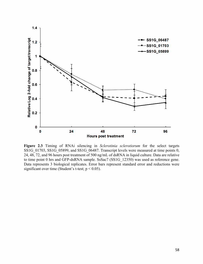
58
Figure 2.3 Timing of RNAi silencing in Sclerotinia sclerotiorum for the select targets SS1G_01703, SS1G_05899, and SS1G_06487. Transcript levels were measured at time points 0, 24, 48, 72, and 96 hours post treatment of 500 ng/mL of dsRNA in liquid culture. Data are relative to time point 0 hrs and GFP-dsRNA sample. SsSac7 (SS1G_12350) was used as reference gene. Data represents 3 biological replicates. Error bars represent standard error and reductions were significant over time (Student’s t-test; p < 0.05).

59
Figure 2.4 Effect of dsRNA dose on Sclerotinia sclerotiorum grown in liquid cultures for the select targets SS1G_01703, SS1G_05899, SS1G_06487, and SS1G_11912. Transcript levels were measured 3 days post treatment of 100, 200, 500, or 1000 ng/mL dose of dsRNA. Data relative to corresponding GFP-dsRNA (non-targeting control) dose and SsSac7 (SS1G_12350) reference gene. Data represents 3 biological replicates. Error bars represent standard error and all treatments were significant (Student’s t-test, p < 0.05).

60
2.4.4 Foliar applications of dsRNA reduce S. sclerotiorum infection in B. napus
Having confirmed that dsRNAs could reduce transcript abundance in S. sclerotiorum for
at least 96 hpi using relatively low concentrations, the level of protection imparted to B. napus
plants using a petal infection assay that facilitated aggressive S. sclerotiorum infection was then
assessed (Girard et al., 2017). A total of 61 S. sclerotiorum genes targets were selected from i)
the Database of Essential Genes (DEG; Luo et al., 2014), ii) literature searches (Liang et al.,
2014), and iii) the Target Identification Pipeline (TIP; Supplementary Table 2.2). TIP genes were
nominated based on a range of selection criteria, including: common significant expression
within the RNA-seq dataset; biological function; moderate expression levels (between 10 and
500 FPKM); fold changes between -0.5 and 4 (infection relative to in vitro); and biological
processes summarized in Supplementary Table 2.2. The dsRNAs were applied to leaves, and S.
sclerotiorum-infected petals were then placed over the dsRNA-treated surfaces. Fungal lesion
size was scored 2 dpi, and of the 61 dsRNAs tested, 24 showed a significant reduction in lesion
size, ranging from 24 to 85 % (one way ANOVA, p < 0.05; Fig. 2.5b; Table 2.1; Additional file
2). Of these 24 dsRNA molecule treatments, 19 conformed to the criteria outlined in TIP.
Some of the dsRNA molecules nominated using the TIP selection criteria included genes
involved in such biological processes as toxin biosynthesis (SS1G_01703), ROS response
(SS1G_02495), and cell cycle regulation (SS1G_09897). Targeting these genes’ transcripts with
dsRNA resulted in significant reductions in lesion size by 85%, 71%, and 45% respectively (one
way ANOVA, p<0.001 for all 3 dsRNA treatments; Fig. 2.5b; Table 2.1). Similarly, A. fumatigus
essential gene homologues associated with ribosomal biogenesis (SS1G_07873) and
mitochondrial protein import (SS1G_06487) also significantly reduced fungal lesion formation
by 64% (p<0.001) and 85% (p<0.001), respectively (Fig. 2.5b; Table 2.1). Leaf surfaces treated

61
Table 2.1 Selected list of Sclerotinia. sclerotiorum target genes for RNA interference testing in liquid culture, and infection assays on Brassica napus and A. thaliana.
Source Gene FPKM in vitro
FPKM Westar 24 hpi
FPKM ZY821 24
hpi Process
Literature 1 SS1G_08218 7913.2 5211.0 6743.8 Oxaloacetate acetylhydrolase KEGG 2 SS1G_00543 782.7 411.3 350.2 SCF complex subunitSkp1
Essential Genes 3 SS1G_05899 268.7 259.1 244.4 Thioredoxin reductase Essential genes 3 SS1G_06487 87.3 82.0 114.3 TIM44 Essential genes 3 SS1G_07873 47.1 41.5 47.8 pre-40S ribosomal particle Low expression
values SS1G_00509 1.8 6.6 3.2 Hydrolase activity, carbohydrate metabolic process
Low expression values SS1G_06055 0.0 0.8 2.0 Prenyltransferase activity
Low expression values SS1G_13720 0.0 1.4 0.5 MFS sugar transporter
Low expression values SS1G_13982 0.1 0.2 2.0 Triacylglycerol lipase
Moderate expression values SS1G_09261 14.20 22.56 22.80 Bub1-Bub3 complex localization to
kinetochore
High accumulation values SS1G_10167 14126.6 19992.7 24179.3 Endo-polygalacturonase
Positive expression fold change 4 SS1G_02791 2.0 19.1 23.8 Transcription factor activity
Positive expression fold change 4 SS1G_04551 1.9 12.6 16.4 Pectinesterase
Positive expression fold change 4 SS1G_14298 17.9 31.6 23.0 bHLH transcription factor
TIP 5 SS1G_01703 45.1 307.0 164.7 Aminoacyl-tRNA ligase activity / alfatoxin biosynthesis
TIP 5 SS1G_02495 61.2 336.1 353.7 Peroxidase activity TIP 5 SS1G_03208 11.5 51.4 48.3 Pre-mRNA splicing factor 8 TIP 5 SS1G_03991 16.7 39.5 39.4 Srb8 component of mediator complex TIP 5 SS1G_04966 30.6 64.3 59.5 Histone modification TIP 5 SS1G_06830 13.7 101.8 71.3 Aminoacyl permease TIP 5 SS1G_09680 50.8 61.2 70.3 60S ribosome biogenesis TIP 5 SS1G_09897 28.53 61.36 59.24 Cdc25 TIP 5 SS1G_11912 24.5 551.2 359.3 Necrosis/ethylene inducing peptide 2 TIP 5 SS1G_12021 149.3 397.4 363.8 1,3 glucan synthase TIP 5 SS1G_12640 53.1 114.2 130.1 Protein disulphide oxidoreductase process
TIP 5 SS1G_12992 13.5 57.5 39.0 Transglutaminase protein modification in ER
TIP 5 SS1G_13702 127.1 98.2 101.9 TIM23 TIP 5 SS1G_13746 15.1 33.7 32.5 Peroxisome
(1) Liang et al. (2014); (2) Kyoto Encyclopedia of Genes and Genomes (www.genome.jp/kegg/ ;Kanehisa et al., 2017); (3) Database of Essential Genes (http://www.essentialgene.org/ ;Luo et al., 2014); (4) Positive fold change in response to in planta growth (5) TIP = Target identification pipeline (Table S2)

62
with either water + Silwet L-77 or a non-fungal dsRNA + Silwet L-77 (GFP-dsRNA) equally
resulted in rapid S. sclerotiorum infection that developed large necrotic lesions by 2 dpi (Fig.
2.5a; Supplementary Figure 2.1). In contrast, when the leaf surface was treated with an
application of S. sclerotiorum–specific dsRNAs, dramatic reductions in lesion size and
morphology was observed for these aforementioned dsRNAs (Fig. 2.5a).
Interestingly, 6 of the dsRNAs tested significantly increased (p<0.05) the lesion size
compared to the control treatment (Fig. 2.5b). Two of these dsRNAs targeted S. sclerotiorum
transcription factor genes (SS1G_14298; SS1G_02791), and lesion size increased 64 and 88%,
respectively (one way ANOVA, p<0.05). The dsRNA targeting SsSkp1 (SS1G_00543) caused an
increase in lesion size by 133% (one way ANOVA, P<0.01). As SKP1 plays a role in both
chromosome separation and ubiquitin-mediated protein degradation (Kanehisa et al., 2017),
RNAi-mediated knockdown was expected to adversely affect fungal growth, but on closer
inspection of the RNA-seq data, SsSkp1 transcripts were not observed to accumulate to a
significant extent during infection on B. napus (Table 2.1), which may explain a lack of fungal
suppression with this particular dsRNA. In contrast, SS1G_14298 and SS1G_02791, which had
increased expression during infection conditions, were possibly nonessential to the infection
process or facilitated pathogenicity and thus failed to reduce fungal pressure in planta.
Furthermore, 31 genes targeted by some dsRNA molecules caused no significant impact
on fungal lesion sizes on the leaves; these dsRNAs targeted genes involved in a range of cellular
processes, including carbohydrate catabolism (SS1G_00509; endo-1,4-beta-xylanase), hydrolysis
(SS1G_13982; triacylglycerol lipase), and kinetochore functions (SS1G_09261). Even a
previously characterized genetic deletion target(Liang et al., 2014), SS1G_08218 (Ssoah;
oxaloacetate acetylhydrolase) had no significant impact on lesion size

63
Figure 2.5 DsRNA targeting Sclerotinia sclerotiorum suppresses lesion size on Brassica napus susceptible cultivar Westar. (a) Sclerotinia sclerotiorum infection lesions on B. napus cv. Westar following a treatment of 200 ng dsRNA targeting S. sclerotiorum genes at 2 dpi. (b) Average lesion size (n = 10 leaves) relative to control (black bar) with standard error bars. Significant difference (One-way ANOVA; p < 0.05) from control represented by asterix (*).

64
(one way ANOVA, p = 0.7) under the conditions tested. While these targets did not show
significant modulations in lesion size, they helped define criteria for TIP.
2.4.5 Topical dsRNAs mitigate S. sclerotiorum infection on A. thaliana
Given the extensive host range of S. sclerotiorum, it was of interest to determine whether
this fungus could be similarly suppressed using dsRNAs in another plant species. Using the
model plant A. thaliana, 10 new S. sclerotiorum genes, nominated using TIP selection criteria
(Table 2.1; Supplementary Table 2.2), were assessed in parallel with 6 of the best performing
targets in B. napus (SS1G_01703, SS1G_02495, SS1G_05899, SS1G_06487, SS1G_07873, and
SS1G_11912; Fig. 5b). Significant reductions in lesion size between 34 – 66 % were observed
(one-way ANOVA, p < 0.05; Fig. 2.6b) by targeting genes associated with processes such as
mRNA splicing (SS1G_03208), ribosome biogenesis (SS1G_09680), protein disulphide
oxidoreductase (SS1G_12640) and a peroxisomal protein (SS1G_13746). DsRNA treatments
reduced the fungal progression on the spore-inoculated leaves, suggesting that dsRNA used to
protect one plant species can also be applied to protect another with near-equivalent success.
The most effective dsRNA treatments on B. napus leaves were also effective at
reducing infection on A. thaliana significantly, such as SS1G_11912, which reduced lesion sizes
by 64-66% in both plant species. However, there were noticeable differences in efficacy of the
other five dsRNAs. Specifically, the dsRNAs targeting SS1G_02495 and SS1G_07873, which
reduced lesions 71 and 64%, respectively in B. napus, only reduced lesions by 34 and 46% in A.
thaliana, respectively. The disparities in lesion size between B. napus and A. thaliana may be
attributed to differences in infection rates, defence responses, or leaf structures.

65
Figure 2.6 DsRNA treatment on Arabidopsis thaliana leaves reduces Sclerotinia sclerotiorum lesion area. (a) Sclerotinia sclerotiorum spore inoculation on A. thaliana leaves after a foliar application 200 ng of specific dsRNA spread over the entire leaf surface at 4 dpi. (b) Average lesion relative to control (darker bar) with standard error bars of 3 bio-replicates of 10 leaves (n =30). All targeting dsRNA treatment were significantly different from control (One-way ANOVA, p < 0.05).

66
2.4.6 DsRNAs targeting homologues in Botrytis cinerea attenuate fungal infection
Botrytis cinerea is a phytopathogenic fungus closely related to S. sclerotiotum, and
hence, it was interest to determine whether some of the S. sclerotiotum target genes identified
during earlier screens could be similarly used as RNAi targets to suppress B. cinerea
infections on B. napus. Botrytis cinerea-specific dsRNA molecules were designed to target five
of the most effective targets identified in S. sclerotiotum (Additional file 2). Leaves treated with
dsRNA targeting BC1G_04775 (SS1G_06487 homologue) and BC1G_01592 (SS1G_05899
homologue) formed smaller brown necrotic regions than the control leaves (Fig. 2.7a).
Treatments of dsRNA targeting BC1G_04955 (SS1G_02495 homologue), BC1G_04775,
BC1G_01592, BC1G_07805 (SS1G_07873 homologue), and BC1G_10306 (SS1G_11912
homologue), on average, reduced lesion sizes by 66% (Fig. 2.7b; one-way ANOVA, p<0.05 for
all treatments). Interestingly, the dsRNA targeting the SS1G_11912 homologue, BC1G_010306,
showed only moderate reductions in lesion area, suggesting that the gene product may be more
important for S. sclerotiorum infection rather than B. cinerea. A complete summary of foliar
results can be found in Additional file 2.
2.5 DISCUSSION
Transcriptome interrogation of the B. napus-S. sclerotiorum interface uncovered selective
targets of fungal growth and pathogenicity that helped guide the development of an RNAi-based
control target identification pipeline (TIP; Supplementary Table 2.2). The demonstrated dsRNA-
mediated knockdown of S. sclerotiorum transcripts provided evidence of the utility of RNAi to
selectively and specifically target individual genes in this agriculturally important fungal
pathogen. Topical applications of dsRNAs targeting S. sclerotiorum transcripts significantly

67
Figure 2.7 Botrytis cinerea homologues targeted with foliar dsRNA controls fungal infection. (a) Botrytis cinerea spore inoculation of Brassica napus cv. Westar leaves after an application of 500 ng of dsRNA targeting B. cinerea genes was spread over a 4 cm2 area at 4 dpi. (b) Average lesion size compared to control (darker bar)(n = 40 leaves) with standard error bars. All targeting dsRNA treatment were significantly different from GFP-dsRNA control (One-way ANOVA, p < 0.05).

68
reduced the size of fungal lesions in both B. napus and the model plant system A.
thaliana. Extension of this technology to homologous transcripts of B. cinerea revealed
unprecedented
cross-species control of fungal pathogenesis using topical applications of dsRNAs targeting a
single gene.
When dsRNAs were incubated with S. sclerotiorum in vitro, transcript level reductions
were observed for all genes tested, providing clear evidence of fungal dsRNA uptake and an
RNAi response. Although the dsRNA uptake mechanism has yet to be established in fungi, the
results are consistent with other eukaryotic organisms, where knockdown of transcript levels
occurred within 48 hours (Gong et al., 2014; Neumann et al., 2006; Štefanic et al., 2010;
Zimmermann et al., 2006). In addition, the dose-response for individual transcripts varied, with
each individual gene responding differently, and very high doses failing to elicit further
transcript reductions, which has been observed in previous insect and flatworm research (Asokan
et al., 2014; Killiny et al., 2014; Meyering-Vos and Müller, 2007; Štefanic et al., 2010).
Differences in knockdown response may be attributed to mRNA secondary structure, mRNA
turnover rate, GC content, or other properties associated with dsRNA structure (Chan et al.,
2009; Larsson et al., 2010; Shao et al., 2007; Vermeulen et al., 2005). Without modifying the
delivery or chemistry of the dsRNA molecules, notable reductions in infection symptoms were
achieved. With recent developments utilizing modified nucleotides and novel delivery methods
(Joga et al., 2016; Love et al., 2010; Semple et al., 2010), future chemistries could increase
silencing potency of RNAi-based control strategies.
Interrogation of global changes in gene expression at the host-pathogen interface in both
susceptible and tolerant hosts of S. sclerotiorum identified novel targets for fungal control using

69
RNAi-based biotechnologies. Using transgenic techniques, endogenously-produced dsRNAs
within fungi targeting single genes can suppress fungal growth (Andrade et al., 2016; Ghag et
al., 2014). In contrast, previous efforts to develop topically-applied dsRNA-based fungicides
have been effectively limited in application and gene specificity by requiring multiple target
gene knockdowns to reduce pathogen progression (Koch et al., 2016; Wang et al., 2016). No
previous research has provided a clear and rational link between transcript profiling and effective
dsRNA molecule design. The identification of single RNAi targets allows for broader agriculture
applications, through the flexible design of dsRNA molecules targeting selective fungal genes
using the target identification pipeline, and the specificity to control a single gene from a single
target organism.
Global RNA profiling and GO term enrichment highlighted specific genes and biological
processes associated with cell division (e.g. SS1G_09897), cell wall modification (e.g.
SS1G_12021), oxidoreductase (e.g. SS1G_05899), and peroxidase activity (e.g. SS1G_02495) as
novel targets for fungal control and selection criteria for TIP. By suppressing gene expression of
processes such as cell wall modification, required for fungal growth and maintenance of cellular
integrity (Werner et al., 2007), and fungal ROS response proteins, required to protect the
advancing fungal hyphae from plant respiratory ROS defense (Heller and Tudzynski, 2011),
gene silencing impeded fungus establishment on the leaf surface. Previous genetic deletions of
thioredoxin reductase and peroxidases in Magnaporthe oryzae Couch striked parallels to the
dsRNA treatment of SS1G_05899 (thioredoxin reductase) and SS1G_02495 (peroxidase)
(Fernandez and Wilson, 2014; Mir et al., 2015), which also restricted fungal advance.
Furthermore, by identifying homologues of essential genes from the model organism A.
fumatigus (Luo et al., 2014), despite the overall processes being downregulated during infection,

70
flexibility was added to TIP-guided selection for dsRNA control. By exploring fungal
transcriptional reprogramming, a more complete understanding of the genes and cellular
processes associated with pathogenicity was gained and provided a guide to the flexible and
adaptable design of dsRNA molecules targeting key processes using TIP.
RNAi of a single gene product may not always significantly limit fungal infection, as
evidenced by efforts to knockdown 32 genes that failed to prevent fungal lesion expansion.
During infection, S. sclerotiorum secretes a diverse arsenal of pathogenicity factors including
hydrolases and nutrient acquisition enzymes to secure nutrient sources during host infection,
while maintaining a constant store of carbohydrates (Heard et al., 2015; Jobic et al., 2007; Lyu et
al., 2015). Consequently, when targeting processes such as carbohydrate catabolism (e.g.
SS1G_00509), cell wall degrading enzymes (e.g. SS1G_04551) and hydrolase activity (e.g.
SS1G_13982), despite being upregulated in the infected tissues, no significant impacts on lesion
sizes were observed, highlighting the fact that not all cellular processes can provide good RNAi
control targets. Similarly, a high level of gene regulation may also reduce the effect of RNAi-
based control. For example, the pathogenicity factor Ssoah (SS1G_08218; oxaloacetate
acetylhydrolase), although essential for pathogenicity, is also known to be heavily regulated by
SsPac1 (Rollins, 2003). Thus, the RNAi-induced reduction in Ssoah gene activity may be
rescued by the intrinsic regulatory network responsible for S. sclerotiorum pathogenesis.
Additionally, other factors such as protein half-life, which could outlast an RNAi silencing event,
and negative pathogenicity regulation, already observed in S. sclerotiorum, could fail to elicit an
observable physiological response despite successful transcript knockdown (Pan et al., 2015;
Rinkevich and Scott, 2013). Thus, by screening a great variety of genes sharing single biological
processes, poor targets can be excluded from the RNAi target selection.

71
Transcript accumulation levels could have affected the efficiency of RNAi silencing in S.
sclerotiorum and thus was an important consideration for the construction of TIP. For the genes
SS1G_13720, SS1G_06055, and SS1G_13982, which accumulated at low levels under all
conditions, no significant changes in lesion formation were observed. Previously, low transcript
accumulation levels resulted in poor siRNA-mediated knockdown in human cells, suggesting
that a threshold level of target mRNA must be present for the activation of the RISC complexes
(Hong et al., 2014; Hu et al., 2004). In contrast, transcripts of the genes Ssoah and SS1G_10167
accumulated at high levels under all conditions, also failed to respond to the administered
dsRNA in the infection assays. In other organisms, genes with high transcription rates have
similarly been difficult to knock down using RNAi, presumably because the dose of dsRNA was
insufficient to eliminate enough of the target transcripts (Dornseifer et al., 2015; Larsson et al.,
2010). At the dsRNA dose tested, the majority of significant reductions in lesion size were
associated with dsRNAs that targeted moderately expressing genes (10-500 FPKM); genes that
showed large deviations in expression levels (up or down-regulated) in the infected plants,
relative to the in vitro –grown fungus, were not significantly affected by the dsRNA treatments.
Effective RNAi can be achieved when targeting genes that cannot reliably increase transcription
in response to a silencing event. Transcriptomic analyses are useful to uncover which genes’
transcripts are induced to increase under infection conditions, as observed by drastic transcript
fold-change levels. A higher accumulation of mRNA would result from stronger promoter
induction and activity under infection conditions (Coradetti et al., 2012; Kawahara et al., 2012).
Therefore, a gene with the capability to elicit a strong transcriptional response may have the
capacity to buffer RNAi-induced perturbations upon exposure to dsRNA. Using RNA-seq,

72
highly induced genes can be avoided, and genes with moderate upregulation during infection can
be preferentially selected.
Synthesizing the results into TIP, 80% of the dsRNA targets, which reduced lesion sizes
in B. napus significantly, adhere to the TIP protocol. Furthermore, all of the ten additional
dsRNAs designed utilizing the TIP criteria, mitigated disease on an alternate plant, A. thaliana,
validating TIP as a novel fungal RNAi identification guide for control of the devastating
phytopathogen S. sclerotiorum. Additionally, six dsRNAs previously assayed on B. napus also
protected A. thaliana. The protection imparted by the dsRNA molecules on both plant species
suggests a common fungal infection strategy is being deployed by S. sclerotiorum and similar
target knockdown may be responsible for improved plant protection. While there was a level of
protection shared between the two host species, the differences in the extent of protection may be
attributed to variations in leaf architecture, such as larger cell size, cell wall and thicker cuticle of
B. napus, which together, can structurally limit fungal growth (Miedes et al., 2014; Serrano et
al., 2014; Uloth et al., 2015). Furthermore, in the closely related fungus, B. cinerea, the same
topical dsRNA protected a variety of horticultural species (Wang et al., 2016). Taken together,
the array of dsRNAs that produced strong transcript reductions could be sufficient to confer
protection for other major agronomic crops. Identifying a variety genes provides multiple tactics
for successful phytoprotection regardless of potential differences in host architecture.
A recent report by Wang et al. (2016) showed that dsRNAs targeting transcripts encoding
both Dicer-like protein 1 and 2, two proteins responsible for small RNA processing in eukaryotic
cells, were able to suppress B. cinerea and Verticillium dahliae Kleb infection of transgenic A.
thaliana containing a construct to produce dsRNA molecules targeting both species (Wang et al.,
2016). Here, the results demonstrated that a single dsRNA, when applied to the leaf surface, was
73
able to suppress the expansion of leaf lesions of both S. sclerotiorum and B. cinerea. While most
of the tested dsRNAs of S. sclerotiorum and B. cinerea could significantly reduce disease
symptoms in B. napus, some targets showed greater reductions in disease severity. Despite both
necrotrophic pathogens likely sharing a common cluster of genes to control pathogenicity in a
broad range of host plant species, B. cinerea likely operates through alternative pathogenic
pathways to S. sclerotiorum to control infection in different host plants (Amselem et al., 2011;
Billon-Grand et al., 2012). Thus, additional target discovery of any host-pathogen system is
warranted to identify the best targets for RNAi-based control of fungal pathogenesis.
RNA-seq technology offers comprehensive insights into the genes and processes
involved in fungal pathogenesis for the design of specific RNAi biotechnology. Identifying
numerous alternative target transcripts allows for greater design flexibility, such that topical
applications of dsRNA can be extended to control other fungal pathogens and protect other
agronomically important plant species. The target identification pipeline provides a new,
adaptable platform for the design of RNAi control and marks a substantial evolution of next-
generation fungal phytopathogenic control. Further examinations of pathosystems will identify
additional efficient RNAi targets across multiple fungi to improve broad fungal control.
2.6 ACKNOWLEDGEMENTS
We would like to thank Dr. Nicolas Lapalu (Versailles, France) for the generous access to
S. sclerotiorum GO terms, Dr. Andrew Fire (Stanford, US) for the gift of the pL4440 vector, and
Dr. Yangdou Wei (University of Saskatchewan, Canada) for B. cinerea. This work was generously
supported by grants from the province of Manitoba Agricultural Rural Development Initiative, the
Canola Council of Canada, and the Western Grains Research Foundation to SW and MFB. AM

74
was supported by a National Science and Engineering Research Council Graduate Masters
Scholarship and the Manitoba government Tri Council Top-Up award.
2.7 REFERENCES
Amselem, J., Cuomo, C.A., Kan, J.A.L. van, et al. (2011) Genomic Analysis of the
Necrotrophic Fungal Pathogens Sclerotinia sclerotiorum and Botrytis cinerea. PLoS Genet.
7, e1002230.
Anders, S. and Huber, W. (2010) Differential expression analysis for sequence count data.
Genome Biol. 11, 1–12.
Andrade, C.M., Tinoco, M.L.P., Rieth, A.F., Maia, F.C.O. and Aragão, F.J.L. (2016) Host-
induced gene silencing in the necrotrophic fungal pathogen Sclerotinia sclerotiorum. Plant
Pathol. 65, 626–632.
Asokan, R., Sharath Chandra, G., Manamohan, M., Krishna Kumar, N.K. and Sita, T.
(2014) Response of various target genes to diet-delivered dsRNA mediated RNA
interference in the cotton bollworm, Helicoverpa armigera. J. Pest Sci. (2004). 87, 163–
172.
Bateman, A., Martin, M.J., O’Donovan, C., et al. (2015) UniProt: A hub for protein
information. Nucleic Acids Res.
Becker, M.G., Walker, P.L., Pulgar-Vidal, N.C. and Belmonte, M.F. (2017) SeqEnrich: A
tool to predict transcription factor networks from co-expressed Arabidopsis and Brassica
napus gene sets. PLoS One 12, 1–13.
Billmyre, R.B., Calo, S., Feretzaki, M., Wang, X. and Heitman, J. (2013) RNAi function,
diversity, and loss in the fungal kingdom. Chromosom. Res. 21, 561–572.

75
Billon-Grand, G., Ve, Rascle, C., Droux, M., Rollins, J.A. and Poussereau, N. (2012) pH
modulation differs during sunflower cotyledon colonization by the two closely related
necrotrophic fungi Botrytis cinerea and Sclerotinia sclerotiorum. Mol. Plant Pathol. 13,
568–578.
Bolger, A.M., Lohse, M. and Usadel, B. (2014) Trimmomatic: A flexible trimmer for Illumina
sequence data. Bioinformatics 30, 2114–2120.
Bolton, M.D., Thomma, B.P.H.J. and Nelson, B.D. (2006) Sclerotinia sclerotiorum (Lib.) de
Bary: Biology and molecular traits of a cosmopolitan pathogen. Mol. Plant Pathol. 7, 1–16.
Bradley, C. a., Lamey, H. a., Endres, G.J., Henson, R. a., Hanson, B.K., McKay, K.R.,
Halvorson, M., LeGare, D.G. and Porter, P.M. (2006) Efficacy of Fungicides for Control
of Sclerotinia Stem Rot of Canola. Plant Dis. 90, 1129–1134.
Campo, S., Gilbert, K.B. and Carrington, J.C. (2016) Small RNA-Based Antiviral Defense in
the Phytopathogenic Fungus Colletotrichum higginsianum. PLoS Pathog. 12, 1–36.
Chan, C.Y., Carmack, C.S., Long, D.D., Maliyekkel, A., Shao, Y., Roninson, I.B. and Ding,
Y. (2009) A structural interpretation of the effect of GC-content on efficiency of RNA
interference. BMC Bioinformatics 10 Suppl 1, S33.
Chen, Y., Gao, Q., Huang, M., Liu, Y., Liu, Z., Liu, X. and Ma, Z. (2015) Characterization of
RNA silencing components in the plant pathogenic fungus Fusarium graminearum. Sci.
Rep. 5, 12500.
Coradetti, S.T., Craig, J.P., Xiong, Y., Shock, T., Tian, C. and Glass, N.L. (2012) Conserved
and essential transcription factors for cellulase gene expression in ascomycete fungi. Proc.
Natl. Acad. Sci. 109, 7397–7402.
Derbyshire, M.C. and Denton-Giles, M. (2016) The control of Sclerotinia stem rot on oilseed

76
rape ( Brassica napus ): Current practices and future opportunities. Plant Pathol. 65, 859–
877.
Dornseifer, S., Willkomm, S., Far, R.K.K., et al. (2015) RNAi revised - Target mRNA-
dependent enhancement of gene silencing. Nucleic Acids Res. 43, 10623–10632.
Fernandez, J. and Wilson, R.A. (2014) Characterizing roles for the glutathione reductase,
thioredoxin reductase and thioredoxin peroxidase-encoding genes of Magnaporthe oryzae
during rice blast disease. PLoS One 9.
Garg, H., Li, H., Sivasithamparam, K., Kuo, J. and Barbetti, M.J. (2010) The infection
processes of Sclerotinia sclerotiorum in cotyledon tissue of a resistant and a susceptible
genotype of Brassica napus. Annu. Bot., 897–908.
Ghag, S.B., Shekhawat, U.K.S. and Ganapathi, T.R. (2014) Host-induced post-transcriptional
hairpin RNA-mediated gene silencing of vital fungal genes confers efficient resistance
against Fusarium wilt in banana. Plant Biotechnol. J. 12, 541–53.
Girard, I.J., Tong, C., Mao, X., Becker, M.G., Huang, J., Kievit, T. De, Dilantha Fernando,
W.G., Liu, S. and Belmonte, M.F. (2017) RNA sequencing of Brassica napus reveals
cellular redox control of Sclerotinia infection. J. Exp. Bot.
Gong, Y.H., Yu, X.R., Shang, Q.L., Shi, X.Y. and Gao, X.W. (2014) Oral Delivery Mediated
RNA Interference of a Carboxylesterase Gene Results in Reduced Resistance to
Organophosphorus Insecticides in the Cotton Aphid, Aphis gossypii Glover. PLoS One 9,
23–25.
Heard, S., Brown, N.A. and Hammond-kosack, K. (2015) An Interspecies Comparative
Analysis of the Predicted Secretomes of the Necrotrophic Plant Pathogens Sclerotinia
sclerotiorum and Botrytis cinerea. PLoS One, 1–27.

77
Heller, J. and Tudzynski, P. (2011) Reactive Oxygen Species in Phytopathogenic Fungi:
Signaling, Development, and Disease. Annu. Rev. Phytopathol. 49, 369–390.
Hong, S.W., Jiang, Y., Kim, S., Li, C.J. and Lee, D. (2014) Target gene abundance contributes
to the efficiency of siRNA-mediated gene silencing. Nucleic Acid Ther. 24, 192–8.
Hu, W., Sillaots, S., Lemieux, S., et al. (2007) Essential Gene Identification and Drug Target
Prioritization in Aspergillus fumigatus. 3.
Hu, X., Hipolito, S., Lynn, R., Abraham, V., Ramos, S. and Wong-Staal, F. (2004) Relative
gene-silencing efficiencies of small interfering RNAs targeting sense and antisense
transcripts from the same genetic locus. Nucleic Acids Res. 32, 4609–4617.
Hulsen, T., Vlieg, J. de and Alkema, W. (2008) BioVenn - a web application for the
comparison and visualization of biological lists using area-proportional Venn diagrams.
BMC Genomics 9, 1–6.
Jobic, C., Boisson, A.M., Gout, E., Rascle, C., Fèvre, M., Cotton, P. and Bligny, R. (2007)
Metabolic processes and carbon nutrient exchanges between host and pathogen sustain the
disease development during sunflower infection by Sclerotinia sclerotiorum. Planta 226,
251–265.
Joga, M.R., Zotti, M.J., Smagghe, G. and Christiaens, O. (2016) RNAi efficiency, systemic
properties, and novel delivery methods for pest insect control: What we know so far. Front.
Physiol. 7, 1–14.
Kamal, M.M., Savocchia, S., Lindbeck, K.D. and Ash, G.J. (2016) Biology and biocontrol of
Sclerotinia sclerotiorum (Lib.) de Bary in oilseed Brassicas. Australas. Plant Pathol. 45, 1–
14.
Kamesh Krishnamoorthy, K., Sankaralingam, A. and Nakkeeran, S. (2016) Standardization

78
of Culture Media and pH for the Rapid Growth of Sclerotinia sclerotiorum causing Head
Rot Disease of Cabbage Standardization of Culture Media and pH for the Rapid Growth of
Sclerotinia sclerotiorum causing Head Rot Disease of Cabbage. Adv. Life Sci. 5(22), 10659–
10661.
Kanehisa, M., Furumichi, M., Tanabe, M., Sato, Y. and Morishima, K. (2017) KEGG: New
perspectives on genomes, pathways, diseases and drugs. Nucleic Acids Res. 45, D353–
D361.
Kawahara, Y., Oono, Y., Kanamori, H., Matsumoto, T., Itoh, T. and Minami, E. (2012)
Simultaneous RNA-seq analysis of a mixed transcriptome of rice and blast fungus
interaction. PLoS One 7, e49423.
Killiny, N., Hajeri, S., Tiwari, S., Gowda, S. and Stelinski, L.L. (2014) Double-stranded RNA
uptake through topical application, mediates silencing of five CYP4 genes and suppresses
insecticide resistance in Diaphorina citri. PLoS One 9, 1–8.
Koch, A., Biedenkopf, D., Furch, A., et al. (2016) An RNAi-Based Control of Fusarium
graminearum Infections Through Spraying of Long dsRNAs Involves a Plant Passage and
Is Controlled by the Fungal Silencing Machinery. PLoS Pathog. 12, 1–22.
Kumar, R., Ichihashi, Y., Kimura, S., Chitwood, D.H., Headland, L.R., Peng, J., Maloof,
J.N. and Sinha, N.R. (2012) A High-Throughput Method for Illumina RNA-Seq Library
Preparation. Front. Plant Sci. 3, 1–10.
Larsson, E., Sander, C. and Marks, D. (2010) mRNA turnover rate limits siRNA and
microRNA efficacy. Mol. Syst. Biol. 6, 433.
Liang, X., Liberti, D., Li, M., Kim, Y.-T., Hutchens, A., Wilson, R. and Rollins, J. a (2014)
Oxaloacetate acetylhydrolase gene mutants of Sclerotinia sclerotiorum do not accumulate
79
oxalic acid, but do produce limited lesions on host plants. Mol. Plant Pathol. 16, 1–13.
Livak, K.J. and Schmittgen, T.D. (2001) Analysis of relative gene expression data using real-
time quantitative PCR and. Methods 25, 402–408.
Llanos, A., François, J. and Parrou, J. (2015) Tracking the best reference genes for RT-qPCR
data normalization in filamentous fungi. BMC Genomics 16, 71.
Love, K.T., Mahon, K.P., Christopher, G., et al. (2010) Lipid-like materials for low-dose, in
vivo gene silencing. Proc. Natl. Acad. Sci. 107, 9915–9915.
Luo, H., Lin, Y., Gao, F., Zhang, C.T. and Zhang, R. (2014) DEG 10, an update of the
database of essential genes that includes both protein-coding genes and noncoding genomic
elements. Nucleic Acids Res. 42, 574–580.
Lyu, X., Shen, C., Fu, Y., Xie, J., Jiang, D., Li, G. and Cheng, J. (2015) Comparative
genomic and transcriptional analyses of the carbohydrate-active enzymes and secretomes of
phytopathogenic fungi reveal their significant roles during infection and development. Sci.
Rep. 5, 15565.
Majumdar, R., Rajasekaran, K. and Cary, J.W. (2017) RNA Interference (RNAi) as a
Potential Tool for Control of Mycotoxin Contamination in Crop Plants: Concepts and
Considerations. Front. Plant Sci. 8, 200.
Meyering-Vos, M. and Müller, A. (2007) RNA interference suggests sulfakinins as satiety
effectors in the cricket Gryllus bimaculatus. J. Insect Physiol. 53, 840–848.
Miedes, E., Vanholme, R., Boerjan, W. and Molina, A. (2014) The role of the secondary cell
wall in plant resistance to pathogens. Front. Plant Sci. 5, 358.
Mir, A.A., Park, S.-Y., Sadat, M.A., Kim, S., Choi, J., Jeon, J. and Lee, Y.-H. (2015)
Systematic characterization of the peroxidase gene family provides new insights into fungal

80
pathogenicity in Magnaporthe oryzae. Sci. Rep. 5, 11831.
Mocellin, S. and Provenzano, M. (2004) RNA interference: learning gene knock-down from
cell physiology. J. Transl. Med. 2, 39.
Mumbanza, F.M., Kiggundu, A., Tusiime, G., Tushemereirwe, W.K., Niblett, C. and
Bailey, A. (2013) In vitro antifungal activity of synthetic dsRNA molecules against two
pathogens of banana, Fusarium oxysporum f. sp. cubense and Mycosphaerella fijiensis. Pest
Manag. Sci. 69, 1155–1162.
Neumann, B., Held, M., Liebel, U. and Erfle, H. (2006) High-throughput RNAi screening by
time-lapse imaging of live human cells. Nat. Methods 3, 385–390.
Nicolás, F.E. and Garre, V. (2016) RNA Interference in Fungi : Retention and Loss. , 1–15.
Oliveros, J.C. (2015) Venny. An interactive tool for comparing lists with Venn’s diagrams.
Pan, Y., Xu, Y., Li, X., Yao, C. and Gao, Z. (2015) SsPemG1 encodes an elicitor-homologous
protein and regulates pathogenicity in Sclerotinia sclerotiorum. Physiol. Mol. Plant Pathol.
92, 70–78.
Price, D.R.G. and Gatehouse, J.A. (2008) RNAi-mediated crop protection against insects.
Trends Biotechnol. 26, 393–400.
Qu, X., Yu, B., Liu, J., et al. (2014) MADS-box transcription factor SsMADS is involved in
regulating growth and virulence in Sclerotinia sclerotiorum. Int. J. Mol. Sci. 15, 8049–8062.
Rinkevich, F.D. and Scott, J.G. (2013) Limitations of RNAi of α6 nicotinic acetylcholine
receptor subunits for assessing the in vivo sensitivity to spinosad. Insect Sci. 20, 101–108.
Rollins, J. A (2003) The Sclerotinia sclerotiorum pac1 gene is required for sclerotial
development and virulence. Mol. Plant. Microbe. Interact. 16, 785–795.
San Miguel, K. and Scott, J.G. (2016) The next generation of insecticides: DsRNA is stable as
81
a foliar-applied insecticide. Pest Manag. Sci. 72, 801–809.
Schwartz, H.F. and Singh, S.P. (2013) Breeding common bean for resistance to white mold: A
review. Crop Sci. 53, 1832–1844.
Semple, S.C., Akinc, A., Chen, J., et al. (2010) Rational design of cationic lipids for siRNA
delivery. Nat Biotechnol 28, 172–176.
Serrano, M., Coluccia, F., Torres, M., L’Haridon, F. and Métraux, J.-P. (2014) The cuticle
and plant defense to pathogens. Front. Plant Sci. 5, 274.
Shao, Y., Chan, C.Y., Maliyekkel, A., Lawrence, C.E., Roninson, I.B. and Ding, Y. (2007)
Effect of target secondary structure on RNAi efficiency. RNA 13, 1631–1640.
Sharma, P., Meena, P.D., Verma, P.R., Saharan, G.S., Mehta, N., Singh, D. and Kumar, A.
(2015) Sclerotinia sclerotiorum ( Lib .) de Bary causing Sclerotinia rot in oilseed Brassicas :
A review. J. Oilseed Brassica 6, 1–44.
Štefanic, S., Dvořák, J., Horn, M., et al. (2010) RNA interference in Schistosoma mansoni
schistosomula: Selectivity, sensitivity and operation for larger-scale screening. PLoS Negl.
Trop. Dis. 4.
Trapnell, C., Roberts, A., Goff, L., et al. (2012) Differential gene and transcript expression
analysis of RNA-seq experiments with TopHat and Cufflinks. Nat. Protoc. 7, 562–78.
Uloth, M.B., Clode, P.L., You, M.P. and Barbetti, M.J. (2015) Attack modes and defence
reactions in pathosystems involving Sclerotinia sclerotiorum , Brassica carinata , B. juncea
and B. napus. Ann. Bot., mcv150.
United States Environmental Protection Agency (2017) EPA Registers Innovative Tool to
Control Corn Rootworm.
Untergasser, A., Cutcutache, I., Koressaar, T., Ye, J., Faircloth, B.C., Remm, M. and

82
Rozen, S.G. (2012) Primer3-new capabilities and interfaces. Nucleic Acids Res. 40, 1–12.
Vermeulen, A., Behlen, L., Reynolds, A., Wolfson, A., Marshall, W.S., Karpilow, J. and
Khvorova, A. (2005) The contributions of dsRNA structure to Dicer specificity and
efficiency. RNA 11, 674–82.
Wang, M., Weiberg, A., Lin, F.-M., Thomma, B.P.H.J., Huang, H.-D. and Jin, H. (2016)
Bidirectional cross-kingdom RNAi and fungal uptake of external RNAs confer plant
protection. Nat. Plants 2, 16151.
Werner, S., Sugui, J. A, Steinberg, G. and Deising, H.B. (2007) A chitin synthase with a
myosin-like motor domain is essential for hyphal growth, appressorium differentiation, and
pathogenicity of the maize anthracnose fungus Colletotrichum graminicola. Mol. Plant.
Microbe. Interact. 20, 1555–1567.
Whyard, S., Singh, A.D. and Wong, S. (2009) Ingested double-stranded RNAs can act as
species-specific insecticides. Insect Biochem. Mol. Biol. 39, 824–832.
Yu, Y., Jiang, D., Xie, J., Cheng, J., Li, G., Yi, X. and Fu, Y. (2012) Ss-Sl2 , a Novel Cell
Wall Protein with PAN Modules , Is Essential for Sclerotial Development and Cellular
Integrity of Sclerotinia sclerotiorum. PLoS One 7.
Yu, Y., Xiao, J., Yang, Y., Bi, C., Qing, L. and Tan, W. (2015) Physiological and Molecular
Plant Pathology Ss-Bi1 encodes a putative BAX inhibitor-1 protein that is required for full
virulence of Sclerotinia sclerotiorum. Physiol. Mol. Plant Pathol. 90, 115–122.
Zhu, W., Wei, W., Fu, Y., Cheng, J., Xie, J., Li, G. and Yi, X. (2013) A Secretory Protein of
Necrotrophic Fungus Sclerotinia sclerotiorum That Suppresses Host Resistance. 8.
Zimmermann, T.S., Lee, A.C.H., Akinc, A., et al. (2006) RNAi-mediated gene silencing in
non-human primates. 441, 111–114.

83
Supplementary Table 2.1 . Mapping summary for all biological replicates of in-vitro and in-planta samples. Raw RNA-sequencing reads trimmed using Trimmomatic 0.33 and aligned to the Sclerotinia sclerotiorum genome using TopHat v2.1.0.
in-
vitro (1)
in-vitro (2)
in-vitro (3)
ZY821 24hpi
(1)
ZY821 24hpi
(2)
ZY821 24hpi
(3)
Westar 24hpi
(1)
Westar 24hpi
(2)
Westar 24hpi
(3)
Trimmed reads 6537111 6339049 11826235 31473436 44428372 20453088 40287633 37020385 52025758
Mapped reads 5388394 5216455 10273216 6282525 6724081 3061161 7810054 6478810 8125640
% Mapped 82.4 82.3 86.9 20 15.1 15 19.4 17.5 15.6
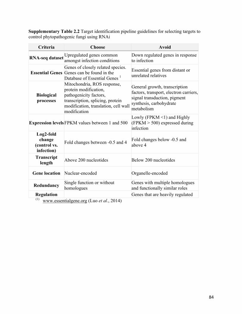
84
Supplementary Table 2.2 Target identification pipeline guidelines for selecting targets to control phytopathogenic fungi using RNAi
Criteria Choose Avoid
RNA-seq dataset Upregulated genes common amongst infection conditions
Down regulated genes in response to infection
Essential Genes Genes of closely related species. Genes can be found in the Database of Essential Genes 1
Essential genes from distant or unrelated relatives
Biological processes
Mitochondria, ROS response, protein modification, pathogenicity factors, transcription, splicing, protein modification, translation, cell wall modification
General growth, transcription factors, transport, electron carriers, signal transduction, pigment synthesis, carbohydrate metabolism
Expression levels FPKM values between 1 and 500 Lowly (FPKM <1) and Highly (FPKM > 500) expressed during infection
Log2-fold change
(control vs. infection)
Fold changes between -0.5 and 4 Fold changes below -0.5 and above 4
Transcript length Above 200 nucleotides Below 200 nucleotides
Gene location Nuclear-encoded Organelle-encoded
Redundancy Single function or without homologues
Genes with multiple homologues and functionally similar roles
Regulation Genes that are heavily regulated (1) www.essentialgene.org (Luo et al., 2014)

85
Supplementary Figure 2.1 Sclerotinia sclerotiorum infection on Brassica napus leaves after treatment of water or GFP-dsRNA. No significant difference was observed at 2 dpi with a sample size of n =10 (p = 0.6, one-way ANOVA).

86
CHAPTER 3 HOST-INDUCED GENE SILENCING AS AN RNA INTERFERENCE
BIOTECHNOLOGY FOR SCLEROTINIA SCLEROTIORUM MANAGEMENT
3.1 ABSTRACT
Sclerotinia sclerotiorum infects over 500 plant species and causes agronomic losses
globally. Previous breeding strategies in most crops have been laboriously lengthy and have not
yet uncovered completely effective host resistance against this pathogen. Recently, plants were
engineered to produce hairpin RNA molecules, which invoke an intrinsic cellular defence
mechanism, RNA interference, within the assailing pest or pathogen and results in targeted
mRNA destruction. With plant transformation techniques, host-induced gene silencing (HIGS)
could provide a more efficient solution for developing host resistance against S. sclerotiorum.
Previously, I demonstrated the capacity of long dsRNA targeting SS1G_01703 (aflatoxin
biosynthesis) and SS1G_05899 (thioredoxin reductase) to impart protection against S.
sclerotiorum infection on the surface of Arabidopsis thaliana leaves. To test HIGS as an
implementation for host resistance, I created two transgenic A. thaliana lines, SS1G01703RNAi
and A. thaliana SS1G05899RNAi, using Agrobacterium tumefaciens. Infection on both A. thaliana
SS1G01703RNAi and A. thaliana SS1G05899RNAi lines demonstrated reductions in lesion size,
fungal DNA presence, and fungal transcript levels. Furthermore, I assessed host cell death and
defence markers to search for differences between transformed and wild-type A. thaliana.
Overall, HIGS could be a new solution for developing host resistance against S. sclerotiorum.

87
3.2 INTRODUCTION
Canola (Brassica napus L.), is threatened by the devastating necrotrophic fungus,
Sclerotinia sclerotiorum (Lib.) de Bary (Canola Council of Canada, 2017; Kamal et al., 2016).
Traditional breeding strategies involved lengthy germplasm screens that have proven to be
largely ineffective in developing host resistance (Garg et al., 2010; Taylor et al., 2015).
Arguably, more tolerant cultivars with increased oxidative responses, intense lignification, and
greater cell wall deposition have been bred to physically restrain fungal colonization at the
cellular level (Garg et al., 2013; Uloth et al., 2015). However, without a truly resistant cultivar,
producers rely on chemical fungicides, which are neither environmentally-benign nor
economical when managed ineffectively. (Derbyshire and Denton-Giles, 2016; Huzar-
Novakowiski et al., 2017; Smalling et al., 2013). Due to the toxicological mode of action, some
fungicides are classified as general biocides (Maltby et al., 2009). These compounds have been
observed to cause mitochondrial respiratory uncoupling in bumblebees (Bombus terrestris L.),
induced necrosis of epithelial cells in earthworms (Lumbricus terrestris L.), and increased
mortality in Blanchard’s cricket frogs (Acris blanchardi) (Cusaac et al., 2016; Kılıç, 2011;
Syromyatnikov et al., 2017). Furthermore, due to the high costs of traditional chemical
treatments, growers may apply diluted fungicidal products to their crops, that have been
implicated in the rise of chemical resistance (Amaradasa and Everhart, 2016; Penaud and
Walker, 2015). Thus, new strategies for developing resistance are required.
New technologies that take advantage of an innate cellular defense pathway, RNA
interference (RNAi), may offer a novel solution to traditional agrochemicals and have been
applied successfully to control agronomic pests (Baum et al., 2007; Bonfim et al., 2007; Papolu
et al., 2013). Recently, two crops, maize (Zea mays L.) and pinto beans (Phaseolus vulgaris L.),

88
used Host-induced gene silencing (HIGS) to control corn rootworm (Diabrotica virgifera
virgifera LeConte) and Bean Golden Mosaic Virus and were approved for commercial
production in the United States and Brazil, respectively (Tollefson, 2011; United States
Environmental Protection Agency, 2017). This technology functions to protect plants against
pests and pathogens through the host plant’s RNAi pathway. Plant cells are transformed with
RNAi constructs to produce hairpin RNA (hpRNA) with sequence homology to a specific
pathogen transcript. These hpRNA molecules act as molecular mimics of the RNAi-inducing
double stranded RNA (dsRNA) and invoke RNAi (Nunes and Dean, 2012). After transcription
and export of the hpRNA into the host cytoplasm, DICER proteins process the molecules into
small interfering RNA (siRNA), 21 – 25 nucleotides in length. These siRNAs are then delivered
to the pathogen cell and are incorporated into ARGONAUTE, forming the RNA-induced
silencing complex (RISC) (Chapter 1, Fig. 1.1d; Nunes and Dean, 2012; Panwar et al., 2017).
Using the internal siRNA as a guide, RISC targets messenger RNA (mRNA) with a
complementary sequence for degradation within the attacking pathogen, resulting in gene
silencing (Chapter 1, Fig. 1.1e-g; Nunes and Dean, 2012; Panwar et al., 2013). Therefore, the
sequence specificity of the mechanism allows RNAi technologies to be species-specific, unlike
most traditional chemical approaches.
HIGS implementations have been successful in controlling various plant pathogenic fungi
(Koch et al., 2013; Tiwari et al., 2017; Zhang et al., 2016). The main advantage of HIGS is the
continual production, and henceforth protection, across the lifecycle of the host plant. For
example, banana plants (Musa spp.) expressing hpRNA targeting either Fusarium transcription
factor 1 or velvet resisted Fusarium oxysporum f. sp. cubense (E.F. Smith) Snyder & Hansen
infection 8 months post-exposure (Ghag et al., 2014). A similar employment of in planta hpRNA
89
expression targeting S. sclerotiorum chitin synthase, critical for fungal cell wall synthesis,
confirmed the use of HIGS for S. sclerotiorum host resistance. Transgenic Nicotiana tabacum L.
reduced lesion size on the leaf surface and diminished S. sclerotiorum transcript levels
compared to the wild-type infection (Andrade et al., 2016). While advances in the development
of HIGS transgenic plants have been successful, there is still no knowledge of the use and
functionality of this defence process in the control of S. sclerotiorum using the model plant,
Arabidopsis thaliana. The use of this model organism could provide insight into how defence
interactions with necrotrophic fungi may occur amongst a variety of important crop species,
especially after the successful formulation of topical dsRNAs to mitigate infection on the leaf
surface (Chapter 2, Fig. 2.6).
Previously, topical sprays of dsRNA targeting two Botrytis cinerea Dicer-like proteins
and three Fusarium graminearum cytochrome P450 lanosterol C-14α demethylases showed
improved levels of protection against fungal infection (Koch et al., 2016; Wang et al., 2016).
However, HIGS technology was concurrently or previously implemented to demonstrate equal or
greater levels of host protection against B. cinerea or F. graminearum infection (Koch et al.,
2013; Wang et al., 2016). Since topical applications of RNAi technologies to control S.
sclerotiorum proved effective (Chapter 2), I was now interested in developing novel traits in A.
thaliana that would protect plants against the same fungus. In this chapter, I transformed A.
thaliana with a RNAi-expression vector targeting either SS1G_01703, encoding a putative
versiconal hemiacetal acetate esterase involved in the aflatoxin biosynthesis (Kanehisa et al.,
2017), or SS1G_05899, a thioredoxin reductase (Luo et al., 2014). Both transgenic lines
restricted lesion progression compared to the wild-type control. Furthermore, using quantitative
PCR (qPCR), I demonstrated reduced fungal colonization and reduction in target transcript
90
accumulation providing unparalleled evidence that HIGS is responsible for improved plant
performance following infection with S. sclerotiorum. Finally, I began exploring potential
differences in host plant cell death and defense between the HIGS and wild-type A. thaliana. In
summary, HIGS engineers resistance against S. sclerotiorum and provides a valuable alternative
for RNAi-based control to manage this devastating fungal disease.
3.3 METHODS
3.3.1 Growth Conditions for Arabidopsis thaliana
A. thaliana seeds were surface sterilized and plated on Murashige and Skoog (MS) basal
media (4.4g/L Murashige and Skoog with vitamins (Caisson Labs, Smithfield, UT, USA) + 2%
sucrose (Sigma Aldrich, St. Louis, Missouri, USA)) supplemented with 150 µg/mL of
kanamycin (Sigma Aldrich, St. Louis, Missouri, USA). Seeds were then vernalized for 3 days at
4°C to synchronize germination rates and placed under constant light. Seedlings were then
transferred to SunshineÒ soil mix #1 (Sungro Horticulture, Agawam, Massachusetts, US) and
stored at 21°C with an alternating light cycle of 16 hours light, 8 hours dark, until the plants
reached maturity.
3.3.2 Transformation and Identification of A. thaliana transformants
Preparation of Agrobacterium tumefaciens
Primers for cloning SS1G_01703 and SS1G_05899 can be found in Table 3.1. Cloning
into Agrobacterium tumefaciens followed the Gateway cloning protocol (Invitrogen, Carlsbad,
CA, US). Target gene sequences were amplified using Phusion Taq (Thermo Scientific, Waltham,
MA, US) under the following conditions: 98°C for 30 s; 35 cycles of: 98°C for 10s, 57°C for 30s,
and 72°C for 30s; and a final extension of 72 °C for 5 min. Amplicons were gel purified (New

91
England Biolabs, Ipswich, MA, US) and digested using FastDigest KpnI and XhoI (Thermo
Scientific, Waltham, MA, US) according to the manufacturer’s protocols. The products were
ligated into the similarly digested pENTR4 vector (Invitrogen, Carlsbad, CA, US) using T4 ligase
(Invritogen, Carlsbad, CA, US) according to the manufacturer’s protocol. Prepared plasmids were
used to transform E. coli MachI cells (Thermo Scientific, Waltham, MA, US), selected using 50
mg/L chlorophenicol (MP Biomedicals Inc., Santa Ana, CA, USA) + LB solid media (Difco
Laboratories, Inc, Detroit, MI, USA), and sequence inserts were confirmed using Sanger
sequencing (The Centre for Applied Genomics, Toronto, ON, Canada). Once sequence was
confirmed, Gateway LR clonase II reactions (Invritogen, Carlsbad, CA, US) with an input ratio of
4:1 (pENTR4:pHellsgate8) were used to shuttle inserts into pHellsgate (Invitrogen, Carlsbad, CA,
US) and prepared plasmids were used to transformed E. coli MachI cells (Thermo Scientific,
Waltham, MA, US). Colonies were selected using 50 mg/L spectinomycin (MP Biomedicals Inc.,
Santa Ana, CA, USA). To confirm transformation, colony PCR screens were conducted using
GoTaq® Green Master Mix (Promega, Madison WI, USA) with the following conditions: 94°C
for 2 min.; 35 cycles of: 94°C for 30 s, 55°C for 30 s., 72°C for 35 s.; final extension of 72°C for
5 min. Plasmids were isolated from positive colonies using Monarch Miniprep kit (New England
Biolabs, Ipswich, MA, US) and then FastDigest XbaI or XhoI separately (Thermo Scientific,
Waltham, MA, US) according to the manufacturer’s protocols to confirm double recombination.
Plasmids were transformed into Agrobacterium tumefaciens GV3201 (Thermo Scientific,
Waltham, MA, US) and selected using 50 mg/L spectinomycin and 50 mg/L gentamycin to
maintain the pMP90 plasmid (MP Biomedicals Inc., Santa Ana, CA, US). A colony PCR was
performed as stated above with the initial 94°C step for 10 min to confirm transformation.
Transformation of Arabidopsis thaliana

92
Agrobacterium tumefaciens cultures were grown in 100 mL of yeast extract peptone
broth (Difco Laboratories, Inc, Detroit, MI, US) until an OD600 of 1.5 to 2 was achieved. Cells
were collected using centrifugation at 4000g for 10 min at 21°C. Cells were re-suspended in 100
mL of MS medium with 0.02% Silwet L-77 (Lehle Seeds, Round Rock, TX, US). Inflorescences
of A. thaliana (Col-0) were dipped at the flowering stage into bacterial resuspension for 10 s and
kept under high humidity for 2 days. Dipping was repeated 3 times before plants were dried and
seeds collected.
Tissue Collection/DNA Extraction and Genotyping
DNA was extracted from leaf samples of 14 day old A. thaliana using a GenoGrinder
2000 (Spex CertiPrep, Metuchen, New Jersey, USA) and DNA extraction buffer [0.25M NaCl,
1M 2-amino-2-(hydroxymethyl)-1,3-propanediol (TRIS)-HCl pH 7.5, 25mM EDTA pH 8, 0.5%
SDS] as described by Edwards et al. (1991). DNA was precipitated with isopropanol, washed
with 75% ethanol, and suspended in water. PCR was performed using fungal gene specific
primers (Table 3.1) using GoTaq® Green Master Mix (Promega Corporation, Madison, WI, US)
to according to the manufacturer’s instructions. Thermocycler conditions were as follows: 94°C
for 2 min.; 35 cycles of: 94°C for 30 s., 55°C for 30 s., 72°C for 35 s.; final extension of 72°C for
5 min. Product sizes were assessed using gel electrophoresis.
3.3.3 Arabidopsis Infection Assays, fungal load quantification, and target gene
knockdown
The surface of 20 day old leaves of A. thaliana were point inoculated with a10 µL droplet
of 1 x 10⁶ S. sclerotiorum spores/mL suspended in sterilized media (24 g/L potato dextrose broth
+ 10 g/L peptone; Garg et al., 2013). Lesion size was scored 3 days post inoculation (dpi) using
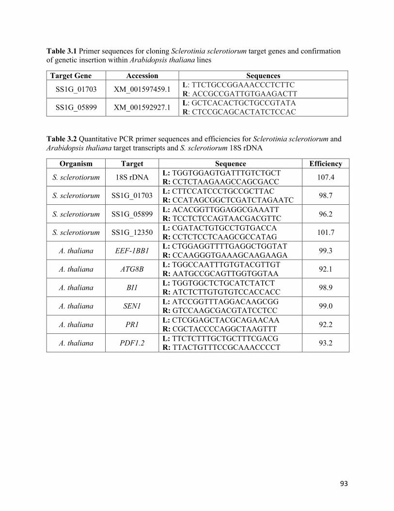
93
Table 3.1 Primer sequences for cloning Sclerotinia sclerotiorum target genes and confirmation of genetic insertion within Arabidopsis thaliana lines
Target Gene Accession Sequences
SS1G_01703 XM_001597459.1 L: TTCTGCCGGAAACCCTCTTC R: ACCGCCGATTGTGAAGACTT
SS1G_05899 XM_001592927.1 L: GCTCACACTGCTGCCGTATA R: CTCCGCAGCACTATCTCCAC
Table 3.2 Quantitative PCR primer sequences and efficiencies for Sclerotinia sclerotiorum and Arabidopsis thaliana target transcripts and S. sclerotiorum 18S rDNA
Organism Target Sequence Efficiency
S. sclerotiorum 18S rDNA L: TGGTGGAGTGATTTGTCTGCT R: CCTCTAAGAAGCCAGCGACC 107.4
S. sclerotiorum SS1G_01703 L: CTTCCATCCCTGCCGCTTAC R: CCATAGCGGCTCGATCTAGAATC 98.7
S. sclerotiorum SS1G_05899 L: ACACGGTTGGAGGCGAAATT R: TCCTCTCCAGTAACGACGTTC 96.2
S. sclerotiorum SS1G_12350 L: CGATACTGTGCCTGTGACCA R: CCTCTCCTCAAGCGCCATAG 101.7
A. thaliana EEF-1BB1 L: CTGGAGGTTTTGAGGCTGGTAT R: CCAAGGGTGAAAGCAAGAAGA 99.3
A. thaliana ATG8B L: TGGCCAATTTGTGTACGTTGT R: AATGCCGCAGTTGGTGGTAA 92.1
A. thaliana BI1 L: TGGTGGCTCTGCATCTATCT R: ATCTCTTGTGTGTCCACCACC 98.9
A. thaliana SEN1 L: ATCCGGTTTAGGACAAGCGG R: GTCCAAGCGACGTATCCTCC 99.0
A. thaliana PR1 L: CTCGGAGCTACGCAGAACAA R: CGCTACCCCAGGCTAAGTTT 92.2
A. thaliana PDF1.2 L: TTCTCTTTGCTGCTTTCGACG R: TTACTGTTTCCGCAAACCCCT 93.2

94
ImageJ software (imagej.nih.gov) and normalized to A. thaliana wild-type control. Infected
tissue was collected and pooled with 3 leaves per biological replicate and stored at -80°C.
Collected tissue was ground in liquid nitrogen and DNA extraction was performed using
a modified fungal DNA extraction buffer [0.5M KCl, 1M 2-amino-2-(hydroxymethyl)-1,3-
propanediol (TRIS)-HCl pH 7.5, 125 mM EDTA pH 8] (Chi et al., 2009). Fungal load was
quantified using Quantitative PCR (qPCR) and the Bio-Rad CFX96 Connect Real Time System.
10 µL reactions were performed using PerfeCTa SYBRⓇ Green SuperMix (Quanta Bio,
Beverly, MA, US) and 1 ng of DNA in each reaction and the relevant primers, listed in Table
3.2. Conditions used were as follows: 95°C for 30 s, and 40 cycles of: 95°C for 5 s, and 60°C for
30 s. Fungal load was assessed relative to wildtype infected tissue (Becker et al., 2017; Livak
and Schmittgen, 2001).
Transcript knockdown was measured 3 dpi using quantitative real time PCR (qRT-PCR).
RNA from each infected leaf sample (3 leaves per biological replicate) was isolated using the
Invitrogen Plant RNA Isolation and Ambion Turbo DNase treatment protocol (Thermo Fisher,
Waltham, MA, USA). Complementary DNA strand synthesis was performed using qScript
cDNA synthesis kit (QuantaBio, Beverly, MA, US) following the manufacturer’s protocol, using
a 10 µL reaction volume and 1000 ng of RNA. Thermocycler conditions for cDNA synthesis
were as follows: 25°C for 10 min., 50°C for 15 min., 85°C for 5 min. Using the Bio-Rad CFX96
Connect Real Time System, 10 µL qRT-PCR reactions were performed using PerfeCTa SYBRⓇ
Green SuperMix and the primers listed in Table 3.2. Conditions used were as follows: 95°C for
30 s; 40 cycles of: 95°C for 5s and 60°C for 30s. Transcript levels were measured in reference to
the SsSac7 (SS1G_12350) housekeeping gene using the ΔΔCq method (Livak and Schmittgen,
2001; Llanos et al., 2015).

95
Plant cell death and defense markers of infected samples used for measuring fungal
transcript were also assessed using qRT-PCR. Non-infected leaf tissue for each line was also
collected. RNA extraction and cDNA preparation was performed as described above. 10µL qRT-
PCR reactions were run on the Bio-Rad CFX96 Connect Real Time System using Perfecta
SYBR GreenⓇ SuperMix under the aforementioned conditions and primers in Table 3.2.
Transcript levels were referenced to the A. thaliana ELONGATION FACTOR 1B BETA (EEF-
1BB1) housekeeping gene and compared infected to non-infected using the ΔΔCq method
(Kabbage et al., 2013; Komaki et al., 2010; Livak and Schmittgen, 2001).
3.4 RESULTS
3.4.1 Host-induced gene silencing technology lessens lesion progression
To assess the extent of host resistance, A. thaliana leaves were inoculated with an
aggressive S. sclerotiorum spore inoculation technique (Garg et al., 2013). A. thaliana
SS1G01703RNAi and A. thaliana SS1G05899RNAi decreased lesion size 76 and 64 %, respectively,
compared to the wild-type control (one-way ANOVA, p < 0.01; Fig. 3.1b). The reduction of
infection was greater by 26% and 27% on A. thaliana SS1G01703RNAi and A. thaliana
SS1G05899RNAi compared to dsRNA foliar applications of SS1G_01703 and SS1G_05899,
respectively (student’s t-test, p<0.05; Chapter 2, Fig. 2.6). The data suggest HIGS may be a more
successful application of RNAi technology to control fungal pathogens.
Phenotypic differences between the pathogenic progression on the wild-type and the
RNAi lines were also observed (Fig. 3.1a). The control, non-hpRNA producing plants had
advanced lesions, covering the majority of the leaf surface. Evidence of systemic fungal

96
Figure 3.1 Sclerotinia sclerotiorum infection was limited on hpRNA-producing Arabidopsis thaliana. (a) Infected leaf tissue 3 dpi after spore inoculation. (b) Average lesion size of A. thaliana plants 3 dpi relative to wildtype control plants with standard error bars. Significant reductions of lesion size were observed and denoted with “*” (one-way ANOVA, p<0.05).
97
infection was also apparent as the lesions appeared to progress into the vasculature of the petiole.
In contrast, the infected A. thaliana SS1G01703RNAi and A. thaliana SS1G05899RNAi leaves had
smaller, localized lesions on the surface of the leaf. The lesion appeared drier compared to the
damp lesions on the wild-type control
3.4.2 Infection under host–induced gene silencing decreases the presence of fungal DNA
The relative amount of S. sclerotiorum 18S rDNA extracted from infected leaf tissue
between the wild-type and the transgenic lines was assessed using qPCR to confirm the
magnitude of fungal colonization. Fungal load of infected A. thaliana SS1G01703RNAi and A.
thaliana SS1G05899RNAi was reduced by 96 and 49 %, respectively, compared to the wild-type
tissue (student’s t-test p < 0.05; Fig 3.2). The difference in lesion size and fungal load are in
concordance with the phenotypic observations of the infected leaves and suggest the infection
was impeded in A. thaliana SS1G01703RNAi and A. thaliana SS1G05899RNAi. The greater
reduction of fungal load in the A. thaliana SS1G01703RNAi suggests that the infection may have
been superficial with limited colonization within the interior of the leaf. In contrast, the A.
thaliana SS1G05899RNAi leaves developed more prominent lesions. The reduction in fungal DNA
confirmed the infection was less developed in comparison to the wild-type plants and further
strengthens the evidence that hpRNA expression was sufficient to suppress fungal invasion into
the leaf’s tissues.
3.4.3 Specific fungal transcripts were reduced during infection on A. thaliana
SS1G01703RNAi and A. thaliana SS1G05899RNAi
Next, transcript levels of SS1G_01703 and SS1G_05899 were measured during infection
on A. thaliana SS1G01703RNAi, A. thaliana SS1G05899RNAi, and wild-type lines to assess hpRNA
production indirectly. S. sclerotiorum-treated A. thaliana SS1G01703RNAi and

98
Figure 3.2 Relative presence of Sclerotinia sclerotiorum 18S rDNA was reduced on Arabidopsis thaliana SS1G01703RNAi and A. thaliana SS1G05899RNAi compared to wild-type A. thaliana. DNA was extracted from spore inoculated tissue 3 days post inoculation. DNA levels were assessed using quantitative PCR. Data represent 3 biological replicates relative to wild-type tissue collection and error bars represent standard error. Significance is denoted with “*” (student’s t-test, p<0.05).
99
A. thaliana SS1G05899RNAi demonstrated transcript reductions of 83% and 75% for
SS1G_01703, and SS1G_05899, respectively, compared to the non-transformed lines (student’s
t-test p < 0.01; Fig.3.3). Thus, A. thaliana SS1G01703RNAi and A. thaliana SS1G05899RNAi were
likely expressing hpRNA targeting SS1G_01703 and SS1G_05899 respectively, which delivered
dsRNA to the fungus infecting the host tissue, and resulted in RNAi-mediated mRNA
knockdown within the fungal hyphae.
3.4.4 Defence markers are elevated and cell death markers are repressed differentially
during infection of hpRNA producing and non-hpRNA producing A. thaliana
qRT-PCR was used to compare mRNA levels of the infected and non-infected states of
the following cell death and defence markers: AUTOPHAGY 8B (ATG8B; autophagy), BAX-
INHIBITOR 1 (BI1; negative regulator of BAX-induced cell death), PLANT DEFENSIN 1.2
(PDF1.2; jasmonic acid (JA) mediated defence), PATHOGENESIS-RELATED GENE 1 (PR1;
salicylic acid (SA) response) and SENESCENCE 1 (SEN1; senescence). Compared to the non-
infected state, infected A. thaliana SS1G01703RNAi, A. thaliana SS1G05899RNAi, and wild-type A.
thaliana demonstrated significant differences in all markers, except for ATG8B and BI1 in wild-
type leaves (student’s t-test, p <0.05; Fig. 3.4). Both A. thaliana SS1G01703RNAi and A. thaliana
SS1G05899RNAi demonstrated significant differences in the death and defence markers compared
to the infection on the wild-type A. thaliana (Fig 3.4). The A. thaliana SS1G01703RNAi line
showed decreases in ATG8B (2-fold; student’s t test p < 0.01), BI1 (1.5-fold; p < 0.01), PDF1.2
(1.7-fold; p < 0.01), PR1 (4.2-fold; p < 0.01), and SEN1 (0.7-fold; p < 0.05) compared to the
wildtype (Fig. 3.4). Similarly, A. thaliana SS1G05899RNAi line demonstrated decreases of the
markers ATG8B (2-fold; p < 0.01), BI1 (1.6-fold; p < 0.01), PDF1.2 (1.7-fold; p < 0.01), and
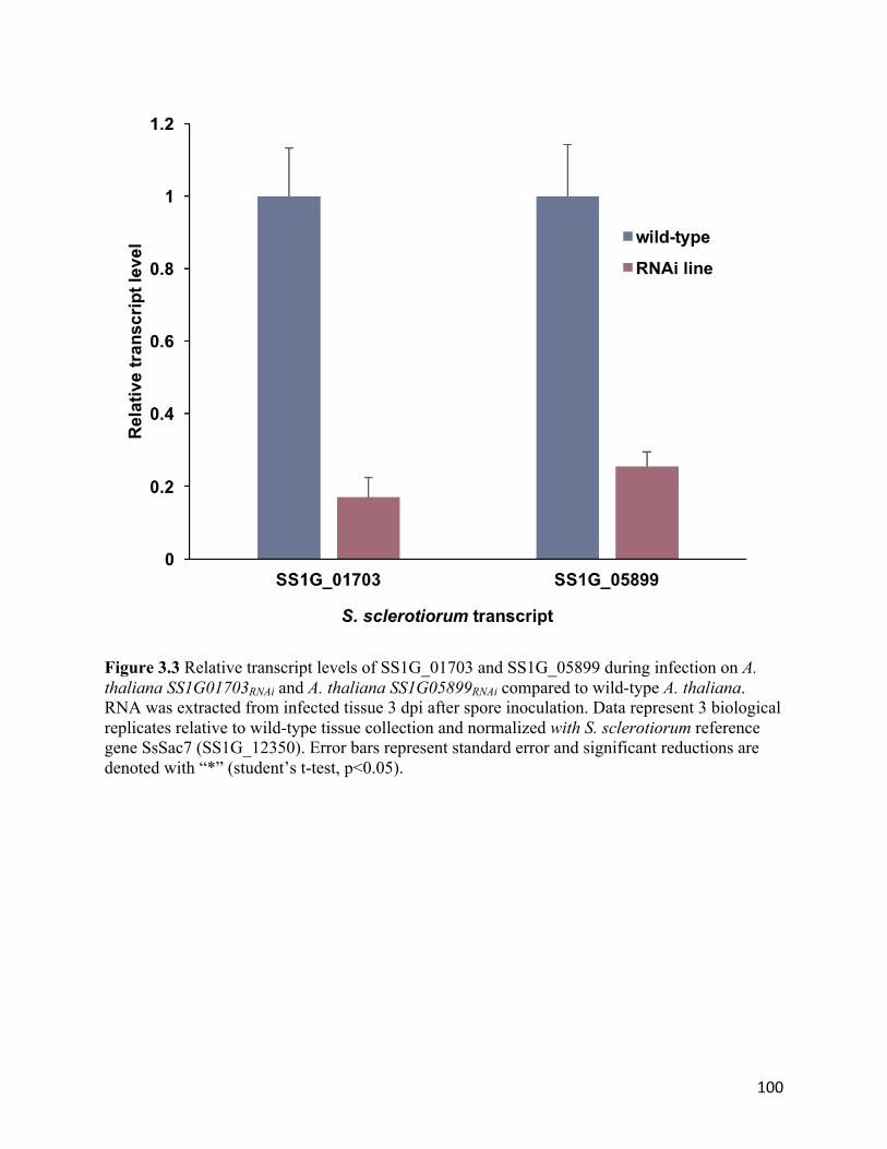
100
Figure 3.3 Relative transcript levels of SS1G_01703 and SS1G_05899 during infection on A. thaliana SS1G01703RNAi and A. thaliana SS1G05899RNAi compared to wild-type A. thaliana. RNA was extracted from infected tissue 3 dpi after spore inoculation. Data represent 3 biological replicates relative to wild-type tissue collection and normalized with S. sclerotiorum reference gene SsSac7 (SS1G_12350). Error bars represent standard error and significant reductions are denoted with “*” (student’s t-test, p<0.05).

101
SEN1 (2.6-fold; p < 0.01). However, PR1 was increased by 0.6-fold (p < 0.05) compared to the
infected wildtype (Fig. 3.4b).
In general, A. thaliana SS1G01703RNAi, A. thaliana SS1G05899RNAi, and wild-type A.
thaliana all showed a strong increase in the JA marker PDF1.2, which is characteristic of
necrotrophic infections, as well as elevated PR1 and SEN1 levels during fungal colonization
(Nováková et al., 2014). However, A. thaliana SS1G01703RNAi and A. thaliana SS1G05899RNAi
responded similarly by decreasing mRNA levels of ATG8B and BI1, unlike the wild-type plants.
Between the transgenic lines, the A. thaliana SS1G01703RNAi had decreased PR1 levels, while the
A. thaliana SS1G05899RNAi demonstrated a weaker SEN1 induction.
3.5 DISCUSSION
The capability of HIGS to suppress S. sclerotiorum disease pressure on A. thaliana
through the production of hpRNA was demonstrated. Both A. thaliana SS1G01703RNAi and A.
thaliana SS1G05899RNAi lines decreased foliar disease severity effectively. The levels of fungal
transcript and DNA were reduced during infection on the RNAi plants, further suggesting A.
thaliana SS1G01703RNAi and A. thaliana SS1G05899RNAi actively attenuated disease symptoms.
Using established markers for cell death and defence, plant response to fungal infection was
explored. Although the defence and death patterns between the RNAi line and the wild-type
plants appeared rather similar, alternative processes are potentially cooperating with HIGS to
repress fungal disease.
SS1G_01703 encodes a putative versiconal hemiacetal acetate esterase, an enzyme involved in
the fungal aflatoxin biosynthesis pathway, and may be involved in the production of secondary
metabolites in S. sclerotiorum (Azevedo et al., 2016; Kanehisa et al., 2017). To

102
Figure 3.4 Relative Arabidopsis thaliana transcript levels of cell death (ATG8B, BI1, SEN1) and defense markers (PDF1.2, PR1) during Sclerotinia sclerotiorum colonization as assessed by quantitative real-time PRC. (a) Infection of A. thaliana SS1G01703RNAi line (purple) and wild-type line (grey). (b) Infection of A. thaliana SS1G05899RNAi (green) and wild-type line (grey). RNA was extracted from infected tissue 3 dpi after spore inoculation. Data represent 3 biological replicates with mRNA levels compared to uninfected control with all mRNA levels normalized to A. thaliana EEF-1BB1 reference transcript. Error bars represent standard error and significant pairwise comparisons are denoted with “*” (student’s t-test, p<0.05)
103
mitigate the effect of these putative toxins, I developed a RNAi vector targeting SS1G_01703
through genetic transformation of A. thaliana. Previously, HIGS was used to prevent mycotoxin
accumulation in maize and peanut (Arachis hypogaea) (Arias et al., 2015; Masanga et al., 2015;
Thakare et al., 2017). When maize and peanut were challenged with Aspergillus flavus, the
pathogen was unable to produce aflatoxins that would contaminate the kernels of the host plant.
Despite the elimination of toxigenic capacity, the fungus did not show reductions in pathogenesis
when targeting this pathway (Arias et al., 2015; Masanga et al., 2015; Thakare et al., 2017). In
contrast, the A. thaliana SS1G01703RNAi demonstrated efficacious suppression of fungal disease
on the leaf surface. Likely, the production of the S. sclerotiorum contributes to the pathogenic
biology and potentially the necrotrophic nature of this fungus. Recently, Roze et al. (2015)
suggested that blocking aflatoxin biosynthesis may limit the production of reactive oxygen
species (ROS) and decrease tolerance to oxidative damage. Since host defence creates an
oxidative environment, such as the respiratory burst, I predicted knocking down levels of
SS1G_01703 mRNA, could contribute to the pathogen’s sensitivity to host counterattack (Heller
and Tudzynski, 2011; Roze et al., 2015). The dramatic reduction in fungal load further supports
the diminished fungal proliferation in the host tissue. Taken together, directed targeting of
SS1G_01703 through the RNAi pathway improves plant performance following S. sclerotiorum
infection. While there is a growing body of evidence that targeting this gene in S. sclerotiorum is
beneficial to the plant, the specific cellular mechanism remains unclear. However, I speculate
that the mode of action is likely through the suppression of host poisoning and increased
sensitivity to oxidative damage.
I was also successful in reducing fungal pressure in A. thaliana line by targeting a fungal
thioredoxin reductase, a gene essential for Aspergillus fumatigus (Hu et al., 2007). Thioredoxin
104
reductase is involved in sulphur tolerance, ROS scavenging, and the reduction of disulphide
bonds (Breitenbach et al., 2015; Montibus et al., 2015; Sato et al., 2011). Previous genetic
deletions of thioredoxin reductase in Aspergillus nidulans resulted in abnormal development
(Thön et al., 2007). Similarly, in the rice pathogen Magnaporthe oryzae, the thioredoxin
reductase system was determined to be essential for appressorium development, intercellular
growth, and oxidative tolerance using genetic deletions. The modified strain was unable to
severely infect rice leaves (Fernandez and Wilson, 2014). During fungal colonization, the host
plant will invoke respiratory bursts and produce ROS that will create an oxidative environment
and damage the molecular structure of cellular components (Heller and Tudzynski, 2011; Torres
et al., 2006). Therefore, the reduction in thioredoxin reductase by an RNAi-induced silencing
event could prevent ROS scavenging and innate cellular responses, leading to an accumulation of
oxidized proteins and nucleic acids within advancing hyphae. The host-induced damage may
have led to the observed reduction in fungal DNA and reduced lesions, suggesting limited fungal
replication within the host tissue. A. thaliana SS1G05899RNAi is the first demonstration of HIGS
to control fungal infection on a plant species targeting the fungal thioredoxin reductase system.
Transgenic A. thaliana expressing hpRNA directed at SS1G_01703 or SS1G_05899
demonstrated greater control of S. sclerotiorum compared to a topical treatment of 200 ng of
dsRNA to the leaf surface (Chapter 2). Previously, Koch et al. (2013) showed dramatic
reductions in F. graminearum infection using HIGS barley lines (Hordeum vulgare L.), which
proved to be more effective than the later study attempting to suppress fungal pressure using
topical dsRNA of the same target (Koch et al., 2016). Furthermore, the size of the molecules,
whether long dsRNA or sRNA, did not have an effect on the closely related fungus B. cinerea
(Wang et al., 2016). Based on the data, there are two potential mechanisms that could explain
105
the higher potency of HIGS plants. First, the host plant cells process the transcribed dsRNA into
siRNA, which would be delivered to the assailing pathogen (Koch et al., 2013; Panwar et al.,
2017). Since the RNAi mechanism is a multi-step process, siRNA could be more efficient as it
skips processing by DCL (Knip et al., 2014; Chapter 1, Fig. 1.1). Second, application of dsRNA
to the surface of the leaf represents a finite amount of dsRNA compared to the RNAi silencing
vectors that constantly produce hpRNA driven by the 35S Cauliflower Mosaic Virus promoter
(Khalid et al., 2017). Thus, the plant would provide a constant supply of processed dsRNAs to
the advancing fungus, sustaining optimal silencing conditions of the target gene despite changes
in fungal cell metabolism or replication. Therefore to complement the diminution of pathogen
transcript levels with the production of hpRNA, northern blots should be done to detect both the
presence and processing of the hpRNA in planta (Koch et al., 2013; Panwar et al., 2017). Gel
visualization would provide supporting evidence linking HIGS technology with the attenuation
of fungal pathogenesis. Taken together, engineering of HIGS in planta could provide a more
efficacious RNAi management strategy to mitigate fungal pressure and could be applied to
globally important cropping systems where transformation protocols are available.
Differences in disease severity observed between the two RNAi lines suggest the plant is
responding to the fungus and fungal attack through different cellular mechanisms. Previous
research using HIGS technology against fungal pathogens failed to explore the host response
during infection (Ghag et al., 2014; Koch et al., 2013; Wang et al., 2016). To better understand
these differences, we quantified changes in gene activity associated with selected cell death and
defense markers. JA-induced senescence could explain the induction of both SEN1 and PDF1.2
and provides a possible explanation into the improved plant protection observed in the transgenic
lines compared to wild-type plants (Balbi and Devoto, 2008). Furthermore, Kabbage et al.
106
(2013) explored the interaction of a non-pathogenic strain of S. sclerotiorum A2 during infection
on wild-type A. thaliana. During host colonization, S. sclerotiorum A2 induced greater
production of PDF1.2 and ATG8B in host A. thaliana. Likely, the JA defense response promoted
cellular autophagy instead of an apoptotic-like programmed cell death, leading to fungal non-
pathogenesis (Kabbage et al., 2013). Despite the evidence, the qRT-PCR results do not support
the hypothesis that autophagy was operating as the primary mode of defence against fungal
infection, as the autophagy marker was downregulated during infection. Thus, other mechanisms
may be responsible for improved plant performance.
Downregulation of BI1, a BAX antagonist, suggests the innate defense response was
likely independent of BAX-induced programmed cell death, usually invoked by H2O2,
temperature stress, SA, and biotic factors (Chen et al., 2013; Watanabe and Lam, 2009).
Recently, the expression of BI1 and autophagy was linked through interactions via
AUTOPHAGY 6 (ATG6) (Xu et al., 2017). Thus, the reduction of both BI1 and ATG8B may
operate via an ATG6-mediated interaction between autophagy and cell death. Other effectors
have also been implicated in necrotrophic resistance and cell death, including ethylene
signalling, cell wall modification, secondary compound synthesis, or nitric oxide (Broekgaarden
et al., 2015; Khare et al., 2017; Miedes et al., 2014; Perchepied et al., 2010). Based on these
results and the differential accumulation of defense markers like PR1, there exists a possibility
that the underlying cellular mechanisms responsible for plant health in A. thaliana
SS1G01703RNAi and A. thaliana SS1G05899RNAi are different, despite leading to the same
beneficial outcome. Moreover, the RNAi construct integrates preferentially into euchromatic
regions in the host genome such as promoters and coding regions (Gelvin and Kim, 2007; Lim et
al., 2007). The interruption of genetic expression and regulation could have also contributed to

107
the differences within A. thaliana SS1G01703RNAi and A. thaliana SS1G05899RNAi. Therefore,
global profiling of RNA populations through RNA-seq analysis should provide the necessary
information to dissect how plants expressing dsRNAs that target the fungus respond to fungal
attack.
When transformation protocols are available, HIGS biotechnology provides a more
potent implementation of an RNAi-based strategy that imparts species-specific resistance from
necrotrophic pathogens. The production of hpRNA in planta disables specific cellular processes
within the invading pathogen and leads to minimal colonization within host tissue. Although
unclear, the underlying molecular processes in the transformed plants contributed to positive host
outcomes, allowing for survival instead of death. Taken together, HIGS could be a sentinel of the
next-generation of fungal management strategies utilizing RNAi that improve upon traditional
protocols for developing host resistance.
3.6 REFERENCES
Amaradasa, B.S. and Everhart, S.E. (2016) Effects of Sublethal Fungicides on Mutation Rates
and Genomic Variation in Fungal Plant Pathogen, Sclerotinia sclerotiorum. PLoS One 11,
e0168079.
Andrade, C.M., Tinoco, M.L.P., Rieth, A.F., Maia, F.C.O. and Aragão, F.J.L. (2016) Host-
induced gene silencing in the necrotrophic fungal pathogen Sclerotinia sclerotiorum. Plant
Pathol. 65, 626–632.
Arias, R.S., Dang, P.M. and Sobolev, V.S. (2015) RNAi-mediated Control of Aflatoxins in
Peanut: Method to Analyze Mycotoxin Production and Transgene Expression in the
Peanut/Aspergillus Pathosystem. J. Vis. Exp. 2015, 1–11.

108
Azevedo, L., Chagas-Paula, D.A., Kim, H., Roque, A.C.M., Dias, K.S.T., Machado, J.C.,
Soares, M.G. and Mertens-Talcott, S.U. (2016) White mold (Sclerotinia sclerotiorum),
friend or foe: Cytotoxic and mutagenic activities in vitro and in vivo. Food Res. Int. 80, 27–
35.
Balbi, V. and Devoto, A. (2008) Jasmonate signalling network in Arabidopsis thaliana : crucial
regulatory nodes and new physiological scenarios. , 301–318.
Baum, J. a, Bogaert, T., Clinton, W., et al. (2007) Control of coleopteran insect pests through
RNA interference. Nat. Biotechnol. 25, 1322–6.
Becker, M.G., Zhang, X., Walker, P.L., et al. (2017) Transcriptome analysis of the Brassica
napus–Leptosphaeria maculans pathosystem identifies receptor, signaling and structural
genes underlying plant resistance. Plant J. 90, 573–586.
Bonfim, K., Faria, J.C., Nogueira, E.O.P.L., Mendes, É.A., Aragão, F.J.L., Recursos, E.,
Norte, P.W. and Brasília, U. De (2007) RNAi-Mediated Resistance to Bean golden mosaic
virus in Genetically Engineered Common Bean ( Phaseolus vulgaris ). 20, 717–726.
Breitenbach, M., Weber, M., Rinnerthaler, M., Karl, T. and Breitenbach-Koller, L. (2015)
Oxidative stress in fungi: Its function in signal transduction, interaction with plant hosts,
and lignocellulose degradation. Biomolecules 5, 318–342.
Broekgaarden, C., Caarls, L., Vos, I.A., Pieterse, C.M.J. and Wees, S.C.M. Van (2015)
Ethylene: traffic controller on hormonal crossroads to defense. Plant Physiol. 169,
pp.01020.2015.
Canola Council of Canada (2017) Economic Impact of the Canola Industry.
Chen, X.H., Yu, H., Deng, H.J., Chen, J.X., Mi, H.B. and Mao, L.C. (2013) Cucumber BAX
inhibitor-1 , a conserved cell death suppressor and a negative programmed cell death

109
regulator under cold stress. 57, 684–690.
Chi, M.H., Park, S.Y. and Lee, Y.H. (2009) A quick and safe method for fungal DNA
extraction. Plant Pathol. J. 25, 108–111.
Cusaac, J.P.W., Morrison, S.A., Belden, J.B., Smith, L.M. and McMurry, S.T. (2016) Acute
toxicity of HeadlineⓇ fungicide to Blanchard’s cricket frogs (Acris blanchardi).
Ecotoxicology 25, 447–455.
Derbyshire, M.C. and Denton-Giles, M. (2016) The control of Sclerotinia stem rot on oilseed
rape ( Brassica napus ): Current practices and future opportunities. Plant Pathol. 65, 859–
877.
Edwards, K., Johnstone, C. and Thompson, C. (1991) A simple and rapid method for the
preparation of plant genomic DNA for PCR analysis. Nucleic Acids Res. 19, 1349.
Fernandez, J. and Wilson, R.A. (2014) Characterizing roles for the glutathione reductase,
thioredoxin reductase and thioredoxin peroxidase-encoding genes of Magnaporthe oryzae
during rice blast disease. PLoS One 9.
Garg, H., Li, H., Sivasithamparam, K. and Barbetti, M.J. (2013) Differentially Expressed
Proteins and Associated Histological and Disease Progression Changes in Cotyledon Tissue
of a Resistant and Susceptible Genotype of Brassica napus Infected with Sclerotinia
sclerotiorum. PLoS One 8.
Garg, H., Li, H., Sivasithamparam, K., Kuo, J. and Barbetti, M.J. (2010) The infection
processes of Sclerotinia sclerotiorum in cotyledon tissue of a resistant and a susceptible
genotype of Brassica napus. Annu. Bot., 897–908.
Gelvin, S.B. and Kim, S.I. (2007) Effect of chromatin upon Agrobacterium T-DNA integration
and transgene expression. Biochim. Biophys. Acta - Gene Struct. Expr. 1769, 410–421.
110
Ghag, S.B., Shekhawat, U.K.S. and Ganapathi, T.R. (2014) Host-induced post-transcriptional
hairpin RNA-mediated gene silencing of vital fungal genes confers efficient resistance
against Fusarium wilt in banana. Plant Biotechnol. J. 12, 541–53.
Heller, J. and Tudzynski, P. (2011) Reactive Oxygen Species in Phytopathogenic Fungi:
Signaling, Development, and Disease. Annu. Rev. Phytopathol. 49, 369–390.
Hu, W., Sillaots, S., Lemieux, S., et al. (2007) Essential Gene Identification and Drug Target
Prioritization in Aspergillus fumigatus. 3, e24.
Huzar-Novakowiski, J., Paul, P.A. and Dorrance, A.E. (2017) Host resistance and chemical
control for management of Sclerotinia stem rot of soybean in Ohio. Phytopathology 107,
937–949.
Kabbage, M., Williams, B. and Dickman, M.B. (2013) Cell Death Control: The Interplay of
Apoptosis and Autophagy in the Pathogenicity of Sclerotinia sclerotiorum. PLoS Pathog. 9.
Kamal, M.M., Savocchia, S., Lindbeck, K.D. and Ash, G.J. (2016) Biology and biocontrol of
Sclerotinia sclerotiorum (Lib.) de Bary in oilseed Brassicas. Australas. Plant Pathol. 45, 1–
14.
Kanehisa, M., Furumichi, M., Tanabe, M., Sato, Y. and Morishima, K. (2017) KEGG: New
perspectives on genomes, pathways, diseases and drugs. Nucleic Acids Res. 45, D353–
D361.
Khalid, A., Zhang, Q., Yasir, M. and Li, F. (2017) Small RNA based genetic engineering for
plant viral resistance: Application in crop protection. Front. Microbiol. 8, 1–11.
Khare, D., Choi, H., Huh, S.U., Bassin, B., Kim, J., Martinoia, E., Sohn, K.H., Paek, K.-H.
and Lee, Y. (2017) Arabidopsis ABCG34 contributes to defense against necrotrophic
pathogens by mediating the secretion of camalexin. Proc. Natl. Acad. Sci. 114, E5712–

111
E5720.
Kılıç, G.A. (2011) Histopathological and biochemical alterations of the earthworm (Lumbricus
terrestris) as biomarker of soil pollution along Porsuk River Basin (Turkey). Chemosphere
83, 1175–1180.
Knip, M., Constantin, M.E. and Thordal-Christensen, H. (2014) Trans-kingdom Cross-Talk:
Small RNAs on the Move. PLoS Genet. 10.
Koch, A., Biedenkopf, D., Furch, A., et al. (2016) An RNAi-Based Control of Fusarium
graminearum Infections Through Spraying of Long dsRNAs Involves a Plant Passage and Is
Controlled by the Fungal Silencing Machinery. PLoS Pathog. 12, 1–22.
Koch, A., Kumar, N., Weber, L., Keller, H., Imani, J. and Kogel, K. (2013) Host-induced
gene silencing of cytochrome P450 lanosterol C14 α -demethylase – encoding genes confers
strong resistance to Fusarium species. 110, 19324–19329.
Komaki, S., Abe, T., Coutuer, S., Inzé, D., Russinova, E. and Hashimoto, T. (2010) Nuclear-
localized subtype of end-binding 1 protein regulates spindle organization in Arabidopsis.
Lim, P.O., Kim, H.J. and Gil Nam, H. (2007) Leaf Senescence. Annu. Rev. Plant Biol. 58,
115–136.
Livak, K.J. and Schmittgen, T.D. (2001) Analysis of relative gene expression data using real-
time quantitative PCR and. Methods 25, 402–408.
Llanos, A., François, J. and Parrou, J. (2015) Tracking the best reference genes for RT-qPCR
data normalization in filamentous fungi. BMC Genomics 16, 71.
Luo, H., Lin, Y., Gao, F., Zhang, C.T. and Zhang, R. (2014) DEG 10, an update of the
database of essential genes that includes both protein-coding genes and noncoding genomic
elements. Nucleic Acids Res. 42, 574–580.

112
Maltby, L., Brock, T.C.M. and Brink, P.J. Van Den (2009) Fungicide risk assessment for
aquatic ecosystems: Importance of interspecific variation, toxic mode of action, and
exposure regime. Environ. Sci. Technol. 43, 7556–7563.
Masanga, J.O., Matheka, J.M., Omer, R.A., Ommeh, S.C., Monda, E.O. and Alakonya,
A.E. (2015) Downregulation of transcription factor aflR in Aspergillus flavus confers
reduction to aflatoxin accumulation in transgenic maize with alteration of host plant
architecture. Plant Cell Rep. 34, 1379–1387.
Miedes, E., Vanholme, R., Boerjan, W. and Molina, A. (2014) The role of the secondary cell
wall in plant resistance to pathogens. Front. Plant Sci. 5, 358.
Montibus, M., Pinson-Gadais, L., Richard-Forget, F., Barreau, C. and Ponts, N. (2015)
Coupling of transcriptional response to oxidative stress and secondary metabolism
regulation in filamentous fungi. Crit. Rev. Microbiol. 41, 295–308.
Nováková, M., Šašek, V., Dobrev, P.I., Valentová, O. and Burketová, L. (2014) Plant
hormones in defense response of Brassica napus to Sclerotinia sclerotiorum - Reassessing
the role of salicylic acid in the interaction with a necrotroph. Plant Physiol. Biochem. 80,
308–317.
Nunes, C.C. and Dean, R.A. (2012) Host-induced gene silencing: A tool for understanding
fungal host interaction and for developing novel disease control strategies. Mol. Plant
Pathol. 13, 519–529.
Panwar, V., Jordan, M., McCallum, B. and Bakkeren, G. (2017) Host-induced silencing of
essential genes in Puccinia triticina through transgenic expression of RNAi sequences
reduces severity of leaf rust infection in wheat. Plant Biotechnol. J.
Panwar, V., McCallum, B. and Bakkeren, G. (2013) Host-induced gene silencing of wheat

113
leaf rust fungus Puccinia triticina pathogenicity genes mediated by the Barley stripe mosaic
virus. Plant Mol. Biol. 81, 595–608.
Papolu, P.K., Gantasala, N.P., Kamaraju, D., Banakar, P., Sreevathsa, R. and Rao, U.
(2013) Utility of host delivered RNAi of two FMRF amide like peptides, flp-14 and flp-18,
for the management of root knot nematode, Meloidogyne incognita. PLoS One 8, 1–16.
Penaud, A. and Walker, A.-S. (2015) Oilseed Rape Pathogens in France. In Fungicide
Resistance in Plant Pathogens: Principles and a Guide to Practical Management. (Ishii, H.
and Hollomon, D.W., eds), pp. 389–399. Tokyo: Springer Japan.
Perchepied, L., Balagué, C., Riou, C., Claudel-Renard, C., Rivière, N., Grezes-Besset, B.
and Roby, D. (2010) Nitric oxide participates in the complex interplay of defense-related
signaling pathways controlling disease resistance to Sclerotinia sclerotiorum in Arabidopsis
thaliana. Mol. Plant. Microbe. Interact. 23, 846–60.
Roze, L. V., Laivenieks, M., Hong, S.Y., Wee, J., Wong, S.S., Vanos, B., Awad, D., Ehrlich,
K.C. and Linz, J.E. (2015) Aflatoxin biosynthesis is a novel source of reactive oxygen
species—a potential redox signal to initiate resistance to oxidative stress? Toxins (Basel). 7,
1411–1430.
Sato, I., Shimatani, K., Fujita, K., Abe, T., Shimizu, M., Fujii, T., Hoshino, T. and Takaya,
N. (2011) Glutathione reductase/glutathione is responsible for cytotoxic elemental sulfur
tolerance via polysulfide shuttle in fungi. J. Biol. Chem. 286, 20283–20291.
Smalling, K.L., Reilly, T.J., Sandstrom, M.W. and Kuivila, K.M. (2013) Occurrence and
persistence of fungicides in bed sediments and suspended solids from three targeted use
areas in the United States. Sci. Total Environ. 447, 179–185.
Syromyatnikov, M.Y., Kokina, A. V., Lopatin, A. V., Starkov, A.A. and Popov, V.N. (2017)
114
Evaluation of the toxicity of fungicides to flight muscle mitochondria of bumblebee
(Bombus terrestris L.). Pestic. Biochem. Physiol. 135, 41–46.
Taylor, A., Coventry, E., Jones, J.E. and Clarkson, J.P. (2015) Resistance to a highly
aggressive isolate of Sclerotinia sclerotiorum in a Brassica napus diversity set. , 932–940.
Thakare, D., Zhang, J., Wing, R.A., Cotty, P.J. and Schmidt, M.A. (2017) Aflatoxin-free
transgenic maize using host-induced gene silencing. , 1–9.
Thön, M., Al-Abdallah, Q., Hortschansky, P. and Brakhage, A.A. (2007) The thioredoxin
system of the filamentous fungus Aspergillus nidulans: Impact on development and
oxidative stress response. J. Biol. Chem. 282, 27259–27269.
Tiwari, I.M., Jesuraj, A., Kamboj, R., Devanna, B.N., Botella, J.R. and Sharma, T.R.
(2017) Host Delivered RNAi, an efficient approach to increase rice resistance to sheath
blight pathogen (Rhizoctonia solani). Sci. Rep. 7, 7521.
Tollefson, J. (2011) Brazil cooks up transgenic bean. Nature 478, 168–168.
Torres, M.A., Jones, J.D.G. and Dangl, J.L. (2006) Reactive oxygen species signaling in
response to pathogens. Plant Physiol. 141, 373–378.
Uloth, M.B., Clode, P.L., You, M.P. and Barbetti, M.J. (2015) Attack modes and defence
reactions in pathosystems involving Sclerotinia sclerotiorum , Brassica carinata , B. juncea
and B. napus. Ann. Bot., mcv150.
United States Environmental Protection Agency (2017) EPA Registers Innovative Tool to
Control Corn Rootworm.
Wang, M., Weiberg, A., Lin, F.-M., Thomma, B.P.H.J., Huang, H.-D. and Jin, H. (2016)
Bidirectional cross-kingdom RNAi and fungal uptake of external RNAs confer plant
protection. Nat. Plants 2, 16151.

115
Watanabe, N. and Lam, E. (2009) Bax inhibitor-1, a conserved cell death suppressor, is a key
molecular switch downstream from a variety of biotic and abiotic stress signals in plants.
Int. J. Mol. Sci. 10, 3149–3167.
Xu, G., Wang, S., Han, S., Xie, K., Wang, Y., Li, J. and Liu, Y. (2017) Plant Bax Inhibitor-1
interacts with ATG6 to regulate autophagy and programmed cell death. Autophagy 13,
1161–1175.
Zhang, T., Jin, Y., Zhao, J.-H., Gao, F., Zhou, B.-J., Fang, Y.Y. and Guo, H.-S. (2016) Host-
Induced Gene Silencing of the Target Gene in Fungal Cells Confers Effective Resistance to
the Cotton Wilt Disease Pathogen Verticillium dahliae. Mol. Plant, 1–4.

116
CHAPTER 4 : CONCLUSIONS AND FUTURE DIRECTIONS
Throughout my thesis, I have gathered evidence for the utility of RNAi-based management
of fungal phytopathogens. I developed two successful implementations of RNAi biotechnology
to control S. sclerotiorum and B. cinerea. RNA sequencing allowed for the largest and most
comprehensive identification of RNAi targets for phytopathogenic management. Synthesizing
the data from the functional screens performed in Chapter 2 allowed for the creation of a novel
target identification pipeline (TIP), providing the first guide for choosing RNAi-based control for
fungi. With further usage, the research community will be able to adjust and fine-tune the criteria
for effective selection of RNAi targets within fungi. For example, I extended my techniques to
the related fungus, B. cinerea, to demonstrate homologous targets could offer efficacious
protection in Chapter 2. Additional targeting of homologous genes in other fungi could
illuminate how different fungal species initiate and sustain pathogenesis. A deeper understanding
of fungal infection would be indispensable for future plant engineering.
The experiments performed in Chapter 3 represent the beginning of further explorations
into HIGS as a durable strategy for phytopathogenic control. I would like to further evaluate
other lines transformed with RNAi vectors that I created based on molecule performance during
my foliar screens. However, some key biological questions remain. For example, I would like to
use whole genome sequencing to examine which genes were interrupted by the Agrobacterium
tumefaciens-mediated transformation, and thereby determine how target site integration
differences may have affected the RNAi efficacy in the different lineages. Additionally, to
validate the lesion phenotype and differential mRNA levels within the host during infection, I
would also like to perform RNA-seq to understand what defence and cell death responses could
be implicated in the phenotypic differences observed between the RNAi lines. This could be of

117
interest for plant breeders looking to develop resistance by modulating these pathways.
Furthermore, exploring uninfected tissues could uncover how the production of hpRNA and the
insertion event effect the basal state of the plant tissue. Pairing RNA-seq with sRNA-seq could
also reveal a more detailed view of RNA processing within cells and develop a greater functional
knowledge of HIGS biotechnology.
I explored interaction of the dsRNA or hpRNA-producing plants targeting S.
sclerotiorum or B. cinerea (Chapters 2 and 3). My thesis focused on the ‘on-target’ effects of the
dsRNA molecules. The level of homology for a dsRNA to elicit an RNAi response and the
severity of the RNAi response has not been teased apart in relation to potential ‘off-target’
effects. Therefore, it will be interesting to investigate the effect of S. sclerotiorum targeting
dsRNA on infection and growth on a variety of other fungal species. QRT-PCR could be
performed to explore whether transcript levels change significantly from the controls, which
would suggest an effect within another organism. Such an exploration is crucial for ecological
and molecular design if RNAi technology will advance further towards commercialization.
Together, these additional experiments would add important details to complete a
compelling investigation of the potency of RNAi-based approaches for phytopathogenic
management. While there are still many unanswered questions concerning the broader impacts of
exogenous dsRNA on plants, my thesis nevertheless provides functional evidence that a new
generation of sustainable technologies to overcome today’s pressing challenges using RNAi
biotechnologies is entirely possible.
![Genetically Modified Organism-Free RNA Interference · Genetically Modified Organism-Free RNA Interference: Exogenous Application of RNA Molecules in Plants1[OPEN] Athanasios Dalakouras,a,b,2,3](https://static.fdocuments.net/doc/165x107/605cd1054dc5810cd70565f5/genetically-modiied-organism-free-rna-genetically-modiied-organism-free-rna.jpg)