
The Cytochemistry of the Amoebocytes and Intestinal ... · 60 Zacks—Cytochemistry of Venus...
17
57 The Cytochemistry of the Amoebocytes and Intestinal Epithelium of Venus mercenaria (Lamellibranchiata), with Remarks on a Pigment resembling Ceroid By SUMNER I. ZACKS Jeffries Wyman Scholar, Harvard University, 1952-3 (From the Department of Anatomy, Harvard Medical School, Boston 15, Mass., U.S.A.) With two plates (figs. 1 and 2) SUMMARY The properties of the amoebocytes and intestinal epithelium of Venus mercenaria were studied by a variety of cytochemical procedures designed to demonstrate pro- teins, enzymes, carbohydrates, and lipids. The cytoplasm of the amoebocytes contains specific granules which are constantly present and which are interpreted as being atypical mitochondria. Identification of their mitochondrial nature rests on their staining with Janus green B, their positive reaction for phospholipid by Baker's test, and the presence of dehydrogenase activity. Unlike typical mitochondria, the specific granules are eosinophil. Protein-bound carbonyl groups and disulphide and sulphydryl groups are present in both the specific granules and the cytoplasm. The sulphydryl groups may in part be associated with the presence of dehydrogenase, lipase, and serum cholinesterase. Amoebocytes also contain glycogen and a material that is resistant to diastase and positive to the periodic acid / Schifftest; this material may be a neutral polysaccharide, unsaturated lipid, or mucoprotein. Cytoplasmic structures which are inconstantly present in amoebocytes include sudanophil droplets, neutral red vacuoles, metachromatic granules, and granules of an excretory pigment resembling ceroid. The sudanophil droplets may be stored neutral fat or lipid associated with the Golgi apparatus. The neutral red vacuoles are not preformed inclusions, but form as the dye accumulates within the cells. Meta- chromatic granules, which are confined solely to the intestinal amoebocytes, consist of phagocytosed intestinal mucus liberated from goblet cells. The histochemical reactions of the columnar intestinal epithelium suggest that these cells may be active in the digestion and absorption of nutrients, since eosinophil granules, lipid droplets, alkaline phosphatase, lipase, and serum cholinesterase are present in them. Masses of a ceroid-like excretory pigment and goblet cells containing mucus are present between the columnar intestinal epithelial cells. The pigment contains phos- pholipid and apparently arises as an oxidized end-product of lipid metabolism. MOEBOCYTES are granulocytes which are found in large numbers in the \ blood, connective tissues, gills, cardiac muscle, and intestinal mucosa of invertebrates. They have been the subject of numerous morphological and chemical studies and many speculations have been advanced concerning their nature and functions (Metschnikoff, 1884; Haughton, 1934; Takatsuki, 1934; Ohuye, 1938; Yonge, 1923, 1926a, 19266, 1946). [Quarterly Journal of Microscopical Science, Vol. 96, part 1, pp. 57-71, March 1955.]
-
Upload
nguyencong -
Category
Documents
-
view
222 -
download
0
Transcript of The Cytochemistry of the Amoebocytes and Intestinal ... · 60 Zacks—Cytochemistry of Venus...

57
The Cytochemistry of the Amoebocytes and IntestinalEpithelium of Venus mercenaria (Lamellibranchiata),
with Remarks on a Pigment resembling Ceroid
By SUMNER I. ZACKSJeffries Wyman Scholar, Harvard University, 1952-3
(From the Department of Anatomy, Harvard Medical School, Boston 15, Mass., U.S.A.)
With two plates (figs. 1 and 2)
SUMMARY
The properties of the amoebocytes and intestinal epithelium of Venus mercenariawere studied by a variety of cytochemical procedures designed to demonstrate pro-teins, enzymes, carbohydrates, and lipids.
The cytoplasm of the amoebocytes contains specific granules which are constantlypresent and which are interpreted as being atypical mitochondria. Identification oftheir mitochondrial nature rests on their staining with Janus green B, their positivereaction for phospholipid by Baker's test, and the presence of dehydrogenase activity.Unlike typical mitochondria, the specific granules are eosinophil. Protein-boundcarbonyl groups and disulphide and sulphydryl groups are present in both the specificgranules and the cytoplasm. The sulphydryl groups may in part be associated withthe presence of dehydrogenase, lipase, and serum cholinesterase. Amoebocytes alsocontain glycogen and a material that is resistant to diastase and positive to the periodicacid / Schifftest; this material may be a neutral polysaccharide, unsaturated lipid, ormucoprotein.
Cytoplasmic structures which are inconstantly present in amoebocytes includesudanophil droplets, neutral red vacuoles, metachromatic granules, and granules ofan excretory pigment resembling ceroid. The sudanophil droplets may be storedneutral fat or lipid associated with the Golgi apparatus. The neutral red vacuoles arenot preformed inclusions, but form as the dye accumulates within the cells. Meta-chromatic granules, which are confined solely to the intestinal amoebocytes, consistof phagocytosed intestinal mucus liberated from goblet cells.
The histochemical reactions of the columnar intestinal epithelium suggest that thesecells may be active in the digestion and absorption of nutrients, since eosinophilgranules, lipid droplets, alkaline phosphatase, lipase, and serum cholinesterase arepresent in them.
Masses of a ceroid-like excretory pigment and goblet cells containing mucus arepresent between the columnar intestinal epithelial cells. The pigment contains phos-pholipid and apparently arises as an oxidized end-product of lipid metabolism.
MOEBOCYTES are granulocytes which are found in large numbers in the\ blood, connective tissues, gills, cardiac muscle, and intestinal mucosa of
invertebrates. They have been the subject of numerous morphological andchemical studies and many speculations have been advanced concerning theirnature and functions (Metschnikoff, 1884; Haughton, 1934; Takatsuki, 1934;Ohuye, 1938; Yonge, 1923, 1926a, 19266, 1946).[Quarterly Journal of Microscopical Science, Vol. 96, part 1, pp. 57-71, March 1955.]

58 Zacks—Cytochemistry of Venus mercenaria
The present study concerns the cytology and cytochemistry of the amoebo-cytes and columnar intestinal epithelium of the quahog, Venus mercenaria. Aceroid-like excretory pigment found in these cells as well as in other tissuesof Venus, was also investigated. Interest in these topics arose from a previousinvestigation of esterolytic enzymes in V. mercenaria (Zacks and Welsh, 1953).
The individual amoebocyte is a nucleated cell 9-12/x in diameter, which,in recently drawn blood, rounds up and appears as a sphere with small pro-jecting spikes i-2/x long. After remaining in contact with the surface of a glassslide for several minutes, or in tissue-spreads, the amoebocytes extend pseudo-pods ; by means of these they are capable of slow motion. Within their cyto-plasm, small hyaline granules as well as yellow, refractile granules of excretorypigment are seen. The numerous, even-sized, hyaline granules are constantlypresent in Venus amoebocytes and therefore will be termed specific granulesto distinguish them from granules of excretory pigment and other cytoplasmicinclusions which are inconstant constituents.
The chemical nature of the cytoplasmic granules of these amoebocytes hasinterested a few investigators, but no one appears before to have studied thegranules by cytochemical methods. By these procedures it was hoped thatinformation might be obtained concerning the chemical nature of the granulesand their relations to the enzymatic activities of the cells. The chemical cyto-logy of the columnar intestinal epithelium was investigated with particularreference to its role in digestion and absorption of nutrients. The nature ofthe excretory pigment which is present in the amoebocytes, intestinal epi-thelium, and other tissues of Venus was similarly investigated by histochemicalmeans. This pigment, which occurs in the form of granules in the amoebo-cytes and amorphous clumps in the intestine, has been described as a de-gradation product of chlorophyll (MacMunn, 1900) and as echinochrome(Durham, 1891). Since the chemical nature of this pigment is poorly under-stood, it was hoped that histochemical tests might contribute to its charac-terization.
MATERIALS AND METHODS
Fresh specimens of V. mercenaria were purchased from a fish dealer. Wholehearts were excised and placed either in sea-water for supravital staining or invarious fixatives. Hearts prepared in this way were convenient objects for thestudy of both amoebocytes and intestinal epithelium, since the heart iswrapped around a segment of intestine.
Supravital methods. For supravital staining, whole hearts were placed insea-water containing Janus green B at 1:10,000 or neutral red at 1:10,000 andstained for 2 hours at 29° C. A fragment of atrial wall was then spread undera coverslip on a glass slide for examination. This preparation was chosenbecause the thin network of atrial muscle fibres enmeshed and supported theamoebocytes, thus allowing a favourable opportunity for observing pseudopodformation and amoeboid motion.
Zacks—Cytochemistry of Venus mercenaria 59
Methods for the staining of proteins. Basiphil structures were studied inZenker-fixed material. Deparaffinized sections were stained in methylene bluesolutions buffered at pH 2-3, 4-3, 6-4, and 7-4 according to the method ofSinger and Morrison (1948). Similar sections were also stained by Mallory'seosin and methylene blue. The Feulgen procedure, followed by counter-staining with light green, was applied to deparaffinized sections to detectdesoxypentose-nucleic acid (DNA). Protein-bound sulphydryl and disulphidegroups were sought in deparaffinized sections of hearts fixed in 80 per cent,alcohol containing 1 per cent, trichloracetic acid (Barrnett and Seligman,1952). Sections were stained with and without prior reduction in (NH4)2S, toallow identification of both sulphydryl and disulphide groups.
Methods for the detection of enzymes. Gomori's acid and alkaline phospha-tase methods (1939, 1941)1 as modified by Dempsey and Deane (1946), wereapplied to alcohol- and acetone-fixed deparaffinized sections as well as tofresh frozen sections. Sections were incubated for 30 minutes, 1, 3, 6, and24 hours in solutions of glycerophosphate buffered to pH 4-5 and 9-5.
Dehydrogenase activity was detected in fresh hearts after incubation for20 minutes in blue tetrazolium chloride (pH 7-4), a method modified fromthat of Rutenburg, Gofstein, and Seligman (1950).
Substances capable of reducing ferricyanide to ferrocyanide were studied byapplying the ferric-ferricyanide reaction of Lillie and Burtner (1953) to freshand formalin-fixed sections.
Methods for the detection of carbohydrates and mucoproteins. Glycogen andpolysaccharides were stained by the periodic acid / Schiff (PAS) method ofMcManus (1946) and Hotchkiss (1948), after fixation of hearts in Rossman'sfluid. Control sections were treated with diastase before application of thisprocedure. Metachromasia was investigated by staining with 0-5 per cent,toluidine blue after 4 per cent, lead acetate fixation (Holmgren, 1940; Jorpes,Holmgren, and Wilander, 1937).
Methods for the detection of lipids. The distribution of lipids was studiedafter staining frozen sections of fresh and formalin-fixed hearts with Sudanblack B. Phospholipids were identified in frozen sections of hearts fixed informaldehyde-calcium and in weak Bouin's fluid (Baker, 1946). Carbonylgroups were sought in frozen sections of material fixed in buffered 10 per cent,formalin and stained by the method of Ashbel and Seligman (1949). Controlsections were extracted with acetone to remove lipid-containing carbonylgroups. Cholesterol and cholesteryl esters were investigated in frozen sectionsby the Schultz procedure and acid-fast substances were identified in materialfixed in formalin, alcohol, and Zenker's fluid, by the carbol-fuchsin procedureof Lillie (1948).
Methods for the detection of birefringence and fluorescence. Birefringence andfluorescence were studied in fresh-frozen and formalin-fixed sections.
Method for the detection of iron salts. To detect the presence of iron saltsmaterial fixed in 80 per cent, alcohol and in Zenker's fluid was embedded inparaffin, sectioned, and treated with potassium ferrocyanide (Lillie, 1948).

60 Zacks—Cytochemistry of Venus mercenaria
OBSERVATIONS
Supravital staining. Spreads of atrial muscle stained in i : 10,000 Janusgreen B exhibited amoebocytes which were uniformly filled with deep blue-green granules I-2JU. in diameter. The harmlessness of this dye was shown bythe slow extension of pseudopods from the cells. Withdrawal of pseudopodswas never observed in supravitally-stained or fresh, unstained preparations.After 3-5 minutes, the specific granules appeared red-violet in colour, andeventually assumed the red colour of the reduction-product of Janus green B,diethyl safranin. Recolouring of the granules did not occur when the atrialfragments were re-exposed to atmospheric oxygen.
The cut ends of the intestine appeared deep blue-green after staining inJanus green B and this staining was almost entirely confined to the intestinalepithelium. A faint blue-green colour was observed in the ventricular muscle,but the intestinal muscle was nearly devoid of staining.
When neutral red (1:10,000) was applied supravitally for 2 hours, theamoebocytes were filled with deep brick-red inclusions of various sizes(fig. 1, A). The majority of the neutral red inclusions were larger and lessuniform in size than the granules stained by Janus green B. The brick-redcolour of the neutral red inclusions suggested that their pH was close toneutrality.
Excretory pigment was unstained by Janus green B and neutral red.Basiphilia. In sections stained in methylene blue at pH 7-4 and 6-4, cardiac
and intestinal muscle fibres, goblet cell mucus, and excretory pigment werestained deep blue, whereas the cytoplasm of the amoebocytes was moderatelystained blue. After staining with methylene blue at pH 4-3, the intestinalmuscle fibres were coloured grey-blue, amoebocyte cytoplasm was stainedlight blue, and masses of excretory pigment appeared blue-green. Gobletcell mucus was metachromatically stained deep violet at pH 3, but excretorypigment and cardiac and intestinal muscle fibres and amoebocyte granuleswere unstained. However, the cell plasma of the amoebocytes was faintlystained blue. At pH 2, nothing was stained.
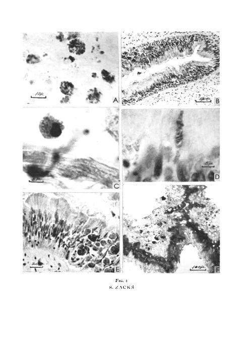
FIG. I (plate), A, atrial muscle spread, illustrating the appearance of amoebocytes supra-vitally stained by neutral red. Large and small irregular vacuoles are present in each cell.
B, transverse section through the intestine of Venus mercenaria, stained by the Feulgenreaction and counterstained by light green. The typical ciliated columnar epithelium, as wellas numerous amoebocytes and accumulations of excretory substance, are illustrated. Numerousamoebocytes are scattered between the intestinal muscle fibres.
c, an amoebocyte stained by the Feulgen procedure and counterstained with light green.The cytoplasm is packed with green-stained specific granules.
D, a goblet cell after application of the Feulgen procedure. The goblet cell contains red-stained mucus and the nuclear chromatin is coloured red-violet.
E, higher magnification of the field shown in B. Masses of excretory pigment composed ofglobules of yellow or brown material embedded in a green-stained matrix are illustrated.
F, transverse, alcohol-fixed section of Venus intestine after 3 hours' incubation in glycero-phosphate at pH 95. Alkaline phosphatase activity is located in the distal portion of theepithelium. Excretory pigment is unreactive yet some clumps of this material appear dark inthe photograph due to the intrinsic colour of the pigment.
A!§#:>4*'.[-r:':-:'
Fio. i
S. ZACKS

FIG. 2
S. ZACKS
Zacks—Cytochemistry of Venus mercenaria 61
Eosin and methylene blue. After staining with eosin and methylene blue(pH 5-3), the specific granules of the amoebocytes were stained red. Occa-sional amoebocytes contained a few blue granules of various size. The oval,peripherally placed nucleus contained strongly basiphil clumps of chromatin.
The intestinal epithelium was composed of tall, ciliated, pseudostratifiedcolumnar cells which rested on a thick basement membrane. The proximaland middle portions of each epithelial cell were basiphil and numerous dust-like eosinophil granules were present in the distal zone. The intestinal surfaceof the epithelial cells was equipped with cilia which were 7-10/1 in length.The nucleus was located in the middle or proximal part of the cells. Amoebo-cytes containing specific granules were gathered in great numbers on the base-ment membrane in spaces between the epithelial cells. Numerous amoebocyteswere also present between the smooth muscle fibres of the intestinal muscula-ture. Occasional goblet cells, containing deeply basiphil mucus, were presentbetween the columnar epithelial cells.
Masses of various sizes (7-30/x) of amorphous, strongly basiphil excretorypigment were also present in the spaces between the epithelial cells. Smallgranules of the same material were scattered between the cardiac musclefibres.
Desoxyribonucleoprotein. The Feulgen procedure stained the nuclei of themuscle fibres, epithelial cells (fig. 1, B) and amoebocytes, but the specificgranules of the amoebocytes were not coloured (fig. 1, c). The cytoplasm ofthe goblet cells was stained deep red in contrast with the violet colour of thenuclei (fig. 1, D). The cytoplasm of the amoebocytes and portions of theexcretory pigment masses were stained by the acid dye, light green, used ascounterstain. The excretory pigment masses in the intestinal epithelium werecomposed of unstained yellow or orange globules embedded in a green-stainedmatrix (fig. i, E).
Protein-bound sulphydryl and disulphide groups. After (NH4)2S reduction, theintestinal epithelium, intestinal muscle cells, and cardiac muscle reacted posi-tively. The amoebocytes also contained sulphydryl groups, as indicated by apositive reaction of their cytoplasm. It could not be ascertained whether thereactive material was localized in both the specific granules and cell plasma ofthe amoebocytes, or only in the cell plasma.
Amoebocytes, intestinal epithelium, and ventricular muscle also reacted
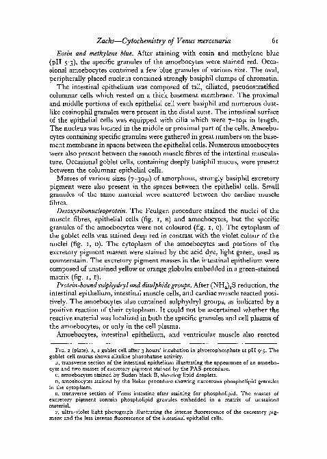
FIG. 2 (plate), A, a goblet cell after 3 hours' incubation in glycerophosphate at pH 9-5. Thegoblet cell mucus shows alkaline phosphatase activity.
B, transverse section of the intestinal epithelium illustrating the appearance of an amoebo-cyte and two masses of excretory pigment stained by the PAS-procedure.
C, amoebocytes stained by Sudan black B, showing lipid droplets.D, amoebocytes stained by the Baker procedure showing numerous phospholipid granules
in the cytoplasm.E, transverse section of Venus intestine after staining for phospholipid. The masses of
excretory pigment contain phospholipid granules embedded in a matrix of unstainedmaterial.
F, ultra-violet light photograph illustrating the intense fluorescence of the excretory pig-ment and the less intense fluorescence of the intestinal epithelial cells.

62 Zacks—Cytochemistry of Venus mercenaria
positively when reduction with (NH4)2S had been omitted, thus indicating thepresence of free-SH groups as well as S-S groups in these cells. By both pro-cedures, the excretory pigment was unreactive.
Alkaline and acid phosphatases. Alkaline phosphatase activity was moreintense in fresh-frozen sections than' in alcohol-fixed sections. After 30minutes' incubation, the distal ends of the columnar epithelial cells werestained brown (fig. 1, F) but the amoebocytes and excretory pigment wereunreactive. After 6 hours of incubation the intestinal epithelium was intenselystained, but only an occasional amoebocyte could be found which containedone or two brown granules. In alcohol-fixed sections, the goblet cells werestained brown after 3 hours of incubation (fig. 2, A).
Acid phosphatase activity was not demonstrable in any of the tissues in-vestigated.
Dehydrogenase. After 20 minutes' incubation in blue tetrazolium chloride,granules of blue formazan pigment were present in the amoebocytes and thecardiac and intestinal muscle fibres.
Ferric-ferricyanide reaction. After 10 minutes in this reagent, the cardiacmuscle and intestinal epithelium were tinged light green, the amoebocyteswere colourless, and the masses of excretory pigment were coloured deepblue-green.
Periodic acid j Schiff reaction. The PAS procedure produced intense redstaining of the cardiac and intestinal muscle fibres, goblet cells, and excretorypigment. The amoebocytes were filled with many red granules of varioussizes. Fig. 2, B illustrates the appearance of an amoebocyte and masses ofexcretory pigment stained by this procedure. After the exposure to the actionof diastase to remove glycogen, staining of both cardiac and intestinal musclefibres and amoebocytes was considerably reduced. However, goblet cells andexcretory pigment were deeply stained despite exposure to diastase.
Staining with toluidine blue [metachromasia). Muscle fibres, the specificgranules of the amoebocytes, and the excretory pigment were stained faintlyblue.
Metachromatic granules and amorphous clumps were abundantly presentin the intestinal lumen and epithelium as well as in amoebocytes located be-tween the epithelium. However, large numbers of amoebocytes located in theintestinal and cardiac musculature were devoid of metachromatic granules.
Lipids. After the staining of atrial spreads in Sudan black B, each amoebo-cyte contained up to a dozen black droplets of variable size. The larger drop-lets were occasionally U shaped and associated with cytoplasmic vacuoles. Atendency toward perinuclear localization occurred (fig. 2, c). After 15 minutes'extraction in acetone or hot alcohol (6o° C), the sudanophil droplets couldno longer be demonstrated.
The amorphous masses of excretory pigment were stained green-black orgrey-green by Sudan black B and small black sudanophil droplets werepresent in the apices of the intestinal epithelial cells.
Phospholipids. Each amoebocyte contained 2-50 black granules of uniform

Zacks—Cytochemistry of Venus mercenaria 63
size (fig. 2, D) after staining by Baker's method for phospholipids. In manyamoebocytes, these granules were not as numerous as the specific granuleswhich were stained by supravital Janus green B or eosin. After pyridine ex-traction, the nuclei of the amoebocytes and muscle cells were stained, but thegranules were unstained. These results indicate that the amoebocyte granulescontain phospholipid. It should be emphasized, however, that in manyamoebocytes, the phospholipid granules represented but a small fraction ofthe full complement of granules stained by Janus green B or eosin.
Black-stained granules and clumps were embedded in the yellow masses ofexcretory pigment present in the intestinal columnar epithelium which wasotherwise unstained (fig. 2, E).
Carbonyl groups. The distal halves of the columnar intestinal epithelial cellsreacted strongly for carbonyl groups, but the basal halves stained only faintly.The musculatures of both heart and intestine were unreactive. The specificgranules as well as the cell plasma of the amoebocytes were moderatelystained. In control preparations extracted with acetone, the outer portions ofthe epithelial cells remained reactive, but the masses of excretory pigmentwere stained red-orange, a result attributable to non-specific solution of theblue azo dye in the lipid component of the excretory pigment (Nachlas andSeligman, 1949). Carbonyl-staining was reduced in amoebocytes after acetoneextraction.
The Schultz test for cholesterol and cholesterol esters was negative in all ofthe cell elements studied.
Acid-fast substances. After several hours' extraction in dilute hydrochloricacid, red masses of excretory substance and occasional small granules in amoe-bocytes were present.
Birefringence and fluorescence. Neither epithelium, excretory pigment, noramoebocytes were birefringent when examined under the polarization micro-scope.
In ultra-violet light, the middle and basal portions of the columnar in-testinal epithelial cells showed light blue fluorescence, whereas the apical,granular portion of the cells- exhibited red-violet fluorescence. The large,amorphous masses of excretory pigment situated on the basement membraneand between the epithelial cells (fig. 2, F), as well as occasional small granules inthe amoebocytes, showed intense yellow fluorescence. Similar fluorescent gran-ules were scattered between the intestinal muscle fibres. Much of this materialseemed to be free, but a smaller fraction was located within amoebocytes.
Ferrocyanide reaction {iron salts). Sections stained by this means were com-pletely negative, no blue or green deposits of Prussian blue being observed.
DISCUSSION
The nature and properties of the various structures in the cytoplasm of amoebocytes
A variety of cytoplasmic inclusions which exhibit distinctive histochemicalproperties and enzymatic reactions were present in the amoebocytes of

64 Zacks—Cytochemistry of Venus mercenaria
V. mercenaria. These included specific granules (constantly present), neutralred vacuoles, sudanophil droplets, pigment granules, and metachromatic andPAS-positive materials, all of which were inconstantly present. Besides thereactions shown by these various inclusions the cell plasma itself manifestedcertain staining properties.
Cytochemical reactions of specific granules in amoebocytes
Supravital staining with Janus green B showed great numbers of even-sized specific granules which reduced the dye to diethyl safranin, thus indicat-ing the presence of hydrogen-donor enzymes. That this reaction was notreversible was seen in the failure of the specific granules to become colouredagain when exposed to atmospheric oxygen. The fact that these granulesstained with Janus green B and were capable of reducing this dye whenoxygen was excluded, indicated that they are of mitochondrial nature. Further-more, dehydrogenase activity associated with the specific granules was indi-cated by the oxidation of blue tetrazolium chloride. The mitochondrial natureof the specific granules was also suggested by the fact that many of thegranules reacted positively with Baker's test for phospholipid. However, thespecific granules were eosinophil, a staining reaction which does not occur intypical mitochondria. The foregoing observations suggested that amoebocytespecific granules represent an atypical variety of mitochondria.
Diastase-labile as well as diastase-resistant PAS-positive granules werepresent in the amoebocytes. The substances responsible for this reaction arethought to be compounds containing 1, 2 glycol linkages which are oxidizedby periodic acid to form aldehyde groups which then react with the Schiffreagent (Hotchkiss, 1948). According to Leblond (1950), only substancesinsoluble in water and fat substances can be considered to persist after ex-posure of the tissues to the reagents employed in fixation and paraffin em-bedding. These substances include glycogen, which can be removed bypretreatment of the sections with diastase, and mucopolysaccharides andmucoproteins which are not removed by diastase. Lillie (1950), Wolman(1950), and Pearse (1953) have shown that certain lipids also yield a positivePAS-reaction which is retained after exposure to the action of diastase. Un-saturated lipids or other substances containing hydroxyl and amino groupson two adjacent carbon atoms might be expected to yield a positive PAS-test.
Since much of the material which was stained by the PAS procedure wasremoved by the action of diastase, it appears that these cells contain glycogen.However, amoebocytes also contained granules of diastase-resistant material.
The nature of the diastase-resistant material in amoebocytes is not clear.This material may be neutral polysaccharide, since evidence of stronglyacidic groups of acid mucopolysaccharides is lacking, or mucoprotein, phos-pholipid, or other unsaturated lipid. The presence of neutral fat and phos-pholipid as well as PAS-positive excretory pigment has been demonstrated inVenus amoebocytes. Furthermore, since serum cholinesterase is present inamoebocytes (Zacks and Welsh, 1953), it is of interest that cholinesterase is

Zacks—Cytochemistry of Venus mercenaria 65
thought to be a mucoprotein and that practically all sites possessing cholin-esterase activity are PAS-positive (Gomori, 1951). Thus the diastase-resistantmaterial may be unsaturated lipid or mucoprotein.
The presence of ketonic carbonyl groups of proteins or proteo-lipids wasindicated rather than ketonic groups associated with lipids (Seligman andAshbel, 1951) since the amoebocytes were still reactive after acetone ex-traction.
Sulphydryl and disulphide groups were present in both the specific granulesand cell plasma of amoebocytes. These groups are important in binding pro-teins and prosthetic groups (Barron, 1951), cell division and growth (Brachet,1950), cell permeability (Lefevre, 1948), and in enzymatic activity (Barron,1951). Several sulphydryl enzymes have been demonstrated in amoebocytes.The presence of lipase in the amoebocytes of several molluscs has been shownby Yonge (19266) and Takatsuki (1934), and recently by histochemical pro-cedures by Zacks and Welsh (1953). Serum cholinesterase is also present inthe amoebocytes of Venus as indicated by carbonaphthoxycholine iodidehydrolysis (Zacks and Welsh, 1953). In addition, Takatsuki (1934) has demon-strated enzymes capable of attacking starch, glycogen, maltose, lactose,sucrose, salicine, and gelatine in extracts of Ostrea amoebocytes.
Among the numerous enzymes requiring SH-groups for their activity arecholinesterase, lipase, esterase, /3-amylase, and carboxypeptidase (Barron,1951). Of this group, cholinesterase, lipase, and an unidentified dehydro-genase have been detected in Venus amoebocytes, and amylase, protease, andlipase have been demonstrated in the amoebocytes of other species (Yonge,19266; Takatsuki, 1934). Since several sulphydryl enzymes are present inamoebocytes, it seems that the sulphydryl groups demonstrable in these cellsmay be partially attributed to these enzymes.
Metachromasia in amoebocytes. The absence of metachromatic granules inamoebocytes outside the intestine suggests that intestinal amoebocytes whichcontain metachromatic granules had phagocytosed some of the goblet cellmucus. Metachromatic staining is regarded as evidence for the presence ofacid mucopolysaccharides (Holmgren and Wilander, 1937; Wislocki, Bunting,and Dempsey, 1947), an important constituent of mucus from several sources.Mucus is used by lamellibranchs to trap food particles to aid their ingestionby amoebocytes and digestive diverticula (Yonge, 19266).
Sudanophil droplets in amoebocytes. Sudanophil droplets of various sizewere present in nearly all the amoebocytes. In every case these droplets couldbe distinguished from the specific granules by their relatively small numberand by their size and position within the cell. Since the sudanophil dropletswere extractable by cold acetone and hot alcohol, they appeared to consist ofneutral fat.
The presence of lipids in lamellibranch amoebocytes has been investigatedby Yonge (1926, a and b), Takatsuki (1934), and others. Yonge believes thatamoebocytes and digestive diverticula play an exclusive role in fat ingestionand digestion and that no fat is digested extracellularly in the stomach. Thus,

66 Zacks—Cytochemistry of Venus mercenaria
lipid droplets present in amoebocytes may represent ingested fat globules.Another interpretation is that of Gatenby and Hill (1934), who regard similarsudanophil droplets in Helix amoebocytes as elements of the Golgi apparatus.The perinuclear position frequently assumed by the sudanophil droplets inVenus amoebocytes might suggest a similar interpretation.
Neutral red vacuoles in amoebocytes. In addition to specific granules andlipid droplets, amoebocytes contained large numbers of various sizes, cyto-plasmic inclusions which were stained by neutral red. That these inclusionswere not the same as the specific granules stained by Janus green B was indi-cated by their lack of uniformity of size and shape. Also the large numbers ofgranules staining with Janus green B precluded the possibility that both thesegranules and neutral red bodies could both be present in the cell as preformedinclusions. Furthermore, it is generally agreed that supravital Janus green Brarely, if ever, stains structures which are stained by neutral red. Bensley(1911) observed that Janus green B stained mitochondria in acinar cells of theguinea-pig pancreas, whereas neutral red stained granules of prozymogen andzymogen. Gatenby (1931) stated that many cells collect and aggregate neutralred into vacuoles while it is passing through the cytoplasm, and Gatenby andHill (1934) concluded that the neutral red inclusions of Helix amoebocyteswere not pre-existent structures. In the case of Venus amoebocytes, the objectswhich stained with neutral red appear to be vacuoles filled with dye, ratherthan preformed cytoplasmic inclusions.
Pigment granules in amoebocytes. Amoebocytes frequently contained highlyrefractile yellow granules which appear to be identical with the excretory pig-ment described by Yonge (1923, 1926, a and b) and others in various lamelli-branchs. These pigment granules were easily distinguished from the specificgranules of these cells. Larger granules and clumps of this material were alsofound scattered throughout the intestinal and cardiac musculature and inespecially large masses in the intestinal epithelium. The histochemistry of theexcretory pigment will be considered more fully in the discussion of theintestinal epithelium.
The nature and properties of Venus intestinal epithelium. The central impor-tance of intracellular digestion in the amoebocytes in lamellibranch nutritionhas been questioned by Nelson (1933), Mansour (1946), and Mansour-Bek(1946). These workers believe that extracellular digestion occurs in the ali-mentary tract of these animals. Yonge (1926a) states that 'no evidence of anyabsorption in the epithelium of the gut or any free surface in the mantlecavity, other than by the agency of phagocytes was found'. Observations madeon the cytochemistry of Venus intestinal epithelium may contribute to thisquestion.
Red-violet granules resembling secretory granules were observed in thedistal zone of the columnar epithelial cells in sections stained by eosin andmethylene blue, and this region was marked by red-violet fluorescence inultra-violet light. The eosinophil, fluorescent granules of each cell corre-sponded in position with the major site of alkaline phosphatase activity. This

Zacks—Cytochemistry of Venus mercenaria 67
enzyme functions in the dephosphorylation of several organic phosphatesincluding hexose diphosphate, nucleic acid, lecithin, and glycerophosphate.Deane and Dempsey (1945) described the localization of alkaline phosphatasein the apical zone of duodenal epithelium in several vertebrate species afterincubation of sections in glycerophosphate. Alkaline phosphatase activity wasalso seen in the supranuclear or Golgi region of these cells. These authorsconcluded that their studies supported the concept that intestinal epitheliumcontains enzymes capable of dephosphorylating intermediate substances innormal metabolism. Kosman, Kaulbersz, and Freeman (1943) reported thatalkaline phosphatase was secreted by the dog duodenum and jejunum andthat it probably functioned in the digestion of monophosphoric esters of food.Lecithin phosphatase is also present in duodenal epithelium and is thoughtto be of importance in digestion of phospholipids (Dempsey and Deane,1946). Furthermore, glycerophosphatase is thought to be involved in fatabsorption, since glycerophosphatase is believed to be an intermediate statein the breakdown and resynthesis of neutral fat (Bloor, 1943). Thus, it appearsthat the alkaline phosphatase activity detectable in the intestinal epitheliumof Venus may be associated with possible digestive and absorptive functionsof these cells. Further evidence for the absorptive role of the epithelium wasseen in the sudanophil droplets which were present in the distal portion ofthe epithelium and ketone-containing lipid which was demonstrated in themiddle portion of the epithelium. Gutheil (1912) observed fat globules in theintestinal epithelium of Anodonta and concluded that the epithelium func-tions in absorption, and Yonge (19266) observed fat globules in the stomachand midgut epithelium of Ostrea after feeding on diatoms, but concluded thatthe amoebocytes transmitted the fat to the epithelial cells for storage. AlthoughYonge (1926, a and b) has denied the existence of digestive and absorptiveactivity in the epithelium, the presence of eosinophil granules, sudanophildroplets, alkaline phosphatase, lipase, and serum cholinesterase suggests thatthese functions may be present.
The nature of the goblet cell mucus. Since the goblet cells of the intestinalepithelium were strongly basiphil and retained their stainability by methyleneblue in solutions buffered below pH 4 and were metachromatic as well asPAS-positive after diastase digestion, it appears that they contain typicalmucus. The goblet cells were stained red by the Feulgen procedure, a colourunlike the violet stain produced in the nuclei. The basis of this atypicalFeulgen reaction is obscure, since this reaction is quite specific for DNA.
The nature and properties of excretory substance. Numerous investigatorshave noted the presence of yellow, brown, or green pigment granules in amoe-bocytes and other tissues (Metschnikoff, 1884; Grobben, 1887; MacMunn,1900; Yonge, 1926, a and b). MacMunn (1900) referred to this material asenterochlorophyll, which he believed to be a derivative of ingested chlorophyll,and Durham (1891) suggested that this pigment was an excretory productcomposed of degraded echinochrome. Since the appearance of the pigmentvaried in different cells, Durham concluded that transformation of the

68 Zacks—Cytochemistry of Venus mercenaria
pigment occurred in the amoebocytes. Similar pigments were observed byJ. H. List (1890) and T. List (1902). J. H. List (1890) reported that the pigmentmasses were not constant and that their nature depended on the food ingestedby the animal since starved animals had few pigment inclusions. Yonge(19266) observed that globules of green and brown material appeared inamoebocytes after diatoms were ingested but disappeared during starvation.The brown colour of the pigment was attributed to ingested chlorophyll andproducts of chlorophyll degradation. In the amoebocytes and free in the in-testinal and cardiac musculature of Venus, excretory pigment appeared assmall yellow granules, but in the intestinal epithelium this material occurredin large yellow or yellow-brown heterogenous aggregates which were com-posed of pigment granules embedded in a matrix.
TABLE I . Comparisons of the histochemical reactions of ceroid and excretorypigment
Test Ceroid Excretory pigment
ColourResistance to fat solventsBasiphiliaAcidolphiliaSudanophiliaAcid-fast.Reduction of ferric-ferricyanideIron (Prussian blue) .Ultra-violet fluorescencePAS after exposure to diastase
Yellow Yellow
Ceroid-like nature of the excretory substance. The description of ceroid byEndicott and Lillie (1944) is strongly reminiscent of the excretory pigmentfound in amoebocytes and intestinal epithelium of Venus. These authorsidentify ceroid by the following characteristics:
1. Golden yellow colour in unstained sections.2. Resistant to fat solvents.3. Stained by basic dyes.4. Not stained by acid dyes.5. Sudanophil in paraffin sections.6. Acid fast.7. Reduces alkaline silver nitrate, but not the ferric-ferricyanide reagent.8. Iron-negative.
In addition Lee (1950) observed that ceroid in rat and mouse livers isvariably PAS-positive, and Popper, Gyorgy, and Goldblatt (1944) reportedthat ceroid possesses bright yellow or golden brown fluorescence whenexamined in ultra-violet light. Table 1 compares the histochemical propertiesof ceroid and excretory pigment.
The excretory material appears to be very similar to ceroid as described byEndicott and Lillie (1944) and others with one exception, namely, that unlike

Zacks—Cytochemistry of Venus mercenaria 69
excretory pigment, ceroid fails to stain when exposed to the ferric-ferricyanidereagent (Endicott and Lillie, 1944). However, Pearse (1953) states that thisproperty may be acquired during the oxidation of lipids.
The origin and nature of ceroid. Ceroid was first discovered in cirrhotic liversof nutritionally deficient rats by Lillie and his associates (1941, 1942), whobelieved that it was a product of abnormal metabolism. It was found thatceroid accumulation was inhibited by feeding on casein, choline, and methio-nine. Subsequently, other workers concluded that ceroid was a lipoproteinderived from the necrotic remnants of liver parenchymal cells (Gyorgy andGoldblatt, 1942; Lee, 1950), and that its presence was due to vitamin E defi-ciency (Victor and Pappenheimer, 1945). This pigment is found in manyvertebrate tissues other than cirrhotic liver (Firminger, 1952; Deane andFawcett, 1952; Lillie, 1941, 1942; Wolf and Pappenheimer, 1945).
Ceroid is believed to be a mixture of several substances (Lee, 1950; Pearse,1953). Pearse (1953) regards ceroid as a member of a general group of pig-ments termed lipofuscins which are derived by oxidation from lipids orlipoproteins. Different staining reactions are given as the lipoidal materialbecomes progressively oxidized. The PAS-reaction is positive in the inter-mediate stages of the oxidation of lipids and Schiff-reacting aldehydes areproduced from unsaturated phosphatides by periodic acid.
Casselman (1951) observed the formation of ceroid-like substances in vitrofrom unsaturated fat and from fatty acids and their esters, but never fromsaturated fats or hydrocarbons. The production of the pigment was preventedby the presence of antioxidants such as a-tocopherol and hydroquinone.Casselman concluded that 'whenever conditions are such that unsaturatedfats accumulate in tissues to such an extent that a relative lack of biologicalantioxidant results, autoxidation of the fats and their conversion to ceroidpigment are favored, and that ceroid and the lipofuscin pigment of vitaminE deficiency may be fundamentally similar'.
The occurrence of a ceroid-like pigment in the amoebocytes and intestineof Venus was unexpected. The excretory pigment appears to be a lipofuscinclosely related to ceroid observed in cirrhotic and vitamin E deficient animals.This material is formed either as an oxidized by-product of lipid metabolismin the intestine or as a by-product of intracellular digestion in the amoebo-cytes. The relation of excretory pigment to digestion is indicated by thedisappearance of this pigment in starved animals. In the case of Venus, theexcretory pigment may arise in part from the oxidation of phospholipidswhich can be demonstrated within the masses of excretory pigment.
It is a pleasure to acknowledge the constant aid and encouragement givenby Dr. George B. Wislocki during the course of these studies.
REFERENCESASHBEL, R., and SELIGMAN, A. M., 1949. 'A new reagent for the histochemical demonstration
of active carbonyl groups. A new method for staining ketonic steroids.' Endocrinology,44, 565-

70 Zacks—Cytochemistry of Venus mercenaria
BAKER, J. R., 1946. 'The histochemical recognition of lipine.' Quart. J. micr. Sci., 87, 441.BARRNETT, R. J., and SELIGMAN, A. M., 1952. 'Histochemical demonstration of protein-
bound sulphydryl groups.' Science, 116, 323.BARRON, E. S. G., 1951. 'Thiol groups of biological importance.' Recent Adv. Enzymol.,
11, 201.
BENSLEY, R. R., 1911. 'Studies on the pancreas of the guinea pig.' Amer. J. Anat., 12, 297.BLOOR, W. R., 1943. Biochemistry of the fatty acids. New York (Reinhold).BRACHET, J., 1950. Chemical embryology (trans, by L. G. Barth). New York. (Interscience
Publishers.)CASSELMAN, W. G. B., 1951. 'The in vitro preparation and histochemical properties of sub-
stances resembling ceroid.' J. exp. Med., 94, 549.DEANE, H. W., and FAWCETT, D. W., 1952. 'Pigmented interstitial cells showing "brown
degenertion" in the ovaries of old mice.' Anat. Rec, 113, 239.and DEMPSEY, E. W., 1945. 'The localization of phosphatases in the Golgi region of
intestinal and other epithelial cells.' Anat. Rec, 93, 401.DEMPSEY, E. W., and DEANE, H. W., 1946. 'The cytological localization, substrate specificity,
and pH optima of phosphatases in the duodenum of the mouse.' J. cell, and comp.Physiol., 27, 159.
DURHAM, H. E., 1891. 'On wandering cells in echinoderms, etc., more especially with regardto excretory functions.' Quart. J. micr. Sci., 33, 81.
ENDICOTT, K. M., and LILLIE, R. D., 1944. 'Ceroid and the pigment of dietary cirrhosis inrats, its characteristics and its differentiation from hemofuscin.' Amer. J. Path., 20, 149.
FIRMINGER, H. E., 1952. 'Apparent identity of pigmented lipoid in cells in adrenal gland andinterstitium of testes of mice following administration of stilbesterol.' J. Nat. CancerInst., 13, 225 (abstract).
GATENBY, J. B., 1931. 'The prozymogen granules ("vacuome") of R. R. Bensley in the Pseudo-triton pancreas, and the modern neutral red cytology.' Amer. J. Anat., 48, 421.and HILL, J. C, 1934. 'Improved technique for non-aseptic tissue culture of Helix
aspersa, with notes on molluscan cytology.' Quart. J. micr. Sci., 76, 331.GOMORI, G., 1939. 'Microtechnical demonstration of phosphatase in tissue sections.' Proc.
Soc. exp. Biol. N.Y., 42, 23.1941- 'Distribution of acid phosphatase in the tissue under normal and under patho-
logical conditions.' Arch. Path., 32, 189.1951. 'Histochemistry of esterases.' J. Natl. Cancer Inst., 12, 247.
GROBBEN, C, 1887-8. 'Die Pericardialdriise der Lamellibranchiaten.' Arb. aus Zool. Inst.Wien, 7, 355.
GUTHEIL, F., 1912. 'Uber den Darmkanal und die Mitteldarmdriise von Anodonta cellensis,Schrot.' Z. wiss. Zool., 99, 444.
GYORGY, P., and GOLDBLATT, H., 1942. 'Observations on the conditions of dietary hepaticinjury (necrosis, cirrhosis) in rats.' J. exp. Med., 75, 355.
HAUGHTON, I., 1934. 'XVII. Amoebocytes and allied cells in invertebrates.' J. Roy. micr. Soc,54, 246.
HOLMGREN, H., 1940. 'Studien iiber Verbreitung und Bedeutung der chromotropen Substanz.'Z. f. mikr.-anat. Forsch., 47, 489.and WILANDER, O., 1937. 'Beitrag zur Kenntnis der Chemie und Funktion der Ehrlich-
schen Mastzellen.' Ibid., 42, 242.HOTCHKISS, R. D., 1948. 'A microchemical reaction resulting in the staining of polysaccharide
structures in fixed tissue preparations.' Arch. Biochem., 16, 131.JORPES, E., HOLMGREN, H., and WILANDER, O., 1937. 'Uber das Vorkommen von Heparin in
den Gefasswanden und den Augen.' Z. f. mikr-anat. Forsch., 42, 279.KOSMAN, A. J., KAULBERSZ, J. W., and FREEMAN, S., 1943. 'The secretion of alkaline phos-
phatase by the dog's intestine.' Amer. J. Physiol., 138, 236.LEBLOND, C. P., 1950. 'Distribution of periodic acid-reactive carbohydrates in the adult rat.'
Amer. J. Anat., 86, 1.LEE, C. S., 1950. 'Histochemical studies of the ceroid pigment of rats and mice and its relation
to necrosis.' J. Nat. Cancer Inst., n , 339.LEFEVRE, P. G., 1948. 'Evidence of active transfer of certain nonelectrolytes across the human
red cell membrane.' J. gen. Physiol., 31, 505.LILLIE, R. D., DAFT, F. S., and SEBRELL, W. H., 1941. 'Cirrhosis of liver in rats on a deficient
diet and the effect of alcohol.' U.S. Pub. Health Rep., 56, 1255.

Zacks—Cytochemistry of Venus mercenaria 71
LILLIE, R. D., ASHBURN, L. L., SEBRELL, W. H., DAFT, F. S., and LOWRY, J. V., 1942.'Histogenesis and repair of the hepatic cirrhosis in rats, produced on low protein dietsand preventable with choline.' Ibid., S7i 5°2.
LILLIE, R. D., 1948. Histopathologic technic. Philadelphia (Blakiston).1950. 'Further exploration of the HIO4-Schiff reaction with remarks on its significance.'
Anat. Rec, 108, 239.and BURTNER, H. J., 1953. 'The ferric ferricyanide test in histochemistry.' J. Histochem.
and Cytochem., 1, 87.LIST, J. H., 1890. 'Uber die Herkunft des Pigmentes in der Oberhaut.' Biol. Centbl., 10, 22.LIST, T., 1902. 'Die Mytiliden.' Fauna und Flora des Golfes von Neapel, 27, 253.MCMANUS, J. F. A., 1946. 'Histological demonstration of mucin after periodic acid.' Nature,
158, 202.MACMUNN, C. A., 1900. 'On the gastric gland of Mollusca and decapod Crustacea: its struc-
ture and functions.' Phil. Trans. Roy. Soc. Lond., 193, 1.MANSOUR, K., 1946. 'Food and digestive organs of lamellibranchs.' Nature, 158, 378.MANSOUR-BEK, J. J., 1946. 'Extracellular proteolytic and lipolytic enzymes of some lamelli-
branchs.' Ibid.METSCHNIKOFF, E., 1884. 'Researches on the intracellular digestion of invertebrates.' Quart.
J. micr. Sci., 24, 89.NACHLAS, M. M., and SELIGMAN, A. M., 1949. 'Histochemical demonstration of esterase.'
J. Natl. Cancer Inst., 9, 415.NELSON, T. C, 1933. 'On the digestion of animal forms by the oyster.' Proc. Soc. exp. Biol.
and Med., 30, 128.OHUYE, T., 1938. 'On corpuscles in the body fluids of some invertebrates. XII. General con-
sideration of the results obtained by the preceding investigations.' Sci. Rep. Toh6ku Imp.Univ. Biol., 13, 359.
PEARSE, A. G. E., 1953. Histochemistry theoretical and applied. Boston. (Little Brown).POPPER, A., GYORGY, P., and GOLDBLATT, H., 1944. 'Fluorescent material (ceroid) in experi-
mental nutritional cirrhosis.' Arch. Path., 37, 161.RUTENBURG, A. M., GOFSTEIN, R., and SELIGMAN, A. M., 1950. 'Preparation of a new tetra-
zolium salt which yields a blue pigment on reduction and its use in the demonstrationof enzymes in normal and neoplastic tissues.' Cancer Research, 10, 113.
SELIGMAN, A. M., and ASHBEL, R., 1951. 'Histochemical demonstrations of latent activecarbonyl groups in normal and neoplastic nervous tissue.' Cancer, 4, 579.
SINGER, M., and MORRISON, R. P., 1948. 'The influence of pH, dye and salt concentrationon the dye binding of modified and unmodified fibrin.' J. biol. Chem., 175, 133.
TAKATSUKI, S., 1934. 'On the nature and functions of the amoebocytes of Ostrea ednlis.'Quart. J. micr. Sci., 76, 379.
VICTOR, J., and PAPPENHEIMER, A. M., 1945. 'The influence of choline, cystine and a toco-pherol upon the occurrence of ceroid pigment in dietary cirrhosis of rats.' J. exp. Med.,82, 375-
WISLOCKI, G. B., BUNTING, H., and DEMPSEY, E. W., 1947. 'Metachromasia in mammaliantissues and its relationship to mucopolysaccharides.' Amer. J. Anat., 81, 1.
WOLF, A., and PAPPENHEIMER, A. M., 1945. 'Occurrence and distribution of acid fast pigmentin the central nervous system.' J. Neuropath, and exp. Neurol., 4, 402.
WOLMAN, M., 1950. 'Staining lipids by the periodic-acid-Schiff method.' Proc. Soc. exp.Biol. and Med., 75, 583.
YONGE, C. M., 1923. 'Studies on the comparative physiology of digestion. Mechanism offeeding, digestion, and assimilation in the lamellibranch, Mya.' Br. J. exp. Biol., 1, 15.1926a. 'The digestive diverticula in the lamellibranchs.' Trans. Roy. Soc. Edinburgh,
54, 7O3-19266. 'Structure and physiology of the organs of feeding in Ostrea edulis.' J. mar. biol.
Assoc. U.K., 14, 295.1946. 'Digestion of animals by lamellibranchs.' Nature, 157, 729
ZACKS, S. I., and WELSH, J. H., 1953. 'Cholinesterase and lipase in the amoebocytes, intestinalepithelium and heart-muscle of the quahog, Venus mercenaria.' Biol. Bull., 105, 200.

![[PPT]Clam Dissection · Web viewDissection of the Clam Venus mercenaria * copyright cmassengale * * * * * * copyright cmassengale Resource * * * * * * * * * * * * * * * * * * * * *](https://static.fdocuments.net/doc/165x107/5aa6232f7f8b9a7c1a8e5555/pptclam-dissection-viewdissection-of-the-clam-venus-mercenaria-copyright-cmassengale.jpg)


