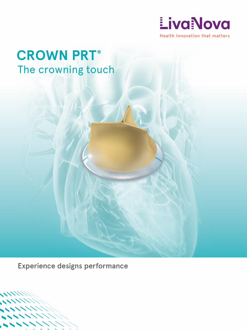
The crowning touch
18
Experience designs performance The crowning touch CROWN PRT ®
Transcript of The crowning touch
Experience designs performance
The crowning touchCROWN PRT®

2
In the treatment of aortic valve disease, the replacement of the native aortic valve with a stented bioprosthesis represents a standard technique to provide older patients with a performing and durable solution, while relieving them from the discomfort of lifelong oral anticoagulation therapy.
CROWN PRT is the latest advancement in stented aortic bioprosthesis technology, featuring surgeon-friendly design and state-of-the-art performance.
CROWN PRT
Crowning Achievements
Continuous InnovationLong Term Clinical Evidence
Global Reach
CrowningDesign
Smooth HandlingStraightforward Implantability
Ample Versatility
CrowningPerformance
Optimal HemodynamicsMinimized Patient Prosthesis Mismatch
Proven Durability
CROWN PRT®

3
CROWN PRT is part of LivaNova's cardiac surgery offering,
a full suite of innovative solutions, supported by strong clinical evidence and
adopted by healthcare providers worldwide.
Pioneering innovative clinical solutionsCrowning achievements
CROWN PRT®
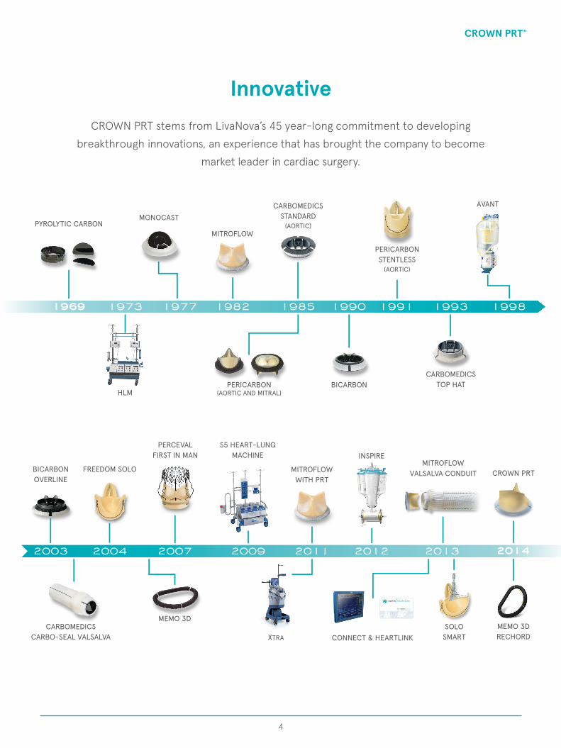
19821977 1985 19911990 1993 19981969 1973
PYROLYTIC CARBONMONOCAST
MITROFLOW
CARBOMEDICSSTANDARD
(AORTIC)
PERICARBONSTENTLESS
(AORTIC)
AVANT
HLMPERICARBON
(AORTIC AND MITRAL)BICARBON
CARBOMEDICSTOP HAT
2003 2004 201320092007 2011 2012
BICARBONOVERLINE
FREEDOM SOLO
PERCEVALFIRST IN MAN
S5 HEART-LUNG MACHINE
MITROFLOWWITH PRT
INSPIREMITROFLOW
VALSALVA CONDUIT
CARBOMEDICSCARBO-SEAL VALSALVA
MEMO 3D
XTRA CONNECT & HEARTLINKSOLO
SMART
InnovativeCROWN PRT stems from LivaNova’s 45 year-long commitment to developing
breakthrough innovations, an experience that has brought the company to become market leader in cardiac surgery.
4
2014
MEMO 3DRECHORD
CROWN PRT
CROWN PRT®

5
*LivaNova aortic prostheses to date.
Proven
CROWN PRT is part of LivaNova's array of Aortic Solutions which encompassboth mechanical and biological prostheses.
LivaNova's mechanical and biological stented solutionshave been successfully implanted since the eighties while its more recent
biological stentless and sutureless solutions since the ninetiesand the turn of the century.
The proven clinical performance of these products is substantiated by the remarkable number of study trials and patient treatments reported in literature.
100 Study Trials*40,000 Patient Treatments*
CROWN PRT®

6
Global
LivaNova's stented biological solutions are successfully usedin more than 80 countries throughout the world.
LivaNova's R&D structures in Canada and Italy systematically integrate the progress of science and technology into products with the objective of developing
efficient and safe therapies for patients and healthcare providers.
CROWN PRT®

CROWN PRT's surgeon-friendly design further supports LivaNova’s commitmentto continuous innovation.
The visible markers provide precise positioning.The radiographic markers allow sharp visualization during X-ray imaging.
Surgeon friendly design for straightforwardimplantability and versatility
Crowning design
7
CROWN PRT®
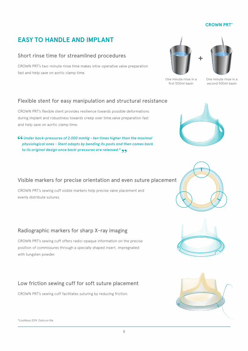
EASY TO HANDLE AND IMPLANT
*LivaNova 2014. Data on file.
One minute rinse in afirst 500ml basin
One minute rinse in a second 500ml basin
+
Under back-pressures of 2.000 mmHg - ten times higher than the maximal physiological ones - Stent adapts by bending its posts and then comes back to its original design once back-pressures are released.*
CROWN PRT’s flexible stent provides resilience towards possible deformations
during implant and robustness towards creep over time.valve preparation fast
and help save on aortic clamp time.
Flexible stent for easy manipulation and structural resistance
CROWN PRT’s sewing cuff visible markers help precise valve placement and
evenly distribute sutures.
Visible markers for precise orientation and even suture placement
CROWN PRT’s sewing cuff facilitates suturing by reducing friction.
Low friction sewing cuff for soft suture placement
CROWN PRT’s sewing cuff offers radio-opaque information on the precise
position of commissures through a specially shaped insert, impregnated
with tungsten powder.
Radiographic markers for sharp X-ray imaging
CROWN PRT’s two-minute rinse time makes intra-operative valve preparation
fast and help save on aortic clamp time.
Short rinse time for streamlined procedures
8
CROWN PRT®
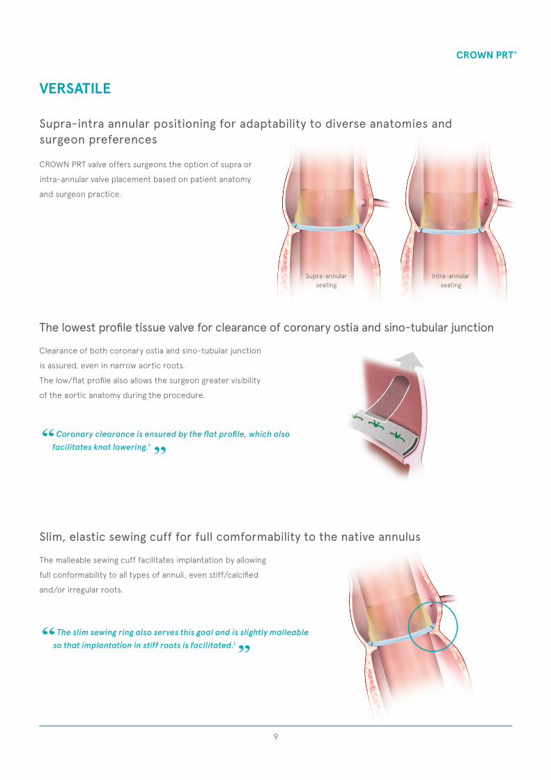
Supra-annularseating
Intra-annularseating
VERSATILE
Coronary clearance is ensured by the flat profile, which also facilitates knot lowering.1
The slim sewing ring also serves this goal and is slightly malleableso that implantation in stiff roots is facilitated.1
CROWN PRT valve offers surgeons the option of supra or
intra-annular valve placement based on patient anatomy
and surgeon practice.
Supra-intra annular positioning for adaptability to diverse anatomies andsurgeon preferences
Clearance of both coronary ostia and sino-tubular junction
is assured, even in narrow aortic roots.
The low/flat profile also allows the surgeon greater visibility
of the aortic anatomy during the procedure.
The malleable sewing cuff facilitates implantation by allowing
full conformability to all types of annuli, even stiff/calcified
and/or irregular roots.
The lowest profile tissue valve for clearance of coronary ostia and sino-tubular junction
Slim, elastic sewing cuff for full comformability to the native annulus
9
CROWN PRT®

CROWN PRT's state-of-the-art performance is the resultof LivaNova's long standing experience in heart valve design and cutting edge
science of materials.The wide open design provides optimal hemodynamics.
The patented Phospholipid Reduction Treatment (PRT) is intended to enhance durability through mitigation of calcium absorption.*
Optimal hemodynamics and proven durabilityCrowning performance
*LivaNova 2014. Data on file.
10
CROWN PRT®

DESIGNED TO OPEN WIDE
Smallest tissuevalve on the market
CROWN PRT’s single bovine pericardium layer is mounted outside the stent. This optimizes hemodynamic performance by
maximizing the flow area through a synchronous and unimpeded opening of the leaflets.2
Single bovine pericardium outer layer for maximized flow areas
By providing favorable transvalvular pressure gradients even in small aortic annuli, CROWN PRT minimizes occurrence of
patient-prosthesis mismatch and thus represents an alternative to aortic root enlargement, which is known to be associated
with an increase of surgical risk.3
Smallest tissue valve on the market as an alternative option to aortic root enlargement
11
CROWN PRT®
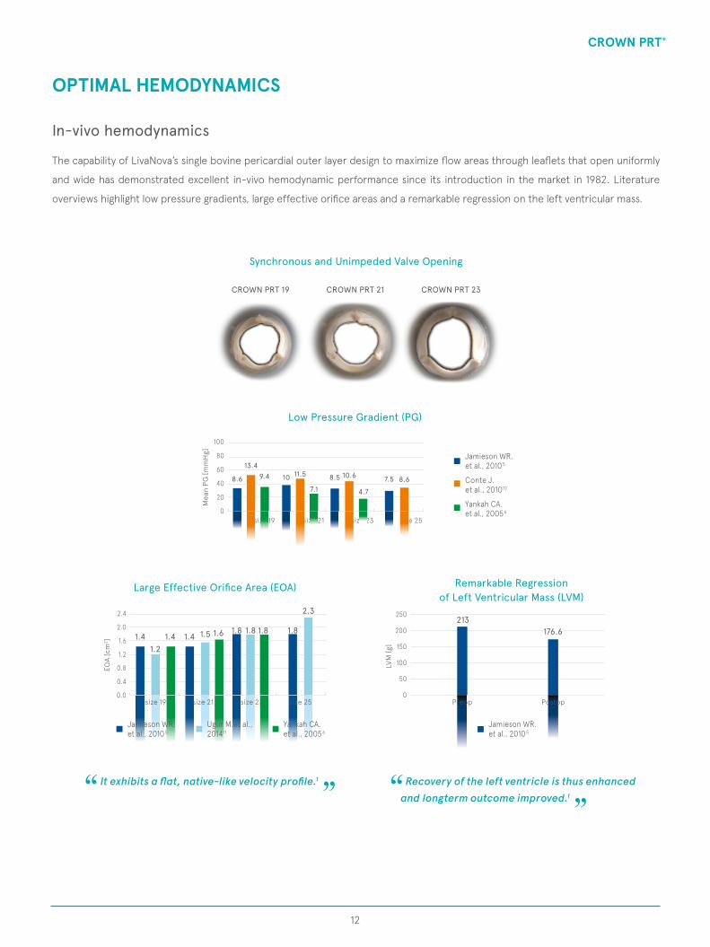
Large Effective Orifice Area (EOA)
2.4
2.0
1.6
1.2
0.8
0.4
0.0size 19 size 21 size 25size 23
1.21.41.41.4 1.5 1.6 1.8 1.81.8 1.8
2.3
EOA
[cm
2 ]
Remarkable Regressionof Left Ventricular Mass (LVM)
250
200
150
100
50
0
LVM
[g]
176.6213
Jamieson WR.et al., 2010 5
Preop Postop
OPTIMAL HEMODYNAMICS
It exhibits a flat, native-like velocity profile.1 Recovery of the left ventricle is thus enhanced and longterm outcome improved.1
Synchronous and Unimpeded Valve Opening
Low Pressure Gradient (PG)
CROWN PRT 23CROWN PRT 19 CROWN PRT 21
size 19 size 21 size 25size 23
100
80
60
40
20
0
Mea
n PG
[mm
Hg]
13.4
8.6 9.4 10 11.5 10.68.5
4.7
8.67.57.1
Jamieson WR.et al., 2010 5
Conte J.et al., 201010
Yankah CA.et al., 20056
Jamieson WR.et al., 2010 5
Ugur M.et al., 201411
Yankah CA.et al., 20056
The capability of LivaNova’s single bovine pericardial outer layer design to maximize flow areas through leaflets that open uniformly
and wide has demonstrated excellent in-vivo hemodynamic performance since its introduction in the market in 1982. Literature
overviews highlight low pressure gradients, large effective orifice areas and a remarkable regression on the left ventricular mass.
In-vivo hemodynamics
12
CROWN PRT®
OPTIMAL HEMODYNAMICS
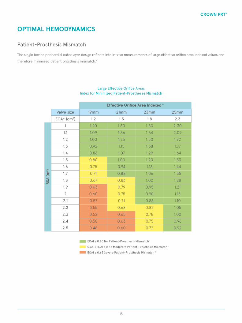
Large Effective Orifice AreasIndex for Minimized Patient-Prostheses Mismatch
EOAI ≥ 0.85 No Patient-Prosthesis Mismatch 4
0.65 < EOAI < 0.85 Moderate Patient-Prosthesis Mismatch4
EOAI ≤ 0.65 Severe Patient-Prosthesis Mismatch 4
Effective Orifice Area Indexed 11
BSA
(m2 )
1.2 1.5 1.8 2.3
19mm 21mm 23mm 25mmValve size
EOA* (cm2)
1
1.1
1.2
1.3
1.4
1.5
1.6
1.7
1.8
1.9
2
2.1
2.2
2.3
2.4
2.5
1.50
1.36
1.25
1.15
1.07
1.00
0.94
0.88
0.83
0.79
0.75
0.71
0.68
0.65
0.63
0.60
2.30
2.09
1.92
1.77
1.64
1.53
1.44
1.35
1.28
1.21
1.15
1.10
1.05
1.00
0.96
0.92
1.20
1.09
1.00
0.92
0.86
0.80
0.75
0.71
0.67
0.63
0.60
0.57
0.55
0.52
0.50
0.48
1.80
1.64
1.50
1.38
1.29
1.20
1.13
1.06
1.00
0.95
0.90
0.86
0.82
0.78
0.75
0.72
The single bovine pericardial outer layer design reflects into in-vivo measurements of large effective orifice area indexed values and
therefore minimized patient prosthesis mismatch.4
Patient-Prosthesis Mismatch
13
CROWN PRT®

The presence By optimally distributing stress on the commissures, pressure forces on the valve posts are relieved
during the cardiac cycle.
DESIGNED AND TREATED TO LAST
Cushioned leaflet contact reduces pericardium wear, enhancing valve durability.
One-seam knit polyester for smooth contact surface between pericardium andsynthetic material
Cross-stitch pattern for even stress distribution on commissures
14
CROWN PRT®

Ca Calcium
Phospholipid
Ca Calcium
Phospholipid
The PRT process removes phospholipids using Octanediol, a long chain alcohol that possesses a lipid-soluble tail to aid
its solubility in phospholipids’s heads and a water-soluble head to allow removal by rinsing pericardial layers before valve
manufacturing.
Lipid-soluble tail Water-soluble
head
Phospholipid
Octanediol molecule
Octanediol molecule
PhospholipidPhospholipidThe Octanediol’s lipid-
soluble tail interacts with the phospholipid’s head
An untreated pericardial layer is going to be treated
using Octanediol molecules
The Octanediol’s water-soluble head makes the phospholipid soluble, namely removable
by rinsing pericardium layersbefore manufacturing
The presence of phospholipids in the pericardial tissue play a key
role in the calcification process of bioprostheses as their phosphate
heads are potential binding sites for circulating calcium ions.
Phospholipid Reduction Treatment for mitigated calcium absorption
DESIGNED AND TREATED TO LAST
*LivaNova 2014. Data on file.
Crown PRT features LivaNova’s patented Phospholipid Reduction
Treatment (PRT) which has proved to decrease phospholipid content
in pericardial tissue leading to a remarkable reduction of calcium
uptake compared to control tissue.*
15
CROWN PRT®

Conte et al., 2010Asch et al., 2012Joshi et al., 2014Jamieson WR. et al., 20097
Lootens, RE0210235/BWilbring M. et al., 20133
Piccardo et al., 2015 12
ISTHMUS Investigators., 20118
Yankah CA. et al., 20089
15 yrs 18 yrs 20 yrs
62.3
85.395
Years after operation
5 yrs
85.692.597.8
89.3
3 yrs
99.2
11 yrs
100100
80
60
40
20
0
%
Freedom from structural valve degeneration at endpoint During the whole follow-up, which ranged up to 11 years, we recorded no reoperation because of failed prosthesis.3
10 yrs
PROVEN DURABILITY
*LivaNova 2014. Data on file.
Freedom from structural valve degeneration (as per ISO 5840)
CROWN PRT 19mm
CROWN PRT 23mm
CROWN PRT 27mm
100
80
60
40
20
05Years Equivalent
200 600 1.000 1.5001.320 1.600 1.700400 800Milion Cycles:2515 37.510 3320 40 42.5
%
The Phospholipid Reduction Treatment (PRT) process is intended to enhance already proven resilient durability by reducing calcium absorption up to 97% compared to control.*
27.57
0.72
30,00
20,00
10,00
0,00
µgC
a/m
g D
W
CROWN PRTUntreated
No failures up to 1.5 billion cycles,equivalent to 37.5 years. Over 6 timesthe minimum requirement.*
CROWN PRT’s unique design shows outstanding resistance during accelerated wear testing at ISO 5840 conditions.
In-vitro durability
LivaNova’s single bovine pericardial outer layer design has over 21 years of published clinical data proving already excellent durability
without anti-calcification treatment.
In-vivo durability
By reducing the nucleation sites for calcium deposition - the phospholipids in the pericardial tissue - the Phospholipid Reduction
Treatment (PRT) addresses directly the origin of tissue calcification which may lead to an enhanced valve durability.
In-animal calcium absorption mitigation testing
Tests in subcutaneous rat implants at 60 days demonstrate a significant reduction of calcium content in PRT-treated bovine
pericardium patches compared to control.
16
CROWN PRT®
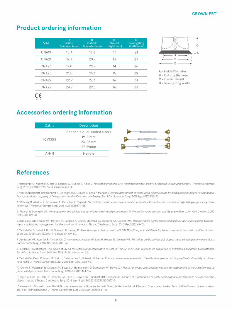
Product ordering information
Accessories ordering information
Cat. #
ICV1353
AH-11
Bendable dual-ended sizers
Handle
19-21mm23-25mm27-29mm
Description
A = Inside DiameterB = Outside DiameterC = Overall HeightD = Sewing Ring Width
ABD
C
17
CNA19
CNA21
CNA23
CNA25
CNA27
CNA29
18.6
20.7
22.7
25.1
27.3
29.5
15.4
17.3
19.0
21.0
22.9
24.7
21
23
26
29
31
33
11
13
14
15
16
16
SizeA B C D
InsideDiameter (mm)
OverallHeight (mm)
OutsideDiameter (mm)
Sewing RingWidth (mm)
CROWN PRT®
References1. Hartrumpf M, Kuehnel R, Erb M, Loladze G, Mueller T, Albes J. Favorable gradients with the mitroflow aortic valve prosthesis in everyday surgery. Thorac Cardiovasc Surg. 2012 Jul;60(5):326-33; discussion 333-4.
2. von Knobelsdorff-Brenkenhoff F, Dieringer MA, Greiser A, Schulz-Menger J. In vitro assessment of heart valve bioprostheses by cardiovascular magnetic resonance: four-dimensional mapping of flow patterns and orifice area planimetry. Eur J Cardiothorac Surg. 2011 Sep;40(3):736-42.
3. Wilbring M, Alexiou K, Schumann E, Matschke K, Tugtekin SM. Isolated aortic valve replacement in patients with small aortic annulus-a high-risk group on long-term follow-up. Thorac Cardiovasc Surg. 2013 Aug;61(5):379-85.
4. Pibarot P, Dumesnil JG. Hemodynamic and clinical impact of prosthesis-patient mismatch in the aortic valve position and its prevention. J Am Coll Cardiol. 2000 Oct;36(4):1131-41.
5. Jamieson WR, Forgie WR, Hayden RI, Langlois Y, Ling H, Stanford EA, Roberts KA, Dolman WB. Hemodynamic performance of mitroflow aortic pericardial biopros-thesis - optimizing management for the small aortic annulus. Thorac Cardiovasc Surg. 2010 Mar;58(2):69-75.
6. Yankah CA, Schubel J, Buz S, Siniawski H, Hetzer R. Seventeen-year clinical results of 1,037 Mitroflow pericardial heart valve prostheses in the aortic position. J Heart Valve Dis. 2005 Mar;14(2):172-9; discussion 179-80.
7. Jamieson WR, Koerfer R, Yankah CA, Zittermann A, Hayden RI, Ling H, Hetzer R, Dolman WB. Mitroflow aortic pericardial bioprosthesis clinical performance. Eur J Cardiothorac Surg. 2009 Nov;36(5):818-24.
8. ISTHMUS Investigators. The Italian study on the Mitroflow postoperative results (ISTHMUS): a 20-year, multicentre evaluation of Mitroflow pericardial bioprosthesis. Eur J Cardiothorac Surg. 2011 Jan;39(1):18-26; discussion 26.
9. Yankah CA, Pasic M, Musci M, Stein J, Detschades C, Siniawski H, Hetzer R. Aortic valve replacement with the Mitroflow pericardial bioprosthesis: durability results up to 21 years. J Thorac Cardiovasc Surg. 2008 Sep;136(3):688-96.
10. Conte J, Weissman N, Dearani JA, Bavaria J, Heimansohn D, Dembitsky W, Doyle D. A North American, prospective, multicenter assessment of the Mitroflow aortic pericardial prosthesis. Ann Thorac Surg. 2010 Jul;90(1):144-152.
11. Ugur M, Suri RM, Daly RC, Dearani JA, Park SJ, Joyce LD, Burkhart HM, Greason KL, Schaff HV. Comparison of early hemodynamic performance of 3 aortic valve bioprostheses. J Thorac Cardiovasc Surg. 2014 Jan 15. pii: S0022-5223(14)00027-0.
12. Alessandro Piccardo, Jean David Blossier, Alexandre Le Guyader, Isabelle Orsel, Seiffedine Sekkal, Elisabeth Cornu, Marc Laskar. Fate of Mitroflow aortic bioprosthe-ses: a 18-year experience. J Thorac Cardiovasc Surg 2016 Mar;151(3):754-761.

© 2018 LivaNova all rights reserved.
Please always refer to the Instructions For Use (IFU) manual provided with each product for detailed information, warnings, precautions and possible adverse side effects.
03/1
8 RE
0210
200/
FTh
is b
roch
ure
is n
ot in
tend
ed fo
r dis
trib
utio
n in
USA
and
Can
ada.
Sorin Group Italia SrlA wholly-owned subsidiary of LivaNova PLC Via Crescentino - 13040 Saluggia (VC) ItalyТеl: +39 0161 487472 - Fax: +39 0161 [email protected]
Manufactured by:LivaNova Canada Corp.Heart Valves Manufacturing Operations5005 North Fraser WayBurnaby BC V5J5M1 - CanadaTel: 1-604-412-5650