The Critical Role of PTEN/PI3K/AKT Signaling Pathway in ...
13
Cell Physiol Biochem 2018;47:981-993 981 Cellular Physiology and Biochemistry Cellular Physiology and Biochemistry Original Paper Accepted: April 17, 2018 This article is licensed under the Creative Commons Attribution-NonCommercial-NoDerivatives 4.0 Interna- tional License (CC BY-NC-ND) (http://www.karger.com/Services/OpenAccessLicense). Usage and distribution for commercial purposes as well as any distribution of modified material requires written permission. DOI: 10.1159/000490142 Published online: May 30, 2018 © 2018 The Author(s) Published by S. Karger AG, Basel www.karger.com/cpb The Critical Role of PTEN/PI3K/AKT Signaling Pathway in Shikonin-Induced Apoptosis and Proliferation Inhibition of Chronic Myeloid Leukemia Yu Chen a,b Tongtong Wang b Jing Du d Yanchun Li e Xin Wang b Yi Zhou b XingXing Yu f Weimin Fan g Qiaojuan Zhu b Xiangmin Tong a,b Ying Wang a,b,c a Laboratory Medical College, Hangzhou Medical College, Hangzhou, Zhejiang, b Clinical Research Institute, Zhejiang Provincial People’s Hospital, People’s Hospital of Hangzhou Medical College, Hangzhou, c Department of Blood Transfusion Medicine, Zhejiang Provincial People’s Hospital, People’s Hospital of Hangzhou Medical College, Hangzhou, d Department of Laboratory Medicine, Zhejiang Provincial People’s Hospital, People’s Hospital of Hangzhou Medical College, Hangzhou, e Department of Pathology, Tongde Hospital of Zhejiang Province, Hangzhou, f Graduate Department, BengBu Medical College, BengBu, g Department of Second Clinical Medical College, Zhejiang Chinese Medical University, Hangzhou, China. Key Words Shikonin • Chronic myeloid leukemia • Proliferation • Apoptosis • PTEN Abstract Background/Aims: Chronic myeloid leukemia (CML) is a myeloproliferative neoplasm. Tyrosine kinase inhibitors (TKIs) are commonly used to treat CML; however, drug resistance of CML cells to TKIs has limited their clinical application. Shikonin, a traditional Chinese herb, has long been used to treat leukemia in China, but the roles and related molecular mechanisms of shikonin treatment in CML remain unclear. Here, we aimed to evaluate the effects of shikonin on the proliferation, apoptosis, and migration of K562 cells, a CML cell line. Methods: Firstly, K562 cell proliferation and apoptosis were tested by CCK8 assay and flow cytometry with Annexin V-FITC/PI staining. Cell migration was measured by Transwell migration assay. In addition, western blot was performed to determine the proteins (PI3K, Bax, Bcl-2, cleaved caspase-3, PTEN, p-AKT, AKT, CXCR4, SDF-1, CD44) involved in the mechanism of action of shikonin. Finally, neutrophils from peripheral blood of CML patients were obtained, and cell proliferation and apoptosis were tested by CCK8 assay and flow cytometry. Results: Shikonin reduced the proliferation of K562 cells in a time- and dose-dependent manner and promoted the apoptosis of K562 cells. Moreover, shikonin increased the PTEN level and inactivated the PI3K/AKT signaling pathway, subsequently upregulating BAX in K562 cells. In addition, shikonin could block K562 cell migration via the CXCR4/SDF-1 axis. Finally, shikonin significantly inhibited the proliferation and promoted the apoptosis of neutrophils from CML Ying Wang and Xiangmin Tong Laboratory Medical College, Hangzhou Medical College, Hangzhou Zhejiang, 310053, (China) E-Mail [email protected] / [email protected]
Transcript of The Critical Role of PTEN/PI3K/AKT Signaling Pathway in ...
Cell Physiol Biochem 2018;47:981-993DOI: 10.1159/000490142Published online: May 30, 2018 981
Cellular Physiology and Biochemistry
Cellular Physiology and Biochemistry
© 2018 The Author(s). Published by S. Karger AG, Baselwww.karger.com/cpb
Chen et al.: Mechanisms Underlying the Effect of Shikonin on K562 Cells
Original Paper
Accepted: April 17, 2018
This article is licensed under the Creative Commons Attribution-NonCommercial-NoDerivatives 4.0 Interna-tional License (CC BY-NC-ND) (http://www.karger.com/Services/OpenAccessLicense). Usage and distribution for commercial purposes as well as any distribution of modified material requires written permission.
DOI: 10.1159/000490142Published online: May 30, 2018
© 2018 The Author(s) Published by S. Karger AG, Baselwww.karger.com/cpb
The Critical Role of PTEN/PI3K/AKT Signaling Pathway in Shikonin-Induced Apoptosis and Proliferation Inhibition of Chronic Myeloid LeukemiaYu Chena,b Tongtong Wangb Jing Dud Yanchun Lie Xin Wangb Yi Zhoub XingXing Yuf Weimin Fang Qiaojuan Zhub Xiangmin Tonga,b Ying Wanga,b,c
aLaboratory Medical College, Hangzhou Medical College, Hangzhou, Zhejiang, bClinical Research Institute, Zhejiang Provincial People’s Hospital, People’s Hospital of Hangzhou Medical College, Hangzhou, cDepartment of Blood Transfusion Medicine, Zhejiang Provincial People’s Hospital, People’s Hospital of Hangzhou Medical College, Hangzhou, dDepartment of Laboratory Medicine, Zhejiang Provincial People’s Hospital, People’s Hospital of Hangzhou Medical College, Hangzhou, eDepartment of Pathology, Tongde Hospital of Zhejiang Province, Hangzhou, fGraduate Department, BengBu Medical College, BengBu, gDepartment of Second Clinical Medical College, Zhejiang Chinese Medical University, Hangzhou, China.
Key WordsShikonin • Chronic myeloid leukemia • Proliferation • Apoptosis • PTEN
AbstractBackground/Aims: Chronic myeloid leukemia (CML) is a myeloproliferative neoplasm. Tyrosine kinase inhibitors (TKIs) are commonly used to treat CML; however, drug resistance of CML cells to TKIs has limited their clinical application. Shikonin, a traditional Chinese herb, has long been used to treat leukemia in China, but the roles and related molecular mechanisms of shikonin treatment in CML remain unclear. Here, we aimed to evaluate the effects of shikonin on the proliferation, apoptosis, and migration of K562 cells, a CML cell line. Methods: Firstly, K562 cell proliferation and apoptosis were tested by CCK8 assay and flow cytometry with Annexin V-FITC/PI staining. Cell migration was measured by Transwell migration assay. In addition, western blot was performed to determine the proteins (PI3K, Bax, Bcl-2, cleaved caspase-3, PTEN, p-AKT, AKT, CXCR4, SDF-1, CD44) involved in the mechanism of action of shikonin. Finally, neutrophils from peripheral blood of CML patients were obtained, and cell proliferation and apoptosis were tested by CCK8 assay and flow cytometry. Results: Shikonin reduced the proliferation of K562 cells in a time- and dose-dependent manner and promoted the apoptosis of K562 cells. Moreover, shikonin increased the PTEN level and inactivated the PI3K/AKT signaling pathway, subsequently upregulating BAX in K562 cells. In addition, shikonin could block K562 cell migration via the CXCR4/SDF-1 axis. Finally, shikonin significantly inhibited the proliferation and promoted the apoptosis of neutrophils from CML
Ying Wang and Xiangmin Tong
Laboratory Medical College, Hangzhou Medical College, HangzhouZhejiang, 310053, (China)E-Mail [email protected] / [email protected]
Cell Physiol Biochem 2018;47:981-993DOI: 10.1159/000490142Published online: May 30, 2018 982
Cellular Physiology and Biochemistry
Cellular Physiology and Biochemistry
© 2018 The Author(s). Published by S. Karger AG, Baselwww.karger.com/cpb
Chen et al.: Mechanisms Underlying the Effect of Shikonin on K562 Cells
patients. Conclusion: These results demonstrated that shikonin inhibits CML proliferation and migration and induces apoptosis by the PTEN/PI3K/AKT pathway, revealing the effects of shikonin therapy on CML.
Introduction
Chronic myeloid leukemia (CML) is a myeloproliferative neoplasm induced by the BCR-ABL1 (breakpoint cluster region-Abelson murine leukemia viral oncogene homolog 1) fusion gene with an incidence of 1–2 cases per 100, 000 people and accounts for 15% of newly diagnosed cases of leukemia in adults [1]. Tyrosine kinase inhibitors (TKIs) that target BCR-ABL1 are the standard treatment for CML. Although TKIs are extensively used to treat CML patients, serious drug–drug interactions and emerging resistance of CML cells to TKIs through multiple mechanisms have limited their clinical application [2]. Hence, new therapeutic strategies with less toxicity and higher efficacy are urgently required, especially for relapsing and chemo-resistant CML patients.
Lithospermum erythrorhizon, referred to as “Zicao” in Chinese, is a plant that is used as a “heat-clearing and blood-cooling” medicine. The extracts of Zicao root have been used in traditional Chinese medicine as a cancer treatment for many years [3]. Shikonin (C16H16O5) is a naturally occurring naphthoquinone compound in the root of herb Lithospermum erythrorhizon and shows pleiotropic effects on some diseases, including anti-tumor, anti-ischemic, anti-inflammatory, and anti-bacterial effects as well as acceleration of tissue granulation proliferation [4]. Shikonin also inhibited cells viability and induced apoptosis of Cholangiocarcinoma cells, effects enhanced by TRAIL treatment via ROS mediated JNK signalling pathways [5]. Previous reports suggested that shikonin could induce oxidative activities and apoptotic effects in HL-60 cells [6]. In addition, shikonin could exert anti-leukemic activity through inhibiting the c-MYC and related ERK/JNK/MAPK pathways in U937 [7]. Moreover, shikonin, as a generator of ROS, could induce oxidative injury in apoptotic signaling cascades and may be a promising agent for the treatment of CML [8].
Phosphatases and tensin homolog (PTEN) plays an important role in mature organisms as a tumor suppressor. PTEN gene inactivation by mutation or deletion is common in pediatric T-cell acute lymphoblastic leukemia (T-ALL) [9]. The major substrate of PTEN is phosphatidylinositol-3, 4, 5-triphosphate (PIP3), which is produced by the action of phosphoinositide-3-kinase (PI3K) [10]. The PI3K/AKT signaling pathway plays an important role in the development of resistance to carcinoma therapy, and inhibition of the PI3K/AKT signaling pathway may suppress cancer cell growth and induce apoptosis in various cancers [11-13]. PTEN is the main negative regulator of the PI3K/AKT signaling pathway and is implicated in regulating proliferation and survival of T-cell progenitors [14].
Although the efficacy of shikonin has been reported, its precise anti-CML role and underlying molecular mechanism remain to be elucidated. Here, we evaluated the therapeutic efficacy of shikonin in K562 cells (CML cell line) and neutrophils from CML patients. Understanding the efficacy and mode of action of shikonin will provide a foundation for its potential use in clinical settings.
Materials and Methods
ReagentsShikonin was purchased from Sigma Chemical Company (St. Louis, MO, USA) and dissolved in
dimethyl sulfoxide (DMSO, Sigma-Aldrich, St. Louis, MO, USA). Potassium bisperoxo (1, 10-phenanthroline) oxovanadate (BPV) (Sigma-Aldrich), a PTEN inhibitor, was used at 1 µM in DMSO. Wortmanin (Sigma-Aldrich), a specific PI3K inhibitor, was used at 50 nM in DMSO.
Cell culture and primary samplesThe leukemia cell lines K562 (chronic myelocytic leukemia), KU812 (basophilic leukemic cells in
human peripheral blood), and MEG-01 (human megakaryocyte leukemia cells) were grown in RPMI
© 2018 The Author(s)Published by S. Karger AG, Basel
Cell Physiol Biochem 2018;47:981-993DOI: 10.1159/000490142Published online: May 30, 2018 983
Cellular Physiology and Biochemistry
Cellular Physiology and Biochemistry
© 2018 The Author(s). Published by S. Karger AG, Baselwww.karger.com/cpb
Chen et al.: Mechanisms Underlying the Effect of Shikonin on K562 Cells
1640 supplemented with 10% heat-inactivated fetal bovine serum (FBS) and treated with shikonin at the indicated concentrations.
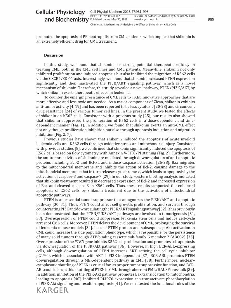
Neutrophils from peripheral blood of CML patients were obtained with a human peripheral blood neutrophil separation solution kit (Haoyang, Tianjin, China) as follows: 5 mL of neutrophil separation solution I was added to a 15-mL conical tube. Human peripheral blood was overlaid on the neutrophil separation solution I and centrifuged continuously for 30 min at 500 g at 25 °C. After centrifugation, two cell layers appeared in the centrifuge tube; the upper-layer cells were mononuclear cells, and the lower-layer cells were neutrophils. The neutrophils were collected, identified with a blood film (Fig. 8I, 8J), grown in RPMI 1640 supplemented with 20% FBS, and treated with shikonin. Control cells were treated with 0.1% DMSO.
Cell viability assayCell viability was assessed by the CCK8 colorimetric assay (Beyotime, Hangzhou, China) as previously
described [15]. Briefly, cells were plated in 96-well plates (2×10³ cells/well) and incubated at 37 °C with 5% CO2 for 24 h, 48 h, and 72 h, and the absorbance was recorded at 450 nm using a micro-plate reader (BioTek, Winooski, VT, USA). The results were presented as viability rates. Cell viability was calculated according to the following formula: Cell viability (%) = (OD treatment–OD blank)/(OD control–OD blank).
Apoptosis assayCell apoptosis was detected using the Annexin V-FITC/PI Apoptosis Detection Kit (Beyotime, Haimen,
China) following the manufacturer’s instructions after drug treatment. Briefly, cells at a density of 5×10⁵ cells/mL were resuspended and mixed in 500 μL binding buffer with 5 μL Annexin V-FITC and 5 μL propidium iodide (PI). After incubation for 15 min, cell apoptosis was detected by flow cytometry (NovoCyte, ACEA Biosciences, San Diego, CA, USA).
Western blotCells were lysed with RIPA lysis buffer (Beyotime) to extract the total protein. Then, the concentration
of total protein was quantitated by a bicinchoninic acid (BCA) protein assay kit (Beyotime). Equal amounts of protein were separated by SDS-polyacrylamide gel electrophoresis (PAGE) and transferred onto polyvinylidene fluoride (PVDF) membranes (Millipore, Bedford, MA, USA). After being blocked with 5% non-fat milk for 1 h, the membranes were probed with specific primary antibodies against PI3K, Bax, Bcl-2, cleaved caspase-3, PTEN, p-AKT, AKT, CXCR4, SDF-1, CD44, GAPDH, and actin (1:1, 000, Cell Signaling Technology, Danvers, MA, USA) at 4 °C overnight and subsequently incubated with the corresponding secondary antibodies (1:5, 000, Beyotime) for 1 h at room temperature. An enhanced chemiluminescence (ECL) solution (Qihai Biotec, Shanghai, China) was used to visualize the target bands, and the Gel-Pro Analyzer software (Media Cybernetics, Bethesda, MD, USA) was employed to measure relative band intensities. GAPDH or actin served as an internal control, respectively.
Reverse transcription-PCR (RT-PCR) assayK562 cells were cultured with shikonin at 10, 20, or 40 μM for 24 h, while the control cells were
treated with 0.1% DMSO. After shikonin incubation, the mRNA from K562 cells was extracted (MagnaPure LC RNA Isolation Kit; Roche Applied Science, Penzberg, Germany) and reverse transcribed into cDNA with the Transcription High Fidelity cDNA Synthesis Kit (Roche Applied Science) following the corresponding manufacturer’s instructions. With template DNA prepared, the parameters of amplification reaction were as follows: 3 min at 95 °C, followed by 40 cycles for 10 sec at 95 °C, and 30 sec at 60 °C. The primer pairs used are listed in Table 1.
Transwell migration assayCell migration was determined
using the Transwell migration assay (Corning Life Sciences, Acton, MA, USA), according to the manufacturer’s instructions and as previously described [16]. Briefly, K562 cells
Table 1. Primer sequence of PTEN, PI3K, Akt, GAPDH
Gene Forward Reverse PTEN CAGAGCGAGGGGCATCAG GCAGGAAATCCCATAGCAATAA PI3K ATGGGGATGATTTACGGC TCTCCTTTGTTCTTGTCTTTGA Akt TGAGCGACGTGGCTATTG CAGTCTGGATGGCGGTT GAPDH CATCAATGGAAATCCCATCA GACTCCACGACGTACTCAGC
Cell Physiol Biochem 2018;47:981-993DOI: 10.1159/000490142Published online: May 30, 2018 984
Cellular Physiology and Biochemistry
Cellular Physiology and Biochemistry
© 2018 The Author(s). Published by S. Karger AG, Baselwww.karger.com/cpb
Chen et al.: Mechanisms Underlying the Effect of Shikonin on K562 Cells
were treated with shikonin (0, 10, 20, or 40 μM) for 12 h prior to the migration assay. K562 cells without FBS were added to the upper chamber (1×10⁵ cells/well) and allowed to migrate for 24 h at 37 °C toward the lower chamber, which contained RPMI 1640 with 10% FBS. The cells that migrated to the lower chambers were counted by cytometry with Countstar IC1000 (Inno-Alliance Biotech, Wilmington, DE, USA).
StatisticsStatistical analysis was performed using GraphPad Prism v. 5.0 software (GraphPad Software, Inc., La
Jolla, CA, USA). Data are expressed as means ± standard deviation (SD). All data presented represent results from at least three independent experiments. One-way analysis of variance followed by Tukey’s multiple comparison test was used to compare differences between groups. Statistical significance was defined as P < 0.05.
Results
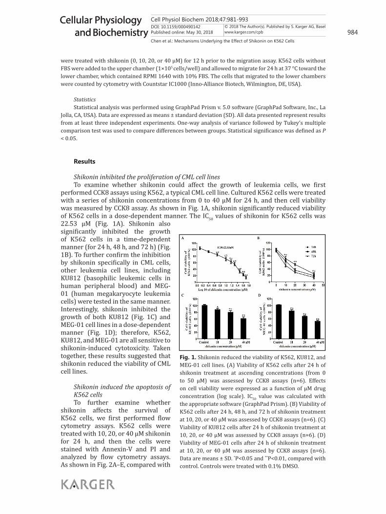
Shikonin inhibited the proliferation of CML cell linesTo examine whether shikonin could affect the growth of leukemia cells, we first
performed CCK8 assays using K562, a typical CML cell line. Cultured K562 cells were treated with a series of shikonin concentrations from 0 to 40 μM for 24 h, and then cell viability was measured by CCK8 assay. As shown in Fig. 1A, shikonin significantly reduced viability of K562 cells in a dose-dependent manner. The IC50 values of shikonin for K562 cells was 22.53 μM (Fig. 1A). Shikonin also significantly inhibited the growth of K562 cells in a time-dependent manner (for 24 h, 48 h, and 72 h) (Fig. 1B). To further confirm the inhibition by shikonin specifically in CML cells, other leukemia cell lines, including KU812 (basophilic leukemic cells in human peripheral blood) and MEG-01 (human megakaryocyte leukemia cells) were tested in the same manner. Interestingly, shikonin inhibited the growth of both KU812 (Fig. 1C) and MEG-01 cell lines in a dose-dependent manner (Fig. 1D); therefore, K562, KU812, and MEG-01 are all sensitive to shikonin-induced cytotoxicity. Taken together, these results suggested that shikonin reduced the viability of CML cell lines.
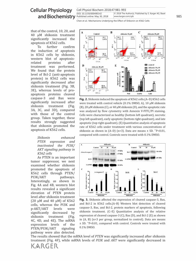
Shikonin induced the apoptosis of K562 cellsTo further examine whether
shikonin affects the survival of K562 cells, we first performed flow cytometry assays. K562 cells were treated with 10, 20, or 40 μM shikonin for 24 h, and then the cells were stained with Annexin-V and PI and analyzed by flow cytometry assays. As shown in Fig. 2A–E, compared with
Figure 1
Figure 2
Fig. 1. Shikonin reduced the viability of K562, KU812, and MEG-01 cell lines. (A) Viability of K562 cells after 24 h of shikonin treatment at ascending concentrations (from 0 to 50 μM) was assessed by CCK8 assays (n=6). Effects on cell viability were expressed as a function of μM drug concentration (log scale). IC50 value was calculated with the appropriate software (GraphPad Prism). (B) Viability of K562 cells after 24 h, 48 h, and 72 h of shikonin treatment at 10, 20, or 40 μM was assessed by CCK8 assays (n=6). (C) Viability of KU812 cells after 24 h of shikonin treatment at 10, 20, or 40 μM was assessed by CCK8 assays (n=6). (D) Viability of MEG-01 cells after 24 h of shikonin treatment at 10, 20, or 40 μM was assessed by CCK8 assays (n=6). Data are means ± SD. *P<0.05 and **P<0.01, compared with control. Controls were treated with 0.1% DMSO.
Cell Physiol Biochem 2018;47:981-993DOI: 10.1159/000490142Published online: May 30, 2018 985
Cellular Physiology and Biochemistry
Cellular Physiology and Biochemistry
© 2018 The Author(s). Published by S. Karger AG, Baselwww.karger.com/cpb
Chen et al.: Mechanisms Underlying the Effect of Shikonin on K562 Cells
that of the control, 10, 20, and 40 μM shikonin treatment significantly increased the apoptosis of K562 cells.
To further confirm the induction of apoptosis in K562 cells by shikonin, western blot of apoptosis-related proteins after treatment was performed. We found that the protein level of Bcl-2 (anti-apoptosis protein) in K562 cells was significantly decreased after shikonin treatment (Fig. 3B, 3E), whereas levels of pro-apoptosis proteins cleaved caspase-3 and Bax were significantly increased after shikonin treatment (Fig. 3A, 3C, and 3D), compared with those of the control group. Taken together, these results strongly suggested that shikonin promoted the apoptosis of K562 cells.
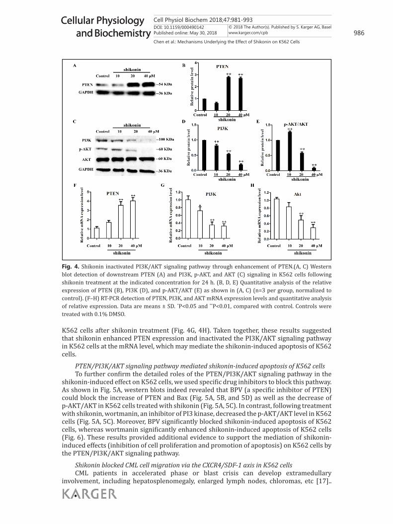
Shikonin enhanced PTEN expression and inactivated the PI3K/AKT signaling pathway in K562 cellsAs PTEN is an important
tumor suppressor, we next examined whether shikonin promoted the apoptosis of K562 cells through PTEN/PI3K/AKT pathways. Interestingly, as shown in Fig. 4A and 4B, western blot results revealed a significant elevation of PTEN protein level after shikonin treatment (20 μM and 40 μM) of K562 cells, whereas the PI3K and p-AKT/AKT levels were significantly decreased by shikonin treatment (Fig. 4C, 4D, and 4E). The mRNA expression levels of the PTEN/PI3K/AKT signaling pathway were also detected. The results showed that the mRNA level of PTEN was significantly increased after shikonin treatment (Fig. 4F), while mRNA levels of PI3K and AKT were significantly decreased in
Fig. 3. Shikonin affected the expression of cleaved caspase-3, Bax, and Bcl-2 in K562 cells.(A–B) Western blot detection of cleaved caspase-3, Bax, and Bcl-2, protein markers of apoptosis, following shikonin treatment. (C–E) Quantitative analysis of the relative expression of cleaved caspase-3 (C), Bax (D), and Bcl-2 (E) as shown in (A, B) (n=3 per group, normalized to control). Data are means ± SD. **P<0.01, compared with control. Controls were treated with 0.1% DMSO.
Figure 3
Figure 1
Figure 2
Fig. 2. Shikonin induced the apoptosis of K562 cells.(A–D) K562 cells were treated with control vehicle (0.1% DMSO, A), 10 μM shikonin (B), 20 μM shikonin (C), or 40 μM shikonin (D), and the apoptotic rate was analyzed by flow cytometry with Annexin V-FITC/PI staining. Cells were characterized as healthy (bottom left quadrant), necrotic (top left quadrant), early apoptotic (bottom right quadrant), and late apoptotic (top right quadrant). (E) Quantitative analysis of apoptosis rate of K562 cells under treatment with various concentrations of shikonin as shown in (A–D) (n=3). Data are means ± SD. **P<0.01, compared with control. Controls were treated with 0.1% DMSO.
Cell Physiol Biochem 2018;47:981-993DOI: 10.1159/000490142Published online: May 30, 2018 986
Cellular Physiology and Biochemistry
Cellular Physiology and Biochemistry
© 2018 The Author(s). Published by S. Karger AG, Baselwww.karger.com/cpb
Chen et al.: Mechanisms Underlying the Effect of Shikonin on K562 Cells
K562 cells after shikonin treatment (Fig. 4G, 4H). Taken together, these results suggested that shikonin enhanced PTEN expression and inactivated the PI3K/AKT signaling pathway in K562 cells at the mRNA level, which may mediate the shikonin-induced apoptosis of K562 cells.
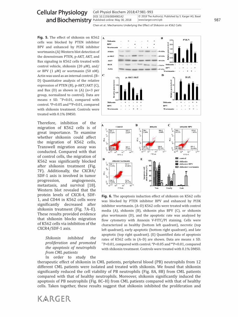
PTEN/PI3K/AKT signaling pathway mediated shikonin-induced apoptosis of K562 cellsTo further confirm the detailed roles of the PTEN/PI3K/AKT signaling pathway in the
shikonin-induced effect on K562 cells, we used specific drug inhibitors to block this pathway. As shown in Fig. 5A, western blots indeed revealed that BPV (a specific inhibitor of PTEN) could block the increase of PTEN and Bax (Fig. 5A, 5B, and 5D) as well as the decrease of p-AKT/AKT in K562 cells treated with shikonin (Fig. 5A, 5C). In contrast, following treatment with shikonin, wortmanin, an inhibitor of PI3 kinase, decreased the p-AKT/AKT level in K562 cells (Fig. 5A, 5C). Moreover, BPV significantly blocked shikonin-induced apoptosis of K562 cells, whereas wortmanin significantly enhanced shikonin-induced apoptosis of K562 cells (Fig. 6). These results provided additional evidence to support the mediation of shikonin-induced effects (inhibition of cell proliferation and promotion of apoptosis) on K562 cells by the PTEN/PI3K/AKT signaling pathway.
Shikonin blocked CML cell migration via the CXCR4/SDF-1 axis in K562 cellsCML patients in accelerated phase or blast crisis can develop extramedullary
involvement, including hepatosplenomegaly, enlarged lymph nodes, chloromas, etc [17]..
Fig. 4. Shikonin inactivated PI3K/AKT signaling pathway through enhancement of PTEN.(A, C) Western blot detection of downstream PTEN (A) and PI3K, p-AKT, and AKT (C) signaling in K562 cells following shikonin treatment at the indicated concentration for 24 h. (B, D, E) Quantitative analysis of the relative expression of PTEN (B), PI3K (D), and p-AKT/AKT (E) as shown in (A, C) (n=3 per group, normalized to control). (F–H) RT-PCR detection of PTEN, PI3K, and AKT mRNA expression levels and quantitative analysis of relative expression. Data are means ± SD. *P<0.05 and **P<0.01, compared with control. Controls were treated with 0.1% DMSO.
Figure 3
Figure 4:
Cell Physiol Biochem 2018;47:981-993DOI: 10.1159/000490142Published online: May 30, 2018 987
Cellular Physiology and Biochemistry
Cellular Physiology and Biochemistry
© 2018 The Author(s). Published by S. Karger AG, Baselwww.karger.com/cpb
Chen et al.: Mechanisms Underlying the Effect of Shikonin on K562 Cells
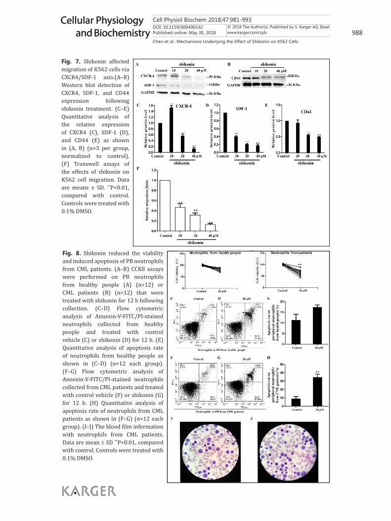
Therefore, inhibition of the migration of K562 cells is of great importance. To examine whether shikonin could affect the migration of K562 cells, Transwell migration assay was conducted. Compared with that of control cells, the migration of K562 was significantly blocked after shikonin treatment (Fig. 7F). Additionally, the CXCR4/SDF-1 axis is involved in tumor progression, angiogenesis, metastasis, and survival [18]. Western blot revealed that the protein levels of CXCR-4, SDF-1, and CD44 in K562 cells were significantly decreased after shikonin treatment (Fig. 7A–E). These results provided evidence that shikonin blocks migration of K562 cells via inhibition of the CXCR4/SDF-1 axis.
Shikonin inhibited the proliferation and promoted the apoptosis of neutrophils from CML patientsIn order to study the
therapeutic effect of shikonin in CML patients, peripheral blood (PB) neutrophils from 12 different CML patients were isolated and treated with shikonin. We found that shikonin significantly reduced the cell viability of PB neutrophils (Fig. 8A, 8B) from CML patients compared with that of healthy neutrophils. Moreover, shikonin significantly induced the apoptosis of PB neutrophils (Fig. 8C–H) from CML patients compared with that of healthy cells. Taken together, these results suggest that shikonin inhibited the proliferation and
Fig. 5. The effect of shikonin on K562 cells was blocked by PTEN inhibitor BPV and enhanced by PI3K inhibitor wortmanin.(A) Western blot detection of the downstream PTEN, p-AKT, AKT, and Bax signaling in K562 cells treated with control vehicle, shikonin (20 μM), and/or BPV (1 μM) or wortmanin (50 nM). Actin was used as an internal control. (B–D) Quantitative analysis of the relative expression of PTEN (B), p-AKT/AKT (C), and Bax (D) as shown in (A) (n=3 per group, normalized to control). Data are means ± SD. **P<0.01, compared with control. #P<0.05 and ##P<0.01, compared with shikonin treatment. Controls were treated with 0.1% DMSO.
Figure 5
Fig. 6. The apoptosis induction effect of shikonin on K562 cells was blocked by PTEN inhibitor BPV and enhanced by PI3K inhibitor wortmanin. (A–D) K562 cells were treated with control media (A), shikonin (B), shikonin plus BPV (C), or shikonin plus wortmanin (D), and the apoptotic rate was analyzed by flow cytometry with Annexin V-FITC/PI staining. Cells were characterized as healthy (bottom left quadrant), necrotic (top left quadrant), early apoptotic (bottom right quadrant), and late apoptotic (top right quadrant). (E) Quantified data of apoptosis rates of K562 cells in (A–D) are shown. Data are means ± SD. **P<0.01, compared with control. #P<0.05 and ##P<0.01, compared with shikonin treatment. Controls were treated with 0.1% DMSO.
Figure 6
Cell Physiol Biochem 2018;47:981-993DOI: 10.1159/000490142Published online: May 30, 2018 988
Cellular Physiology and Biochemistry
Cellular Physiology and Biochemistry
© 2018 The Author(s). Published by S. Karger AG, Baselwww.karger.com/cpb
Chen et al.: Mechanisms Underlying the Effect of Shikonin on K562 Cells
Fig. 7. Shikonin affected migration of K562 cells via CXCR4/SDF-1 axis.(A–B) Western blot detection of CXCR4, SDF-1, and CD44 expression following shikonin treatment. (C–E) Quantitative analysis of the relative expression of CXCR4 (C), SDF-1 (D), and CD44 (E) as shown in (A, B) (n=3 per group, normalized to control). (F) Transwell assays of the effects of shikonin on K562 cell migration. Data are means ± SD. **P<0.01, compared with control. Controls were treated with 0.1% DMSO.
Figure 7
Fig. 8. Shikonin reduced the viability and induced apoptosis of PB neutrophils from CML patients. (A–B) CCK8 assays were performed on PB neutrophils from healthy people (A) (n=12) or CML patients (B) (n=12) that were treated with shikonin for 12 h following collection. (C–D) Flow cytometric analysis of Annexin-V-FITC/PI-stained neutrophils collected from healthy people and treated with control vehicle (C) or shikonin (D) for 12 h. (E) Quantitative analysis of apoptosis rate of neutrophils from healthy people as shown in (C–D) (n=12 each group). (F–G) Flow cytometric analysis of Annexin-V-FITC/PI-stained neutrophils collected from CML patients and treated with control vehicle (F) or shikonin (G) for 12 h. (H) Quantitative analysis of apoptosis rate of neutrophils from CML patients as shown in (F–G) (n=12 each group). (I–J) The blood film information with neutrophils from CML patients. Data are mean ± SD **P<0.01, compared with control. Controls were treated with 0.1% DMSO.
Figure 8
Cell Physiol Biochem 2018;47:981-993DOI: 10.1159/000490142Published online: May 30, 2018 989
Cellular Physiology and Biochemistry
Cellular Physiology and Biochemistry
© 2018 The Author(s). Published by S. Karger AG, Baselwww.karger.com/cpb
Chen et al.: Mechanisms Underlying the Effect of Shikonin on K562 Cells
promoted the apoptosis of PB neutrophils from CML patients, which implies that shikonin is an extremely efficient drug for CML treatment.
Discussion
In this study, we found that shikonin has strong potential therapeutic efficacy in treating CML, both in the CML cell lines and CML patients. Meanwhile, shikonin not only inhibited proliferation and induced apoptosis but also inhibited the migration of K562 cells via the CXCR4/SDF-1 axis. Interestingly, we found that shikonin increased PTEN expression significantly and then inactivated the PI3K/AKT signaling pathway, which is a novel mechanism of shikonin. Therefore, this study revealed a novel pathway, PTEN/PI3K/AKT, by which shikonin exerts therapeutic effects on leukemia.
To counter the emerging resistance of CML cells to TKIs, innovative approaches that are more effective and less toxic are needed. As a major component of Zicao, shikonin exhibits anti-tumor activity [4, 19] and has been reported to be less cytotoxic [20-23] and circumvent drug resistance [24] of various tumor cell lines. In the present study, we tested the effects of shikonin on K562 cells. Consistent with a previous study [25], our results also showed that shikonin suppressed the proliferation of K562 cells in a dose-dependent and time-dependent manner (Fig. 1). In addition, we found that shikonin exerts an anti-CML effect not only though proliferation inhibition but also through apoptosis induction and migration inhibition (Fig. 2, 7).
Previous studies have shown that shikonin induced the apoptosis of acute myeloid leukemia cells and K562 cells through oxidative stress and mitochondria injury. Consistent with previous studies [8], we confirmed that shikonin significantly induced the apoptosis of K562 cells based on flow cytometry with Annexin V-FITC/PI staining (Fig. 2). Furthermore, the antitumor activities of shikonin are mediated through downregulation of anti-apoptotic proteins including Bcl-2 and Bcl-xL and induce caspase activation [26-28]. Bax migrates to the mitochondrial membrane and inhibits the action of Bcl-2, causing damage to the mitochondrial membrane that in turn releases cytochrome-c, which leads to apoptosis by the activation of caspase-3 and caspase-7 [29]. In our study, western blotting analysis indicated that shikonin treatment resulted in decreased expression of Bcl-2 and increased expression of Bax and cleaved caspase-3 in K562 cells. Thus, these results supported the enhanced apoptosis of K562 cells by shikonin treatment due to the activation of mitochondrial apoptotic pathways.
PTEN is an essential tumor suppressor that antagonizes the PI3K/AKT anti-apoptotic pathway [30, 31]. Thus, PTEN could affect cell growth, proliferation, and survival through counteracting PI3K and downregulating the PI3K/AKT signaling pathway [32]. It has previously been demonstrated that the PTEN/PIK3/AKT pathways are involved in tumorigenesis [31, 33]. Overexpression of PTEN could suppresses leukemia stem cells and induce cell-cycle arrest of CML cells. Moreover, PTEN delays the development of CML, prolonging the survival of leukemia mouse models [34]. Loss of PTEN protein and subsequent p-Akt activation in CML could increase the side population phenotype, which is responsible for the persistence of many solid tumors through ATP-binding cassette sub-family G member 2 (ABCG2) [35]. Overexpression of the PTEN gene inhibits K562 cell proliferation and promotes cell apoptosis via downregulation of the PI3K/Akt pathway [36]. However, in high BCR-ABL-expressing cells, although downregulation of PTEN increases AKT activity, the cell-cycle inhibitor p21WAF-1, which is associated with AKT, is PI3K independent [37]. BCR-ABL promotes PTEN downregulation through a MEK-dependent pathway in CML [38]. Furthermore, nuclear–cytoplasmic shuttling of PTEN is crucial for its proper tumor suppressive function, and BCR-ABL could disrupt this shuttling of PTEN in CML through aberrant PML/HAUSP crosstalk [39]. In addition, inhibition of the PI3K-Akt pathway promotes Bax translocation to mitochondria, leading to apoptosis [40]. Inhibited RLIP76 expression can transactivate phosphorylation of PI3K-Akt signaling and result in apoptosis [41]. We next tested the functional roles of the
Cell Physiol Biochem 2018;47:981-993DOI: 10.1159/000490142Published online: May 30, 2018 990
Cellular Physiology and Biochemistry
Cellular Physiology and Biochemistry
© 2018 The Author(s). Published by S. Karger AG, Baselwww.karger.com/cpb
Chen et al.: Mechanisms Underlying the Effect of Shikonin on K562 Cells
potential signaling pathway in shikonin-induced apoptosis. We found that shikonin increased PTEN expression significantly and in turn decreased PI3K and p-AKT/AKT levels by western blot (Fig. 4A–E). The mRNA level of PTEN was significantly increased (Fig. 4F), while mRNA levels of PI3K and AKT were significantly decreased in K562 cells after shikonin treatment (Fig. 4G, 4H). Based on this cumulative evidence, we suggested that shikonin induced apoptosis and inhibited proliferation of K562 cells through the PTEN/PI3K/AKT pathway. Furthermore, in this study, we found that PTEN inhibitor BPV blocked the shikonin-induced apoptosis, whereas PI3K inhibitor wortmanin enhanced the shikonin-induced apoptosis of K562 cells treated with shikonin (Fig. 5, 6). These results strongly implied that the shikonin-induced apoptosis of K562 cells occurred through enhancement of PTEN and inhibition of the PI3K/AKT signaling pathways and may provide a novel tool for treatment of CML.
CXCR4 (chemokine C-X-C motif receptor 4), a chemokine G protein-coupled receptor [42], is elevated in a variety of cancers including CML [43, 44]. CXCR4 overexpression could affect tumor growth, invasion, angiogenesis, metastasis, relapse, and therapeutic resistance [45]. SDF-1, also known as CXCL12 (chemokine C-X-C motif ligand 12), is a stromal cell-derived factor 1, which is the sole ligand of CXCR4 and a homeostatic chemokine, overexpressed in many human cancers [45, 46]. Chemotaxis driven by CXCR4 and SDF-1 interactions has been shown to control various biological functions including cell adhesion, migration, and invasion [47]. Moreover, treatment with AMD3100, a selective CXCR4 antagonist, resulted in increased tumor apoptosis and knockdown of SDF-1, reducing cell proliferation and tumor growth [48]. In both murine and human prostate cancer cells, the PTEN loss-activated PI3K/Akt pathway induces CXCL12/CXCR4 expression, and the Akt1-induced CXCR4 expression affects cellular invasion and tumor growth [49]. The CD44 hyaluronic acid receptor is involved in homing of leukemic cells. Crosstalk existing between the CXCR4/SDF-1 axis and CD44 in normal hematopoietic cells can block acute myeloid leukemia cell homing [50]. In this study, we found that shikonin treatment decreased the expression of CXCR4, SDF-1, and CD44. Furthermore, K562 cell migration was inhibited by shikonin treatment (Fig. 7). These results indicated that shikonin affects migration of K562 cells via the CXCR4/SDF-1 axis and may involve the PTEN/PI3K/AKT pathway. The findings of this study suggest a novel therapeutic approach for CML.
Since the previous studies of shikonin were focused on cell lines [6-8, 51], to investigate the effects of shikonin on CML patients, PB neutrophils from 12 different CML patients were collected and treated with shikonin. PB neutrophils from 12 healthy people were employed as a control. We found that shikonin could also inhibit the proliferation and induce the apoptosis of PB neutrophils from CML patients. These results implied that shikonin has very promising clinical potential for treatment of CML patients. Moreover, shikonin did not affect proliferation and apoptosis of PB neutrophils from healthy people, which suggested that shikonin is a safe medication with few side effects for future clinical use.
In summary, the present study demonstrated that shikonin could inhibit the proliferation and induce the apoptosis of K562 cells and PB neutrophils from CML patients, which is mediated by the PTEN/PI3K/AKT signaling pathway. In addition, shikonin could block K562 cell migration via the CXCR4/SDF-1 axis. Our results revealed the novel effects of shikonin in leukemia therapy and elucidated the detailed mechanisms involving PTEN/PI3K/AKT.
Acknowledgements
This study was supported by the Education Foundation of Zhejiang province (Y201636954), Zhejiang Provincial Natural Science Foundation (Q17H090034, LY15C090006), Science and Technology Planning Project of Zhejiang Province (2017C33197), Medical and Health Science and Technology Project of Zhejiang Province (2017209265, 2018277310), Zhejiang Medical College Youth Dr. start-up funding (2015B07), and Outstanding Youth Foundation of Zhejiang Provincial People’s Hospital (ZRY2016B007, ZRY2016A003).

Cell Physiol Biochem 2018;47:981-993DOI: 10.1159/000490142Published online: May 30, 2018 991
Cellular Physiology and Biochemistry
Cellular Physiology and Biochemistry
© 2018 The Author(s). Published by S. Karger AG, Baselwww.karger.com/cpb
Chen et al.: Mechanisms Underlying the Effect of Shikonin on K562 Cells
Disclosure Statement
The authors declare no conflicts of interest.
References
1 Jabbour E, Kantarjian H: Chronic myeloid leukemia: 2014 update on diagnosis, monitoring, and management. Am J Hematol 2012;89:1037-1045.
2 Li H, Li M, Wang G, Shao F, Chen W, Xia C, Wang S, Li Y, Zhou G, Liu Z: EM23, A Natural Sesquiterpene Lactone fromElephantopus mollis, Induces Apoptosis in Human Myeloid Leukemia Cells through Thioredoxin- and Reactive Oxygen Species-Mediated Signaling Pathways. Front Pharmacol 2016;7:77.
3 Papageorgiou VP, Assimopoulou AN, Couladouros EA, Hepworth D, Nicolaou KC: The Chemistry and Biology of Alkannin, Shikonin, and Related Naphthazarin Natural Products. Angew Chem Int Edit 1999;38:270-301.
4 Deng B, Feng Y, Deng B: TIPE2 Mediates the Suppressive Effects of Shikonin on MMP13 in Osteosarcoma Cells. Cell Physiol Biochem 2015;37:2434.
5 Zhou G, Yang Z, Wang X, Tao R, Zhou Y: TRAIL Enhances Shikonin Induced Apoptosis through ROS/JNK Signaling in Cholangiocarcinoma Cells. Cell Physiol Biochem 2017;42:1073-1086.
6 Zhang B, Chen N, Chen H, Wang Z, Zheng Q: The critical role of redox homeostasis in shikonin-induced HL-60 cell differentiation via unique modulation of the Nrf2/ARE pathway. Oxid Med Cell Longev 2012;2012:781516.
7 Zhao Q, Assimopoulou AN, Klauck SM, Harilaos D, Ioanna C, Nadine K, José-Luis R, Papageorgiou VP, Rudolf B, Thomas E: Inhibition of c-MYC with involvement of ERK/JNK/MAPK and AKT pathways as a novel mechanism for shikonin and its derivatives in killing leukemia cells. Oncotarget 2015;6:38934-38951.
8 Mao X, Yu CR, Li WH, Li WX: Induction of apoptosis by shikonin through a ROS/JNK-mediated process in Bcr/Abl-positive chronic myelogenous leukemia (CML) cells. Cell Res 2008;18:879-888.
9 Jenkinson S, Kirkwood AA, Goulden N, Vora A, Linch DC, Gale RE: Impact ofPTENabnormalities on outcome in pediatric patients with T-cell acute lymphoblastic leukemia treated on the MRC UKALL2003 trial. Leukemia 2016;30:39.
10 Kishimoto H, Hamada K, Saunders M, Backman S, Sasaki T, Nakano T, Mak TW, Suzuki A: Physiological functions of Pten in mouse tissues. Cell Struct Funct 2003;28:11-21.
11 Cheng TC, Lai CS, Chung MC, Kalyanam N, Majeed M, Ho CT, Ho YS, Pan MH: Potent anti-cancer effect of 3’-hydroxypterostilbene in human colon xenograft tumors. PLoS One 2014;9:e111814.
12 Gowda R, Madhunapantula SV, Desai D, Amin S, Robertson GP: Simultaneous targeting of COX-2 and AKT using selenocoxib-1-GSH to inhibit melanoma. Mol Cancer Ther 2013;12:3-15.
13 Hodgson MC, Deryugina EI, Suarez E, Lopez SM, Lin D, Xue H, Gorlov IP, Wang Y, Agoulnik IU: INPP4B suppresses prostate cancer cell invasion. Cell Commun Signal 2014;12:61.
14 Ciofani M, Zúñigapflücker JC: Notch promotes survival of pre-T cells at the beta-selection checkpoint by regulating cellular metabolism. Nat Immunol 2005;6:881.
15 Zhang M, Pan Y, Dorfman RG, Chen Z, Liu F, Zhou Q, Huang S, Zhang J, Yang D, Liu J: AR-42 induces apoptosis in human hepatocellular carcinoma cells via HDAC5 inhibition. Oncotarget 2016;7:22285-22294.
16 Azab A, Runnels J, C, Moreau A, Azab F, Leleu X, Jia X, Wright R, Ospina B, Carlson A, Alt C: CXCR4 inhibitor AMD3100 disrupts the interaction of multiple myeloma cells with the bone marrow microenvironment and enhances their sensitivity to therapy. Blood 2009;113:4341.
17 Sawyers CL, Hochhaus A, Feldman E, Goldman JM, Miller CB, Ottmann OG, Schiffer CA, Talpaz M, Guilhot F, Deininger MWN: Imatinib induces hematologic and cytogenetic responses in patients with chronic myelogenous leukemia in myeloid blast crisis: results of a phase II study. Blood 2002;99:3530.
18 Teicher BA, Fricker SP: CXCL12 (SDF-1)/CXCR4 pathway in cancer. Clin Cancer Res 2010;16:2927-2931.19 Efferth T, Miyachi H, Bartsch H: Pharmacogenomics of a traditional Japanese herbal medicine (Kampo) for
cancer therapy. Cancer Genomics Proteomics 2007;4:81-91.20 Yang Q, Li S, Fu Z, Lin B, Zhou Z, Wang Z, Hua Y, Cai Z: Shikonin promotes adriamycin-induced apoptosis by
upregulating caspase-3 and caspase-8 in osteosarcoma. Mol Med Report 2017;16:1347-1352.

Cell Physiol Biochem 2018;47:981-993DOI: 10.1159/000490142Published online: May 30, 2018 992
Cellular Physiology and Biochemistry
Cellular Physiology and Biochemistry
© 2018 The Author(s). Published by S. Karger AG, Baselwww.karger.com/cpb
Chen et al.: Mechanisms Underlying the Effect of Shikonin on K562 Cells
21 Hasenoehrl C, Schwach G, Ghaffari-Tabrizy-Wizsy N, Fuchs R, Kretschmer N, Bauerr R, Pfragner R: Anti-tumor effects of shikonin derivatives on human medullary thyroid carcinoma cells. Endocr Connect 2017;6:53-62.
22 Wei Y, Li M, Cui S, Wang D, Zhang CY, Zen K, Li L: Shikonin Inhibits the Proliferation of Human Breast Cancer Cells by Reducing Tumor-Derived Exosomes. Molecules 2016;21:777.
23 Vališ K, Talacko P, Grobárová V, J Č, Novák P: Shikonin regulates C-MYC and GLUT1 expression through the MST1-YAP1-TEAD1 axis. Exp Cell Res 2016;349:273.
24 Han W, Li L, Qiu S, Lu Q, Pan Q, Gu Y, Luo J, Hu X: Shikonin circumvents cancer drug resistance by induction of a necroptotic death. Mol Cancer Ther 2007;6:1641-1649.
25 Wang P, Ran F, Jiang J, Zhang B, Sun X, Zheng Q: Shikonin Induced K562 Cell Apoptosis through Oxidative Stress. Medicinal Plant 2013;4:5-8.
26 Wang X, Wang Y: Ginsenoside Rh2 Mitigates Pediatric Leukemia Through Suppression of Bcl-2 in Leukemia Cells. Cell Physiol Biochem 2015;37:641-650.
27 PC H, Huang YT, Tsai ML, Wang YJ, Lin JK, Pan MH: Induction of apoptosis by shikonin through coordinative modulation of the Bcl-2 family , p27 , and p53 , release of cytochrome c , and sequential activation of caspases in human colorectal carcinoma cells. J Agr Food Chem 2009;52:6330-6337.
28 Yeh CC, Kuo HM, Li TM, Lin JP, Yu FS, Lu HF, Chung JG, Yang JS: Shikonin-induced apoptosis involves caspase-3 activity in a human bladder cancer cell line (T24). In vivo 2007;21:1011-1019.
29 Kumar S, Eroglu E, Rd SJ, Scissumgunn K, Saldanha SN, Singh UP, Manne U, Ponnazhagan S, Mishra MK: Resveratrol induces mitochondria-mediated, caspase-independent apoptosis in murine prostate cancer cells. Oncotarget 2017;8:20895-20908.
30 Hopkins BD, Hodakoski C, Barrows D, Mense SM, Parsons RE: PTEN function: the long and the short of it. Trends Biochem Sci 2014;39:183-190.
31 Lim HJ, Crowe P, Yang JL: Current clinical regulation of PI3K/PTEN/Akt/mTOR signalling in treatment of human cancer. J Cancer Res Clin Oncol 2015;141:671-689.
32 Shi Y, Wang J, Chandarlapaty S, Cross J, Thompson C, Rosen N, Jiang X: PTEN is a protein tyrosine phosphatase for IRS1. Nat Struct Mol Biol 2014;21:522-527.
33 Liu W, Zhou Y, Reske SN, Shen C: PTEN mutation: many birds with one stone in tumorigenesis. Anticancer Res 2008;28:3613-3619.
34 Peng C, Chen Y, Yang Z, Zhang H, Osterby L, Rosmarin AG, Li S: PTEN is a tumor suppressor in CML stem cells and BCR-ABL-induced leukemias in mice. Blood 2010;115:626-635.
35 Huang FF, Zhang L, Wu DS, Yuan XY, Chen FP, Zeng H, Yu YH, Zhao XL: PTEN Regulates BCRP/ABCG2 and the Side Population through the PI3K/Akt Pathway in Chronic Myeloid Leukemia. Plos One 2014;9:e88298.
36 Cheng ZY, Liang WT, Niu ZY, Li YJ, Shang XF, Yang N, Jiao T, Pan L: Regulationary of PTEN/PI3K/Akt pathway on apoptosis of K562 cells. Cancer Res Prev Treat 2009;36:828-832.
37 Keeshan K, Cotter TG, Mckenna SL: Bcr-Abl upregulates cytosolic p21WAF-1/CIP-1 by a phosphoinositide-3-kinase (PI3K)-independent pathway. Br J Haematol 2003;123:34.
38 Panuzzo C, Crivellaro S, Carrà G, Guerrasio A, Saglio G, Morotti A: BCR-ABL promotes PTEN downregulation in chronic myeloid leukemia. Plos One 2014;9:e110682.
39 Morotti A, Panuzzo C, Crivellaro S, Pergolizzi B, Familiari U, Berger AH, Saglio G, Pandolfi PP: BCR-ABL disrupts PTEN nuclear-cytoplasmic shuttling through phosphorylation-dependent activation of HAUSP. Leukemia 2014;28:1326-1333.
40 Tsuruta F, Masuyama N, Gotoh Y: The phosphatidylinositol 3-kinase (PI3K)-Akt pathway suppresses Bax translocation to mitochondria. J Biol Chem 2002;277:14040.
41 Yang J, Song Q, Cai Y, Wang P, Wang M, Zhang D: RLIP76-dependent suppression of PI3K/AKT/Bcl-2 pathway by miR-101 induces apoptosis in prostate cancer. Biochem Bioph Res Co 2015;463:900-906.
42 Keshava Prasad TS, Goel R, Kandasamy K, Keerthikumar S, Kumar S, Mathivanan S, Telikicherla D, Raju R, Shafreen B, Venugopal A: Human Protein Reference Database—2009 update. Nucleic Acids Res 2009;37:767-772.
43 Weisberg E, Azab AK, Manley PW, Kung AL, Christie AL, Bronson R, Ghobrial IM, Griffin JD: Inhibition of CXCR4 in CML cells disrupts their interaction with the bone marrow microenvironment and sensitizes them to nilotinib. Leukemia 2012;26:985.
44 Balkwill F: The significance of cancer cell expression of the chemokine receptor CXCR4. Semin Cancer Biol 2004;14:171-179.

Cell Physiol Biochem 2018;47:981-993DOI: 10.1159/000490142Published online: May 30, 2018 993
Cellular Physiology and Biochemistry
Cellular Physiology and Biochemistry
© 2018 The Author(s). Published by S. Karger AG, Baselwww.karger.com/cpb
Chen et al.: Mechanisms Underlying the Effect of Shikonin on K562 Cells
45 Chatterjee S, Behnam AB, Nimmagadda S: The intricate role of CXCR4 in cancer. Adv Cancer Res 2014;124:31-82.
46 Burger JA, Bürkle A: The CXCR4 chemokine receptor in acute and chronic leukaemia: a marrow homing receptor and potential therapeutic target. Br J Haematol 2007;137:288–296.
47 Guo F, Wang Y, Liu J, Mok SC, Xue F, Zhang W: CXCL12/CXCR4: a symbiotic bridge linking cancer cells and their stromal neighbors in oncogenic communication networks. Oncogene 2016;35:816.
48 Righi E, Kashiwagi S, Yuan J, Santosuosso M, Leblanc P, Ingraham R, Forbes B, Edelblute B, Collette B, Xing D: CXCL12/CXCR4 blockade induces multimodal antitumor effects that prolong survival in an immunocompetent mouse model of ovarian cancer. Cancer Res 2011;71:5522.
49 Conleylacomb MK, Saliganan A, Kandagatla P, Chen YQ, Cher ML, Chinni SR: PTEN loss mediated Akt activation promotes prostate tumor growth and metastasis via CXCL12/CXCR4 signaling. Mol Cancer 2013;12:85.
50 Darwish NH, Sudha T, Godugu K, Elbaz O, Abdelghaffar HA, Hassan EE, Mousa SA: Acute myeloid leukemia stem cell markers in prognosis and targeted therapy: potential impact of BMI-1, TIM-3 and CLL-1. Oncotarget 2016;7:57811-57820.
51 Trivedi R, Müller GA, Rathore MS, Mishra DP, Dihazi H: Anti-Leukemic Activity of Shikonin: Role of ERP57 in Shikonin Induced Apoptosis in Acute Myeloid Leukemia. Cell Physiol Biochem 2016;39:604-616.