Diagnosis of bile duct hepatocellular carcinoma thrombus ...
Romanian Journal of Morphology and Embryology 2010, 51(1):141–144
OORRIIGGIINNAALL PPAAPPEERR
The common bile duct: size, course, relations DANA BLIDARU1), M. BLIDARU2), C. POP1), CARMEN CRIVII1),
ANDREEA SECELEANU1)
1)Department of Anatomy and Embryology 2)Department of Pathophysiology
“Iuliu Haţieganu” University of Medicine and Pharmacy, Cluj-Napoca
Abstract The common bile duct may present a number of anatomical peculiarities regarding its size, course and relations, which should be taken into consideration by the anatomists and by the surgeons as well, during the surgery of the gallbladder, pancreas and duodenum. In the present study, we have analyzed the anatomical peculiarities of the common bile duct in 150 adult corpses of both sexes from the Anatomy Department and 22 human fetuses from the Pathology Department, University of Medicine and Pharmacy Cluj-Napoca. Keywords: common bile duct, relations, size, proper hepatic artery.
Introduction
Regarding the anatomy of the common bile duct, there are a lot of data in literature, but only few studies present the anatomical peculiarities and their distribu-tion on sexes. This study analyzed the size, the course and the relations of the common bile duct on human corpses, adults and fetuses, and also, made a global study and a differentiated study by sexes.
Material and Methods
The material consists of 150 adult dissected corpses of both sexes (93 males and 57 females) and 22 human fetuses (12 males and 10 females). The common bile ducts were measured in length and diameter, the course and its relations being investigated as well. The data was tested for significance with the t-test.
Results
Dimensions
The common bile duct is formed at the junction of the common hepatic duct with the cystic duct.
At adults, the length was between 55–150 mm and the diameter between 4–14 mm, for males, and the length was between 50–95 mm and the diameter between 4–8 mm, for females. The mean, the standard deviation and the standard error for the dimensions of the common bile duct are presented in Table 1.
Table 1 – Dimensions of the common bile duct in adult corpses, both sexes
Length [mm] Diameter [mm]
Males Females Males Females Mean
± SD ± SE
73.90 ± 11.55 ± 1.19
68.96 ± 11.00 ± 1.45
5.34 ± 1.46 ± 0.15
5.10 ± 0.90 ± 0.11
Mean ± SD ± SE
72.02 ± 11.56 ± 0.94
5.25 ± 1.28 ± 0.10
The results of the common bile duct study in fetuses are presented in Table 2.
Table 2 – Dimensions of the common bile duct in fetuses
Fetal age [weeks]
14 15 16 17
Length [mm] Mean
± SD ± SE
4.80 ± 1.30 ± 0.58
5.83 ± 1.47 ± 0.60
5.6 ± 1.67 ± 0.74
7 ± 0.89 ± 0.36
Diameter [mm]Mean
±SD ± SE
1.20 ± 0.44 ± 0.20
1.25 ± 0.27 ± 0.11
1.20 ± 0.27 ± 0.12
1.58 ± 0.37 ± 0.15
Course and relations
Continuing the course of the hepatic duct, the common bile duct has an arciform course with a right and anterior concavity, going to the descending segment of the duodenum where, after the joining with the main pancreatic duct, it will open on the hepato-pancreatic ampulla, in the middle third of this duodenal segment. The common bile duct consists of three parts: retro-duo-denal, retro-pancreatic and intra-parietal. In 53 corpses, the common bile duct had also a supra-duodenal segment with a length of 5–20 mm.
A. The supra-duodenal segment
The supra-duodenal segment founded in the right border of the lesser omentum is a part of the hepatic pedicle. In this case, the supra-duodenal segment has posterior relations with the portal vein, via a thin layer of connective tissue. This layer, in pathological cases, can compromise the separation of the two structures, with the possible damage of the portal vein. On the left side of the common bile duct arises the proper hepatic artery.
B. The retroduodenal segment The retroduodenal segment is a part of the anterior
wall of the Winslow’s hiatus. The common bile duct
Dana Blidaru et al.
142
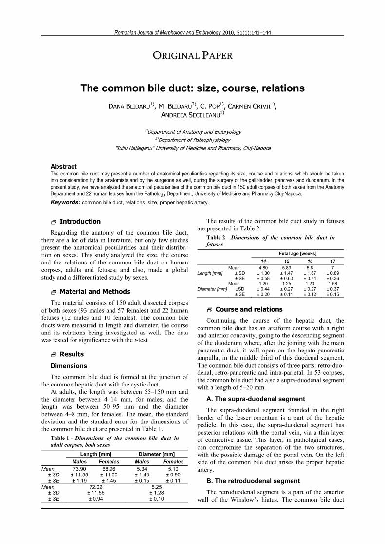
descends posterior to the superior segment of the duodenum, in relation with the superior duodenal flexure, and describes an arch with anterior concavity. This segment has important vascular relations (Figure 1A): the portal vein, posterior; the common hepatic artery, on the left side of the common bile duct and it will continue with the proper hepatic artery; gastroduodenal artery, which descends anterior to the portal vein and to the left of the common bile duct. At this level, the superior right pancreatico-duodenal artery, with origin in the gastro-duodenal artery, crosses the anterior side of the common bile duct, over the pancreas. Then, it passes on the dorsal side of the pancreas, between the common bile duct and the second segment of the duodenum, and ends with an anastomosis with the superior branch of the left pancre-atico-duodenal artery; the superior right pancreatico-duodenal artery passes posterior to the common bile duct and opens on the right side of the portal vein. The portal vein is passing through the triangle forms by the superior border of the pancreas, the common hepatic artery to the left and the gastro-duodenal artery to the right. In this triangle and on the left side of the common bile duct there are present one or two lymph nodes. There, it is also present the end of the gastric coronary vein, which passes downward and it opens in the portal or splenic vein, posterior to the pancreas (Figure 1B).
Figure 1 – Relations of the retroduodenal common bile duct.
C. The retropancreatic segment
The common bile duct descends through a groove or
a canal placed on the posterior side of the pancreas. In this segment, the common bile duct has the following relations (Figure 2): anterior – the head of the pancreas and the superior posterior pancreatico-duodenal artery (Gray H) or superior right pancreatico-duodenal artery (Testut L), which, after it passes anterior to the common bile duct from left to right, will pass on the right margin of the common bile duct, and then crosses again the duct, this time on its posterior aspect.
Figure 2 – Relations of the retro-pancreatic common bile duct – posterior view (original sketch).
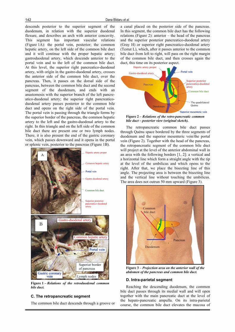
The retropancreatic common bile duct passes through Quénu space bordered by the three segments of duodenum and the superior mesenteric vein/the portal vein (Figure 2). Together with the head of the pancreas, the retropancreatic segment of the common bile duct will project at the level of the anterior abdominal wall in an area with the following borders [1, 2]: a vertical and a horizontal line which form a straight angle with the tip at the level of the umbilicus and which opens to the right. After that, we place the bisecting line of this angle. The projecting area is between the bisecting line and the vertical line without touching the umbilicus. The area does not outrun 50 mm upward (Figure 3).
Figure 3 – Projection area on the anterior wall of the abdomen of the pancreas and common bile duct.
D. Intra-parietal segment
Reaching the descending duodenum, the common bile duct passes through its medial wall and will open together with the main pancreatic duct at the level of the hepato-pancreatic ampulla. On its intra-parietal course, the common bile duct elevates the mucosa of
The common bile duct: size, course, relations
143
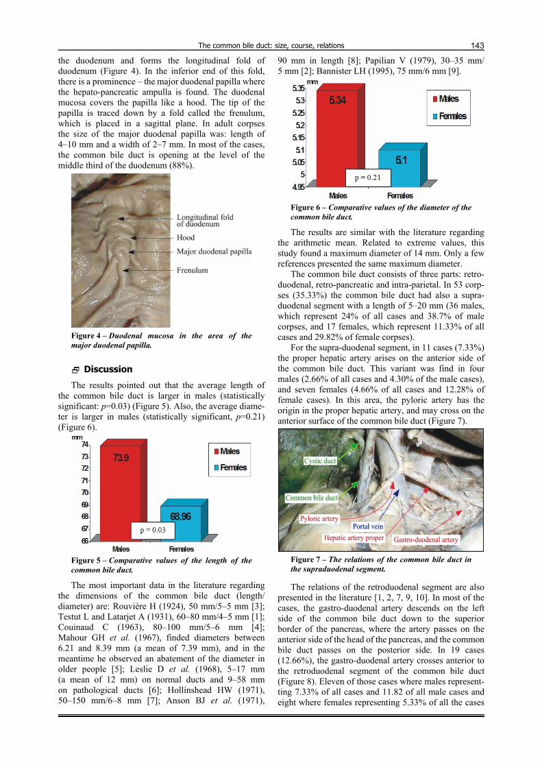
the duodenum and forms the longitudinal fold of duodenum (Figure 4). In the inferior end of this fold, there is a prominence – the major duodenal papilla where the hepato-pancreatic ampulla is found. The duodenal mucosa covers the papilla like a hood. The tip of the papilla is traced down by a fold called the frenulum, which is placed in a sagittal plane. In adult corpses the size of the major duodenal papilla was: length of 4–10 mm and a width of 2–7 mm. In most of the cases, the common bile duct is opening at the level of the middle third of the duodenum (88%).
Figure 4 – Duodenal mucosa in the area of the major duodenal papilla.
Discussion
The results pointed out that the average length of the common bile duct is larger in males (statistically significant: p=0.03) (Figure 5). Also, the average diame-ter is larger in males (statistically significant, p=0.21) (Figure 6).
Figure 5 – Comparative values of the length of the common bile duct.
The most important data in the literature regarding the dimensions of the common bile duct (length/ diameter) are: Rouvière H (1924), 50 mm/5–5 mm [3]; Testut L and Latarjet A (1931), 60–80 mm/4–5 mm [1]; Couinaud C (1963), 80–100 mm/5–6 mm [4]; Mahour GH et al. (1967), finded diameters between 6.21 and 8.39 mm (a mean of 7.39 mm), and in the meantime he observed an abatement of the diameter in older people [5]; Leslie D et al. (1968), 5–17 mm (a mean of 12 mm) on normal ducts and 9–58 mm on pathological ducts [6]; Hollinshead HW (1971), 50–150 mm/6–8 mm [7]; Anson BJ et al. (1971),
90 mm in length [8]; Papilian V (1979), 30–35 mm/ 5 mm [2]; Bannister LH (1995), 75 mm/6 mm [9].
Figure 6 – Comparative values of the diameter of the common bile duct.
The results are similar with the literature regarding the arithmetic mean. Related to extreme values, this study found a maximum diameter of 14 mm. Only a few references presented the same maximum diameter.
The common bile duct consists of three parts: retro-duodenal, retro-pancreatic and intra-parietal. In 53 corp-ses (35.33%) the common bile duct had also a supra-duodenal segment with a length of 5–20 mm (36 males, which represent 24% of all cases and 38.7% of male corpses, and 17 females, which represent 11.33% of all cases and 29.82% of female corpses).
For the supra-duodenal segment, in 11 cases (7.33%) the proper hepatic artery arises on the anterior side of the common bile duct. This variant was find in four males (2.66% of all cases and 4.30% of the male cases), and seven females (4.66% of all cases and 12.28% of female cases). In this area, the pyloric artery has the origin in the proper hepatic artery, and may cross on the anterior surface of the common bile duct (Figure 7).
Figure 7 – The relations of the common bile duct in the supraduodenal segment.
The relations of the retroduodenal segment are also presented in the literature [1, 2, 7, 9, 10]. In most of the cases, the gastro-duodenal artery descends on the left side of the common bile duct down to the superior border of the pancreas, where the artery passes on the anterior side of the head of the pancreas, and the common bile duct passes on the posterior side. In 19 cases (12.66%), the gastro-duodenal artery crosses anterior to the retroduodenal segment of the common bile duct (Figure 8). Eleven of those cases where males represent-ting 7.33% of all cases and 11.82 of all male cases and eight where females representing 5.33% of all the cases

Dana Blidaru et al.
144
and 14.03% of all the female cases. We did not find any data in the literature regarding the incidence of this relation between the gastro-duodenal artery and anterior aspect of the common bile duct.
Figure 8 – The gastro-duodenal artery crossing the anterior side of the retroduodenal segment of the common bile duct.
In the retropancreatic segment, in four cases (2.66%) the superior posterior pancreatico-duodenal artery descends on the left side of the common bile duct and doesn’t cross it; posterior – the Treitz’s fascia, through which it comes in relation with the inferior vena cava, right renal vein and right testicular (ovarian) vein.
Conclusions
The common bile duct has an average length of 72.02 mm and an average diameter of 5.25 mm.
The average length and the average diameter of the common bile duct are larger in males. The common bile duct presents a series of anatomical variations regarding its course and relations. Those peculiarities are impor-tant because they should be known not only by the anatomists but also by the surgeons during the surgery of the gallbladder, pancreas and duodenum.
References [1] TESTUT L, LATARJET A, Traité d’anatomie humaine, 8ème
edition, Librairie Octave Doin, Gaston Doin et Cie, Éditeurs, Paris, 1931, 639–678.
[2] PAPILIAN V, Anatomia omului, vol. II, ediţia a V-a, Ed. Didactică şi Pedagogică, Bucureşti, 1979, 145–150.
[3] ROUVIERE H, Anatomie humaine, descriptive et topogra-phique, Masson et Cie, Éditeurs, Paris, 1924, 810–817.
[4] COUINAUD C, Anatomie de l’abdomen, tome II, Gaston Doin et Cie, Éditeurs, Paris, 1963, 446–447.
[5] MAHOUR GH, WAKIM KG, FERRIS DO, The common bile duct in man: its diameter and circumference, Ann Surg, 1967, 165(e):415–419.
[6] LESLIE D, The width of the common bile duct, Surg Gynecol Obstet, 1968, 126(4):761–764.
[7] HOLLINSHEAD HW, Anatomy for surgeons. Volume 2: The thorax, abdomen, and pelvis, Harper & Row Publishers, New York, 1971, 333–374.
[8] ANSON BJ, MCVAY CB, Surgical anatomy, vol. 1, 5th edition, WB Saunders Co., Philadelphia, 1971, 614–618.
[9] BANNISTER LH (ed), Alimentary system. In: WILLIAMS PL, BANNISTER LH, BERRY MM, COLLINS P, DYSON M, DUSSEK JE, FERGUSON MWJ (eds), Gray’s anatomy: the anatomical basis of medicine and surgery, 38th edition, Churchill Livingstone, New York, 1995, 1795–1812.
[10] BLIDARU DANA, Clinical anatomy of the extrahepatic bile ducts, PhD thesis, “Iuliu Haţieganu” University of Medicine and Pharmacy, Cluj-Napoca, 2001, 74–99.
Corresponding author Dana Blidaru, Assistant Professor, MD, PhD, Department of Anatomy and Embryology, Faculty of Medicine, “Iuliu Haţieganu” University of Medicine and Pharmacy, 2–4 Victor Babeş Street, 400012 Cluj-Napoca, Romania; Phone +40740–338 185, e-mail: [email protected] Received: January 20th, 2009
Accepted: February 15th, 2010