the clinical utility of bronchoalveolar lavage cellular analysis
98
1 THE CLINICAL UTILITY OF BRONCHOALVEOLAR LAVAGE CELLULAR ANALYSIS IN INTERSTITIAL LUNG DISEASE: AN ATS CLINICAL PRACTICE GUIDELINE On-Line Supplement Chairs: ▪ Keith C. Meyer, MD, MS, University of Wisconsin School of Medicine and Public Health, Madison, WI, USA ▪ Ganesh Raghu, MD, University of Washington School of Medicine, University of Washington Medical Center, Seattle, WA, USA Committee Members: ▪ Robert P. Baughman, MD, University of Cincinnati Medical Center, Cincinnati, OH, USA ▪ Kevin K. Brown, MD, National Jewish Center, Denver, CO, USA ▪ Ulrich Costabel, MD, Ruhrlandklinik, Universitätsklinikum, Essen, Germany ▪ Roland M. du Bois, MD, Imperial College, London, UK ▪ Marjolein Drent, MD, Maastricht University Medical Centre, The Netherlands ▪ Patricia L. Haslam, PhD, National Heart and Lung Institute & Royal Brompton Hospital, London, UK ▪ Dong Soon Kim, MD, Asan Medical Center, University of Ulsan, Seoul, Republic of Korea ▪ Sonoko Nagai, MD, Kyoto University, Japan ▪ Paola Rottoli, MD, University of Siena, Italy ▪ Cesare Saltini, MD, University of Roma Tor Vergata Hospital, Rome, Iltaly ▪ Moises Selman, MD, Instituto Nacional de Enfermedades Respiratorias Ismael Cosio Villegas, México City, México ▪ Charlie Strange, MD, Medical University of South Carolina, Charleston, SC, USA ▪ Brent Wood, PhD, University of Washington Medical Center, Seattle, WA, USA Key Words: bronchoscopy, bronchoalveolar lavage fluid, lung diseases, pulmonary fibrosis, cell differential count Corresponding Author: Keith C. Meyer, MD, MS Professor of Medicine University of Wisconsin School of Medicine and Public Health K4/910 Clinic Science Center 600 Highland Avenue Madison, WI 53792-9988
Transcript of the clinical utility of bronchoalveolar lavage cellular analysis
1
THE CLINICAL UTILITY OF BRONCHOALVEOLAR LAVAGE CELLULAR ANALYSIS IN INTERSTITIAL LUNG DISEASE:
AN ATS CLINICAL PRACTICE GUIDELINE On-Line Supplement Chairs: ▪ Keith C. Meyer, MD, MS, University of Wisconsin School of Medicine and Public Health, Madison,
WI, USA ▪ Ganesh Raghu, MD, University of Washington School of Medicine, University of Washington
Medical Center, Seattle, WA, USA Committee Members: ▪ Robert P. Baughman, MD, University of Cincinnati Medical Center, Cincinnati, OH, USA ▪ Kevin K. Brown, MD, National Jewish Center, Denver, CO, USA ▪ Ulrich Costabel, MD, Ruhrlandklinik, Universitätsklinikum, Essen, Germany ▪ Roland M. du Bois, MD, Imperial College, London, UK ▪ Marjolein Drent, MD, Maastricht University Medical Centre, The Netherlands ▪ Patricia L. Haslam, PhD, National Heart and Lung Institute & Royal Brompton Hospital, London, UK ▪ Dong Soon Kim, MD, Asan Medical Center, University of Ulsan, Seoul, Republic of Korea ▪ Sonoko Nagai, MD, Kyoto University, Japan ▪ Paola Rottoli, MD, University of Siena, Italy ▪ Cesare Saltini, MD, University of Roma Tor Vergata Hospital, Rome, Iltaly ▪ Moises Selman, MD, Instituto Nacional de Enfermedades Respiratorias Ismael Cosio Villegas, México
City, México ▪ Charlie Strange, MD, Medical University of South Carolina, Charleston, SC, USA ▪ Brent Wood, PhD, University of Washington Medical Center, Seattle, WA, USA
Key Words: bronchoscopy, bronchoalveolar lavage fluid, lung diseases, pulmonary fibrosis, cell differential count Corresponding Author: Keith C. Meyer, MD, MS Professor of Medicine University of Wisconsin School of Medicine and Public Health K4/910 Clinic Science Center 600 Highland Avenue Madison, WI 53792-9988
2
1. BRIEF HISTORY OF BRONCHOALVEOLAR LAVAGE AND ITS APPLICATION TO ILD 2. DEFINITIONS
3. TECHNICAL CONSIDERATIONS AND PROCEDURE DETAILS FOR BAL CELLULAR ANALYSIS
o 3.1. Technique for performing BAL 3.1.1. Position of the patient 3.1.2. Area (bronchopulmonary segment) that is lavaged in the lung 3.1.3. Number of areas in the lung lavaged 3.1.4. Suction pressure during the procedure 3.1.5. Total volume of normal saline instilled /number of aliquots/ volume of saline
instilled in each aliquot for BAL 3.1.6. Handling the first aliquot separately 3.1.7. Variability of lavage return 3.1.8. Safety aspects of BAL procedure 3.1.9. Summary of specific suggestions for the bronchoscopist performing the BAL
procedure o 3.2. Standardized request form for BAL cellular analyses o 3.3. Specimen handling, transport to the laboratory and processing of the retrieved BAL fluid
specimen (BALF ) 3.3.1. Specific suggestions for handling and transport of BALF specimen to the
laboratory 3.3.2. Specific suggestions for handling and processing the BALF specimen by laboratory
technicians • 3.3.2a. Handling of aspirated BAL fluid • 3.3.2b. Centrifugation for cell concentration • 3.3.2c.Summary of specificsuggestions for the laboratory technician
3.3.3. Specific suggestions for additional analysis based upon clinical setting or initial BAL cell analyses
3.3.4. The storage of residual BAL fluid after cellular analyses
4. USING BAL AND CELLULAR ANALYSES FOR DIAGNOSTIC EVALUATION OF ILD o 4.1. Gross analysis o 4.2. Technical details for cell count and differential cellular analysis in retrieved BAL fluid o 4.3. Infection screening o 4.4. Flow cytometry o 4.5. Reporting of BAL cellular analysis
4.5.1. The effect of underlying disease processes or tobacco smoking on BAL cellular analysis
4.5.2. The reporting of cellular components of the BAL fluid 4.5.3. The reporting of acelluar components 4.5.4. A formal standardized report form for BAL cellular analysis
5. CLINICAL APPLICATION OF BAL CELLULAR FINDINGS TO SPECIFIC TYPES OF ILD
o 5.1. Idiopathic interstitial pneumonia 5.1.1 Idiopathic pulmonary fibrosis (IPF) 5.1.2. Nonspecific interstitial pneumonia (NSIP) 5.1.3. Cryptogenic organizing pneumonia (COP) 5.1.4. Eosinophilic interstitial lung Diseases 5.1.5. Desquamative interstitial pneumonia (DIP) and respiratory bronchiolitis with ILD
(RBILD)
3
5.1.6. Acute interstitial pneumonia (AIP) 5.1.7. Lymphoid interstitial pneumonia (LIP)
o 5.2. Sarcoidosis o 5.3. Hypersensitivity pneumonitis o 5.4. Connective tissue disease (CTD) /collagen vascular disease (CVD)
5.4.1. Systemic sclerosis 5.4.2. Rheumatoid arthritis 5.4.3. Primary Sjögren’s syndrome 5.4.4. Dermatomyositis/polymyositis 5.4.5. Systemic lupus erythematosus 5.4.6. Undifferentiated connective tissue disease 5.4.7. Conclusions regarding the clinical utility of BAL cellular analyses for all
CTD/CVD-associated ILD o 5.5. Occupational interstitial lung disease
5.5.1. Coal Workers Pneumoconiosis 5.5.2. Silicosis 5.5.3. Asbestosis 5.5.4. Chronic beryllium disease 5.5.5. Hard metal disease
o 5.6. Other specific non-IIP interstitial lung diseases 5.6.1. Pulmonary Langerhans cell histiocytosis 5.6.2. Pulmonary alveolar proteinosis 5.6.3. Alveolar hemorrhage syndromes 5.6.4. Drug-induced interstitial lung disease 5.6.5. Radiation pneumonitis
7. OTHER ILD 8. CLINICAL UTILITY OF BAL CELLULAR ANALYSIS IN THE EVALUATION OF ACUTE ONSET ILD 9. FUTURE DIRECTIONS
4
1. BRIEF HISTORY OF BRONCHOALVEOLAR LAVAGE AND ITS APPLICATION TO ILD
Bronchial irrigation with saline solution via a catheter passed through a rigid bronchoscope was
first reported in 1927 (1), and the term “bronchial lavage” was coined by Stitt in 1932 (2) . Although
initially used as a therapy for septic lung disease or pulmonary alveolar proteinosis (3, 4), lung lavage was
subsequently employed to study lower respiratory tract immunity in various animal models in the 1960s
(5, 6) and applied to the study of the human lung in the late 1960s and 1970s (7, 8). A seminal article
reporting the application of BAL to the study of secretions obtained from the human lung was published
in 1974 when saline lavage of a portion of the lung via a flexible bronchoscope was introduced as a
research tool by Reynolds and Newball (9). Saline lavage of a defined area of the lung became known as
bronchoalveolar lavage, and hundreds of articles on BAL appeared in the literature in the 1980s and
1990s as clinical use of the flexible fiberoptic bronchoscope expanded.
The technique rapidly gained acceptance, and a large number of centers began using the
technique to obtain cells and proteins from the lower respiratory tract (10). However, many centers used
their own technique for performing lavage, and concern arose that differences in technique could lead to
significant differences in results obtained from BAL fluid analysis and the interpretation of such findings.
Consequently, several groups established standardized methods for performing BAL. The European
Respiratory Society has provided statements on the use of BAL for the clinical evaluation of patients with
ILD including recommendations concerning the technical aspects of performing BAL (11-13) and on
standardization of the BAL procedure (14), and a multi-center, NIH-sponsored investigation conducted
under the auspices of the American Thoracic Society in the United States examined BAL cell profiles in
patients with ILD and normal volunteer subjects (15). Specific recommendations regarding various
aspects of performing and analyzing BAL were subsequently published. However, these statements were
published prior to the use of high-resolution computed tomography (HRCT) of the chest as a routine
clinical tool for the evaluation of patients with ILD and before the current classification of different forms
of idiopathic interstitial pneumonia (IIP) had evolved (16) to become the system that is currently
accepted. Despite the promotion of BAL standardization in Europe, the technique has never been
standardized on a global basis, and considerable variation in technique and in analysis persists among
bronchoscopists and analytical laboratories around the world.
Although BAL provides a means of retrieving secretions that coat the apical surfaces of bronchial
and alveolar epithelium of normal individuals or patients with infectious or inflammatory lower
respiratory tract disorders, these secretions are considerably diluted by the saline used to perform BAL.
Many factors can affect the amount of fluid retrieved and/or the cellular and acellular profile of the
retrieved bronchoalveolar epithelial surface fluid. Over the past quarter century that BAL has been
applied to the clinical evaluation of patients with various forms of lung disease, its diagnostic potential for

5
ILD has been hampered by a relative lack of specificity. Although patterns in differential counts of BAL
nucleated immune cells have correlated fairly well with certain forms of ILD such as sarcoidosis, their
suboptimal specificity has limited its utility as a diagnostic tool in ILD.
Over the past decade, HRCT of the chest has greatly improved the clinician’s ability to arrive at a
likely diagnosis in the majority of cases of ILD. Nonetheless, tissue sampling is often performed via
bronchoscopic transbronchial lung biopsy (TBLB) or surgical lung biopsy (SLB) to confirm or secure an
accurate and confident ILD diagnosis. Transbronchial lung biopsy, while frequently diagnostic in certain
forms of ILD such as granulomatous lung disease also has its limitations; tissue sampling may be
inadequate and non-diagnostic, and the risk of a complication, while relatively low, is increased compared
to bronchoscopy with BAL only (17, 18). Surgical lung biopsy allows sampling of lung tissue that is
usually diagnostic, but the risk of complications, including death, is not negligible (19, 20).
Bronchoalveolar lavage (BAL) is a safe, easily performed and well-tolerated procedure.
Importantly, BAL has rarely been reported to precipitate acute exacerbations or progression of ILD and
can be safely performed by experienced bronchoscopists. BAL typically reveals variations in cellular
(nucleated immune cells) and acellular components in patients with ILD that differ from normal control
subjects, and many clinicians currently use BAL cellular analysis as a guide for narrowing the differential
diagnosis of ILD. However, the role of BAL in routine clinical management has been a subject of
ongoing debate and controversy. Although BAL should not be considered a stand-alone diagnostic test,
when BAL cellular analysis is applied according to standardized protocols and considered in the
appropriate clinical setting and context of other information obtained from ancillary diagnostic testing
such as HRCT, it may allow the clinician to greatly narrow the broad differential diagnoses and avoid
more invasive diagnostic procedures that entail some risk to the patient.
2. DEFINITIONS
Interstitial lung diseases (ILD) are defined as bilateral infiltrative diseases that are characterized
by abnormalities in both lungs on chest radiographic imaging studies. These disorders typically manifest
in immunocompetent adults with exertional dyspnea, abnormal pulmonary physiology and gas transfer
without clinical suspicion of infection or malignancy and display an accumulation of inflammatory and
immune effector cells and abnormal extracellular matrix in the distal airways and alveolar walls including
the interstitium. The ILDs usually evolve over a period of months to years and include disorders of
known cause such as the pneumoconioses and hypersensitivity pneumonitis (HP) or of unknown cause
such as sarcoidosis, rheumatologic lung disease, and idiopathic interstitial pneumonia (IIP).
Idiopathic interstitial pneumonia (IIP) is a term that encompasses a heterogeneous group of ILD
of unknown etiology that include idiopathic pulmonary fibrosis (IPF), non-specific interstitial pneumonia
6
(NSIP), desquamative interstitial pneumonia (DIP), respiratory bronchiolitis with interstitial lung disease
(RBILD), acute interstitial pneumonia (AIP), cryptogenic organizing pneumonia (COP), and lymphoid
interstitial pneumonia (LIP). In the appropriate clinical setting, IPF is a distinctive clinical entity
characterized by the pattern of usual interstitial pneumonia (UIP) in HRCT and/or surgical lung biopsy
(16) . In the absence of surgical lung biopsy evidence of UIP, it has been suggested that a clinical
diagnosis of IPF can be made by fulfilling all of the four major and three of the four minor criteria as set
forth by an International Consensus Statement (21). Certain IIP pathologies may coexist in the same
patient when multiple regions of the lung are biopsied; especially UIP and NSIP (22), and these patterns
also occur in other ILDs and thus are not disease-specific.
Although most forms of ILD are chronic, some rare forms of ILD can present acutely. The committee
defined acute ILD as a respiratory illness ≤4 weeks duration that is characterized by shortness of breath,
hypoxemia, and diffuse infiltrates in a previously healthy patient who has no known lung disease and no
obvious risk factors for ARDS such as sepsis, trauma, drowning, or overt aspiration. These include acute
interstitial pneumonia (AIP), acute eosinophilic pneumonia (AEP), acute hypersensitivity pneumonitis
(AHP), diffuse alveolar hemorrhage (DAH), cryptogenic organizing pneumonia (COP), drug reactions,
and acute exacerbations of IPF or other forms of ILD. BAL can be performed safely under appropriate
conditions and facilitate an expedient diagnosis of a few specific forms of acute ILD. Bronchoscopy with
BAL has been safely performed in acutely ill patients with acute respiratory distress syndrome (ARDS)
and is contraindicated (relative) if the patient has respiratory –cardio-pulmonary instability or a severe
hemorrhagic diathesis.
3. TECHNICAL CONSIDERATIONS AND PROCEDURE DETAILS FOR BAL CELLULAR
ANALYSIS
Reports in the literature that pertain to the performance of BAL document the considerable
variability in technical aspects of performing BAL.This may in part be the reason of apparent reluctancy
among pulmonologists worldwide to subject patients for BAL in the diagnostic evaluation of patients
with ILD. This section reviews the features of obtaining and processing BAL that appear to affect the
results of BAL cellular abnormalities and provides guidelines with the anticipation of standardizing the
performance of the BAL procedure and analyses of BAL fluid (BALF) retrieved during BAL procedure
plus subsequent reporting and interpretation of the results.
An underlying disease process or smoking may affect BAL bronchoscopic technique or analysis
in several ways. Airway obstruction leads to changes in BAL fluid return. This is due to the mechanical
problem of aspirating fluid through narrowed airways that collapse when negative pressure is applied to
7
facilitate fluid retrieval. In patients with chronic obstructive lung disease, the airway collapse is variable;
the greater the negative pressure used to aspirate the fluid, significant airway collapse is more likely to
occur and adversely influence the BALF return. In this regard, FEV-1/FVC ratio has been correlated with
the yield of BALF return; the lower the ratio, the lower the proportion of BAL return (23, 24) Airway
obstruction in asthmatics can be induced by the procedure itself (25, 26). A major concern in asthma is
the risk of the procedure, and recommendations for performing BAL in asthmatics have been made (26,
27). Bronchoalveolar lavage can be performed in acute respiratory distress syndrome (ARDS) patients
(28). However the procedure can lead to significant hypoxia (29), and modifications of the procedure,
such as increasing inspired oxygen concentration, may be necessary to maintain oxygen saturation of
>90% during the procedure (28). Nonetheless, BAL has been performed in various respiratory disorders;
however, it is unknown if specific ILD and/or the severity of functional impairment of specific forms of
ILD influence the BALF return during the BAL.
3.1. BAL Technique
3.1.1. Position of the patient:
Gravity may influence the lavage process. Because suction pressure affects airways, fluid
flowing back toward the bronchoscope (when aliquot instillation is completed and aspiration is initiated)
is relied upon. The position of the patient can affect the lavage, and gravity may impede lavage return
from more gravity-dependent lung regions. There are no studies comparing the effects of upright, semi-
recumbent, or supine positioning on BAL. Positioning should be determined according to the clinical
situation affecting the patient and should be noted in the procedure report.
3.1.2. Area (bronchopulmonary segment) that is lavaged in the lung:
Another important consideration is the choice of the most appropriate area or areas of the lungs
for lavage. When BAL was first introduced into clinical investigation in the 1970s, a main focus of
research initially was to investigate immune and inflammatory mechanisms in IPF because of the
especially poor prognosis and lack of effective therapies. Because infiltrates in IPF tend to be more
prominent in the lung bases on thoracic imaging, some investigators have performed the BAL from the
right lower lobe (30). Others, however, have preferred to sample the right middle lobe due to its easier
access and higher fluid recoveries versus lower lobe lavages (9). Obtaining BAL from the lingula was
generally avoided because biopsies from the tip of the lingula were often unsuitable for diagnostic
purposes. A large number of reports in the 1970s and 1980s of BAL studies in the main groups of ILDs
soon showed that differential BAL cell count findings were very similar from different centres using the
8
right middle lobe or the right lower lobe as their standard lavage site. There was discussion about whether
an upper lobe site should be used for diseases with more prominent upper zone shadowing, but this
approach was criticized at the time because of lack of a standardized procedure. This omission from
earlier recommendations for performing BAL relates to the fact that such recommendations were made
prior to the subsequent re-classification of the IIPs (16), and it is unknown if this necessitates a new
approach to the choice of the site for lavage.
Significant differences have been reported among different lobes for both sarcoidosis (31) and
idiopathic pulmonary fibrosis (32) . The use of HRCT in recent years and the importance of HRCT
patterns in the new classification of IIPs, raises the possibility that differences in HRCT patterns in the
lungs of individual patients may prove useful to identify target areas for BAL that may more accurately
reflect the inflammatory cell profile that is associated with a specific ILD. For example, areas of ground-
glass opacity may reflect areas of more active inflammation and represent good target areas for lavage.
Further research is needed to better clarify whether different HRCT patterns are associated with different
BAL cell profiles in the same lung and whether this would lead to major differences in the diagnostic
interpretation compared with the current approach to use a standardized lavage site. While this document
is intended for management of non-infectious ILDs , there is evidence that lavage site variation is an
important consideration in evaluation of respiratory infections; earlier reports have supported the BALF
to be retrieved from the site of maximal involvement .In opportunistic lung infections (with diffuse
pulmonary infiltrates), it is clear that when BAL was performed in patients with P. carinii pneumonia,
lavage in the upper lobes had a higher yield of organisms than the traditional right middle lobe or lingual,
and the yield was the same from the upper lobe and the more traditional right middle lobe or lingula (33).
While it is unclear whether BAL from selective sites (directed by HRCT abnornalities) increases
the diagnostic yield of BAL cellular analyses in the evaluation of patients with ILD, the additional
concern of lack of standardization for BAL from different bronchopulmonary segments warrants
additional well designed studies to investigate this further. While BAL can be performed in any area of
the lung in ILD, the area or areas of the lung lavaged should be specified in the BAL request form (see
below).
3.1.3. Number of areas in the lung to be lavaged:
Generally, only one geographic area (lung segment) is lavaged when evaluating ILD. If multiple
areas are lavaged, the specific lung regions that are lavaged should be noted. Some investigators have
used multiple sites to evaluate ILD and pooled all the areas lavaged to get a higher number of cells and a
more generalized representation of lung airspaces (32). However, occasionally a comparison of returns
between lavage sites or correlation of lavage site and HRCT appearance is desired, and in this context the
9
returns will need to be kept separate. Pooling multiple lavages from different lavage sites also has the
disadvantage of introducing greater variability into the procedure and compromising the efforts to
improve BAL standardization, which is required for many research purposes. Using single aliquots to
lavage different areas has the further disadvantage that such ‘first aliquots’ have been shown to contain a
greater proportion of bronchial components than subsequent aliquots from the same site. If for any reason
a non-standard site is used, it is recommended that the reason for this be stated and that the number of
areas lavaged and whether lavage aspirates from different lung regions are pooled be noted and
commented in the BAL request and report forms (see below).
3.1.4. Suction pressure during the procedure:
Airways collapse when the suction pressure is too high, and this should be apparent to the
bronchoscopist if it occurs during the BAL procedure The majority of physicians in one survey used low
pressure wall suction (<60 mm Hg). Some prefer negative pressure generated by a hand-held syringe,
adjusting pressure based on visualization of the airways. Regardless of specific technique, visual
examination of the airway allows the bronchoscopist to monitor for airway collapse during the aspiration
process. It is suggested that negative suction pressure remain at least below 100 mm Hg and adjusted to
avoid visible airway collapse.
3.1.5. Total volume of normal saline instilled /number of aliquots/ volume of saline instilled in each
aliquot for BAL:
The volume of normal saline instilled during the BAL affects the BALF return and the results.
This has been shown for both the cellular and protein contents of the BAL sample (34-36). The major
changes seem to occur during the first 100 ml of the process. The fluid aspirated after 60 ml instilled is
significantly different than obtained after 120 ml has been instilled. After two aliquots of 60 ml have been
instilled and aspirated, the cells and protein returns appear to become consistent. In one study of ILD and
control subjects, differences were not appreciated until at least 120 ml of fluid were instilled (37). This
effect of volume on ILDs has been noted by others (38). Cellular analyses from BAL specimens collected
from sequential BAL returns separately and analyzed separately from individual aliquots indicate that the
BAL sample collected from the first aliquot tends to represent cells (epithelial cells) and protein
(lactoferrin) from bronchiolar surfaces (23, 39, 40). It is suggested that at least a total volume of 100 ml of
saline should be instilled to adequately sample cells and proteins on bronchoalveolar epithelial surfaces.
Although some experts have suggested a specific number of aliquots be used for BAL (11, 14),
there is considerable variability in the number and volume of aliquots used by pulmonologists within and
outside the United States. The choice of aliquot size can easily be standardized as it is apparent that this
10
is often determined merely by the size of the syringes available to the bronchoscopist in the local facility
(usually either 20 or 60 ml). As noted above, the volume of the lavage is crucial for BAL sampling. In
this regard 6 x 20 ml aliquots could provide similar returns to 2 x 60 ml lavages or other subdivisions of
120 ml totals. Larger volumes instilled into a broncho-alveolar units (spread over a larger ‘alveolar’
relative to ‘bronchial’ area than smaller instilled volumes) and use of larger volumes in humans has
shown better alveolar sampling, with an increased proportion of cells and solutes found in the alveolar
spaces (40). Larger total instilled volumes of a minimum of 100 ml and a recommended standard 240 ml
using standard 4 x 60 ml aliquots have therefore been recommended by the European Respiratory Society
(ERS) to improve standardization when more efficient alveolar sampling and accurate quantitative
measurements are required (14, 40). This approach to standardization is important for investigators using
the BAL samples for research as well as routine clinical purposes. However, when BAL is being
performed in centers for routine clinical purposes as an aid to diagnosis of specific ILD, the BAL
differential counts are not influenced by dilution and there is no evidence that they are influenced
substantially by the variability in total introduction volumes or aliquot numbers and size used by different
workers. A potential disadvantage of smaller instilled volumes (e.g.20 ml) is that they can recover a
relatively higher proportion of ‘bronchial components’, thus this approach may be more useful for those
studying airways diseases rather than ILDs.
An ERS Task Force has produced a comprehensive report on the many causes of variability in
BAL, including variable dilution, and recommends better standardization of BAL procedure, including
volume and number of aliquots instilled, to reduce this variability (14). Quantitative measurements of cell
counts per ml and acellular components per ml are profoundly influenced by variable dilution, which may
explain why their clinical utility remains poorly defined compared to differential cell counts that are not
influenced by dilution. To facilitate future clinical developments, recommendations for standardization
have been defined, which include the use of a standard total instilled volume of 200-240 ml introduced in
4 aliquots (14, 40).
3.1.6. Handling the first aliquot separately:
The aspirated fluid after the first 20 ml has been reported to be distinctly different from additional
aliquots (39). Therefore, some investigators have proposed that the first aliquot be handled in a different
manner (39) or even discarded if one is studying diffuse ILDs. Alternatively, the first aliquot could be
sent to microbiology laboratory for stains and cultures of infectious pathogen/s. However, the numbers of
cells in the later aliquots are much larger than those retrieved after the earlier volumes of lavage (34, 37).
In one study, it was determined that combining aliquots of fluid retrieved after the first 60 ml aliquot did
not significantly change the overall lavage results (34). The initial fluid can be handled separately,
11
especially if diseases that originate in the larger airways are being studied. However, this committee felt
that it is acceptable to pool all the aspirated fluid for routine cellular analyses, but the technique that is
used should be specified in the BAL request form (see below).
3.1.7. Variability of lavage return:
The percent of instilled saline that is retrieved is also variable. If less than 5% of the volume
instilled is retrieved, alveolar sampling is probably inadequate. In one study of nonbronchoscopic lavage,
it was shown that patients with a greater than 5% return of instilled fluid have a higher diagnostic yield
(41). This committee recommends that at least 30% of the instilled volume be aspirated to be considered
an optimal alveolar sample, although smaller volumes may still retrieve diagnostic material. Small
volume returns (especially those <10% of the total instilled volume) should not be considered true BAL
samples.
For safety reasons, the lavage should be stopped if the volume instilled and retained within the
lung becomes too large (e.g. persistent recovery of <5% per aliquot). Because this decision will depend
on the volume of the instilled aliquots, the bronchoscopist should be aware of the total instilled and
aspirated volumes after each aliquot is instilled. If the difference between instilled and aspirated volumes
becomes considerable (e.g. greater than 100 ml), instilling additional aliquots in the area of this lavage is
not recommended, but a second lavage in a separate area could be undertaken if the patient is tolerating
the procedure.
3.1.8. Safety aspects of BAL procedure:
Bronchoalveolar lavage procedure has a well-proven safety record in both human research and in
clinical applications that range from evaluating patients with asthma and chronic obstruc tive pulmonary
disease to patients with acute respiratory distress syndrome and ventilator-associated pneumonia. The
two most common complications linked to BAL procedure are fever and hypoxemia (42, 43), but these
BAL-associated adverse events are almost always self-limited and have been linked, to some degree, to
the use of larger total lavage volumes (44) Nonetheless, the BAL procedure can precipitate acute phase
responses with an increase in circulating pro-inflammatory cytokines, an increase in peripheral blood
neutrophils, an influx and transient sequestration of neutrophils into the lung, and alterations in iron
homeostasis (45-47). Despite the concern that BAL procedure can induce procedure associated acute
inflammatory responses, fever, and hypoxemia, it has a well established safety record when used to
evaluate patients with VAP (48), patients with ARDS (28, 49), lung transplant recipients (50),
immunocompromised patients with acute infiltrates (51), patients with impaired coagulation (52) , or
patients with evolving respiratory failure (53). Although pneumothorax and significant bleeding have
12
been reported (28, 52, 54, 55), such complications are exceedingly rare when bronchoscopy and BAL are
performed without an accompanying biopsy. BAL has been implicated to precipitate an acute respiratory
decline immediately following the procedure; two reports of acute exacerbation of IPF have been
associated with to the BAL procedure in the clinical setting (56, 57).
Although BAL appears to be quite safe, adequate safety protocols should be utilized and be in
place whenever bronchoscopy and BAL are performed to minimize the possibility of complications; the
bronchoscopist should be prepared to identify and treat potential complications. A detailed history of
complications associated with other procedures or trauma should be obtained, and the use of medications
(e.g. clopidrogel, aspirin, coumadin, heparin) and/or medical conditions (e.g. uremia) that increase
bleeding risk should be noted in keeping with good clinical practice. Coagulation studies (international
normalized ratio, partial thromboplastin time, platelet count) may be performed to determine whether a
significant coagulopathy is present that may increase the risk of bleeding complications. Intravenous
access should be established in all patients who undergo conscious sedation, and personnel involved in
the procedure should be adequately trained in conscious sedation protocols. Heart rate and
oxyhemoglobin saturation should be monitored continuously throughout the procedure, and patients
should not have an unstable medical condition such as recent myocardial infarction or unstable angina,
Although adequate amounts of topical lidocaine (e.g. multiple 1-2 ml aliquots of 1% lidocaine) should be
given to optimize patient comfort during the procedure, the total lidocaine dose should be kept to the
minimum amount needed to achieve patient comfort and control cough, and contamination of BAL fluid
with excessive lidocaine should be avoided to minimize the potentials of poor cell viability at the time of
cellular analyses. Adherence to a stringent safety protocol has been shown to diminish and minimize the
risk of complications in lung transplant recipients subjected to bronchoscopy (50). Similarly, adequate
safety regimens should be in place and followed for every bronchoscopy procedure.
3.1.9. Summary of specific suggestions for the bronchoscopist performing the BAL procedure
►Routine clinical evaluation prior to performing BAL should include enquiry and appropriate testing for
bleeding tendencies.
► For patients with suspected ILD in whom it has been decided that a BAL can be tolerated and will be
performed, the BAL target site should be chosen on the basis of a HRCT performed prior to the
procedure , rather than choosing a traditional BAL site.
►A safety protocol, standard for fiberoptic bronchoscopy procedure should be followed to minimize the
likelihood of procedure-related complications.
13
►BAL should be performed with a fiberoptic bronchoscope in a wedge position in the selected
bronchopulmonary segment.
►The total instilled volume should be no less than 100 ml and should not exceed 300 ml, and 3 to 5
aliquots should be used.
►Total unretrieved volume of instilled BAL fluid should not be excessive (e.g. greater than 100 ml from
one segment).
►Negative suction pressure should not exceed 100 mm Hg and should be adjusted to avoid visible
airway collapse.
► For optimal sampling of distal airspaces the total volume (pooled aliquots) retrieved should be ≥30%
of the total instilled volume. A total volume of retrieved fluid less than 30% may provide a
misleading cell differential, especially if total retrieved volume is less than 10% of total instilled
volume. If <5% of each instilled aliquot volume is recovered during the procedure due to retention
of most of the fluid in the lavaged segment, the procedure should be aborted to avoid increased risk
of tissue disruption and/or inflammatory mediator release due to overdistention of the lavaged
segment..
► If <5% of each instilled aliquot volume is recovered during the procedure due to retention of most of
the fluid in the lung, the procedure should be aborted to avoid increased risk to the patient.
►A minimal volume of 5 ml (optimal volume = 10-20 ml) of a pooled BAL sample is recommended for
BAL cellular analysis (the rest of the sample should be used for microbiology, virology, and
malignant cell cytology laboratory testing as clinically indicated).
► It is acceptable to pool all aliquots of the aspirated fluid for routine analyses (including the first
retrieved aliquot), but the technique used should be specified in the BAL request form (see below).
3.2. Standardized Request Form for BAL cellular analyses
The committee recommends that a request form be completed and sent to the laboratory with the
BAL fluid specimen for BAL cellular analysis (see Request form online appendix that should be
utilized). This is useful for appropriate interpretation (see interpretation, below) and should include:
• Time and date of the procedure
• Smoking history
• Age and gender
• Pertinent clinical features (clinical suspicion of underlying diagnosis)
• Specific area(s) lavaged
14
• Total instilled volume
• Gross appearance of BAL fluid
• Patient position during the procedure
• Checkboxes for routine analysis and specific additional testing
3.3. Suggestions for specimen handling, transport to the laboratory and processing of the retrieved
BAL fluid (BALF) specimen
3.3.1. Specific suggestions for handling and transport of BALF specimen to the laboratory ►BALF specimens should be collected in containers /aliquots that do not promote cell adherence to
container surfaces (e.g. silicone-coated glass or polypropylene or other plastics that are designed for
suspension tissue culture).
►BALF specimens can be transported ‘fresh’ at room temperature if the analytical laboratory is located
within the same facility and there is minimal delay between BAL fluid retrieval and delivery to the
laboratory to increase the likelihood of preservation of cell viability at the time of analyses trained
and designated laboratory personnel.
► For anticipated delay in delivery to the laboratory beyond 30-60 minutes following retrieval of the
fresh BALF, specimens should be transported at 4°C (e.g. on ice) and delivered within 1 hour.
►BALF specimens retrieved should not be frozen and/or not transported with dry ice for BAL cellular
analyses.
►Transport periods greater than 1 hour in the original lavage saline are not recommended; the cells
should be transferred to an appropriate tissue culture medium to preserve them for longer periods
(e.g. MEM+25 mM HEPES or RPMI 1640+25mM HEPES).
►Rapid processing by designated laboratory technician trained in handling of BALF samples is ideal (i.e
immediately after the specimen is delivered to the laboratory) and will provide optimal results; if
significant delay in processing is unavoidable, viability of BAL cells are better preserved in nutrient-
supplemented media (e.g. MEM+25mM HEPES or RPMI 1640+25mM HEPES) for up to 24 hrs. In
selected facilities, such nutrient-supplemented media may be added to the container of the BALF
specimen collected shortly after the procedure is completed; this will require appropriate
coordination and elective arrangements between designated laboratory staff and clinical personnel
handling the transport of the BALF specimen.
15
3.3.2. Specific suggestions for handling and processing the BALF specimen by laboratory technicians
3.3.2.a. Handling of aspirated BAL fluid:
The aspirated fluid can contain mucus material as well as the fluid itself. The handling of this
fluid can affect the results of the BAL cellular analyses. Filtering BAL fluid though gauze prior to
analysis has been used by some groups to remove the mucus. However, the cells in the BAL fluid may
variably adhere to the gauze and adherent mucus. This may affect cell retrieval and the cellular profile of
the BAL sample (30). The committee recommends that filtering BAL fluid with gauze should be avoided
unless excessive amounts of mucus are present overtly. If filtration is performed, it should be specified in
the report form (see below). Macrophages retrieved by BAL can be quite adherent to glass or similar
surfaces, and macrophage adherence may be altered in various conditions, including cigarette smoking
(58). Storage of cells at the time of the lavage should be in containers (e.g. polypropylene, other plastics
designed for suspension tissue culture or silicone-coated glass) that do not promote cell adherence.
3.3.2.b. Centrifugation for cell concentration:
Techniques used to concentrate proteins and cells may lead to loss of cells (59-61). A
centrifugation speed of 250-300g for 10 minutes is recommended and should yield a cell pellet that can be
readily resuspended with gentle agitation. Cell counts can be made on unconcentrated samples. If
concentration is performed prior to cell counting, the method should be specified.
3.3.2.c. Summary of specific suggestions for the laboratory technicians: ►BAL cellular analysis should be performed within one hour if in nutrient-poor media (e.g. saline).
►If BAL cellular analysis cannot be performed within 1 hour, cells can be transferred to tissue culture
medium ( e.g. MEM+25mM HEPES or RPMI 1640+25mM HEPES) for processing that should be
performed within 2-3 hours for optimal results.
►BAL fluid and BAL-derived cell suspensions should be processed in labware that do not promote cell
adherence to container surfaces (e.g. silicone-coated glass or polypropylene or other plastics -
designed for suspension tissue culture).
►Mucus can be dissolved (e.g. dithiothreitol) if necessary.
►Specimens with gross mucus can be strained through 4x4 loose gauze if necessary.
►The BAL fluid should be centrifuged at an appropriate speed (e.g. 250-300g for 10 minutes) to
maintain cellular integrity and allow uniform resuspension.
►Cell suspensions should be refrigerated at 4°C and resuspended in tissue culture medium (eg.
MEM+25mM HEPES) if not analyzed immediately.
16
►Nucleated cell counts should be obtained via a hemocytometer.
►Cell viability should be determined (Trypan blue exclusion) and reported.
►Specimens obtained more than 24 hrs prior to analysis are not suitable for analysis.
►Differential cell counts should be performed via cytocentrifugation with staining (Wright-Giemsa or
May-Grunwald-Giemsa) and enumeration of at least 400 cells.
►A Diff-Quick stain is not recommended, as mast cells will not be identified.
3.3.3.Specific suggestions for additional analysis based upon clinical setting or initial BAL cell
analyses
► For patients with suspected ILD in whom BAL is performed, we suggest that lymphocyte subset
analysis not be a routine component of BAL cellular analysis.
►Flow cytometry or immunocytochemistry for lymphocyte markers (B or T lymphocytes, Langerhans
cells) should be performed as clinically indicated or when suspected from initial BAL cellular
findings and the laboratory be alerted to the possibility of this additional analysis.
►Periodic Acid Schiff staining should be performed (alveolar proteinosis sediment) if Primary alveolar
proteinosis is suspected clinically or based on initial BAL appearances.
►Oil red O staining should be performed if aspiration is suspected.
►Hemosiderin stain should be performed if hemorrhage is suspected and/or if initial BAL appearances
raise suspicion of haemosiderin-laden macrophages.
►A separate report by a hematopathologist or cytopathologist is mandated for interpretation of isolated
cells that are suspicious for malignancy.
►Inorganic dust bodies or particles within macrophages can be characterized by energy dispersive
electron microprobe analysis if BAL cellular and microscopic appearances indicate their presence.
3.3.4. The storage of residual BAL fluid after cellular analyses:
Proper storage of BAL samples is crucial for the subsequent measurement of certain markers.
Cells stored at 4° C can be analyzed up to 24 hours after the procedure without significant changes in the
total cell count and differentials (62) However, neutrophil apoptosis is known to have commenced by 9
hours and such cells can be rapidly phagocytosed by macrophages(63, 64).
Certain proteins may be temperature sensitive and require that BAL aliquots be stored at -80° C
until analysis is performed. The handling of many of the proteins measured in the lung lining fluid are
17
detailed elsewhere (14). The committee recommends that the conditions under which BAL samples are
stored be specified in the BAL report form (see below).
4. BALF AND CELLULAR ANALYSIS FOR DIAGNOSTIC EVALUATION IN ILD
4.1. Gross analysis:
The appearance of BAL fluids can give important clues concerning the cause of ILD. The
presence of blood with a progressive increase in the intensity of bloody discoloration in the retrieved BAL
fluid with sequential aliquots during the BAL procedure strongly suggests a pulmonary/alveolar
hemorrhage syndrome (65). If BAL fluid is grossly cloudy /’milky’appearance with a light brown/beige
color appearance and contains whitish flocculent material that settles to the bottom of a container when
left to stand, the diagnosis of pulmonary alveolar proteinosis is suggested (66). If cloudy material is
present but low-speed centrifugation is required to clear the fluid, the diagnosis of microlithiasisis
suggested (67). If oily material layers out on the top of the BAL fluid, the presence of lipid material and
lipoid pneumonia is suggested. The presence of a black cell pellet after centrifugation suggests that the
subject is a likely smoker or has been exposed to significant amounts of carbonaceous material in inhaled
air.
4.2. Technical details for performing cell count and BAL cell differential counts in retrieved BALF:
Identification and enumeration of cellular elements is an important component of the evaluation
of BAL fluid (11-13), although relatively few published studies regarding the technical aspects of
specimen preparation exist. The single most critical prerequisite for successful cellular analysis is
maintenance of cell viability following collection. Saline solutions, as are used for washing of the alveolar
space when a BAL is performed, have low pH and are nutrient poor and incapable of sustaining cells for
more than a limited time, i.e. less than one hour. If the sample will not be examined immediately, the
collection and replacement of the saline cell-free supernatant with tissue culture medium such as MEM
with 25 mM HEPES buffer or RPMI 1640 with HEPES buffer prior to transport provides much improved
sample stability, which may not be provided by merely keeping the specimen at 4°C. Centrifugation of
the sample, removal of the supernatant, and resuspension in medium is the preferred method of
supplementation. Refrigeration of the sample will also retard cellular degradation and is advisable if the
delay in specimen analysis is greater than 8 hours (e.g. overnight). Samples greater than 24 hours old are
generally not suitable for analysis, even with supplementation. It is important that the fluid volume be
recorded in the BAL request/report form prior to supplementation with any medium.
18
Following receipt in the laboratory, the cellular elements may need to be separated from the
mucus contained in the fluid, if excessive mucus is overtly present on gross inspection of the fluid.
Although the commonly used method to remove mucus is to strain the sample through a single layer of
sterile gauze, this is not recommended by the committee because some loss of cell types may occur. For
the occasional samples with very excessive mucus, treatment of the sedimented cell pellet with
dithiothreitol after centrifugation to remove and collect the fluid supernatant is recommended as an
alternative method to dissolve mucus without loss of other material. The cellular elements should then be
washed with MEM+25mM HEPES of a balanced electrolyte solution such as Hank’s Balanced Salt
Solution (HBSS) or phosphate buffered saline (PBS) which lack calcium and magnesium followed by
centrifugation at 250-300g for 5 minutes. Following careful aspiration of the supernatant, it is suggested
that the cells can be resuspended in a known volume of tissue culture media by gentle agitation.
Vigorous vortexing should be avoided.
The committee suggests that a minimum volume of 5 ml of BAL fluid should be used for cell
analysis; a larger amount (10-20 ml), however, provides a more optimal specimen. A nucleated cell count
should then be obtained manually in duplicate using a hemocytometer, either in the well-mixed original
fluid or after the first wash with the caveat that cells are progressively lost with repeated washing. The
presence of increased numbers of red blood cells should be noted, but need not be routinely enumerated.
Automated cell counters are unlikely to provide accurate cell counts, as they are unable to correctly
enumerate epithelial cells and other non-hematopoietic cellular elements. Stabilized cellular control
materials are now commercially available for the quality control of body fluid cell counts and these
should be used for the quality control of BAL cell counts. It is important to use pipettes and tubes for
specimen handling that do not promote cell adherence (siliconized glass or polypropylene), as plain glass
surfaces can cause macrophages to become activated and adherent, resulting in their underestimation. The
viability of the nucleated cells may be estimated by using Trypan Blue in a counting fluid (i.e. Trypan
blue exclusion), especially if specimens have not been freshly obtained and analyzed. The absolute
number of nucleated cells, cellular concentration and percent viability in the original specimen can then
be calculated, assuming the original and post-wash volumes are known. A viability of greater than 90%
is considered acceptable, and less than 80% suggests the possibility of significant specimen compromise
and thus be considered suboptimal for cellular analyses and may underestimate the true cellular
components of the ILD within the alveolar walls.
For the identification and enumeration of cellular subpopulations, the committee suggests that six
cytocentrifuge preparations should be made. Although, it is recognized that the cytocentrifuge technique
may underestimate lymphocytes, particularly if their numbers are high (61, 68), cytocentrifugation is a
technique commonly available in clinical laboratories, is rapid, and provides improved cellullar detail
19
compared with other methods. Two of the air-dried preparations should be stained with either Wright-
Giemsa or May-Grunwald-Giemsa and subsequently protected by a coverslip. A Diff-Quick stain is
suboptimal, as mast cells are poorly identified. Under low power magnification (200x), the presence of
the morphologic abnormalities may be identified, e.g. dust particles, microorganisms, clusters of
malignant cells, or acellular aggregates suggestive of alveolar proteinosis. Under high power
magnification (500x), a differential count of the nucleated BAL immune cells should be performed, to
include: macrophages, lymphocytes, neutrophils, and eosinophils. To provide a statistically meaningful
estimate for populations representing less than 10% of the total, 400 cells should be evaluated, as is
currently recommended for differential WBC counts in peripheral blood (69). The cells evaluated should
be equally divided between the two slides and a random search method used to minimize processing and
distributional artifacts. For cytocentrifuge preparations, the speed of the centrifuge, the area of the slide,
and the number of cells counted have all been shown to lead to differences in counts (70). The presence of
bacteria or other organisms as well as malignant cells should also be noted. The other remaining
unstained slides should be retained for potential use for more specialized stains as indicated by clinical
history or morphologic findings (e.g. iron stain for hemosiderin-laden macrophages, PAS for alveolar
proteinosis, Oil Red O for aspiration, silver stain for fungal infection, etc). Specimens that are considered
to accurately reflect distal airspace inflammatory cell patterns should not contain more than 5% epithelial
cells.
Specific cutoffs that define abnormal increases in BAL cell differential counts are not supported
by strong evidence, but differential counts in non-smoking, clinically normal individuals have been stated
to show lymphocytes ≤ 15%, neutrophils ≤3%, eosinophils ≤ 0.5%, mast cells ≤0.5%, and macrophages >
80%(11) . Other studies in which BAL was performed in large cohorts of patients have used slightly
different cutoffs to define BAL eosinophilia (eosinophils > 2%, BAL neutrophilia (neutrophils > 4%), or
BAL lymphocytosis (BAL lymphocytes >14%) (71). Additionally, percentages of neutrophils and
lymphocytes tend to be somewhat higher for clinically normal, elderly individuals compared to younger
adults (72, 73).
Our analysis of the data presented in various published reports of BAL in normal individuals
with or without smoking as an additional variable and in reports of BAL performed in patients with lung
disease suggest that reasonable thresholds for increases in BAL cell differential counts are >15% for
lymphocytes, >3% for neutrophils, and >1% for eosinophils. Healthy smokers can have up to a 10 fold
increase in the number of macrophages per ml of BAL fluid but no increase in lymphocytes compared
with healthy non-smokers. This results in their having slightly, but significantly, higher differential
percentage counts of macrophages (≥85%) and slightly lower percentages of lymphocytes compared with
healthy non-smokers.
20
4.3. Infection screening:
Pulmonary infection may present as a diffuse infiltrative lung disease, or infection may
complicate the course of ILD. Because fungal or mycobacterial infections can masquerade or coexist
with various forms of ILD, BAL fluid should be examined for infectious agents as clinically indicated
when used in the evaluation of diffuse infiltrative lung disease. For patients with possible granulomatous
diseases such as sarcoidosis, the committee suggests that minimal screening of BAL should include
testing to identify mycobacterial and fungal infection in addition to BAL cellular component analysis.
Other testing should also be performed as clinically indicated.
4.4. Flow cytometry:
In some instances, it may be desirable to enumerate the percentage of T cells, T cell subsets, or
expression of other antigens on specific cellular subsets. In particular, assessment of the CD4/CD8 ratio
could be considered in the presence of ≥15% lymphocytes. However, the determination of T lymphocyte
subsets on specimens that contain less than 15% lymphocytes is unlikely to yield useful information.
These studies can be easily accomplished by standard flow cytometric techniques using the same cell
preparation described above. In patients having known hematopoietic neoplasms, flow cytometry can
provide evidence of pulmonary involvement by the identification and quantitation of abnormal blast or
neoplastic lymphocyte populations with a high degree of sensitivity and specificity. For Langerhans cell
histiocytosis, immunophenotying by either immunocytochemical or flow cytometric techniques can be
important to support the diagnosis. Although flow cytometry may provide useful diagnostic clues in
certain situations, the committee does not recommend analyses for T cell subsets by flow cytometry as a
routine component for the cellular analysis of all BAL specimens.
4.5. Reporting of BAL Cellular Analysis
4.5.1. The effect of underlying disease processes or tobacco smoking on BAL cellular analysis:
Cigarette smoking can also have profound effects on the cell populations found in the BAL fluid
(11). The greatest effect is an increase in the number of macrophages, which are often 10-fold greater in
numbers than in nonsmokers. Neutrophils also appear in increased numbers. Because of this, it is
essential to establish control ranges not only for healthy non-smokers but also for ex-smokers and current
smokers for routine and research purposes. If a patient is a current cigarette smoker, changes in the BAL
cellular profile that are attributed to a disease process may actually be due to inflammatory changes
caused by the smoking habits of the patient. Because of the potential effects of cigarette smoking and
21
underlying lung disease on technical aspects of BAL and WBC total cell counts and profiles, the
underlying disease of the patient and cigarette smoking history should be included in the BAL request
form for appropriate interpretation of the BAL cellular analyses.
4.5.2. The reporting of cellular components of the BAL fluid:
The committee suggests that the results of the BAL cellular analysis be reported in a
standardized manner; this report should include: patient demographic information, clinical information
supporting the indication for performing the BAL, the initial specimen volume, color and turbidity, %
viability for the nucleated cells, absolute total nucleated cell numbers, and immune cell differential
percentages. The columnar ciliated epithelial cells should be reported as a percentage of the total
nucleated cells, with less than 5% ciliated columnar epithelial cells (generally simply stated as ‘epithelial
cells’) characteristic of adequate alveolar sampling. The presence of greater than 5% ciliated columnar
epithelial cells suggests increased proximal airway sampling. Such samples should be considered
suboptimal, and the interpretation qualified as potentially not being representative of the alveolar space.
The differential cell counts should be reported as a percentage of total viable immune/inflammatory cells,
excluding epithelial cells, and reference ranges should be included (15). The presence of other specific
morphologic findings (e.g. malignant cells, lipoprotein bodies, etc.) should also be reported, and the
results of any special stains should be described. If T cell subset analysis or other immunophenotypic
studies are performed, the CD4/CD8 ratio or other findings should be reported. An interpretation of the
quality of the study and significance of the laboratory findings in the patient’s clinical context should also
be provided, but requires that the appropriate clinical information be supplied in the request form for BAL
cellular analyses (see below). The committee suggests that the report of the BAL cellular pattern should
be accompanied by a formal interpretation by an expert in ILD or pathologist familiar with ILD.
4.5.3. The reporting of acellular components:
While the clinical significance of the acellular component of the BAL fluid is unknown, this has
been the topic of a specific ERS report (14). One aspect of that report was an analysis of the limitations of
the currently available markers of BAL dilution (40). Instilled fluid is mixed with the endogenous fluid in
the alveoli (epithelial lining fluid) during the lavage process. The aspirated fluid contains a mixture of the
instilled and epithelial lining fluid. Determining the percentage of epithelial lining fluid, and hence the
concentration of these constituents in the lung lining fluid, has been estimated by using a dilution marker.
The most common endogenous marker has been urea (74). Measurements of urea in the
peripheral blood and epithelial lining fluid are assumed to be the same. Knowing the concentration of
urea in the blood and in the aspirated BAL fluid, one can calculate the dilution. This does provide a
22
relatively good marker of dilution in many conditions. However, the amount of urea in the lung rises
during theBAL procedure and the dwell time during the bronchoscopy itself affects the measurements
(75). Conditions, which increase epithelial permeability, such as ARDS and pneumonia, will also affect
the results using this internal marker. Water has been shown to rapidly pass into the lung during the BAL
procedure (76). Thus, it is unclear whether any internal marker can be reliably used to measure all the
changes that occur during BAL (77).
The use of an external marker has also several limitations. While methylene blue was reported to
be a relatively useful marker for some diseases (78), methylene blue is also taken up by the cells in the
airway, reducing the concentration in the aspirated BAL fluid. To date, all external markers have been
taken up to a varying degree by the biologically active cells, such as alveolar macrophages. One
suggested method is to report the cells per ml of aspirated fluid (40). Using this correction method has
allowed clinicians to quantitate the number of bacteria in the alveolar space and to therefore diagnose
bacterial pneumonia (79, 80). Although this technique may fail to detect mild differences between groups,
it should be hypothesized to detect changes that are one hundred fold or greater. Because all methods
used to estimate the initial volume (prior to dilution with instilled normal saline and retrieval of the
diluted specimen) of bronchoalveolar surface (“alveolar lining fluid”) are problematic and may introduce
considerable error, the committee recommends that total nucleated cell count can be reported as per ml of
fluid aspirated. To improve the reliability of quantitative measurements of BAL acellular components,
the committee acknowledges the ERS recommendations be followed using a standard total instilled
volume of 200-240ml and a standard number of 4 aliquots (40)
The committee did not identify any benefit for analyzing and reporting acellular components of
the BAL for routine clinical management of patients with ILD.
4.5.4. A formal standardized report form for BAL cellular analysis:
The committee suggests that the interpretation of the cellular analyses be reported in a formal
report form.The report should include/note the following:
• Clinical suspicion of the specific differential diagnosis of the ILD in question
• Cigarette smoking history
• Radiographic HRCT pattern
• Drug use (especially corticosteroids and other immunosuppressants)
The above should be sought and provided in the form requesting cellular analyses
• Volume and gross appearance (color and turbidity) of uncentrifuged BAL fluid
• Percent viability of nucleated cells (if performed)
23
• Absolute total nucleated cell number per ml of retrieved, pooled BAL
• BAL immune cell differential percentages
• Percentage of epithelial cells that comprise total nucleated cells
• Other specific morphologic findings (e.g. plasma cells, malignant cells, lipoprotein bodies,
unusual appearance such as foamy AM, presence of foreign bodies, birefringent material)
• Quantitative measurements of BAL cells or acellular components should be expressed as per ml
of aspirated fluid
The committee suggests that all of the above essential clinical and cellular details should be
integrated in a formal report (see on line appendix) interpretated by an expert in ILD and/or
specialist with adequate familiarity with hematopoietic cellular morphology and ILD as well
as technical aspects of cellular analyses of the BAL fluid that has been properly retrieved
from the lung.
24
5. CLINICAL APPLICATION OF BAL CELLULAR FINDINGS TO SPECIFIC TYPES OF ILD
5.1 Idiopathic Interstitial Pneumonia
5.1.1. Idiopathic Pulmonary Fibrosis
Idiopathic pulmonary fibrosis (IPF), the most common of the idiopathic interstitial pneumonias
(IIP) and the classic progressive fibrosing interstitial lung disease, has been studied extensively with
BAL. Knowledge of IPF has increased significantly over the last decade. Various definitions pertaining to
ILD have evolved, and the clinical manifestations of different forms of ILD have been recognized.
Initially the term IPF (or cryptogenic fibrosing alveolitis [CFA]) was liberally used to describe essentially
all of the progressive, fibrosing non-granulomatous lung diseases of unknown cause. As the definition
evolved, it was used to describe the disorders we now call the IIP. We now know that
clinicopathologically and prognostically distinct disorders exist within the group of IIP (16), and that they
can be separated by the pathologic patterns seen on surgical lung biopsy. IPF is now recognized as a
distinct clinical entity manifested in adults and charecterized by the presence of the usual interstitial
pneumonia (UIP) pattern, on surgical lung biopsy and /or HRCT scan of chest (16). Because of this
diagnostic evolution, and the evolved knowledge of IIP over the last decade, many studies performed
before 2000 are difficult to interpret. Therefore this section focuses on those studies where subjects met
the ATS/ERS criteria published in 2000 (21) for diagnosis by surgical lung biopsy or clinical,
radiographic, physiologic, and bronchoscopic data. All responses to PubMed searches using BAL and
IPF or CFA were reviewed to assure the use of a surgical lung biopsy of UIP or a confident clinical
diagnosis utilizing current criteria.
The typical BAL cellular profile in IPF consists predominantly of macrophages together with
moderately increased percentages of neutrophils and eosinophils while lymphocyte percentages are
generally normal (15, 81). However, these findings are regularly seen in a wide variety of fibrosing lung
diseases other than IPF and therefore nonspecific (82). Neutrophil counts greater than 5% and sometimes
up to 30% are seen in up to 90% of patients. The numbers of neutrophils may be directly proportional to
the extent of disease seen on HRCT (83). Eosinophil counts greater than 5% are seen in up to 60% of
patients. Atypical BAL cellular profiles include eosinophil counts greater than 20% or lymphocyte
counts greater than 15%. With atypical findings like these, differential diagnoses such as eosinophilic
lung disease (84), NSIP, COP, infection, hypersensitivity pneumonitis, and sarcoidosis (16) should be
considered.
The value of BAL cell counts and patterns as a prognostic tool for an individual patient is limited.
While increases in BAL neutrophils or eosinophils have been associated with shortened survival, not all
studies have confirmed this (85). The presence of increased numbers of lymphocytes has been associated
25
with greater responsiveness to corticosteroid therapy and an improved survival; however, most of these
studies were performed prior to the exclusion of NSIP from the diagnosis of IPF. Nevertheless, a recent
paper on IPF (defined according to currently accepted diagnostic criteria) demonstrated that an increased
baseline BAL neutrophil percentage was an independent predictor of early mortality, whereas lymphocyte
and eosinophil percentages were not (86). This study supported the results of earlier studies that used
older criteria for the diagnosis of IPF. A recent study of a large cohort of patients who met consensus
criteria for the diagnosis of IPF revealed significant BAL lymphocytosis (>30%) in 6 of 74 patients (87),
and the diagnosis for these 6 patients was ultimately determined to be NSIP or HP rather than IPF.
Overall, the data published in the literature to date have significant limitations and the BAL
techniques utilized in the studies are highly variable to make a recommendation for the use of BAL
cellular analysis to provide prognostically useful information for an individual patient at the time of
diagnosis. Additionally, there is assuredly no role for serial BAL for the sole purpose of assessing
response to therapy or prognosis.
The committee concludes the following regarding the clinical utility of BAL cellular analyses for
IPF:
AS AN AID FOR THE DIAGNOSIS:
▪ The criteria for the diagnosis of IPF should be in accordance with current published guidelines for the
clinical management of IPF
▪ BAL cellular profile alone cannot establish a definitive diagnosis of IPF
▪ Typical BAL cellular findings that do not suggest an alternative diagnosis may provide a more confident
diagnosis of IPF when combined with a HRCT pattern that is suggestive of UIP (possible UIP
pattern) plus supporting clinical criteria as described in current published guidelines for the clinical
management of IPF
▪ Significant BAL lymphocytosis (> 30 %) suggests a diagnosis other than IPF
FOR PROGNOSIS
▪ Literature to date do not convincingly support the use of BAL cell profiles as prognostic indicators
5.1.2. Nonspecific Interstitial Pneumonia (NSIP)
In 1994, Katzenstein and Fiorelli reported that NSIP could be distinguished histopathologically
from other forms of IIP (88). Several subsequent reports and reviews have confirmed the characterization
of NSIP, and the clinico-radiological-pathological features of idiopathic NSIP (patients who have no
rheumatologic disease such as scleroderma) have helped to discriminate IPF/UIP from other forms of IIP
26
(21, 89-93). NSIP is now recognized as an IIP with a relatively favorable prognosis compared to IPF.
Recent studies have shown that a histological pattern of NSIP is rather common (93), and it can exhibit
both interlobar and intralobar variability (22). Its histopathology is variable (cellular, mixed, or fibrotic),
and clinical presentation ranges from subacute to chronic modes of onset. Although two studies have
described an unfavorable prognosis in some NSIP patients (16, 93), IPF/UIP patients have poorer survival
at 5 years after diagnosis compared to patients with NSIP, though this difference is considerably reduced
at 10 years after detection (16, 93). Fibrotic changes in NSIP are temporally homogeneous (92), and
fibroblastic foci with dense fibrosis are not prominently found on specimens obtained from surgical lung
biopsy.
The BAL fluid cell profile in patients with cellular NSIP typically has an increased lymphocyte
percentage with a reduced CD4/CD8 ratio. These characteristics are of limited diagnostic use in the
absence of other findings, as both are common to a variety of disease processes such as infection,
collagen vascular diseases, hypersensitivity pneumonitis, drug-induced reaction, and malignancy. When
observed in association with clinico-radiological features compatible with NSIP, however, they may
strongly suggest the presence of NSIP. Although BALF from patients with cellular NSIP usually exhibit
an increase in the lymphocyte percentage with a reduced CD4/CD8 ratio (94), patients with fibrotic NSIP
rarely show these features. This discrepancy limits the utility of the BAL fluid cell profile as a sole
diagnostic criterion for differentiating NSIP from other forms of IIP. Among patients with fibrotic NSIP
(Katzenstein's group 2 and 3), some show BALF CD8 T lymphocytosis, while others exhibit BALF fluid
cell profiles similar to those observed in IPF/UIP patients (low lymphocyte percentage but frequent
increases in neutrophils or eosinophils (90, 94) . This BAL cell profile is similar to that observed for
NSIP associated with collagen vascular disease (90, 92, 94-96). Anecdotal findings have shown
resolution of BAL fluid lymphocytosis with clinical improvement.
The committee concludes the following regarding the clinical utility of BAL cellular analyses for
NSIP:
AS AN AID FOR DIAGNOSIS:
▪ BAL cell profiles usually show an increase in lymphocyte percentage with cellular NSIP, and the
CD4/CD8 ratio tends to be decreased if BAL lymphocyte subsets are characterized
▪ BAL lymphocytosis is usually not observed with fibrotic NSIP
▪ The finding of BAL lymphocytosis combined with diffuse ground glass opacities on HRCT supports a
diagnosis of cellular NSIP, but other entities such as HP, COP, drug toxicity, or sarcoidosis are also
associated with BAL lymphocytosis.
FOR PROGNOSIS: Unknown; no convincing reports
27
5.1.3. Cryptogenic Organizing Pneumonia (COP)
Cryptogenic organizing pneumonia is an idiopathic interstitial pneumonia of unknown cause (16)
that has also been studied with BAL. Previously called idiopathic bronchiolitis obliterans with organizing
pneumonia (BOOP), the disease is differentiated from organizing pneumonias that occur with inhalation
injuries, radiation, connective tissue diseases, transplant rejection, various drugs and viral pneumonias
(97); (98); (99).
Because the radiograph and clinical course are similar to many pulmonary diseases, diagnostic
bronchoscopy is usually performed using BAL and biopsy to exclude fungal, mycobacterial, and
malignant diseases. The BAL cellular profile in COP is mixed, consisting of lymphocytes, neutrophils,
eosinophils, plasma cells, mast cells, and foamy macrophages in variable degrees. Although some studies
have attempted to correlate the radiographic presentation with expected BAL profile (100), there is no
consensus that any cell profile is diagnostic for COP. Some estimation of disease chronicity helps define
whether the BAL is compatible with COP. Lymphocytes typically predominate in chronic cases.
Fulminant COP typically has higher BAL neutrophil percentages (101). Eosinophil percentages are
usually between 2% and 25% of cells. Higher percentages should prompt consideration of eosinophilic
pneumonia. Because COP has a high proclivity for relapse after corticosteroids are stopped, BAL profiles
have been compared between patients who relapse and those who continue to improve after
corticosteroids; no differences have been found (102).
When compared to other interstitial lung diseases, BAL from COP has increased numbers of
lymphocytes (103, 104). A mouse model of COP has been developed in CBA/J mice infected with 1 x
106 plaque-forming units of reovirus 1/L (105). These mice develop corticosteroid responsive follicular
bronchiolitis and intraalveolar fibrosis similar to human COP. Depletion of either CD4 (+) or CD8(+)
lymphocytes has been shown to limit fibrosis and development of the COP lesion (105). Although HP
and sarcoidosis may also have an increased lymphocyte percentage, the clinical presentations and HRCT
are usually different from COP, and an increase in BAL eosinophils is usually absent in sarcoidosis.
The committee concludes the following regarding the clinical utility of BAL cellular analyses for
COP:
AS AN AID FOR THE DIAGNOSIS:
▪ The cellular profile of excess BAL lymphocytes in the setting of peripheral alveolar infiltrates in which
infectious and malignant disease has been excluded by bronchoscopy strongly suggests COP
FOR PROGNOSIS
28
▪ BAL cellular analysis is not helpful in determining the response to therapy, predicting relapse, or
monitoring the course of disease
5.1.4. Eosinophilic Interstitial Lung Diseases
Eosinophilic lung diseases (ELD) are a heterogeneous group of disorders defined by prominent
accumulations of eosinophils. These include drug-induced pulmonary eosinophilia, acute or chronic
eosinophilic pneumonia, parasitic infections, hypereosinophilic syndrome, allergic angiitis and
granulomatosis (Churg-Strauss syndrome), and allergic bronchopulmonary aspergillosis. Additionally,
prominent eosinophil infiltration may occur with malignancy, pulmonary Langerhans cell histiocytosis
(PLCH), other systemic vasculitides, or sarcoidosis. Peripheral blood and tissue eosinophilia have been
noted in various inflammatory disorders: skin conditions (eczema, dermatitis, and generalized drug
reactions), malignancies (Hodgkin's disease and lung cancer), chronic granulomatous disorders
(tuberculosis, sarcoidosis), fungal diseases (coccidioidomycosis, aspergillosis), drug and chemical-related
conditions, and idiopathic pulmonary infiltrates with eosinophilia syndromes. In adults, drug-induced
eosinophilic pneumonia (drug-EP) constitutes a frequent cause of pulmonary infiltrate with or without
eosinophilia and/or alveolar eosinophils.
The introduction of BAL in the diagnostic evaluation of ILD has expanded the list of ELD
substantially by identifying disorders characterized by an increase in the number of eosinophils in BAL
fluid but not necessarily in blood eosinophils. The diagnostic value of BAL eosinophilia may be
particularly helpful if the level of blood eosinophils is normal. The diagnosis of pulmonary eosinophilia
can be made if BALF shows 25% or more eosinophils on the cell differential count (106). Because ELD
may be mistaken for other diseases, especially severe community-acquired pneumonia, the diagnosis may
be missed or delayed (107). Because of the diversity of clinical conditions that cause pulmonary
eosinophilia, the differential diagnostic process is often tedious and difficult. A working diagnosis can
usually be reached by obtaining a complete clinical history (including occupations, hobbies, drug
exposures, and recreational activities), a thorough physical examination, a chest radiograph, skin testing,
and serologic studies for fungi and paragonimiasis. An increased number of eosinophils in BAL fluid can
be seen in a variety of inflammatory disorders, including chronic granulomatous disord ers (e.g.
tuberculosis or sarcoidosis) (108), fungal diseases (e.g. coccidiodomycosis or aspergillosis), extrinsic
allergic alveolitis (EAA), hypersensitivity pneumonitis (HP) (109, 110), drug- and chemical-related
conditions (111), idiopathic pulmonary fibrosis (IPF) (112, 113), lung involvement with collagen vascular
disease (15, 106, 112, 114), and P. carinii pneumonia (115, 116). However, these conditions rarely
include more than 10% eosinophils. Patients with a higher percentage of eosinophils most likely have
29
ELD. The clinical presentation of a chronic eosinophilic pneumonia (CEP) may mimic IPF, but CEP has
a more favourable outcome.
Acute eosinophilic pneumonia (EP) is an acute febrile illness that can result in life-threatening
respiratory failure, and the presence of eosinophils in BAL is often striking and can be of diagnostic
value. The diagnosis of lung eosinophilia can usually be made quickly and safely with examination of
BAL fluid (BAL fluid differential with ≥ 25% eosinophils) together with clinical information in most
cases even when the patient is already critically ill. Moreover, lung biopsy is often not an option in
patients who are acutely ill due to acute EP. Furthermore, the presence of BAL fluid findings consistent
with diffuse alveolar damage (DAD) may strengthen the suspicion of acute EP (117). Intervention with
corticosteroids results in rapid complete recovery without relapse. Chronic eosinophilic pneumonia
(CEP) is an illness of unknown aetiology occurring predominantly in women (106, 118). BAL fluid
analysis demonstrates high percentages of eosinophils in the acute stage of CEP with normalization of the
BALF cell profile after treatment with corticosteroids. A typical clinical presentation, consisting of an
increase of the percentage eosinophils in BAL fluid and a prompt response to corticosteroids, can be
diagnostic of CEP in most cases and obviate the need for lung biopsy.
The committee concludes the following regarding the clinical utility of BAL cellular analyses for
ELD:
AS AN AID FOR THE DIAGNOSIS:
▪ ELD is usually associated with a prominent increase in BAL eosinophils
▪ The finding of a large increase in BAL eosinophil differential percentage (≥25%) combined with
peripheral radiographic opacities or consolidation on CXR or HRCT strongly supports a diagnosis of
AEP, CEP, or drug-induced ELD
▪ When combined with consistent clinical data, the finding of BAL eosinophilia provides strong support
for a confident diagnosis of an ELD syndrome
FOR PROGNOSIS: unknown
5.1.5. Desquamative Interstitial Pneumonia and Respiratory Bronchiolitis with Interstitial Lung
Disease
Respiratory bronchiolitis (RB), respiratory bronchiolitis-associated interstitial lung disease (RB-
ILD), and desquamative interstitial pneumonia (DIP) have overlapping features and are all highly
associated with smoking. RB-ILD has a histopathologic pattern indistinguishable from smoking-related
RB, which is a relatively common finding on histopathologic examination of lungs from heavy smokers.
RB-ILD is characterized by accumulation of pigmented macrophages and mild inflammatory changes
30
centering on respiratory bronchioles and neighbouring alveoli, mild alveolar septal thickening, and
fibrosis (119-122). The diagnosis of RB-ILD is based upon clinical features (symptoms, clinical signs,
pulmonary function changes and radiological or preferably HRCT abnormalities). Variability in the
classification of patients in some previous studies as having RB versus RB-ILD poses problems in
interpreting studies of RB-ILD. RB is generally differentiated from RB-ILD by a relative lack of
symptoms and imaging studies (HRCT) that do not show diffuse parenchymal changes that are seen in
RB-ILD. The histological features of DIP are similar to RB-ILD. Lung tissue is characterized by greatly
increased numbers of tan-brown macrophages in alveolar spaces with mild alveolar septal thickening,
with or without mild fibrosis, but bronchiolar inflammation is not prominent as in RB-ILD(123). In some
studies DIP and RB-ILD have been considered distinct pathological conditions, but in others these entities
are described as extremes of a spectrum of the same smoking-related process (81, 119-121, 123, 124).
There are relatively few BAL data in the literature for RB-ILD and DIP, probably due to the
rarity of these diseases combined with the difficulty of studying patients with well-defined clinical
features. In the ATS/ERS consensus on IIP (16) an increase in alveolar macrophages containing brown
golden or black pigmented inclusions, indistinguishable from those found in normal smokers, is reported
in RB-ILD. The absence of this finding should prompt the consideration of other diagnoses. A slight
increase in neutrophils may also be observed. The BAL cellular pattern typically shows an increase in
pigmented macrophages in DIP, but it has been associated with increases in neutrophils, eosinophils and
lymphocytes as well. However, these descriptions of BAL findings have been based upon relatively little
data for these forms of IIP (125-128). Since the ATS statement on IIP was published, comprehensive
reviews on RB-ILD and DIP (119-122, 129, 130) and on the role of BAL in ILD (131-135) have appeared
in the literature. A role for BAL in the diagnosis of RB-ILD is suggested with characteristic BAL
findings consisting of a considerable increase in brown pigmented macrophages accompanied by a
modest increase in neutrophils, but not eosinophils or lymphocytes (130). Clearly, there is an overlap
between RB-ILD and DIP not only for BAL but also for histological, HRCT and clinical features (136).
BAL cannot be considered diagnostic for DIP or RB-ILD, but BAL cellular analysis may aid in the
differential diagnosis. The recent classification of RB-ILD and DIP opens new perspectives for future
BAL data, improving characterization of patients with more accurate and precise criteria (16, 137).
The committee concludes the following regarding the clinical utility of BAL cellular analyses for
DIP and RB-ILD
AS AN AID FOR THE DIAGNOSIS:
▪ Large numbers of pigmented macrophages in BAL fluid are consistent with a diagnosis of DIP or RB-
ILD and may be accompanied by increased numbers of neutrophils and/or eosinophils
31
FOR PROGNOSIS: unknown
5.1.6. Acute Interstitial Pneumonia (AIP)
The currently recognized entity of AIP (16) corresponds to the majority of the cases of rapidly
progressive, fibrotic pneumonitis that were first described by Hamman and Rich in 1935 (138). The
initial reports by Hamman and Rich drew attention to ILD and pulmonary fibrosis, and the eponym,
Hamman-Rich syndrome, was used to describe progressive pulmonary fibrosis for which no cause could
be identified. Although the term Hamman-Rich syndrome was at one time used synonymously with IPF
and thought to be a variant of UIP in Liebow’s classification system as described in 1975 (139), the
Hamman-Rich syndrome was eventually linked to the histopathologic finding of DAD (140, 141), and the
clinical descriptor, AIP, was chosen as the name for the clinical entity. AIP is now recognized as a form
of IIP that is distinct from IPF/UIP and other forms of IIP (16). The cardinal features of AIP include the
rapid onset of symptoms, the absence of an identifiable cause, and the presence of DAD on examination
of lung tissue (16).
Bronchoalveolar lavage cellular findings may vary according to the time that it is performed
during the natural course of AIP, but generally an increase in total cells and neutrophils, which may be
accompanied by evidence of hemorrhage (presence of RBCs and/or hemosiderin), has been reported (127,
142). However, very little data from a cohort that is larger than a case series or case reports exists in the
literature, in contrast to BAL data for ARDS or other ILD entities. Although BAL cellular findings in
AIP are non-specific, the cellular pattern can provide supportive data and assist in the differential
diagnosis by identifying or excluding other diagnoses such as acute EP, acute HP, infection, or DAH.
Nonetheless, performance of a surgical lung biopsy is advocated if BAL does not provide a definitive
diagnosis and the clinical presentation does not fit with a diagnosis of ARDS.
The committee concludes the following regarding the clinical utility of BAL cellular analyses for
AIP :
AS AN AID FOR THE DIAGNOSIS :
▪ BAL cellular findings are non-specific and not diagnostic of AIP
▪ A prominent increase in neutrophils in BALF that cannot be explained by pulmonary infection is
consistent with a diagnosis of AIP in the appropriate clinical setting
▪ A surgical lung biopsy may be required to detect histopathologic changes that characterize this entity
and make a confident clinical diagnosis
FOR PROGNOSIS : unknown
32
5.1.7. Lymphoid Interstitial Pneumonia (LIP)
The term LIP has persisted as a clinicopathologic entity from the time that it was initially coined
by Liebow and Carrington in 1969 (143). However, our understanding of this disorder has changed since
its initial description and classification, and many experts would classify LIP as a pulmonary
lymphoproliferative disorder and possibly a preneoplastic lesion. The development of
immunohistochemical methods, however, has allowed the differentiation of LIP from truly neoplastic
lesions including mucosa-associated lymphoid tissue (MALT) lesions, which are low-grade B cell
lymphomas, and has shown that malignant transformation is unusual in LIP (144-146). The ATS
consensus statement on IIP (16) suggested that LIP should be “regarded as a histologic variant of diffuse
pulmonary lymphoid hyperplasia with predominantly interstitial changes” that is related to follicular
bronchiolitis, a peribronchiolar lymphocytic infiltrate that has germinal centers (147). Whatever its
etiology, its histologic pattern is that of an IIP, although its incidence is very low and some cases have
been reexamined and reclassified as cellular NSIP (16).
Due to the rarity of LIP, especially idiopathic LIP that is not associated with infection or
autoimmunity, there is relatively little literature concerning BAL in LIP. Many publications are only case
reports or pertain exclusively to children. Taking into consideration the paucity of literature reports on
BAL in LIP, some investigators have reported a significant increase in both total lymphocytes and the
differential percentage of lymphocytes in BAL fluid from patients with LIP (148-150). The largest case
series of patients with LIP that was not associated with infection consisted of 15 subjects of whom three
had idiopathic LIP. BAL was performed in 6 patients, and the mean percentage of lymphocytes on
differential cell count was 31% (148). Examination of immunoglobulin gene expression by BAL
lymphocytes can assist in diagnosis. The detection of monoclonal immunoglobulin gene rearrangement
of BAL lymphocytes suggests the presence of pulmonary lymphoma rather than LIP (149-151).
The committee concludes the following regarding the clinical utility of BAL cellular analyses for
LIP:
AS AN AID FOR THE DIAGNOSIS:
▪ BAL cellular analysis by itself cannot establish a diagnosis of LIP
▪ BAL lymphocytosis in combination with clinical features and HRCT findings that are suggestive of LIP
can be supportive of this diagnosis
FOR PROGNOSIS: unknown
5.2. Sarcoidosis
Sarcoidosis is a multisystem disease of unknown etiology, histopathologically defined by the
presence of epithelioid cell granulomata in multiple organs. These granulomas are characterized by a
33
surrounding rim of lymphocytes. Bronchoalveolar lavage allows recovery of airspace lymphocytes which
have been shown to be predominantly activated T-cells of the Th-1-phenotype that typically accumulate
in all involved organs. Phenotypically, these cells are characterized by expression of CD4, HLA-DR and
other activation markers. The increased CD4/CD8 ratio at sites of involvement has been included in the
definition of sarcoidosis in 1991(152), whereas the more recent update of the definition in the ATS/ERS
Statement of 1999 replaced the CD4/CD8 ratio with “heightened Th-1 immune response” (153). The
reduction in lymphocyte numbers that is frequently observed in peripheral blood of patients with
sarcoidosis has been attributed to the localized T-cell accumulation in tissues affected by granulomatous
inflammation.
BAL has added immensely to our understanding of the immunopathogenesis of sarcoidosis (12,
132, 154-173), and a number of studies have shown a good correlation between the type and number of
inflammatory cells obtained by BAL and those observed in histological sections of lung biopsy specimens
as well as cells derived from mechanically disaggregated lung tissue (174-176). In sarcoidosis, BAL
shows an excess of lymphocytes in 90% of patients at the time of diagnosis. The total cell count in BAL
is usually only mildly elevated in pulmonary sarcoidosis, in contrast to the marked elevation in
hypersensitivity pneumonitis, (157-159). The relative proportion of lymphocytes is higher in clinically
active disease (range 20 – 80, mean around 40 %), but clinically inactive sarcoidosis patients have a lower
percentage (mean of 30 %). There is considerable overlap in lymphocytes between active and inactive
disease, and BAL cell counts may be normal in 10 – 15 % of patients (176). In late or advanced
sarcoidosis, neutrophils may also be increased, as well as the number of mast cells (177).
Certain BAL parameters other than lymphocytosis can also be helpful in differentiating
sarcoidosis from other entities. A normal or only mildly elevated total cell count with a predominance of
lymphocytes, a normal percentage of eosinophils and neutrophils, and the lack of plasma cells or foamy
alveolar macrophages is characteristic for sarcoidosis. Hypersensitivity pneumonitis, in contrast, usually
shows an additional mild increase in BAL neutrophils and eosinophils plus the presence of plasma cells
(178-180). In IPF, the lymphocyte count is normal or only mildly elevated. Recently, Drent and
colleagues were able to differentiate between the three most prevalent forms of ILD (sarcoidosis, IPF, and
hypersensitivity pneumonitis) via computer program for analyzing BAL data, using a discriminate
analysis of logistic regression (181).
The BAL CD4/CD8 ratio is increased in about 50 – 60 % of patients with sarcoidosis. The
diagnostic value of this ratio has been debated recently because of the high variability in sarcoidosis (181,
182). The ratio may even be decreased in 15 % of patients. Nevertheless, three independent groups found
almost identical values for the sensitivity and specificity of an elevated ratio for diagnosing sarcoidosis
(183-185). The sensitivity was rather low, reaching 55 %. The specificity was high at 95 %. The
34
specificity of the CD4/CD8 ratio was higher than the specificity of transbronchial biopsy in one study
(184), and an elevated CD4/CD8 T cell ratio may have a diagnostic specificity that is similar to the
histopathological demonstration of granulomata in patients with a compatible clinical and radiological
profile that fits with the diagnosis of sarcoidosis. A recent study sought to quantify how the likelihood for
a given diagnosis changes with the knowledge of BAL cell differentials and the CD4/CD8 ratio. Welker
et al, found that, when lymphocytes were combined with the CD4/CD8, the probability of sarcoidosis was
doubled if the CD4/CD8 ratio was high (186). They were able to demonstrate an added informative value
of the CD4/CD8 ratio, especially in sarcoidosis and hypersensitivity pneumonitis.
The presence of abnormal BAL cell counts (i.e increased) has been considered as an ‘alveolitis’
in early reports of BAL cellular findings. Because the BAL procedure collects cells from airways and
alveoli, the term "alveolitis" has remained controversial, and the use of this term is generally discouraged.
It had been suggested that patients with a ‘high intensity alveolitis’ showed clinical deterioration, but
patients with a ‘low intensity alveolitis’ remained stable (187). However, the prognostic value of BAL
lymphocytes and its value as an indicator for corticosteroid therapy was subsequently shown to be
questionable. Many subsequent studies have shown that the degree of lymphocytosis at time of diagnosis
is of no prognostic significance (177, 188-191). Additionally, patients with a good prognosis and high
likelihood of spontaneous remission, such as those with acute sarcoidosis (Löfgren’s syndrome), can have
very high CD4/CD8 ratios (192). Therefore, the quantification and also the subtyping of lymphocytes in
BAL fluid from patient with sarcoidosis have not fulfilled the early promise of a useful marker of disease
activity in sarcoidosis. However, the finding of an asociated increased BAL neutrophils may have
prognostic value. Two independent groups have shown that increased neutrophils in BAL correlated with
clinical deterioration during follow-up (172, 173), but additional prospective studies are needed to
confirm this finding.
The committee concludes the following regarding the clinical utility of BAL cellular analyses for
sarcoidosis:
AS AN AID FOR THE DIAGNOSIS:
▪ BAL fluid analysis usually reveals a prominent increase in lymphocytes on the differential cell count,
although a BAL lymphocytosis is not always present
▪ The finding of an increase in the CD4/CD8 ratio combined with a BAL lymphocytosis strongly supports
a diagnosis of sarcoidosis, particularly when combined with consistent clinical and radiographic
findings
FOR PROGNOSIS:
35
▪ BAL cellular findings such as a very high proportion of lymphocytes (‘intense lymphocytic alveolitis’)
or CD4/CD8 lymphocyte ratio do not have prognostic value
5.3. Hypersensitivity Pneumonitis
Hypersensitivity pneumonitis(HP), also known as extrinsic allergic alveolitis, is an
immunologically mediated lung disease that results from repeated inhalation of a variety of finely
dispersed antigens such as organic dusts and low molecular weight chemical compounds (193). The
disease is characterized by a diffuse lymphocytic inflammation affecting the small airways and
pulmonary parenchyma. HP has been classified into acute, subacute and chronic on the basis of its
clinical presentation. Long-term outcome of the subacute/chronic forms is variable, but the disease may
evolve to fibrosis or emphysematous changes and lead to irreversible lung damage (193). The clinical
presentation seems to depend on several factors, including the amount and duration of exposure to the
antigen, the nature of the inhaled dust, and the host response. HP occurs more frequently in nonsmokers
than in smokers, despite the same exposure risk (194-197). The reasons for the apparent protection
provided by smoking is unknown, but it is possibly related to the local immunosuppressive effect of
cigarette smoking, which may decrease the immune reaction against the antigen, thus preventing the
exaggerated response necessary to develop HP (198, 199). Although cigarette smoking has an apparent
suppressive effect on the development of HP, smoking has no suppressive effect on disease progression
once the disease is established (196). Moreover, when the disease occurs in smokers, the clinical course
seems to be more insidious and chronic, suggesting that cigarette smoking worsens the clinical behavior
of the disease (200, 201). It is important to emphasize that the characteristics of the inhaled antigen (i.e.
fungi versus proteins), the clinical form of presentation (acute versus subacute/chronic) and the smoking
status of patients can affect the BAL findings, and they should be considered when BAL cells and non-
cellular BAL constituents are evaluated.
The BAL cell profile in HP is characterized by a significant increase in the total cell count,
especially a remarkable elevation in the percentage of lymphocytes, which often exceeds 50%. This
marked BAL lymphocytosis is the most consistent finding in this disease, and it has been reported in all
the clinical presentations (202-205). In pigeon breeder´s disease, BAL lymphocytes in subacute (< 3
months of symptoms before diagnosis) and chronic (> 12 months) cases are usually over 50%, although
levels are significantly higher in subacute compared with chronic HP (206). In general, BAL lymphocyte
percentage of less than 30% makes the diagnosis of HP uncertain, and it is important to recognize that
BAL lymphocytosis may be also found in a number of asymptomatic individuals who are exposed to the
antigen but do not develop the disease (207). It is unknown whether the latter finding represents an
36
appropriate, normal inflammatory response or whether the apparent “normal” individuals exposed to
antigens that can induce HP have a subclinical, low-intensity lower airway inflammation.
Drent and coworkers evaluated BAL inflammatory cell profiles from 59 nonsmoking HP patients
at various time-points after termination of antigen inhalation (109). When patients were lavaged soon
after exposure (less than 24 hours), BAL contained increased numbers of neutrophils, lymphocytes,
eosinophils and mast cells, with a concomitant reduction of alveolar macrophages. At 2-7 days after
antigen exposure, BAL was primarily characterized by an increase in lymphocytes, plasma cells and mast
cells. Although higher than controls, a significant drop in BAL neutrophils was observed compared with
the group of patients studied in the first 24 hours. In BAL obtained more than one week after exposure,
the distribution of cell types displayed a tendency to return to normal values, with the exception of the
lymphocytes.
The majority of the BAL lymphocytes are T cells that express a variety of activation markers
such as the p75 chain of interleukin (IL)-2 receptor, “very late activation antigen” (VLA-1), and class II
antigens of the major histocompatibility complex (HLA)-DR (208, 209). Flow cytometry and polymerase
chain reaction analyses of T cell receptor β chain variable regions have demonstrated an overexpression
of cells bearing Vβ2, Vβ3, Vβ5, Vβ6, and Vβ8 gene segments (210). Evaluation of CD4+/CD8+ T
lymphocyte subpopulations has given contradictory results. It is the general belief that HP is characterized
by an exaggerated accumulation of CD8+ T cells with a decrease in the CD4+/CD8+ ratio, and a number of
studies have supported this notion (209-213). Nevertheless, some authors have found that both subsets are
increased without changes in the CD4+/CD8+ ratio, while others have reported an opposite finding with a
predominance of CD4+ on the surface phenotypes of BAL T cells and an increased CD4+/CD8+ ratio (194,
204, 205). This apparent discrepancy may, in part, be related to the antigen that causes the HP. Ando et
al.(214) found significant differences between summer-type HP (CD4+/CD8+ ratio: 0.6 + 0.1) versus
farmer´s lung (4.4 + 0.7) and also reported intermediate values in bird fancier´s lung and ventilation
pneumonitis (~2.0). An additional finding was that, in general, smokers with HP exhibited higher levels
of CD4 with an increased CD4+/CD8+ ratio, suggesting that exposure to tobacco smoke may elevate this
ratio in patients with HP (214). An increase in the CD4+ subpopulation has been also reported in chronic
cases that may evolve to fibrosis (215).
Thus, BAL CD4+/CD8+ ratio in HP is quite variable and is probably affected by the clinical form
(acute versus chronic), exposure to tobacco smoke, the type and dose of inhaled antigen, and, possibly,
the time elapsed since antigen exposure. The influence of other cofactors such as viral infection,
nonspecific irritants, immunological conditions, and genetic background is unknown. A predominant
increase in CD8+ appears to be observed primarily in nonsmokers with acute HP, while a prevalence of
CD4+ T cells is frequently found in smokers or those with chronic/fibrotic forms of the disease (205).
37
Additionally, increased natural killer cells, nonmajor histocompatibility complex (MHC)-restricted
cytotoxic lymphocytes, and lymphokine-activated killer cells are usually detected in BAL from patients
with HP (208).
Other changes in BAL cellular composition in addition to a T cell lymphocytosis have been
reported. High titres of BAL specific antibodies and a lung tissue B cell expansion are also found in HP
(216), and a number of studies have documented a modest but significant increase of plasma cells (217).
In a study in which 1,260 BAL fluids from patients with different interstitial lung diseases were analyzed,
plasma cells were found more often in patients with HP and drug-induced pneumonitis (218).
Interestingly, for HP patients with plasma cells in BAL, the plasma cells are higher at 2-7 days after the
last antigen exposure and then progressively decrease over time, and the plasma cells are accompanied by
increased numbers of lymphocytes, eosinophils and mast cells and a lower CD4+/CD8+ ratio as compared
with HP patients without BAL plasma cells (217).
Although the percentage of BAL macrophages in HP patients is very low, the absolute number of
macrophages is comparable to those in controls because of the increase in total BAL cells. Alveolar
macrophages are in a high state of activation and display a higher intensity expression of HLA-DQ and
HLA-DP than those from patients with IPF or from healthy subjects (219). Likewise, alveolar
macrophages from HP patients overexpress intercellular adhesion molecule (ICAM)-1, an important cell
surface glycoprotein that functions as an adhesion molecule (220). In addition, an increase in the
percentage of AM containing surfactant protein A (SP-A) has been found in HP patients (221).
A moderate, but significant increase of the percentage of neutrophils has been found after
inhalation challenge (acute transient increase in neutrophils), and BAL neutrophilia is found in about half
of the patients with chronic bird breeder´s disease (109, 180, 222, 223). Acute episodes of HP are usually
associated with an increase in the number of circulating neutrophils with the subsequent migration of
these cells into the lungs (223-225). Because most patients are lavaged days and even weeks after the last
exposure, the early increase of neutrophils is only revealed if BAL is performed soon after antigen
challenge, although BAL neutrophils can remain higher than control subjects even one month after
antigen challenge (109). In a study of chronic HP, Haslam and coworkers (180) evaluated 15 patients
with chronic changes, most of whom were exposed to the antigen up to the time of BAL. A significant
increase of neutrophils was observed in half of them. A more recent study has shown an increase in BAL
neutrophils in one third of patients with chronic pigeon breeder´s disease (226). Currently, it is not clear if
this increase in BAL neutrophils is related to a recent exposure or is part of the chronic response.
Mast cells play an important role in inflammation, fibrosis and remodeling. Several studies have
documented a small but significant increase in HP (180, 227, 228), and electron microscopy suggests that
these cells resemble bronchial subepithelial tissue mast cells rather than those from dermal connective
38
tissue and alveolar interstitial tissue (180). Additionally, several ultrastructural changes suggestive of
activation and degranulation have been describe (180, 228), and histamine is present in BAL supernatants
(227). A recent study including patients with sarcoidosis, IPF, COP, rheumatoid lung, RB-ILD and HP,
showed that mast cells are increased in COP and HP (229), but no correlations were found with smoking
history or the lymphocyte surface markers CD3, CD4, and CD8. Interestingly, increased mast cells have
not only been reported in acute farmer´s lung, but also in a number of ex-farmer´s lung patients who had
quit farming and even in some normal farmers (230).
The committee concludes the following regarding the clinical utility of BAL cellular analyses for
hypersensitivity pneumonitis:
AS AN AID FOR THE DIAGNOSIS:
▪ BAL lymphocytosis supports the diagnosis.
▪ The diagnosis of HP can be confidently made in a patient with an interstitial lung disease that is
accompanied by: a) an antecedent exposure and positive serum antibodies against the putative antigen;
b) the presence of small poorly defined nodules, ground glass and a mosaic attenuation pattern on
HRCT; and c) greater than 50% lymphocytes in the BAL differential cell count.
▪ Alternative diagnoses should be suspected if BAL lymphocytosis is absent.
▪ Non-smoking patients with an acute or subacute presentation of HP may display an increase of CD8+ T-
cells with a decrease in the CD4/CD8 T lymphocyte ratio, whereas patients who are active smokers or
have a chronic presentation usually have an increased CD4/CD8 ratio.
FOR PROGNOSIS: Uncertain
5.4. Connective Tissue Disease (CTD) /Collagen Vascular Disease (CVD)
Our understanding of the rheumatological diseases, particularly the different patterns of diffuse
lung disease that occur in these diseases, has evolved significantly over recent years. Much of this has
been stimulated by the discussion around, and publication of, the document on classification of IIP (16).
Although the statement on idiopathic disease cannot be applied directly to rheumatological diseases, it is
very clear that a number of common patterns exist. Importantly, in many of the rheumatological diseases,
different patterns of diffuse lung disease may occur in different patients and, of relevance, a combination
of airway or other intrathoracic disease may co-exist with diffuse lung disease in the same patient, and
subclinical disease can be identified in individuals with no pulmonary symptoms when their lungs are
investigated as part of routine evaluation. The pathological patterns of UIP, NSIP, LIP, DAD, COP and
DIP can all be found in patients with rheumatological disease, and these pathological patterns of diffuse
lung disease can coexist with airway diseases including bronchiolitis obliterans and bronchiectasis.
39
Historic data on BAL cell patterns do not take into account more recent detailed knowledge of the
different patterns of diffuse lung disease that occur in the rheumatological diseases. Additionally, the
absence of prospective studies of treatment (with the exception of systemic sclerosis) does not allow any
statement of the prognostic significance of BAL cell findings to be made in most circumstances, and
correlations of lung histopathology with BAL cell profiles have been fairly limited, although some data
are available for systemic sclerosis (SSc). There are little data concerning the clinical significance of
BAL cell patterns and CTD-associated ILD in the context of currently available HRCT imaging, although
this has been studied to some degree in SSc. Although there are a number of studies that have explored
BAL cellular findings longitudinally, these findings have not been correlated with treatment response,
prognosis, or the HRCT pattern. In historic series, the presence of excess neutrophils, eosinophils and
lymphocytes is reported in a small number of patients with “collagen vascular disease” (231).
In the Clinical Guidelines and Indications for Bronchoalveolar Lavage that was published by the
task force on BAL in 1992 (13), CTD cases were subdivided into those with and without “ILD.” The
predominant abnormality reported was an excess of BAL neutrophils, with eosinophils reported as
increased in SSc only while neutrophils were elevated in rheumatoid arthritis (RA), systemic lupus
erythematosus (SLE) and Sjögren’s syndrome. This document stated that there was little diagnostic value
of BAL in CTD except to differentiate associated lung disorders such as infection, pulmonary
hemorrhage, alveolar proteinosis or a superimposed drug-induced disorder. Similarly, statements about
prognosis need to be somewhat more guarded now than those reported in previous studies when an
increase in lymphocytes was thought to be predictive of a relatively good prognosis but predominant
neutrophils and eosinophils indicative of a poorer prognosis, because of our currently more refined
appreciation of the varied patterns of diffuse lung disease and combinations with airway disease that can
be observed in patients with CVD. In the American Review of Respiratory Disease Supplement on BAL
(15) BAL cell counts were reported in 14 patients with “connective tissue disease,” and the major
differences were between the numbers of neutrophils and eosinophils in comparison to controls. More
recent studies have focused on individual rheumatological diseases and some clearer messages are
emerging.
5.4.1. Systemic sclerosis
There have been a number of studies of BAL in SSc. Frigieri (232) performed bilateral BAL in the right
middle and left upper lobe and found a combination of lymphocytes, eosinophils and neutrophils in the
BAL cell differential. They concluded that a single lavage performed in the right middle lobe was able to
accurately identify lung inflammation associated with SSc. In the largest study of BAL cell findings in
SSc, Bouros et al (95) reported BAL findings in 73 patients, and increases in numbers of neutrophils
40
and/or eosinophils and/or lymphocytes were observed. More eosinophils were present in those
individuals with an NSIP pattern of diffuse lung disease in contrast to those with a UIP/end stage lung
pattern of disease. Neutrophil and lymphocyte numbers did not differ between these two subgroups.
More lymphocytes were present in individuals whose biopsies showed cellular NSIP versus those with a
fibrotic NSIP pattern. In separate studies from the same group it has been shown that
increased BAL neutrophils are associated with the finding of extensive changes on HRCT, especially
reticular abnormalities (83). Lymphocytes were present irrespective of whether there was any disease
observed on HRCT. These findings are consistent with a number of other studies of BAL cellular
analysis in patients with SSc.
BAL differential cell counts in SSc are of questionable prognostic value. The presence of excess
granulocytes was associated with greater impairment of lung function in one study (233), and Schnabel et
al (234) confirmed the studies of Silver et al (233, 235) in reporting reduction in BAL neutrophils
following cyclophosphamide treatment, which coincided with improvement in lung function. However,
in another study of cyclophosphamide therapy given intravenously, this lavage cell response was not seen
even though some of the patients were found to have improvement in lung function. Excess numbers of
neutrophils has been described by some authors to predict more progressive disease (233, 236, 237).
However, it has been shown that an increase in neutrophils reflects an increase in the extent of disease on
HRCT, particularly the reticular pattern, and this increase is, therefore, likely a marker of more extensive
disease rather than an independent index of progressive disease (238). In one retrospective, uncontrolled
study, a decreased likelihood of response to treatment was associated with the presence of neutrophils
(239). In another study (240) BAL cellular analysis showing excess neutrophils or eosinophils was used
as the index to initiate treatment with immunosuppressive therapy, and these patients were allocated to
receive treatment or no treatment. Patients with increased inflammatory cells in their BAL had a better
outcome with treatment than those without treatment, but a significant proportion of patients with normal
BAL cell profiles deteriorated. Furthermore, increased inflammatory cells in BAL may be present in
individuals with normal chest radiography and pulmonary function tests (232). Because many patients
with apparently normal BAL cellular profiles may exhibit progressive disease, the presence of an
abnormal BAL cell profile as the only index for treatment with immunosuppressive therapy is not
supported. Additionally, serial BAL as a monitor of disease activity cannot be recommended as a clinical
tool and and should be reserved for research purposes, although one study has shown that a abnormal
BAL cell profile was associated with lung function deterioration (233). Lastly, two recent, large clinical
trials that included BAL cellular analysis did not find useful correlation of BAL cell profiles with disease
progression or response to treatment (71, 241).
41
The committee concludes the following regarding the clinical utility of BAL cellular analyses for
SSc:
AS AN AID FOR THE DIAGNOSIS:
▪ The predominant lavage pattern in SSc consists of neutrophilia with or without increased eosinophils.
▪ Some patients have a lymphocytosis, especially those with a cellular NSIP pattern.
FOR PROGNOSIS:
▪ BAL cell profiles should not be used to determine whether treatment should be started or to monitor
disease activity.
▪ BAL cell profiles do not predict response to therapy or prognosis.
5.4.2. Rheumatoid arthritis:
Twenty-four patients were evaluated in one of the first studies of rheumatoid arthritis (242). Nine
patients with clinical and chest imaging evidence of diffuse lung had an increased percentage of
neutrophils in BAL. Five patients who lacked clinical evidence of lung disease but had imaging or lung
function changes showed an increase in BAL lymphocytes in comparison to a third group of patients that
had no evidence of lung disease and lacked BAL cellular changes. The findings were interpreted as
showing that subclinical lung disease was characterized by BAL lymphocytosis, whereas the presence of
diffuse lung disease was associated with an increase in BAL neutrophils. However, the co-existence of
airway and diffuse lung disease was not considered. In a second study in 1990 of 39 patients (243), 25
individuals had an excess of lymphocytes and 8 an excess of granulocytes. The abnormal BAL cell
profile was independent of the presence or absence of rheumatoid factor. Another study from Japan (244)
showed increased BAL neutrophils 55% of the cohort but an increase in lymphocytes in only 25%.
Gabbay et al. reported BAL findings in 36 patients who underwent intensive imaging, lung function
testing, and BAL. Evidence of lung inflammation (defined as elevations in neutrophils, eosinophils
and/or lymphocytes) were reported for 52% of the patients, and more than one cell type was increased in
7 patients. An abnormal BAL cell profile was present in all 5 individuals who had symptoms combined
with chest imaging and physiological abnormalities as compared to 10 of 13 asymptomatic patients with
radiological and physiological abnormality and 0 of 11 who were asymptomatic and had no abnormalities
on chest imaging or pulmonary function testing.
The committee concludes the following regarding the clinical utility of BAL cellular analyses for
RA :
AS AN AID FOR THE DIAGNOSIS:
▪ Lymphocytes are a more prominent feature in RA than in SSc.
42
▪ Good studies to correlate BAL with HRCT patterns of disease are lacking
FOR PROGNOSIS: unknown
5.4.3. Primary Sjögren’s syndrome.
There have been few studies of this condition using BAL. Delavanga, et al. (245) performed
BAL on 23 patients and found that the major abnormality was an increase in lymphocytes.
Patients with higher numbers of lymphocytes had more frequent respiratory symptoms and chest
radiographic evidence of lung disease. Deheinzeiln et al. (246) demonstrated an excess of
lymphocytes and/or polymorphonuclear leukocytes in 13 of 20 patients who had BAL. Lung
biopsies that were performed in 12 of these patients showed a wide range of abnormalities that
likely accounted for the variability in the BAL cell profile. A longitudinal study (247) of
pulmonary involvement in primary Sjögren’s syndrome examined 18 individuals who had a
previous documentation of an abnormal BAL cell profile consistent with inflammation (14,
lymphocytes alone; 4, lymphocytes together with neutrophils) and were lavaged a second time
after a two-year interval. Six of the 14 patients with lymphocyte excess when first lavaged had
normal differential cell counts, and only one of the remaining 8 had developed an increase in
neutrophils. The pattern of mixed BAL inflammatory cell patterns did not change in 4 patients
with a mixed BAL cell pattern when initially lavaged, and all five patients with an abnormal
HRCT (subpleural lines in 3 patients, ground glass opacities in one and ground glass opacities
with subpleural lines in the fifth patient), showed an excess of neutrophils and lymphocytes in
BAL. The presence of alveolar neutrophils was associated with a greater rate of decline in gas
transfer.
The committee concludes the following regarding the clinical utility of BAL cellular analyses for
Primary Sjögren’s syndrome:
AS AN AID FOR THE DIAGNOSIS:
▪ Lymphocytosis is prominent and usually associated with good outcome.
▪ Excessive neutrophils, when present, are associated with persistent disease.
FOR PROGNOSIS: unknown
5.4.4. Dermatomyositis/Polymyositis.
43
Inreased neutrophils and/or lymphocytes in BAL have been reported by a number of investigators. An
earlier, small study showed an increased lymphocyte count in 3 out of 5 patients in whom the procedure
was performed (248). Komosci et al (249) found that of eight patients subjected to BAL, four had
elevated neutrophils and lymphocytes, while an additional patient had lymphocytosis only. These BAL
cell changes were observed despite patients being on treatment that was sufficiently adequate to control
the myositis. Schnabel et al (250) screened 63 consecutive patients with polymyositis/dermatomyositis
and identified diffuse lung disease in 20 of the 63. The clinical and serological features of the anti-Jo1
syndrome were present in the majority of patients. Ten of the patients with progressive diffuse lung
disease had BAL neutrophilia and extensive ground glass opacities on HRCT. An abnormal BAL cell
differential was, however, not demonstrated for the remaining 10 individuals without rapidly progressive
disease.
The committee concludes the following regarding the clinical utility of BAL cellular analyses for
dermatomyositis/polymyositis:
AS AN AID FOR THE DIAGNOSIS:
▪ Increased lymphocytes and neutrophils represent the most common BAL abnormality.
FOR PROGNOSIS:
▪ Increased neutrophils have been associated with disease progression.
5.4.5. Systemic lupus erythematosus.
There have been very few studies of BAL in SLE. Therefore, the committee is unable to make any clear
statement on the use of BAL analysis in diagnosis or management of SLE.
5.4.6. Undifferentiated connective tissue disease. Todate, there have been three reports of
bronchoalveolar lavage in undifferentiated connective tissue disease (251-253). In a study by
Kumanovics (253), 13 patients with signs of lung disease were investigated. Eleven of the 13 had an
increase in total cell numbers in BAL. Lymphocytes were increased in six, neutrophils were increased in
two, and a mixed BAL cell pattern (lymphocyte and neutrophil increase) was identified in one. No
conclusions regarding BAL cellular analysis in undifferentiated connective tissue disease can be drawn.
5.4.7. Conclusions regarding the clinical utility of BAL cellular analyses for all CTD/CVD-associated
ILD:
▪ There is a paucity of studies of BAL in CTD-ILD
44
▪ Many of the studies combine different rheumatological diseases and very few make any effort to
correlate lavage findings with HRCT or lung biopsy, with the exception of SSc
▪ The prevalence of mixed patterns of disease in the lungs of patients with rheumatological diseases
makes any precise interpretation of the predominant pattern of altered BAL cell profiles uncertain
▪ Subclinical lung inflammation as reflected by abnormal BAL cell patterns is frequently present.
▪ Eosinophilia appears to be an infrequent finding in BAL.
▪ BAL lymphocytosis tends to be a predominant finding in Sjögren’s syndrome and RA.
▪ Neutrophilia appears to be more common when the HRCT pattern is suggestive of fibrosis, and the
numbers of neutrophils increase with increasing amount of lung involvement on HRCT.
▪ There is a striking lack of knowledge concerning
(1) the significance of an abnormal BAL cell profile in subclinical disease
(2) the progression of different patterns of altered BAL cell profiles and the significance of such
changes
(3) whether BAL reflects disease the specific inflammatory cell types involved in disease
pathogenesis
(4) the relationship BAL findings, HRCT, and/or biopsy for most rheumatological diseases
(5) the role of BAL cell subtype analysis
(6) whether BAL cell profiles can be used, together with HRCT, to classify the predominant pattern
of lung disease
Thus, the role of BAL cellular analyses in management of ILD associated with CTD/CVD is unclear.
5.5. Occupational Interstitial Lung Disease
Environmental exposures at work place can cause specific ILD in a genetically predisposed
person .Such ‘occupation induced ILD’ includes the pneumoconioses, a group of chronic lung diseases
caused by the accumulation of asbestos, silica, silicate or coal dust in the distal respiratory tract and
pulmonary parenchyma combined with the tissue reaction to their presence, metal-induced granulomatous
disorders (induced by beryllium, aluminum, titanium, and man-made mineral fibers), hard metal disease
(HMD) and, less frequently, fibrotic changes due to exposure to iron, silver, barium and rare earth metals.
The diagnosis of coal worker pneumoconiosis (CWP), silicosis and asbestosis is generally based upon a
relevant exposure history, a compatible clinical picture and typical roentgenologic abnormalities.
Diagnostic criteria for chronic beryllium disease (CBD) include a history of beryllium exposure and a
positive response to an in vitro peripheral blood or BAL T-cell proliferationtest (Be lymphocyte
proliferation test, BeLPT). Tissue examination is usually required for the exclusion of other ILD.
45
Significant limitations in published reports to date include 1) differences in BAL technical
procedures; 2) use of the International Labour Organization (ILO) chest X-ray classification (which has
been demonstrated to have low sensitivity and specificity in ILO score when correlated with HRCT
studies) to categorize study subjects as exposed or diseased; 3) the potential for study bias because the
inclusion and exclusion criteria, recruitment and sampling modalities and current treatments were
reported only in a limited number of studies; and 4) the potential for test review bias as most studies did
not include blind interpretation of BAL results with respect to the clinical and radiological data that were
reported. Data duplication may also have occurred, because many studies that originated from a limited
number of research groups. Because of these limitations, the overall level of evidence for the diagnostic
accuracy of BAL in dust induced lung diseases is very low.
5.5.1. Coal Workers Pneumoconiosis.
Coal dust inhaled by miners and other coal workers can lead to the development of simple CWP or
progressive massive fibrosis (PMF), which are defined by the presence of multiple radiographic shadows
smaller or greater than one cm in diameter, respectively. Risk is thought to increase with the cumulative
exposure to coal dust. CWP lesions are usually found in the upper lobes with a posterior predominance in
the early stages. The pathological hallmark of CWP is a densely pigmented dust macula resulting from
accumulation of carbon dust-laden macrophages around the respiratory bronchiole associated with
minimal collagenous scarring and focal emphysema. The enlargement and coalescence of adjoining
macules lead to nodular lesions, in which clear fibrosis and vascular destruction occur. Nodules may
subsequently coalesce into the large opacities that are characteristic of PMF.
BAL cellular profiles of CWP have been described in several studies (254-261) of which six are
from the same research groups in Northern France. They report an increase in the total cell counts with
increased numbers of alveolar macrophages, lymphocytes and polymorphonuclear leukocytes. An
elevated proportion of macrophages that have engulfed numerous black and bright-brown polyhedral and
large rectangular particles was reported in one study (257). These changes are seen in current smokers,
non-smokers and former smokers, but a dramatic increase in the percentages and total numbers of
alveolar macrophages seen in current smokers may obscure the effect of coal dust. Progression to PMF
does not seem to modify the BAL cell pattern. A slight increase in lymphocyte percentage has been
reported in simple pneumoconiosis versus PMF patients in one study (257). Several studies have reported
that exaggerated amounts of macrophage mediators capable of injuring lung parenchimal cells and
promoting lung fibrosis are released in the lower respiratory tract of coal workers and CWP affected
subjects. Higher levels of interferon-γ have been found in the BAL fluid of both exposed and CWP
affected workers, and exaggerated levels of fibronectin, IL-1, MCP-1, PDGF, TGF-β and IGF-1 have

46
been found in individuals with CWP, but not in exposed, unaffected workers. In summary, BAL cell
profiles indicative of excess numbers of macrophages may suggest CWP or PMF in a non-smoking
worker.
The committee concludes the following regarding the clinical utility of BAL cellular analyses for
CWP:
AS AN AID FOR THE DIAGNOSIS:
▪ BAL cell profiles indicative of increased numbers of macrophages and elevated proportion of coal dust-
laden macrophages are suggestive of CWP or progressive massive fibrosis (PMF).
FOR PROGNOSIS: Unknown
5.5.2. Silicosis. Silicosis is caused by the exposure to respirable particles of crystalline silica in a variety
of occupational settings. The histologic hallmark of silicosis is the silicotic nodule that is formed by dust-
laden macrophages (which contain silica particles that can be identified via polarized light microscopy)
and hyalinized connective tissue surrounded by fibrous tissue in an onion-like pattern. Silicotic nodules
may coalesce and form large conglomerates involving the pulmonary arteries and the airways, ultimately
leading to PMF.
BAL cellular data on silicosis patients have been reported in numerous original articles (262-
274). The BAL cellular profiles of silica-exposed subjects are characterized by increased numbers of
macrophages in non-smoking subjects. This macrophage pattern is associated with moderately increased
percentages of lymphocytes and neutrophils in patients with silicosis. Lymphocyte CD4/CD8 ratios of 1.4
and 0.8 were reported in two studies on mixed dust pneumoconiosis (275, 276). Some studies have
shown that fibroblast products (e.g. type III procollagen) and macrophage lysosomal enzymes (e.g.
alkaline phosphatase) were present at higher levels in the BAL fluid of silicosis patients, and higher levels
of immunoglobulins were found both in the exposed and the affected subjects. Some studies have
assessed the levels of silica exposure using particle quantification by optical or electron microscopy (265,
266, 270, 272, 274). Although different approaches have been used for light microscopy particle counts,
these studies found an increased number of particles within the macrophages in silica exposed and
silicosis affected subjects compared to non-exposed controls. However, significant differences in dust
burden for exposed individuals versus those with silicosis were not detected. In summary, BAL profiles
of silica-exposed workers and workers with silicosis are characterized by an excess of BAL macrophages
that is appreciable in non-smokers. Increased numbers of lymphocytes and neutrophils seem to
characterize the inflammatory process associated with progression to silicosis.
47
The committee concludes the following regarding the clinical utility of BAL cellular analyses for
silicosis :
AS AN AID FOR THE DIAGNOSIS:
▪ BAL profiles of silica-exposed workers and workers with silicosis are characterized by an excess in
BAL macrophages and an increased silica particle burden of macrophages that is appreciable in non-
smokers
FOR PROGNOSIS
▪ Increased numbers of lymphocytes and neutrophils have been associated with progression to silicosis.
5.5.3. Asbestosis:
Exposure to fibrous silicates, which have been widely used for industrial purposes including construction
of buildings and homes, is the cause of asbestosis. Despite the decline in its use in the past decades,
asbestos, due to its environmental persistence, is still a serious health hazard in most industrialized
countries. Asbestosis is characterized by a process of interstitial lung fibrosis associated with retention of
asbestos fibers and formation of ferruginous bodies. Four grades of severity of asbestosis have been
described: starting with (1) the presence of discrete foci of fibrosis limited to the walls of first order
respiratory bronchioles, (2) extension of fibrosis into adjacent alveolar ducts and septa, (3) coalescence of
fibrotic foci involving the respiratory bronchioles and surrounding alveoli, and (4) the obliteration of the
parenchyma with fibrous tissue and honeycombing. The distribution of abnormalities in early or mild
disease is subpleural and predominates in lower lung lobes, but it may subsequently spread to the central
lung zones and to the upper lung fields.
BAL cellular profile data have been reported in numerous studies (275, 277-312). Although the
BAL protocols employed in the published studies varied considerably in terms of lavaged areas and
instilled fluid volumes, BAL cellular profiles of asbestos-exposed individuals were characterized by
increased numbers of macrophages and, to a lesser degree, increased neutrophils. Individuals with
asbestosis tend to have increased macrophages acompanied by a smaller increase in neutrophils and
eosinophils. Lymphocyte numbers are also increased in the non-smoking asbestos-exposed subjects and
in patients with asbestosis. When lymphocyte CD4/CD8 subpopulations data were reported, the
CD4/CD8 ratios ranged from 1.0 to 3.9 in asbestos-exposed subjects and from 0.8 to 6.9 in patients with
asbestosis. Quantification of soluble mediators in BAL fluid and in ex vivo BAL cell cultures showed
increased levels of oxidants, neutrophil products, proinflammatory cytokines and fibroblast growth-
related factors in the lower respiratory tract of asbestos-exposed and subjects with asbestosis. Inhaled
asbestos fibers deposited in the respiratory and terminal bronchioles become coated by an iron-rich
protein layer and form brownish, club-shaped structures called asbestos bodies (AB) whose thin,
48
transparent and colorless cores distinguishes them from other ferruginous bodies. Although many studies
have been published that measured AB counts in BAL fluid (285, 292, 293, 303-305, 309, 312), these
studies have not convincingly demonstrated significant differences in AB concentration between
asbestos-exposed and patients with asbestosis.
In summary, non-smoking, asbestos-exposed individuals and patients with asbestosis can have a
BAL cell profile that is characterized by a dramatic increase in the numbers of all BAL cell types.
Macrophage activation soluble products are increased both in exposed individuals and in subjects with
lung disease, while markers related to collagen production are increased in those with lung disease. AB
counts in BAL do not discriminate between individuals with significant asbestos exposure versus those
with asbestosis.
The committee concludes the following regarding the clinical utility of BAL cellular analyses for
asbestosis:
AS AN AID FOR THE DIAGNOSIS:
▪ BAL profiles in the non-smoking asbestos-exposed individuals and in patients with asbestosis are
characterized by an increase in all BAL cell types
▪ Asbestos body/fiber counts in BAL do not discriminate between exposure and disease
FOR PROGNOSIS: Unknown
5.5.4. Chronic beryllium disease. BAL cellular profiles in beryllium (Be) exposed workers have been
reported in numerous manuscripts (313-330). These studies evaluated individuals with beryllium
exposure and evidence of sensitization (i.e., with a positive Be-lymphocyte proliferation test (Be-LPT)
but no granulomatous histology) or individuals with CBD. BAL profiles in CBD are characterized by a
moderate increase in cell counts that can be entirely ascribed to the dramatic expansion of lymphocytes.
This lymphocytic BAL cell pattern is associated with the presence of mononuclear cell interstitial
infiltrates and granulomas in the lung tissue. A positive correlation has been found between ground glass
and nodule extension on HRCT scans and the lymphocyte numbers (317). All the studies analysing T cell
subsets reported an increase in the CD4/CD8 ratio that ranged from 3.7 to 7.2.
Beryllium sensitization is usually detected by performing the Be-LPT on peripheral blood
mononuclear cells and routinely used for screening of Be-exposed subjects. Results are expressed as peak
stimulation index (peak SI), and higher values of peak SI have been reported for BAL Be-LPT compared
to peripheral blood Be-LPT. BAL studies have shown increased levels of TNF-α and IL-6 in the lavage
fluids of CBD patients compared to normal controls or non-diseased, sensitized subjects (315, 330). Th1
cytokines are found in Be-stimulated but not in unstimulated BAL cell cultures (329, 330). Soluble BAL
49
markers of inflammation are increased in CBD compared to Be sensitized subjects, however these
determinations are performed only in research settings. Therefore, the immunocytological BAL profile
can provide support to a CBD diagnosis and may help with obtaining a firm diagnosis in subjects not
undergoing biopsy, and increased soluble mediator levels may be suggestive of active disease. A positive
BAL Be-LPT may be diagnostic of CBD in blood Be-LPT negative individuals.
The committee concludes the following regarding the clinical utility of BAL cellular analyses for
CBD:
AS AN AID FOR THE DIAGNOSIS:
▪ A BAL lymphocytosis is the hallmark of CBD
▪ The immunocytological BAL profile provides support to a CBD diagnosis and may help with obtaining
a firm diagnosis in subjects not undergoing biopsy or in whom granulomas are not obtained on TBB.
▪ A BAL Be-LPT may be positive even when blood Be-LPT testing is negative. A positive BAL BeLPT
can replace blood BeLPT as one of the dfining criteria for CBD.
FOR PROGNOSIS: Uunknown
5.5.5. Hard metal disease.
Hard metals include cemented alloys of tungsten carbide with cobalt and other metals such as titanium,
tantalum, molybdenum or nickel, used in the production of cutting tools and other machine parts.
Sporadically, workers manufacturing or using hard metal tools may develop an occupational asthma
syndrome and/or a giant cell interstitial pneumonitis dominated by the infiltration of mononuclear and
multinucleated, foreign body type giant cells with aspects of desquamative alveolitis and interstitial
fibrosis, defined as hard metal disease (HMD). The pathogenesis of HMD is still unclear, but it may be
related to a hypersensitivity reaction to aerosolized cobalt dust. BAL data are available from 30 HMD
patients and from 19 HM-exposed subjects reported as case reports (331-342). BAL in HMD patients
shows a marked increase in BAL cell numbers, reaching 2500-3000 cells/ml in three patients. The BAL
cell pattern is mixed (lymphocytic, neutrophilic, and eosinophilic) with variable proportions of these three
cell types. Different BAL cell patterns have been ascribed to different phases of the disease. CD4/CD8
ratios were available for 7 patients and ranged from 0.1 to 1.6 (336-338). Giant cell counts have been
published for 20 subjects with a range of 0.1 to 10% (331-333, 337). Although the disease is
characterized by high BAL cell counts with giant cells considered to represent the disease hallmark, there
is not sufficient evidence to support their diagnostic usefulness in the clinical setting.
The committee concludes the following regarding the clinical utility of BAL cellular analyses for
hard metal disease:
50
AS AN AID FOR THE DIAGNOSIS:
▪ Although giant cells represent the disease hallmark of hard metal disease, current evidence does not
support a diagnostic role for detection of giant cells in BAL for diagnosis.
FOR PROGNOSIS: Unknown
5.6. Other Specific Non-IIP Interstitial Lung Diseases
5.6.1. Pulmonary Langerhans Cell Histiocytosis
Langerhans cell histiocytosis (LCH) is a rare, chronic granulomatous disorder involving the
monocyte–macrophage cell lineage. Although LCH is one disease, its clinical features vary from an
isolated bone lesion to multiple organ involvement (343-346). Localized disease has been previously
called eosinophilic granuloma, and multisystem forms were often referred to by eponyms such as
Hand-Schüller-Christian disease, histicytosis X and Letterer–Siwe disease, but the terminology has
now been simplified to LCH (347). The clinical features of pulmonary LCH (PLCH) are variable. The
majority of patients are smoking adults who have nonspecific respiratory symptoms, and recurrent
pneumothorax is one of the characteristic manifestations. Immunohistochemical studies are useful in
recognizing Langerhans cells (LC), which stain for the S-100 protein, CD1a, and HLA-DR (347-350).
Because LCs can be identified in several pathologic pulmonary processes, the mere presence of LCs is
not diagnostic of PLCH. Histologic diagnosis of pulmonary LCH in adults rests on the identification of
typical lung lesions, coupled with reliable demonstration of increased numbers of LCs (346).
The central histopathological feature of LCH is the abnormal proliferation of LCs (351, 352),
which are differentiated cells of monocyte–macrophage lineage that function as antigen-presenting
cells. There are two unique morphologic features for the definitive identification of LCs: the presence
of Birbeck granules, which appear as pentalaminar (five-layered), tennis racquet-shaped intracellular
structures on electron microscopy (343) and the strong presence of the CD1a antigen on the cell
surface, which is not observed for other cells of histiocytic origin (346, 349). Langerhans cells are
widely distributed in normal tissues including epidermis, interdigitating cells of lymph nodes, thymus,
mucosal epithelium of the gastrointestinal systems and the cervix, and lung (353-356). Cigarette
smoke causes an increase in LCs in the lungs of patients with PLCH as well as in asymptomatic
smokers (353). The origin of the expanded population of LCs is unknown, although locally produced
cytokines, such as TNF- and granulocyte–macrophage colony-stimulating factor (GM-CSF), may
have a central role in the recruitment of LCs into the lung (357).
According to the “Histiocyte Society “ the diagnosis of LCH is regarded as “presumptive” when
the typical morphological characteristics of Langerhans’ cells are seen with light microscopy, and as
51
“designated” when additional stains (eg, protein S-100) are positive (358). Diagnosis is “definite” if
stains for CD1a antigen are positive and/or intracytoplasmic organelles (Birbeck granules) are seen with
electron microscopy (358, 359). Pulmonary LCH is one of the few diseases in which BAL has a high
diagnostic value and can replace lung biopsy (132, 360). The BAL cell examination often shows
increased numbers of total cells and macrophages with modest elevation of neutrophils and eosinophilis
(361-363). These are not diagnostic and may be related to smoking history of most patients, because in
nonsmoking LCH patients, the number of macrophages were not increased (363). In the active phase,
lymphocyte number also increases with reduction in CD4/CD8 ratio (364). Because LCH is an abnormal
proliferation of LC, the demonstration of LC in BAL fluid supports the diagnosis in an appropriate
clinicoradiologic setting (361). Even though the demonstration of Birbeck granule is highly specific for
LC, the ultrastructural analysis is time consuming and the immunohistochemical study with monoclonal
antibody is much easier (365-368). Positive staining for S-100 protein is far less specific for LC (350).
However, the presence of LC even with more specific CD1 (OKT6) positivity is not, in itself, diagnostic,
because they were found in normal smokers and the patients with other diseases like ILD or
bronchoalveolar carcinoma (353, 354, 356, 369). The quantity of LC in BAL fluid is a discriminating
feature that differentiates PLCH from other entities, but it is not yet certain how much LC should be
present to allow a definitive diagnosis of PLCH to be made on the basis of BAL analysis. It was reported
that all patients with PLCH had LC more than 5% of BAL cells in contrast to much less value (< 3.6%) in
all other conditions (365). By reviewing all the published literatures (354, 356, 363, 365-368, 370-373)
using this 5% as a cutoff value, among 125 normal controls and 571 patients with other types of ILD,
only two cases (one with hypersensitivity pneumonitis and the other with chronic bronchitis) had 5% CD1
positive cells (371); all others had a much lower value. Therefore, LC more than 5% of BAL cells is
highly suggestive of LCH. Although flow cytometry can be used to quantitate LCs in BAL,
autofluorescence of macrophages from smokers can interfere with accurate flow cytometric analysis for
positive cells. Smoking cessagion rapidly reduces cell recovery in bronchoalveolar lavage fluid, while
alveolar macrophage fluorescence remains high and immunocytochemistry would provide more accurate
quantitation (374). However low numbers of LC cannot exclude PLCH, because the lesions are patchy
and the number of LC is dependent on the stage and decrease as the disease progresses.
The committee concludes the following regarding the clinical utility of BAL cellular analyses for
PLCH:
AS AN AID FOR THE DIAGNOSIS:
▪ When HRCT and clinical data suggest the diagnosis of PLCH, BAL cellular findings can be supportive
and help confirm the diagnosis.
52
▪ When the HRCT is nondiagnostic, the finding of elevated levels of CD1a staining (>5 percent of cells)
in the BAL fluid strongly supports the diagnosis.
▪ Non diagnostic BAL findings (<5% CD1 cells) do not exclude PLCH in otherwise clinically
suspected cases and surgical lung biopsy should be considered in such situations for establishing
or eliminating the diagnosis of PLCH.
FOR PROGNOSIS: Unknown
5.6.2. Pulmonary Alveolar Proteinosis
Pulmonary alveolar proteinosis (PAP) is a rare disease in which alveoli are filled with
phospholipid-rich proteinacious material. More than 90% of cases occur as a primary acquired disorder of
unknown etiology, but occasionally it occurs in association with other conditions, and congenital PAP is a
distinct familial disorder that is transmitted in an autosomal recessive manner (375-379). Secondary PAP
has been described in association with (1) lung infections (377, 378), (2) hematologic malignancies and
other conditions altering a patient’s immune status (376), and (3) exposure to inhaled chemicals or
minerals, such as silica, aluminum dust, insecticides, and titanium (380-383). Most patients are men
(male:female ratio 2.65:1.0) and smoke, and the age distribution at diagnosis closely resembles that of the
population as a whole (377). PAP usually presents with the gradual onset of progressive dyspnea, a
minimally productive cough, or fatigue which are variably associated with weight loss and low-grade
fever. Marked fever usually indicates the presence of a complicating infection. Chest X-ray findings are
suggestive of the “butterfly” or “bat wing” appearance of pulmonary edema without other radiographic
signs of left heart failure (384). HRCT scanning has a characteristic appearance of patchy or geographic
air-space "ground-glass" opacities or consolidation with some thickening of the interlobular septa, often
in polygonal shapes, resulting in a "crazy-paving" appearance (385). Hypoxemia is common, and
elevated serum LDH has been reported (386). PAP is a disease in which BAL has one of the highest
diagnostic yields, making surgical lung biopsy unnecessary in most cases (387). Additionally, lung
lavage has a major value in the treatment.
A recent major advance is the discovery of animal models of PAP based on deletion of genes for
GM-CSF itself or the GM-CSF receptor. Mice lacking either GM-CSF (GM-/-) or ßc (ßc-/-) receptor have
impaired surfactant clearance by alveolar macrophages, leading to a condition that resembles human PAP
(388, 389). Bone marrow transplant with the ßc-chain-repleted stem cells corrected the PAP in the ßc-
deleted mice (390). Thus deficiency of GM-CSF is suspected as a pathogenetic mechanism for acquired
primary PAP in adults. In the patients with PAP, GM-CSF level in the BAL fluid was actually higher
than normal (391, 392) and the demonstration of neutralizing anti–GM-CSF antibodies in the serum and
53
BAL fluid has suggested that the disease may have an autoimmune pathogenesis (391, 393-395) with the
pulmonary disease resulting from a blockade of endogenous GM-CSF.
When BAL is performed, the retrieved fluid is grossly cloudy or milky and contains dense, white
flocculent material that settles out fairly rapidly when left to stand (66, 396). Stained cytospins show
pauci-cellular fluid with an amorphous basophilic granular background material and the characteristic
(although not specific) dense, globular structures, which stain pink with PAS but do not stain with Alcian
Blue, differentiating them from mucins (66, 397-400). Morphologically abnormal "foamy" macrophages
engorged with diastase-resistant PAS-positive intracellular inclusions are also present. Ultrastructurally,
the BAL sediment is identical to the intra-alveolar material seen in biopsy specimens: abundant lamellar
bodies and cellular debris are present (401). The alveolar macrophages are usually enlarged with
abundant intracellular bodies and lipid droplets or empty vacuoles. The major constituent of the lavage
fluid is lecithin, a major phospholipid component of surfactant (402, 403). Also increased concentration
(10- to 50-fold) of surfactant protein A (SP-A) and surfactant protein D was found in BAL fluid and
serum (404-407) . These findings in conjunction with electron microscopic features and sequential reports
demonstrating the restoration of normal surface activity after ethyl alcohol extraction confirm that the
accumulated alveolar material is surfactant-derived (377). Biochemical analysis of BAL fluid has shown a
higher protein and phospholipid concentration with qualitative abnormalities in phospholipid composition
(408). Various tumor markers, including carcinoembryonic antigen and CA 19-9 have been detected in
the BAL fluid and serum of patients with PAP (409). However, they were also found in other pulmonary
diseases (410) and the specificity of these findings for PAP compared to other diffuse lung disorders
remains to be determined. One recent study suggested that elevated pulmonary SP-D levels in BAL fluid
compared to levels in the serum could be highly specific for PAP (406). Such tests, however, are currently
experimental and their clinical value needs to be confirmed in larger populations. Cells are usually sparse
in BAL fluid, and the differential cell count of the lavage fluid is usually not helpful in the diagnosis
(383); decreased percentage and absolute number with defective function of macrophages was reported.
However, both a lymphocyte-predominant pattern (411) and a macrophage-predominant pattern (407)
have been reported in small series of patients. The CD4/CD8 ratio among the lymphocytes in the BAL
fluid was similarly variable and both low and high ratios found (411, 412).
A number of other conditions may be considered for differential diagnosis of PAP. Exogenous
lipoid pneumonia frequently has opaque/milky BAL fluid and Pneumocystis carinii is a fairly common
potential cause of a false-positive diagnosis of PAP (66). Special stains such as lipid staining, Gomori’s
methenamine silver (GMS) or fluorescent stain for P. carinii should be done. Amiodorone and
chlorphenteramine toxicity may show lamellated inclusion bodies within macrophages, which mimic PAP
(399, 413, 414). Although rare, amyloid has reported to be found in BAL fluid, but it can be easily
54
differentiated and identified by special stains such as Congo red and electron microscopy showing parallel
and interlacing arrays of fibrils (415). Sometimes resolving pneumonia with collections of eosinophilic
globular extracellular material, which represented fibrin, can lead to a false-positive diagnosis of PAP.
Surfactant may accumulate in conjunction with tuberculosis(416), Pneumocystis carinii (417), or
endogenous lipid pneumonia associated with an obstructed bronchus (418). Thus in the absence of large
amounts of surfactant and supporting clinical and radiologic findings, the diagnosis of PAP should be
made with caution.
Although the gross apperance and the microscopic features of the BAL fluid are very
charcteristic, in the future, additional tests on the BAL fluid, particularly for the presence of specific
pulmonary surfactant apoproteins such as SP-D, may further increase the specificity in diagnosing PAP.
Serologic diagnosis by demonstration of autoantibodies against GM-CSF has been reported to be by
highly sensitive (100%) and specific (98%) (419). Gene therapy may be a future treatment option for
patients with specific genetic defects and for other adult patients with possible autoimmune-mediated
PAP, and future pathophysiologic insights will likely lead to targeted therapeutic strategies.
The committee concludes the following regarding the clinical utility of BAL cellular analyses for
PAP:
AS AN AID FOR THE DIAGNOSIS:
▪ BAL fluid retrieved from patients with PAP is typically cloudy or milky in nature and contains dense,
white flocculent material that rapidly settles with gravity. This amorphous material, particularly its
globular structures, stain positive with PAS staining but negative with Alcian Blue, and cytospins
usually show relatively few inflammatory cells.
▪ BAL fluid analysis in combination with typical HRCT findings and clinical presentation can strongly
support a diagnosis of PAP.
▪ Lung biopsy may not be required for a confident diagnosis.
FOR PROGNOSIS: Unknown
5.6.3. Alveolar Hemorrhage Syndromes
Diffuse alveolar hemorrhage (DAH) refers to a distinct subset of pulmonary hemorrhage with
widespread bleeding into distal airspaces, presumably because of microvascular injury associated with an
immunologic or autoimmune disorder (420-422). It can occur in association with a variety of conditions
or as a disease isolated to lung (420-428). Because extensive DAH can be life threatening, early
recognition of DAH is critical and may decrease the likelihood of respiratory failure and end-stage renal
disease. Unfortunately, the diagnosis is frequently delayed due to the absence of hemoptysis (429, 430).
55
Furthermore, alveolar hemorrhage is not an uncommon problem in immunocompromised patients,
including bone marrow or stem cell transplantation (431-436), in whom invasive diagnostic procedures
are risky because of the high likelihood of the presence of a bleeding diathesis. In this situation, BAL has
the advantage of not only being less invasive and less risky, but also being very effective in the diagnosis
of infections, which were often identified in these patients (433, 435, 437).
No clear definition of DAH has been unanimously accepted from BAL data, but several
definitions have been proposed in the literature. One definition holds that grossly bloody BAL fluid in all
aliquots, or progressively bloodier BAL fluid with each instilled aliquot from at least three different
samples identifies DAH (438), and this has been confirmed pathologically at autopsy in some patients
(432). However, discrepancies have been found between this gross finding of bloody BAL fluid and the
histology (433, 439). In a study that examined 77 patients who died of pulmonary complications, 4
among 8 patients with confirmed DAH at autopsy had grossly normal BAL fluid, but 7 of 13 patients
without DAH also had hemorrhagic BAL fluid (439). Additionally, no correlation of the bloody
appearance of BAL fluid or the presence of large numbers of RBCs in BAL specimens with either an
elevated hemosiderin score or the presence of alveolar hemorrhage in tissue specimens was reported
(440). Although bloody BAL fluid is suggestive, it is not in itself a definite sign of DAH, especially in
the presence of thrombocytopenia or other coagulopathy. Furthermore, BAL fluid that is clear and
appears free of significant hemorrhage does not exclude the possibility of DAH.
Hemosiderin has been used as a key marker to define DAH, and the presence of numerous
hemosiderin laden macrophages is probably more specific, or at least a reasonable marker for AH.
However, the absence of hemosiderin in alveolar macrophages does not exclude the possibility of recent
(less than 48 hour) or remote (greater than 12 days) alveolar hemorrhage (441), because animal
experiments and human studies have shown that it requires at least 48–72 hours after the onset of
hemorrhage into the airspaces for hemosiderin (a hemoglobin degradation product) to be detected in BAL
macrophages (442-444). Total clearance of hemosiderin is possible within 2 to 4 weeks after an acute
hemorrhage, but it may not be cleared for up to 2 months (444). In chronic bleeding the siderophages tend
to accumulate in the alveolar spaces for a longer time and can be observed in BAL fluid for several
months after bleeding ceases (441). Furthermore, the mere presence of siderophages without their
quantification is insufficient to diagnose acute hemorrhage, because normal individuals, especially
smokers (up to 3 %) and patients with interstitial lung diseases may have up to 5% siderophages in their
alveoli (427, 445, 446). In order to quantify the alveolar macrophage (AM) hemosiderin content in BAL
samples, Golde and colleagues proposed a numerical scale based on the blue intensity of AM on an iron
stain (Prussian blue or Perls) graded from 0-4 (0 - no color; 1 - faint blue or trace of hemosiderin in a
minor portion of cytoplasm; 2 - deep blue in a minor portion of the cell or intermediate color throughout
56
the cytoplasm; 3 - deep blue in most areas of cytoplasm; 4 - deep blue throughout the cell or macrophages
filled with hemosiderin). The mean score for 100 AM was calculated (Golde score) (430, 447). The
efficacy of this Golde scale was validated 12 years later by Kahn in 51 immunosuppressed and 8 non-
immunocompromised patients with new pulmonary infiltrates. It correlated closely with the histologic
severity of DAH in 26 patients from whom a lung biopsy or autopsy specimens were available. Golde
score greater than 100 correlated with severe DAH and 20-100 with moderate hemorrhage (440). Similar
validation via correlations with histologic diagnosis was provided by others (430, 448). Although the
quantification of the hemosiderin content of AM improved the specificity of the diagnosis of DAH by
BAL (446), this laboratory technique is very time-consuming and difficult for routine use. However,
good correlation between the percentage of siderophages and the Golde score has been reported (429),
and a percentage of siderophages equal or greater than 20% is sufficient and is an easier determinant of
the diagnosis of DAH than the Golde score; in normal individuals, the siderophage count is always lower
than 10% of BAL AM. However, smoking can lead to increased numbers of iron-positive macrophages
(429), and a minority (less than 5%) of the patients with collagen vascular disease and vasculitis had iron-
positive AM (427). However special consideration is needed in case of thrombocytopenia (<
50,000/mm3), coagulopathy or drugs (e.g. heparin or aspirin), renal failure, and heart transplantation due
to chronic heart failure (447, 448).
Most patients with DAH present with dyspnea and bilateral diffuse alveolar infiltrates on chest
roentgenogram. Hemoptysis is usually present, but not always. Anemia that may be a feature of low-
grade, chronic alveolar hemorrhage further supports the diagnosis; however anemia is usually not present
in acute DAH. Therefore, any diffuse bilateral alveolar infiltrate on chest roentgenogram should prompt
the consideration of DAH, and diagnostic bronchoscopy and BAL should be done early. The
progressively bloodier fluid retrieved in each aliquot is suggestive, even though not definitely diagnostic
in itself. BAL fluid should be sent for the microbiologic studies for possible infection and cytospin slides
should be stained for iron (Prussian blue or Perls). A siderophage count more than 20% of BAL cells or
Golde (hemosiderin) score more than 100 is indicative of significant AH. However, Maldonado and
colleagues (449) have reported that >20% hemosiderin-laden macrophages can be seen in some patients
with diffuse alveolar damage (DAD), and DAD must be considered in the differential diagnosis.
Similarly, increased numbers of hemosiderin-laden macrophages have been observed in biopsy specimens
from patients with IPF accompanied by secondary pulmonary hypertension, although an increase in
hemosidering-laden macrophages in BAL from such patients has not yet been reported (450).
Additionally, interpretation should be performed cautiously in cases with a bleeding tendency. Once the
diagnosis of AH is established, careful clinical evaluation is essential for the differential diagnosis of
underlying disease, especially renal diseases, collagen-vascular diseases, and systemic vasculitis, with
57
serologic studies including anti –GBM antibody and ANCA. However, the results of these serologic tests
are generally too late for early decision-making, and tissue biopsy results (including direct
immunofluorescence studies) may be more quickly available. Furthermore, histopathologic diagnoses
lend justification to the long-term immunosuppression required for many patients with DAH.
The committee concludes the following regarding the clinical utility of BAL cellular analyses for
Alveolar hemorrhage syndromes:
AS AN AID FOR THE DIAGNOSIS:
▪ The finding of progressively bloody BAL fluid with sequential aliquots, especially if accompanied by
the identification of a large number of BAL macrophages containing hemosiderin, supports a
diagnosis of DAH.
▪ BAL cannot identify specific causes of DAH.
FOR PROGNOSIS: Unknown
5.6.4. Drug-Induced Interstitial Lung Disease
A large number of drugs are potentially pneumotoxic and capable of inducing radiologic
infiltrates. Drug-induced ILD syndromes, which can range from acute to chronic in onset and clinical
presentation, encompass a wide range of pulmonary pathologies (451) and associated radiologic patterns
(452) that include acute noncardiogenic pulmonary edema, alveolar hemorrhage, diffuse alveolar damage,
eosinophilic pneumonia, hypersensitivity pneumonitis, drug-induced lupus, and pulmonary fibrosis.
However, there are no generally accepted criteria that one can use to absolutely determine if radiologic
infiltrates are caused by exposure to a drug, and drug-induced pneumotoxicity is often an idiosyncratic
reaction. The two most common patterns of drug-induced ILD are that of (1) a relatively acute
hypersensitivity response that may be associated with compartimentalized airspace or peripheral blood
eosinophilia or (2) a subacute to chronic form that may mimic some forms of IIP and have some degree of
fibrosis (453, 454).
Numerous published studies have examined BAL cell profiles in drug-induced ILD (455-464).
Although total cell count may be normal, it is usually increased. Increased numbers of neutrophils,
eosinophils, and/or lymphocytes may be present in BAL, and mast cells and/or plasma cells may also be
identified. Lymphocytes have frequently been examined for some ILD induced by drugs such as
methotrexate, amiodarone, nitrofurantoin, or bleomycin, and a low CD4 to CD8 T lymphocyte ratio is
frequently present, even if total lymphocytes/ml BAL fluid is not elevated (459, 462). However, a
predominance of CD4 T lymphocytes has been reported with drugs such as methotrexate or nitrofurantoin
(460, 465). The inflammatory cells that flux into the lung and that are retrieved in BAL fluids likely
58
reflect the type of inflammation induced by a particular drug combined with pro-inflammatory gene
expression (i.e. cytokines and chemokines) of a given individual in response to a toxic drug reaction in
the lung. Amiodarone causes characteristic changes in the alveolar macrophage population with foamy
intracytoplasmic alterations corresponding to a form of phospholipidosis (462, 466).
The use of prescribed and over-the-counter medications needs to be carefully examined when
patients are evaluated for ILD. The differential diagnosis is frequently complicated by the presence of
disorders such as connective tissue diseases that can cause ILD, and pneumotoxicity may be
superimposed on ILD that is already present. Nonetheless, radiologic evaluation including HRCT
scanning can establish a pattern that suggests acute inflammation (ground glass attenuation), pulmonary
edema, pulmonary hemorrhage, or chronic fibrotic changes caused by specific drug exposure.
Bronchoalveolar lavage and BAL fluid analysis can establish the presence of pulmonary hemorrhage
and/or lower tract infection. It can also detect an expansion of inflammatory cell populations that
supports the diagnosis of a pneumotoxic drug reaction. The detection of large numbers of eosinophils or
lymphocytes suggests inflammation that may respond particularly well to anti-inflammatory therapy.
The committee concludes the following regarding the clinical utility of BAL cellular analyses for
DILD:
AS AN AID FOR THE DIAGNOSIS:
▪ BAL cell profiles (e.g. BAL lymphocytosis or eosinophilia) in an appropriate clinical setting, especially
when combined with HRCT findings, can support a diagnosis of drug-induced ILD.
▪ BAL analysis can be used to rule out other potential entities such as pulmonary hemorrhage or infection.
FOR PROGNOSIS: UNKNOWN
5.6.5. Radiation Pneumonitis
Therapeutic irradiation of thoracic malignancies can be complicated by radiation pneumonitis and
radiation fibrosis. Symptomatic radiation pneumonitis usually manifests after 1 to 3 months of
completion of radiotherapy and is observed in approximately 7 to 8 percent of patients (467, 468).
Radiation fibrosis usually evolves over a 6 to 24 month period and may occur in patients who did not
have manifestations of acute pneumonitis (467). Early histopathologic changes following lung
irradiation, which generally occur within the first 2 months, are characterized by capillary and small
vessel injury, vascular congestion, and increased capillary permeability with hyaline membrane formation
(469). An intermediate stage at 2 to 9 months is characterized by pulmonary capillary obstruction and
interstitial fibrosis with septal thickening which can subside with mild radiation injury. A chronic phase
may arise, typically after nine months, with severe injury and is characterized by progressive septal
59
thickening and vascular sclerosis (470). Endothelial cell damage is thought to play a key role in
pulmonary radiation injury, and host factors, such as impaired production of plasminogen activator by
endothelial cells, may predispose to radiation fibrosis (471). Animal models of radiation pneumonitis
generally show increased neutrophils and lymphocytes in BAL fluid (472, 473), and a study in patients
demonstrated increased lymphocytes and eosinophils in BAL fluid from those who developed radiation
pneumonitis (474). Increased BAL lymphocytes have been found in the controlateral lung following
thoracic irradiation for breast cancer (475), suggesting that radiation pneumonitis may be, at least in part,
immunologically mediated, perhaps as a response to autoantigens generated by the effect of ionizing
radiation on lung tissue.
Because individuals who receive thoracic irradiation may have other causes of radiologic
infiltrates, bronchoscopy with BAL may be necessary to exclude other causes of symptomatic pulmonary
dysfunction such as pulmonary hemorrhage, diffuse spread of malignancy, pulmonary infection, or drug
toxicity. The presence of increased inflammatory cells and exclusion of the aforementioned entities
strongly supports radiation pneumonitis if the appropriate temporal association with thoracic irradiation is
present. However, patients given radiotherapy often receive drugs that can induce lung inflammation and
damage with associated increases in BAL inflammatory cells, making it difficult to determine the specific
cause of perturbations in BAL cell differential counts.
The committee concludes the following regarding the clinical utility of BAL cellular analyses for
Radiation Pneumonitis:
AS AN AID FOR THE DIAGNOSIS:
▪ BAL can be useful in evaluating patients with ILD that may be linked to radiation injury by excluding
confounding entities such as hemorrhage or infection.
▪ The presence of significantly increased numbers of lymphocytes cells combined with an appropriate
clinical setting and HRCT findings can lend support to the diagnosis.
FOR PROGNOSIS: unknown
7. OTHER ILD
Various other disorders can alter lung parenchyma and cause diffuse changes on thoracic
imaging, but these entities are relatively uncommon causes of ILD. These include lipoid pneumonia,
chronic aspiration pneumonitis, lymphangitic or bronchoalveolar carcinoma and
lymphangioleiomyomatosis (LAM). Diffuse pulmonary infiltrates may also be associated with,
amyloidosis, pulmonary alveolar microlithiasis, bronchiolitis, giant cell pneumonitis, various metabolic
60
disorders (e.g. lysosomal storage diseases or Hermansky-Pudlak syndrome), or gastrointestinal disease
(e.g. inflammatory bowel disease). Although BAL studies have been performed in patients with these
latter entities, the data are relatively limited, and BAL analysis appears to be of limited diagnostic value.
Exogenous lipoid pneumonia, which is most commonly associated with mineral oil aspiration,
can cause diffuse radiographic infiltrates and may be associated with granulomatous or fibrotic
histopathology (476). Fat globules in alveolar spaces and AM are typically seen, and BAL will usually
retrieve AM that have vacuolated cytoplasm and stain positive for fat with Sudan or oil red O stains (476,
477).
Pulmonary fibrosis has been associated with chronic, subclinical aspiration of gastric contents
that occurs in patients with esophageal problems such as gastroesophageal reflux and/or hiatal hernia
(476), and gastroesophageal reflux has been identified in many patients with IPF (478). Although
semiquantative scoring of BAL AM via staining with oil red O has been described and associated with
chronic aspiration (479), the use of this technique and scoring of staining intensity has not been validated.
Although some investigators have reported a lipid-laden AM scoring method as useful to identify
repetitive aspiration in children (480, 481), others have found such scoring to be non-specific and of
limited utility in identifying children with chronic silent aspiration (482). Other markers, such as pepsin-
like activity, may prove more valuable in the diagnosis of aspiration of gastric secretions (483).
However, no studies have validated the utility of BAL pepsin concentrations in the diagnosis of gastric
aspiration.
Bronchoalveolar carcinoma, lymphangitic carcinoma, or infiltration of the lung with bone-
marrow-derived malignant cells can all give interstitial patterns on chest radiographic imaging.
Bronchoalveolar carcinoma can be diagnosed via sputum cytology, examination of BAL fluid, or
transbronchial lung biopsy, and surgical lung biopsy is usually unnecessary (476, 484). Lymphangitic
carcinomatosis, which is usually caused by metastasis of adenocarcinomas from various organs, typically
gives an interstitial pattern on chest radiography and often causes considerable thickening of interlobular
septae on HRCT (485). Bronchial brushings, BAL analysis, or bronchoscopic lung biopsies usually show
positive cytopathology or histopathology (486).
Lymphangioleiomyomatosis (LAM), which is either sporadic or associated with tuberous
sclerosis and occurs almost exclusively in females, is a rare disorder characterized by smooth muscle
proliferation leading to progressive respiratory failure due to cyst formation throughout the lungs (487).
A presumptive clinical diagnosis of LAM can be made on the characteristic appearance of cysts on
HRCT, particularly when extrapulmonary manifestations of LAM or angiomyolipoma are present (487,
488). The diagnosis can be unequivocally demonstrated by surgical lung biopsy or transbronchial biopsy
that shows characteristic histologic changes combined with positive staining of smooth muscle cells with
61
the monoclonal antibody, HMB45 (487, 489, 490). Bronchoalveolar lavage does not contribute to the
diagnosis and appears to have no role when LAM is clinically suspected, although it can be used to rule
out other possibilities.
The committee concludes the following regarding the clinical utility of BAL cellular analyses for
other ILD:
AS AN AID FOR THE DIAGNOSIS:
Lipoid pneumonia
▪ BAL will usually retrieve AM that have vacuolated cytoplasm and stain positive for fat with Sudan or
oil red O stains.
Chronic aspiration pneumonitis
▪ Semiquantative scoring of BAL AM via oil red O staining has been described and associated with
chronic aspiration, but the use of this technique and scoring of staining intensity has not been
validated.
▪ Some investigators have reported a lipid-laden AM scoring method as useful to identify repetitive
aspiration in children, but others have found such scoring to be non-specific and of limited utility in
identifying children with chronic silent aspiration.
▪ Other markers, such as pepsin-like activity, may prove more valuable in the diagnosis of aspiration of
gastric secretions.
Bronchoalveolar carcinoma
▪ Can be diagnosed via sputum cytology, examination of BAL fluid, or transbronchial lung biopsy.
▪ Surgical lung biopsy is usually unnecessary for diagnosis.
Lymphangitic carcinomatosis
▪ Bronchial brushings, BAL analysis, or bronchoscopic lung biopsies usually show the malignant cells.
Lymphangioleiomyomatosis
▪ BAL does not contribute to the diagnosis and appears to have no role when LAM is clinically suspected.
▪ BAL can be helpful in ruling out other possible diagnoses.
Amyloidosis
▪ BAL does not provide diagnostic cell patterns.
Pulmonary alveolar microlithiasis
▪ BAL may be cloudy and clear only with low-speed centrifugation.
▪ Microliths may be seen in the BAL cell pellet.
Bronchiolitis
▪ BAL does not provide diagnostic cell patterns.
62
FOR PROGNOSIS: Unknown
8. CLINICAL UTILITY OF BAL CELLULAR ANALYSIS IN THE EVALUATION OF ACUTE
ONSET ILD
Bronchoalveolar lavage findings can assist in the diagnosis of acute onset ILD. Acute onset of
ILD was defined by the committee as illness ≤4 weeks duration, shortness of breath, hypoxemia, and
diffuse radiographic infiltrates in a patient with no history of prior lung disease and no obvious risk
factors for ARDS (e.g. sepsis, trauma, aspiration). Diagnostic considerations include infection, non-
infectious ILD (AIP, acute EP, DAH, acute HP, acute COP, pneumotoxic drug reactions, or radiation
injury), or acute exacerbation of previously undiagnosed IPF. Examination of BAL fluid can detect
infection or hemorrhage, and the BAL cell profile may show large numbers of eosinophils, which is
strongly supportive of a diagnosis of acute EP. Additionally, large numbers of lymphocytes would
suggest acute HP or drug toxicity, especially if accompanied by plasma cells and an appropriate exposure
history. Lung injury due to severe pneumonia or DAD is associated with desquamation of type II
pneumocytes (491), and reactive type II pneumocytes in BAL are commonly observed with drug
reactions, respiratory infection, or other forms of acute lung injury (492, 493). Bronchoscopy with BAL
at the time of acute presentation may facilitate diagnosis and minimize procedural risk if a diagnosis can
be made that obviates the need to progress to a surgical lung biopsy.
The committee concludes the following regarding the clinical utility of BAL cellular analyses for
diagnostic evaluation of acute onset ILD:
▪ Marked BAL eosinophilia supports the diagnosis of acute EP.
▪ BAL is useful in detecting alveolar hemorrhage syndromes or infection.
▪ Marked BAL lymphocytosis suggests HP or drug toxicity as the etiology in the appropriate clinical
setting.
9. FUTURE DIRECTIONS:
Since its introduction into clinical investigation in the 1970's, it is clear that BAL has been an
extremely valuable research tool for the retrieval and study of cells involved in lung inflammation, lower
respiratory tract infections, and extra-cellular components in epithelial surface liquid. Research utilizing
BAL has not only generated extensive data concerning the identity and functions of immune cells in the
peripheral airspaces, but also identified mediators and other products derived from inflammatory cells and
diseased lung tissue that play important roles in disease pathogenesis. Unfortunately, only a small portion
63
of this mass of research information has yet been shown to be of value to the practicing clinician in the
diagnosis or management of ILD.
The fact that methods are still lacking to accurately assess disease activity, stage various diseases
and render a prognosis, aid the selection of optimal approaches to treatment, or monitor response to
therapeutic interventions constitutes a major problem that needs to be resolved through additional clinical
investigation. Identifying specific and sensitive markers that can address these problems remains an
elusive but vitally important target. Universal adoption of a reasonably standardized BAL procedure will
undoubtedly improve the chance of identifying clinically relevant differences between disorders and
stages of disease. It will also enable centers to pool information to achieve the large patient groups
necessary to more adequately investigate the value of BAL markers to indicate clinical disease activity,
detect disease progression, provide a prognosis, and guide treatment strategies.
Another important clinical problem is the evolving importance of histopathologic patterns within
the spectrum of IIP to define subgroups that constitute distinct disease entities that indicate different
management approaches. Because of differences in prognosis and treatment, a large number of patients
with possible IIP may need to undergo definitive surgical lung biopsy unless a reasonably confident
diagnosis can be reached via other diagnostic testing. Under these circumstances, it is clearly important,
whenever possible, to identify other diagnostic groups, such as those related to known etiologic agents,
using less invasive procedures that avoid the use of the higher risk procedure of surgical lung biopsy. To
minimize risk further, an important objective for research is to seek less invasive approaches to
differentiate the distinct subgroups within the idiopathic forms of ILD. Multi-center studies that
prospectively compare BAL with HRCT and histological patterns in large groups of patients will be
facilitated by improvements in BAL standardization. Such studies have the potential to better define the
underlying inflammatory and histological events associated with different HRCT appearances, which may
reduce the need for biopsies and provide information that can guide effective therapy.
Breakthroughs in knowledge that can lead to a better understanding of pathogenetic mechanisms
in ILD will undoubtedly be facilitated by the identification of full genomic sequence information by the
human genome project. Rapidly expanding computational capabilities combined with evolving high-
throughput gene expression technologies are now being exploited to establish gene expression profiles for
various specific disease states including ILD. These cutting edge methods may eventually prove
extremely useful in making an accurate diagnosis, identifying useful biomarkers that determine patients
response to therapeutic interventions and predict clinical course, choosing and implementing effective
therapies, monitoring disease activity, and assessing the effect of therapeutic interventions. Acquisition
of new knowledge of the pathobiology of various lung diseases is being aided by the recent development
of DNA microarray and other technologies that identify and monitor expression patterns of vast numbers
64
of genes (494, 495). In addition, recent advances in proteomics that allow mapping and identification of
multiple protein expression patterns by 2D gel electophoresis, image analysis, protein spot transfer and
mass spectrometry may allow the identification and quantatitation of products of genes linked to disease
pathogenesis (496).
Some early investigations that utilized electrophoretic techniques to examine protein profiles in
BAL from patients with ILD were able to show some differences between IPF and sarcoidosis (497, 498),
and later studies using two-dimensional electrophoretic techniques allowed enhanced fingerprinting of
digested proteins from BAL supernatant fluids and demonstrated different profiles for IPF, sarcoidosis,
and HP (499, 500). Rottoli et al. (501) used protein mapping via 2-dimensional electrophoresis to
construct protein maps from BAL fluid that could profile differences among IPF, systemic sclerosis, and
sarcoidosis, and Sabounchi-Schüutt et al. (502) have used similar techniques to identify inflammatory
serum markers of patients with sarcoidosis and speculated that proteins in BAL fluid and/or peripheral
blood may eventually provide disease-specific markers. The development of techniques that employ (1)
mass spectrometry combined with ionization of peptides via matrix-assisted laser-desorption ionization
(MALDI) or electrospray ionization (ESI) or (2) mass spectrometry combined with the use of surface
chromatography to capture proteins on a chip surface (SELDI) coupled with greatly enhanced
computational abilities and proteomics databases hold considerable promise for the study of ILD via this
emerging technology. Although these newer techniques for proteome analysis are still being developed
and have yet to be widely applied to the study of ILD, they have already been applied to the study of
markers in BAL fluid in patients with lung inflammation (503).
A global analysis approach has identified many genes with increased expression in IPF(495), and
certain gene products that may play an important role in IPF pathogenesis and/or serve as disease
biomarkers, such as pigment epithelium-derived factor (504) matrilysin (505), and osteopontin, have been
identified using these methods. Thonhofer et al. (506) examined gene expression by stimulated BAL
cells from patients with sarcoidosis and showed that over a thousand genes were up- or down-regulated,
including selective up-regulation of B-MYB, a potent growth factor for lymphocytes and regulator of
apoptosis, and FABP4, a regulator of lipid metabolism and arachidonic acid uptake by macrophages. Not
only can microarray genetic analysis provide characteristic gene expression patterns for specific ILD, but
distinct expression patterns may differentiate one ILD from another. Validation of this concept has been
provided by Selman et al. (507), who demonstrated distinct gene expression patterns in lung tissue for
patients with IPF versus HP, and an exciting application of microarray-derived gene expression patterns
to BAL cells has now been used to differentiate patients with chronic rejection versus those without via
sequential profiling of BAL cells based on extracted RNA as the indicator of gene activity (508). In lung
transplantation, although the diagnosis of chronic rejection currently depends upon spirometry as a

65
surrogate marker to detect and grade it (509), techniques to diagnose chronic rejection via microarray
analysis of BAL cell gene expression (508) or BAL fluid protein analysis via MALDI-TOF profiles (510)
have been reported and may provide a sensitive and accurate means of detecting the onset of obliterative
bronchiolitis. Similar techniques are being investigated to detect acute cellular rejection (511).
In addition to profiling of lung tissue and/or BAL cells, comparisons of BAL cell gene expression
with peripheral blood may allow the identification of signatures that may give diagnostic information,
provide an index of disease activity, or assist in the selection of specific pharmacologic interventions for
effective treatment of the disorder. Microarray investigations of peripheral blood mononuclear cells from
patients with acute sarcoidosis have demonstrated that expression of the Bcl-2 family of genes shows a
pro-survival profile, while NFkB is up-regulated in sarcoidosis patents with progressive disease (512).
Additionally, Rutherford et al. (513) have demonstrated specific peripheral blood gene expression
patterns that correlate with self-limited disease. Such studies, especially when combined with
examination of biopsy specimens and/or BAL cells, may allow the identification of circulating
biomarkers, such as osteopontin in peripheral blood (514) that may eventually prove useful in clinical
practice.
Despite significant diagnostic potential, very few patients have been investigated so far using
gene and protein mapping approaches. The applications of the powerful techniques of gene and protein
mapping to the study of interstitial lung diseases are still at a very early stage. To exploit their full
potential in the future will require access to large collections of well-characterized clinical samples.
Collections of blood, biopsies and BAL samples can all make important contributions to our
understanding and differentiation of various forms of ILD.
Standardization of the clinical procedure of BAL will undoubtedly contribute greatly to the use of
BAL in the diagnosis of specific ILD and allow comparison of BAL data among centers around the
world. The Committee strongly encourages the adoption of the technical aspects of performing and
analyzing BAL fluid for cellular analyses outlined in this statement by pulmonologists and laboratories
worldwide.
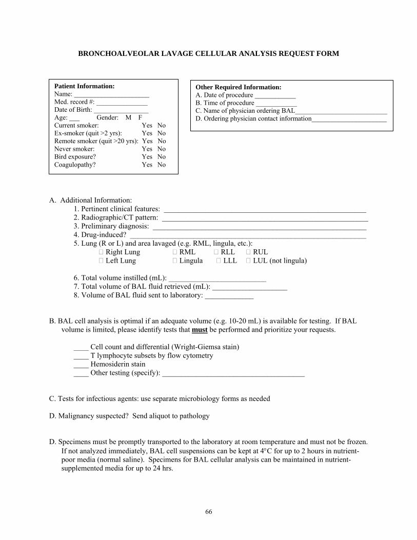
66
BRONCHOALVEOLAR LAVAGE CELLULAR ANALYSIS REQUEST FORM A. Additional Information: 1. Pertinent clinical features: ______________________________________________________ 2. Radiographic/CT pattern: _______________________________________________________ 3. Preliminary diagnosis: _________________________________________________________ 4. Drug-induced? _______________________________________________________________ 5. Lung (R or L) and area lavaged (e.g. RML, lingula, etc.): Right Lung RML RLL RUL Left Lung Lingula LLL LUL (not lingula) 6. Total volume instilled (mL): __________________________ 7. Total volume of BAL fluid retrieved (mL): ____________________ 8. Volume of BAL fluid sent to laboratory: _____________ B. BAL cell analysis is optimal if an adequate volume (e.g. 10-20 mL) is available for testing. If BAL
volume is limited, please identify tests that must be performed and prioritize your requests. ____ Cell count and differential (Wright-Giemsa stain) ____ T lymphocyte subsets by flow cytometry ____ Hemosiderin stain ____ Other testing (specify): ______________________________________ C. Tests for infectious agents: use separate microbiology forms as needed D. Malignancy suspected? Send aliquot to pathology D. Specimens must be promptly transported to the laboratory at room temperature and must not be frozen.
If not analyzed immediately, BAL cell suspensions can be kept at 4°C for up to 2 hours in nutrient-poor media (normal saline). Specimens for BAL cellular analysis can be maintained in nutrient-supplemented media for up to 24 hrs.
Other Required Information: A. Date of procedure ____________ B. Time of procedure ____________ C. Name of physician ordering BAL___________________________ D. Ordering physician contact information______________________
Patient Information: Name: ______________________ Med. record #: _______________ Date of Birth: ________________ Age: ___ Gender: M F Current smoker: Yes No Ex-smoker (quit >2 yrs): Yes No Remote smoker (quit >20 yrs): Yes No Never smoker: Yes No Bird exposure? Yes No Coagulopathy? Yes No
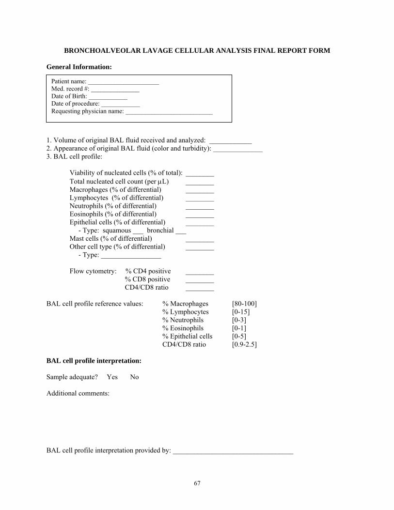
67
BRONCHOALVEOLAR LAVAGE CELLULAR ANALYSIS FINAL REPORT FORM General Information: 1. Volume of original BAL fluid received and analyzed: ____________ 2. Appearance of original BAL fluid (color and turbidity): ______________ 3. BAL cell profile:
Viability of nucleated cells (% of total): ________ Total nucleated cell count (per μL) ________ Macrophages (% of differential) ________ Lymphocytes (% of differential) ________ Neutrophils (% of differential) ________ Eosinophils (% of differential) ________ Epithelial cells (% of differential) ________ - Type: squamous ___ bronchial ___ Mast cells (% of differential) ________ Other cell type (% of differential) ________ - Type: _________________
Flow cytometry: % CD4 positive ________ % CD8 positive ________ CD4/CD8 ratio ________
BAL cell profile reference values: % Macrophages [80-100] % Lymphocytes [0-15] % Neutrophils [0-3] % Eosinophils [0-1] % Epithelial cells [0-5] CD4/CD8 ratio [0.9-2.5] BAL cell profile interpretation: Sample adequate? Yes No Additional comments: BAL cell profile interpretation provided by: __________________________________
Patient name: ______________________ Med. record #: _______________ Date of Birth: ____________ Date of procedure: ____________ Requesting physician name: ___________________________
68
REFERENCES 1. Stitt HL. Bronchial aspiration and irrigation with a hypertonis saline solution. J Med 1927;5:112-117. 2. Stitt HL. Bronchial lavage. Bull StLouis Medical Society 1932;26:246-249. 3. Kylstra JA, Rausch DC, Hall KD, Spock A. Volume-controlled lung lavage in the treatment of asthma, bronchiectasis, and mucoviscidosis. Am Rev Respir Dis 1971;103:651-665. 4. Ramirez J, Schultz RB, Dutton RE. Pulmonary alveolar proteinosis: A new technique and rationale for treatment. Arch Intern Med 1963;112:419-431. 5. Myrvik Q. ESL, and B. Fariss. Studies on pulmonary alveolar macrophages from the normal rabbit: A technique to procure them in a high state of purity. J Immunol 1961;101:128-132. 6. Collins FMaGBM. Delayed hypersensitivity and arthus reactivity in relation to host resistance in salmonella infected mice. J Immunol 1968;101:830-845. 7. Reynolds HYaRET. Pulmonary host defences. I. Analysis of protein and lipids in bronchial secretions and antibody responses after vaccination with pseudomonas aeruginosa. J Immunol 1973;111:358-368. 8. Reynolds HYaRET. Pulmonary host defenses. Ii. Interaction of respiratory antibodies with pseudomonas aeruginosa and alveolar macrophages. J Immunol 1974;111:369-380. 9. Reynolds HYaHHN. Analysis of proteins and respiratory cells obtained from human lungs by bronchial lavage. J Lab Clin Med 1974;84:559-573. 10. Reynolds HY. Use of bronchoalveolar lavage in humans-past necessity and future imperative. Lung 2000;178:271-293. 11. Klech H, W. Pohl and eds. Technical recommendations and guidelines for bronchoalveolar lavage (bal). Report of the european society of pneumology task group. Eur Respir J 1989;2:561-585. 12. Klech H, Hutter, and eds. Clinical guidelines and indications for bronchoalveolar lavage (bal); report of the european society of pneumonolgy task group on bal. Eur Respir J 1990;3:937-976. 13. Klech H, C. Hutter, U Costabel and eds. Clinical guidelines and indications for bronchoalveolar lavage (bal); report of the european society of pneumology task group on bal. European Respiratory Review 1992;2:47-127. 14. Haslam PL, Baughman RP. Report of ers task force: Guidelines for measurement of acellular components and standardization of bal. Eur Respir J 1999;14:245-248. 15. Committee TBC-oS. Bronchoalveolar lavage consituents in healthy individuals, idopathic pulmonary fibrosis, and selected comparison groups. Am Rev Respir Dis 1990;141:S169-S202. 16. Travis WD, T.E. King, Bateman, and et al. American thoracic society/european respiratory society international multidisciplinary concensus classification of the idiopathic interstitial pneumonias. This joint statement of the american thoracic society (ats), and the european respiratory society (ers) was adopted by the ats board of directors, june 2001 and by the ers executive committee, june 2001. Am J Respir Crit Care Med 2002;165:277-304. 17. Ahmad M, Livingston DR, Golish JA, Mehta AC, Wiedemann HP. The safety of outpatient transbronchial biopsy. Chest 1986;90:403-405. 18. Simpson FG, Arnold AG, Purvis A, Belfield PW, Muers MF, Cooke NJ. Postal survey of bronchoscopic practice by physicians in the united kingdom. Thorax 1986;41:311-317.
69
19. Kramer MR, Berkman N, Mintz B, Godfrey S, Saute M, Amir G. The role of open lung biopsy in the management and outcome of patients with diffuse lung disease. Ann Thorac Surg 1998;65:198-202. 20. Utz JP, Ryu JH, Douglas WW, Hartman TE, Tazelaar HD, Myers JL, Allen MS, Schroeder DR. High short-term mortality following lung biopsy for usual interstitial pneumonia. Eur Respir J 2001;17:175-179. 21. American thoracic society. Idiopathic pulmonary fibrosis: Diagnosis and treatment. International consensus statement. American thoracic society (ats), and the european respiratory society (ers). Am J Respir Crit Care Med 2000;161:646-664. 22. Flaherty KR, Travis WD, Colby TV, Toews GB, Kazerooni EA, Gross BH, Jain A, Strawderman RL, Flint A, Lynch JP, Martinez FJ. Histopathologic variability in usual and nonspecific interstitial pneumonias. Am J Respir Crit Care Med 2001;164:1722-1727. 23. Martin TR, Raghu G, Maunder RJ, Springmeyer SC. The effects of chronic bronchitis and chronic air-flow obstruction on lung cell populations recovered by bronchoalveolar lavage. Am Rev Respir Dis 1985;132:254-260. 24. Baughman RP. Asthma and other diseases. In baughman rp. St. Louis: Mosby; 1992. 25. Metzger WJ, Zavala D, Richerson HB, Moseley P, Iwamota P, Monick M, Sjoerdsma K, Hunninghake GW. Local allergen challenge and bronchoalveolar lavage of allergic asthmatic lungs. Description of the model and local airway inflammation. Am Rev Respir Dis 1987;135:433-440. 26. Metzger WJ, Nugent K, Richerson HB, Moseley P, Lakin R, Zavala D, Hunninghake GW. Methods for bronchoalveolar lavage in asthmatic patients following bronchoprovocation and local antigen challenge. Chest 1985;87:16S-19S. 27. Health NIo. Workshop summary. Summary and recommendations of a workshop on the investigative use of fiberoptic bronchoscopy and bronchoalveolar lavage in individuals with asthma. J Allergy Clin Immunol 1985;76:145-147. 28. Steinberg KP, Mitchell DR, Maunder RJ, Milberg JA, Whitcomb ME, Hudson LD. Safety of bronchoalveolar lavage in patients with adult respiratory distress syndrome. Am Rev Respir Dis 1993;148:556-561. 29. Guerra LFaRPB. Use of bronchoalveolar lavage to dignose bacterial pneumonia in mechanically ventilated patients. Crit Care Med 1990;18:169-173. 30. Kelly C, Ward C, Bird G, Hendrick D, Walters H. The effect of filtration on absolute and differential cell counts in fluid obtained at bronchoalveolar lavage. Respir Med 1989;83:107-110. 31. Peterson MW, Nugent KM, Jolles H, Monick M, Hunninghake GW. Uniformity of bronchoalveolar lavage in patients with pulmonary sarcoidosis. Am Rev Respir Dis 1988;137:79-84. 32. Garcia JG, Wolven RG, Garcia PL, Keogh BA. Assessment of interlobar variation of bronchoalveolar lavage cellular differentials in interstitial lung diseases. Am Rev Respir Dis 1986;133:444-449. 33. Baughman RP, Dohn MN, Shipley R, Buchsbaum JA, Frame PT. Increased pneumocystis carinii recovery from the upper lobes in pneumocystis pneumonia. The effect of aerosol pentamidine prophylaxis. Chest 1993;103:426-432. 34. Dohn MN, Baughman RP. Effect of changing instilled volume for bronchoalveolar lavage in patients with interstitial lung disease. Am Rev Respir Dis 1985;132:390-392. 35. Davis GS, Giancola MS, Costanza MC, Low RB. Analyses of sequential bronchoalveolar lavage samples from healthy human volunteers. Am Rev Respir Dis 1982;126:611-616.
70
36. Merrill W, O'Hearn E, Rankin J, Naegel G, Matthay RA, Reynolds HY. Kinetic analysis of respiratory tract proteins recovered during a sequential lavage protocol. Am Rev Respir Dis 1982;126:617-620. 37. Helmers RA, Dayton CS, Floerchinger C, Hunninghake GW. Bronchoalveolar lavage in interstitial lung disease: Effect of volume of fluid infused. J Appl Physiol 1989;67:1443-1446. 38. Thompson AB, Bohling T, Payvandi F, Rennard SI. Lower respiratory tract lactoferrin and lysozyme arise primarily in the airways and are elevated in association with chronic bronchitis. J Lab Clin Med 1990;115:148-158. 39. Rennard SI, Ghafouri M, Thompson AB, Linder J, Vaughan W, Jones K, Ertl RF, Christensen K, Prince A, Stahl MG, et al. Fractional processing of sequential bronchoalveolar lavage to separate bronchial and alveolar samples. Am Rev Respir Dis 1990;141:208-217. 40. Baughman RPRSI. Bronchoalveolar lavage: General approaches to correct for variability of dilution and lung permeability. Eur Respir J 1999:66, 28-31. 41. Baughman RP, Spencer RE, Kleykamp BO, Rashkin MC, Douthit MM. Ventilator associated pneumonia: Quality of nonbronchoscopic bronchoalveolar lavage sample affects diagnostic yield. Eur Respir J 2000;16:1152-1157. 42. Strumpf IJ, Feld MK, Cornelius MJ, Keogh BA, Crystal RG. Safety of fiberoptic bronchoalveolar lavage in evaluation of interstitial lung disease. Chest 1981;80:268-271. 43. Pingleton SK, Harrison GF, Stechschulte DJ, Wesselius LJ, Kerby GR, Ruth WE. Effect of location, ph, and temperature of instillate in bronchoalveolar lavage in normal volunteers. Am Rev Respir Dis 1983;128:1035-1037. 44. Dhillon DP, Haslam PL, Townsend PJ, Primett Z, Collins JV, Turner-Warwick M. Bronchoalveolar lavage in patients with interstitial lung diseases: Side effects and factors affecting fluid recovery. Eur J Respir Dis 1986;68:342-350. 45. Krause A, Hohberg B, Heine F, John M, Burmester GR, Witt C. Cytokines derived from alveolar macrophages induce fever after bronchoscopy and bronchoalveolar lavage. Am J Respir Crit Care Med 1997;155:1793-1797. 46. Standiford TJ, Kunkel SL, Strieter RM. Elevated serum levels of tumor necrosis factor-alpha after bronchoscopy and bronchoalveolar lavage. Chest 1991;99:1529-1530. 47. Huang YC, Bassett MA, Levin D, Montilla T, Ghio AJ. Acute phase reaction in healthy volunteers after bronchoscopy with lavage. Chest 2006;129:1565-1569. 48. Fagon JY, Chastre J, Wolff M, Gervais C, Parer-Aubas S, Stephan F, Similowski T, Mercat A, Diehl JL, Sollet JP, Tenaillon A. Invasive and noninvasive strategies for management of suspected ventilator-associated pneumonia. A randomized trial. Ann Intern Med 2000;132:621-630. 49. Hertz MI, Woodward ME, Gross CR, Swart M, Marcy TW, Bitterman PB. Safety of bronchoalveolar lavage in the critically ill, mechanically ventilated patient. Crit Care Med 1991;19:1526-1532. 50. Dransfield MT, Garver RI, Weill D. Standardized guidelines for surveillance bronchoscopy reduce complications in lung transplant recipients. J Heart Lung Transplant 2004;23:110-114. 51. Peikert T, Rana S, Edell ES. Safety, diagnostic yield, and therapeutic implications of flexible bronchoscopy in patients with febrile neutropenia and pulmonary infiltrates. Mayo Clin Proc 2005;80:1414-1420. 52. Weiss SM, Hert RC, Gianola FJ, Clark JG, Crawford SW. Complications of fiberoptic bronchoscopy in thrombocytopenic patients. Chest 1993;104:1025-1028.
71
53. Antonelli M, Conti G, Riccioni L, Meduri GU. Noninvasive positive-pressure ventilation via face mask during bronchoscopy with bal in high-risk hypoxemic patients. Chest 1996;110:724-728. 54. Bulpa PA, Dive AM, Mertens L, Delos MA, Jamart J, Evrard PA, Gonzalez MR, Installe EJ. Combined bronchoalveolar lavage and transbronchial lung biopsy: Safety and yield in ventilated patients. Eur Respir J 2003;21:489-494. 55. Cazzadori A, Di Perri G, Bonora S, Lanzafame M, Allegranzi B, Concia E. Fatal pneumothorax complicating bal in a bone marrow transplant recipient with bronchiolitis obliterans. Chest 1997;111:1468-1469. 56. Hiwatari N, Shimura S, Takishima T, Shirato K. Bronchoalveolar lavage as a possible cause of acute exacerbation in idiopathic pulmonary fibrosis patients. Tohoku J Exp Med 1994;174:379-386. 57. Kim DS, Park JH, Park BK, Lee JS, Nicholson AG, Colby T. Acute exacerbation of idiopathic pulmonary fibrosis: Frequency and clinical features. Eur Respir J 2006;27:143-150. 58. Schaberg T, Lauer C, Lode H, Fischer J, Haller H. Increased number of alveolar macrophages expressing adhesion molecules of the leukocyte adhesion molecule family in smoking subjects. Association with cell-binding ability and superoxide anion production. Am Rev Respir Dis 1992;146:1287-1293. 59. Mordelet Dambrine M, A. Arnoux, G. Stanislas-Leguern, D. Sandron, J. Chretien, and G. Huchon. Processing of lung lavage fluid causes variability in bronchoalveolar cell count. Am Rev Respir Dis 1984:305-306. 60. Willcox M, Kervitsky A, Watters LC, King TE, Jr. Quantification of cells recovered by bronchoalveolar lavage. Comparison of cytocentrifuge preparations with the filter method. Am Rev Respir Dis 1988;138:74-80. 61. Saltini C, Hance AJ, Ferrans VJ, Basset F, Bitterman PB, Crystal RG. Accurate quantification of cells recovered by bronchoalveolar lavage. Am Rev Respir Dis 1984;130:650-658. 62. Rankin J, G.P. Nagegal, and H.Y. Reynolds. . Use of a central laboratory for analysis of bronchoalveolar lavage fluid. Am Rev Respir Dis 1986:186-190. 63. Hebert MJ, Takano T, Holthofer H, Brady HR. Sequential morphologic events during apoptosis of human neutrophils. Modulation by lipoxygenase-derived eicosanoids. J Immunol 1996;157:3105-3115. 64. Savill JS, Wyllie AH, Henson JE, Walport MJ, Henson PM, Haslett C. Macrophage phagocytosis of aging neutrophils in inflammation. Programmed cell death in the neutrophil leads to its recognition by macrophages. J Clin Invest 1989;83:865-875. 65. Robbins RA, Linder J, Stahl MG, Thompson AB, 3rd, Haire W, Kessinger A, Armitage JO, Arneson M, Woods G, Vaughan WP, et al. Diffuse alveolar hemorrhage in autologous bone marrow transplant recipients. Am J Med 1989;87:511-518. 66. Maygarden SJ, Iacocca MV, Funkhouser WK, Novotny DB. Pulmonary alveolar proteinosis: A spectrum of cytologic, histochemical, and ultrastructural findings in bronchoalveolar lavage fluid. Diagn Cytopathol 2001;24:389-395. 67. Mariotta S, Guidi L, Papale M, Ricci A, Bisetti A. Pulmonary alveolar microlithiasis: Review of italian reports. Eur J Epidemiol 1997;13:587-590. 68. Fleury-Feith J, Escudier E, Pocholle MJ, Carre C, Bernaudin JF. The effects of cytocentrifugation on differential cell counts in samples obtained by bronchoalveolar lavage. Acta Cytol 1987;31:606-610.
72
69. (NCCLS) NCfCLS. Reference leukocyte differential count (proportional) and evaluation of instrumental methods.; 1992. 70. De Brauwer EI, Drent M, Mulder PG, Bruggeman CA, Wagenaar SS, Jacobs JA. Differential cell analysis of cytocentrifuged bronchoalveolar fluid samples affected by the area counted. Anal Quant Cytol Histol 2000;22:143-149. 71. Goh NS, Veeraraghavan S, Desai SR, Cramer D, Hansell DM, Denton CP, Black CM, du Bois RM, Wells AU. Bronchoalveolar lavage cellular profiles in patients with systemic sclerosis-associated interstitial lung disease are not predictive of disease progression. Arthritis Rheum 2007;56:2005-2012. 72. Meyer KC, Rosenthal NS, Soergel P, Peterson K. Neutrophils and low-grade inflammation in the seemingly normal aging human lung. Mech Ageing Dev 1998;104:169-181. 73. Meyer KC SP. Bronchoalveolar lymphocyte phenotypes change in the normal aging human lung. Thorax 1999:697-700. 74. Rennard SI, Basset G, Lecossier D, O'Donnell KM, Pinkston P, Martin PG, Crystal RG. Estimation of volume of epithelial lining fluid recovered by lavage using urea as marker of dilution. J Appl Physiol 1986;60:532-538. 75. Marcy TW, Merrill WW, Rankin JA, Reynolds HY. Limitations of using urea to quantify epithelial lining fluid recovered by bronchoalveolar lavage. Am Rev Respir Dis 1987;135:1276-1280. 76. Kelly CA, Fenwick JD, Corris PA, Fleetwood A, Hendrick DJ, Walters EH. Fluid dynamics during bronchoalveolar lavage. Am Rev Respir Dis 1988;138:81-84. 77. Baughman RP. The uncertainties of bronchoalveolar lavage. Eur Respir J 1997:1940-1942. 78. Baughman RP, Bosken CH, Loudon RG, Hurtubise P, Wesseler T. Quantitation of bronchoalveolar lavage with methylene blue. Am Rev Respir Dis 1983;128:266-270. 79. Cantral DE, Tape TG, Reed EC, Spurzem JR, Rennard SI, Thompson AB. Quantitative culture of bronchoalveolar lavage fluid for the diagnosis of bacterial pneumonia. Am J Med 1993;95:601-607. 80. Thorpe JE, Baughman RP, Frame PT, Wesseler TA, Staneck JL. Bronchoalveolar lavage for diagnosing acute bacterial pneumonia. J Infect Dis 1987;155:855-861. 81. Cushley M, Davidson, A, DuBois, R and et al. . The diagnosis, assessment and treatment of diffuse parenchymal lung disease in adults. Thorax 1999:S1-S30. 82. U. C. Atlas of bronchoalveolar lavage. Phildaelphia, PA: Chapman and Hall; 1998. 83. Wells AU, Hansell DM, Rubens MB, Cullinan P, Haslam PL, Black CM, Du Bois RM. Fibrosing alveolitis in systemic sclerosis. Bronchoalveolar lavage findings in relation to computed tomographic appearance. Am J Respir Crit Care Med 1994;150:462-468. 84. Allen JN, Davis WB. Eosinophilic lung diseases. Am J Respir Crit Care Med 1994;150:1423-1438. 85. Tabuena RP, Nagai S, Tsutsumi T, Handa T, Minoru T, Mikuniya T, Shigematsu M, Hamada K, Izumi T, Mishima M. Cell profiles of bronchoalveolar lavage fluid as prognosticators of idiopathic pulmonary fibrosis/usual interstitial pneumonia among japanese patients. Respiration 2005;72:490-498. 86. Kinder BW, Brown KK, Schwarz MI, Ix JH, Kervitsky A, King TE, Jr. Baseline bal neutrophilia predicts early mortality in idiopathic pulmonary fibrosis. Chest 2008;133:226-232.
73
87. Ohshimo S, Bonella F, Cui A, Beume M, Kohno N, Guzman J, Costabel U. Significance of bronchoalveolar lavage for the diagnosis of idiopathic pulmonary fibrosis. Am J Respir Crit Care Med 2009;179:1043-1047. 88. Katzenstein AL, Fiorelli RF. Nonspecific interstitial pneumonia/fibrosis. Histologic features and clinical significance. Am J Surg Pathol 1994;18:136-147. 89. Bjoraker JA, Ryu JH, Edwin MK, Myers JL, Tazelaar HD, Schroeder DR, Offord KP. Prognostic significance of histopathologic subsets in idiopathic pulmonary fibrosis. Am J Respir Crit Care Med 1998;157:199-203. 90. Nagai S, Kitaichi M, Itoh H, Nishimura K, Izumi T, Colby TV. Idiopathic nonspecific interstitial pneumonia/fibrosis: Comparison with idiopathic pulmonary fibrosis and boop. Eur Respir J 1998;12:1010-1019. 91. Cottin V, Donsbeck AV, Revel D, Loire R, Cordier JF. Nonspecific interstitial pneumonia. Individualization of a clinicopathologic entity in a series of 12 patients. Am J Respir Crit Care Med 1998;158:1286-1293. 92. Daniil ZD, Gilchrist FC, Nicholson AG, Hansell DM, Harris J, Colby TV, du Bois RM. A histologic pattern of nonspecific interstitial pneumonia is associated with a better prognosis than usual interstitial pneumonia in patients with cryptogenic fibrosing alveolitis. Am J Respir Crit Care Med 1999;160:899-905. 93. Nicholson AG, Colby TV, du Bois RM, Hansell DM, Wells AU. The prognostic significance of the histologic pattern of interstitial pneumonia in patients presenting with the clinical entity of cryptogenic fibrosing alveolitis. Am J Respir Crit Care Med 2000;162:2213-2217. 94. Shimizu S, Yoshinouchi T, Ohtsuki Y, Fujita J, Sugiura Y, Banno S, Yamadori I, Eimoto T, Ueda R. The appearance of s-100 protein-positive dendritic cells and the distribution of lymphocyte subsets in idiopathic nonspecific interstitial pneumonia. Respir Med 2002;96:770-776. 95. Bouros D, Wells AU, Nicholson AG, Colby TV, Polychronopoulos V, Pantelidis P, Haslam PL, Vassilakis DA, Black CM, du Bois RM. Histopathologic subsets of fibrosing alveolitis in patients with systemic sclerosis and their relationship to outcome. Am J Respir Crit Care Med 2002;165:1581-1586. 96. Yamadori I, Fujita J, Kajitani H, Bandoh S, Tokuda M, Ohtsuki Y, Yoshinouchi T, Okahara M, Yamaji Y, Tanimoto Y, Sato Y, Ishida T. Lymphocyte subsets in lung tissues of interstitial pneumonia associated with untreated polymyositis/dermatomyositis. Rheumatol Int 2001;21:89-93. 97. Crestani B, Valeyre D, Roden S, Wallaert B, Dalphin JC, Cordier JF. Bronchiolitis obliterans organizing pneumonia syndrome primed by radiation therapy to the breast. The groupe d'etudes et de recherche sur les maladies orphelines pulmonaires (germ"o"p). Am J Respir Crit Care Med 1998;158:1929-1935. 98. Majori M, Poletti V, Curti A, Corradi M, Falcone F, Pesci A. Bronchoalveolar lavage in bronchiolitis obliterans organizing pneumonia primed by radiation therapy to the breast. J Allergy Clin Immunol 2000;105:239-244. 99. Nagai S, Satake N, Kitaichi M, Izumi T. [interstitial pneumonia associated with collagen vascular diseases: Histological findings, and cells in bronchoalveolar lavage fluid]. Nihon Kyobu Shikkan Gakkai Zasshi 1995;33 Suppl:258-263.
74
100. Poletti V, Cazzato S, Minicuci N, Zompatori M, Burzi M, Schiattone ML. The diagnostic value of bronchoalveolar lavage and transbronchial lung biopsy in cryptogenic organizing pneumonia. Eur Respir J 1996;9:2513-2516. 101. Cohen AJ, King TE, Jr., Downey GP. Rapidly progressive bronchiolitis obliterans with organizing pneumonia. Am J Respir Crit Care Med 1994;149:1670-1675. 102. Lazor R, Vandevenne A, Pelletier A, Leclerc P, Court-Fortune I, Cordier JF. Cryptogenic organizing pneumonia. Characteristics of relapses in a series of 48 patients. The groupe d'etudes et de recherche sur les maladles "orphelines" pulmonaires (germ"o"p). Am J Respir Crit Care Med 2000;162:571-577. 103. Costabel U, Teschler H, Guzman J. Bronchiolitis obliterans organizing pneumonia (boop): The cytological and immunocytological profile of bronchoalveolar lavage. Eur Respir J 1992;5:791-797. 104. Forlani S, Ratta L, Bulgheroni A, Cascina A, Paschetto E, Cervio G, Luinetti O, Fietta AM, Meloni F. Cytokine profile of broncho-alveolar lavage in boop and uip. Sarcoidosis Vasc Diffuse Lung Dis 2002;19:47-53. 105. Majeski EI, Paintlia MK, Lopez AD, Harley RA, London SD, London L. Respiratory reovirus 1/l induction of intraluminal fibrosis, a model of bronchiolitis obliterans organizing pneumonia, is dependent on t lymphocytes. Am J Pathol 2003;163:1467-1479. 106. Allen JN, Davis WB, Pacht ER. Diagnostic significance of increased bronchoalveolar lavage fluid eosinophils. Am Rev Respir Dis 1990;142:642-647. 107. Pope-Harman AL, Davis WB, Allen ED, Christoforidis AJ, Allen JN. Acute eosinophilic pneumonia. A summary of 15 cases and review of the literature. Medicine (Baltimore) 1996;75:334-342. 108. Drent M, Wagenaar SS, Mulder PH, van Velzen-Blad H, Diamant M, van den Bosch JM. Bronchoalveolar lavage fluid profiles in sarcoidosis, tuberculosis, and non-hodgkin's and hodgkin's disease. An evaluation of differences. Chest 1994;105:514-519. 109. Drent M, van Velzen-Blad H, Diamant M, Wagenaar SS, Hoogsteden HC, van den Bosch JM. Bronchoalveolar lavage in extrinsic allergic alveolitis: Effect of time elapsed since antigen exposure. Eur Respir J 1993;6:1276-1281. 110. Drent M, Grutters JC, Mulder PG, van Velzen-Blad H, Wouters EF, van den Bosch JM. Is the different t helper cell activity in sarcoidosis and extrinsic allergic alveolitis also reflected by the cellular bronchoalveolar lavage fluid profile? Sarcoidosis Vasc Diffuse Lung Dis 1997;14:31-38. 111. Camus PH, Foucher P, Bonniaud PH, Ask K. Drug-induced infiltrative lung disease. Eur Respir J Suppl 2001;32:93s-100s. 112. Schnabel A, Csernok E, Braun J, Gross WL. Inflammatory cells and cellular activation in the lower respiratory tract in churg-strauss syndrome. Thorax 1999;54:771-778. 113. Peterson MW, Monick M, Hunninghake GW. Prognostic role of eosinophils in pulmonary fibrosis. Chest 1987;92:51-56. 114. Schnabel A, Reuter M, Gloeckner K, Muller-Quernheim J, Gross WL. Bronchoalveolar lavage cell profiles in wegener's granulomatosis. Respir Med 1999;93:498-506. 115. Jacobs JA, Dieleman MM, Cornelissen EI, Groen EA, Wagenaar SS, Drent M. Bronchoalveolar lavage fluid cytology in patients with pneumocystis carinii pneumonia. Acta Cytol 2001;45:317-326.
75
116. Djamin RS, Drent M, Schreurs AJ, Groen EA, Wagenaar SS. Diagnosis of pneumocystis carinii pneumonia in hiv-positive patients. Bronchoalveolar lavage vs. Bronchial brushing. Acta Cytol 1998;42:933-938. 117. Trisolini R, Cancellieri A, Bonaccorsi A, Poletti V. Bronchoalveolar lavage suggesting diffuse alveolar damage in a patient with acute eosinophilic pneumonia. Sarcoidosis Vasc Diffuse Lung Dis 2001;18:311-312. 118. Durieu J, Wallaert B, Tonnel AB. Long-term follow-up of pulmonary function in chronic eosinophilic pneumonia. Groupe d'etude en pathologie interstitielle de la societe de pathologie thoracique du nord. Eur Respir J 1997;10:286-291. 119. Fraig M, Shreesha U, Savici D, Katzenstein AL. Respiratory bronchiolitis: A clinicopathologic study in current smokers, ex-smokers, and never-smokers. Am J Surg Pathol 2002;26:647-653. 120. Ryu JH, Myers JL, Swensen SJ. Bronchiolar disorders. Am J Respir Crit Care Med 2003;168:1277-1292. 121. Wells AU, Nicholson AG, Hansell DM, du Bois RM. Respiratory bronchiolitis-associated interstitial lung disease. Semin Respir Crit Care Med 2003;24:585-594. 122. Davies G, Wells AU, du Bois RM. Respiratory bronchiolitis associated with interstitial lung disease and desquamative interstitial pneumonia. Clin Chest Med 2004;25:717-726, vi. 123. Ryu JH, Colby TV, Hartman TE, Vassallo R. Smoking-related interstitial lung diseases: A concise review. Eur Respir J 2001;17:122-132. 124. Heyneman LE, Ward S, Lynch DA, Remy-Jardin M, Johkoh T, Muller NL. Respiratory bronchiolitis, respiratory bronchiolitis-associated interstitial lung disease, and desquamative interstitial pneumonia: Different entities or part of the spectrum of the same disease process? AJR Am J Roentgenol 1999;173:1617-1622. 125. Hunninghake GW, Crystal RG. Cigarette smoking and lung destruction. Accumulation of neutrophils in the lungs of cigarette smokers. Am Rev Respir Dis 1983;128:833-838. 126. King TE, Jr. Respiratory bronchiolitis-associated interstitial lung disease. Clin Chest Med 1993;14:693-698. 127. Nagai S, Kitaichi M, Izumi T. Classification and recent advances in idiopathic interstitial pneumonia. Curr Opin Pulm Med 1998;4:256-260. 128. Marques LJ, Teschler H, Guzman J, Costabel U. Smoker's lung transplanted to a nonsmoker. Long-term detection of smoker's macrophages. Am J Respir Crit Care Med 1997;156:1700-1702. 129. Craig PJ, Wells AU, Doffman S, Rassl D, Colby TV, Hansell DM, Du Bois RM, Nicholson AG. Desquamative interstitial pneumonia, respiratory bronchiolitis and their relationship to smoking. Histopathology 2004;45:275-282. 130. Ryu JH, Myers JL, Capizzi SA, Douglas WW, Vassallo R, Decker PA. Desquamative interstitial pneumonia and respiratory bronchiolitis-associated interstitial lung disease. Chest 2005;127:178-184. 131. Raghu G. Is bronchoalveolar lavage clinically useful for everyday practice in interstitial lung disease? Journal of Bronchology 1999:217-221. 132. Costabel U, Guzman J. Bronchoalveolar lavage in interstitial lung disease. Curr Opin Pulm Med 2001;7:255-261. 133. Baughman RP, Drent M. Role of bronchoalveolar lavage in interstitial lung disease. Clin Chest Med 2001;22:331-341.
76
134. Rottoli P, Bargagli E. Is bronchoalveolar lavage obsolete in the diagnosis of interstitial lung disease? Curr Opin Pulm Med 2003;9:418-425. 135. Meyer KC. The role of bronchoalveolar lavage in interstitial lung disease. Clin Chest Med 2004;25:637-649, v. 136. Veeraraghavan S, Latsi PI, Wells AU, Pantelidis P, Nicholson AG, Colby TV, Haslam PL, Renzoni EA, du Bois RM. Bal findings in idiopathic nonspecific interstitial pneumonia and usual interstitial pneumonia. Eur Respir J 2003;22:239-244. 137. Raghu G, Brown KK. Interstitial lung disease: Clinical evaluation and keys to an accurate diagnosis. Clin Chest Med 2004;25:409-419, v. 138. Hamman LaAR, Rich. Fulminating diffuse interstitial fibrosis of the lungs. Trans Am Prog Climat Assoc 1935:154-163. 139. Liebow AA. Definition and classification of the interstitial pneumonias in human lung. Prog Respir Res 1975:1-31. 140. Askin FB. Back to the future: The hamman-rich syndrome and acute interstitial pneumonia. Mayo Clin Proc 1990;65:1624-1626. 141. Katzenstein AL, Myers JL, Mazur MT. Acute interstitial pneumonia. A clinicopathologic, ultrastructural, and cell kinetic study. Am J Surg Pathol 1986;10:256-267. 142. Bonaccorsi A, Cancellieri A, Chilosi M, Trisolini R, Boaron M, Crimi N, Poletti V. Acute interstitial pneumonia: Report of a series. Eur Respir J 2003;21:187-191. 143. Liebow AAaCBC. The interstitial pneumonias. In: M. Simon EJPaML, editor. Frontiers of pulmonary radiology, 1st ed. New York: Grune & Stratton; 1969. p. 102-141. 144. Koss MN, Hochholzer L, Langloss JM, Wehunt WD, Lazarus AA. Lymphoid interstitial pneumonia: Clinicopathological and immunopathological findings in 18 cases. Pathology 1987;19:178-185. 145. Nicholson AG, Wotherspoon AC, Diss TC, Hansell DM, Du Bois R, Sheppard MN, Isaacson PG, Corrin B. Reactive pulmonary lymphoid disorders. Histopathology 1995;26:405-412. 146. Kradin RL, Mark EJ. Benign lymphoid disorders of the lung, with a theory regarding their development. Hum Pathol 1983;14:857-867. 147. Yousem SA, Colby TV, Carrington CB. Follicular bronchitis/bronchiolitis. Hum Pathol 1985;16:700-706. 148. Cha SI, Fessler MB, Cool CD, Schwarz MI, Brown KK. Lymphoid interstitial pneumonia: Clinical features, associations and prognosis. Eur Respir J 2006;28:364-369. 149. Pisani RJ, Witzig TE, Li CY, Morris MA, Thibodeau SN. Confirmation of lymphomatous pulmonary involvement by immunophenotypic and gene rearrangement analysis of bronchoalveolar lavage fluid. Mayo Clin Proc 1990;65:651-656. 150. Philippe B, Delfau-Larue MH, Epardeau B, Autran B, Clauvel JP, Farcet JP, Couderc LJ. B-cell pulmonary lymphoma: Gene rearrangement analysis of bronchoalveolar lymphocytes by polymerase chain reaction. Chest 1999;115:1242-1247. 151. Swigris JJ, Berry GJ, Raffin TA, Kuschner WG. Lymphoid interstitial pneumonia: A narrative review. Chest 2002;122:2150-2164. 152. Poulter LW, Rossi GA, Bjermer L, Costabel U, Israel-Biet D, Klech H, Pohl W, Velluti G. The value of bronchoalveolar lavage in the diagnosis and prognosis of sarcoidosis. Eur Respir J 1990;3:943-944. 153. Hunninghake GW, Costabel U, Ando M, Baughman R, Cordier JF, du Bois R, Eklund A, Kitaichi M, Lynch J, Rizzato G, Rose C, Selroos O, Semenzato G, Sharma OP. Ats/ers/wasog
77
statement on sarcoidosis. American thoracic society/european respiratory society/world association of sarcoidosis and other granulomatous disorders. Sarcoidosis Vasc Diffuse Lung Dis 1999;16:149-173. 154. Yeager H, Jr., Williams MC, Beekman JF, Bayly TC, Beaman BL. Sarcoidosis: Analysis of cells obtained by bronchial lavage. Am Rev Respir Dis 1977;116:951-954. 155. Health NIo. Pulmonary sarcoidosis: A disease characterized and perpetuated by activated lung t lymphocytes. Ann Intern Med 1981:73-94. 156. Costabel U, Bross KJ, Matthys H. Pulmonary sarcoidosis: Assessment of disease activity by lung lymphocyte subpopulations. Klin Wochenschr 1983;61:349-356. 157. Hunninghake GW, Kawanami O, Ferrans VJ, Young RC, Jr., Roberts WC, Crystal RG. Characterization of the inflammatory and immune effector cells in the lung parenchyma of patients with interstitial lung disease. Am Rev Respir Dis 1981;123:407-412. 158. Hunninghake GW, Bedell GN, Zavala DC, Monick M, Brady M. Role of interleukin-2 release by lung t-cells in active pulmonary sarcoidosis. Am Rev Respir Dis 1983;128:634-638. 159. Muller-Quernheim J, Saltini C, Sondermeyer P, Crystal RG. Compartmentalized activation of the interleukin 2 gene by lung t lymphocytes in active pulmonary sarcoidosis. J Immunol 1986;137:3475-3483. 160. Robinson BW, McLemore TL, Crystal RG. Gamma interferon is spontaneously released by alveolar macrophages and lung t lymphocytes in patients with pulmonary sarcoidosis. J Clin Invest 1985;75:1488-1495. 161. Semenzato G, Agostini C, Trentin L, Zambello R, Chilosi M, Cipriani A, Ossi E, Angi MR, Morittu L, Pizzolo G. Evidence of cells bearing interleukin-2 receptor at sites of disease activity in sarcoid patients. Clin Exp Immunol 1984;57:331-337. 162. Moller DR, Forman JD, Liu MC, Noble PW, Greenlee BM, Vyas P, Holden DA, Forrester JM, Lazarus A, Wysocka M, Trinchieri G, Karp C. Enhanced expression of il-12 associated with th1 cytokine profiles in active pulmonary sarcoidosis. J Immunol 1996;156:4952-4960. 163. Prasse A, Georges CG, Biller H, Hamm H, Matthys H, Luttmann W, Virchow JC, Jr. Th1 cytokine pattern in sarcoidosis is expressed by bronchoalveolar cd4+ and cd8+ t cells. Clin Exp Immunol 2000;122:241-248. 164. Wahlstrom J, Katchar K, Wigzell H, Olerup O, Eklund A, Grunewald J. Analysis of intracellular cytokines in cd4+ and cd8+ lung and blood t cells in sarcoidosis. Am J Respir Crit Care Med 2001;163:115-121. 165. Shigehara K, Shijubo N, Ohmichi M, Yamada G, Takahashi R, Okamura H, Kurimoto M, Hiraga Y, Tatsuno T, Abe S, Sato N. Increased levels of interleukin-18 in patients with pulmonary sarcoidosis. Am J Respir Crit Care Med 2000;162:1979-1982. 166. Shigehara K, Shijubo N, Ohmichi M, Takahashi R, Kon S, Okamura H, Kurimoto M, Hiraga Y, Tatsuno T, Abe S, Sato N. Il-12 and il-18 are increased and stimulate ifn-gamma production in sarcoid lungs. J Immunol 2001;166:642-649. 167. Bachwich PR, Lynch JP, 3rd, Larrick J, Spengler M, Kunkel SL. Tumor necrosis factor production by human sarcoid alveolar macrophages. Am J Pathol 1986;125:421-425. 168. Eden E, Turino GM. Interleukin 1 secretion from human alveolar macrophages in lung disease. J Clin Immunol 1986;6:326-333. 169. Hunninghake GW. Release of interleukin-1 by alveolar macrophages of patients with active pulmonary sarcoidosis. Am Rev Respir Dis 1984;129:569-572.
78
170. Ziegenhagen MW, Rothe ME, Zissel G, Muller-Quernheim J. Exaggerated tnfalpha release of alveolar macrophages in corticosteroid resistant sarcoidosis. Sarcoidosis Vasc Diffuse Lung Dis 2002;19:185-190. 171. Ziegenhagen MW, Benner UK, Zissel G, Zabel P, Schlaak M, Muller-Quernheim J. Sarcoidosis: Tnf-alpha release from alveolar macrophages and serum level of sil-2r are prognostic markers. Am J Respir Crit Care Med 1997;156:1586-1592. 172. Ziegenhagen MW, Rothe ME, Schlaak M, Muller-Quernheim J. Bronchoalveolar and serological parameters reflecting the severity of sarcoidosis. Eur Respir J 2003;21:407-413. 173. Drent M, Jacobs JA, de Vries J, Lamers RJ, Liem IH, Wouters EF. Does the cellular bronchoalveolar lavage fluid profile reflect the severity of sarcoidosis? Eur Respir J 1999;13:1338-1344. 174. Semenzato G, Chilosi M, Ossi E, Trentin L, Pizzolo G, Cipriani A, Agostini C, Zambello R, Marcer G, Gasparotto G. Bronchoalveolar lavage and lung histology. Comparative analysis of inflammatory and immunocompetent cells in patients with sarcoidosis and hypersensitivity pneumonitis. Am Rev Respir Dis 1985;132:400-404. 175. Campbell DA, Poulter LW, du Bois RM. Immunocompetent cells in bronchoalveolar lavage reflect the cell populations in transbronchial biopsies in pulmonary sarcoidosis. Am Rev Respir Dis 1985;132:1300-1306. 176. Paradis IL, Dauber JH, Rabin BS. Lymphocyte phenotypes in bronchoalveolar lavage and lung tissue in sarcoidosis and idiopathic pulmonary fibrosis. Am Rev Respir Dis 1986;133:855-860. 177. Bjermer L, Rosenhall L, Angstrom T, Hallgren R. Predictive value of bronchoalveolar lavage cell analysis in sarcoidosis. Thorax 1988;43:284-288. 178. Costabel U. The alveolitis of hypersensitivity pneumonitis. Eur Respir J 1988;1:5-9. 179. Costabel U, Bross KJ, Ruhle KH, Lohr GW, Matthys H. Ia-like antigens on t-cells and their subpopulations in pulmonary sarcoidosis and in hypersensitivity pneumonitis. Analysis of bronchoalveolar and blood lymphocytes. Am Rev Respir Dis 1985;131:337-342. 180. Haslam PL, Dewar A, Butchers P, Primett ZS, Newman-Taylor A, Turner-Warwick M. Mast cells, atypical lymphocytes, and neutrophils in bronchoalveolar lavage in extrinsic allergic alveolitis. Comparison with other interstitial lung diseases. Am Rev Respir Dis 1987;135:35-47. 181. Drent M, van Nierop MA, Gerritsen FA, Wouters EF, Mulder PG. A computer program using balf-analysis results as a diagnostic tool in interstitial lung diseases. Am J Respir Crit Care Med 1996;153:736-741. 182. Kantrow SP, Meyer KC, Kidd P, Raghu G. The cd4/cd8 ratio in bal fluid is highly variable in sarcoidosis. Eur Respir J 1997;10:2716-2721. 183. Costabel U, A. Zaiss, and J. Guzman. Sensitivity and specificity of bal findings in sarcoidosis. Sarcoidosis 1992;Suppl 1:211-214. 184. Winterbauer RH, Lammert J, Selland M, Wu R, Corley D, Springmeyer SC. Bronchoalveolar lavage cell populations in the diagnosis of sarcoidosis. Chest 1993;104:352-361. 185. Thomeer MaMD. Predictive value of cd4/cd8 ratio in bronchoalveolar lavage in the diagnosis of sarcoidosis (abstract). Sar Vase Diffuse Lung Dis 1997;Suppl 1:36. 186. Welker L, Jorres RA, Costabel U, Magnussen H. Predictive value of bal cell differentials in the diagnosis of interstitial lung diseases. Eur Respir J 2004;24:1000-1006.
79
187. Keogh BA, Hunninghake GW, Line BR, Crystal RG. The alveolitis of pulmonary sarcoidosis. Evaluation of natural history and alveolitis-dependent changes in lung function. Am Rev Respir Dis 1983;128:256-265. 188. Israel-Biet D, Venet A, Chretien J. Persistent high alveolar lymphocytosis as a predictive criterion of chronic pulmonary sarcoidosis. Ann N Y Acad Sci 1986;465:395-406. 189. Verstraeten A, Demedts M, Verwilghen J, van den Eeckhout A, Marien G, Lacquet LM, Ceuppens JL. Predictive value of bronchoalveolar lavage in pulmonary sarcoidosis. Chest 1990;98:560-567. 190. Laviolette M, La Forge J, Tennina S, Boulet LP. Prognostic value of bronchoalveolar lavage lymphocyte count in recently diagnosed pulmonary sarcoidosis. Chest 1991;100:380-384. 191. Baughman RP, Shipley R, Eisentrout CE. Predictive value of gallium scan, angiotensin-converting enzyme level, and bronchoalveolar lavage in two-year follow-up of pulmonary sarcoidosis. Lung 1987;165:371-377. 192. Ward K, O'Connor C, Odlum C, Fitzgerald MX. Prognostic value of bronchoalveolar lavage in sarcoidosis: The critical influence of disease presentation. Thorax 1989;44:6-12. 193. Selman M. Hypersensitivity pneumonitis. In: King MSaT, editor. Interstitial lung disease, 4th ed. Hamilton: B. C. Decker; 2003. p. 452-484. 194. Depierre A, Dalphin JC, Pernet D, Dubiez A, Faucompre C, Breton JL. Epidemiological study of farmer's lung in five districts of the french doubs province. Thorax 1988;43:429-435. 195. Warren CP. Extrinsic allergic alveolitis: A disease commoner in non-smokers. Thorax 1977;32:567-569. 196. Arima K, M. Ando, K. Ito, T. Sakata, T. Yamaguchi, S. Araki and M. Futatsuka. Effect of cigarette smoking on prevalence of summer type hypersensitivity pneumonitis caused by trichosporon cutaneum. Arch Environ Health 1992:274-278. 197. Dalphin JC, Debieuvre D, Pernet D, Maheu MF, Polio JC, Toson B, Dubiez A, Monnet E, Laplante JJ, Depierre A. Prevalence and risk factors for chronic bronchitis and farmer's lung in french dairy farmers. Br J Ind Med 1993;50:941-944. 198. Hughes DA, Haslam PL. Effect of smoking on the lipid composition of lung lining fluid and relationship between immunostimulatory lipids, inflammatory cells and foamy macrophages in extrinsic allergic alveolitis. Eur Respir J 1990;3:1128-1139. 199. Moszczynski P, Zabinski Z, Moszczynski P, Jr., Rutowski J, Slowinski S, Tabarowski Z. Immunological findings in cigarette smokers. Toxicol Lett 2001;118:121-127. 200. Ohtsuka Y, Munakata M, Tanimura K, Ukita H, Kusaka H, Masaki Y, Doi I, Ohe M, Amishima M, Homma Y, et al. Smoking promotes insidious and chronic farmer's lung disease, and deteriorates the clinical outcome. Intern Med 1995;34:966-971. 201. Cormier Y, Gagnon L, Berube-Genest F, Fournier M. Sequential bronchoalveolar lavage in experimental extrinsic allergic alveolitis. The influence of cigarette smoking. Am Rev Respir Dis 1988;137:1104-1109. 202. Cormier Y, Israel-Assayag E, Desmeules M, Lesur O. Effect of contact avoidance or treatment with oral prednisolone on bronchoalveolar lavage surfactant protein a levels in subjects with farmer's lung. Thorax 1996;51:1210-1215. 203. Ratjen F, Costabel U, Griese M, Paul K. Bronchoalveolar lavage fluid findings in children with hypersensitivity pneumonitis. Eur Respir J 2003;21:144-148. 204. Yoshizawa Y, Ohtani Y, Hayakawa H, Sato A, Suga M, Ando M. Chronic hypersensitivity pneumonitis in japan: A nationwide epidemiologic survey. J Allergy Clin Immunol 1999;103:315-320.
80
205. Pardo A, Smith KM, Abrams J, Coffman R, Bustos M, McClanahan TK, Grein J, Murphy EE, Zlotnik A, Selman M. Ccl18/dc-ck-1/parc up-regulation in hypersensitivity pneumonitis. J Leukoc Biol 2001;70:610-616. 206. Barrera L, Mendoza F, Zuniga J, Estrada A, Zamora AC, Melendro EI, Ramirez R, Pardo A, Selman M. Functional diversity of t-cell subpopulations in subacute and chronic hypersensitivity pneumonitis. Am J Respir Crit Care Med 2008;177:44-55. 207. Cormier Y, Belanger J, Laviolette M. Persistent bronchoalveolar lymphocytosis in asymptomatic farmers. Am Rev Respir Dis 1986;133:843-847. 208. Semenzato G, Trentin L, Zambello R, Agostini C, Cipriani A, Marcer G. Different types of cytotoxic lymphocytes recovered from the lungs of patients with hypersensitivity pneumonitis. Am Rev Respir Dis 1988;137:70-74. 209. Trentin L, Migone N, Zambello R, di Celle PF, Aina F, Feruglio C, Bulian P, Masciarelli M, Agostini C, Cipriani A, et al. Mechanisms accounting for lymphocytic alveolitis in hypersensitivity pneumonitis. J Immunol 1990;145:2147-2154. 210. Trentin L, Zambello R, Facco M, Tassinari C, Sancetta R, Siviero M, Cerutti A, Cipriani A, Marcer G, Majori M, Pesci A, Agostini C, Semenzato G. Selection of t lymphocytes bearing limited tcr-vbeta regions in the lung of hypersensitivity pneumonitis and sarcoidosis. Am J Respir Crit Care Med 1997;155:587-596. 211. Semenzato G. Immunology of interstitial lung diseases: Cellular events taking place in the lung of sarcoidosis, hypersensitivity pneumonitis and hiv infection. Eur Respir J 1991;4:94-102. 212. Lohmeyer J, Friedrich J, Grimminger F, Maus U, Tenter R, Morr H, Velcovsky HG, Seeger W, Rosseau S. Expression of mucosa-related integrin alphaebeta7 on alveolar t cells in interstitial lung diseases. Clin Exp Immunol 1999;116:340-346. 213. Tsushima K, Fujimoto K, Yamazaki Y, Takamizawa A, Amari T, Koizumi T, Kubo K. Hypersensitivity pneumonitis induced by spores of lyophyllum aggregatum. Chest 2001;120:1085-1093. 214. Ando M, Konishi K, Yoneda R, Tamura M. Difference in the phenotypes of bronchoalveolar lavage lymphocytes in patients with summer-type hypersensitivity pneumonitis, farmer's lung, ventilation pneumonitis, and bird fancier's lung: Report of a nationwide epidemiologic study in japan. J Allergy Clin Immunol 1991;87:1002-1009. 215. Murayama J, Yoshizawa Y, Ohtsuka M, Hasegawa S. Lung fibrosis in hypersensitivity pneumonitis. Association with cd4+ but not cd8+ cell dominant alveolitis and insidious onset. Chest 1993;104:38-43. 216. McSharry C. B lymphocytes in allergic alveolitis. Clin Exp Allergy 2003;33:159-162. 217. Dai H, Guzman J, Bauer PC, Costabel U. Elevated levels of soluble tnf receptors in bronchoalveolar lavage fluid in extrinsic allergic alveolitis. Clin Exp Allergy 1999;29:1209-1213. 218. Drent M, van Velzen-Blad H, Diamant M, Wagenaar SS, Donckerwolck-Bogaert M, van den Bosch JM. Differential diagnostic value of plasma cells in bronchoalveolar lavage fluid. Chest 1993;103:1720-1724. 219. Haslam PL PD, Townsend PJ. Increases in hla-dq, dp, dr and transferrin receptors on alveolar macrophages in sarcoidosis and allergic alveolitis compared with fibrosing alveolitis. Chest 1990;97:651-661. 220. Pforte A, Schiessler A, Gais P, von Kress S, Beer B, Riethmuller G, Ziegler-Heitbrock HW. Expression of the adhesion molecule icam-1 on alveolar macrophages and in serum in extrinsic allergic alveolitis. Respiration 1993;60:221-226.
81
221. Guzman J, Wang YM, Kalaycioglu O, Schoenfeld B, Hamm H, Bartsch W, Costabel U. Increased surfactant protein a content in human alveolar macrophages in hypersensitivity pneumonitis. Acta Cytol 1992;36:668-673. 222. Fournier E, Tonnel AB, Gosset P, Wallaert B, Ameisen JC, Voisin C. Early neutrophil alveolitis after antigen inhalation in hypersensitivity pneumonitis. Chest 1985;88:563-566. 223. Ohtani Y, Kojima K, Sumi Y, Sawada M, Inase N, Miyake S, Yoshizawa Y. Inhalation provocation tests in chronic bird fancier's lung. Chest 2000;118:1382-1389. 224. Vogelmeier C, Krombach F, Munzing S, Konig G, Mazur G, Beinert T, Fruhmann G. Activation of blood neutrophils in acute episodes of farmer's lung. Am Rev Respir Dis 1993;148:396-400. 225. Behr J, Degenkolb B, Beinert T, Krombach F, Vogelmeier C. Pulmonary glutathione levels in acute episodes of farmer's lung. Am J Respir Crit Care Med 2000;161:1968-1971. 226. Pardo A, Barrios R, Gaxiola M, Segura-Valdez L, Carrillo G, Estrada A, Mejia M, Selman M. Increase of lung neutrophils in hypersensitivity pneumonitis is associated with lung fibrosis. Am J Respir Crit Care Med 2000;161:1698-1704. 227. Miadonna A, Pesci A, Tedeschi A, Bertorelli G, Arquati M, Olivieri D. Mast cell and histamine involvement in farmer's lung disease. Chest 1994;105:1184-1189. 228. Ishida T, Matsui Y, Matsumura Y, Fujimori N, Furutani M. Bronchoalveolar mast cells in summer-type hypersensitivity pneumonitis: Increase in numbers and ultrastructural evidence of degranulation. Intern Med 1995;34:357-363. 229. Schildge J, Klar B, Hardung-Backes M. [mast cells in bronchoalveolar lavage fluid of patients with interstitial lung diseases]. Pneumologie 2003;57:202-207. 230. Laviolette M, Cormier Y, Loiseau A, Soler P, Leblanc P, Hance AJ. Bronchoalveolar mast cells in normal farmers and subjects with farmer's lung. Diagnostic, prognostic, and physiologic significance. Am Rev Respir Dis 1991;144:855-860. 231. Weinberger SE, Kelman JA, Elson NA, Young RC, Jr., Reynolds HY, Fulmer JD, Crystal RG. Bronchoalveolar lavage in interstitial lung disease. Ann Intern Med 1978;89:459-466. 232. Frigieri L, Mormile F, Grilli N, Mancini D, Ciappi G, Pagliari G, Magaro M, Flamini G. Bilateral bronchoalveolar lavage in progressive systemic sclerosis: Interlobar variability, lymphocyte subpopulations, and functional correlations. Respiration 1991;58:132-140. 233. Silver RM, Miller KS, Kinsella MB, Smith EA, Schabel SI. Evaluation and management of scleroderma lung disease using bronchoalveolar lavage. Am J Med 1990;88:470-476. 234. Schnabel A, Reuter M, Gross WL. Intravenous pulse cyclophosphamide in the treatment of interstitial lung disease due to collagen vascular diseases. Arthritis Rheum 1998;41:1215-1220. 235. Silver RM, Metcalf JF, Stanley JH, LeRoy EC. Interstitial lung disease in scleroderma. Analysis by bronchoalveolar lavage. Arthritis Rheum 1984;27:1254-1262. 236. Miller KS, Smith EA, Kinsella M, Schabel SI, Silver RM. Lung disease associated with progressive systemic sclerosis. Assessment of interlobar variation by bronchoalveolar lavage and comparison with noninvasive evaluation of disease activity. Am Rev Respir Dis 1990;141:301-306. 237. Behr J, Vogelmeier C, Beinert T, Meurer M, Krombach F, Konig G, Fruhmann G. Bronchoalveolar lavage for evaluation and management of scleroderma disease of the lung. Am J Respir Crit Care Med 1996;154:400-406.
82
238. Wells AU, Hansell DM, Haslam PL, Rubens MB, Cailes J, Black CM, du Bois RM. Bronchoalveolar lavage cellularity: Lone cryptogenic fibrosing alveolitis compared with the fibrosing alveolitis of systemic sclerosis. Am J Respir Crit Care Med 1998;157:1474-1482. 239. Wells AU, Hansell DM, Corrin B, Harrison NK, Goldstraw P, Black CM, du Bois RM. High resolution computed tomography as a predictor of lung histology in systemic sclerosis. Thorax 1992;47:738-742. 240. White B, Moore WC, Wigley FM, Xiao HQ, Wise RA. Cyclophosphamide is associated with pulmonary function and survival benefit in patients with scleroderma and alveolitis. Ann Intern Med 2000;132:947-954. 241. Strange C, Bolster MB, Roth MD, Silver RM, Theodore A, Goldin J, Clements P, Chung J, Elashoff RM, Suh R, Smith EA, Furst DE, Tashkin DP. Bronchoalveolar lavage and response to cyclophosphamide in scleroderma interstitial lung disease. Am J Respir Crit Care Med 2008;177:91-98. 242. Garcia JG, Parhami N, Killam D, Garcia PL, Keogh BA. Bronchoalveolar lavage fluid evaluation in rheumatoid arthritis. Am Rev Respir Dis 1986;133:450-454. 243. Popp W, Ritschka L, Scherak O, Braun O, Kolarz G, Rauscher H, Zwick H. Bronchoalveolar lavage in rheumatoid arthritis and secondary sjogren's syndrome. Lung 1990;168:221-231. 244. Ishioka S, Inyaku K, Shirotani Y, Nakamura K, Oyama T, Hozawa S, Takahashi K, Yamakido M. [bronchoalveolar lavage fluid findings in rheumatoid arthritis]. Nihon Kyobu Shikkan Gakkai Zasshi 1992;30:614-618. 245. Dalavanga YA, Voulgari PV, Georgiadis AN, Leontaridi C, Katsenos S, Vassiliou M, Drosos AA, Constantopoulos SH. Lymphocytic alveolitis: A surprising index of poor prognosis in patients with primary sjogren's syndrome. Rheumatol Int 2006;26:799-804. 246. Deheinzelin D, Capelozzi VL, Kairalla RA, Barbas Filho JV, Saldiva PH, de Carvalho CR. Interstitial lung disease in primary sjogren's syndrome. Clinical-pathological evaluation and response to treatment. Am J Respir Crit Care Med 1996;154:794-799. 247. Salaffi F, Manganelli P, Carotti M, Baldelli S, Blasetti P, Subiaco S, Binci MC, Bichi Secchi E, Amici F, Cervini C. A longitudinal study of pulmonary involvement in primary sjogren's syndrome: Relationship between alveolitis and subsequent lung changes on high-resolution computed tomography. Br J Rheumatol 1998;37:263-269. 248. Grau JM, Miro O, Pedrol E, Casademont J, Masanes F, Herrero C, Haussman G, Urbano-Marquez A. Interstitial lung disease related to dermatomyositis. Comparative study with patients without lung involvement. J Rheumatol 1996;23:1921-1926. 249. Komocsi A, Kumanovics G, Zibotics H, Czirjak L. Alveolitis may persist during treatment that sufficiently controls muscle inflammation in myositis. Rheumatol Int 2001;20:113-118. 250. Schnabel A, Reuter M, Biederer J, Richter C, Gross WL. Interstitial lung disease in polymyositis and dermatomyositis: Clinical course and response to treatment. Semin Arthritis Rheum 2003;32:273-284. 251. Enomoto K, Takada T, Suzuki E, Ishida T, Moriyama H, Ooi H, Hasegawa T, Tsukada H, Nakano M, Gejyo F. Bronchoalveolar lavage fluid cells in mixed connective tissue disease. Respirology 2003;8:149-156. 252. Pesci A, Bertorelli G, Manganelli P, Ambanelli U. Bronchoalveolar lavage analysis of interstitial lung disease in crest syndrome. Clin Exp Rheumatol 1986;4:121-124.
83
253. Kumanovics G, Magyarlaki T, Komocsi A, Szekeres G, Czirjak L. Simultaneous presence of neutrophil alveolitis and ki-67 positivity of alveolar macrophages in dermato-/polymyositis and systemic sclerosis. Rheumatol Int 2003;23:6-10. 254. Lassalle P, Gosset P, Aerts C, Fournier E, Lafitte JJ, Degreef JM, Wallaert B, Tonnel AB, Voisin C. Abnormal secretion of interleukin-1 and tumor necrosis factor alpha by alveolar macrophages in coal worker's pneumoconiosis: Comparison between simple pneumoconiosis and progressive massive fibrosis. Exp Lung Res 1990;16:73-80. 255. Lesur OJ, Mancini NM, Humbert JC, Chabot F, Polu JM. Interleukin-6, interferon-gamma, and phospholipid levels in the alveolar lining fluid of human lungs. Profiles in coal worker's pneumoconiosis and idiopathic pulmonary fibrosis. Chest 1994;106:407-413. 256. Sablonniere B, Scharfman A, Lafitte JJ, Laine A, Aerts C, Hayem A. Enzymatic activities of bronchoalveolar lavages in coal workers pneumoconiosis. Lung 1983;161:219-228. 257. Vallyathan V, Goins M, Lapp LN, Pack D, Leonard S, Shi X, Castranova V. Changes in bronchoalveolar lavage indices associated with radiographic classification in coal miners. Am J Respir Crit Care Med 2000;162:958-965. 258. Vanhee D, Gosset P, Marquette CH, Wallaert B, Lafitte JJ, Gosselin B, Voisin C, Tonnel AB. Secretion and mrna expression of tnf alpha and il-6 in the lungs of pneumoconiosis patients. Am J Respir Crit Care Med 1995;152:298-306. 259. Vanhee D, Gosset P, Wallaert B, Tonnel AB. Role of macrophage-derived cytokines in coal workers' pneumoconiosis. Ann N Y Acad Sci 1994;725:183-192. 260. Vanhee D, Gosset P, Wallaert B, Voisin C, Tonnel AB. Mechanisms of fibrosis in coal workers' pneumoconiosis. Increased production of platelet-derived growth factor, insulin-like growth factor type i, and transforming growth factor beta and relationship to disease severity. Am J Respir Crit Care Med 1994;150:1049-1055. 261. Wallaert B, Lassalle P, Fortin F, Aerts C, Bart F, Fournier E, Voisin C. Superoxide anion generation by alveolar inflammatory cells in simple pneumoconiosis and in progressive massive fibrosis of nonsmoking coal workers. Am Rev Respir Dis 1990;141:129-133. 262. Begin RO, Cantin AM, Boileau RD, Bisson GY. Spectrum of alveolitis in quartz-exposed human subjects. Chest 1987;92:1061-1067. 263. Calhoun WJ, Christman JW, Ershler WB, Graham WG, Davis GS. Raised immunoglobulin concentrations in bronchoalveolar lavage fluid of healthy granite workers. Thorax 1986;41:266-273. 264. Capelli A, Lusuardi M, Cerutti CG, Donner CF. Lung alkaline phosphatase as a marker of fibrosis in chronic interstitial disorders. Am J Respir Crit Care Med 1997;155:249-253. 265. Christman JW, Emerson RJ, Graham WG, Davis GS. Mineral dust and cell recovery from the bronchoalveolar lavage of healthy vermont granite workers. Am Rev Respir Dis 1985;132:393-399. 266. Falchi M, Paoletti L, Mariotta S, Giosue S, Guidi L, Biondo L, Scavalli P, Bisetti A. Non-fibrous inorganic particles in bronchoalveolar lavage fluid of pottery workers. Occup Environ Med 1996;53:762-766. 267. Grobbelaar JP, Bateman ED. Hut lung: A domestically acquired pneumoconiosis of mixed aetiology in rural women. Thorax 1991;46:334-340. 268. Inoue Y, Hashimoto A, Takada Y, Nishimura K, Hiwada K, Kokubu T. Angiotensin converting enzyme in sarcoidosis and in silicosis. Clin Exp Hypertens A 1987;9:481-485.
84
269. Lusuardi M, Capelli A, Carli S, Donner CF. Inflammatory and immune reactions associated with inorganic dust exposure: Comparison between patients with and without clinical lung involvement. Eur Respir J 1990;3:365-367. 270. Lusuardi M, Capelli A, Donner CF, Capelli O, Velluti G. Semi-quantitative x-ray microanalysis of bronchoalveolar lavage samples from silica-exposed and nonexposed subjects. Eur Respir J 1992;5:798-803. 271. Sharma SK, Pande JN, Verma K. Bronchoalveolar lavage fluid (balf) analysis in silicosis. Indian J Chest Dis Allied Sci 1988;30:257-261. 272. Christman JW, Emerson RJ, Hemenway DR, Graham WG, Davis GS. Effects of work exposure, retirement, and smoking on bronchoalveolar lavage measurements of lung dust in vermont granite workers. Am Rev Respir Dis 1991;144:1307-1313. 273. Larivee P, Cantin A, Dufresne A, Begin R. Enzyme activities of lung lavage in silicosis. Lung 1990;168:151-158. 274. Monso E, Carreres A, Tura JM, Ruiz J, Fiz J, Xaus C, Llatjos M, Morera J. Electron microscopic microanalysis of bronchoalveolar lavage: A way to identify exposure to silica and silicate dust. Occup Environ Med 1997;54:560-565. 275. Costabel U, Bross KJ, Huck E, Guzman J, Matthys H. Lung and blood lymphocyte subsets in asbestosis and in mixed dust pneumoconiosis. Chest 1987;91:110-112. 276. Fireman E, Greif J, Schwarz Y, Man A, Ganor E, Ribak Y, Lerman Y. Assessment of hazardous dust exposure by bal and induced sputum. Chest 1999;115:1720-1728. 277. Wallace JM, Oishi JS, Barbers RG, Batra P, Aberle DR. Bronchoalveolar lavage cell and lymphocyte phenotype profiles in healthy asbestos-exposed shipyard workers. Am Rev Respir Dis 1989;139:33-38. 278. Gellert AR, Langford JA, Winter RJ, Uthayakumar S, Sinha G, Rudd RM. Asbestosis: Assessment by bronchoalveolar lavage and measurement of pulmonary epithelial permeability. Thorax 1985;40:508-514. 279. Takemura T, Rom WN, Ferrans VJ, Crystal RG. Morphologic characterization of alveolar macrophages from subjects with occupational exposure to inorganic particles. Am Rev Respir Dis 1989;140:1674-1685. 280. al Jarad N, Gellert AR, Rudd RM. Bronchoalveolar lavage and 99mtc-dtpa clearance as prognostic factors in asbestos workers with and without asbestosis. Respir Med 1993;87:365-374. 281. Begin R, Drapeau G, Boileau R, Vezina Y, Cantin A, Desmarais Y, Martel M. Enzyme activities of lung lavage in asbestosis. Clin Biochem 1986;19:240-243. 282. Callahan KS, Griffith DE, Garcia JG. Asbestos exposure results in increased lung procoagulant activity in vivo and in vitro. Chest 1990;98:112-119. 283. Cantin A, Allard C, Begin R. Increased alveolar plasminogen activator in early asbestosis. Am Rev Respir Dis 1989;139:604-609. 284. Cantin AM, Larivee P, Martel M, Begin R. Hyaluronan (hyaluronic acid) in lung lavage of asbestos-exposed humans and sheep. Lung 1992;170:211-220. 285. Corhay JL, Delavignette JP, Bury T, Saint-Remy P, Radermecker MF. Occult exposure to asbestos in steel workers revealed by bronchoalveolar lavage. Arch Environ Health 1990;45:278-282. 286. Cullen MR, Merrill WW. Association between neutrophil concentration in bronchoalveolar lavage fluid and recent losses in diffusing capacity in men formerly exposed to asbestos. Chest 1992;102:682-687.
85
287. Di Menza L, Hirsch A, Sebastien P, Gaudichet A, Bignon J. Assessment of past asbestos exposure in patients: Occupational questionnaire versus monitoring in broncho-alveolar lavage. IARC Sci Publ 1980:609-614. 288. Galani V, Constantopoulos S, Manda-Stachouli C, Frangou-Lazaridis M, Mavridis A, Vassiliou M, Dalavanga Y. Additional proteins in bal fluid of metsovites environmentally exposed to asbestos: More evidence of "protection" against neoplasia? Chest 2002;121:273-278. 289. Garcia JG, Griffith DE, Cohen AB, Callahan KS. Alveolar macrophages from patients with asbestos exposure release increased levels of leukotriene b4. Am Rev Respir Dis 1989;139:1494-1501. 290. Gellert AR, Macey MG, Uthayakumar S, Newland AC, Rudd RM. Lymphocyte subpopulations in bronchoalveolar lavage fluid in asbestos workers. Am Rev Respir Dis 1985;132:824-828. 291. Gellert AR, Perry D, Langford JA, Riches PG, Rudd RM. Asbestosis. Bronchoalveolar lavage fluid proteins and their relationship to pulmonary epithelial permeability. Chest 1985;88:730-735. 292. Jaurand MC, Gaudichet A, Atassi K, Sebastien P, Bignon J. Relationship between the number of asbestos fibres and the cellular and enzymatic content of bronchoalveolar fluid in asbestos exposed subjects. Bull Eur Physiopathol Respir 1980;16:595-606. 293. Karjalainen A, Anttila S, Mantyla T, Taskinen E, Kyyronen P, Tukiainen P. Asbestos bodies in bronchoalveolar lavage fluid in relation to occupational history. Am J Ind Med 1994;26:645-654. 294. Kronenberg RS, Levin JL, Dodson RF, Garcia JG, Griffith DE. Asbestos-related disease in employees of a steel mill and a glass bottle-manufacturing plant. Ann N Y Acad Sci 1991;643:397-403. 295. Lenz AG, Costabel U, Maier KL. Oxidized bal fluid proteins in patients with interstitial lung diseases. Eur Respir J 1996;9:307-312. 296. Lesur O, Bernard AM, Begin RO. Clara cell protein (cc-16) and surfactant-associated protein a (sp-a) in asbestos-exposed workers. Chest 1996;109:467-474. 297. Pairon JC, Martinon L, Iwatsubo Y, Vallentin F, Billon-Galland MA, Bignon J, Brochard P. Retention of asbestos bodies in the lungs of welders. Am J Ind Med 1994;25:793-804. 298. Robinson BW, Rose AH, Thompson PJ, Hey A. Comparison of bronchoalveolar lavage helper/suppressor t-cell ratios in sarcoidosis versus other interstitial lung diseases. Aust N Z J Med 1987;17:9-15. 299. Roggli VL, Coin PG, MacIntyre NR, Bell DY. Asbestos content of bronchoalveolar lavage fluid. A comparison of light and scanning electron microscopic analysis. Acta Cytol 1994;38:502-510. 300. Rom WN. Accelerated loss of lung function and alveolitis in a longitudinal study of non-smoking individuals with occupational exposure to asbestos. Am J Ind Med 1992;21:835-844. 301. Rom WN, Bitterman PB, Rennard SI, Cantin A, Crystal RG. Characterization of the lower respiratory tract inflammation of nonsmoking individuals with interstitial lung disease associated with chronic inhalation of inorganic dusts. Am Rev Respir Dis 1987;136:1429-1434. 302. Scharfman A, Hayem A, Davril M, Marko D, Hannothiaux MH, Lafitte JJ. Special neutrophil elastase inhibitory activity in bal fluid from patients with silicosis and asbestosis. Eur Respir J 1989;2:751-757.
86
303. Schwartz DA, Galvin JR, Burmeister LF, Merchant RK, Dayton CS, Merchant JA, Hunninghake GW. The clinical utility and reliability of asbestos bodies in bronchoalveolar fluid. Am Rev Respir Dis 1991;144:684-688. 304. Schwartz DA, Galvin JR, Merchant RK, Dayton CS, Burmeister LF, Merchant JA, Hunninghake GW. Influence of cigarette smoking on bronchoalveolar lavage cellularity in asbestos-induced lung disease. Am Rev Respir Dis 1992;145:400-405. 305. Sebastien P, Armstrong B, Monchaux G, Bignon J. Asbestos bodies in bronchoalveolar lavage fluid and in lung parenchyma. Am Rev Respir Dis 1988;137:75-78. 306. Sprince NL, Oliver LC, McLoud TC, Eisen EA, Christiani DC, Ginns LC. Asbestos exposure and asbestos-related pleural and parenchymal disease. Associations with immune imbalance. Am Rev Respir Dis 1991;143:822-828. 307. Sprince NL, Oliver LC, McLoud TC, Ginns LC. T-cell alveolitis in lung lavage of asbestos-exposed subjects. Am J Ind Med 1992;21:311-319. 308. Spurzem JR, Saltini C, Rom W, Winchester RJ, Crystal RG. Mechanisms of macrophage accumulation in the lungs of asbestos-exposed subjects. Am Rev Respir Dis 1987;136:276-280. 309. Teschler H, Friedrichs KH, Hoheisel GB, Wick G, Soltner U, Thompson AB, Konietzko N, Costabel U. Asbestos fibers in bronchoalveolar lavage and lung tissue of former asbestos workers. Am J Respir Crit Care Med 1994;149:641-645. 310. Gellert AR, Langford JA, Uthayakumar S, Rudd RM. Bronchoalveolar lavage and clearance of 99m-tc-dtpa in asbestos workers without evidence of asbestosis. Br J Dis Chest 1985;79:251-257. 311. Hayes AA, Rose AH, Musk AW, Robinson BW. Neutrophil chemotactic factor release and neutrophil alveolitis in asbestos-exposed individuals. Chest 1988;94:521-525. 312. De Vuyst P, Dumortier P, Moulin E, Yourassowsky N, Roomans P, de Francquen P, Yernault JC. Asbestos bodies in bronchoalveolar lavage reflect lung asbestos body concentration. Eur Respir J 1988;1:362-367. 313. Barna BP, Dweik RA, Farver CF, Culver D, Yen-Lieberman B, Thomassen MJ. Nitric oxide attenuates beryllium-induced ifngamma responses in chronic beryllium disease: Evidence for mechanisms independent of il-18. Clin Immunol 2002;103:169-175. 314. Bauer RA, Sawyer RT, Daniloff E, Balkissoon R, Rose CS, Newman LS. Bronchoalveolar lavage macrophage-lymphocyte clusters in granulomatous disease are linked to lymphocytosis. Sarcoidosis Vasc Diffuse Lung Dis 2000;17:174-180. 315. Bost TW, Riches DW, Schumacher B, Carre PC, Khan TZ, Martinez JA, Newman LS. Alveolar macrophages from patients with beryllium disease and sarcoidosis express increased levels of mrna for tumor necrosis factor-alpha and interleukin-6 but not interleukin-1 beta. Am J Respir Cell Mol Biol 1994;10:506-513. 316. Cullen MR, Kominsky JR, Rossman MD, Cherniack MG, Rankin JA, Balmes JR, Kern JA, Daniele RP, Palmer L, Naegel GP, et al. Chronic beryllium disease in a precious metal refinery. Clinical epidemiologic and immunologic evidence for continuing risk from exposure to low level beryllium fume. Am Rev Respir Dis 1987;135:201-208. 317. Daniloff EM, Lynch DA, Bartelson BB, Newell JD, Jr., Bernstein SM, Newman LS. Observer variation and relationship of computed tomography to severity of beryllium disease. Am J Respir Crit Care Med 1997;155:2047-2056. 318. Fontenot AP, Canavera SJ, Gharavi L, Newman LS, Kotzin BL. Target organ localization of memory cd4(+) t cells in patients with chronic beryllium disease. J Clin Invest 2002;110:1473-1482.
87
319. Fontenot AP, Kotzin BL, Comment CE, Newman LS. Expansions of t-cell subsets expressing particular t-cell receptor variable regions in chronic beryllium disease. Am J Respir Cell Mol Biol 1998;18:581-589. 320. Inoue Y, Barker E, Daniloff E, Kohno N, Hiwada K, Newman LS. Pulmonary epithelial cell injury and alveolar-capillary permeability in berylliosis. Am J Respir Crit Care Med 1997;156:109-115. 321. Kittle LA, Sawyer RT, Fadok VA, Maier LA, Newman LS. Beryllium induces apoptosis in human lung macrophages. Sarcoidosis Vasc Diffuse Lung Dis 2002;19:101-113. 322. Maier LA, Raynolds MV, Young DA, Barker EA, Newman LS. Angiotensin-1 converting enzyme polymorphisms in chronic beryllium disease. Am J Respir Crit Care Med 1999;159:1342-1350. 323. Maier LA, Sawyer RT, Bauer RA, Kittle LA, Lympany P, McGrath D, Dubois R, Daniloff E, Rose CS, Newman LS. High beryllium-stimulated tnf-alpha is associated with the -308 tnf-alpha promoter polymorphism and with clinical severity in chronic beryllium disease. Am J Respir Crit Care Med 2001;164:1192-1199. 324. Maier LA, Sawyer RT, Tinkle SS, Kittle LA, Barker EA, Balkissoon R, Rose C, Newman LS. Il-4 fails to regulate in vitro beryllium-induced cytokines in berylliosis. Eur Respir J 2001;17:403-415. 325. Newman LS, Bobka C, Schumacher B, Daniloff E, Zhen B, Mroz MM, King TE, Jr. Compartmentalized immune response reflects clinical severity of beryllium disease. Am J Respir Crit Care Med 1994;150:135-142. 326. Newman LS, Kreiss K, King TE, Jr., Seay S, Campbell PA. Pathologic and immunologic alterations in early stages of beryllium disease. Re-examination of disease definition and natural history. Am Rev Respir Dis 1989;139:1479-1486. 327. Rossman MD, Kern JA, Elias JA, Cullen MR, Epstein PE, Preuss OP, Markham TN, Daniele RP. Proliferative response of bronchoalveolar lymphocytes to beryllium. A test for chronic beryllium disease. Ann Intern Med 1988;108:687-693. 328. Saltini C, Winestock K, Kirby M, Pinkston P, Crystal RG. Maintenance of alveolitis in patients with chronic beryllium disease by beryllium-specific helper t cells. N Engl J Med 1989;320:1103-1109. 329. Tinkle SS, Kittle LA, Schumacher BA, Newman LS. Beryllium induces il-2 and ifn-gamma in berylliosis. J Immunol 1997;158:518-526. 330. Tinkle SS, Newman LS. Beryllium-stimulated release of tumor necrosis factor-alpha, interleukin-6, and their soluble receptors in chronic beryllium disease. Am J Respir Crit Care Med 1997;156:1884-1891. 331. Cugell DW, Morgan WK, Perkins DG, Rubin A. The respiratory effects of cobalt. Arch Intern Med 1990;150:177-183. 332. Davidson AG HP, Corrin B, Coutts II, Dewar A, Riding WD, Studdy PR, Newman-Taylor AJ. Interstitial lung disease and asthma in hard metal workers; bronchoalveolar lavage, ultrastructural, and analytical findings and results of bronchial provocation tests. Thorax 1983;2:119-128. 333. Della Torre F, Cassani M, Segale M, Scarpazza G, Pietra R, Sabbioni E. Trace metal lung diseases: A new fatal case of hard metal pneumoconiosis. Respiration 1990;57:248-253. 334. Demedts M, Gheysens B, Nagels J, Verbeken E, Lauweryns J, van den Eeckhout A, Lahaye D, Gyselen A. Cobalt lung in diamond polishers. Am Rev Respir Dis 1984;130:130-135.
88
335. Forni A. Bronchoalveolar lavage in the diagnosis of hard metal disease. Sci Total Environ 1994;150:69-76. 336. Michetti G, Mosconi G, Zanelli R, Migliori M, Gaffuri G, Villa R, Michetti L. Bronchoalveolar lavage and its role in diagnosing cobalt lung disease. Sci Total Environ 1994;150:173-178. 337. Migliori M, Mosconi G, Michetti G, Belotti L, D'Adda F, Leghissa P, Musitelli O, Cassina G, Motta T, Seghizzi P, et al. Hard metal disease: Eight workers with interstitial lung fibrosis due to cobalt exposure. Sci Total Environ 1994;150:187-196. 338. Mosconi G, Zanelli R, Migliori M, Seghizzi P, Michetti G, Nicoli D, Poma M. Study of lung reactions in six asymptomatic workers occupationally exposed to hard metal dusts. Med Lav 1991;82:131-136. 339. Rizzato G, Fraioli P, Sabbioni E, Pietra R, Barberis M. Multi-element follow up in biological specimens of hard metal pneumoconiosis. Sarcoidosis 1992;9:104-117. 340. Rizzato G, Fraioli P, Sabbioni E, Pietra R, Barberis M. The differential diagnosis of hard metal lung disease. Sci Total Environ 1994;150:77-83. 341. Rizzato G, Lo Cicero S, Barberis M, Torre M, Pietra R, Sabbioni E. Trace of metal exposure in hard metal lung disease. Chest 1986;90:101-106. 342. Schwarz Y, Kivity S, Fischbein A, Abraham JL, Fireman E, Moshe S, Dannon Y, Topilsky M, Greif J. Evaluation of workers exposed to dust containing hard metals and aluminum oxide. Am J Ind Med 1998;34:177-182. 343. Egeler RM, D'Angio GJ. Langerhans cell histiocytosis. J Pediatr 1995;127:1-11. 344. Friedman PJ, Liebow AA, Sokoloff J. Eosinophilic granuloma of lung. Clinical aspects of primary histiocytosis in the adult. Medicine (Baltimore) 1981;60:385-396. 345. Sundar KM, Gosselin MV, Chung HL, Cahill BC. Pulmonary langerhans cell histiocytosis: Emerging concepts in pathobiology, radiology, and clinical evolution of disease. Chest 2003;123:1673-1683. 346. Vassallo R, Ryu JH, Colby TV, Hartman T, Limper AH. Pulmonary langerhans'-cell histiocytosis. N Engl J Med 2000;342:1969-1978. 347. Favara BE, Feller AC, Pauli M, Jaffe ES, Weiss LM, Arico M, Bucsky P, Egeler RM, Elinder G, Gadner H, Gresik M, Henter JI, Imashuku S, Janka-Schaub G, Jaffe R, Ladisch S, Nezelof C, Pritchard J. Contemporary classification of histiocytic disorders. The who committee on histiocytic/reticulum cell proliferations. Reclassification working group of the histiocyte society. Med Pediatr Oncol 1997;29:157-166. 348. Soler P, Chollet S, Jacque C, Fukuda Y, Ferrans VJ, Basset F. Immunocytochemical characterization of pulmonary histiocytosis x cells in lung biopsies. Am J Pathol 1985;118:439-451. 349. Tazi A, Bonay M, Grandsaigne M, Battesti JP, Hance AJ, Soler P. Surface phenotype of langerhans cells and lymphocytes in granulomatous lesions from patients with pulmonary histiocytosis x. Am Rev Respir Dis 1993;147:1531-1536. 350. Flint A, Lloyd RV, Colby TV, Wilson BW. Pulmonary histiocytosis x. Immunoperoxidase staining for hla-dr antigen and s100 protein. Arch Pathol Lab Med 1986;110:930-933. 351. Coppes-Zantinga A, Egeler RM. The langerhans cell histiocytosis x files revealed. Br J Haematol 2002;116:3-9. 352. Nezelof C, Basset F, Rousseau MF. Histiocytosis x histogenetic arguments for a langerhans cell origin. Biomedicine 1973;18:365-371.
89
353. Casolaro MA, Bernaudin JF, Saltini C, Ferrans VJ, Crystal RG. Accumulation of langerhans' cells on the epithelial surface of the lower respiratory tract in normal subjects in association with cigarette smoking. Am Rev Respir Dis 1988;137:406-411. 354. Kawanami O, Basset F, Ferrans VJ, Soler P, Crystal RG. Pulmonary langerhans' cells in patients with fibrotic lung disorders. Lab Invest 1981;44:227-233. 355. Leonidas JC, Guelfguat M, Valderrama E. Langerhans' cell histiocytosis. Lancet 2003;361:1293-1295. 356. Soler P, Moreau A, Basset F, Hance AJ. Cigarette smoking-induced changes in the number and differentiated state of pulmonary dendritic cells/langerhans cells. Am Rev Respir Dis 1989;139:1112-1117. 357. Caux C, Dezutter-Dambuyant C, Schmitt D, Banchereau J. Gm-csf and tnf-alpha cooperate in the generation of dendritic langerhans cells. Nature 1992;360:258-261. 358. Egeler RM, Willman CL. Is langerhans cell histiocytosis a myeloid dendritic stem cell disorder related to myelodysplastic disorders? Med Pediatr Oncol 2000;35:426-427. 359. de Graaf JH, Egeler RM. New insights into the pathogenesis of langerhans cell histiocytosis. Curr Opin Pediatr 1997;9:46-50. 360. Danel C, D. Isreal-Biet, U. Costabel, B. Wallert, and H. Klech The clinical role of bal in rare pulmonary diseases. Eur Respir J 1991:83-88. 361. Basset F, Soler P, Jaurand MC, Bignon J. Ultrastructural examination of broncho-alveolar lavage for diagnosis of pulmonary histiocytosis x: Preliminary report on 4 cases. Thorax 1977;32:303-306. 362. Bonnet D, Kermarec J, Marotel C, L'Her P, Levagueresse R, Heyraud JD, Natali F, de Muizon H, Allard P. [data of broncho-alveolar lavage and pulmonary histiocytosis x]. Rev Pneumol Clin 1987;43:121-130. 363. Hance AJ, Basset F, Saumon G, Danel C, Valeyre D, Battesti JP, Chretien J, Georges R. Smoking and interstitial lung disease. The effect of cigarette smoking on the incidence of pulmonary histiocytosis x and sarcoidosis. Ann N Y Acad Sci 1986;465:643-656. 364. Harari S, Comel A. Pulmonary langerhans cell histiocytosis. Sarcoidosis Vasc Diffuse Lung Dis 2001;18:253-262. 365. Auerswald U, Barth J, Magnussen H. Value of cd-1-positive cells in bronchoalveolar lavage fluid for the diagnosis of pulmonary histiocytosis x. Lung 1991;169:305-309. 366. Chollet S, Soler P, Dournovo P, Richard MS, Ferrans VJ, Basset F. Diagnosis of pulmonary histiocytosis x by immunodetection of langerhans cells in bronchoalveolar lavage fluid. Am J Pathol 1984;115:225-232. 367. Refabert L, Rambaud C, Mamou-Mani T, Scheinmann P, de Blic J. Cd1a-positive cells in bronchoalveolar lavage samples from children with langerhans cell histiocytosis. J Pediatr 1996;129:913-915. 368. Uebelhoer M, Bewig B, Sternberg K, Rabe K, Nowak D, Magnussen H, Barth J. Alveolar macrophages from bronchoalveolar lavage of patients with pulmonary histiocytosis x: Determination of phenotypic and functional changes. Lung 1995;173:187-195. 369. Hammar S, Bockus D, Remington F, Bartha M. The widespread distribution of langerhans cells in pathologic tissues: An ultrastructural and immunohistochemical study. Hum Pathol 1986;17:894-905. 370. Morell F, Reyes L, Majo J, Orriols R, Roman A. [langerhans cell histiocytosis. Clinical longitudinal study of 21 patients]. Med Clin (Barc) 2000;115:60-64.
90
371. Sledziewska J, Roginska E, Oblakowski P, Slodkowska J, Hawrylkiewicz I, Kus J, Pawlicka L, Pirozynski M, Rowinska-Zakrzewska E. [usefulness of cd1 expression on surfaces of cells in bronchoalveolar fluid for diagnosis of histiocytosis x--our experience]. Pneumonol Alergol Pol 1999;67:311-317. 372. Teschler H, Y.M. Wang, N. Konietzko, and U. Costabel. Bronchoalveolaere lavage: Stellenwert in der diagnostik seltener lungenerkrankungen. Atemw Lunggenkrkh Jahrgang 1989:625-630. 373. Xaubet A, Agusti C, Picado C, Guerequiz S, Martos JA, Carrion M, Agusti-Vidal A. Bronchoalveolar lavage analysis with anti-t6 monoclonal antibody in the evaluation of diffuse lung diseases. Respiration 1989;56:161-166. 374. Skold CM, Hed J, Eklund A. Smoking cessation rapidly reduces cell recovery in bronchoalveolar lavage fluid, while alveolar macrophage fluorescence remains high. Chest 1992;101:989-995. 375. Nogee LM, de Mello DE, Dehner LP, Colten HR. Brief report: Deficiency of pulmonary surfactant protein b in congenital alveolar proteinosis. N Engl J Med 1993;328:406-410. 376. Prakash UB, Barham SS, Carpenter HA, Dines DE, Marsh HM. Pulmonary alveolar phospholipoproteinosis: Experience with 34 cases and a review. Mayo Clin Proc 1987;62:499-518. 377. Seymour JF, Presneill JJ. Pulmonary alveolar proteinosis: Progress in the first 44 years. Am J Respir Crit Care Med 2002;166:215-235. 378. Shah PL, Hansell D, Lawson PR, Reid KB, Morgan C. Pulmonary alveolar proteinosis: Clinical aspects and current concepts on pathogenesis. Thorax 2000;55:67-77. 379. Teja K, Cooper PH, Squires JE, Schnatterly PT. Pulmonary alveolar proteinosis in four siblings. N Engl J Med 1981;305:1390-1392. 380. Buechner HA, Ansari A. Acute silico-proteinosis. A new pathologic variant of acute silicosis in sandblasters, characterized by histologic features resembling alveolar proteinosis. Dis Chest 1969;55:274-278. 381. Cordonnier C, Fleury-Feith J, Escudier E, Atassi K, Bernaudin JF. Secondary alveolar proteinosis is a reversible cause of respiratory failure in leukemic patients. Am J Respir Crit Care Med 1994;149:788-794. 382. Miller RR, Churg AM, Hutcheon M, Lom S. Pulmonary alveolar proteinosis and aluminum dust exposure. Am Rev Respir Dis 1984;130:312-315. 383. Wang BM, Stern EJ, Schmidt RA, Pierson DJ. Diagnosing pulmonary alveolar proteinosis. A review and an update. Chest 1997;111:460-466. 384. Kariman K, Kylstra JA, Spock A. Pulmonary alveolar proteinosis: Prospective clinical experience in 23 patients for 15 years. Lung 1984;162:223-231. 385. Holbert JM, Costello P, Li W, Hoffman RM, Rogers RM. Ct features of pulmonary alveolar proteinosis. AJR Am J Roentgenol 2001;176:1287-1294. 386. Hoffman RM, Rogers RM. Serum and lavage lactate dehydrogenase isoenzymes in pulmonary alveolar proteinosis. Am Rev Respir Dis 1991;143:42-46. 387. Costabel U GJ, Bonella F, Oshimo S. Bronchoalveolar lavage in other interstitial lung diseases. Semin Respir Crit Care Med 2007;28:514-524. 388. Dranoff G, Crawford AD, Sadelain M, Ream B, Rashid A, Bronson RT, Dickersin GR, Bachurski CJ, Mark EL, Whitsett JA, et al. Involvement of granulocyte-macrophage colony-stimulating factor in pulmonary homeostasis. Science 1994;264:713-716.
91
389. Stanley E, Lieschke GJ, Grail D, Metcalf D, Hodgson G, Gall JA, Maher DW, Cebon J, Sinickas V, Dunn AR. Granulocyte/macrophage colony-stimulating factor-deficient mice show no major perturbation of hematopoiesis but develop a characteristic pulmonary pathology. Proc Natl Acad Sci U S A 1994;91:5592-5596. 390. Nishinakamura R, Wiler R, Dirksen U, Morikawa Y, Arai K, Miyajima A, Burdach S, Murray R. The pulmonary alveolar proteinosis in granulocyte macrophage colony-stimulating factor/interleukins 3/5 beta c receptor-deficient mice is reversed by bone marrow transplantation. J Exp Med 1996;183:2657-2662. 391. Baughman R, D. Romberger, J. A. Whitsett, and A. Kurdowska. Increased granulocyte macrophage -- colony stimulating factor concentration in bronchoalveolar lavage fluid from patients with pulmonary alveolar proteinosis. J Bronch 2002:96-101. 392. Carraway MS, Ghio AJ, Carter JD, Piantadosi CA. Detection of granulocyte-macrophage colony-stimulating factor in patients with pulmonary alveolar proteinosis. Am J Respir Crit Care Med 2000;161:1294-1299. 393. Kitamura T, Tanaka N, Watanabe J, Uchida, Kanegasaki S, Yamada Y, Nakata K. Idiopathic pulmonary alveolar proteinosis as an autoimmune disease with neutralizing antibody against granulocyte/macrophage colony-stimulating factor. J Exp Med 1999;190:875-880. 394. Tanaka N, Watanabe J, Kitamura T, Yamada Y, Kanegasaki S, Nakata K. Lungs of patients with idiopathic pulmonary alveolar proteinosis express a factor which neutralizes granulocyte-macrophage colony stimulating factor. FEBS Lett 1999;442:246-250. 395. Thomassen MJ, Yi T, Raychaudhuri B, Malur A, Kavuru MS. Pulmonary alveolar proteinosis is a disease of decreased availability of gm-csf rather than an intrinsic cellular defect. Clin Immunol 2000;95:85-92. 396. Heppleston AG, Young AE. Alveolar lipo-proteinosis: An ultrastructural comparison of the experimental and human forms. J Pathol 1972;107:107-117. 397. Burkhalter A, Silverman JF, Hopkins MB, 3rd, Geisinger KR. Bronchoalveolar lavage cytology in pulmonary alveolar proteinosis. Am J Clin Pathol 1996;106:504-510. 398. Chou CW, Lin FC, Tung SM, Liou RD, Chang SC. Diagnosis of pulmonary alveolar proteinosis: Usefulness of papanicolaou-stained smears of bronchoalveolar lavage fluid. Arch Intern Med 2001;161:562-566. 399. Mermolja M, Rott T, Debeljak A. Cytology of bronchoalveolar lavage in some rare pulmonary disorders: Pulmonary alveolar proteinosis and amiodarone pulmonary toxicity. Cytopathology 1994;5:9-16. 400. Mikami T, Yamamoto Y, Yokoyama M, Okayasu I. Pulmonary alveolar proteinosis: Diagnosis using routinely processed smears of bronchoalveolar lavage fluid. J Clin Pathol 1997;50:981-984. 401. Hook GE, Gilmore LB, Talley FA. Multilamelled structures from the lungs of patients with pulmonary alveolar proteinosis. Lab Invest 1984;50:711-725. 402. Ramirez J, Harlan WR, Jr. Pulmonary alveolar proteinosis. Nature and origin of alveolar lipid. Am J Med 1968;45:502-512. 403. Sahu S, DiAugustine RP, Lynn WS. Lipids found in pulmonary lavage of patients with alveolar proteinosis and in rabbit lung lamellar organelles. Am Rev Respir Dis 1976;114:177-185. 404. Crouch E, Persson A, Chang D. Accumulation of surfactant protein d in human pulmonary alveolar proteinosis. Am J Pathol 1993;142:241-248.
92
405. Honda Y, Takahashi H, Shijubo N, Kuroki Y, Akino T. Surfactant protein-a concentration in bronchoalveolar lavage fluids of patients with pulmonary alveolar proteinosis. Chest 1993;103:496-499. 406. Honda Y, Kuroki Y, Matsuura E, Nagae H, Takahashi H, Akino T, Abe S. Pulmonary surfactant protein d in sera and bronchoalveolar lavage fluids. Am J Respir Crit Care Med 1995;152:1860-1866. 407. Kuroki Y, Tsutahara S, Shijubo N, Takahashi H, Shiratori M, Hattori A, Honda Y, Abe S, Akino T. Elevated levels of lung surfactant protein a in sera from patients with idiopathic pulmonary fibrosis and pulmonary alveolar proteinosis. Am Rev Respir Dis 1993;147:723-729. 408. Kogishi K, Kurozumi M, Fujita Y, Murayama T, Kuze F, Suzuki Y. Isolation and partial characterization of human low molecular weight protein associated with pulmonary surfactant. Am Rev Respir Dis 1988;137:1426-1431. 409. Hirakata Y, Kobayashi J, Sugama Y, Kitamura S. Elevation of tumour markers in serum and bronchoalveolar lavage fluid in pulmonary alveolar proteinosis. Eur Respir J 1995;8:689-696. 410. Ishii H, Mukae H, Kadota J, Kaida H, Nagata T, Abe K, Matsukura S, Kohno S. High serum concentrations of surfactant protein a in usual interstitial pneumonia compared with non-specific interstitial pneumonia. Thorax 2003;58:52-57. 411. Milleron BJ, Costabel U, Teschler H, Ziesche R, Cadranel JL, Matthys H, Akoun GM. Bronchoalveolar lavage cell data in alveolar proteinosis. Am Rev Respir Dis 1991;144:1330-1332. 412. Schoch OD, Schanz U, Koller M, Nakata K, Seymour JF, Russi EW, Boehler A. Bal findings in a patient with pulmonary alveolar proteinosis successfully treated with gm-csf. Thorax 2002;57:277-280. 413. Liu FL, Cohen RD, Downar E, Butany JW, Edelson JD, Rebuck AS. Amiodarone pulmonary toxicity: Functional and ultrastructural evaluation. Thorax 1986;41:100-105. 414. Smith P, Heath D, Hasleton PS. Electron microscopy of chlorphentermine lung. Thorax 1973;28:559-566. 415. Hirschl S. Electron microscopic analysis of human amyloid. J Ultrastruct Res 1969;29:281-292. 416. Steer A. Focal pulmonary alveolar proteinosis in pulmonary tuberculosis. Arch Pathol 1969;87:347-352. 417. Tran VN, A.M. Vojtek, J.F. Bernaudin, E. Escudier, and J. Fleury-Feith. Pulmonary alveolar proteinosis associated with pneumocystis carinii. Ultrastructural identificationin bronchoalveolar lavage in aids and immunocompromised non-aids patients. Chest 1990:801-805. 418. Verbeken EK, Demedts M, Vanwing J, Deneffe G, Lauweryns JM. Pulmonary phospholipid accumulation distal to an obstructed bronchus. A morphologic study. Arch Pathol Lab Med 1989;113:886-890. 419. Kitamura T, Uchida K, Tanaka N, Tsuchiya T, Watanabe J, Yamada Y, Hanaoka K, Seymour JF, Schoch OD, Doyle I, Inoue Y, Sakatani M, Kudoh S, Azuma A, Nukiwa T, Tomita T, Katagiri M, Fujita A, Kurashima A, Kanegasaki S, Nakata K. Serological diagnosis of idiopathic pulmonary alveolar proteinosis. Am J Respir Crit Care Med 2000;162:658-662. 420. Colby TV, Fukuoka J, Ewaskow SP, Helmers R, Leslie KO. Pathologic approach to pulmonary hemorrhage. Ann Diagn Pathol 2001;5:309-319. 421. Green RJ, Ruoss SJ, Kraft SA, Duncan SR, Berry GJ, Raffin TA. Pulmonary capillaritis and alveolar hemorrhage. Update on diagnosis and management. Chest 1996;110:1305-1316.
93
422. Leatherman JW, Davies SF, Hoidal JR. Alveolar hemorrhage syndromes: Diffuse microvascular lung hemorrhage in immune and idiopathic disorders. Medicine (Baltimore) 1984;63:343-361. 423. Specks U. Diffuse alveolar hemorrhage syndromes. Curr Opin Rheumatol 2001;13:12-17. 424. Jennings CA, King TE, Jr., Tuder R, Cherniack RM, Schwarz MI. Diffuse alveolar hemorrhage with underlying isolated, pauciimmune pulmonary capillaritis. Am J Respir Crit Care Med 1997;155:1101-1109. 425. Lauque D, Cadranel J, Lazor R, Pourrat J, Ronco P, Guillevin L, Cordier JF. Microscopic polyangiitis with alveolar hemorrhage. A study of 29 cases and review of the literature. Groupe d'etudes et de recherche sur les maladies "orphelines" pulmonaires (germ"o"p). Medicine (Baltimore) 2000;79:222-233. 426. Mark EJ, Ramirez JF. Pulmonary capillaritis and hemorrhage in patients with systemic vasculitis. Arch Pathol Lab Med 1985;109:413-418. 427. Schnabel A, Reuter M, Csernok E, Richter C, Gross WL. Subclinical alveolar bleeding in pulmonary vasculitides: Correlation with indices of disease activity. Eur Respir J 1999;14:118-124. 428. Schwarz MI, R.M. Cherniak, and T.E. King. Diffuse alveolar hemorrhage and other rare infilter disorders. In: Nadel JFMaJA, editor. Textbook of respiratory medicine, 3rd ed. Philadelphia: Saunders; 2000. p. 1733-1755. 429. De Lassence A, Fleury-Feith J, Escudier E, Beaune J, Bernaudin JF, Cordonnier C. Alveolar hemorrhage. Diagnostic criteria and results in 194 immunocompromised hosts. Am J Respir Crit Care Med 1995;151:157-163. 430. Drew WL, Finley TN, Golde DW. Diagnostic lavage and occult pulmonary hemorrhage in thrombocytopenic immunocompromised patients. Am Rev Respir Dis 1977;116:215-221. 431. Ben-Abraham R, Paret G, Cohen R, Szold O, Cividalli G, Toren A, Nagler A. Diffuse alveolar hemorrhage following allogeneic bone marrow transplantation in children. Chest 2003;124:660-664. 432. Heggen J, West C, Olson E, Olson T, Teague G, Fortenberry J, Yeager AM. Diffuse alveolar hemorrhage in pediatric hematopoietic cell transplant patients. Pediatrics 2002;109:965-971. 433. Huaringa AJ, Leyva FJ, Signes-Costa J, Morice RC, Raad I, Darwish AA, Champlin RE. Bronchoalveolar lavage in the diagnosis of pulmonary complications of bone marrow transplant patients. Bone Marrow Transplant 2000;25:975-979. 434. Sisson JH, Thompson AB, Anderson JR, Robbins RA, Spurzem JR, Spence PR, Reed EC, Armitage JO, Vose JM, Arneson MA, et al. Airway inflammation predicts diffuse alveolar hemorrhage during bone marrow transplantation in patients with hodgkin disease. Am Rev Respir Dis 1992;146:439-443. 435. Stover DE, Zaman MB, Hajdu SI, Lange M, Gold J, Armstrong D. Bronchoalveolar lavage in the diagnosis of diffuse pulmonary infiltrates in the immunosuppressed host. Ann Intern Med 1984;101:1-7. 436. Vincent B, Flahault A, Antoine M, Wislez M, Parrot A, Mayaud C, Cadranel J. Aids-related alveolar hemorrhage: A prospective study of 273 bal procedures. Chest 2001;120:1078-1084.
94
437. Golden JA, Hollander H, Stulbarg MS, Gamsu G. Bronchoalveolar lavage as the exclusive diagnostic modality for pneumocystis carinii pneumonia. A prospective study among patients with acquired immunodeficiency syndrome. Chest 1986;90:18-22. 438. Afessa B, Tefferi A, Litzow MR, Krowka MJ, Wylam ME, Peters SG. Diffuse alveolar hemorrhage in hematopoietic stem cell transplant recipients. Am J Respir Crit Care Med 2002;166:641-645. 439. Agusti C, Ramirez J, Picado C, Xaubet A, Carreras E, Ballester E, Torres A, Battochia C, Rodriguez-Roisin R. Diffuse alveolar hemorrhage in allogeneic bone marrow transplantation. A postmortem study. Am J Respir Crit Care Med 1995;151:1006-1010. 440. Kahn FW, Jones JM, England DM. Diagnosis of pulmonary hemorrhage in the immunocompromised host. Am Rev Respir Dis 1987;136:155-160. 441. Grebski E, Hess T, Hold G, Speich R, Russi E. Diagnostic value of hemosiderin-containing macrophages in bronchoalveolar lavage. Chest 1992;102:1794-1799. 442. Epstein CE, Elidemir O, Colasurdo GN, Fan LL. Time course of hemosiderin production by alveolar macrophages in a murine model. Chest 2001;120:2013-2020. 443. Meyer TS, Fedde MR, Gaughan EM, Langsetmo I, Erickson HH. Quantification of exercise-induced pulmonary haemorrhage with bronchoalveolar lavage. Equine Vet J 1998;30:284-288. 444. Sherman JM, Winnie G, Thomassen MJ, Abdul-Karim FW, Boat TF. Time course of hemosiderin production and clearance by human pulmonary macrophages. Chest 1984;86:409-411. 445. Linder J, R.A. Robbins, and S.I. Rennard. Cytological criteria for diffuse alveolar hemorrhage. Acta Cytol 1988:763. 446. Perez-Arellano JL, Losa Garcia JE, Garcia Macias MC, Gomez Gomez F, Jimenez Lopez A, de Castro S. Hemosiderin-laden macrophages in bronchoalveolar lavage fluid. Acta Cytol 1992;36:26-30. 447. Finley TN, Aronow A, Cosentino AM, Golde DW. Occult pulmonary hemorrhage in anticoagulated patients. Am Rev Respir Dis 1975;112:23-29. 448. Moumouni H, Lamotte F, Anthonioz P. [sideroses of alveolar macrophages. Analysis of a continuous series of 360 bronchoalveolar lavages]. Pathol Biol (Paris) 1993;41:604-609. 449. Maldonado F, Parambil JG, Yi ES, Decker PA, Ryu JH. Haemosiderin-laden macrophages in the bronchoalveolar lavage fluid of patients with diffuse alveolar damage. Eur Respir J 2009;33:1361-1366. 450. Kim KH, Maldonado F, Ryu JH, Eiken PW, Hartman TE, Bartholmai BJ, Decker PA, Yi ES. Iron deposition and increased alveolar septal capillary density in nonfibrotic lung tissue are associated with pulmonary hypertension in idiopathic pulmonary fibrosis. Respir Res 2010;11:37. 451. Smith GJ. The histopathology of pulmonary reactions to drugs. Clin Chest Med 1990;11:95-117. 452. Erasmus JJ, McAdams HP, Rossi SE. High-resolution ct of drug-induced lung disease. Radiol Clin North Am 2002;40:61-72. 453. Israel-Biet D, Labrune S, Huchon GJ. Drug-induced lung disease: 1990 review. Eur Respir J 1991;4:465-478. 454. Zitnik RJaRAM. Drug - induced lung disease. In: King MISaTE, editor. Interstitial lung disease. Hamilton, ONT: BC Decker; 1998. p. 432-449.
95
455. Akoun GM, Milleron BJ, Mayaud CM, Tholoniat D. Provocation test coupled with bronchoalveolar lavage in diagnosis of propranolol-induced hypersensitivity pneumonitis. Am Rev Respir Dis 1989;139:247-249. 456. Akoun GM, Liote HA, Liote F, Gauthier-Rahman S, Kuntz D. Provocation test coupled with bronchoalveolar lavage in diagnosis of drug (nilutamide)-induced hypersensitivity pneumonitis. Chest 1990;97:495-498. 457. Akoun GM, Cadranel JL, Milleron BJ, D'Ortho MP, Mayaud CM. Bronchoalveolar lavage cell data in 19 patients with drug-associated pneumonitis (except amiodarone). Chest 1991;99:98-104. 458. Akoun GM, Cadranel JL, Blanchette G, Milleron BJ, Mayaud CM. Bronchoalveolar lavage cell data in amiodarone-associated pneumonitis. Evaluation in 22 patients. Chest 1991;99:1177-1182. 459. Akoun GM, Cadranel JL, Rosenow EC, 3rd, Milleron BJ. Bronchoalveolar lavage cell data in drug-induced pneumonitis. Allerg Immunol (Paris) 1991;23:245-252. 460. Brutinet WaWM. Chronic nitrofurantoin reaction associated with t-lymphocyte alveolitis. Chest 1986:150-152. 461. Costabel U, M. Schmitz-Schumann, and H. Matthys. Bronchoalveolar t-cell subsets in gold lung. Chest 1985:135-136. 462. Israel-Biet D, Venet A, Caubarrere I, Bonan G, Danel C, Chretien J, Hance AJ. Bronchoalveolar lavage in amiodarone pneumonitis. Cellular abnormalities and their relevance to pathogenesis. Chest 1987;91:214-221. 463. Salmeron S, Brochard L, Rain B, Herve P, Brenot F, Simonneau G, Duroux P. Early neutrophil alveolitis after rechallenge in drug induced alveolitis. Thorax 1988;43:647-648. 464. Velcke Y, R. Pauwels, and M. van der Straeten. Bronchoalveolar lavage in acute hypersensitivity pneumonitis caused by sulfasalazine. Chest 1987:572-573. 465. White DA, Rankin JA, Stover DE, Gellene RA, Gupta S. Methotrexate pneumonitis. Bronchoalveolar lavage findings suggest an immunologic disorder. Am Rev Respir Dis 1989;139:18-21. 466. Costabel U, Uzaslan E, Guzman J. Bronchoalveolar lavage in drug-induced lung disease. Clin Chest Med 2004;25:25-35. 467. Movsas B, Raffin TA, Epstein AH, Link CJ, Jr. Pulmonary radiation injury. Chest 1997;111:1061-1076. 468. Roach M, 3rd, Gandara DR, Yuo HS, Swift PS, Kroll S, Shrieve DC, Wara WM, Margolis L, Phillips TL. Radiation pneumonitis following combined modality therapy for lung cancer: Analysis of prognostic factors. J Clin Oncol 1995;13:2606-2612. 469. Gross NJ. Pulmonary effects of radiation therapy. Ann Intern Med 1977;86:81-92. 470. Roswit B, White DC. Severe radiation injuries of the lung. AJR Am J Roentgenol 1977;129:127-136. 471. Ward WF, Sharplin J, Franko AJ, Hinz JM. Radiation-induced pulmonary endothelial dysfunction and hydroxyproline accumulation in four strains of mice. Radiat Res 1989;120:113-120. 472. Giri PG, Kimler BF, Giri UP, Cox GG, Reddy EK. Comparison of single, fractionated and hyperfractionated irradiation on the development of normal tissue damage in rat lung. Int J Radiat Oncol Biol Phys 1985;11:527-534. 473. Rosiello RA, Merrill WW. Radiation-induced lung injury. Clin Chest Med 1990;11:65-71.
96
474. Nakayama Y, Makino S, Fukuda Y, Min KY, Shimizu A, Ohsawa N. Activation of lavage lymphocytes in lung injuries caused by radiotherapy for lung cancer. Int J Radiat Oncol Biol Phys 1996;34:459-467. 475. Roberts CM, Foulcher E, Zaunders JJ, Bryant DH, Freund J, Cairns D, Penny R, Morgan GW, Breit SN. Radiation pneumonitis: A possible lymphocyte-mediated hypersensitivity reaction. Ann Intern Med 1993;118:696-700. 476. Schwarz MI. Miscellaneous interstitial lung diseases. In: King MISaTEJ, editor. Interstitial lung disease. Hamilton: BC Decker; 2003. p. 877-916. 477. Silverman JF, Turner RC, West RL, Dillard TA. Bronchoalveolar lavage in the diagnosis of lipoid pneumonia. Diagn Cytopathol 1989;5:3-8. 478. Tobin RW, Pope CE, 2nd, Pellegrini CA, Emond MJ, Sillery J, Raghu G. Increased prevalence of gastroesophageal reflux in patients with idiopathic pulmonary fibrosis. Am J Respir Crit Care Med 1998;158:1804-1808. 479. Corwin RW, Irwin RS. The lipid-laden alveolar macrophage as a marker of aspiration in parenchymal lung disease. Am Rev Respir Dis 1985;132:576-581. 480. Ahrens P, Noll C, Kitz R, Willigens P, Zielen S, Hofmann D. Lipid-laden alveolar macrophages (llam): A useful marker of silent aspiration in children. Pediatr Pulmonol 1999;28:83-88. 481. Bauer ML, Lyrene RK. Chronic aspiration in children: Evaluation of the lipid-laden macrophage index. Pediatr Pulmonol 1999;28:94-100. 482. Knauer-Fischer S, Ratjen F. Lipid-laden macrophages in bronchoalveolar lavage fluid as a marker for pulmonary aspiration. Pediatr Pulmonol 1999;27:419-422. 483. Ward C, Forrest IA, Brownlee IA, Johnson GE, Murphy DM, Pearson JP, Dark JH, Corris PA. Pepsin like activity in bronchoalveolar lavage fluid is suggestive of gastric aspiration in lung allografts. Thorax 2005;60:872-874. 484. Springmeyer SC, Hackman R, Carlson JJ, McClellan JE. Bronchiolo-alveolar cell carcinoma diagnosed by bronchoalveolar lavage. Chest 1983;83:278-279. 485. Collins J. Ct signs and patterns of lung disease. Radiol Clin North Am 2001;39:1115-1135. 486. Poletti V, Poletti G, Murer B, Saragoni L, Chilosi M. Bronchoalveolar lavage in malignancy. Semin Respir Crit Care Med 2007;28:534-545. 487. Kristoff AS, and J. Moss. Lymphangioleimyomatosis. In: Kings MISaTEJ, editor. Interstitial lung disease. Hamilton: BC Decker; 2003. p. 851-864. 488. Aberle DR, Hansell DM, Brown K, Tashkin DP. Lymphangiomyomatosis: Ct, chest radiographic, and functional correlations. Radiology 1990;176:381-387. 489. Bonetti F, Chiodera PL, Pea M, Martignoni G, Bosi F, Zamboni G, Mariuzzi GM. Transbronchial biopsy in lymphangiomyomatosis of the lung. Hmb45 for diagnosis. Am J Surg Pathol 1993;17:1092-1102. 490. Guinee DG, Jr., Feuerstein I, Koss MN, Travis WD. Pulmonary lymphangioleiomyomatosis. Diagnosis based on results of transbronchial biopsy and immunohistochemical studies and correlation with high-resolution computed tomography findings. Arch Pathol Lab Med 1994;118:846-849. 491. Grigoriu B, Jacobs F, Beuzen F, El Khoury R, Axler O, Brivet FG, Capron F. Bronchoalveolar lavage cytological alveolar damage in patients with severe pneumonia. Crit Care 2006;10:R2.
97
492. Jacobs JA, De Brauwer EI, Ramsay G, Cobben NA, Wagenaar SS, van der Ven AJ, Bruggeman CA, Drent M. Detection of non-infectious conditions mimicking pneumonia in the intensive care setting: Usefulness of bronchoalveolar fluid cytology. Respir Med 1999;93:571-578. 493. Linssen KC, Jacobs JA, Poletti VE, van Mook W, Cornelissen EI, Drent M. Reactive type ii pneumocytes in bronchoalveolar lavage fluid. Acta Cytol 2004;48:497-504. 494. Agostini C, Miorin M, Semenzato G. Gene expression profile analysis by DNA microarrays: A new approach to assess functional genomics in diseases. Sarcoidosis Vasc Diffuse Lung Dis 2002;19:5-9. 495. Kaminski N, Rosas IO. Gene expression profiling as a window into idiopathic pulmonary fibrosis pathogenesis: Can we identify the right target genes? Proc Am Thorac Soc 2006;3:339-344. 496. Bowler RP, Ellison MC, Reisdorph N. Proteomics in pulmonary medicine. Chest 2006;130:567-574. 497. Lenz AG, Meyer B, Costabel U, Maier K. Bronchoalveolar lavage fluid proteins in human lung disease: Analysis by two-dimensional electrophoresis. Electrophoresis 1993;14:242-244. 498. Uebelhoer M, Bewig B, Oldigs M, Nowak D, Magnussen H, Petermann W, Barth J. Protein profile in bronchoalveolar lavage fluid from patients with sarcoidosis and idiopathic pulmonary fibrosis as revealed by sds-page electrophoresis and western blot analysis. Scand J Clin Lab Invest 1993;53:617-623. 499. Magi B, Bini L, Perari MG, Fossi A, Sanchez JC, Hochstrasser D, Paesano S, Raggiaschi R, Santucci A, Pallini V, Rottoli P. Bronchoalveolar lavage fluid protein composition in patients with sarcoidosis and idiopathic pulmonary fibrosis: A two-dimensional electrophoretic study. Electrophoresis 2002;23:3434-3444. 500. Wattiez R, Hermans C, Cruyt C, Bernard A, Falmagne P. Human bronchoalveolar lavage fluid protein two-dimensional database: Study of interstitial lung diseases. Electrophoresis 2000;21:2703-2712. 501. Rottoli P, Magi B, Perari MG, Liberatori S, Nikiforakis N, Bargagli E, Cianti R, Bini L, Pallini V. Cytokine profile and proteome analysis in bronchoalveolar lavage of patients with sarcoidosis, pulmonary fibrosis associated with systemic sclerosis and idiopathic pulmonary fibrosis. Proteomics 2005;5:1423-1430. 502. Sabounchi-Schutt F, Mikko M, Eklund A, Grunewald J, J AS. Serum protein pattern in sarcoidosis analysed by a proteomics approach. Sarcoidosis Vasc Diffuse Lung Dis 2004;21:182-190. 503. de Torre C, Ying SX, Munson PJ, Meduri GU, Suffredini AF. Proteomic analysis of inflammatory biomarkers in bronchoalveolar lavage. Proteomics 2006;6:3949-3957. 504. Cosgrove GP, Brown KK, Schiemann WP, Serls AE, Parr JE, Geraci MW, Schwarz MI, Cool CD, Worthen GS. Pigment epithelium-derived factor in idiopathic pulmonary fibrosis: A role in aberrant angiogenesis. Am J Respir Crit Care Med 2004;170:242-251. 505. Zou F, N. Kaminski, E. Eugui, J. Allard, Z. Yakhini, A. Ben-Dor, L. Lollini, D. Morris, Y. Kim, B. DeLustro, D. Sheppard, A. Pardo, M. Selman, and R.A. Heller. Gene expression analysis reveals matrilysin as a key regulator of pulmonary fibrosis in mice and humans. Proc natl Acad Sci USA 2002:6292-6297. 506. Thonhofer R, Maercker C, Popper HH. Expression of sarcoidosis related genes in lung lavage cells. Sarcoidosis Vasc Diffuse Lung Dis 2002;19:59-65.
98
507. Selman M, Pardo A, Barrera L, Estrada A, Watson SR, Wilson K, Aziz N, Kaminski N, Zlotnik A. Gene expression profiles distinguish idiopathic pulmonary fibrosis from hypersensitivity pneumonitis. Am J Respir Crit Care Med 2006;173:188-198. 508. Lu BS, Yu AD, Zhu X, Garrity ER, Jr., Vigneswaran WT, Bhorade SM. Sequential gene expression profiling in lung transplant recipients with chronic rejection. Chest 2006;130:847-854. 509. Bowdish ME, Arcasoy SM, Wilt JS, Conte JV, Davis RD, Garrity ER, Hertz ML, Orens JB, Rosengard BR, Barr ML. Surrogate markers and risk factors for chronic lung allograft dysfunction. Am J Transplant 2004;4:1171-1178. 510. Zhang Y, Wroblewski M, Hertz MI, Wendt CH, Cervenka TM, Nelsestuen GL. Analysis of chronic lung transplant rejection by maldi-tof profiles of bronchoalveolar lavage fluid. Proteomics 2006;6:1001-1010. 511. Gimino VJ, Lande JD, Berryman TR, King RA, Hertz MI. Gene expression profiling of bronchoalveolar lavage cells in acute lung rejection. Am J Respir Crit Care Med 2003;168:1237-1242. 512. Rutherford RM, Kehren J, Staedtler F, Chibout SD, Egan JJ, Tamm M, Gilmartin JJ, Brutsche MH. Functional genomics in sarcoidosis--reduced or increased apoptosis? Swiss Med Wkly 2001;131:459-470. 513. Rutherford RM, Staedtler F, Kehren J, Chibout SD, Joos L, Tamm M, Gilmartin JJ, Brutsche MH. Functional genomics and prognosis in sarcoidosis--the critical role of antigen presentation. Sarcoidosis Vasc Diffuse Lung Dis 2004;21:10-18. 514. Kadota J, Mizunoe S, Mito K, Mukae H, Yoshioka S, Kawakami K, Koguchi Y, Fukushima K, Kon S, Kohno S, Saito A, Uede T, Nasu M. High plasma concentrations of osteopontin in patients with interstitial pneumonia. Respir Med 2005;99:111-117.


![Bronchoalveolar Lavage and Lung Tissue Digestion [Abstract] … · 2017. 10. 19. · 11. Cytospin cytocentrifuge (Thermo Fisher Scientific/Shandon, model: A7830002) 12. Microscope](https://static.fdocuments.net/doc/165x107/60d0b4531ab8432f5309d74e/bronchoalveolar-lavage-and-lung-tissue-digestion-abstract-2017-10-19-11.jpg)


