The Circulatory System - Biology Building...
47
The Circulatory System The Heart, Blood Vessels, Blood Types
Transcript of The Circulatory System - Biology Building...

The Circulatory System
The Heart, Blood Vessels, Blood Types

The Closed Circulatory System •Humans have a closed circulatory system, typical of all vertebrates, in which blood is confined to vessels and is distinct from the interstitial fluid.
–The heart pumps blood into large vessels that branch into smaller ones leading into the organs.
–Materials are exchanged by diffusion between the blood and the interstitial fluid bathing the cells.
The Cardiovascular System
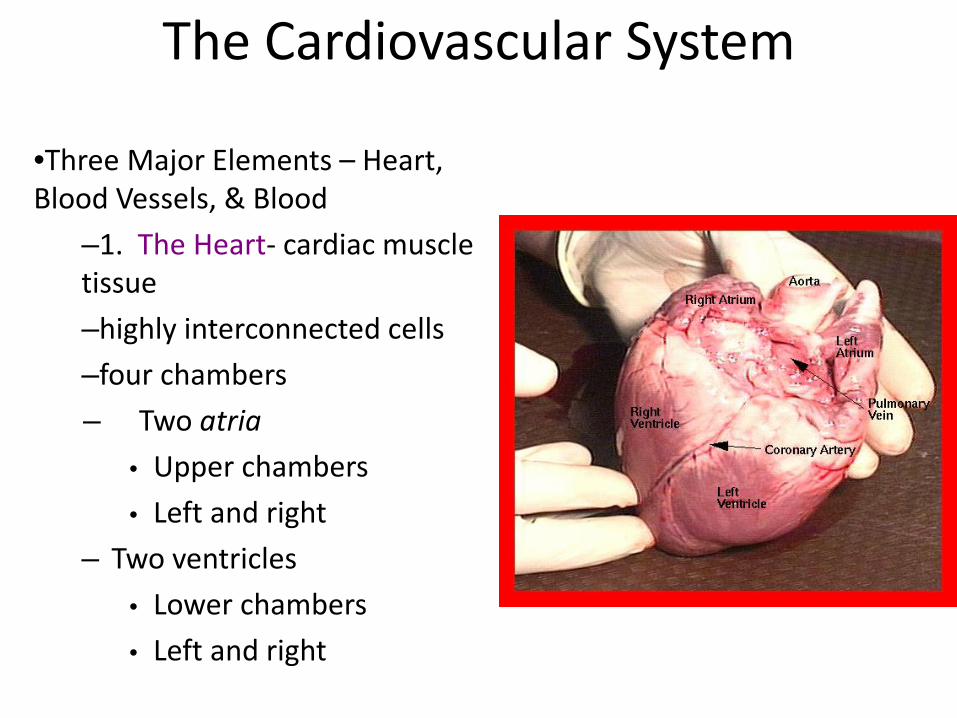
•Three Major Elements – Heart, Blood Vessels, & Blood
–1. The Heart- cardiac muscle tissue –highly interconnected cells –four chambers – Two atria
• Upper chambers • Left and right
– Two ventricles • Lower chambers • Left and right


Heart: Structures
• Heart coverings – Pericardium
• Covers the heart and large blood vessels attached to the heart
• Visceral pericardium – Innermost layer – Directly on the heart
• Parietal pericardium – Layer on top of the visceral pericardium

Heart Wall
• Three layers of tissue – Epicardium: This serous membrane of smooth
outer surface of heart – Myocardium: Middle layer composed of
cardiac muscle cell and responsibility for heart contracting
– Endocardium: Smooth inner surface of heart chambers

Heart: Structures

Pathway of the blood •Superior Vena Cava •Right Atrium •Tricuspid Valve •Right Ventricle •Pulmonary Semilunar Valve •Lungs •Pulmonary Vein •Bicuspid Valve •Left Ventricle •Aortic Semilunar Valve •Aorta •To the bodies organs & cells

Pathway of the blood
Right Atrium
Right Ventricle
Pulmonary Semilunar Valve
Left Atrium
Bicuspid Valve
Left Ventricle
Pulmonary Valve
Tricuspid Valve
Aortic Semilunar Valve
Lungs Body

Heart: External Anatomy •Four chambers
–2 atria –2 ventricles
•Auricles •Major veins
–Superior vena cava –Pulmonary veins
•Major arteries –Aorta –Pulmonary trunk

External Anatomy

Heart: Valves
•Prevent blood from flowing back

Heart Valves

Function of the Heart Valves

Heart: Cardiac Muscle
• Elongated, branching cells containing 1-2 centrally located nuclei • Contains actin and myosin myofilaments • Intercalated disks: Specialized cell-cell contacts • Desmosomes hold cells together and gap junctions allow action potentials • Electrically, cardiac muscle behaves as single unit
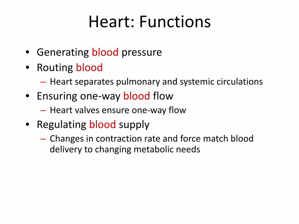
Heart: Functions • Generating blood pressure • Routing blood
– Heart separates pulmonary and systemic circulations • Ensuring one-way blood flow
– Heart valves ensure one-way flow • Regulating blood supply
– Changes in contraction rate and force match blood delivery to changing metabolic needs

Cardiac Cycle
• Heart is two pumps that work together, right and left half
• Repetitive contraction (systole) and relaxation (diastole) of heart chambers
• Blood moves through circulatory system from areas of higher to lower pressure. – Contraction of heart produces the pressure

The Heart: Cardiac Cycle
27-18
One heartbeat = one cardiac cycle Atria contract and relax
• Right atrium contracts – Tricuspid valve opens – Blood fills right ventricle
• Left atrium contracts – Bicuspid valve opens – Blood fills left ventricle
Ventricles contract and relax • Right ventricle contracts
– Tricuspid valve closes – Pulmonary semilunar valve opens – Blood flows into pulmonary artery
• Left ventricle contracts – Bicuspid valve closes – Aortic semilunar valve opens – Blood pushed into aorta

Cardiac Cycle

The Heart: Cardiac Cycle (cont.)
• Influenced by – Exercise – Parasympathetic nerves – Sympathetic nerves – Cardiac control center – Body temperature – Potassium ions – Calcium ions
27-20

Heart: Electrical Properties
• Resting membrane potential (RMP) present – Cardiac Cell at rest are considered polarized, meaning no electrical activity
takes place. • Electrical impulses are generated by automaticity of specialized cardiac cells
• A certain amount of electrical impulse needed to release action potential
• Once an electrical cell generates an electrical impulse, this electrical impulse
causes the ions to cross the cell membrane and causes the action potential, also called depolarization
• The movement of ions across the cell membrane through sodium, potassium and calcium channels, is the drive that causes contraction of the cardiac cells/muscle
• Depolarization with corresponding contraction of myocardial muscle moves as a wave through the heart
• Repolarization is the return of the ions to their previous resting state, which corresponds with relaxation of the myocardial muscle
• Depolarization and repolarization are electrical activities which cause muscular activity
• The action potential curve shows the electrical changes in the myocardial cell during the depolarization – repolarization cycle
• This electrical activity is what is detected on ECG, not the muscular activity

Action Potentials in Skeletal and Cardiac Muscle

SA Node Action Potential

Electrocardiogram • Action potentials through
myocardium during cardiac cycle produces electric currents than can be measured
• Pattern – P wave
• Atria depolarization
– QRS complex • Ventricle depolarization • Atria repolarization
– T wave: • Ventricle repolarization

Heart: Sounds
• First heart sound or “lubb” – Atrioventricular valves and surrounding fluid vibrations as valves
close at beginning of ventricular systole
• Second heart sound or “dupp” – Results from closure of aortic and pulmonary semilunar valves at
beginning of ventricular diastole, lasts longer
• Third heart sound (occasional) – Caused by turbulent blood flow into ventricles and detected near
end of first one-third of diastole

Heart: Mean Arterial Pressure (MAP) • Average blood pressure in aorta • MAP=CO x PR
– CO is amount of blood pumped by heart per minute
• CO=SV x HR – SV: Stroke volume of blood pumped during each heart beat – HR: Heart rate or number of times heart beats per minute
• Cardiac reserve: Difference between CO at rest and maximum CO
– PR is total resistance against which blood must be pumped
Heart: Effects of Aging • Gradual changes in heart function, minor under
resting condition, more significant during exercise • Hypertrophy of left ventricle • Maximum heart rate decreases • Increased tendency for valves to function
abnormally and arrhythmias to occur • Increased oxygen consumption required to pump
same amount of blood

The Cardiovascular System 2. Blood Vessels -A network of tubes
–Arteriesarterioles move away from the heart
•Elastic Fibers •Circular Smooth Muscle
–Capillaries – where gas exchange takes place.
•One cell thick •Serves the Respiratory System
–VeinsVenules moves towards the heart
•Skeletal Muscles contract to force blood back from legs •One way values •When they break - varicose veins form


Circuits
•Pulmonary circuit –The blood pathway between the right side of the heart, to the lungs, and back to the left side of the heart.
•Systemic circuit –The pathway between the left and right sides of the heart.

Circulation
• Pulmonary circuit right atrium right ventricle pulmonary arteries lungs pulmonary veins heart (left atrium)
• Systemic circuit left atrium left ventricle aorta arteries
arterioles capillaries venules veins vena cava heart (right atrium)
27-32

27-33
Circulation (cont.)
• Arterial system – Carry oxygen-rich blood
away from the heart
– Pulmonary arteries carry oxygen-poor blood
– Paired – left and right artery of the same name

Circulation (cont.)
• Venous system – Carries oxygen-
poor blood toward the heart
• Except pulmonary veins
– Most large veins have the same names as the arteries they are next to
27-34
Hepatic portal system Collection of veins
carrying blood to the liver
Click for Larger View

The Cardiovascular System
3. The Blood A. Plasma Liquid portion of the blood.
Contains clotting factors, hormones, antibodies, dissolved gases, nutrients and waste

The Cardiovascular System
•The Blood
B. Erythrocytes - Red Blood Cells
–Carry hemoglobin and oxygen. Do not have a nucleus and live only about 120 days.
–Cannot repair themselves.

The Cardiovascular System •The Blood
C. Leukocytes – White Blood cells
–Fight infection and are formed in the bone marrow
–Five types
•Neutrophils •Lymphocytes •Eosinophils •Basophils •Monocytes

The Cardiovascular System The Blood •D. Thrombocytes – Platelets.
–These are cell fragment that are formed in the bone marrow from magakaryocytes.
–Clot Blood by sticking together – via protein fibers called fibrin.

According to the ABO blood typing system there are four different kinds of blood types: A, B, AB or O (null).
ABO Blood Grouping System

Blood group A If you belong to the blood group A, you have A antigens on the surface of your RBCs and B antibodies in your blood plasma.
Blood group B If you belong to the blood group B, you have B antigens on the surface of your RBCs and A antibodies in your blood plasma.
AB0 blood grouping system

Blood group AB If you belong to the blood group AB, you have both A and B antigens on the surface of your RBCs and no A or B antibodies at all in your blood plasma.
Blood group O If you belong to the blood group O (null), you have neither A or B antigens on the surface of your RBCs but you have both A and B antibodies in your blood plasma.

People with blood group O are called "universal donors" and people with blood group AB are called "universal receivers."
Blood transfusions – who can receive blood from whom?

Blood Group
Antigens Antibodies Can give blood to
Can receive
blood from
AB
A
B
O

Blood Group
Antigens Antibodies Can give blood to
Can receive
blood from
AB A and B None AB AB, A, B, O
A A B A and AB A and O
B B A B and AB B and O
O None A and B AB, A, B, O O

Disorders of the Circulatory System • Anemia - lack of iron in the blood, low RBC count
• Leukemia - white blood cells proliferate wildly, causing anemia
• Hemophilia - bleeder’s disease, due to lack of fibrinogen in thrombocytes
• Heart Murmur - abnormal heart beat, caused by valve problems
• Heart attack - blood vessels around the heart become blocked with plaque, also called myocardial infarction

Cardiac Arrhythmias
• Tachycardia: Heart rate in excess of 100bpm • Bradycardia: Heart rate less than 60 bpm • Sinus arrhythmia: Heart rate varies 5% during
respiratory cycle and up to 30% during deep respiration
• Premature atrial contractions: Occasional shortened intervals between one contraction and succeeding, frequently occurs in healthy people