The Cell - storage.googleapis.com Cell All living things are ... A eukaryotic cells contains a...
96
The Cell All living things are composed of cells. The cell is smallest unit of living things that can carry out the activities necessary for life. many forms of life exist as single celled organisms such as bacteria. More complex organisms, including plants and animals, are multicellular. There are two distinct types of cells: eukaryotic cells and prokaryotic cells.
Transcript of The Cell - storage.googleapis.com Cell All living things are ... A eukaryotic cells contains a...

The Cell
All living things are composed of cells. The cell is smallest unit of living things that can carry out the activities necessary for life. many forms of life exist as single celled organisms such as bacteria. More complex organisms, including plants and animals, are multicellular.
There are two distinct types of cells: eukaryotic cells and prokaryotic cells.

12A

20D

Microscopy
Microscopes are the most important tools of cytology, the
study of cell structure
Three important parameters in microscopy are magnification, contrast, and resolution.
Magnification is the ratio of an object’s image size to its real size. Light microscopes can magnify
effectively to about 1,000 times the actual size of the specimen;
Contrast, accentuates differences in parts of the sample. Improvements in light microscopy have
included new methods for enhancing contrast, such as staining or labeling cell components with
different color to stand out visually.
Resolution is a measure of the clarity of the image; it is the minimum distance two points can be
separated and still be distinguished as two points. the light microscope cannot resolve detail finer than
about 0.2 micrometer (μm), or 200 nanometers (nm), regardless of the magnification.

Microscopes are the most important tools of cytology,
the
study of cell structure.
Electron microscopes have revealed many
organelles and other subcellular structures that were
impossible to resolve with the light microscope. A
disadvantage of electron microscopy is that the
methods used to prepare the specimen kill the cells.
For all microscopy techniques, in fact, specimen
preparation can introduce artifacts, structural features
seen in micrographs that do not exist in the living cell.
But the light microscope offers advantages,
especially in studying living cells. Labeling individual
cellular molecules or structures with fluorescent
markers has made it possible to see such structures
with increasing detail. In addition, both confocal and
deconvolution microscopy have sharpened images of
3-D tissues and cells. Researchers can see the
distinguish subcellular structures as small as 10–20
nm across.
Microscopy


17A
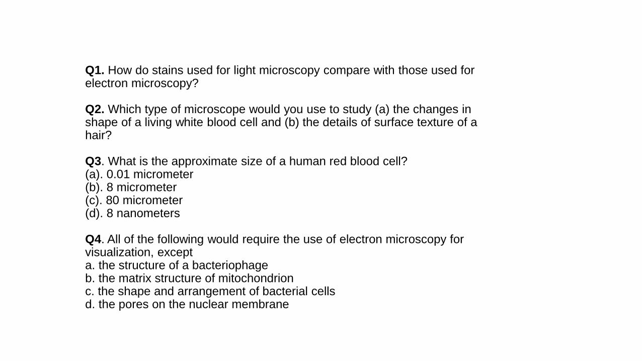
Q1. How do stains used for light microscopy compare with those used for electron microscopy?
Q2. Which type of microscope would you use to study (a) the changes in shape of a living white blood cell and (b) the details of surface texture of a hair?
Q3. What is the approximate size of a human red blood cell?(a). 0.01 micrometer(b). 8 micrometer(c). 80 micrometer(d). 8 nanometers
Q4. All of the following would require the use of electron microscopy for visualization, excepta. the structure of a bacteriophageb. the matrix structure of mitochondrionc. the shape and arrangement of bacterial cellsd. the pores on the nuclear membrane
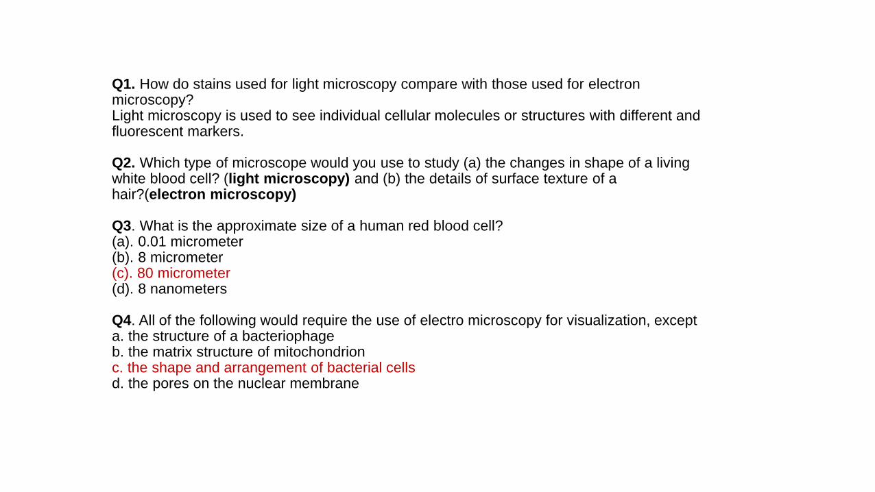
Q1. How do stains used for light microscopy compare with those used for electron microscopy?Light microscopy is used to see individual cellular molecules or structures with different and fluorescent markers.
Q2. Which type of microscope would you use to study (a) the changes in shape of a living white blood cell? (light microscopy) and (b) the details of surface texture of a hair?(electron microscopy)
Q3. What is the approximate size of a human red blood cell?(a). 0.01 micrometer(b). 8 micrometer(c). 80 micrometer(d). 8 nanometers
Q4. All of the following would require the use of electro microscopy for visualization, excepta. the structure of a bacteriophageb. the matrix structure of mitochondrionc. the shape and arrangement of bacterial cellsd. the pores on the nuclear membrane


4B


6C, 8E, 9C

The CellAll living things are composed of cells. The cell is smallest unit of living things that can carry out the activities necessary for life. many forms of life exist as single cell- organisms such as bacteria. More complex organisms, including plants and animals, are multicellular.
There are two distinct types of cells: eukaryotic cells and prokaryotic cells.
A eukaryotic cells contains a membrane bound structure called a nucleus and cytoplasm. Cytoplasm was filled with tiny structures called organelles (endomembrane system). Examples of the eukaryotic cells are Fungi, protists, plants, animals and humans.

Prokaryotic cell, which is a lot smaller than a eukaryotic cells, does
not contain a nucleus and membrane-bound organelles. The DNA
is concentrated in a region that is not membrane-enclosed, called the nucleoid

The interior of either type of cell is called the cytoplasm; in eukaryotic cells, this term
refers only to the region between the nucleus and the plasma membrane. Within the
cytoplasm of a eukaryotic cell, suspended in cytosol, are a variety of organelles
of specialized form and function. These membrane-bounded structures are absent in
prokaryotic cells.
eukaryotic cells
prokaryotic cells

Cell wall
Cell wall is one cell structure not found in animal cells. Plants and algae have cell walls made of cellulose. The cell walls of fungi are usually made of chitins.
The primary cell wall is outside the plasma membrane.
Chitins
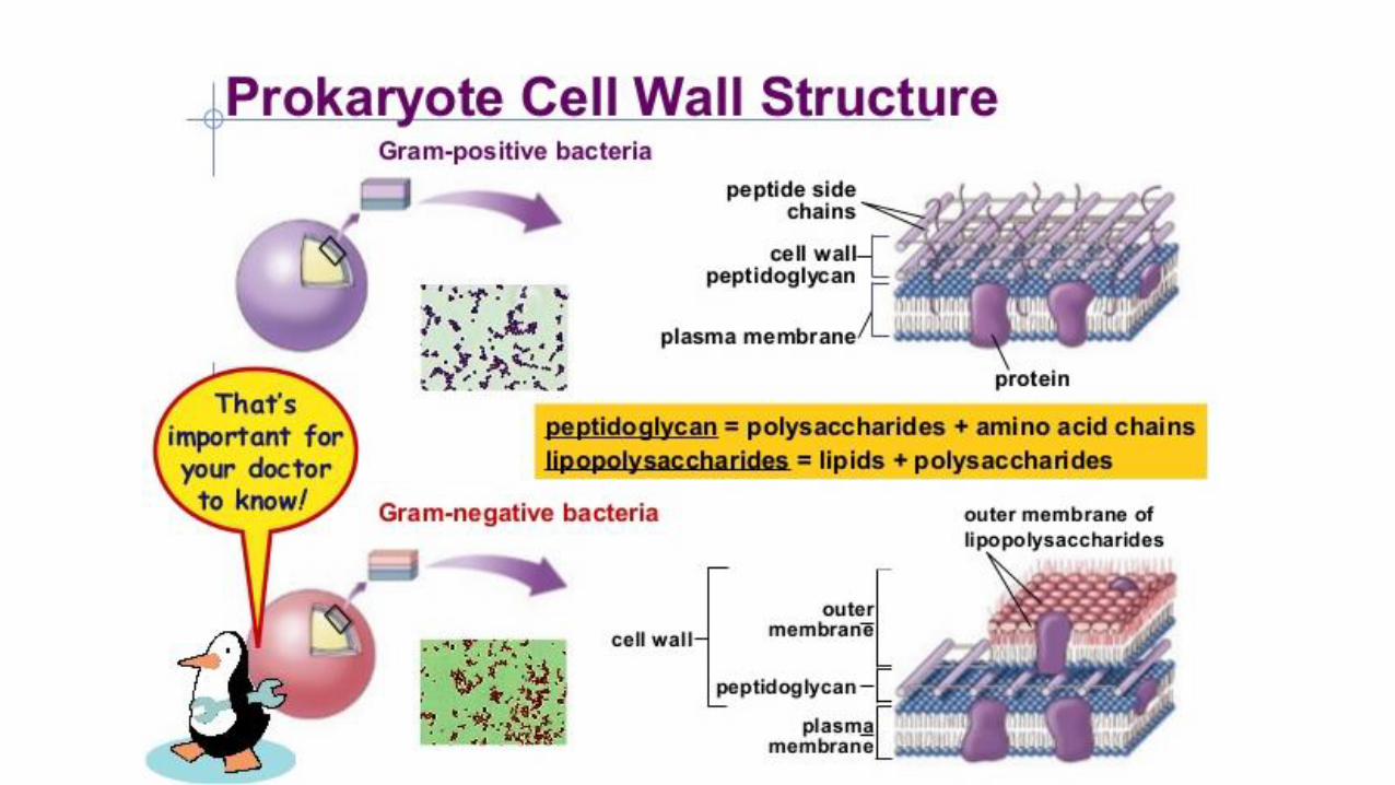

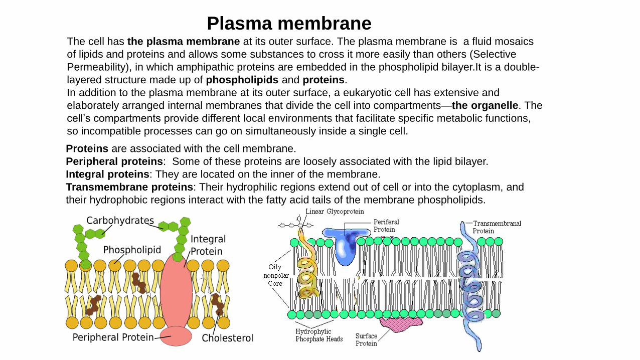
The cell has the plasma membrane at its outer surface. The plasma membrane is a fluid mosaics
of lipids and proteins and allows some substances to cross it more easily than others (Selective
Permeability), in which amphipathic proteins are embedded in the phospholipid bilayer.It is a double-
layered structure made up of phospholipids and proteins.
In addition to the plasma membrane at its outer surface, a eukaryotic cell has extensive and
elaborately arranged internal membranes that divide the cell into compartments—the organelle. The
cell’s compartments provide different local environments that facilitate specific metabolic functions,
so incompatible processes can go on simultaneously inside a single cell.
Plasma membrane
Proteins are associated with the cell membrane.
Peripheral proteins: Some of these proteins are loosely associated with the lipid bilayer.
Integral proteins: They are located on the inner of the membrane.
Transmembrane proteins: Their hydrophilic regions extend out of cell or into the cytoplasm, and
their hydrophobic regions interact with the fatty acid tails of the membrane phospholipids.


UK Biology Olympiad (BBO) 2015, C

Organelles in eukaryotic cells
A eukaryotic cells are like a biological factories. It contains different kinds of organelles, each organelle has its own special task.
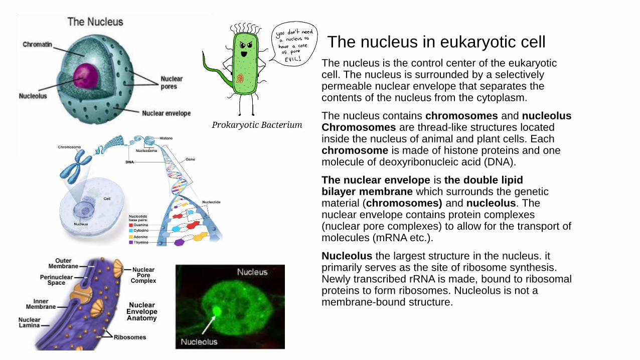
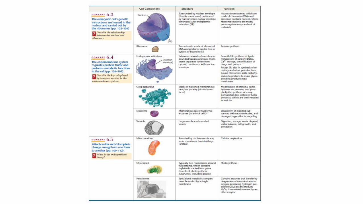
The nucleus in eukaryotic cellThe nucleus is the control center of the eukaryotic cell. The nucleus is surrounded by a selectively permeable nuclear envelope that separates the contents of the nucleus from the cytoplasm.
The nucleus contains chromosomes and nucleolusChromosomes are thread-like structures located inside the nucleus of animal and plant cells. Each chromosome is made of histone proteins and one molecule of deoxyribonucleic acid (DNA).
The nuclear envelope is the double lipid bilayer membrane which surrounds the genetic material (chromosomes) and nucleolus. The nuclear envelope contains protein complexes (nuclear pore complexes) to allow for the transport of molecules (mRNA etc.).
Nucleolus the largest structure in the nucleus. it primarily serves as the site of ribosome synthesis.Newly transcribed rRNA is made, bound to ribosomal proteins to form ribosomes. Nucleolus is not a membrane-bound structure.

rRNA
3B

RibosomesRibosomes are composed RNAs and proteins. they are the protein factories found within all living cells, that serves as the site of biological protein synthesis (translation). Ribosomes link amino acids together to produce proteins in the order specified by messenger RNA (mRNA) molecules.
Ribosome proteins consist of two major components: the small ribosomal subunit, which reads the mRNA, and the large subunit, which joins amino acids to form a polypeptide chain. The transfer ribonucleic acid (tRNA) is a type of RNA molecule that helps decode a mRNA sequence by bring the amino acids to Ribosomes.
Ribosomes can be either free floating in the cells or attached to endoplasmic reticulum (ER).
Ribosome

45B
Canada(CBO) 2016

Endoplasmic reticulum
The Endoplasmic reticulum (ER) is a network of
membrane-enclosed tubules and sacs (cisternae) that
extends from the nuclear membrane throughout the
cytoplasm in eukaryotic cells. there are two distinct types
of ER that perform different functions within the cell.
The rough ER, which is covered by ribosomes on its
outer surface, functions in protein synthesis and
processing. That is why it contains ribosomes.
The smooth ER is not associated with ribosomes and is
involved in 1. lipid metabolism. Assists in synthesis of
lipids, steroids such as sex hormones. 2. detoxifies
drugs and poisons from the body. 3. Store Ca ions in
muscle cells to facilitate normal muscle contractions.

UK2015, 4B
(a membrane that bounds the chief vacuole of a plant cell.)

1C


13D

Golgi apparatusThe Golgi is composed of flattened membrane-enclosed sacs (cisternae) and associated vesicles. A striking feature of the Golgi apparatus is its distinct polarity in both structure and function. Proteins from the ER enter at its cis face (entry face), which is convex and usually oriented toward the nucleus. They are then transported through the Golgi and exit from its concave trans face (exit face). As they pass through the Golgi, proteins are modified and sorted for transport to their eventual destinations within the cell.
Golgi complex, After leaving the ER, many transport vesicles travel to the Golgi apparatus. We can think of the Golgi as a warehouse for receiving, sorting, shipping, and even some manufacturing. Here, products of the ER, such as proteins, are modified and stored and then sent to other destinations: lysosomes, the plasma membrane, or secretion. In addition, glycolipids are synthesized within the Golgi. In plant cells, the Golgi apparatus further serves as the site at which the complex polysaccharides of the cell wall are synthesized. The Golgi apparatus is thus involved in processing the broad range of cellular constituents that travel along the secretory pathway.

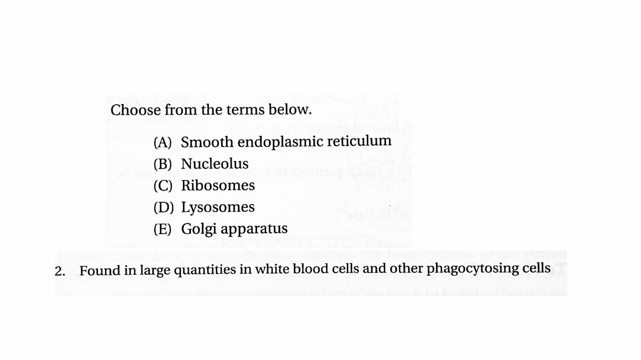
Lysosomes
Lysosomes are membrane-enclosed organelles that contain an array of enzymes capable of breaking down all types of biological polymers—proteins, nucleic acids, carbohydrates, and lipids.
PH in the lysosome is around 5.
Lysosomes function as the digestive system of the cell, serving both to degrade material taken up from outside the cell and to digest obsolete components of the cell itself. In their simplest form, lysosomes are visualized as dense spherical vacuoles, but they can display considerable variation in size and shape as a result of differences in the materials that have been taken up for digestion. Lysosomes thus represent morphologically diverse organelles defined by the common function of degrading intracellular material.
Plant cells do not have lysosomes

PeroxisomesPeroxisomes are organelles and found in both plant and animal cells perform the specialized functions. In animal, they are common in the liver and kidney cells. They contain catalase catalyzes the following chemical reactions and detoxify various substances (Fig).
Peroxisomes contain at least 50 different enzymes, which are involved in a variety of biochemical pathways in different types of cells. Peroxisomes originally were defined as organelles that carry out oxidation reactions leading to the production of hydrogen peroxide (H2O2). Because hydrogen peroxide is harmful to the cell, peroxisomes also contain the enzyme catalase, which decomposes hydrogen peroxide either by converting it to water or by using it to oxidize another organic compound. A variety of substrates are broken down by such oxidative reactions in peroxisomes, including uric acid, amino acids, and fatty acids. The oxidation of fatty acids is a particularly important example, since it provides a major source of metabolic energy. In animal cells, fatty acids are oxidized in both peroxisomes and mitochondria, but in yeasts and plants fatty acid oxidation is restricted to peroxisomes.
Fig (a). Fatty acid oxidation in peroxisomes
Fig (b). Peroxisomes and lysosomes are two brothers and
work together to break down the waste products in the cell
Peroxisome
lysosome

USABO2011, 2C

2D

VacuoleA vacuole is a membrane bound organelle which is present in all plant and fungal cells and some protists, animal and bacterial cells. Vacuoles are essentially enclosed compartments which are filled with water containing inorganic and organic molecules including enzymes in solution.
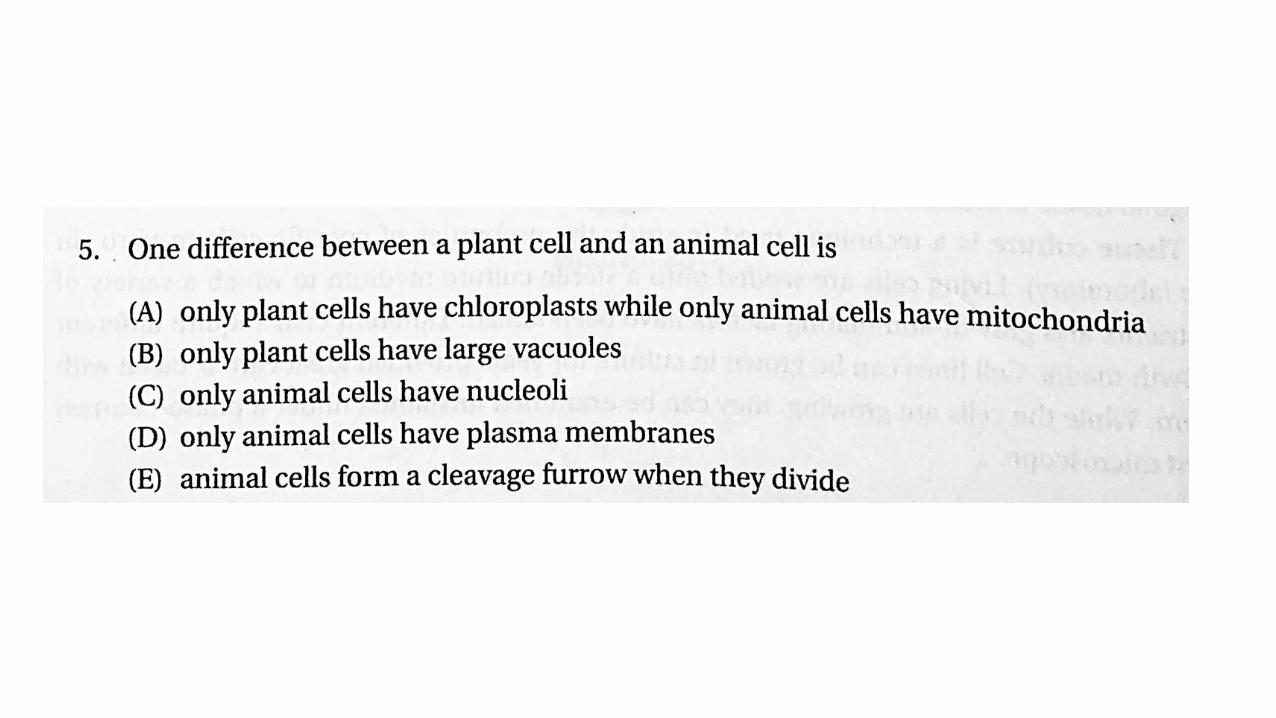
Plant cells contain a large vacuole.
D

Endosomal V-ATPase (vesicular H+-ATPase) is a pH-sensor regulating the degradative pathway. According to our model, V-ATPase is responsible for: (i) the generation of a pH gradient between vesicular membranes; (ii) sensing of intravesicular pH; and (iii) transmitting this information to the cytosolic side of the membrane.
In plants, the V-ATPase is an active component of the vacuole, which in situations such as citrus fruits can reach pH values as low as 2.2, although other proton pumps may
be involved in helping to maintain such a low pH.
The Plant H+-ATPase has a pH optimum of 6.6, it is
well below the physiological pH of the plant cell
cytoplasm (usually around 7.2-7.5). Thus, when ever
protons start accumulating in the cytoplasm, the
activity of the H+-ATPase increases, resulting in the
expulsion of the excess H+ from the cell.

USABO2013, 30D


CytoskeletonThe cytoskeleton can be referred to as a complex network of interlinking microfilaments and microtubules that extend throughout the cytoplasm, from the nucleus to the plasma membrane.
Microtubules are hollow cylinders, they form the centrioles (Fig 5), cilia and flagella(Fig 6). Tubulin participate in cellular division and movement.
Microfilaments are composed of linear polymers of G-actin proteins. They also act as tracks for the movement of myosin molecules in muscle contraction.
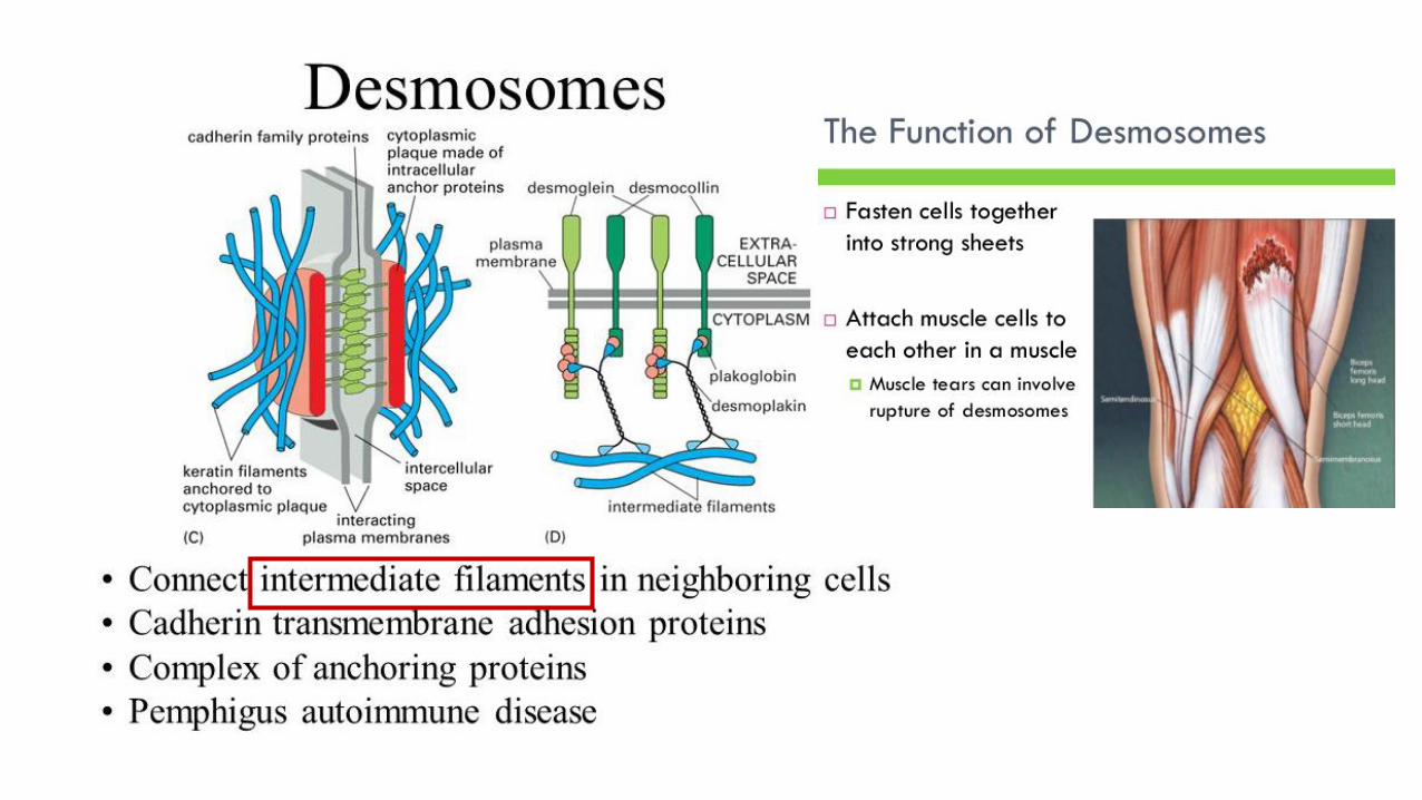
Intermediate filaments support cell shape and fix organelles in place.
Confocal Microscopy ofthe eukaryotic cytoskeleton.Actin filaments are shown in red, microtubules are show in green that supports cell shape and function.
Movement
Structure
6 5

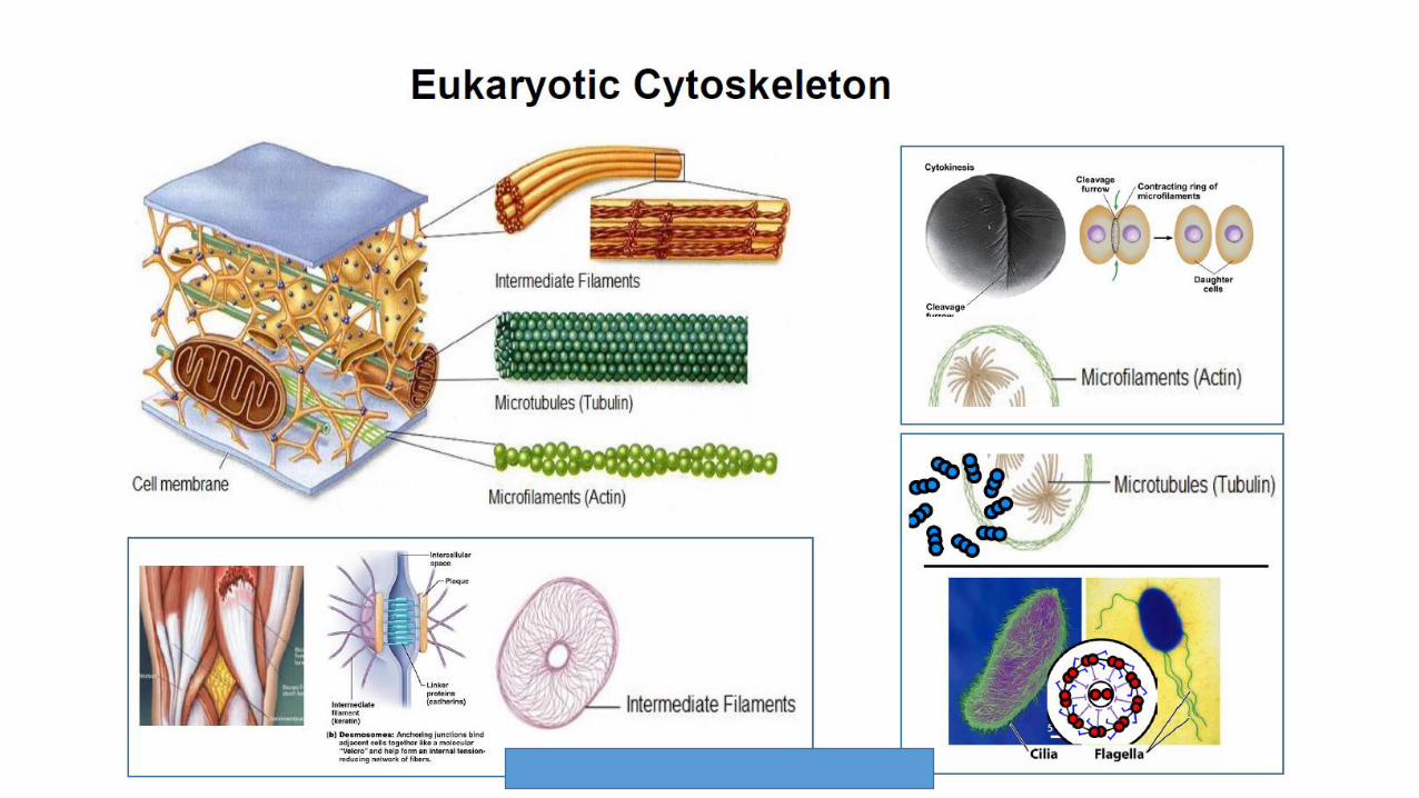
Eukaryotic CytoskeletonThe cytoskeleton can be referred to as a complex network of interlinking microfilaments and microtubules that extend throughout the cytoplasm, from the nucleus to the plasma membrane.
Microtubules are hollow cylinders, they form the centrioles, cilia and flagella. Tubulin participate in cellular division and movement.
Microfilaments are composed of linear polymers of G-actin proteins. They also act as tracks for the movement of myosin molecules in muscle contraction.
Intermediate filaments support cell shape and fix organelles in place.
Movement


Centrioles
Centrioles are small, paired, cylindrical structures that are found within microtubule organizing centers (MTOCs). When a cell is ready to divide, the centrioles produce microtubules, which pull the replicated chromosomes apart and move them to opposite ends of the cell. Centrioles are common in animal cells, they are not found in plant cells
Spindle fibers and centrioles consist of nine triplets of microtubules arranged in circle


Cytokinesis
Plant cells Animal cells
Vesicles originating from Golgi bodies migrate to the plane between the two newly forming nuclei.
Microfilaments form a ring inside the plasma membrane between the two newly forming nuclei.
The division of the cytoplasm takes placed by cell plate formation
The division of the cytoplasm takes place by cleavage
Cell plate formation starts at the center of cell and grows outward.
Cleavage starts at periphery and then movesinward.

Cell Junctions

Mitochondria
Mitochondria are the powerhouses of the cell. Mitochondria are the site of cellular respiration. All cells have many mitochondria. A very active cell could have 2500 of them. Mitochondria have an outer double membrane and an inner series of membranes called cristae. They contain enzymes that converting the energy from organic molecules into energy molecule-adenosine triphosphate (ATP).
Mitochondria are involved in other tasks, such as signaling, cellular differentiation, and cell death.
The mitochondrion has its own independent DNA that shows substantial similarity to bacterial DNA.
The mitochondrion has its own independent ribosomes.
18 C

Chloroplast
Chloroplasts are organelles found in plant cells and eukaryotic algae that conduct photosynthesis. chloroplasts have an outer double membrane
Chloroplasts absorb sunlight and use it in conjunction with water and carbon dioxide gas to produce food for the plant.
They have an outer double membrane, their own independent DNA that shows substantial similarity to bacterial DNA.
They have their own independent ribosomes.

Australia (ABO) 2016, 12D

E


19B

14D, 15A, 16E

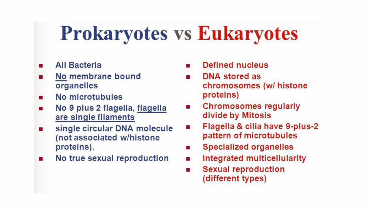
Features of prokaryotic
cells such as bacteria:
Features of eukaryotic cells
Cell wall Cell wall (plants only)
Plasma membrane Plasma membrane
Cytoplasm Cytoplasm
Ribosomes Ribosomes
Nucleoid region
Bacterial chromosome
Flagella and cilia
Capsule
Nucleus and nucleolus
Centrioles (animals only)
Rough endoplasmic reticulum
Smooth endoplasmic reticulum
Golgi apparatus
Mitochondria
Chloroplasts (plants only)
Lysosomes
Peroxisomes
Vacuole
Central vacuole(plants only)
Cytoskeleton
Structure Plant cell Animal Cell
Cell wall Yes No
Mitochondria
/Chloroplasts
Chloroplasts&Mitochondria
Mitochondria
Centrioles No Yes
Central
vacuole
Yes, large No or smaller
Lysosomes No Yes

Canada(CBO) 2016, 46B

5E

17A, 18C
(CBO2016)

7E


10B


Membrane transportMembrane transport refers to the collection of mechanisms that regulate the passage of solutes such as ions and small molecules through biological membranes, which are lipid bilayers that contain proteins embedded in them. The regulation of passage through the membrane is due to selective membrane permeability - a characteristic of biological membranes which allows them to separate substances of distinct chemical nature.
Water molecules are polar and not lipid soluble, but they can rapidly cross a lipid bilayer through aquaporins, which are integral membrane proteins that regulate the flow of water.
Transport can be either active or passive. Active transport requires energy (e.g. ATP ). passive transport requires no energy.
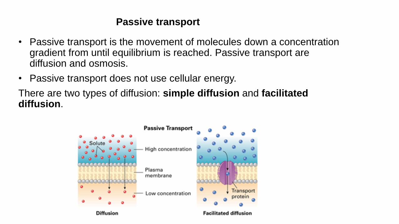
Passive transport
• Passive transport is the movement of molecules down a concentration gradient from until equilibrium is reached. Passive transport are diffusion and osmosis.
• Passive transport does not use cellular energy.
There are two types of diffusion: simple diffusion and facilitated diffusion.

Active transport
Active transport is the movement of molecules across a cell membrane from a region of their lower concentration to a region of their higher concentration in the direction against some gradient or other obstructing factor (often a concentration gradient).
Active transport uses cellular energy to move them against a gradient, polar repulsion, or other resistance.
Active transport is usually associated with accumulating high concentrations of molecules that the cell needs, such as ions, glucose and amino acids. If the process uses chemical energy, such as from ATP, it is termed primary active transport. The best sample of the active transport is a special protein called the Sodium-potassium pump. Secondary active transport involves the use of an electrochemical gradient. Examples of the active transport include the uptake of glucose in the intestines in humans.
Primary active transport. Secondary active transport
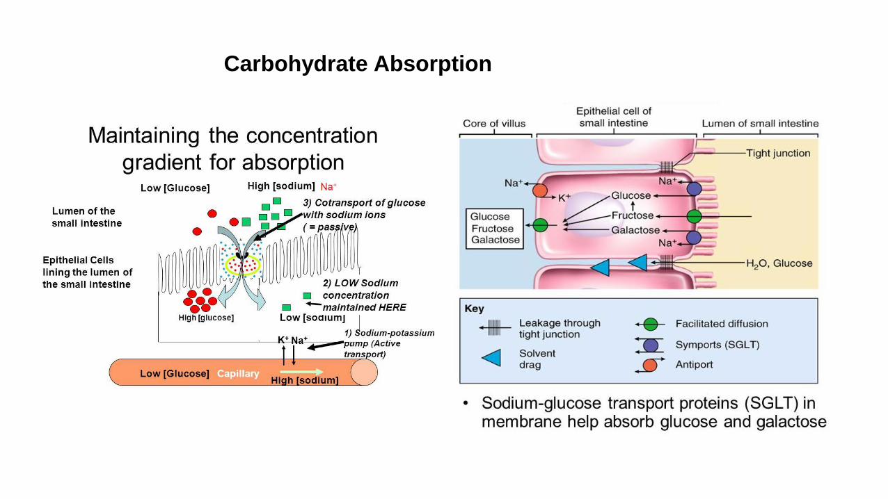
Carbohydrate Absorption

Glucose transport
Canada Biology Olympiad 2016
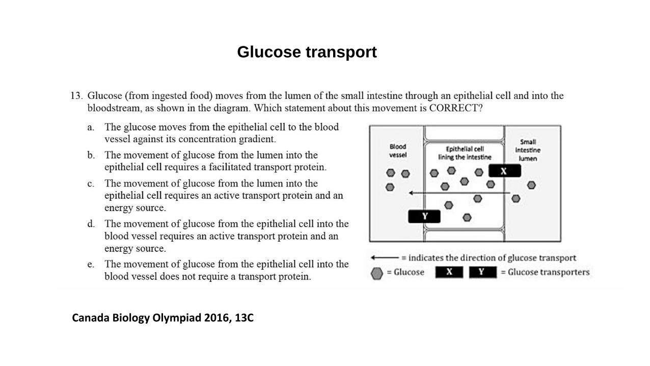
Glucose transport
Canada Biology Olympiad 2016, 13C

Canada (CBO) 2016, 16D

EndocytosisEukaryotic cells are also able to take up macromolecules and particles from the surrounding medium by a distinct process called endocytosis. In endocytosis, the material to be internalized is surrounded by an area of plasma membrane, which then buds off inside the cell to form a vesicle containing the ingested material. The term “endocytosis” was include both the ingestion of large particles (such as bacteria) and the uptake of fluids or macromolecules in small vesicles. The former of these activities is known as phagocytosis (cell eating) and the latter as pinocytosis (cell drinking). A special type of endocytosis , receptor-mediated endocytosis, the macromolecules to be first bind to specific cell surface receptors. These receptors are concentrated in specialized regions of the plasma membrane, called clathrin-coated pits. These pits bud from the membrane to form small clathrin-coated vesicles containing the receptors and their bound macromolecules (ligands). The clathrin-coated vesicles then fuse with early endosomes, in which their contents are sorted for transport to lysosomes or recycling to the plasma membrane. Mammalian cells use receptor-mediated endocytosis to take cholesterol into cells. Cholesterol in the blood is usually found in lipid-protein complexes called low-density lipoproteins (LDLs). LDLs bind to specific
receptor proteins on the cell surface, thereby triggering their uptake by receptor-mediated endocytosis.

UK2015, 5D

Exocytosis
In exocytosis, materials are exported out of the cell via secretory vesicles. In this process, the Golgi complex packages macromolecules into transport vesicles that travel to and fuse with the plasma membrane. This fusion causes the vesicle to spill its contents out of the cell. Exocytosis is important in expulsion of waste materials out of the cell and in the secretion of cellular products such as digestive enzymes or hormones. The movement of macromolecules such as proteins or polysaccharides into or out of the cell is called bulk transport. Exocytosis requires the expenditure of energy (ATP).

UK2015, 10B

Bulk Flow
Bulk flow is one-way movement of fluids brought about by pressure. The movement of blood through a blood vessel or movement of fluid in xylem vessels and phloem tubes of plans are examples of bulk flow.Transport in xylem relies upon the cohesion of water molecules to each other and adhesion to the vessel's wall via hydrogen bonding. If an air bubble forms the flow will be stopped as the column is broken and the pressure difference in the vessel cannot be transmitted; this is called an embolism. Once these embolisms are nucleated, the remaining water in the capillaries begins to turn to water vapor. Plants have physiological mechanisms to reestablish the capillary action within their cells.

Dialysis
Dialysis is the diffusion of solutes across a selectively permeable membrane. A cellophane bag is often used as an artificial membrane to separate small molecules from large molecules. Dialysis is the artificial process of eliminating waste (diffusion) and unwanted water (ultrafiltration) from the blood. Our kidneys do this naturally. Some people, however, may have failed or damaged kidneys which cannot carry out the function properly - they may need dialysis. Kidney dialysis is a life-support treatment that uses a special machine to filter harmful wastes, salt, and excess fluid from the blood. This restores the blood to a normal, healthy balance.
10E

Q7. All the following required ATP Except
a. Na-K pump
b. Cell absorbing oxygen
c. Receptor-mediated endocytosis
d. Amoeboid movement
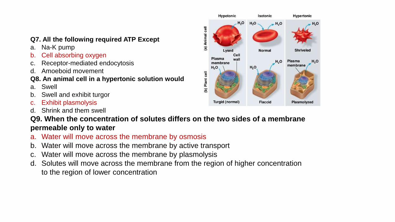
Q8. An animal cell in a hypertonic solution would
a. Swell
b. Swell and exhibit turgor
c. Exhibit plasmolysis
d. Shrink and them swell
Q9. When the concentration of solutes differs on the two sides of a
membrane permeable only to water
a. Water will move across the membrane by osmosis
b. Water will move across the membrane by active transport
c. Water will move across the membrane by plasmolysis
d. Solutes will move across the membrane from the region of higher
concentration to the region of lower concentration
Q7. All the following required ATP Except
a. Na-K pump
b. Cell absorbing oxygen
c. Receptor-mediated endocytosis
d. Amoeboid movement
Q8. An animal cell in a hypertonic solution would
a. Swell
b. Swell and exhibit turgor
c. Exhibit plasmolysis
d. Shrink and them swell
Q9. When the concentration of solutes differs on the two sides of a membrane
permeable only to water
a. Water will move across the membrane by osmosis
b. Water will move across the membrane by active transport
c. Water will move across the membrane by plasmolysis
d. Solutes will move across the membrane from the region of higher concentration
to the region of lower concentration

Q10-13. refer to the following key, each answer in the key may be used more than
once or not at all
a. Active transport
b. Bulk flow
c. osmosis
d. Facilitated diffusion
Q10. Solutes will move across the membrane from the region of higher solute
concentration to the region of lower solute concentration without the aid of
proteins.
Q11. Water will move across the membrane from the region of higher concentration
of water to the region of lower concentration of water without the aid of proteins.
Q12. movement of urine through the urinary tract.
Q13. movement of solutes across a plasma membrane requiring the addition of
energy.

Q14. The movement of molecules during diffusion can be described by all of the following Except
a. Molecular movements are random
b. Net movement of solute molecules is from a region of higher concentration to region of lower concentration
c. Each molecule move independently of other molecules
d. Solution molecules always move down the concentration gradient
Q15. A saturated suspension of starch is enclosed in a dialysis tubing bag, a material through which only
water can pass, but not starch. The bag with starch is placed into a beaker of distilled water. All the
following are expected to occur Except
a. There will be a net movement of water from a hypotonic region to a hypertonic region.
b. There will be a net movement of solute from a hypertonic region to a hypotonic region.
c. The dialysis bag with its contents will gain weight.
d. No starch will be detect outside the dialysis bag.
Q16. The Na-K Pump transports Na ions and K ions across the plasma membrane against their
concentration gradients. This pump is not considered a co-transport because
a. ATP is produced through this transporter.
b. A gradient, not ATP, drives the transport of these ions
c. ATP, not A gradient, drives the transport of these ions.
d. co-transporters go in only one direction, but the Na-K Pump drives ions across the membrane in both
directions.
Q17. The resting membrane potential depends on which of the following?
I. Active transport
II. Selective permeability
III. Differential distribution of ions across the axonal membrane
a. III only
b. I and II only
c. II and III only
d. I, II, and III

Q14. The movement of molecules during diffusion can be described by all of the following Except
a. Molecular movements are random
b. Net movement of solute molecules is from a region of higher concentration to region of lower concentration
c. Each molecule move independently of other molecules
d. Solution molecules always move down the concentration gradient
Q15. A saturated suspension of starch is enclosed in a dialysis tubing bag, a material through which only
water can pass, but not starch. The bag with starch is placed into a beaker of distilled water. All the following
are expected to occur Except
a. There will be a net movement of water from a hypotonic region to a hypertonic region.
b. There will be a net movement of solute from a hypertonic region to a hypotonic region.
c. The dialysis bag with its contents will gain weight.
d. No starch will be detect outside the dialysis bag.
Q16. The Na-K Pump transports Na ions and K ions across the plasma membrane against their concentration
gradients. This pump is not considered a co-transport because
a. ATP is produced through this transporter.
b. A gradient, not ATP, drives the transport of these ions
c. ATP, not A gradient, drives the transport of these ions.
d. co-transporters go in only one direction, but the Na-K Pump drives ions across the membrane in both directions.
Q17. The resting membrane potential depends on which of the following?
I. Active transport
II. Selective permeability
III. Differential distribution of ions across the axonal membrane
a. III only
b. I and II only
c. II and III only
d. I, II, and III


![Lithium-Ion battery SOC estimation...defines the cell’s internal resistance and the cell’s dynamics [14]. Hysteresis is a part of cell behavior and exhibits the dependency of](https://static.fdocuments.net/doc/165x107/5ebb5ced0c2acc01ef418f19/lithium-ion-battery-soc-estimation-deines-the-cellas-internal-resistance.jpg)