The Cardiovascular System The major organs of the cardiovascular system The heart structure and...
32
The Cardiovascular System The major organs of the cardiovascular system The heart structure and function
-
Upload
jacob-gilbert -
Category
Documents
-
view
216 -
download
1
Transcript of The Cardiovascular System The major organs of the cardiovascular system The heart structure and...

The Cardiovascular SystemThe major organs of the cardiovascular system
The heart structure and function

Chapter 13 pp. 329-364
• Name the organs of the cardiovascular system and discuss their functions.
• Name and describe the locations and functions of the major parts of the heart.
• Trace the pathway of the blood through the heart and the vessels of the coronary circulation.

Major organs of the cardiovascular system
• The heart – located in the pericardial cavity, slightly to the left, close to the left lung, and rests on the diaphragm
• Arteries – strong elastic vessels that are adapted for carrying blood away from the heart under high pressure

Major organs of the cardiovascular system
• Arterioles – smaller branches coming from the arteries
• Capillaries- smallest of the artery system, connect the smallest arterioles and the smallest venules

Major organs of the cardiovascular system
• Venules – the smallest vessels of the venous system, that continue from the capillaries and merge to form veins
• Veins- carry blood back to the atria of the heart following pathways that are almost parallel to the arteries. Similar to arteries, but have thinner walls, and generally have flaplike valves. Generally lower pressure than that of the arteries.
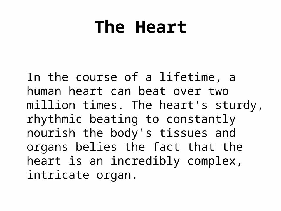
The Heart
In the course of a lifetime, a human heart can beat over two million times. The heart's sturdy, rhythmic beating to constantly nourish the body's tissues and organs belies the fact that the heart is an incredibly complex, intricate organ.

Blood flow through the body

The heart is composed of specially organized tissue surrounding a cartilage foundation.
The heart is divided into four chambers, composed of the left and right atria, and the left and right ventricles.
A sophisticated valve system controls blood flow between the chambers. In fact, it is the latching of the heart valves that creates the beating sound of the heart.

Superior Vena CavaThe superior vena cava is one of the two main veins bringing de-oxygenated blood from the body to the heart. Veins from the head and upper body feed into the superior vena cava, which empties into the right atrium of the heart.

Inferior Vena CavaThe inferior vena cava is one
of the two main veins bringing de-oxygenated blood from the body to the heart. Veins from the legs and lower torso feed into the inferior vena cava, which empties into the right atrium of the heart.

Right Atrium
• The right atrium receives de-oxygenated blood from the body through the superior vena cava and inferior vena cava .

Tricuspid Valve • The tricuspid valve separates
the right atrium from the right ventricle. It opens to allow the de-oxygenated blood collected in the right atrium to flow into the right ventricle. It closes as the right ventricle contracts, preventing blood from returning to the right atrium; thereby, forcing it to exit through the pulmonary valve into the pulmonary artery.
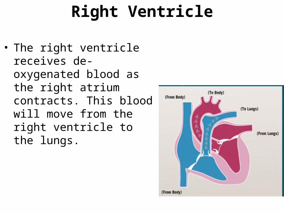
Right Ventricle
• The right ventricle receives de-oxygenated blood as the right atrium contracts. This blood will move from the right ventricle to the lungs.

Pulmonary Valve
• The pulmonary valve separates the right ventricle from the pulmonary artery. As the ventricles contract, it opens to allow the de-oxygenated blood collected in the right ventricle to flow to the lungs. It closes as the ventricles relax, preventing blood from returning to the heart.

Pulmonary Artery
• The pulmonary artery is the vessel transporting de-oxygenated blood from the right ventricle to the lungs. A common misconception is that all arteries carry oxygen-rich blood. It is more appropriate to classify arteries as vessels carrying blood away from the heart.

Cardio-respiratory connection
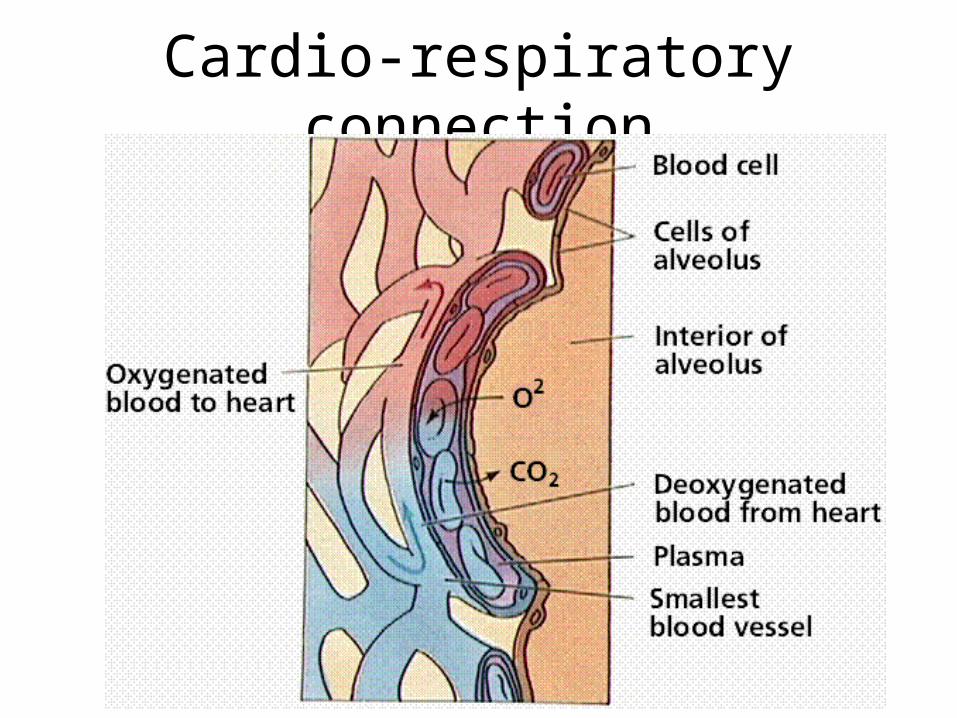
Cardio-respiratory connection

Cardio-respiratory connection

Pulmonary Vein
• The pulmonary vein is the vessel transporting oxygen-rich blood from the lungs to the left atrium. A common misconception is that all veins carry de-oxygenated blood. It is more appropriate to classify veins as vessels carrying blood to the heart.

Left Atrium
• The left atrium receives oxygenated blood from the lungs through the pulmonary vein. As the contraction triggered by the sinoatrial node progresses through the atria, the blood passes through the mitral valve into the left ventricle.

Mitral (Bicuspid) Value
• The mitral valve separates the left atrium from the left ventricle. It opens to allow the oxygenated blood collected in the left atrium to flow into the left ventricle. It closes as the left ventricle contracts, preventing blood from returning to the left atrium; thereby, forcing it to exit through the aortic valve into the aorta.

Left Ventricle
• The left ventricle receives oxygenated blood as the left atrium contracts. The walls of the left ventricle are thicker than the walls of the right ventricle, so that they can generate enough force to push the blood from the left ventricle into the aorta.

Aortic Valve
• The aortic valve separates the left ventricle from the aorta. As the ventricles contract, it opens to allow the oxygenated blood collected in the left ventricle to flow throughout the body. It closes as the ventricles relax, preventing blood from returning to the heart.

Aorta
• The aorta is the largest single blood vessel in the body. It is approximately the diameter of your thumb. This vessel carries oxygen-rich blood from the left ventricle to the various parts of the body.

Papillary Muscles
• The papillary muscles attach to the lower portion of the interior wall of the ventricles. They connect to the chordae tendineae, which attach to the tricuspid valve in the right ventricle and the mitral valve in the left ventricle. The contraction of the papillary muscles opens these valves. When the papillary muscles relax, the valves close.

Chordae Tendineae
• The chordae tendineae are tendons linking the papillary muscles to the tricuspid valve in the right ventricle and the mitral valve in the left ventricle. The chordae tendineae are string-like in appearance and are sometimes referred to as "heart strings."

Ventricular Septum
• is the stout wall separating the lower chambers (the ventricles) of the heart from one another.
• directed obliquely backward and to the right, and is curved toward the right ventricle

Sinoatrial Node (often called the SA node or sinus node)
• serves as the natural pacemaker for the heart.
• Nestled in the upper area of the right atrium, it sends the electrical impulse that triggers each heartbeat.
• The impulse spreads through the atria, prompting the cardiac muscle tissue to contract in a coordinated wave-like manner.

Atrioventricular node (or AV node)
• The impulse that originates from the SA node strikes AV node
• situated in the lower portion of the right atrium.
• The AV node in turn sends an impulse through the nerve network to the ventricles, initiating the same wave-like contraction of the ventricles.
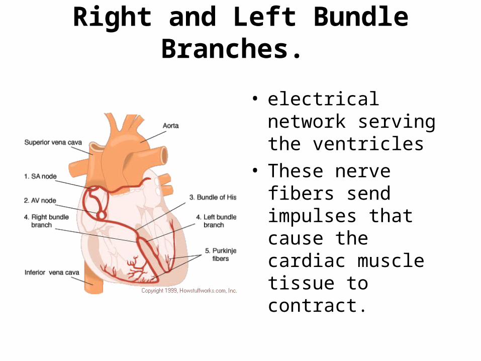
Right and Left Bundle Branches.
• electrical network serving the ventricles
• These nerve fibers send impulses that cause the cardiac muscle tissue to contract.

Right and Left Bundle Branches. • located in the inner
ventricular walls of the heart, just beneath the endocardium.
• These fibers are specialized myocardial fibers that conduct an electrical stimulus or impulse that enables the heart to contract in a coordinated fashion.

Purkinje Fibers
• electrical network serving the ventricles
• These nerve fibers send impulses that cause the cardiac muscle tissue to contract.