The brain on art: intense aesthetic experience activates the
17
ORIGINAL RESEARCH ARTICLE published: 20 April 2012 doi: 10.3389/fnhum.2012.00066 The brain on art: intense aesthetic experience activates the default mode network Edward A. Vessel 1 *, G. Gabrielle Starr 2 and Nava Rubin 3 1 Center for Brain Imaging, New York University, New York, NY, USA 2 Department of English, New York University, New York, NY,USA 3 Center for Neural Science, New York University, New York, NY,USA Edited by: Alvaro Pascual-Leone, Beth Israel Deaconess Medical Center/Harvard Medical School, USA Reviewed by: Alvaro Pascual-Leone, Beth Israel Deaconess Medical Center/Harvard Medical School, USA Luis M. Martinez, Universidade de A Coruña, Spain Mark A. Halko, Beth Israel Deaconess Medical Center, USA *Correspondence: Edward A. Vessel, Center for Brain Imaging, New York University, 4 Washington Pl., Rm. 156, New York, NY 10003, USA. e-mail: [email protected] Aesthetic responses to visual art comprise multiple types of experiences, from sensation and perception to emotion and self-reflection. Moreover, aesthetic experience is highly individual, with observers varying significantly in their responses to the same artwork. Combining fMRI and behavioral analysis of individual differences in aesthetic response, we identify two distinct patterns of neural activity exhibited by different sub-networks. Activity increased linearly with observers’ ratings (4-level scale) in sensory (occipito-temporal) regions. Activity in the striatum (STR) also varied linearly with ratings, with below-baseline activations for low-rated artworks. In contrast, a network of frontal regions showed a step-like increase only for the most moving artworks (“4” ratings) and non-differential activity for all others. This included several regions belonging to the “default mode network” (DMN) previously associated with self-referential mentation. Our results suggest that aesthetic experience involves the integration of sensory and emotional reactions in a manner linked with their personal relevance. Keywords: aesthetics, preference, fmri, visual art, default mode network INTRODUCTION Human beings in every culture seek out a variety of experiences which are classified as “aesthetic”—activities linked to the per- ception of external objects, but not to any apparent functional use these objects might have. Looking at paintings, listening to music, or reading poems—these are hedonic experiences in which humans consistently choose to engage. And although the relevant objects in and of themselves have no immediate or direct value for survival or for the satisfaction of basic needs (food, shelter, repro- duction), they nevertheless accrue great value within human cul- ture. What are the neural underpinnings of aesthetically moving experience? Although the foundation of aesthetic inquiry as a formal schol- arly discipline is relatively recent—the philosopher Alexander Baumgarten introduced the modern use of the term in 1739— musings about the nature of “beauty” date back at least as early as Plato (Plato, 1989) and Confucius, and evidence exists of well-developed artistic traditions in most of the world’s ancient cultures (e.g., China, India, Egypt, Mesopotamia, Persia). But it is only recently that it has become possible to investigate the physiological bases of aesthetic experience. Recent neuroimag- ing studies have identified several brain regions whose activation correlates with a variety of aesthetic experiences—namely loca- tions in the anterior medial prefrontal cortex (aMPFC) and the caudate/striatum, with several additional regions detected in some studies but not others (Blood and Zatorre, 2001; Cela- Conde et al., 2004; Kawabata and Zeki, 2004; Vartanian and Goel, 2004; Jacobsen et al., 2006; Di Dio and Gallese, 2009; Kirk et al., 2009; Ishizu and Zeki, 2011; Lacey et al., 2011; Salimpoor et al., 2011). These findings form the initial basis for the field of neuroaesthetics, but key questions remain. In this study we exam- ined more closely issues surrounding the intensity and diversity of aesthetic responses. A major theme in philosophical inquiry into aesthetic expe- rience is a tension between universality and subjectivity. On one hand, many authors have argued that aesthetic evaluations rely on universal principles. On the other, philosophical inquiry also emphasized the importance of understanding aesthetic responses as strongly subjective. These two views are not, in principle, mutually exclusive: subjective judgment may lead to aesthetic evaluations that are so consistent across individuals as to be termed universal. Indeed, the notion of universal aesthetics relies on the observation of wide agreement among people about the aesthetic value of certain objects or classes of objects (e.g., flow- ers; Scarry, 1999). Yet aesthetic judgments are not only subjective but also highly susceptible to cultural norms, education, and exposure. Thus, while there may be certain items that com- mand consensus in their evaluations, for the majority of artifacts judgments can vary widely. This variation in aesthetic judgments can be used to isolate the neural dimensions of aesthetic responses as opposed to reactions to particular features of a given work of art (e.g., Kawabata and Zeki, 2004; Salimpoor et al., 2011). To date, most studies have used stimuli that generated wide agreement. Putative subjective aspects of an experience were potentially confounded with differ- ences in the stimuli themselves. Another fundamental problem is that using stimuli on whose aesthetic value people tend to agree necessarily gives more weight to common internal factors—be they driven by culture or by evolution—and leaves little room for truly individual aspects of subjective aesthetic experience Frontiers in Human Neuroscience www.frontiersin.org April 2012 | Volume 6 | Article 66 | 1 HUMAN NEUROSCIENCE
Transcript of The brain on art: intense aesthetic experience activates the
ORIGINAL RESEARCH ARTICLEpublished: 20 April 2012
doi: 10.3389/fnhum.2012.00066
The brain on art: intense aesthetic experience activates thedefault mode networkEdward A. Vessel 1*, G. Gabrielle Starr 2 and Nava Rubin3
1 Center for Brain Imaging, New York University, New York, NY, USA2 Department of English, New York University, New York, NY, USA3 Center for Neural Science, New York University, New York, NY, USA
Edited by:
Alvaro Pascual-Leone, Beth IsraelDeaconess Medical Center/HarvardMedical School, USA
Reviewed by:
Alvaro Pascual-Leone, Beth IsraelDeaconess Medical Center/HarvardMedical School, USALuis M. Martinez, Universidade de ACoruña, SpainMark A. Halko, Beth IsraelDeaconess Medical Center, USA
*Correspondence:
Edward A. Vessel, Center for BrainImaging, New York University, 4Washington Pl., Rm. 156, New York,NY 10003, USA.e-mail: [email protected]
Aesthetic responses to visual art comprise multiple types of experiences, from sensationand perception to emotion and self-reflection. Moreover, aesthetic experience is highlyindividual, with observers varying significantly in their responses to the same artwork.Combining fMRI and behavioral analysis of individual differences in aesthetic response, weidentify two distinct patterns of neural activity exhibited by different sub-networks. Activityincreased linearly with observers’ ratings (4-level scale) in sensory (occipito-temporal)regions. Activity in the striatum (STR) also varied linearly with ratings, with below-baselineactivations for low-rated artworks. In contrast, a network of frontal regions showed astep-like increase only for the most moving artworks (“4” ratings) and non-differentialactivity for all others. This included several regions belonging to the “default modenetwork” (DMN) previously associated with self-referential mentation. Our resultssuggest that aesthetic experience involves the integration of sensory and emotionalreactions in a manner linked with their personal relevance.
Keywords: aesthetics, preference, fmri, visual art, default mode network
INTRODUCTIONHuman beings in every culture seek out a variety of experienceswhich are classified as “aesthetic”—activities linked to the per-ception of external objects, but not to any apparent functionaluse these objects might have. Looking at paintings, listening tomusic, or reading poems—these are hedonic experiences in whichhumans consistently choose to engage. And although the relevantobjects in and of themselves have no immediate or direct value forsurvival or for the satisfaction of basic needs (food, shelter, repro-duction), they nevertheless accrue great value within human cul-ture. What are the neural underpinnings of aesthetically movingexperience?
Although the foundation of aesthetic inquiry as a formal schol-arly discipline is relatively recent—the philosopher AlexanderBaumgarten introduced the modern use of the term in 1739—musings about the nature of “beauty” date back at least as earlyas Plato (Plato, 1989) and Confucius, and evidence exists ofwell-developed artistic traditions in most of the world’s ancientcultures (e.g., China, India, Egypt, Mesopotamia, Persia). But itis only recently that it has become possible to investigate thephysiological bases of aesthetic experience. Recent neuroimag-ing studies have identified several brain regions whose activationcorrelates with a variety of aesthetic experiences—namely loca-tions in the anterior medial prefrontal cortex (aMPFC) andthe caudate/striatum, with several additional regions detected insome studies but not others (Blood and Zatorre, 2001; Cela-Conde et al., 2004; Kawabata and Zeki, 2004; Vartanian andGoel, 2004; Jacobsen et al., 2006; Di Dio and Gallese, 2009; Kirket al., 2009; Ishizu and Zeki, 2011; Lacey et al., 2011; Salimpooret al., 2011). These findings form the initial basis for the field of
neuroaesthetics, but key questions remain. In this study we exam-ined more closely issues surrounding the intensity and diversity ofaesthetic responses.
A major theme in philosophical inquiry into aesthetic expe-rience is a tension between universality and subjectivity. On onehand, many authors have argued that aesthetic evaluations relyon universal principles. On the other, philosophical inquiry alsoemphasized the importance of understanding aesthetic responsesas strongly subjective. These two views are not, in principle,mutually exclusive: subjective judgment may lead to aestheticevaluations that are so consistent across individuals as to betermed universal. Indeed, the notion of universal aesthetics relieson the observation of wide agreement among people about theaesthetic value of certain objects or classes of objects (e.g., flow-ers; Scarry, 1999). Yet aesthetic judgments are not only subjectivebut also highly susceptible to cultural norms, education, andexposure. Thus, while there may be certain items that com-mand consensus in their evaluations, for the majority of artifactsjudgments can vary widely.
This variation in aesthetic judgments can be used to isolate theneural dimensions of aesthetic responses as opposed to reactionsto particular features of a given work of art (e.g., Kawabata andZeki, 2004; Salimpoor et al., 2011). To date, most studies haveused stimuli that generated wide agreement. Putative subjectiveaspects of an experience were potentially confounded with differ-ences in the stimuli themselves. Another fundamental problem isthat using stimuli on whose aesthetic value people tend to agreenecessarily gives more weight to common internal factors—bethey driven by culture or by evolution—and leaves little roomfor truly individual aspects of subjective aesthetic experience
Frontiers in Human Neuroscience www.frontiersin.org April 2012 | Volume 6 | Article 66 | 1
HUMAN NEUROSCIENCE
Vessel et al. The brain on art
to emerge. We solved this by using stimuli for which peopleexpressed strongly individual preferences. These large individualdifferences enable us to use the diversity of visual artwork to parseout the different components of aesthetic experience.
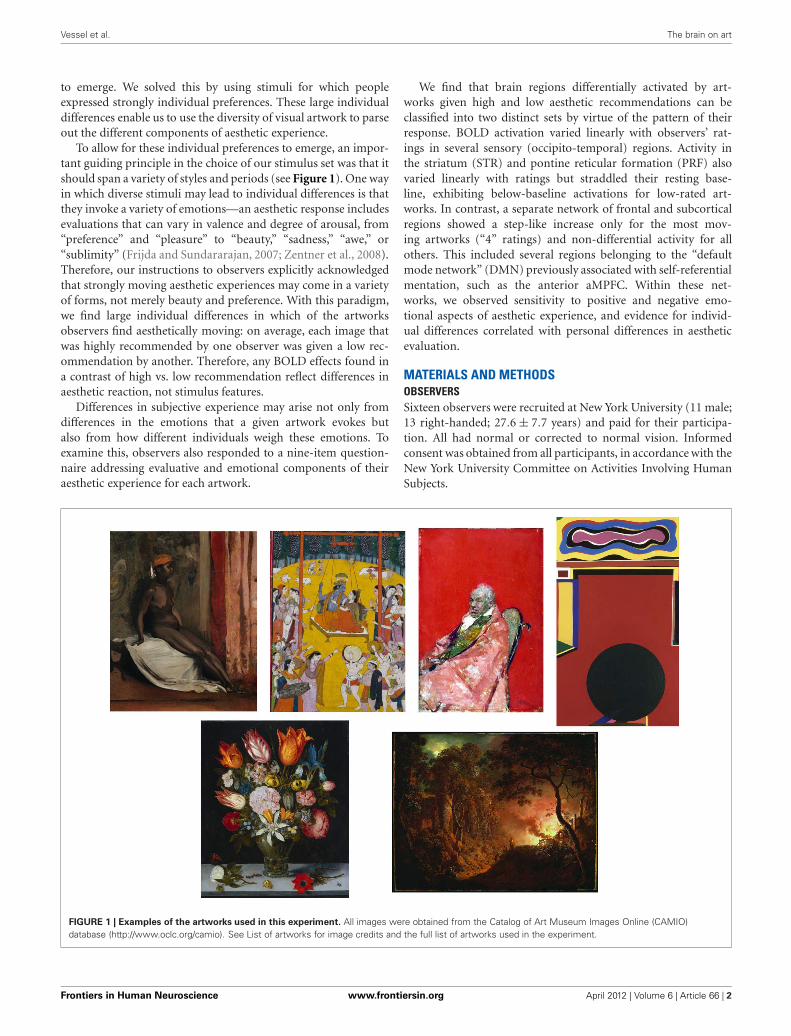
To allow for these individual preferences to emerge, an impor-tant guiding principle in the choice of our stimulus set was that itshould span a variety of styles and periods (see Figure 1). One wayin which diverse stimuli may lead to individual differences is thatthey invoke a variety of emotions—an aesthetic response includesevaluations that can vary in valence and degree of arousal, from“preference” and “pleasure” to “beauty,” “sadness,” “awe,” or“sublimity” (Frijda and Sundararajan, 2007; Zentner et al., 2008).Therefore, our instructions to observers explicitly acknowledgedthat strongly moving aesthetic experiences may come in a varietyof forms, not merely beauty and preference. With this paradigm,we find large individual differences in which of the artworksobservers find aesthetically moving: on average, each image thatwas highly recommended by one observer was given a low rec-ommendation by another. Therefore, any BOLD effects found ina contrast of high vs. low recommendation reflect differences inaesthetic reaction, not stimulus features.
Differences in subjective experience may arise not only fromdifferences in the emotions that a given artwork evokes butalso from how different individuals weigh these emotions. Toexamine this, observers also responded to a nine-item question-naire addressing evaluative and emotional components of theiraesthetic experience for each artwork.
We find that brain regions differentially activated by art-works given high and low aesthetic recommendations can beclassified into two distinct sets by virtue of the pattern of theirresponse. BOLD activation varied linearly with observers’ rat-ings in several sensory (occipito-temporal) regions. Activity inthe striatum (STR) and pontine reticular formation (PRF) alsovaried linearly with ratings but straddled their resting base-line, exhibiting below-baseline activations for low-rated art-works. In contrast, a separate network of frontal and subcorticalregions showed a step-like increase only for the most mov-ing artworks (“4” ratings) and non-differential activity for allothers. This included several regions belonging to the “defaultmode network” (DMN) previously associated with self-referentialmentation, such as the anterior aMPFC. Within these net-works, we observed sensitivity to positive and negative emo-tional aspects of aesthetic experience, and evidence for individ-ual differences correlated with personal differences in aestheticevaluation.
MATERIALS AND METHODSOBSERVERSSixteen observers were recruited at New York University (11 male;13 right-handed; 27.6 ± 7.7 years) and paid for their participa-tion. All had normal or corrected to normal vision. Informedconsent was obtained from all participants, in accordance with theNew York University Committee on Activities Involving HumanSubjects.
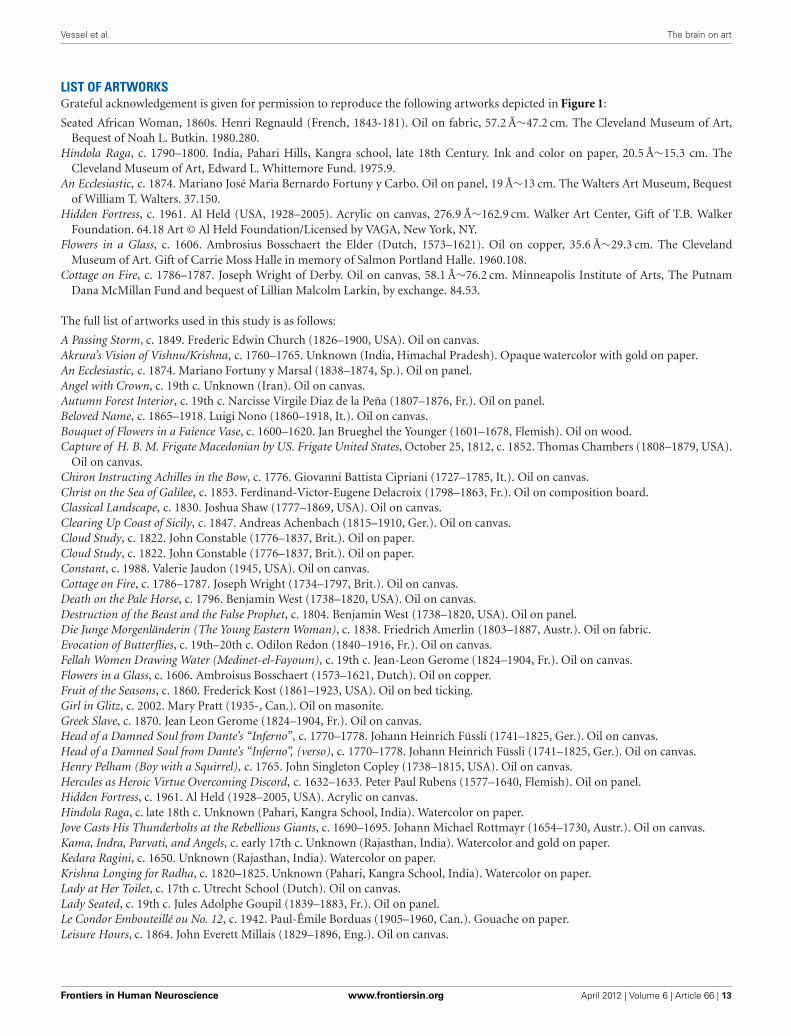
FIGURE 1 | Examples of the artworks used in this experiment. All images were obtained from the Catalog of Art Museum Images Online (CAMIO)database (http://www.oclc.org/camio). See List of artworks for image credits and the full list of artworks used in the experiment.
Frontiers in Human Neuroscience www.frontiersin.org April 2012 | Volume 6 | Article 66 | 2
Vessel et al. The brain on art
STIMULIOne hundred and nine images were selected from the Catalogof Art Museum Images Online database (CAMIO: http://www.oclc.org/camio; Figure 1 and List of Artworks). CAMIO con-tains more than 90,000 images of textiles, paintings, architecture,and sculpture from museum collections around the world. Theworks of art came from a variety of cultural traditions (American,European, Indian, and Japanese) and from a variety of histori-cal periods (from the 15th century to the recent past). Imageswere representational and abstract, and could be roughly classi-fied as either female figure(s) (33), male figure(s) (23), a mixedgroup (20), still life (11), landscape (14), or abstract paint-ing (8). These classifications did not show significant effects onresponses.
Commonly reproduced images were not used, in order to min-imize recognition. Most observers recognized no images, and noobserver recognized more than a very few (3–5) stimulus imagesas reported by survey responses.
Images were scaled such that the largest dimension did notexceed 20◦ of visual angle, and the area did not exceed 75% ofa 20◦ box. Stimulus presentation and response collection werecontrolled using a Macintosh G4 running Matlab 6.5 and thePsychophysics Toolbox (Brainard, 1997).
PROCEDUREObservers were told they would be viewing a set of artworks whilelying in the scanner. They were to use a scale of 1–4 by pressing abutton on a hand-held response box to answer the question “howstrongly does this painting move you?” according to the followinginstructions:
Imagine that the images you see are of paintings that may beacquired by a museum of fine art. The curator needs to knowwhich paintings are the most aesthetically pleasing based on howstrongly you as an individual respond to them. Your job is togive your gut-level response, based on how much you find thepainting beautiful, compelling, or powerful. Note: The paintingsmay cover the entire range from “beautiful” to “strange” or even“ugly.” Respond on the basis of how much this image “moves” you.What is most important is for you to indicate what works you findpowerful, pleasing, or profound.
Each observer viewed all 109 artworks; the order was counterbal-anced across observers to control for possible serial order effects.Observers were instructed prior to entering the magnet and givenpractice trials using artworks not in the stimulus set.
Nine-item evaluative questionnaireAfter the fMRI session, observers were given a short break, andwere then taken to a behavioral lab where they sat in front ofa computer screen to complete a nine-item questionnaire. Theywere shown the same set of paintings in the same order as in thescanner. Each painting was shown for 6 s. Observers were asked torate the intensity with which each artwork evoked the followingevaluative/emotional responses: joy, pleasure, sadness, confusion,awe, fear, disgust, beauty, and the sublime. Responses to this nine-item questionnaire were given using mouse clicks on a visualseven-point scale for each item. These items were presented in
random order on each trial. Observers could respond to the nineitems in any order, but could not change ratings.
Observers ranged from those with novice-level experience ofart and art history to several having completed some undergrad-uate study in the history of art (evaluated using a survey at thetime of the experiment). Before entering the scanner, observerswere also administered the Positive and Negative Affect Schedule(PANAS; Watson et al., 1988). PANAS is a highly stable and inter-nally consistent metric for dispositional affect (mood), used todetermine how frequently an observer experiences positive andnegative affect in a defined time period. Observers in this studywere asked to answer questions with regard to the immediatelypreceding few days.
fMRI SCANNING PROCEDURESfMRI scans were carried out at New York University’s Center forBrain Imaging, using a 3-T Siemens Allegra scanner and a NovaMedical Head coil (NM011 head transmit coil). Artworks wereprojected onto a screen in the bore of the magnet and viewedthrough a mirror mounted on the head coil.
The 109 artworks were divided into four sets (different subsetsper observer, depending on order) and shown over the course offour functional scans using a slow event-related design. Duringthese functional scans, the blood oxygen level dependent (BOLD)signal was measured from the entire brain using thirty-six 3 mmslices aligned approximately parallel to the AC-PC plane (in planeresolution 3 × 3 mm, TR = 2 s, TE = 30 ms, FA = 80◦). Eachtrial began with a 1 s blank period then a blinking fixation pointfor 1 s, followed by the artwork for 6 s, and a blank screen for4 s, during which the observer pressed a key corresponding torecommendation. An additional 0, 2, or 4 s blank interval wasinserted pseudorandomly between trials to jitter trial timing, withan average trial length of 13.14 s.
Observers were also run in a localizer scan containing blocksof objects, scrambled objects, faces, and places. This 320 s scanconsisted of four 18 s blocks of each stimulus type, during whichthe observer performed a “1-back” task (where observers monitorfor exact repeats of an image). Each block contained 16 stimulusimages plus two repeats, each presented for 800 ms with a 200 msinter-stimulus-interval. The full-color images were placed on topof phase-scrambled versions of the same stimuli filling a 500 ×500 pixel square to control for differences in size across stimuluscategories.
A high resolution (1 mm3) anatomical volume (MPRagesequence) was obtained after the functional scans for registrationand spatial normalization.
BEHAVIORAL DATA ANALYSISFor the observers’ recommendations collected during the scan-ning session, a measure of agreement across individuals was com-puted by taking the set of 109 recommendations for every pairof observers and computing the Pearson correlation coefficient.Images with any missing recommendation values were excludedfrom the correlations in a pairwise manner. One observer gave no“4” recommendations, and was, therefore, excluded from subse-quent analyses relying on the contrast of “4” vs. “1” responses.Similarly, a measure of across-observer agreement was computed
Frontiers in Human Neuroscience www.frontiersin.org April 2012 | Volume 6 | Article 66 | 3

Vessel et al. The brain on art
for each item of the nine-item questionnaire collected after thescanning session. For each item, the Pearson correlation coeffi-cient was computed for each pair of observers.
Factor analysis of evaluative questionnaireThe responses on the nine-item questionnaire produced by eachobserver to each artwork (16 × 109 = 1744 trials total) were thenconverted to z-scores within observers and concatenated into asingle large matrix of scores. Principal components extraction wasused to identify factors with eigenvalues greater than one. Twoemotional/evaluative factors survived and were rotated using the“direct oblmin” method, which does not require that the factorsbe orthogonal. Scores on these two factors were computed foreach of the 1744 trials using regression (see Figure 7).
fMRI DATA ANALYSISThe scans were pre-processed using the FMRIB Software Library(FSL; Oxford, UK) to correct for slice-timing and motion,and were high-pass filtered at 0.0125 Hz. Subsequent analy-ses were performed using BrainVoyager QX (Brain Innovation,Maastricht, Netherlands). After alignment to observer-specifichigh-resolution anatomical images, the scans were normalized toTalairach space (Talairach and Tournoux, 1988), blurred with an8 mm Gaussian kernel, and converted to z-scores.
4-vs.-1 whole brain analysisTo identify regions sensitive to observer recommendation, awhole-brain random effects group-level general linear model(GLM) analysis was computed with the responses of eachobserver on each of the four possible recommendation levelscoded as separate regressors (as a 6 s “on” period for each imageconvolved with a standard two-gamma hemodynamic responsefunction, HRF). A contrast of the “4” regressors vs. the “1” regres-sors was computed and the resulting statistical map was correctedfor multiple comparisons at a false discovery rate (FDR) of q <
0.05 (Benjamini and Hochberg, 1995; Genovese et al., 2002) and acluster threshold of 5 3 mm3 voxels. This contrast will be referredto as the 4-vs.-1 whole-brain analysis (see Appendix Table A1 andFigures 3–5).
ROI analysisIn order to compare BOLD activation for all four recommenda-tion levels across these regions, the group-level clusters from the4-vs.-1 analysis were used to draw regions-of-interest (ROIs) fromwhich we extracted timeseries for each observer. Using the aver-age (over voxels in the ROI) of non-blurred, z-scored timeseriesfor each scan, individual observer parameter estimates for each ofthe four recommendation levels were obtained using a GLM witha standard two-gamma HRF convolved with a 6 s “on” periodfor each image (see Figures 3–5). Standard errors were computedacross observers.
4-vs.-321 whole brain analysisTo further isolate processes particular to aesthetic response, wecomputed a second whole-brain contrast relying on the samewhole-brain GLM as above, but with a new contrast of only the“4” recommendations vs. the average of all the other recommen-dation levels, balanced to add to zero [e.g., a linear contrast of
(−1 −1 −1 3) for the 1, 2, 3, and 4 regressors]. The same statisti-cal threshold was used to correct for multiple comparisons – FDRof q < 0.05 and a 5 3 mm3 cluster threshold. This contrast will bereferred to as the 4-vs.-321 whole-brain analysis (see Figure 6).Note that this contrast may lead to the discovery of new activa-tions not found in the original 4-vs.-1 analysis. Given the widelyextended and interconnected nature of the resulting whole-brainmap, we do not report the full set of activation coordinates—mostof the peak activations were coincident with regions reportedfor the 4-vs.-1 contrast. Group-level ROIs were isolated for fourprominent activations not found in the 4-vs.-1 contrast: the ante-rior medial pre-frontal cortex (aMPFC), the left hippocampus(HC), left substantia nigra (SN), and the left posterior cingulatecortex (PCC). It was not possible to draw an isolated ROI for theaMPFC from this contrast given the large swath of activation—we, therefore, drew a more restricted ROI for the aMPFC based onthe 4-vs.-1 whole-brain contrast, but with a statistical threshold ofp < 0.001.
ROI analysis of evaluative factorsThe trial-by-trial scores for the two factors extracted from theprincipal components factor analysis of the nine-item evaluativequestionnaire were used to create BOLD predictors by convolv-ing with a standard 2 gamma HRF with a length of 1 TR (2 s)and a delay of 1 TR relative to image onset. This middle TRwas chosen as a compromise given our uncertainty about when,during a 6 s viewing, an observer was able to integrate enoughinformation across successive fixations of an artwork to generatean affective response. The resulting timecourses were combinedwith an “Image On” predictor and orthonormalized using theGram-Schmidt process before being entered into a GLM predict-ing BOLD activation in each of the ROI’s identified in the wholebrain analysis (see Figure 7).
Individual differences analysis of evaluative questionnaireWe performed an analysis of individual differences in responsesto the nine-item evaluative questionnaire and their relationshipto BOLD activation. Each observer’s recommendations and theirsubsequent responses on the nine items were converted to z-scores, and then concatenated into a single large matrix (16observers × 109 images = 1744 rows). We performed a step-wise regression analysis in SPSS (IBM, Somers, NY) of observers’recommendations against their responses to the nine items toeliminate redundant terms or terms which had no significantpredictive power for recommendations. Individual standardizedbeta weights were then computed for how well each of the itemssurviving this procedure predicted recommendations, enteredin order from most-to-least predictive at the group level (seeAppendix Table A2). The resulting beta weights, which can beconceptualized as reflecting the weight an observer places on aparticular emotion/evaluation when making recommendations,were used to predict the size (across observers) of the 4-vs.-1BOLD effect in the set of ROIs identified in the whole-brainrecommendation-based analysis. This yielded an overall R2 foreach ROI and beta weights for each of the items with associatedconfidence intervals. A significant effect in this analysis wouldindicate that variability across observers in the size of the BOLD
Frontiers in Human Neuroscience www.frontiersin.org April 2012 | Volume 6 | Article 66 | 4

Vessel et al. The brain on art
effect in an ROI is related to variability in how much individualobservers weigh a particular emotion/evaluation when makingrecommendations (see Figure 8).
RESULTSThere was very low agreement in recommendations acrossobservers, as assessed by computing the correlations betweenobservers’ recommendations taken in pairs (Figure 2). The aver-age agreement (0.13 ± 0.17) indicates quite low agreement forvisual art compared to other kinds of stimuli (e.g., Vessel andRubin, 2010). (The mean of this distribution is significantlydifferent from zero by a t-test, t[119] = 8.72, p < 10−13, butCronbach’s alpha, a measure of inter-rater reliability, confirms thevery low agreement, α = 0.709; Cronbach, 1951). This findinghas an important methodological consequence: on average, eachimage highly recommended by one observer was given a low rec-ommendation by another. Therefore, any BOLD effects found ina contrast of high vs. low recommendation reflect differences inaesthetic reaction, not features of the images.
A whole-brain group contrast of trials in which an observergave an image the highest recommendation (“4”) vs. trials inwhich the image was given the lowest recommendation (“1”)revealed a set of posterior, anterior, and subcortical brain regionsthat were correlated with observers’ aesthetic recommendations(Appendix Table A1; see “Materials and Methods, 4-vs.-1 Wholebrain analysis”). Below, we describe further the responses of theseregions, grouped by the nature of the response. The groupingswere based on an analysis beyond that which produced Table A1(4-vs.-1)—specifically, the pattern of responses across all fourrecommendation levels (see below). To examine those patterns,individual regions of interest (ROIs) were created based on the4-vs.-1 whole-brain contrast, and the average timecourses wereanalyzed to estimate the response to each of the four responselevels (see “Materials and Methods, ROI analysis”).
In posterior (occipito-temporal) ROIs, there was a linear rela-tionship between recommendation level and BOLD response
FIGURE 2 | The distribution of pairwise correlations across observers’
recommendations, illustrating highly individual responses. Eachobserver’s recommendations were correlated with every other observer’srecommendations, taken in pairs. This histogram shows the distribution ofall the correlation coefficients.
(Figure 3; left inferior temporal sulcus, ITS: −49, −61, −2; leftparahippocampal cortex, PHC: −31, −32, −15; right superiortemporal gyrus, STG: 52, −10, 7). In left ITS and left PHC BOLDresponse increased in an approximately linear fashion above rest-ing baseline for increasing recommendations. Similarly, BOLDsignal in right STG decreased in an approximately linear fashionbelow resting baseline for decreasing aesthetic reactions.
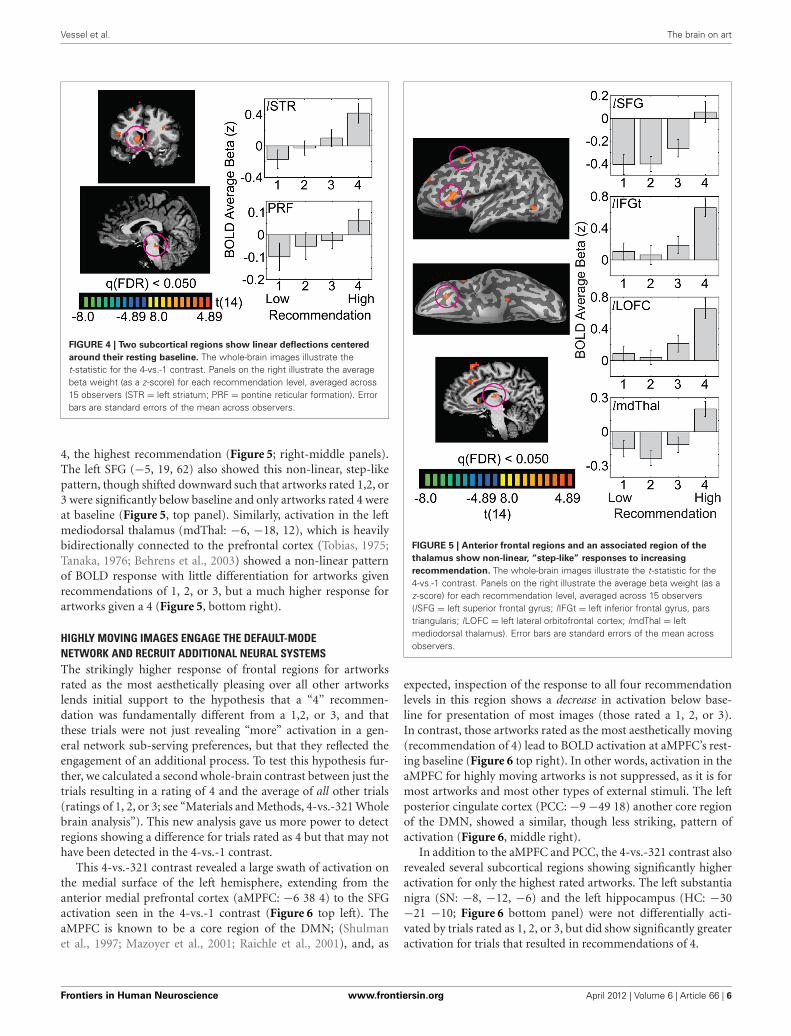
In two subcortical regions, the left striatum (STR) and thepontine reticular formation (PRF), there was also a linear rela-tionship between recommendation and BOLD activation. But incontrast to occipito-temporal ROIs, BOLD response levels strad-dled the resting baseline (Figure 4; STR: −12, 10, 6; PRF: 0, −28,−17). Thus, highly-rated images led to activation greater thanbaseline and low-rated images led to decreases from the restingbaseline.
In contrast with the linear relation between recommenda-tion and BOLD response observed in the occipito-temporal andsubcortical regions above, frontal ROIs identified in the 4-vs.-1contrast (Appendix Table A1) revealed a markedly different pat-tern of responses. In the left inferior frontal gyrus, pars trian-gularis (IFGt), left lateral orbitofrontal cortex (LOFC), and leftsuperior frontal gyrus (SFG) there was a non-linear, “step-like”pattern relating aesthetic recommendation and BOLD response(Figure 5). Activation in left IFGt (−50, 32, 12) and left LOFC(−35, 24, −4) was near baseline for artworks given a 1, 2, or 3recommendation, but was strikingly higher for artworks given a
FIGURE 3 | Posterior occipito-temporal regions of cortex show linear
deflections from baseline with increasing recommendation. Thewhole-brain images illustrate the t-statistic for the 4-vs.-1 contrast. Panelson the right illustrate the average beta weight (as a z-score) for eachrecommendation level, averaged across 15 observers (lITS = leftinferotemporal sulcus; lPHC = left parahippocampal cortex; rSTG = rightsuperior temporal gyrus). Error bars are standard errors of the mean acrossobservers.
Frontiers in Human Neuroscience www.frontiersin.org April 2012 | Volume 6 | Article 66 | 5
Vessel et al. The brain on art
FIGURE 4 | Two subcortical regions show linear deflections centered
around their resting baseline. The whole-brain images illustrate thet-statistic for the 4-vs.-1 contrast. Panels on the right illustrate the averagebeta weight (as a z-score) for each recommendation level, averaged across15 observers (STR = left striatum; PRF = pontine reticular formation). Errorbars are standard errors of the mean across observers.
4, the highest recommendation (Figure 5; right-middle panels).The left SFG (−5, 19, 62) also showed this non-linear, step-likepattern, though shifted downward such that artworks rated 1,2, or3 were significantly below baseline and only artworks rated 4 wereat baseline (Figure 5, top panel). Similarly, activation in the leftmediodorsal thalamus (mdThal: −6, −18, 12), which is heavilybidirectionally connected to the prefrontal cortex (Tobias, 1975;Tanaka, 1976; Behrens et al., 2003) showed a non-linear patternof BOLD response with little differentiation for artworks givenrecommendations of 1, 2, or 3, but a much higher response forartworks given a 4 (Figure 5, bottom right).
HIGHLY MOVING IMAGES ENGAGE THE DEFAULT-MODENETWORK AND RECRUIT ADDITIONAL NEURAL SYSTEMSThe strikingly higher response of frontal regions for artworksrated as the most aesthetically pleasing over all other artworkslends initial support to the hypothesis that a “4” recommen-dation was fundamentally different from a 1,2, or 3, and thatthese trials were not just revealing “more” activation in a gen-eral network sub-serving preferences, but that they reflected theengagement of an additional process. To test this hypothesis fur-ther, we calculated a second whole-brain contrast between just thetrials resulting in a rating of 4 and the average of all other trials(ratings of 1, 2, or 3; see “Materials and Methods, 4-vs.-321 Wholebrain analysis”). This new analysis gave us more power to detectregions showing a difference for trials rated as 4 but that may nothave been detected in the 4-vs.-1 contrast.
This 4-vs.-321 contrast revealed a large swath of activation onthe medial surface of the left hemisphere, extending from theanterior medial prefrontal cortex (aMPFC: −6 38 4) to the SFGactivation seen in the 4-vs.-1 contrast (Figure 6 top left). TheaMPFC is known to be a core region of the DMN; (Shulmanet al., 1997; Mazoyer et al., 2001; Raichle et al., 2001), and, as
FIGURE 5 | Anterior frontal regions and an associated region of the
thalamus show non-linear, “step-like” responses to increasing
recommendation. The whole-brain images illustrate the t-statistic for the4-vs.-1 contrast. Panels on the right illustrate the average beta weight (as az-score) for each recommendation level, averaged across 15 observers(lSFG = left superior frontal gyrus; lIFGt = left inferior frontal gyrus, parstriangularis; lLOFC = left lateral orbitofrontal cortex; lmdThal = leftmediodorsal thalamus). Error bars are standard errors of the mean acrossobservers.
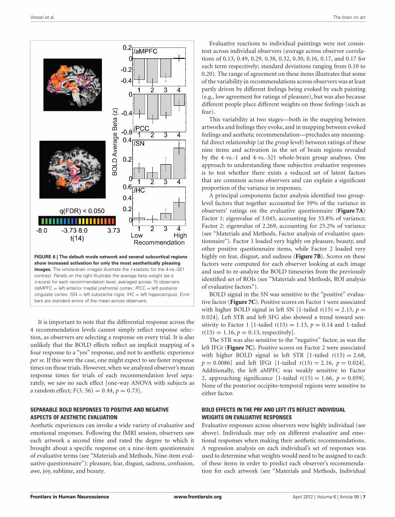
expected, inspection of the response to all four recommendationlevels in this region shows a decrease in activation below base-line for presentation of most images (those rated a 1, 2, or 3).In contrast, those artworks rated as the most aesthetically moving(recommendation of 4) lead to BOLD activation at aMPFC’s rest-ing baseline (Figure 6 top right). In other words, activation in theaMPFC for highly moving artworks is not suppressed, as it is formost artworks and most other types of external stimuli. The leftposterior cingulate cortex (PCC: −9 −49 18) another core regionof the DMN, showed a similar, though less striking, pattern ofactivation (Figure 6, middle right).
In addition to the aMPFC and PCC, the 4-vs.-321 contrast alsorevealed several subcortical regions showing significantly higheractivation for only the highest rated artworks. The left substantianigra (SN: −8, −12, −6) and the left hippocampus (HC: −30−21 −10; Figure 6 bottom panel) were not differentially acti-vated by trials rated as 1, 2, or 3, but did show significantly greateractivation for trials that resulted in recommendations of 4.
Frontiers in Human Neuroscience www.frontiersin.org April 2012 | Volume 6 | Article 66 | 6
Vessel et al. The brain on art
FIGURE 6 | The default mode network and several subcortical regions
show increased activation for only the most aesthetically pleasing
images. The whole-brain images illustrate the t-statistic for the 4-vs.-321contrast. Panels on the right illustrate the average beta weight (as az-score) for each recommendation level, averaged across 15 observers(laMPFC = left anterior medial prefrontal cortex; lPCC = left posteriorcingulate cortex; lSN = left substantia nigra; lHC = left hippocampus). Errorbars are standard errors of the mean across observers.
It is important to note that the differential response across the4 recommendation levels cannot simply reflect response selec-tion, as observers are selecting a response on every trial. It is alsounlikely that the BOLD effects reflect an implicit mapping of afour response to a “yes” response, and not to aesthetic experienceper se. If this were the case, one might expect to see faster responsetimes on those trials. However, when we analyzed observer’s meanresponse times for trials of each recommendation level sepa-rately, we saw no such effect [one-way ANOVA with subjects asa random effect; F(3, 56) = 0.44, p = 0.73].
SEPARABLE BOLD RESPONSES TO POSITIVE AND NEGATIVEASPECTS OF AESTHETIC EVALUATIONAesthetic experiences can invoke a wide variety of evaluative andemotional responses. Following the fMRI session, observers saweach artwork a second time and rated the degree to which itbrought about a specific response on a nine-item questionnaireof evaluative terms (see “Materials and Methods, Nine-item eval-uative questionnaire”): pleasure, fear, disgust, sadness, confusion,awe, joy, sublime, and beauty.
Evaluative reactions to individual paintings were not consis-tent across individual observers (average across observer correla-tions of 0.13, 0.49, 0.29, 0.38, 0.32, 0.30, 0.16, 0.17, and 0.17 foreach term respectively; standard deviations ranging from 0.10 to0.20). The range of agreement on these items illustrates that someof the variability in recommendations across observers was at leastpartly driven by different feelings being evoked by each painting(e.g., low agreement for ratings of pleasure), but was also becausedifferent people place different weights on those feelings (such asfear).
This variability at two stages—both in the mapping betweenartworks and feelings they evoke, and in mapping between evokedfeelings and aesthetic recommendation—precludes any meaning-ful direct relationship (at the group level) between ratings of thesenine items and activation in the set of brain regions revealedby the 4-vs.-1 and 4-vs.-321 whole-brain group analyses. Oneapproach to understanding these subjective evaluative responsesis to test whether there exists a reduced set of latent factorsthat are common across observers and can explain a significantproportion of the variance in responses.
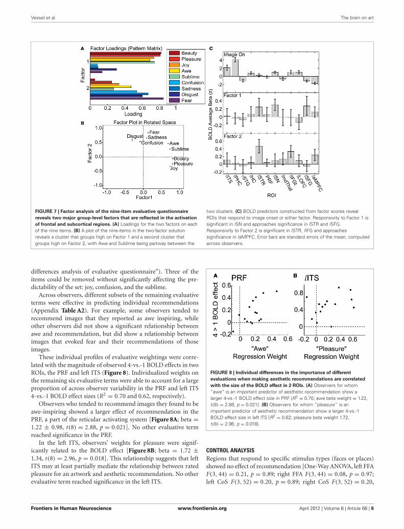
A principal components factor analysis identified two group-level factors that together accounted for 59% of the variance inobservers’ ratings on the evaluative questionnaire (Figure 7A)Factor 1: eigenvalue of 3.045, accounting for 33.8% of variance;Factor 2: eigenvalue of 2.269, accounting for 25.2% of variance(see “Materials and Methods, Factor analysis of evaluative ques-tionnaire”). Factor 1 loaded very highly on pleasure, beauty, andother positive questionnaire items, while Factor 2 loaded veryhighly on fear, disgust, and sadness (Figure 7B). Scores on thesefactors were computed for each observer looking at each imageand used to re-analyze the BOLD timeseries from the previouslyidentified set of ROIs (see “Materials and Methods, ROI analysisof evaluative factors”).
BOLD signal in the SN was sensitive to the “positive” evalua-tive factor (Figure 7C). Positive scores on Factor 1 were associatedwith higher BOLD signal in left SN [1-tailed t(15) = 2.15, p =0.024]. Left STR and left SFG also showed a trend toward sen-sitivity to Factor 1 [1-tailed t(15) = 1.13, p = 0.14 and 1-tailedt(15) = 1.16, p = 0.13, respectively].
The STR was also sensitive to the “negative” factor, as was theleft IFGt (Figure 7C). Positive scores on Factor 2 were associatedwith higher BOLD signal in left STR [1-tailed t(15) = 2.68,p = 0.0086] and left IFGt [1-tailed t(15) = 2.16, p = 0.024].Additionally, the left aMPFC was weakly sensitive to Factor2, approaching significance [1-tailed t(15) = 1.66, p = 0.059].None of the posterior occipito-temporal regions were sensitive toeither factor.
BOLD EFFECTS IN THE PRF AND LEFT ITS REFLECT INDIVIDUALWEIGHTS ON EVALUATIVE RESPONSESEvaluative responses across observers were highly individual (seeabove). Individuals may rely on different evaluative and emo-tional responses when making their aesthetic recommendations.A regression analysis on each individual’s set of responses wasused to determine what weights would need to be assigned to eachof these items in order to predict each observer’s recommenda-tion for each artwork (see “Materials and Methods, Individual
Frontiers in Human Neuroscience www.frontiersin.org April 2012 | Volume 6 | Article 66 | 7
Vessel et al. The brain on art
FIGURE 7 | Factor analysis of the nine-item evaluative questionnaire
reveals two major group-level factors that are reflected in the activation
of frontal and subcortical regions. (A) Loadings for the two factors on eachof the nine items. (B) A plot of the nine-items in the two-factor solutionreveals a cluster that groups high on Factor 1 and a second cluster thatgroups high on Factor 2, with Awe and Sublime being partway between the
two clusters. (C) BOLD predictors constructed from factor scores revealROIs that respond to image onset or either factor. Responsivity to Factor 1 issignificant in lSN and approaches significance in lSTR and lSFG.Responsivity to Factor 2 is significant in lSTR, lIFG and approachessignificance in laMPFC. Error bars are standard errors of the mean, computedacross observers.
differences analysis of evaluative questionnaire”). Three of theitems could be removed without significantly affecting the pre-dictability of the set: joy, confusion, and the sublime.
Across observers, different subsets of the remaining evaluativeterms were effective in predicting individual recommendations(Appendix Table A2). For example, some observers tended torecommend images that they reported as awe inspiring, whileother observers did not show a significant relationship betweenawe and recommendation, but did show a relationship betweenimages that evoked fear and their recommendations of thoseimages.
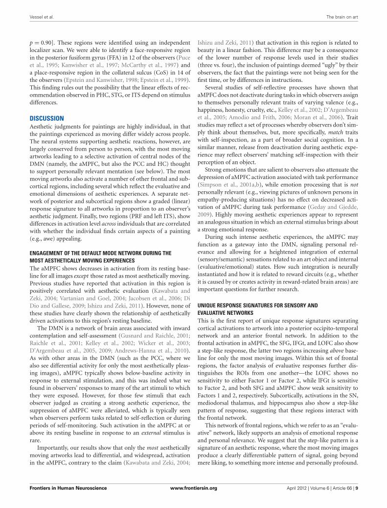
These individual profiles of evaluative weightings were corre-lated with the magnitude of observed 4-vs.-1 BOLD effects in twoROIs, the PRF and left ITS (Figure 8). Individualized weights onthe remaining six evaluative terms were able to account for a largeproportion of across observer variability in the PRF and left ITS4-vs.-1 BOLD effect sizes (R2 = 0.70 and 0.62, respectively).
Observers who tended to recommend images they found to beawe-inspiring showed a larger effect of recommendation in thePRF, a part of the reticular activating system [Figure 8A; beta =1.22 ± 0.98, t(8) = 2.88, p = 0.021]. No other evaluative termreached significance in the PRF.
In the left ITS, observers’ weights for pleasure were signif-icantly related to the BOLD effect [Figure 8B; beta = 1.72 ±1.34, t(8) = 2.96, p = 0.018]. This relationship suggests that leftITS may at least partially mediate the relationship between ratedpleasure for an artwork and aesthetic recommendation. No otherevaluative term reached significance in the left ITS.
FIGURE 8 | Individual differences in the importance of different
evaluations when making aesthetic recommendations are correlated
with the size of the BOLD effect in 2 ROIs. (A) Observers for whom“awe” is an important predictor of aesthetic recommendation show alarger 4-vs.-1 BOLD effect size in PRF [R2 = 0.70; awe beta weight = 1.22,t(8) = 2.88, p = 0.021]. (B) Observers for whom “pleasure” is animportant predictor of aesthetic recommendation show a larger 4-vs.-1BOLD effect size in left ITS [R2 = 0.62; pleasure beta weight 1.72,t(8) = 2.96, p = 0.018].
CONTROL ANALYSISRegions that respond to specific stimulus types (faces or places)showed no effect of recommendation [One-Way ANOVA, left FFAF(3, 44) = 0.21, p = 0.89; right FFA F(3, 44) = 0.08, p = 0.97;left CoS F(3, 52) = 0.20, p = 0.89; right CoS F(3, 52) = 0.20,
Frontiers in Human Neuroscience www.frontiersin.org April 2012 | Volume 6 | Article 66 | 8
Vessel et al. The brain on art
p = 0.90]. These regions were identified using an independentlocalizer scan. We were able to identify a face-responsive regionin the posterior fusiform gyrus (FFA) in 12 of the observers (Puceet al., 1995; Kanwisher et al., 1997; McCarthy et al., 1997) anda place-responsive region in the collateral sulcus (CoS) in 14 ofthe observers (Epstein and Kanwisher, 1998; Epstein et al., 1999).This finding rules out the possibility that the linear effects of rec-ommendation observed in PHC, STG, or ITS depend on stimulusdifferences.
DISCUSSIONAesthetic judgments for paintings are highly individual, in thatthe paintings experienced as moving differ widely across people.The neural systems supporting aesthetic reactions, however, arelargely conserved from person to person, with the most movingartworks leading to a selective activation of central nodes of theDMN (namely, the aMPFC, but also the PCC and HC) thoughtto support personally relevant mentation (see below). The mostmoving artworks also activate a number of other frontal and sub-cortical regions, including several which reflect the evaluative andemotional dimensions of aesthetic experiences. A separate net-work of posterior and subcortical regions show a graded (linear)response signature to all artworks in proportion to an observer’saesthetic judgment. Finally, two regions (PRF and left ITS), showdifferences in activation level across individuals that are correlatedwith whether the individual finds certain aspects of a painting(e.g., awe) appealing.
ENGAGEMENT OF THE DEFAULT MODE NETWORK DURING THEMOST AESTHETICALLY MOVING EXPERIENCESThe aMPFC shows decreases in activation from its resting base-line for all images except those rated as most aesthetically moving.Previous studies have reported that activation in this region ispositively correlated with aesthetic evaluation (Kawabata andZeki, 2004; Vartanian and Goel, 2004; Jacobsen et al., 2006; DiDio and Gallese, 2009; Ishizu and Zeki, 2011). However, none ofthese studies have clearly shown the relationship of aestheticallydriven activations to this region’s resting baseline.
The DMN is a network of brain areas associated with inwardcontemplation and self-assessment (Gusnard and Raichle, 2001;Raichle et al., 2001; Kelley et al., 2002; Wicker et al., 2003;D’Argembeau et al., 2005, 2009; Andrews-Hanna et al., 2010).As with other areas in the DMN (such as the PCC, where wealso see differential activity for only the most aesthetically pleas-ing images), aMPFC typically shows below-baseline activity inresponse to external stimulation, and this was indeed what wefound in observers’ responses to many of the art stimuli to whichthey were exposed. However, for those few stimuli that eachobserver judged as creating a strong aesthetic experience, thesuppression of aMPFC were alleviated, which is typically seenwhen observers perform tasks related to self-reflection or duringperiods of self-monitoring. Such activation in the aMPFC at orabove its resting baseline in response to an external stimulus israre.
Importantly, our results show that only the most aestheticallymoving artworks lead to differential, and widespread, activationin the aMPFC, contrary to the claim (Kawabata and Zeki, 2004;
Ishizu and Zeki, 2011) that activation in this region is related tobeauty in a linear fashion. This difference may be a consequenceof the lower number of response levels used in their studies(three vs. four), the inclusion of paintings deemed “ugly” by theirobservers, the fact that the paintings were not being seen for thefirst time, or by differences in instructions.
Several studies of self-reflective processes have shown thataMPFC does not deactivate during tasks in which observers assignto themselves personally relevant traits of varying valence (e.g.,happiness, honesty, cruelty, etc., Kelley et al., 2002; D’Argembeauet al., 2005; Amodio and Frith, 2006; Moran et al., 2006). Traitstudies may reflect a set of processes whereby observers don’t sim-ply think about themselves, but, more specifically, match traitswith self-inspection, as a part of broader social cognition. In asimilar manner, release from deactivation during aesthetic expe-rience may reflect observers’ matching self-inspection with theirperception of an object.
Strong emotions that are salient to observers also attenuate thedepression of aMPFC activation associated with task performance(Simpson et al., 2001a,b), while emotion processing that is notpersonally relevant (e.g., viewing pictures of unknown persons inempathy-producing situations) has no effect on decreased acti-vation of aMPFC during task performance (Geday and Gjedde,2009). Highly moving aesthetic experiences appear to representan analogous situation in which an external stimulus brings abouta strong emotional response.
During such intense aesthetic experiences, the aMPFC mayfunction as a gateway into the DMN, signaling personal rel-evance and allowing for a heightened integration of external(sensory/semantic) sensations related to an art object and internal(evaluative/emotional) states. How such integration is neurallyinstantiated and how it is related to reward circuits (e.g., whetherit is caused by or creates activity in reward-related brain areas) areimportant questions for further research.
UNIQUE RESPONSE SIGNATURES FOR SENSORY ANDEVALUATIVE NETWORKSThis is the first report of unique response signatures separatingcortical activations to artwork into a posterior occipito-temporalnetwork and an anterior frontal network. In addition to thefrontal activation in aMPFC, the SFG, IFGt, and LOFC also showa step-like response, the latter two regions increasing above base-line for only the most moving images. Within this set of frontalregions, the factor analysis of evaluative responses further dis-tinguishes the ROIs from one another—the LOFC shows nosensitivity to either Factor 1 or Factor 2, while lFGt is sensitiveto Factor 2, and both SFG and aMPFC show weak sensitivity toFactors 1 and 2, respectively. Subcortically, activations in the SN,mediodorsal thalamus, and hippocampus also show a step-likepattern of response, suggesting that these regions interact withthe frontal network.
This network of frontal regions, which we refer to as an “evalu-ative” network, likely supports an analysis of emotional responseand personal relevance. We suggest that the step-like pattern is asignature of an aesthetic response, where the most moving imagesproduce a clearly differentiable pattern of signal, going beyondmere liking, to something more intense and personally profound.
Frontiers in Human Neuroscience www.frontiersin.org April 2012 | Volume 6 | Article 66 | 9
Vessel et al. The brain on art
Additional support for this interpretation comes from a recentstudy in which observers were instructed to view artworks interms of semantic or visual detail (“pragmatically”), as opposedto in terms of color, composition, shapes, mood, and evokedemotion (“aesthetically”). They found an activation in left lateralprefrontal cortex (−44, 37, 7; BA 10) corresponding to what weterm left IFGt, which was selectively engaged in the “aesthetic”condition (Cupchik et al., 2009).
The second signature we observe, a linear response to observerrecommendation, is found in more posterior cortical regions(PHC, ITS, and STS). In all of these areas, BOLD signal respondsto the onset of any image and linearly tracks observers’ aestheticreactions. Several previous reports have also found activations inoccipito-temporal areas for preference judgments of a variety ofstimuli, including artwork, abstract geometric shapes, scenes, andfaces (e.g., Vartanian and Goel, 2004; Jacobsen et al., 2006; Kimet al., 2007; Yue et al., 2007).
These activations likely reflect a stimulus-bound sensoryand semantic analysis of preference that is relatively automatic.Supporting this interpretation is the finding that observers whoserecommendations were well predicted by ratings of image-induced “pleasure” tended to show a larger BOLD effect in the ITS(suggesting that observers differ in the degree to which they valuea sensory/semantic analysis performed by posterior areas versusemotional evocativeness when reacting to aesthetic experiences).It is important to note that the linear effect of aesthetic recom-mendation that we observed in these areas is not due to systematicdifferences in the type of stimuli preferred by the observers, asneither the CoS nor FFA, defined using an independent local-izer task (for places and faces, respectively) showed any effect ofrecommendation.
Subcortical regions STR and PRF, which also show a lin-ear relationship to observer’s recommendations, increased abovebaseline for recommended images and decreased below baselinefor non-recommended images. Given the involvement of a col-umn of areas in the midbrain with arousal functions (Kinomuraet al., 1996; Steriade, 1996), these activations may reflect “reward”valence in STR and arousal level in PRF, two often theorized axesof emotional responsivity (Lang et al., 1990; Low et al., 2008).Although we did not explicitly measure physiological arousal, thefact that the BOLD effect size in PRF was larger for observers whotended to recommend images they found awe-inspiring suggestsa potential association between aesthetic awe and arousal.
INTEGRATION IN THE STRIATUMNot only is STR activity linearly related to aesthetic recommen-dation, it is also sensitive to both emotional/evaluative factors.This suggests that STR may integrate perceptual, evaluative, andreward components of aesthetic response for the purpose ofoutcome selection (the choice of recommendation level). Thispattern, along with the detection of a related response pattern inthe mdThal, is in accord with the established existence of cortico-striato-pallado-thalamic loops (Alexander et al., 1986; Steriadeand Llinás, 1988; Alexander and Crutcher, 1990; Middleton andStrick, 2002; Kelly and Strick, 2004). Further research will beneeded to elucidate the temporal dynamics of the flow of infor-mation between these regions in aesthetic responses.
The location of the observed striatal activation straddles theanatomical division between dorsal and ventral STR, and is sim-ilar to that reported by Vartanian and Goel (2004), though otherstudies of preference have reported more ventral effects (Kimet al., 2007; Lacey et al., 2011). Intriguingly, we did find sig-nificantly greater activation in the right ventral STR for themost highly recommended images (4-vs.-321 contrast, results notshown). The literature on reward posits that the dorsal STR rep-resents the “actor” function of learning and implements habits ordecisions (Maia, 2009), as well as the expectation of reward andpunishment (Delgado et al., 2000, 2003), whereas the ventral STR(along with the amygdala, VTA, and OFC), carries out “critic”functions of representing actual reward and reward-predictionerror (Schultz et al., 1992; Schoenbaum et al., 1998; Hikosakaand Watanabe, 2000; Schultz, 2000; Tremblay and Schultz, 2000;Setlow et al., 2003; Paton et al., 2006; Wan and Peoples, 2006;Simmons et al., 2007). While the locus of our activation in STRdoes not clearly fall in either the ventral or dorsal STR, the factthat STR responds regardless of emotional valence is in agreementwith findings in monetary reward (Delgado et al., 2000, 2003).
Findings in regard to aesthetic reward have suggested a schismbetween desired and achieved reward that maps onto dorsal andventral STR, respectively. Based on a PET study of pleasurableresolution of musical expectation, Salimpoor et al. (2011) havesuggested that the caudate (“dorsal” STR) responds primarily toexpecting a desired reward (“wanting”), while the nucleus accum-bens (ventral STR) is active while experiencing the peak emotionalresponse (“liking”) associated with the resolution of a musicaltheme, line, or phrase. Unlike the novel, static images used in ourstudy, their musical stimuli are temporally extended experiences,enabling listeners to predict the resolution of a musical phrase (andsubsequent pleasure) based on familiarity with musical structureor particular songs. This may partially explain the difference inthe locus of striatal effects following the hypothesized momentof aesthetic reward, given the known involvement of basal gan-glia structures in a variety of temporally sequenced behaviors(Harrington et al., 1998). However, our task and results argueagainst a strict interpretation of striatal activation as reflectinganticipatory “wanting” a predicted reward, as there was no possi-bility of differential anticipatory responses for any of our images.
AREAS FOR FURTHER RESEARCHOur experiment is the first to find activation in the SN in visualaesthetic response, though it has been reported for music (Suzukiet al., 2008). Activation in the left SN for the most highly ratedimages raises the possibility that the efferent dopaminergic con-nections from the SN to the STR offer a mechanism by whichhedonic responses to the most highly moving images might bemodulated. This might be tested in further research.
In this set of observers, recommendation-related BOLDresponse appears primarily as increases in activation in the lefthemisphere. However, it is unclear at this time whether this rep-resents a real difference in the lateralization of aesthetic processesor merely reflects variation in the sensitivity of observing theseeffects at the whole-brain level.
Finally, it remains to be seen to what degree these systems areperturbed by depression or other mood disorders. Intriguingly,
Frontiers in Human Neuroscience www.frontiersin.org April 2012 | Volume 6 | Article 66 | 10

Vessel et al. The brain on art
we found that the size of the BOLD effect in PHC, reflectingsemantic/sensory processing, was larger for observers reportingpositive mood (r = 0.68, p < 0.004 using r to z transform), sug-gesting that mood may act as a gateway to getting pleasure fromsensory/aesthetic experiences.
CONCLUSIONSThe nature of aesthetic experience presents an apparent para-dox. Observers have strong aesthetic reactions to very differentsets of images, and are moved by particular images for very dif-ferent reasons. Yet the ability to be aesthetically moved appearsto be universal. The emerging picture of brain networks under-lying aesthetic experience presents a potential solution to thisparadox. Aesthetic experience involves the integration of neurallyseparable sensory and emotional reactions in a manner linkedwith their personal relevance. Such experiences are universal inthat the brain areas activated by aesthetically moving experiencesare largely conserved across individuals. However, this network
includes central nodes of the DMN that mediate the intenselysubjective and personal nature of aesthetic experiences, alongwith regions reflecting the wide variety of emotional states (bothpositive and negative) that can be experienced as aestheticallymoving.
The linking of intense aesthetic experience and personal rel-evance may have implications for artists and educators alike—further research could explore whether increasing the personalrelevance of aesthetic experiences increases their intensity and theresulting associations.
ACKNOWLEDGMENTSJustin Little, Lizzie Oldfather, Alexander Denker, BrynnHerrschaft, Steven R. Quartz, Damian Stanley, Souheil Inati,and Pablo Velasco. This project was supported by an ADVANCEResearch Challenge Grant funded by the NSF ADVANCE-PAIDaward # HRD-0820202 and by the Andrew W. Mellon Foundation(as a New Directions Fellowship).
REFERENCESAlexander, G. E., and Crutcher, M. D.
(1990). Functional architecture ofbasal ganglia circuits: neural sub-strates of parallel processing. TINS13, 266–271.
Alexander, G. E., DeLong, M. R.,and Strick, P. L. (1986). Parallelorganization of functionally segre-gated circuits linking basal gangliaand cortex. Annu. Rev. Neurosci. 9,357–381.
Amodio, D. M., and Frith, C. D. (2006).Meeting of minds: the medialfrontal cortex and social cognition.Nat. Rev. Neurosci. 7, 268–277.
Andrews-Hanna, J. R., Reidler, J. S.,Sepulcre, J., Poulin, R., and Buckner,R. L. (2010). Functional-anatomicfractionation of the brain’s defaultnetwork. Neuron 65, 550–562.
Behrens, T. E. J., Johansen-Berg, H.,Woolrich, M. W., Smith, S. M.,Wheeler-Kingshott, C. A. M.,Boulby, P. A., Barker, G. J., Sillery,E. L., Sheehan, K., Ciccarelli, O.,Thompson, A. J., Brady, J. M.,and Matthews, P. M. (2003). Non-invasive mapping of connectionsbetween human thalamus andcortex using diffusion imaging. Nat.Neurosci. 6, 750–757.
Benjamini, Y., and Hochberg, Y. (1995).Controlling the false discovery rate:a practical and powerful approachto multiple testing. J. R. Stat. Soc.Ser. B 57, 289–300.
Blood, A. J., and Zatorre, R. J. (2001).Intensely pleasurable responses tomusic correlate with activity inbrain regions implicated in rewardand emotion. Proc. Natl. Acad. Sci.U.S.A. 98, 11818–11823.
Brainard, D. H. (1997). The psy-chophysics toolbox. Spat. Vis. 10,443–446.
Cela-Conde, C. J., Marty, G., Maestú, F.,Ortiz, T., Munar, E., Fernández, A.,Roca, M., Rosselló, J., and Quesney,F. (2004). Activation of the pre-frontal cortex in the human visualaesthetic perception. Proc. Natl.Acad. Sci. U.S.A. 101, 6321–6325.
Chatterjee, A. (2011). Neuroaesthetics:a coming of age story. J. Cogn.Neurosci. 23, 53–62.
Cronbach, L. J. (1951). Coefficientalpha and the internal structure oftests. Psychometrika 16, 297–334.
Cupchik, G. C., Vartanian, O., Crawley,A., and Mikulis, D. J. (2009).Viewing artworks: contributions ofcognitive control and perceptualfacilitation to aesthetic experience.Brain. Cogn. 70, 84–91.
D’Argembeau, A., Collette, F., vander Linden, M., Laureys, S., DelFiore, G., Degueldre, C., Luxen,A., and Salmon, E. (2005). Self-referential reflective activity and itsrelationship with rest: a PET stidu.Neuroimage 25, 616–624.
D’Argembeau, A., Stawarczyk, D.,Majerus, S., Collette, F., van derLinden, M., Feyers, D., Maquet, P.,and Salmon, E. (2009). The neuralbasis of personal goal processingwhen envisioning future events.J. Cogn. Neurosci. 22, 1701–1713.
Delgado, M. R., Locke, H. M., Stenger,V. A., and Fiez, J. A. (2003). Dorsalstriatum responses to reward andpunishment: effects of valenceand magnitude manipulations.Cogn. Affect. Behav. Neurosci. 3,27–38.
Delgado, M. R., Nystrom, L. E., Fissell,C., Noll, D. C., and Fiez, J. A.(2000). Tracking the hemody-namic responses to reward andpunishment in the striatum. J.Neurophysiol. 84, 3072–3077.
Di Dio, C., and Gallese, V. (2009).Neuroaesthetics: a review. Curr.Opin. Neurobiol. 19, 682–687.
Epstein, R., Harris, A., Stanley, D., andKanwisher, N. (1999). The parahip-pocampal place area: recognition,navigation, or encoding? Neuron 23,115–125.
Epstein, R., and Kanwisher, N. (1998).A cortical representation of the localvisual environment. Nature 392,598–601.
Frijda, N. H., and Sundararajan, L.(2007). Emotion refinement: a the-ory inspired by Chinese poetics.Perspect. Psychol. Sci. 2, 227–241.
Geday, J., and Gjedde, A. (2009).Attention, emotion, and deactiva-tion of default activity in infe-rior medial prefrontal cortex. BrainCogn. 69, 344–352.
Genovese, C. R., Lazar, N. A., andNichols, T. (2002). Thresholding ofstatistical maps in functional neu-roimaging using the false discoveryrate. Neuroimage 15, 870–878.
Gusnard, D. A., and Raichle, M. E.(2001). Searching for a baseline:functional imaging and the restinghuman brain. Nat. Rev. Neurosci. 2,685–693.
Harrington, D. L., Haaland, K. Y., andHermanowicz, N. (1998). Temporalprocessing in the basal ganglia.Neuropsychology 12, 3–12.
Hikosaka, K., and Watanabe, M.(2000). Delay activity of orbital
and lateral prefrontal neuronsof the monkey varying with dif-ferent rewards. Cereb. Cortex 10,263–271.
Ishizu, T., and Zeki, S. (2011). Toward abrain-based theory of beauty. PLoSOne 6:e21852. doi: 10.1371/jour-nal.pone.0021852
Jacobsen, T., Schubotz, R. I., Hofel, L.,and Cramon, D. Y. V. (2006). Braincorrelates of aesthetic judgment ofbeauty. Neuroimage 29, 276–285.
Kanwisher, N., McDermott, J., andChun, M. M. (1997). The fusiformface area: a module in humanextrastriate cortex specialized forface perception. J. Neurosci. 17,4302–4311.
Kawabata, H., and Zeki, S. (2004).Neural correlates of beauty. J.Neurophysiol. 91, 1699–1705.
Kelley, W. M., Macrae, C. N., Wyland,C. L., Caglar, S., Inati, S., andHeatherton, T. F. (2002). Findingthe self? An event-related fMRIstudy. J. Cogn. Neurosci. 14,785–794.
Kelly, R. M., and Strick, P. L. (2004).Macro architecture of basal ganglialoops with the cerebral cortex: useof rabies virus to reveal multisy-naptic circuits. Prog. Brain Res. 143,449–459.
Kim, H., Adolphs, R., O’Doherty, J. P.,and Shimojo, S. (2007). Temporalisolation of neural processes under-lying face preference decisions.Proc. Natl. Acad. Sci. U.S.A. 104,18253–18258.
Kinomura, S., Larsson, J., Gulyas, B.,and Rolard, P. E. (1996). Activationby attention of the human retic-ular formation and thalamic
Frontiers in Human Neuroscience www.frontiersin.org April 2012 | Volume 6 | Article 66 | 11

Vessel et al. The brain on art
intralaminar nuclei. Science 271,512–515.
Kirk, U., Skov, M., Christensen, M. S.,and Nygaard, N. (2009). Brain cor-relates of aesthetic expertise: a para-metric fMRI study. Brain Cogn. 69,306–315.
Lacey, S., Hagtvedt, H., Patrick, V. M.,Anderson, A., Stilla, R., Deshpande,G., Hu, X., Sato, J. R., Reddy, S.,and Sathian, K. (2011). Art forreward’s sake: visual art recruitsthe ventral striatum. Neuroimage 55,420–433.
Lang, P. J., Bradley, M. M., andCuthbert, B. N. (1990). Emotion,attention, and startle reflex. Psychol.Rev. 97, 377–398.
Low, A., Lang, P. J., Smith, J. C., andBradley, M. M. (2008). Both preda-tor and prey: emotional arousal inthreat and reward. Psychol. Sci. 19,865–873.
Maia, T. V. (2009). Reinforcementlearning, conditioning, and thebrain: successes and challenges.Cogn. Affect. Behav. Neurosci. 9,243–264.
Mazoyer, B., Zago, L., Mellet, E.,Bricogne, S., Etard, O., Houde,O., Crivello, F., Joiiot, M. Petit, L.,and Tzourio-Mazoyer, N. (2001).Cortical networks for workingmemory and executive functionssustain the conscious resting state inman. Brain Res. Bull. 54, 287–298.
McCarthy, G., Puce, A., Gore, J. C.,and Allison, T. (1997). Face-specificprocessing in the human fusiformgyrus. J. Cogn. Neurosci. 9, 605–610.
Middleton, F. A., and Strick, P. L.(2002). Basal-ganglia ‘projections’to the prefrontal cortex of the pri-mate. Cereb. Cortex 12, 926–935.
Moran, J. M., Macrae, C. N.,Heatherton, T. F., Wyland, C.L., and Kelley, W. M. (2006).Neuroanatomical evidence fordistinct cognitive and affectivecomponents of self. J. Cogn.Neurosci. 18, 1586–1594.
Paton, J. J., Belova, M. A., Morrison, S.E., and Salzman, C. D. (2006). Theprimate amygdala represents thepositive and negative value of visualstimuli during learning. Nature 439,865–870.
Plato. (1989). Symposium (Trans.A. Nehamas and P. Woodruff).Indianapolis, IN: Hackett.
Puce, A., Allison, T., Gore, J. C.,and McCarthy, G. (1995). Face-sensitive regions in humanextrastriate cortex studied byfunctional MRI. J. Neurophysiol. 74,1192–1199.
Raichle, M. E., MacLeod, A. M., Snyder,A. Z., Powers, W. J., Gusnard, D.A., and Shulman, G. L. (2001).A default mode of brain function.Proc. Natl. Acad. Sci. U.S.A. 98,676–682.
Salimpoor, V. N., Benovoy, M., Larcher,K., Dagher, A., and Zatorre, R.J. (2011). Anatomically distinctdopamine release during antic-ipation and experience of peakemotion to music. Nat. Neurosci.14, 257–262.
Scarry, E. (1999). On Beauty andBeing Just. Princeton, NJ: PrincetonUniversity Press.
Schoenbaum, G., Chiba, A. A., andGallagher, M. (1998). Orbito-frontal cortex and basolateralamygdala encode expected outcom-ces during learning. Nat. Neurosci.1, 155–159.
Schultz, W. (2000). Multiple reward sig-nals in the brain. Nat. Rev. Neurosci.1, 199–207.
Schultz, W., Apicella, P., Scarnati, E.,and Ljunberg, T. (1992). Neuronalactivity in monkey ventral striatumrelated to the expectation of reward.J. Neurosci. 12, 4595–4610.
Setlow, B., Schoenbaum, G., andGallagher, M. (2003). Neuralencoding of ventral striatum duringolfactory discrimination learning.Neuron 38, 625–636.
Shulman, G. L., Fiez, J. A., Corbetta,M., Buckner, R. L., Miezin, F. M.,Raichle, M. E., and Petersen, S.E. (1997). Common blood flowchanges across visual tasks: 2.Decreases in cerebral cortex. J.Cogn. Neurosci. 9, 648–663.
Simmons, J. E., Ravel, S., Shidara, M.,and Richmond, B. J. (2007). “Acomparison of reward-contingentneuronal activity in mondkyorbitofrontal cortex and ventralstriatum: guiding acitons toward
rewards,” in Linking Affect toAction: Critical Contributions ofThe Orbitofrontal Cortex, ed G.Schoenbaum, J. A., Gottfried, E.A., Murray and S. J., Ramus (NewYork, NY: New York Academy ofSciences), 674–694.
Simpson, J. R., Snyder, A. Z., Gusnard,D. A., and Raichle, M. E. (2001a).Emotion-induced changes inhuman medial prefrontal cortex: I.During cognitive task performance.Proc. Natl. Acad. Sci. U.S.A. 93,683–687.
Simpson, J. R., Snyder, A. Z., Gusnard,D. A., and Raichle, M. E. (2001b).Emotion-induced changes inhuman medial prefrontal cortex:II. During anticipatory anxiety.Proc. Natl. Acad. Sci. U.S.A. 93,688–693.
Steriade, M. (1996). Arousal: revisit-ing the reticular activating system.Science 272, 225–226.
Steriade, M., and Llinás, R. R. (1988).The functional states of the tha-lamus and the associated neu-ronal interplay. Physiol. Rev. 68,649–742.
Suzuki, M., Okamura, N., Kawachi, Y.,Tashiro, M., and Arao, H. (2008).Discrete cortical regions associatedwith the musical beauty of majorand minor chords. Cogn. Affect.Behav. Neurosci. 8, 126–131.
Talairach, J., and Tournoux, P. (1988).Co-Planar Sterotaxic Atlas of theHuman Brain: 3-DimensionalProportional System – An Approachto Cerebral Imaging. New York, NY:Thieme Medical Publishers.
Tanaka, D. J. (1976). Thalamic pro-jections of the dorsomedial pre-frontal cortex in the rhesus monkey(Macaca mulatta). Brain Res. 110,21–38.
Tobias, T. J. (1975). Afferents to pre-frontal cortex from the thalamicmediodorsal nucleus in the rhesusmonkey. Brain Res. 83, 191–212.
Tremblay, L., and Schultz, W. (2000).Reward-related neuronal activityduring go-nogo task performancein primate orbitofrontal cortex. J.Neurophysiol. 83, 1864–1876.
Vartanian, O., and Goel, V. (2004).Neuroanatomical correlates of
aesthetic preference for paintings.Neuroreport 15, 893–897.
Vessel, E. A., and Rubin, N. (2010).Beauty and the beholder: highlyindividual taste for abstract, but notreal-world images. J. Vis. 10, 14.
Wan, X., and Peoples, L. L. (2006).Firing patterns of accumbal neuronsduring a Pavlovian-conditionedapproach task. J. Neurophysiol. 96,652–660.
Watson, D., Clark, L. A., and Tellegen,A. (1988). Development and vali-dation of brief measures of posi-tive and negative affect: the PANASscales. J. Pers. Soc. Psychol. 54,1063–1070.
Wicker, B., Ruby, P., Royet, J. P.,and Fonlupt, P. (2003). A rela-tion between rest and the selfin the brain? Brain Res. Rev. 43,224–230.
Yue, X., Vessel, E. A., and Biederman, I.(2007). Neural basis of scene prefer-ences. Neuroreport 18, 525–529.
Zentner, M., Grandjean, D., andScherer, K. R. (2008). Emotionsevoked by the sound of music:characterization, classification,and measurement. Emotion 8,494–521.
Conflict of Interest Statement: Theauthors declare that the researchwas conducted in the absence of anycommercial or financial relationshipsthat could be construed as a potentialconflict of interest.
Received: 12 October 2011; accepted: 12March 2012; published online: 20 April2012.Citation: Vessel EA, Starr GG and RubinN (2012) The brain on art: intense aes-thetic experience activates the defaultmode network. Front. Hum. Neurosci.6:66. doi: 10.3389/fnhum.2012.00066Copyright © 2012 Vessel, Starr andRubin. This is an open-access articledistributed under the terms of theCreative Commons Attribution NonCommercial License, which permitsnon-commercial use, distribution, andreproduction in other forums, providedthe original authors and source arecredited.
Frontiers in Human Neuroscience www.frontiersin.org April 2012 | Volume 6 | Article 66 | 12
Vessel et al. The brain on art
LIST OF ARTWORKSGrateful acknowledgement is given for permission to reproduce the following artworks depicted in Figure 1:
Seated African Woman, 1860s. Henri Regnauld (French, 1843-181). Oil on fabric, 57.2 Å∼47.2 cm. The Cleveland Museum of Art,Bequest of Noah L. Butkin. 1980.280.
Hindola Raga, c. 1790–1800. India, Pahari Hills, Kangra school, late 18th Century. Ink and color on paper, 20.5 Å∼15.3 cm. TheCleveland Museum of Art, Edward L. Whittemore Fund. 1975.9.
An Ecclesiastic, c. 1874. Mariano José Maria Bernardo Fortuny y Carbo. Oil on panel, 19 Å∼13 cm. The Walters Art Museum, Bequestof William T. Walters. 37.150.
Hidden Fortress, c. 1961. Al Held (USA, 1928–2005). Acrylic on canvas, 276.9 Å∼162.9 cm. Walker Art Center, Gift of T.B. WalkerFoundation. 64.18 Art © Al Held Foundation/Licensed by VAGA, New York, NY.
Flowers in a Glass, c. 1606. Ambrosius Bosschaert the Elder (Dutch, 1573–1621). Oil on copper, 35.6 Å∼29.3 cm. The ClevelandMuseum of Art. Gift of Carrie Moss Halle in memory of Salmon Portland Halle. 1960.108.
Cottage on Fire, c. 1786–1787. Joseph Wright of Derby. Oil on canvas, 58.1 Å∼76.2 cm. Minneapolis Institute of Arts, The PutnamDana McMillan Fund and bequest of Lillian Malcolm Larkin, by exchange. 84.53.
The full list of artworks used in this study is as follows:
A Passing Storm, c. 1849. Frederic Edwin Church (1826–1900, USA). Oil on canvas.Akrura’s Vision of Vishnu/Krishna, c. 1760–1765. Unknown (India, Himachal Pradesh). Opaque watercolor with gold on paper.An Ecclesiastic, c. 1874. Mariano Fortuny y Marsal (1838–1874, Sp.). Oil on panel.Angel with Crown, c. 19th c. Unknown (Iran). Oil on canvas.Autumn Forest Interior, c. 19th c. Narcisse Virgile Diaz de la Peña (1807–1876, Fr.). Oil on panel.Beloved Name, c. 1865–1918. Luigi Nono (1860–1918, It.). Oil on canvas.Bouquet of Flowers in a Faience Vase, c. 1600–1620. Jan Brueghel the Younger (1601–1678, Flemish). Oil on wood.Capture of H. B. M. Frigate Macedonian by US. Frigate United States, October 25, 1812, c. 1852. Thomas Chambers (1808–1879, USA).
Oil on canvas.Chiron Instructing Achilles in the Bow, c. 1776. Giovanni Battista Cipriani (1727–1785, It.). Oil on canvas.Christ on the Sea of Galilee, c. 1853. Ferdinand-Victor-Eugene Delacroix (1798–1863, Fr.). Oil on composition board.Classical Landscape, c. 1830. Joshua Shaw (1777–1869, USA). Oil on canvas.Clearing Up Coast of Sicily, c. 1847. Andreas Achenbach (1815–1910, Ger.). Oil on canvas.Cloud Study, c. 1822. John Constable (1776–1837, Brit.). Oil on paper.Cloud Study, c. 1822. John Constable (1776–1837, Brit.). Oil on paper.Constant, c. 1988. Valerie Jaudon (1945, USA). Oil on canvas.Cottage on Fire, c. 1786–1787. Joseph Wright (1734–1797, Brit.). Oil on canvas.Death on the Pale Horse, c. 1796. Benjamin West (1738–1820, USA). Oil on canvas.Destruction of the Beast and the False Prophet, c. 1804. Benjamin West (1738–1820, USA). Oil on panel.Die Junge Morgenländerin (The Young Eastern Woman), c. 1838. Friedrich Amerlin (1803–1887, Austr.). Oil on fabric.Evocation of Butterflies, c. 19th–20th c. Odilon Redon (1840–1916, Fr.). Oil on canvas.Fellah Women Drawing Water (Medinet-el-Fayoum), c. 19th c. Jean-Leon Gerome (1824–1904, Fr.). Oil on canvas.Flowers in a Glass, c. 1606. Ambroisus Bosschaert (1573–1621, Dutch). Oil on copper.Fruit of the Seasons, c. 1860. Frederick Kost (1861–1923, USA). Oil on bed ticking.Girl in Glitz, c. 2002. Mary Pratt (1935-, Can.). Oil on masonite.Greek Slave, c. 1870. Jean Leon Gerome (1824–1904, Fr.). Oil on canvas.Head of a Damned Soul from Dante’s “Inferno”, c. 1770–1778. Johann Heinrich Füssli (1741–1825, Ger.). Oil on canvas.Head of a Damned Soul from Dante’s “Inferno”, (verso), c. 1770–1778. Johann Heinrich Füssli (1741–1825, Ger.). Oil on canvas.Henry Pelham (Boy with a Squirrel), c. 1765. John Singleton Copley (1738–1815, USA). Oil on canvas.Hercules as Heroic Virtue Overcoming Discord, c. 1632–1633. Peter Paul Rubens (1577–1640, Flemish). Oil on panel.Hidden Fortress, c. 1961. Al Held (1928–2005, USA). Acrylic on canvas.Hindola Raga, c. late 18th c. Unknown (Pahari, Kangra School, India). Watercolor on paper.Jove Casts His Thunderbolts at the Rebellious Giants, c. 1690–1695. Johann Michael Rottmayr (1654–1730, Austr.). Oil on canvas.Kama, Indra, Parvati, and Angels, c. early 17th c. Unknown (Rajasthan, India). Watercolor and gold on paper.Kedara Ragini, c. 1650. Unknown (Rajasthan, India). Watercolor on paper.Krishna Longing for Radha, c. 1820–1825. Unknown (Pahari, Kangra School, India). Watercolor on paper.Lady at Her Toilet, c. 17th c. Utrecht School (Dutch). Oil on canvas.Lady Seated, c. 19th c. Jules Adolphe Goupil (1839–1883, Fr.). Oil on panel.Le Condor Embouteillé ou No. 12, c. 1942. Paul-Émile Borduas (1905–1960, Can.). Gouache on paper.Leisure Hours, c. 1864. John Everett Millais (1829–1896, Eng.). Oil on canvas.
Frontiers in Human Neuroscience www.frontiersin.org April 2012 | Volume 6 | Article 66 | 13
Vessel et al. The brain on art
Louise de Keroualle, c. 1671. Peter Lely (Peter van der Faes Lilley, 1618–1680, Eng.). Oil on canvas.Lucretia, c. 1534. Jan Gossaert (1478–1532, Flemish). Oil on panel to canvas transfer.Mandala of Shiva and Shakti, c. mid-late 18th c. Unknonwn (Nepal). Colors on cloth.Mars and Venus, Allegory of Peace, c. 1770. Louis-Jean-Francois Lagrenee (1725–1805, Fr.). Oil on canvas.Medusa, c. 1892. Alice Pice Barney (1857–1931, USA). Pastel on canvas.Mount Vesuvius at Midnight, c. 1868. Albert Bierstadt (1830–1902, USA). Oil on canvas.Mrs. Benjamin Hallowell, c. 1766. John Singleton Copley (1738–1815, USA). Oil on canvas.Niagara Falls from Table Rock, c. 1835. Samuel Finley Breese Morse (1791–1872, USA). Oil on canvas.On The Beach, c. 1845. Alfred Pellan (1906–1988, Can.). Oil on canvas.Oranges in Tissue Paper, c. 1890. William J. McCloskey (1859–1941, USA). Oil on canvas.Passage Violet, c. 1967. Fernand Leduc (1916, Can.). Acrylic on canvas.Peasant Girl with Sheep, c. 19th–20th c. Julien Dupre (1851–1910, Fr.). Oil on canvas.Portrait of a Child, c. 1805. Jean Baptiste Greuze (1725–1805, Fr.). Oil on canvas.Portrait of a Farmer’s Wife, c. 20th c. Robert Gwathmey (1903–1988, USA). Oil on canvas.Portrait of a Lady, c. 1771. John Singleton Copley (1738–1815, USA). Oil on canvas.Portrait of a Woman, c. 1660. Jan Mytens (1614–1670, Dutch). Oil on canvas.Portrait of a Woman, c. 1851. Jean-Leon Gerome (1824–1904, Fr.). Oil on canvas.Portrait of a Woman in a Rose-Colored Gown (La Dame en Rose), c. 18th c. Maurice Quentin de la Tour (1704–1788, Fr.). Pastel on
paper.Portrait of Madame X, c. 1907. Giovanni Boldini (1845–1931, It.). Oil on panel.Portrait of Mlle. Lange as Danae, c. 1799. Anne-Louis Girodet de Roucy Trioson (1767–1824, Fr.). Oil on canvas.Prometheus Bound, c. 1846–1847. Thomas Cole (1801–1848, USA). Oil on canvas.Red Sunset, Old Pond Concerto, c. 1972. Alma Thomas (1891–1978, USA). Acrylic on canvas.Rustam’s Seventh Course: He Kills the White Div: Illustration from the Manuscript, the “Tahmasp Shahnama” of Firdausi, c. 1520s–1540s.
Mir Musavvir (1533–1609, Persian). Watercolor, gold, and silver on paper.sans titre no 3, c. 1948. Charles Daudelin (1920–2001, Can.). Gouache on paper.Scene during the Eruption of Vesuvius, c. 1827. Joseph Franque (1774–1833, Fr.). Oil on canvas.Seated African Woman, c. 1860s. Henri Regnault (1843–1871, Fr.). Oil on canvas.Self-Portrait as a Yawning Man, c. 1783. Joseph Ducreux (1735–1802, Fr.). Oil on canvas.Somewhere in America, c. 1933. Robert Brackman (1898–1980, USA). Oil on canvas.Still Life with Flowers and Fruit, c. 1869. Claude Monet (1840–1926, Fr.). Oil on canvas.Still Life with Flowers, Shells, A Shark’s Head, and Petrifications, c. 1819. Antoine Berjon (1754–1843, Fr.). Oil on canvas.Still Life with Oysters and Grapes, c. 1653. Jan Davidsz Heem (1606–1684, Dutch). Oil on panel.The Angels Appearing to Abraham, c. 1750s. Francesco Guardi (1712–1793, It.). Oil on canvas.The Beeches, c. 1845. Asher B. Durand (1796–1886, USA). Oil on canvas.The Betrayal of Christ, c. 1618. Anthony van Dyck (1599–1641, Belgian). Oil on canvas.The Blue Dress (or A Duchess), c. 1861. Alfred Stevens (1823–1906, Belgian). Oil on panel.The Bouquet, c. 1870s. Raimundo Madrazo y Garreta (1841–1920, Sp.). Oil on panel.The Comtesse d’Haussonville (Louise de Broglie), c. 1845. Jean-Auguste-Dominique Ingres (1780–1867, Fr.). Oil on canvas.The Dead Soldier, c. 1789. Joseph Wright (1734–1797, Brit.). Oil on canvas.The Death of Lucretia, c. 1735–1737. Ludovico Mazzanti (1686–1775, It.). Oil on canvas.The Death of Sardanapalus, c. 1844. Ferdinand-Victor-Eugene Delacroix (1798–1863, Fr.). Oil on canvas.The Flagellation, c. 19th–20th c. Georges Rouault (1871–1958, Fr.). Oil on canvas.The Four Seasons (Part 1: Spring), c. 1755. Francois Boucher (1703–1770, Fr.). Oil on canvas.The Great Goddess Durga Slaying the Buffalo Demon (Mahishasuramardini), c. 1750. Unknown (India). Opaque watercolor with gold-
and silver-colored metallic paint on paper.The Madness of the Gopis, c. 1750. unknown (Rajasthan, India). Watercolor with gold on paper.The Magic Room, c. 1994. Patssi Valdez (1951, USA). Acrylic on canvas.The Martyrdom of St. Cecilia, c. 1610. Carlo Saraceni (1579–1620, It.). Oil on canvas.The Morning Visit, c. 19th–20th c. Raimundo Madrazo y Garreta (1841–1920, Sp.). Oil on canvas.The Nightmare, c. 1781. Henry Fuseli (1741–1825, Swiss). Oil on canvas.The Questioner of the Sphinx, c. 1863. Elihu Vedder (1836–1923, USA). Oil on canvas.The Rape of Europa, c. 1726–1727. Noël-Nicolas Coypel (1690–1734, Fr.). Oil on canvas.The Reaper with a Sickle, c. 1838. Jean-Baptiste Camille Corot (1796–1875, Fr.). Oil on canvas.The Roman Daughter, c. 1811. Rembrandt Peale (1778–1860, USA). Oil on canvas.The Salutation of Beatrice, c. 1859. Dante Gabriel Rossetti (1828–1882, Brit.). Oil on wood.The Wave, c. 1869–1870. Gustave Courbet (1819–1877, Fr.). Oil on canvas.
Frontiers in Human Neuroscience www.frontiersin.org April 2012 | Volume 6 | Article 66 | 14
Vessel et al. The brain on art
The Wreck, c. 1854. Eugene-Louis-Gabriel Isabey (1804–1886, Fr.). Oil on canvas.Thirty-Six Immortal Poets, c. 1740–1750. Kagei Tatebayashi (1504–1589, Jap.). Two-fold screen, ink and color on paper.Three Dancers, c. 1940. William H. Johnson (1901–1970, USA). Oil on burlap.Tomatoes, Fruit, and Flowers, c. 1860. Unknown (American). Oil on canvas.Triumphant Child, c. 1946. Walter Quirt (1902–1968, USA). Oil on canvas.Trompe-l’Oeil Still Life with a Flower Garland and a Curtain, c. 1658. Adriaen van der Spelt (1630–1673, Dutch). Oil on panel.Turning Point of Thirst, c. 1934. Victor Brauner (1903–1966, Romanian). Oil on canvas.Unidentified Raga, c. 1775. unknown (Indian). Watercolor and gold on paper.Venus, c. 1518. Lucas (the Elder) Cranach (1472–1553, Ger.). Oil on linden.Vision of the Sage Markandeya, c. 1775–1800. Unknown (Himachal Pradesh, India). Watercolor and gold on paper.Watson and the Shark, c. 1782. John Singleton Copley (1738–1815, USA). Oil on canvas.Woman and Flowers (Opus LIX), c. 1868. Sir Lawrence Alma-Tadema (1836–1912, Dutch). Oil on panel.Yama, King of Hell, c. 1800. Unknown (Tibet). Watercolor on cotton.Young Woman with a Fan, c. 1754–1756. Pietro Rotari (1707–1762, It.). Oil on canvas.Young Woman with a Turban, c. 1780. Jacques Louis David (1748–1825, Fr.). Oil on canvas.Young Women Jumping Rope, c. 1942–1944. Rufino Tamayo (1899–1991, Mex.). Oil on canvas.
Frontiers in Human Neuroscience www.frontiersin.org April 2012 | Volume 6 | Article 66 | 15
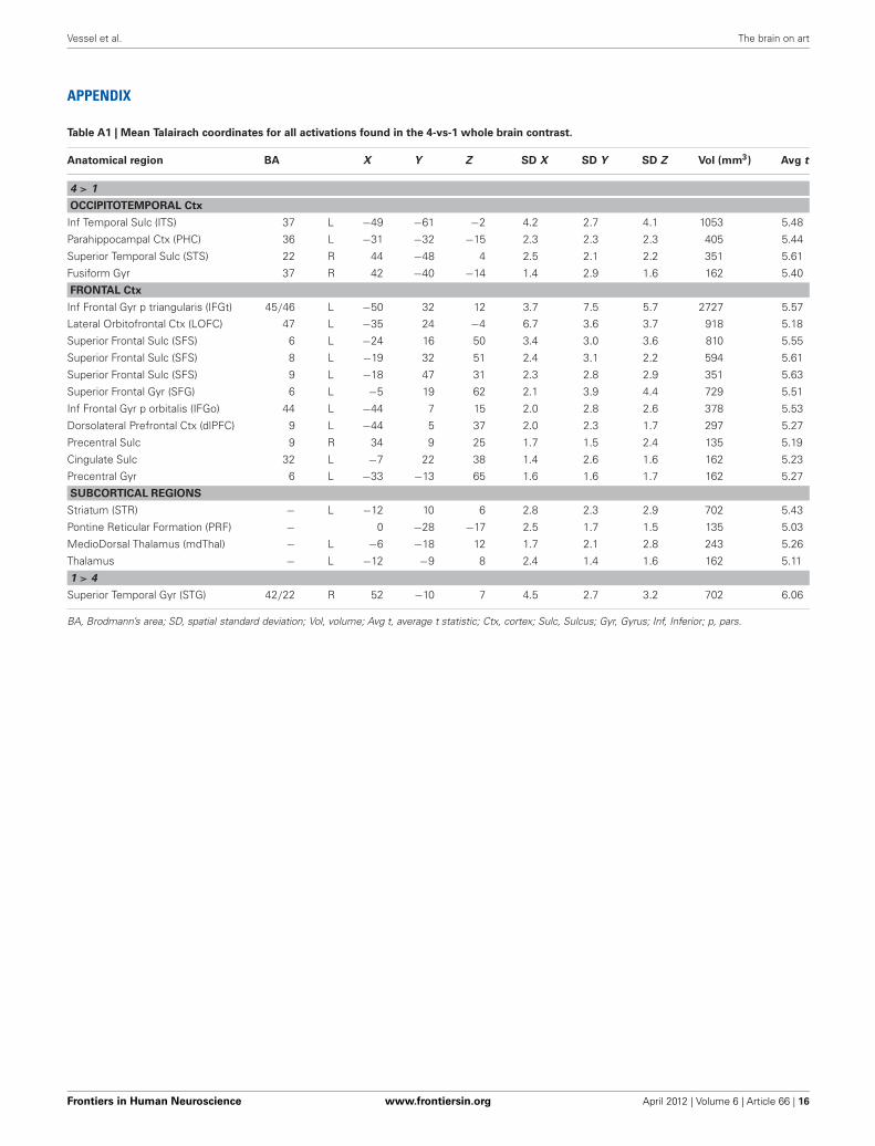
Vessel et al. The brain on art
APPENDIX
Table A1 | Mean Talairach coordinates for all activations found in the 4-vs-1 whole brain contrast.
Anatomical region BA X Y Z SD X SD Y SD Z Vol (mm3) Avg t
4 > 1
OCCIPITOTEMPORAL Ctx
Inf Temporal Sulc (ITS) 37 L −49 −61 −2 4.2 2.7 4.1 1053 5.48
Parahippocampal Ctx (PHC) 36 L −31 −32 −15 2.3 2.3 2.3 405 5.44
Superior Temporal Sulc (STS) 22 R 44 −48 4 2.5 2.1 2.2 351 5.61
Fusiform Gyr 37 R 42 −40 −14 1.4 2.9 1.6 162 5.40
FRONTAL Ctx
Inf Frontal Gyr p triangularis (IFGt) 45/46 L −50 32 12 3.7 7.5 5.7 2727 5.57
Lateral Orbitofrontal Ctx (LOFC) 47 L −35 24 −4 6.7 3.6 3.7 918 5.18
Superior Frontal Sulc (SFS) 6 L −24 16 50 3.4 3.0 3.6 810 5.55
Superior Frontal Sulc (SFS) 8 L −19 32 51 2.4 3.1 2.2 594 5.61
Superior Frontal Sulc (SFS) 9 L −18 47 31 2.3 2.8 2.9 351 5.63
Superior Frontal Gyr (SFG) 6 L −5 19 62 2.1 3.9 4.4 729 5.51
Inf Frontal Gyr p orbitalis (IFGo) 44 L −44 7 15 2.0 2.8 2.6 378 5.53
Dorsolateral Prefrontal Ctx (dlPFC) 9 L −44 5 37 2.0 2.3 1.7 297 5.27
Precentral Sulc 9 R 34 9 25 1.7 1.5 2.4 135 5.19
Cingulate Sulc 32 L −7 22 38 1.4 2.6 1.6 162 5.23
Precentral Gyr 6 L −33 −13 65 1.6 1.6 1.7 162 5.27
SUBCORTICAL REGIONS
Striatum (STR) − L −12 10 6 2.8 2.3 2.9 702 5.43
Pontine Reticular Formation (PRF) − 0 −28 −17 2.5 1.7 1.5 135 5.03
MedioDorsal Thalamus (mdThal) − L −6 −18 12 1.7 2.1 2.8 243 5.26
Thalamus − L −12 −9 8 2.4 1.4 1.6 162 5.11
1 > 4
Superior Temporal Gyr (STG) 42/22 R 52 −10 7 4.5 2.7 3.2 702 6.06
BA, Brodmann’s area; SD, spatial standard deviation; Vol, volume; Avg t, average t statistic; Ctx, cortex; Sulc, Sulcus; Gyr, Gyrus; Inf, Inferior; p, pars.
Frontiers in Human Neuroscience www.frontiersin.org April 2012 | Volume 6 | Article 66 | 16
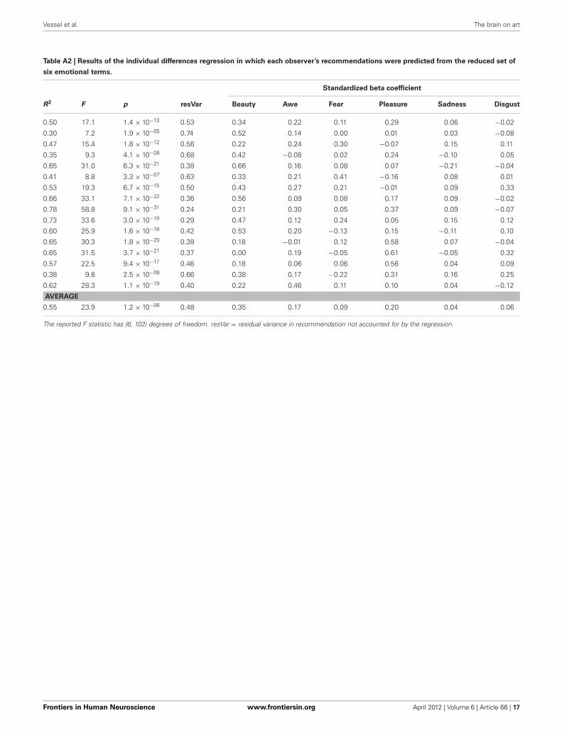
Vessel et al. The brain on art
Table A2 | Results of the individual differences regression in which each observer’s recommendations were predicted from the reduced set of
six emotional terms.
Standardized beta coefficient
R2 F p resVar Beauty Awe Fear Pleasure Sadness Disgust
0.50 17.1 1.4 × 10⁻¹³ 0.53 0.34 0.22 0.11 0.29 0.06 −0.02
0.30 7.2 1.9 × 10−05 0.74 0.52 0.14 0.00 0.01 0.03 −0.08
0.47 15.4 1.8 × 10⁻¹² 0.56 0.22 0.24 0.30 −0.07 0.15 0.11
0.35 9.3 4.1 × 10−08 0.68 0.42 −0.08 0.02 0.24 −0.10 0.05
0.65 31.0 6.3 × 10⁻²¹ 0.38 0.66 0.16 0.08 0.07 −0.21 −0.04
0.41 8.8 3.3 × 10−07 0.63 0.33 0.21 0.41 −0.16 0.08 0.01
0.53 19.3 6.7 × 10⁻¹⁵ 0.50 0.43 0.27 0.21 −0.01 0.09 0.33
0.66 33.1 7.1 × 10⁻²² 0.36 0.56 0.09 0.08 0.17 0.09 −0.02
0.78 58.8 9.1 × 10⁻³¹ 0.24 0.21 0.30 0.05 0.37 0.09 −0.07
0.73 33.6 3.0 × 10⁻¹⁹ 0.29 0.47 0.12 0.24 0.05 0.15 0.12
0.60 25.9 1.6 × 10⁻¹⁸ 0.42 0.53 0.20 −0.13 0.15 −0.11 0.10
0.65 30.3 1.8 × 10⁻²⁰ 0.38 0.18 −0.01 0.12 0.58 0.07 −0.04
0.65 31.5 3.7 × 10⁻²¹ 0.37 0.00 0.19 −0.05 0.61 −0.05 0.32
0.57 22.5 9.4 × 10⁻¹⁷ 0.46 0.18 0.06 0.06 0.56 0.04 0.09
0.38 9.8 2.5 × 10−08 0.66 0.38 0.17 −0.22 0.31 0.16 0.25
0.62 28.3 1.1 × 10⁻¹⁹ 0.40 0.22 0.46 0.11 0.10 0.04 −0.12
AVERAGE
0.55 23.9 1.2 × 10−06 0.48 0.35 0.17 0.09 0.20 0.04 0.06
The reported F statistic has (6, 102) degrees of freedom. resVar = residual variance in recommendation not accounted for by the regression.
Frontiers in Human Neuroscience www.frontiersin.org April 2012 | Volume 6 | Article 66 | 17