The Biotechnology Education Company - EDVOTEK · The Biotechnology Education Company® •...
22
255.111202 The Biotechnology Education Company ® EDVOTEK, Inc. • 1-800-EDVOTEK • www.edvotek.com All components are intended for educational research only. They are not to be used for diagnostic or drug purposes, nor administered to or consumed by humans or animals. 255 EDVO-Kit # Purification & Size Determination of Green (gfp) & Blue (bfp) Fluorescent Proteins Storage: Some components require freezer storage. See page 3 for specific storage instructions. EXPERIMENT OBJECTIVES: Students will learn to partially purify the Green (gfp) and Blue (bfp) fluorescent proteins. In an optional activity, the molecular weights of the proteins will be determined. U p d a t e d R e v i s e d a n d
Transcript of The Biotechnology Education Company - EDVOTEK · The Biotechnology Education Company® •...
255.111202
The Biotechnology Education Company ®
EDVOTEK, Inc. • 1-800-EDVOTEK • www.edvotek.com
All components are intended for educational research only. They are not to be used for diagnostic or drug purposes, nor administered to or consumed by humans or animals.
255EDVO-Kit #
Purifi cation & Size Determinationof Green (gfp) & Blue (bfp) Fluorescent Proteins
Storage: Some components require freezer storage.See page 3 for specifi c storage instructions.
EXPERIMENT OBJECTIVES:
Students will learn to partially purify the Green (gfp) and Blue (bfp) fl uorescent proteins. In an optional activity, the molecular weights of the
proteins will be determined.
Updated
Revised
and

2The Biotechnology Education Company® • 1-800-EDVOTEK • www.edvotek.com
Purifi cation & Size Determination of GFP & BFP
255.111202
Table of Contents Page
Experiment Components 3
Experiment Requirements 3
Background Information 4
Experiment Procedures
Experiment Overview 6
Packing and Equilibrating the Column 7
Partial Purifi cation of gfp and bfp Proteins 8
Sample Preparation for Denaturing SDS-gel Electrophoresis 9
Electrophoresis of Proteins 10
Staining the Gel 13
Determination of Molecular Weights 14
Study Questions 16
Instructor's Guidelines
Notes to the Instructor 17
Pre-Lab Preparations 18
Electrophoresis Hints and Help 20
Idealized Schematic of Results 21
Study Questions and Answers 22
Safety Data Sheets can be found on our website:
www.edvotek.com/safety-data-sheets

3
255255Experiment
Purifi cation & Size Determination of GFP & BFP
255.111202
EDVOTEK - The Biotechnology Education Company® 1-800-EDVOTEK • www.edvotek.com
FAX: 202.370.1501 • email: [email protected]
EXPERIMENT COMPONENTS StorageA Cell Extract containing green protein (gfp) FreezerB Cell Extract containing blue protein (bfp) FreezerC Column Elution Buffer (10x) FreezerD Standard Protein Markers FreezerE Dry Matrix Room temperatureF Protein Denaturing Solution FreezerG 50% Glycerol Solution Freezer
• Tris Glycine SDS Electrophoresis Buffer (10x) Room temperature• Protein InstaStain® sheets Room temperature• Chromatography Columns Room temperature
None of the components have been prepared from human sources.
REQUIREMENTS
• Vertical Gel Electrophoresis Apparatus (optional)• D.C. Power Source (optional)• Automatic Micropipet (5 -50µl) and Tips (Cat. #638 Fine Tip Micropipet Tips recommended)• Balance• Ice Buckets and Ice• Long U.V. lamps (Cat. #969)• Ring stands and column clamps• 1ml pipets and pipet pumps• Microtest tubes• Distilled or deionized water• Three Polyacrylamide gels* (OPTIONAL)• Glacial acetic acid (OPTIONAL)• Methanol (OPTIONAL)
*Polyacrylamide gels are not included for the electrophoresis part of this experiment. The experiment is designed for two student groups to share a gel. A total of 3 polyacrylamide gels (Precast polyacrylamide gels, Cat. #651) are required.
All components are intended for educational research only. They are not to be used for diag-nostic or drug purposes, nor administered to or consumed by humans or animals.
There is enough of each sample for six (6) groups sharing three polyacrylamide gels.
Store componentsA-D, F, G in the
freezer.
InstaStain, EDVOTEK, and The Biotechnology Education Company are registered trademarks of EDVOTEK, Inc.
4
Duplication of this document, in conjunction with use of accompanying reagents, is permitted for classroom/laboratory use only. This document, or any part, may not be reproduced or distributed for any other purpose without the written consent of EDVOTEK, Inc.
Copyright © 1993,1994,1997, 1998, 1999, 2001, 2005, 2011 EDVOTEK, Inc., all rights reserved. 255.111202
The Biotechnology Education Company® • 1-800-EDVOTEK • www.edvotek.com
Purifi cation & Size Determination of GFP & BFP255255 Experiment
Background Information
PURIFICATION & SIZE DETERMINATION OF GREEN & BLUE FLUORESCENT PROTEINS
Bioluminescence from marine microorganisms has been observed by many summer visitors at various beaches around the world. It always fascinates the observer by the repeated parade of both color and light on the sand during the ebb and fl ow. This observation takes second place to the light produced by the bioluminescent jelly fi sh, Aquorea victo-ria. A bright bursting energy of light is observed when energy is transferred to the green fl uorescent protein (gfp) which is located in a specialized photogenic cell located in the base of the jellyfi sh umbrella. There are several variants to gfp protein that have been ge-netically engineered and which dramatically enhance classroom laboratory experiments. An excellent companion to gfp is the blue fl uorescent protein (bfp) which is cloned and well characterized.
This family of proteins had been known for some time and signifi cant research in this area had been published. The fl uorescent proteins have been cloned and expressed. These proteins do not require substrates, other gene products, or cofactors. When exposed to long or short U.V. light, they will emit a bright green or blue light that is clearly visible in bacteria that are transformed by plasmids that contain genes for the gfp or bfp. Likewise, purifi cation of gfp or bfp is simplifi ed by their detection based on fl uorescence.
There are many examples of chimeric proteins that are fusion products using the gfp or bfp fl uorescent proteins as biological tags. Such fusions are at either the N- or C- terminal and often result in no loss in the fl uorescence or biological activity of the chimeric protein. These new biotechnology tools have made it possible to conduct studies that deal with protein localization and traffi cking within cells.
The green fl uorescent protein (gfp) has 238 amino acid residues and has a molecular weight of approximately 40,000 daltons. It appears that most of the intact protein is required for maintaining fl uorescence and only small deletions of a few amino acids are allowed without compromising the integrity of the protein structure. Interestingly, the chromophore responsible for light emission is within the primary structure of the protein and resides in a tripeptide at positions 65 to 67 which is cyclic and is composed of the amino acids Ser-Tyr-Gly. The importance of protein folding is clearly demonstrated with gfp where the protein is fl uorescent only upon proper conformational folding.
The blue fl uorescent protein (bfp) is a derivative variant of the gfp. It has a His-66 sub-stitution at the Tyr-66 position and a second substitution from Tyr-145 to Phe-145. The initial bfp known as P-4 had only the His-66 substitution and was not as bright as the double mutant. With the crystal structure of gfp determined, several other variations of the gfp were genetically engineered using site directed mutagenesis. In such procedures, specifi c mutations are introduced in a protein to determine the impact of the mutation on structure and function of the protein. The set of gfp and bfp proteins can be used as a dramatic tool to visually demonstrate the effect of pivotal amino acid changes on the structure and function of proteins. Amino acid substitution can also be used to demon-strate the effects of accumulated mutations on aging and various cancers.

5
Duplication of this document, in conjunction with use of accompanying reagents, is permitted for classroom/laboratory use only. This document, or any part, may not be reproduced or distributed for any other purpose without the written consent of EDVOTEK, Inc. Copyright © 1993,1994,1997, 1998, 1999, 2001, 2005, 2011 EDVOTEK, Inc., all rights reserved. 255.111202
The Biotechnology Education Company® • 1-800-EDVOTEK • www.edvotek.com
255255Experiment
Purifi cation & Size Determination of GFP & BFPExp
erimen
t Proced
ure
Background Information
In this experiment, bacterial extracts containing gfp or bfp will be fractionated by chro-matography using a molecular sieve matrix. Factors that affect the separation include size, shape and associated non-protein biologicals such as carbohydrate residues. The fl uorescent proteins will be detected on the column and subsequently in the test tubes by examination under long U.V. light. For most proteins, such columns can also be used to determine apparent molecular weights. Accurate estimation of protein polypeptide composition and size(s) are performed by analyzing the fractions that contain the pro-tein of interest by denaturing polyacrylamide gel electrophoresis.

6
Duplication of this document, in conjunction with use of accompanying reagents, is permitted for classroom/laboratory use only. This document, or any part, may not be reproduced or distributed for any other purpose without the written consent of EDVOTEK, Inc.
Copyright © 1993,1994,1997, 1998, 1999, 2001, 2005, 2011 EDVOTEK, Inc., all rights reserved. 255.111202
The Biotechnology Education Company® • 1-800-EDVOTEK • www.edvotek.com
Purifi cation & Size Determination of GFP & BFP255255 Experiment
Exp
erim
ent
Pro
ced
ure
Experiment Overview
EXPERIMENT OBJECTIVE:
In this experiment, students will learn methods and procedures to partially purify the FluoroGreen™ (gfp) and FluoroBlue™ (bfp) fl uorescent proteins. The molecular weights of the proteins will be determined using denatured SDS polyacrylamide gel electrophoresis.
LABORATORY SAFETY
1. Gloves and goggles should be worn routinely as good laboratory practice.
2. Exercise extreme caution when working with equipment that is used in conjunc-tion with the heating and/or melting of reagents.
3. DO NOT MOUTH PIPET REAGENTS - USE PIPET PUMPS.
4. Exercise caution when using any electrical equipment in the laboratory.
5. Always wash hands thoroughly with soap and water after handling reagents or biological materials in the laboratory.
7
Duplication of this document, in conjunction with use of accompanying reagents, is permitted for classroom/laboratory use only. This document, or any part, may not be reproduced or distributed for any other purpose without the written consent of EDVOTEK, Inc. Copyright © 1993,1994,1997, 1998, 1999, 2001, 2005, 2011 EDVOTEK, Inc., all rights reserved. 255.111202
The Biotechnology Education Company® • 1-800-EDVOTEK • www.edvotek.com
255255Experiment
Purifi cation & Size Determination of GFP & BFPExp
erimen
t Proced
ure
The loading of the column and subsequent elution will be done at room tem-perature. The elution buffers and the fractions collected will be stored on ice as they elute from the column.
1. Vertically mount the column on a ring stand. Make sure it is straight.
2. Slide the cap onto the spout at the bottom of the column. Fill about one-third of the column with the elution buffer.
3. Mix the slurry (molecular sieve) thoroughly by swirling or gently stir-ring.
4. Carefully pipet the mixed slurry into the column by letting it stream down the inside walls of the column.
If the fl ow is stopped by an air pocket, stop adding the slurry and fi rmly tap the column until the air is removed and the slurry continues to fl ow down the side of the column.
5. Place an empty beaker under the column to collect wash buffer.
6. Remove the cap from the bottom of the column and allow the matrix to pack into the column.
7. Wash the packed column with 5ml of 1x elution buffer. Do not allow the column to dry.
8. Slide the cap onto the spout and make sure it does not drip.
OPTIONAL STOPPING POINT
Packing and Equilibrating the Column
NOTE:
The loading of the column and subsequent elution wi l l be done at room temperature. The elution buffers and the fractions collected will be stored on ice as they elute from the column.
Do not allow the column to dry.
The prepared DNA sample can be stored in the freezer for electrophoresis at a later time.
���������
8
Duplication of this document, in conjunction with use of accompanying reagents, is permitted for classroom/laboratory use only. This document, or any part, may not be reproduced or distributed for any other purpose without the written consent of EDVOTEK, Inc.
Copyright © 1993,1994,1997, 1998, 1999, 2001, 2005, 2011 EDVOTEK, Inc., all rights reserved. 255.111202
The Biotechnology Education Company® • 1-800-EDVOTEK • www.edvotek.com
Purifi cation & Size Determination of GFP & BFP255255 Experiment
Exp
erim
ent
Pro
ced
ure
Partial Purifi cation of Green & Blue Fluroescent Proteins
DO NOT USE SHORT U.V. LIGHT (USED FOR DNA
WORK). DIRECT VISION WILL CAUSE BURNS AND DAMAGE
TO EYES!
OPTIONAL STOPPING POINT If time does not permit you to continue with the SDS polyacrylamide analysis, you may freeze the fractions at -20°C and perform the assays at a later date.
NOTE:
If you shine long U.V. light (black light) on the column, you will see (gfp) or (bfp) migrating through the column and you can predict the peak tubes that will contain the protein.
COLLECTING COLUMN FRACTIONS OF (gfp) PROTEIN
1. Label the fi rst set of eight microtest tubes #1-8.
2. Slowly load the column with 0.2ml of the gfp extract. Allow the extract to complete-ly enter the column.
3. Elute the column with 1x elution buffer. • Add buffer slowly (several drops at a time) to avoid diluting the protein sample. • Using the graduated marks on the sides of the tubes, collect 0.5ml fractions in
the labeled microcentrifuge tubes. • Store fractions on ice immediately upon collection.
* * * Remember - Do not allow the column to dry. * * *
4. Check all fractions by using long wave U.V. light to identify tubes that contain the fl uorescent gfp or bfp proteins.
5. Save the fractions containing gfp proteins for further analysis by SDS gel electropho-resis (optional).
COLLECTING COLUMN FRACTIONS OF (bfp) PROTEIN
1. Wash the column with 10ml of 1x Elution buffer.
2. Label a second set of eight microtest tubes #9-16. Repeat steps 2-5 (from above) with 0.2ml of the (bfp) extract. Store the 0.5ml fractions on ice immediately upon collection. Do not allow the column to dry.
9
Duplication of this document, in conjunction with use of accompanying reagents, is permitted for classroom/laboratory use only. This document, or any part, may not be reproduced or distributed for any other purpose without the written consent of EDVOTEK, Inc. Copyright © 1993,1994,1997, 1998, 1999, 2001, 2005, 2011 EDVOTEK, Inc., all rights reserved. 255.111202
The Biotechnology Education Company® • 1-800-EDVOTEK • www.edvotek.com
255255Experiment
Purifi cation & Size Determination of GFP & BFPExp
erimen
t Proced
ure
Sample Preparation for Denaturing SDS-Gel Electrophoresis (OPTIONAL)
1. Identify the tube with the highest fl uorescence of the gfp. Transfer 200µl each of the peak extract into two clean microtest tubes. Label one tube "gfp native" and the second tube "gfp denatured".
2. Identify the tube with the highest fl uorescence of the bfp. Transfer 200µl each of the peak extract into two clean microtest tubes. Label one tube "bfp native" and the second tube "bfp denatured".
PREPARING NATIVE PROTEINS (UNBOILED)
The protein in its native form can be shown to fl uoresce with a long wave U.V. light.
3. Add 25µl of 50% glycerol (G) to each of the tubes labeled "gfp native" and "bfp native".
4. Mix and set these tubes aside for later electrophoresis.
PREPARING DENATURED PROTEINS (BOILED)Note: Denaturing the proteins will remove their fl uorescence.
5. To denature the protein samples, add 25µl of protein denaturing solu-tion (F) to each of the tubes labeled "gfp denatured" and "bfp dena-tured". The denaturing solution contains sodium dodecylsulfate (SDS) and 2-mer-captoethanol.
6. Bring a beaker of water, covered with aluminum foil, to a boil.
7. Make sure the sample tubes to be denatured (boiled) are tightly capped and thawed.
8. The bottom of the tubes should be pushed through the foil and immersed in the boiling water for 5 minutes. The tubes should be kept suspended by the foil.
9. Allow the tubes to cool for a few minutes at room temperature.
10. Proceed with Electrophoresis of Proteins as outline on page 10.
DENATURING THE STANDARD PROTEIN MARKERS
• If your standard protein marker has not been rehydrated and boiled by your instruc-tor, add 130 µl of distilled or deionized water to it and allow the sample to rehy-drate for several minutes. Vortex or mix vigorously. Then, proceed to denaturing the standard protein markers as outlined in steps 6-9 above.
• If your standard protein marker has already been denatured, proceed with Electro-phoresis of Proteins as outlined on page 10.
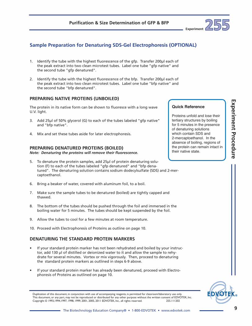
Quick Reference
Proteins unfold and lose their tertiary structures by boiling for 5 minutes in the presence of denaturing solutions which contain SDS and 2-mercaptoethanol. In the absence of boiling, regions of the protein can remain intact in their native state.

10
Duplication of this document, in conjunction with use of accompanying reagents, is permitted for classroom/laboratory use only. This document, or any part, may not be reproduced or distributed for any other purpose without the written consent of EDVOTEK, Inc.
Copyright © 1993,1994,1997, 1998, 1999, 2001, 2005, 2011 EDVOTEK, Inc., all rights reserved. 255.111202
The Biotechnology Education Company® • 1-800-EDVOTEK • www.edvotek.com
Purifi cation & Size Determination of GFP & BFP255255 Experiment
Exp
erim
ent
Pro
ced
ure
Electrophoresis of Proteins (OPTIONAL)
PREPARING THE POLYACRYLAMIDE GEL FOR ELECTROPHORESIS
Precast Polyacrylamide Gels:
Precast polyacrylamide gels will vary slightly in design. Procedures for their use will be similar.
1. Open the pouch containing the gel cassette with scissors. Remove the cassette and place it on the bench top with the front facing up.
Note: The front plate is smaller (shorter) than the back plate.
2. Some cassettes will have tape at the bottom of the front plate. Remove all of the tape to expose the bottom of the gel to allow electrical contact.
3. Insert the Gel Cassette into the electrophoresis chamber.
4. Remove the comb by placing your thumbs on the ridges and pushing (pressing) upwards, carefully and slowly.
The fi gure above shows a polyacrylamide gel cassette in the EDVOTEK® Vertical Electrophoresis Apparatus, Model #MV10.
Negative ElectrodeBlack Dot (-)
Buffer Level
Sample Well
Long Cassette Plate
Polyacrylamide Gel
Viewing Level(cut out area on
front support panel)
Platinum wire
Short Cassette Plate
Gel Cassette Clip
Micropipet withFine tip
PROPER ORIENTATION OF THE GEL IN THE ELECTROPHORESIS UNIT
1. Place the gel cassette in the electrophoresis unit in the proper orientation. Protein samples will not separate in the gel if the cassette is not oriented correctly. Follow the direc-tions accompanying the specifi c apparatus.
2. Add the diluted buffer into the chamber. The sample wells and the back plate of the gel cassette should be submerged under buffer.
3. Rinse each well by squirting electrophoresis buffer into the wells using a transfer pipet.
The gel is now ready for practice gel loading or sample loading.

11
Duplication of this document, in conjunction with use of accompanying reagents, is permitted for classroom/laboratory use only. This document, or any part, may not be reproduced or distributed for any other purpose without the written consent of EDVOTEK, Inc. Copyright © 1993,1994,1997, 1998, 1999, 2001, 2005, 2011 EDVOTEK, Inc., all rights reserved. 255.111202
The Biotechnology Education Company® • 1-800-EDVOTEK • www.edvotek.com
255255Experiment
Purifi cation & Size Determination of GFP & BFPExp
erimen
t Proced
ure
Electrophoresis of Proteins (OPTIONAL)
PRACTICE GEL LOADING
EDVOTEK® Cat. #638, Fine Tip Micropipet Tips are recommended for loading samples into polyacrylamide gels. A regular microtip may damage the cassette and result in the loss of protein samples.
1. Place a fresh fi ne tip on the micropipet. Aspirate 20 µl of practice gel loading solution.
2. Place the lower portion of the fi ne pipet tip between the two glass plates, below the surface of the electrode buffer, directly over a sample well. The tip should be at an angle pointed towards the well. The tip should be partially against the back plate of the gel cassette but the tip opening should be over the sample well, as illustrated in the fi gure on page 9.
Do not try to jam the pipet tip in between the plates of the gel cassette.
4. Eject all the sample by steadily pressing down on the plunger of the automatic pipet.
Do not release the plunger before all the sample is ejected. Premature release of the plunger will cause buffer to mix with sample in the micropipet tip. Release the pipet plunger after the sample has been delivered and the pipet tip is out of the buffer.
5. Before loading protein samples for the actual experiment, the practice gel load-ing solution must be removed from the sample wells.
Do this by fi lling a transfer pipet with buffer and squirting a stream into the sample wells. This will displace the practice gel loading solution, which will be diluted into the buffer and will not interfere with the experiment.
EDVOTEK® Cat. #638, Fine Tip Micropipet Tips are recommended for loading samples into polyacrylamide gels. A regular microtip may damage the cassette and result in the loss of protein samples.

12
Duplication of this document, in conjunction with use of accompanying reagents, is permitted for classroom/laboratory use only. This document, or any part, may not be reproduced or distributed for any other purpose without the written consent of EDVOTEK, Inc.
Copyright © 1993,1994,1997, 1998, 1999, 2001, 2005, 2011 EDVOTEK, Inc., all rights reserved. 255.111202
The Biotechnology Education Company® • 1-800-EDVOTEK • www.edvotek.com
Purifi cation & Size Determination of GFP & BFP255255 Experiment
Exp
erim
ent
Pro
ced
ure
Electrophoresis of Proteins (OPTIONAL)
LOADING PROTEIN SAMPLES
Change pipet tips between loading each sample. Make sure the wells are cleared of all practice loading solution. Gently squirt electrophoresis buffer into the wells with a transfer pipet.
Two student groups can share a gel. Some of the samples contain denaturing solution which contains SDS and 2-mercaptoethanol. The samples should be loaded in the following manner:
First Student Group
Lane 1 20µl of Standard Protein Markers (boiled for 5 minutes)Lane 2 20µl of gfp native (not boiled)Lane 3 20µl of gfp denatured (boiled for 5 minutes)Lane 4 20µl of bfp native (not boiled)Lane 5 20µl of bfp denatured (boiled for 5 minutes)
Second Student Group
Lane 6 20µl of Standard Protein Markers (boiled for 5 minutes)Lane 7 20µl of gfp native (not boiled)Lane 8 20µl of gfp denatured (boiled for 5 minutes)Lane 9 20µl of bfp native (not boiled)Lane 10 20µl of bfp denatured (boiled for 5 minutes)
RUNNING THE GEL
1. After the samples are loaded, carefully snap the cover all the way down onto the electrode terminals. The black plug in the cover should be on the terminal with the black dot.
2. Insert the plug of the black wire into the black input of the power supply (negative input). Insert the plug of the red wire into the red input of the power supply (positive input).
Volts Recommended Time
Minimum Optimal
125 60 min 75 min
Time and Voltage3. Set the power supply at the required voltage and run the electro-
phoresis for the length of time as determined by your instructor. When the current is fl owing, you should see bubbles forming on the electrodes. The sudsing is due to the SDS in the buffer.
4. After the electrophoresis is fi nished, turn off power, unplug the unit, disconnect the leads and remove the cover.
EDVOTEK® Cat. #638, Fine Tip Micropipet Tips are recommended for loading samples into polyacrylamide gels. A regular micro-tip may damage the cassette and result in the loss of protein samples.
READ ME!
NOTE:
Shine the long wave U.V. light on the gel while the native proteins are separating.
13
Duplication of this document, in conjunction with use of accompanying reagents, is permitted for classroom/laboratory use only. This document, or any part, may not be reproduced or distributed for any other purpose without the written consent of EDVOTEK, Inc. Copyright © 1993,1994,1997, 1998, 1999, 2001, 2005, 2011 EDVOTEK, Inc., all rights reserved. 255.111202
The Biotechnology Education Company® • 1-800-EDVOTEK • www.edvotek.com
255255Experiment
Purifi cation & Size Determination of GFP & BFPExp
erimen
t Proced
ure
NOTE:
Polyacrylamide gels are very thin and fragile. Use care in handling to avoid tearing the gel.
Fixative and Destaining Solution for each gel
(100ml)
50 ml Methanol10 ml Glacial Acetic Acid40 ml Distilled Water
STAINING WITH PROTEIN INSTASTAIN® IN ONE EASY STEP
EDVOTEK features a state-of-the-art, proprietary stain for DNA or Protein gels called InstaStain®. Protein Polyacrylamide gels can be stained with Protein InstaStain® cards in one easy step. Staining is rapid, sensitive and Polyacrylamide gels are ready for visualization in 1-3 hours.
InstaStain® Blue and InstaStain® Ethidium Bromide are also available from EDVOTEK for staining of DNA gels.
1. After electrophoresis, turn off the power and remove the gel cassette from the gel electrophoresis apparatus.
2. To remove the gel from the cassette, lay the cassette down and care-fully remove the front plate by placing a coin or a spatula in the slot at the top edge, near the sample wells, and twist to separate the two plates of the cassette.
3. Gently lift the front plate away from the larger back plate. In most cases, the gel will stay on the back plate. If the gel partially sticks to the front plate, let it fall onto the back plate.
4. Pour approximately 100 ml of fi xative solution in a small tray.
5. Transfer the back plate of the cassette (with the gel) into the tray con-taining the fi xative solution. Wet gloved fi ngers with fi xative solution and gently nudge the gel off the back plate and remove the plate, leaving the gel submerged in the fi xative solution.
6. Gently fl oat a sheet of Protein InstaStain® card with the stain side (blue) facing in the liquid. Remove the Protein InstaStain® card after 30 minutes.
7. Cover the staining tray with saran wrap to prevent evaporation.
8. Gently agitate on a rocking platform for 1-3 hours or overnight.
9. After staining, Protein bands will appear medium to dark blue against a light background* and will be ready for excellent photographic results.
* Destaining is usually not required but can be carried out if the gel background is too dark. Gels can be destained in several changes of fresh destaining solution until the appearance and contrast of the protein bands against the background improves.
STORING THE GEL:
Once satisfactory result is achieved, the gel can be stored in distilled or deionized water.
For permanent storage, the gel can be dried between two sheets of cellophane (saran wrap) stretched in an em-broidery hoop. Air-dry the gel for several days until the gel is paper thin. Cut the "extra" saran wrap surrounding the dried gel. Place the dried gel overnight be-tween two heavy books to avoid curling. Tape it into a laboratory book.
Wear gloves and safety goggles
Staining the Gel

14
Duplication of this document, in conjunction with use of accompanying reagents, is permitted for classroom/laboratory use only. This document, or any part, may not be reproduced or distributed for any other purpose without the written consent of EDVOTEK, Inc.
Copyright © 1993,1994,1997, 1998, 1999, 2001, 2005, 2011 EDVOTEK, Inc., all rights reserved. 255.111202
The Biotechnology Education Company® • 1-800-EDVOTEK • www.edvotek.com
Purifi cation & Size Determination of GFP & BFP255255 Experiment
Exp
erim
ent
Pro
ced
ure
Determination of Molecular Weights
If measurements are taken directly from the gel, skip steps 1 and 2.
1. Take a transparent sheet, such as cellulose acetate (commonly used with overhead projectors) and lay it over the wrapped gel.
2. With a felt-tip pen, carefully trace the outlines of the sample wells. Then trace over all the protein bands on the gel.
In the example shown above, the standard molecular weights are: 94,000 30,000 67,000 20,000 38,000 14,000
8
10
76
5
4
3
2
1
9
876
5
4
3
2
1
9
876
5
4
3
2
1
9
100,000
10,000
6 7 8 9 10
Centimeters
Mol
ecul
ar W
eigh
t
Phosphorylase
Bovine Serum Albumin
Ovalbumin
Carbonic anhydrase
5
Soybean Trypsin Inhibitor
Lysozyme
3. Measure the migration distance, in centimeters (to the nearest mil-limeter) of every major band in the gel. (Ignore the faint bands, refer to Idealized Schematic.) All measure-ments should be from the bottom of the sample well to the bottom of the protein band.
4. Using semilog graph paper, plot the migration distance or Rf of each stan-dard protein on the non-logarithmic x-axis versus its molecular weight on the logarithmic y-axis. Choose your scales so that the data points are well spread out. Assume the second cycle on the y-axis represents 10,000 to 100,000 (see example at left).
5. Draw the best average straight line through all the points. This line should roughly have an equal num-ber of points scattered on each side of the line. As an example, refer to the fi gure at left. This method is a linear approximation.
6. Using your standard graph, deter-mine the molecular weight of the three unknown proteins. This can be done by fi nding the Rf (or migra-tion distance) of the unknown band on the x-axis and drawing a straight vertical until the standard line is intersected. A straight line is then made from the intersection across to the y-axis where the approximate molecular weight can be determined.

8
10
7
6
5
4
3
2
1
9
87
6
5
4
3
2
1
9
87
6
5
4
3
2
1
9

16
Duplication of this document, in conjunction with use of accompanying reagents, is permitted for classroom/laboratory use only. This document, or any part, may not be reproduced or distributed for any other purpose without the written consent of EDVOTEK, Inc.
Copyright © 1993,1994,1997, 1998, 1999, 2001, 2005, 2011 EDVOTEK, Inc., all rights reserved. 255.111202
The Biotechnology Education Company® • 1-800-EDVOTEK • www.edvotek.com
Purifi cation & Size Determination of GFP & BFP255255 Experiment
Exp
erim
ent
Pro
ced
ure
1. What is the anticipated difference in apparent molecular weight between pFluoro-Green™ (gfp) and pFluoroBlue™ (bfp) as detected by denatured SDS- polyacrylamide gel analysis?
2. Why is the molecular sieving matrix swelled prior to packing the column?
3. What is the basis of molecular sieve chromatography?
4. Can molecular sieve chromatography columns be used to separate DNA fragments?
Study Questions

17
255255Experiment
Purifi cation & Size Determination of GFP & BFP
255.111202
EDVOTEK - The Biotechnology Education Company® 1-800-EDVOTEK • www.edvotek.com
FAX: 202.370.1501 • email: [email protected]
Instructor’s Guide
HOW THIS EXPERIMENT IS ORGANIZED
This experiment module contains biologicals and reagents for six (6) groups sharing three (3) polyacrylamide gels (2 groups per gel). Enough buffer is included for three (3) vertical electrophoresis units (Model MV-10 or equivalent). Additional electrophoresis buffer is required for more than three units.
Note: Polyacrylamide gels are not included. You may choose to purchase precast gels (Cat. #s 651 or 652).
A variety of factors, such as class size, length of laboratory sessions, and availability of equipment, will infl uence the implementation of this experiment with your students. These guidelines can be adapted to fi t your specifi c set of circumstances.
To facilitate implementation, this experiment can be divided into general experimental activities with logical stopping points. These optional stopping points can occur after each series of procedures.
Your personal preference and schedule will determine how and when the gels will be prepared for use in this experiment.
APPROXIMATE TIME REQUIREMENTS FOR PRE-LAB AND EXPERIMENTAL PROCEDURES
1. Pre-lab preparations will require approximately 20 minutes on the day of the lab.
2. Students will require approximately 15 minutes to heat samples and load the gel. Practice gel loading may require an additional 15 minutes if performed the same day of the lab.
3. Electrophoresis will require approximately 1 to 1.5 hours, depending on the voltage.
PRACTICE GEL LOADING
This experiment kit contains practice gel loading solution. If your students are unfamiliar with vertical gel electrophoresis, it is suggested that they practice loading the sample wells before performing the actual experiment.

18
Duplication of this document, in conjunction with use of accompanying reagents, is permitted for classroom/laboratory use only. This document, or any part, may not be reproduced or distributed for any other purpose without the written consent of EDVOTEK, Inc.
Copyright © 1993,1994,1997, 1998, 1999, 2001, 2005, 2011 EDVOTEK, Inc., all rights reserved. 255.111202
The Biotechnology Education Company® • 1-800-EDVOTEK • www.edvotek.com
255255Experiment
Inst
ruct
or’
s G
uid
eInstructor’s Guide Purifi cation & Size Determination of GFP & BFP
PreLab Preparations
COLUMN ELUTION BUFFER
Dilute the 10x Column Elution Buffer (C) by mixing 30ml of 10x Column Elu-tion Buffer (C) with 270ml of Distilled water. Label this buffer "1x Column Elution Buffer".
PREPARATION OF THE SLURRY
1. Hydrate the Dry Matrix (E) in 40ml of 1x Column Elution buffer (diluted C).
2. Gently stir occasionally for a minimum of 30 to 60 minutes. 3. Aliquot 6ml for each of the six groups.
CELL EXTRACT CONTAINING GFP OR BFP FLUORESCENT PROTEIN
1. Thaw the frozen extracts (A and B) at room temperature and immedi-ately place them on ice.
2. Label 6 tubes "gfp extract". Aliquot 220µl of the extract into the tubes. Place immediately back on ice.
3. Label an additional 6 tubes "bfp extract". Aliquot 220µl of the extract into the tubes. Place immediately back on ice.
For Partial Purifi cation and Sample Preparation, each group requires:
1 Chromatography column 1 Ring stand with clamp 8 Microtest tubes 6ml Slurry (hydrated E) 40ml 1x Column Elution Buffer (diluted C) 220μl "gfp extract" (A) 220μl "bfp extract" (B) 75μl Protein Denaturing Solution (F) 75μl 50% Glycerol Solution (G) Automatic micropipet and tips 5ml pipets and pump

19
Duplication of this document, in conjunction with use of accompanying reagents, is permitted for classroom/laboratory use only. This document, or any part, may not be reproduced or distributed for any other purpose without the written consent of EDVOTEK, Inc. Copyright © 1993,1994,1997, 1998, 1999, 2001, 2005, 2011 EDVOTEK, Inc., all rights reserved. 255.111202
The Biotechnology Education Company® • 1-800-EDVOTEK • www.edvotek.com
255255Experiment
Purifi cation & Size Determination of GFP & BFPIn
structo
r’s Gu
ide
Instructor’s Guide
Concentrated Distilled Total Buffer (10x) + Water = Volume
58 ml 522 ml 580 ml
95 ml 855 ml 950 ml
EDVOTEK Model #
MV10
MV20
Tris-Glycine-SDS Electrophoresis (Chamber) Buffer
Pre-Lab Preparations
PREPARING ELECTROPHORESIS BUFFER
Prepare the electrophoresis buffer by adding and mixing 1 part Tris-Glycine-SDS 10x buffer concen-trate to 9 parts distilled water.
The approximate volume of 1x electrophoresis buffer required for EDVOTEK® Protein Vertical Electrophoresis units are listed in the table below. The buffer should just cover the back plate of the gel cassette.
Volts Recommended Time
Minimum Optimal
125 60 min 75 min
Time and Voltage
ELECTROPHORESIS TIME AND VOLTAGE
Your time requirements will dictate the voltage and the length of time for your samples to separate by electrophoresis. Approximate recom-mended times are listed in the table at right.
RECONSTITUTION OF LYOPHILIZED PROTEIN MOLECULAR WEIGHT STANDARDS
Once rehydrated, the tube of Protein Molecular Weight Standards (D) contains enough material for loading 6 wells. The tube can be boiled in conjunction with the denatured GFP extracts.
1. Add 130 µl of distilled or deionized water to the tube of Potein Molecular Weight Standards (D) and allow the material to hydrate for several minutes. Vortex or mix vigorously.
2. Bring a beaker of water, covered with aluminum foil, to a boil. Remove from heat.
3. Make sure the sample tubes are tightly capped (in screw-cap tubes) and well-labeled. The bottom of the tubes should be pushed through the foil and im-mersed in boiling water for 5 minutes. The tubes should be kept suspended by the foil.
4. The markers can be aliquoted for each of the student groups, or students can share the rehydrated sample stock tube. Have students load samples onto the polyacrylamide gel while the samples are still warm to avoid aggregation. The volume of sample to load per well is 20 µl.
5. Store any unused portion of reconstituted sample at -20°C and repeat steps 2 and 3 when using samples at a later time.
20
Duplication of this document, in conjunction with use of accompanying reagents, is permitted for classroom/laboratory use only. This document, or any part, may not be reproduced or distributed for any other purpose without the written consent of EDVOTEK, Inc.
Copyright © 1993,1994,1997, 1998, 1999, 2001, 2005, 2011 EDVOTEK, Inc., all rights reserved. 255.111202
The Biotechnology Education Company® • 1-800-EDVOTEK • www.edvotek.com
255255Experiment
Inst
ruct
or’
s G
uid
eInstructor’s Guide Purifi cation & Size Determination of GFP & BFP
PREPARING STAINING AND DESTAINING SOLUTIONS
The stock solution is used for staining and destaining with Protein InstaStain®
1. Solution for staining with Protein InstaStain®
• Prepare a stock solution of Methanol and Glacial Acetic Acid by combining 180 ml Methanol, 140 ml Distilled water, and 40 ml Glacial Acetic Acid.
• Staining of Protein Gel(s) is optional.
2. Destaining Solution
• Use the stock solution of Methanol, Glacial Acetic acid and distilled water (in Step 1) to destain the gel(s).
PreLab Preparations
Electrophoresis Hints and Help
AVOIDING COMMON PITFALLS
• Use fresh electrophoresis buffer and stain, correctly diluted and prepared according to instructions.
• Before performing the experiment, practice sample delivery techniques to avoid diluting the sample with buffer during gel loading.
CARE AND MAINTENANCE OF THE ELECTROPHORESIS APPARATUS
To clean the apparatus chamber, rinse well with tap water. If your tap water is high in minerals, give the items a fi nal rinse with distilled water. Let air dry. Do not use de-tergents of any kind, or expose the apparatus to alcohols. Avoid touching the fragile platinum electrodes.
21
Duplication of this document, in conjunction with use of accompanying reagents, is permitted for classroom/laboratory use only. This document, or any part, may not be reproduced or distributed for any other purpose without the written consent of EDVOTEK, Inc. Copyright © 1993,1994,1997, 1998, 1999, 2001, 2005, 2011 EDVOTEK, Inc., all rights reserved. 255.111202
The Biotechnology Education Company® • 1-800-EDVOTEK • www.edvotek.com
255255Experiment
Purifi cation & Size Determination of GFP & BFPIn
structo
r’s Gu
ide
Instructor’s Guide
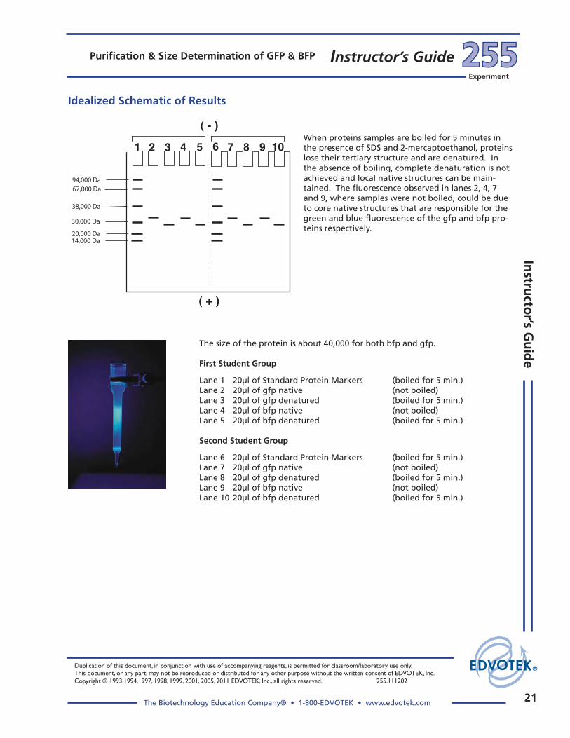
Idealized Schematic of Results
The size of the protein is about 40,000 for both bfp and gfp.
First Student Group
Lane 1 20µl of Standard Protein Markers (boiled for 5 min.)Lane 2 20µl of gfp native (not boiled)Lane 3 20µl of gfp denatured (boiled for 5 min.)Lane 4 20µl of bfp native (not boiled)Lane 5 20µl of bfp denatured (boiled for 5 min.)
Second Student Group
Lane 6 20µl of Standard Protein Markers (boiled for 5 min.)Lane 7 20µl of gfp native (not boiled)Lane 8 20µl of gfp denatured (boiled for 5 min.)Lane 9 20µl of bfp native (not boiled)Lane 10 20µl of bfp denatured (boiled for 5 min.)
When proteins samples are boiled for 5 minutes in the presence of SDS and 2-mercaptoethanol, proteins lose their tertiary structure and are denatured. In the absence of boiling, complete denaturation is not achieved and local native structures can be main-tained. The fl uorescence observed in lanes 2, 4, 7 and 9, where samples were not boiled, could be due to core native structures that are responsible for the green and blue fl uorescence of the gfp and bfp pro-teins respectively.
14,000 Da20,000 Da
30,000 Da
38,000 Da
67,000 Da94,000 Da

22
Duplication of this document, in conjunction with use of accompanying reagents, is permitted for classroom/laboratory use only. This document, or any part, may not be reproduced or distributed for any other purpose without the written consent of EDVOTEK, Inc.
Copyright © 1993,1994,1997, 1998, 1999, 2001, 2005, 2011 EDVOTEK, Inc., all rights reserved. 255.111202
The Biotechnology Education Company® • 1-800-EDVOTEK • www.edvotek.com
255255Experiment
Inst
ruct
or’
s G
uid
eInstructor’s Guide Purifi cation & Size Determination of GFP & BFP
Study Questions and Answers
1. What is the anticipated difference in apparent molecular weight between pFluoro-Green™ (gfp) and pFluoroBlue™ (bfp) as detected by denatured SDS- polyacrylamide gel analysis?
The difference between the two proteins is the substitution of two amino acids. This difference in mass is negligible and therefore the two proteins will be identical in molecular weight as judged by this method of analysis.
2. Why is the molecular sieving matrix swelled prior to packing the column?
When the matrix is swelled in an aqueous buffer it will form spheres which provide the sieving property for the separation of biomolecules.
3. What is the basis of molecular sieve chromatography?
Molecular sieve matrices are based on the separation of biomolecules based on size and shape. Assuming such molecules are globular, then size is the major parameter for the separation. If the biomolecules are too large to penetrate the pores in the molecu-lar sieve, the large molecules will go around the beads and will elute in the exclusion buffer with no separation. By contrast molecules that penetrate the Sephadex beads will be fractionated based on size with large to small molecules eluting sequentially.
4. Can molecular sieve chromatography be used to separate DNA fragments?
Small fragments of DNA (oligonucleotides) can be separated by molecular sieve chromatography. DNA molecules are large for fractionation using most Sephadex matrices and therefore DNA will not penetrate the beads. For example a 1 Kb double stranded DNA fragment is 2000 nucleotides when multiplied with the molecular weight of 500 (for the average of a nucleotide) is 1,000,000 daltons. Unlike DNA, pro-tein molecules tend to be much smaller for example, the gfp and bfp are bout 40,000 daltons and are therefore ideal candidates to be separated by a molecular sieve type of chromatography.