The Biology and Management of Subglottic Hemangioma: Past ...
12
The Laryngoscope Lippincott Williams & Wilkins, Inc. © 2004 The American Laryngological, Rhinological and Otological Society, Inc. The Biology and Management of Subglottic Hemangioma: Past, Present, Future Reza Rahbar, DMD, MD; Richard Nicollas, MD; Gilles Roger, MD; Jean-Michel Triglia, MD; Erea-Noel Garabedian, MD; Trevor J. McGill, MD; Gerald B. Healy, MD Objectives/Hypothesis: Objectives were 1) to re- view the presentation, natural history, and manage- ment of subglottic hemangioma; 2) to assess the affect of five variables (age, gender, degree of subglottic narrowing, location and extent of subglottic heman- gioma, and lack or presence of other hemangioma) and the outcome of six different treatment modalities (conservative monitoring, corticosteroid, laser sur- gery, tracheotomy, laryngotracheoplasty, and inter- feron) in the management of subglottic hemangioma; and 3) to present specific guidelines to help deter- mine the best possible treatment modality at the time of initial presentation. Study Design: Retrospective review in the setting of three tertiary care pediatric medical centers. Methods: Methods included 1) exten- sive review of the literature; 2) a systematic review with respect to age, gender, presentation, associated medical problems, location and degree of subglottic narrowing, initial treatment, need for subsequent treatments, outcome, complications, and prognosis; and 3) statistical analysis to determine the effect of five variables (age, gender, degree of subglottic nar- rowing, location and extent of subglottic hemangi- oma, and lack or presence of other hemangioma) and the outcome of six different treatment modalities (conservative monitoring, corticosteroid, laser sur- gery, tracheotomy, laryngotracheoplasty, and inter- feron). Results: In all, 116 patients with a mean age of 4.7 months were treated. The most common location of subglottic hemangioma was the left side. The range of subglottic narrowing was 10% to 99% (mean per- centage, 65%). Twenty-six patients (22%) were man- aged with a single treatment modality, which in- cluded conservative monitoring (n 13), corticosteroid (n 11), and tracheotomy (n 2). Ninety patients (78%) required multimodality treat- ments. Overall, the treatments included conservative monitoring (n 13), corticosteroid (n 100), trache- otomy (n 32), CO 2 laser (n 66), interferon (n 5), and laryngotracheoplasty (n 25). Complication rates included the following: conservative monitoring (none), corticosteroid (18%), tracheotomy (none), CO 2 laser (12%), interferon (20%), and laryngotracheo- plasty (20%). The following variables showed statisti- cal significance in the outcome of different treatment modality: 1) degree of subglottic narrowing (P < .001), 2) location of subglottic hemangioma (P < .01), and 3) presence of hemangioma in other areas (P < .005). Gender (P > .05) and age at the time of presentation (P > .06) did not show any statistical significance on the outcome of the treatments. Conclusion: Each patient should be assessed comprehensively, and treatment should be individualized based on symptoms, clinical findings, and experience of the surgeon. The authors presented treatment guidelines in an attempt to ra- tionalize the management of subglottic hemangioma and to help determine the best possible treatment modality at the time of initial presentation. Key Words: Airway, hemangioma, corticosteroid, laser surgery, interferon, vincristine. Laryngoscope, 114:1880 –1891, 2004 INTRODUCTION Subglottic hemangioma is a rare condition with the potential to cause life-threatening complications in the newborn period. The most common presenting symptom is biphasic stridor, which is often exacerbated by crying and upper respiratory tract infection. Symptoms frequently present before 6 months of age. The natural history of this entity is characterized by progressive airway obstruction during the proliferative phase followed by resolution of symptoms during the involution phase. Since the early Presented as a Candidate’s Thesis (R.R.) to the Triological Society, Inc. From the Department of Otolaryngology and Communication Disor- ders (R.R., T.J.M., G.B.H.), Children’s Hospital, and the Department of Otol- ogy and Laryngology (R.R., T.J.M., G.B.H.), Harvard Medical School, Boston, Massachusetts, U.S.A.; La Timone Children’s Hospital, University Aix- Marseille II, (J-M.T., R.N.) Aix-Marseille, France; and the Department of Otolaryngology (E.-N. G., G.R), Armand-Trousseau Children’s Hospital, AP- HP, University Paris VI, Paris, France. Editor’s Note: This Manuscript was accepted for publication April 19, 2004. Send Correspondence to Reza Rahbar, DMD, MD, Department of Otolar- yngology and Communication Disorders, Children’s Hospital, 300 Longwood Av- enue, Boston, MA 02155, U.S.A. E-mail: [email protected] DOI: 10.1097/01.mlg.0000147915.58862.27 Laryngoscope 114: November 2004 Rahbar et al.: Subglottic Hemangioma 1880
Transcript of The Biology and Management of Subglottic Hemangioma: Past ...

The LaryngoscopeLippincott Williams & Wilkins, Inc.© 2004 The American Laryngological,Rhinological and Otological Society, Inc.
The Biology and Management of SubglotticHemangioma: Past, Present, Future
Reza Rahbar, DMD, MD; Richard Nicollas, MD; Gilles Roger, MD; Jean-Michel Triglia, MD;Erea-Noel Garabedian, MD; Trevor J. McGill, MD; Gerald B. Healy, MD
Objectives/Hypothesis: Objectives were 1) to re-view the presentation, natural history, and manage-ment of subglottic hemangioma; 2) to assess the affectof five variables (age, gender, degree of subglotticnarrowing, location and extent of subglottic heman-gioma, and lack or presence of other hemangioma)and the outcome of six different treatment modalities(conservative monitoring, corticosteroid, laser sur-gery, tracheotomy, laryngotracheoplasty, and inter-feron) in the management of subglottic hemangioma;and 3) to present specific guidelines to help deter-mine the best possible treatment modality at the timeof initial presentation. Study Design: Retrospectivereview in the setting of three tertiary care pediatricmedical centers. Methods: Methods included 1) exten-sive review of the literature; 2) a systematic reviewwith respect to age, gender, presentation, associatedmedical problems, location and degree of subglotticnarrowing, initial treatment, need for subsequenttreatments, outcome, complications, and prognosis;and 3) statistical analysis to determine the effect offive variables (age, gender, degree of subglottic nar-rowing, location and extent of subglottic hemangi-oma, and lack or presence of other hemangioma) andthe outcome of six different treatment modalities(conservative monitoring, corticosteroid, laser sur-gery, tracheotomy, laryngotracheoplasty, and inter-feron). Results: In all, 116 patients with a mean age of4.7 months were treated. The most common location
of subglottic hemangioma was the left side. The rangeof subglottic narrowing was 10% to 99% (mean per-centage, 65%). Twenty-six patients (22%) were man-aged with a single treatment modality, which in-cluded conservative monitoring (n � 13),corticosteroid (n � 11), and tracheotomy (n � 2).Ninety patients (78%) required multimodality treat-ments. Overall, the treatments included conservativemonitoring (n � 13), corticosteroid (n � 100), trache-otomy (n � 32), CO2 laser (n � 66), interferon (n � 5),and laryngotracheoplasty (n � 25). Complicationrates included the following: conservative monitoring(none), corticosteroid (18%), tracheotomy (none), CO2laser (12%), interferon (20%), and laryngotracheo-plasty (20%). The following variables showed statisti-cal significance in the outcome of different treatmentmodality: 1) degree of subglottic narrowing (P < .001),2) location of subglottic hemangioma (P < .01), and 3)presence of hemangioma in other areas (P < .005).Gender (P > .05) and age at the time of presentation (P> .06) did not show any statistical significance on theoutcome of the treatments. Conclusion: Each patientshould be assessed comprehensively, and treatmentshould be individualized based on symptoms, clinicalfindings, and experience of the surgeon. The authorspresented treatment guidelines in an attempt to ra-tionalize the management of subglottic hemangiomaand to help determine the best possible treatmentmodality at the time of initial presentation. KeyWords: Airway, hemangioma, corticosteroid, lasersurgery, interferon, vincristine.
Laryngoscope, 114:1880–1891, 2004
INTRODUCTIONSubglottic hemangioma is a rare condition with the
potential to cause life-threatening complications in thenewborn period. The most common presenting symptom isbiphasic stridor, which is often exacerbated by crying andupper respiratory tract infection. Symptoms frequentlypresent before 6 months of age. The natural history of thisentity is characterized by progressive airway obstructionduring the proliferative phase followed by resolution ofsymptoms during the involution phase. Since the early
Presented as a Candidate’s Thesis (R.R.) to the Triological Society,Inc.
From the Department of Otolaryngology and Communication Disor-ders (R.R., T.J.M., G.B.H.), Children’s Hospital, and the Department of Otol-ogy and Laryngology (R.R., T.J.M., G.B.H.), Harvard Medical School, Boston,Massachusetts, U.S.A.; La Timone Children’s Hospital, University Aix-Marseille II, (J-M.T., R.N.) Aix-Marseille, France; and the Department ofOtolaryngology (E.-N. G., G.R), Armand-Trousseau Children’s Hospital, AP-HP, University Paris VI, Paris, France.
Editor’s Note: This Manuscript was accepted for publication April19, 2004.
Send Correspondence to Reza Rahbar, DMD, MD, Department of Otolar-yngology and Communication Disorders, Children’s Hospital, 300 Longwood Av-enue, Boston, MA 02155, U.S.A. E-mail: [email protected]
DOI: 10.1097/01.mlg.0000147915.58862.27
Laryngoscope 114: November 2004 Rahbar et al.: Subglottic Hemangioma
1880

1970s, a number of treatment modalities have been advo-cated with variable success and morbidity.
Currently, there are no specific guidelines for thetreatment of subglottic hemangioma, and most patientsundergo several different treatment modalities during thecourse of their management. The purposes of the presentstudy were to review the presentation, diagnosis, andnatural history and to present guidelines in an attempt torationalize the management of subglottic hemangiomaand to help determine the best possible treatment at thetime of initial presentation.
MATERIALS AND METHODSPatients treated for subglottic hemangioma at three pediat-
ric tertiary medical centers between 1980 and 2002 were identi-fied. A systematic chart review was undertaken to determine theage, gender, initial presentation, significant medical and familyhistory, location and degree of airway narrowing, involvement ofother areas, initial treatment, need for subsequent treatments,efficacy of each treatment modality, and complications.
Six different treatment modalities (conservative monitor-ing, corticosteroid, laser surgery, tracheotomy, interferon, andlaryngotracheoplasty [LTP]) were used in the management of ourpatients. We hypothesized that the outcome of each of thesetreatment modalities is based on the following variables: 1) age atthe time of presentation, 2) gender, 3) lack or presence of otherhemangioma, 4) degree of subglottic narrowing, and 5) locationand extent of the subglottic hemangioma. We performed lineardiscriminant analysis1 to determine the effect of these five pre-dictor variables on the outcome of the six different treatmentmodalities just mentioned.
The five predictor variables were coded as follows: gender,male or female; age, months; location of subglottic hemangioma,unilateral � 1, unilateral with posterior extension � 2, bilateral� 3, and circumferential � 4; degree of subglottic narrowing, 0 to100; and other hemangioma, yes or no. Complete data for thepredictor variables were available for 82 patients. SAS softwarewas used for all data management and analyses.2
RESULTSIn all, 116 patients with subglottic hemangioma were
identified. There were 78 female and 38 male patients.Fifty-two patients were the product of a full-term preg-nancy and normal delivery, 8 patients were born at lessthan 36 weeks of gestation, and no information was avail-
able on 56 patients. The age at the time of presentationranged from 1 to 18 months (mean age, 4.7 mo). The mostcommon presentation was biphasic stridor noted in morethan 90% of patients. Other signs and symptoms includedcyanosis, apnea, and retraction. Eighteen patients (16%)presented with other medical problems (Table I).
All patients underwent direct laryngoscopy and bron-choscopy. Diagnosis of subglottic hemangioma was basedon endoscopic appearance of the lesion as a reddish-bluishsubmucosal mass. Lesions were described as bilateral (n �25 [22%]), left-sided and posterior (n � 24 [21%]), circum-ferential (n � 24 [21%]), unilateral and left-sided (n � 19[16%]), unilateral and right-sided (n � 7 [6%]), posterior (n� 6 [5%]), right-sided and posterior (n � 3 [2%]), unknown(n � 8 [7%]). The degree of airway narrowing ranged from10% to 99% (mean percentage, 65%) in 88 patients andwas unknown in 28 patients.
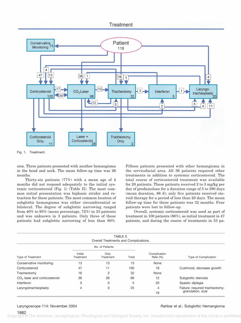
Twenty-six patients (22%) were managed with a sin-gle treatment modality, which included conservative mon-itoring (n � 13), corticosteroid only (n � 11), and trache-otomy only (n � 2). Ninety patients (78%) requiredmultimodality treatments (Fig. 1 and Table II). The over-all management of the 116 patients included conservativemonitoring (n � 13), corticosteroid (n � 100), CO2 lasertherapy (n � 66), tracheotomy (n � 32), interferon (n � 5),and LTP (n � 25).
Conservative Monitoring GroupThirteen patients with a mean age of 6 months pre-
sented in the conservative monitoring group (Fig. 1 andTable II). All patients presented with mild stridor withoutany evidence of apnea, cyanosis, or feeding difficulty. Themost common location of subglottic hemangioma was uni-lateral and left-side with or without posterior extension.None of the patients had a circumferential lesion. Subglot-tic narrowing ranged from 10% to 30% (mean percentage,22%) in 11 patients and was unknown in two patients.Two patients presented with a small cutaneous hemangi-oma in other areas of head and neck. All patients weremonitored conservatively, because of the lack of any majorrespiratory or feeding difficulty. There were no complica-tions with a mean follow-up time of 21 months.
Systemic Corticosteroid GroupThe systemic corticosteroid group (Fig. 1 and Table
II) contained 47 patients (41%) who were treated withcorticosteroid therapy as the initial treatment modality.Thirty-three of these patients were started on a cortico-steroid regimen before our evaluation. The age at the timeof initial presentation ranged from 1 to 10 months (meanage, 4 mo), and degree of airway narrowing ranged from20% to 95% (mean percentage, 66%).
Corticosteroid was successful as the only treatmentmodality in 11 of these patients (Fig. 1) (Table II). These11 patients had a mean age of 4 months at the time ofpresentation with a range of subglottic narrowing of 20%to 75% (mean percentage, 46%). Only two of these patientshad a percentage of subglottic narrowing greater than50%. The most common location of the subglottic heman-gioma was either bilateral or unilateral with posteriorextension. None of the patients had a circumferential le-
TABLE I.Associated Medical Problems.
History No. of Cases
Bronchopulmonary dysplasia 2
Hypospadias 1
Reflux 5
Ventriculoseptal defect 1
Right-side aortic arch 1
Aberrant left-side subclavian 1
Midline sternal cleft 1
Aberrant Innominate artery 1
Hyaline membrane myopathy 3
Left-side vocal cord paralysis 1
Hypoplasia of descending aorta 1
Laryngoscope 114: November 2004 Rahbar et al.: Subglottic Hemangioma
1881
sion. Three patients presented with another hemangiomain the head and neck. The mean follow-up time was 26months.
Thirty-six patients (77%) with a mean age of 4months did not respond adequately to the initial sys-temic corticosteroid (Fig. 1) (Table II). The most com-mon initial presentation was biphasic stridor and re-traction for these patients. The most common location ofsubglottic hemangioma was either circumferential orbilateral. The degree of subglottic narrowing rangedfrom 40% to 95% (mean percentage, 72%) in 33 patientsand was unknown in 3 patients. Only three of thesepatients had subglottic narrowing of less than 60%.
Fifteen patients presented with other hemangioma inthe cervicofacial area. All 36 patients required othertreatments in addition to systemic corticosteroid. Thetotal course of corticosteroid treatment was availablefor 20 patients. These patients received 2 to 3 mg/kg perday of prednisolone for a duration range of 5 to 390 days(mean duration, 86 d); only five patients received ste-roid therapy for a period of less than 20 days. The meanfollow-up time for these patients was 32 months. Fourpatients were lost to follow-up.
Overall, systemic corticosteroid was used as part oftreatment in 100 patients (86%), as initial treatment in 47patients, and during the course of treatments in 53 pa-
TABLE II.Overall Treatments and Complications.
Type of Treatment
No. of Patients
ComplicationRate (%) Type of Complication
InitialTreatment
OnlyTreatment Total
Conservative monitoring 13 13 13 None
Corticosteroid 47 11 100 18 Cushinoid, decrease growth
Tracheotomy 16 2 32 None
CO2 laser and corticosteroid 36 28 66 12 Subglottic stenosis
Interferon 0 0 5 20 Spastic diplegia
Laryngotracheoplasty 4 0 25 4 Failure: required tracheotomy;granulation, scar16
Fig. 1. Treatment.
Laryngoscope 114: November 2004 Rahbar et al.: Subglottic Hemangioma
1882

tients. Complications associated with use of corticosteroidwere noted in 18 patients (18%) and included cushingoidfeatures (n � 8), decreased growth (n � 4), pneumonia (n� 3), and gastric ulcer (n � 3) (Table II).
Tracheotomy GroupSixteen patients (14%) with a mean age of 2.8 months
underwent tracheotomy as the initial treatment modality(Fig. 1 and Table II). Biphasic stridor and retraction werethe most common presentation. The most common loca-tion of subglottic hemangioma was either circumferentialor bilateral. Degree of subglottic narrowing ranged from60% to 99% (mean percentage, 84%) in seven patients andwas unknown in nine patients. One patient presentedwith a cervicofacial hemangioma. Tracheotomy as the onlytreatment modality was successful in two of the patients.Fourteen patients required other treatments because ofconcern for the degree of airway narrowing and the re-serve airway in case of obstructed or displaced tracheot-omy tube (Fig. 1).
In all, 32 patients (28%) underwent tracheotomy aspart of their treatment. Sixteen patients underwent tra-cheotomy as the initial treatment, and 16 patients under-went tracheotomy during the course of treatments. Age atdecannulation ranged from 1 to 3 years (mean age, 22 mo)in 24 patients and was unknown in 7 patients. No compli-cations, subglottic stenosis or death, were reported with amean follow-up time of 36 months.
Laser SurgeryThe laser surgery group (Fig. 1 and Table II) con-
tained 36 patients (31%) with a mean age of 5 months whowere treated with CO2 laser therapy as the initial treat-ment. Degree of airway narrowing ranged from 50% to80% (mean percentage, 68%) in 17 patients and was un-known in 19 patients.
Twenty-eight patients (Fig. 1 and Table II) respondedwell to CO2 laser excision followed with systemic cortico-steroid. A unilateral subglottic hemangioma was the mostcommon presentation for these patients. Degree of airwaynarrowing ranged from 50% to 80% (mean percentage,66%) in 14 patients and was unknown in 14 patients. Thenumber of laser surgeries for these patients ranged from 1to 4 (mean number, 2).
Eight patients (Fig. 1 and Table II) did not respond toCO2 laser surgery and systemic corticosteroid and re-quired either tracheotomy or LTP, or both. The most com-mon location of subglottic hemangioma was either circum-ferential or bilateral. Degree of airway narrowing rangedfrom 70% to 80% (mean percentage, 77%) in three patientsand was unknown in five patients.
Overall, CO2 laser surgery was used in 66 patients(57%) as part of the treatment with a mean number of 2procedures per patient. Eight patients (12%) presentedwith subglottic stenosis following laser surgery, four pa-tients required tracheotomy followed with LTP, and fourpatients were monitored conservatively with resolution ofsymptoms (Table II).
Interferon GroupFive patients (4%) with a mean age of 2.4 months
were treated with interferon (Fig. 1 and Table II). Thelocations of the subglottic hemangioma were circumferen-tial (n � 3), bilateral (n � 1), and unknown (n � 1). Thedegree of subglottic narrowing ranged from 70% to 90%(mean percentage, 80%) in three patients and was un-known in two patients. All five patients presented withextensive hemangioma in other areas of head and neck.All patients required a combination of treatment modali-ties (Fig. 1). All patients responded well to interferon withan average treatment duration of 8 months. One patientpresented with spastic diplegia at 8 months after initia-tion of treatment, which resolved after termination oftherapy. No other complication was noted with a meanfollow-up time of 36 months.
Laryngotracheoplasty GroupThe LTP group (Fig. 1 and Table II) contained 25
patients (22%) with a mean age of 4 months who under-went LTP for excision of their lesion. The most commonlocation of the hemangioma was either circumferential orbilateral (72%). The range of airway narrowing was 60%to 99% (mean percentage, 82%). Thirteen patients (52%)presented with hemangiomas in other areas of head andneck. Twenty-one patients (84%) were treated with eithersystemic corticosteroid or other surgical intervention (CO2
laser, tracheotomy), or both, before LTP (Fig. 1). Twenty-one patients had augmentation of the subglottis usingcostal or conchal cartilage. One patient (4%) failed LTPand required a tracheotomy. Four patients (16%) pre-sented with minimal granulation tissue postoperatively,which was treated with CO2 laser excision. No other com-plication was noted with a mean follow-up time of 47months.
Statistical AnalysisWe are fully aware of inherent difficulties in the
analysis of our data, which include change of treatmentprotocols since the early 1980s, surgeons with differentmanagement philosophies, and improvement of surgicaland medical techniques. We have presented 116 patientswith subglottic hemangioma who underwent six differenttreatments (conservative monitoring, corticosteroid, lasersurgery, tracheotomy, interferon, and LTP). We hypothe-sized that the outcome of a treatment modality is deter-mined by the following variables: age, gender, degree ofsubglottic narrowing, location of subglottic hemangioma,and lack or presence of other hemangioma. We performedlinear discriminant analysis1 to determine the effect ofthese five variables on the outcome of the six differenttreatment modalities. Figure 2 outlines the results of sta-tistical analysis.
The degree of subglottic narrowing caused by hem-angioma was a significant predictor variable (P � .001).All patients who were monitored conservatively had asubglottic narrowing of less than 30% (mean percentage,22%). Patients who were treated successfully with corti-costeroid alone had a mean subglottic narrowing of 45%,and only two of these patients had subglottic narrowing ofmore than 50%. Patients who required further interven-
Laryngoscope 114: November 2004 Rahbar et al.: Subglottic Hemangioma
1883
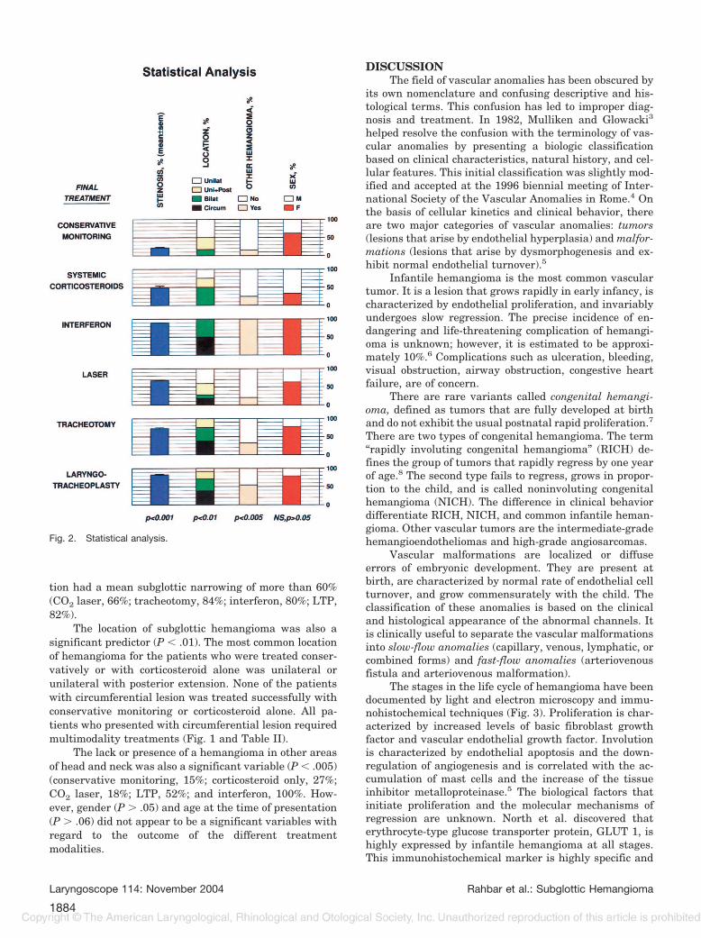
tion had a mean subglottic narrowing of more than 60%(CO2 laser, 66%; tracheotomy, 84%; interferon, 80%; LTP,82%).
The location of subglottic hemangioma was also asignificant predictor (P � .01). The most common locationof hemangioma for the patients who were treated conser-vatively or with corticosteroid alone was unilateral orunilateral with posterior extension. None of the patientswith circumferential lesion was treated successfully withconservative monitoring or corticosteroid alone. All pa-tients who presented with circumferential lesion requiredmultimodality treatments (Fig. 1 and Table II).
The lack or presence of a hemangioma in other areasof head and neck was also a significant variable (P � .005)(conservative monitoring, 15%; corticosteroid only, 27%;CO2 laser, 18%; LTP, 52%; and interferon, 100%. How-ever, gender (P � .05) and age at the time of presentation(P � .06) did not appear to be a significant variables withregard to the outcome of the different treatmentmodalities.
DISCUSSIONThe field of vascular anomalies has been obscured by
its own nomenclature and confusing descriptive and his-tological terms. This confusion has led to improper diag-nosis and treatment. In 1982, Mulliken and Glowacki3
helped resolve the confusion with the terminology of vas-cular anomalies by presenting a biologic classificationbased on clinical characteristics, natural history, and cel-lular features. This initial classification was slightly mod-ified and accepted at the 1996 biennial meeting of Inter-national Society of the Vascular Anomalies in Rome.4 Onthe basis of cellular kinetics and clinical behavior, thereare two major categories of vascular anomalies: tumors(lesions that arise by endothelial hyperplasia) and malfor-mations (lesions that arise by dysmorphogenesis and ex-hibit normal endothelial turnover).5
Infantile hemangioma is the most common vasculartumor. It is a lesion that grows rapidly in early infancy, ischaracterized by endothelial proliferation, and invariablyundergoes slow regression. The precise incidence of en-dangering and life-threatening complication of hemangi-oma is unknown; however, it is estimated to be approxi-mately 10%.6 Complications such as ulceration, bleeding,visual obstruction, airway obstruction, congestive heartfailure, are of concern.
There are rare variants called congenital hemangi-oma, defined as tumors that are fully developed at birthand do not exhibit the usual postnatal rapid proliferation.7
There are two types of congenital hemangioma. The term“rapidly involuting congenital hemangioma” (RICH) de-fines the group of tumors that rapidly regress by one yearof age.8 The second type fails to regress, grows in propor-tion to the child, and is called noninvoluting congenitalhemangioma (NICH). The difference in clinical behaviordifferentiate RICH, NICH, and common infantile heman-gioma. Other vascular tumors are the intermediate-gradehemangioendotheliomas and high-grade angiosarcomas.
Vascular malformations are localized or diffuseerrors of embryonic development. They are present atbirth, are characterized by normal rate of endothelial cellturnover, and grow commensurately with the child. Theclassification of these anomalies is based on the clinicaland histological appearance of the abnormal channels. Itis clinically useful to separate the vascular malformationsinto slow-flow anomalies (capillary, venous, lymphatic, orcombined forms) and fast-flow anomalies (arteriovenousfistula and arteriovenous malformation).
The stages in the life cycle of hemangioma have beendocumented by light and electron microscopy and immu-nohistochemical techniques (Fig. 3). Proliferation is char-acterized by increased levels of basic fibroblast growthfactor and vascular endothelial growth factor. Involutionis characterized by endothelial apoptosis and the down-regulation of angiogenesis and is correlated with the ac-cumulation of mast cells and the increase of the tissueinhibitor metalloproteinase.5 The biological factors thatinitiate proliferation and the molecular mechanisms ofregression are unknown. North et al. discovered thaterythrocyte-type glucose transporter protein, GLUT 1, ishighly expressed by infantile hemangioma at all stages.This immunohistochemical marker is highly specific and
Fig. 2. Statistical analysis.
Laryngoscope 114: November 2004 Rahbar et al.: Subglottic Hemangioma
1884

is not observed in other vascular tumors or vascular mal-formations.9 There is preliminary evidence that the risk ofhemangioma is 10-fold higher in children of women whoundergo chorionic villus sampling.10 Viral origin and ge-netic alteration have also been postulated.5
PresentationSubglottic hemangioma is the most common neo-
plasm of the infant airway. In 1871, Mackenzie was thefirst to describe a hemangioma of the larynx.11 Phillipsand Ruh11 presented the first description of infantile sub-glottic hemangioma in 1913. The most common presenta-tion is biphasic stridor, which is exacerbated by crying andupper respiratory tract infection and often presents before6 months of age. Recurrent episodes of croup may indicatethe possible diagnosis of subglottic hemangioma. Hoarse-ness, cough, dysphasia, and hemoptysis are uncommon.The natural history of subglottic hemangioma is unpre-dictable. The majority of lesions are characterized by pro-gressive airway obstruction shortly after birth during theproliferative phase, followed by resolution of symptomsafter the first year of life during the involutive phase.Although subglottic hemangioma is a benign condition, itcan be associated with a fatal outcome. Without properdiagnosis and treatment, a 30% to 70% mortality rate hasbeen reported.5,12
Cutaneous hemangioma is present in approximately50% of affected children. However, the absence of cutane-ous hemangiomas does not preclude the presence of asubglottic hemangioma.13 Although the biological basisfor the association of cervicofacial and subglottic heman-gioma is unknown, this relationship has been reported inthe past.8 Orlow et al.13 reported a 63% association be-tween the presence of cutaneous hemangiomas distrib-uted in the “beard pattern” (preauricular, chin, lower lip,
neck) and the presence of symptomatic hemangioma inthe upper airway or subglottis (Fig. 4).
EvaluationDiagnosis of subglottic hemangioma is based on the
history and physical examination. Diagnosis is confirmedby endoscopic examination, which reveals a submucosallesion that is compressible in the subglottic area (Fig. 5).The color ranges from red to blue depending on the thick-ness of the overlying mucosa and degree of vascularity.Biopsy is required only if findings on endoscopy are notdiagnostic. The most common location of subglottic hem-angioma is on the left side. It may also present as circum-ferential, bilateral, or unilateral with or without posteriorextension (Fig. 5).
ImagingIt has been reported that asymmetrical narrowing of
the subglottic airway on frontal radiographs of the neck ispathognomonic for hemangioma.14 However, Copper etal.15 reported that only 50% of subglottic hemangiomaspresented asymmetrical narrowing, whereas others pre-sented as symmetric narrowing. We recommend workupwith magnetic resonance imaging (MRI) if there is suspi-cion for cervical or intrathoracic extension of hemangioma(Fig. 6). Hemangioma appears to be solid tissue of inter-mediate intensity on T1-weighted spin-echo images and ofmoderate hyperintensity on T2-weighted spin-echo im-ages. One may also see flow voids within the hemangiomathat could indicate shunting between feeding arteries anddraining veins.5
TreatmentA multitude of medical and surgical modalities has
been proposed for the management of the subglottic hem-angioma, and no single approach has received universalacceptance.
Conservative monitoring. Because of the concernfor possible airway obstruction and the high mortality rateassociated with subglottic hemangioma, it is essential to
Fig. 3. (A) Hemangioma: proliferative phase. Closely packed, plumpendothelial cells and pericytes with small vascular lumina are evi-dent (H&E stain, original magnification �400). (B) Hemangioma:involuting phase. Less closely packed, plump endothelial cells andpericytes with thick vascular basement membrane are evident. Thechannels are separated by delicate connective tissue (H&E stain,original magnification �400).
Fig. 4. (A) Hemangioma involving the cervicofacial area, parotid,and nose. (B) Hemangioma involving the “beard distribution” (peri-auricular area, chin, neck).
Laryngoscope 114: November 2004 Rahbar et al.: Subglottic Hemangioma
1885

intervene with a treatment or initiate close monitoringafter the initial diagnosis. We have presented 13 patientswho were monitored conservatively without any complica-tion. Based on our data, patients who present with asubglottic hemangioma that causes less than 30% subglot-tic narrowing without any significant respiratory or feed-ing difficulty may be monitored conservatively (Fig. 7).However, it is essential that these patients have closefollow-up and access to immediate medical care in case ofchange in symptoms.
Radiation therapy. In 1919, New and Clark16 re-ported the successful application of external-beam radia-tion for the treatment of subglottic hemangioma in twopatients. In 1961, Ferguson and Flake17 advocated the useof tracheotomy and radiation therapy for the managementof subglottic hemangioma. They reported an average re-gression time of 9 months with the age range at decannu-lation of 1 to 2 years in 17 patients. Other studies havealso reported success with the application of radioactivegold implant.18,19 However, because of concerns for asso-ciated risk of damage to the normal airway mucosa andsecondary malignancy, this treatment modality was aban-doned in the late 1970s.
Cryotherapy. In 1972, Schechter and Biller20 re-ported the application of cryotherapy and steroid use intwo patients with only transient benefit. Jokinen et al.21
reported five patients treated with cryotherapy with mi-nor benefit. The depth of tissue destruction using cryo-therapy is unpredictable, which could result in further
complication such as subglottic stenosis. Overall, the ap-plication of cryotherapy in the management of subglottichemangioma has been disappointing, and we do not rec-ommend this treatment modality.
Tracheotomy. Tracheotomy with observation forinvolution was first described by Suehs and Herbur22 in1940. Many studies followed and recommended trache-otomy and waiting for spontaneous resolution.23-25
However, it is important to recognize that tracheotomydoes not influence the natural history of the hemangi-oma and often has to be in place for approximately 2years. When considering tracheotomy, it is imperativeto think about the degree of airway narrowing and thereserve airway because an obstructed or displaced tra-cheotomy tube could be life-threatening. A mortalityrate of 40% to 60% has been reported because of trache-otomy tube plugging and accidental decannulation.12,26
We recommend tracheotomy when the airway is com-promised because of hemangioma in the area of 1) glot-tis and supraglottic, 2) multiple lesions of subglottis andtrachea, and 3) a contraindication to another treatmentmodality (Fig. 7). When considering tracheotomy as atreatment option, factors such as adequate familyteaching, availability of nursing and medical care, andcommunication and speech development must be takeninto account.
Corticosteroids. In 1973, Cohen27 reported on theuse of systemic corticosteroids in the treatment of subglot-tic hemangioma. Many authors reported successful re-
Fig. 5. Subglottic hemangioma. (A)Unilateral lesion causing completeobstruction of subglottic area. (B)Circumferential lesion. (C) Unilateralright-side lesion with posterior exten-sion. (D) Isolated posterior lesion.
Laryngoscope 114: November 2004 Rahbar et al.: Subglottic Hemangioma
1886

gression and resolution of airway symptoms with systemiccorticosteroid.5,12,28 Other investigators have reported atransient response with serious side effects.20,29 It is truly
difficult to properly assess the efficacy of the corticosteroidbecause it is often used in combination with other treat-ments in the management of subglottic hemangiomas.Also, the natural history of involution of hemangiomamakes the picture more confusing when considering theprolonged duration of corticosteroid. When including hem-angiomas of various sites, a positive response is seen in30% to 60% of cases.5 The mechanism of action of cortico-steroid is unclear. However, it may increase the sensitiv-ity of the hemangioma to physiological vasoconstriction.Its function may also be a result of blockage of estradiolreceptors in the hemangioma, which are thought to en-hance growth.
Intralesional injection of corticosteroids followed byendotracheal intubation has also been advocated.30,31 In1990, Meeuwis et al.30 reported on successful treatment ofsix patients with a mean number of 2 injections (range,1–5 injections) and an average intubation period of 19days (range, 7–36 d). In 1997, Hoeve et al.31 reportedsuccess in 14 of the 15 patients with a mean number of 3injections (range, 1–12 injections) and an average intuba-tion period of 37 days (range, 6–129 d). The need forrepeated endoscopy and injections, prolonged intubationtime, and the need for intermittent pharmacological pa-ralysis and immobilization are of concern and may causefurther morbidity.
One hundred of our patients (86%) received systemiccorticosteroid as part of their treatment. Eleven patientsresponded adequately to systemic corticosteroid alone asthe initial treatment. These patients had either a unilat-eral or bilateral lesion with a mean degree of subglotticnarrowing of 45%. Thirteen patients who had circumfer-ential lesion did not respond adequately to corticosteroidalone and required further intervention. Based on ourdata, patients who present with subglottic hemangiomathat causes less than 50% subglottic narrowing and do nothave circumferential involvement respond well to an ini-tial trial of systemic corticosteroid treatment (Fig. 7).
We recommend the dosage of 2 to 3 mg/kg per day ofprednisone or an equivalent corticosteroid. Signs of re-sponsiveness should occur within several days to 1 week ofthe initiation of the treatment.5 One should see dimin-ished rate of growth, softening of the tumor, fading of thecolor, and resolution of symptoms. If there is a response,the dosage should be tapered slowly and terminated whenthere is resolution of airway symptoms. Rebound growthis noted if the dosage is tapered too quickly or terminatedprematurely. However, if corticosteroid fails to control thegrowth of hemangioma and does not result in resolution ofsymptoms in 2 to 3 weeks after treatment, it should betapered quickly and stopped, and other treatment modal-ities should be considered. No factors have been identifiedthat predict the response to corticosteroid treatment, andsome hemangiomas appear to be nonresponders. Also,there are no data to support that higher dose or prolongedtreatment improves response rate. Enjolras et al.32 re-ported rates of 30% accelerated regression, 30% no re-sponse, and 40% equivocal response in 25 infants withlife-threatening hemangiomas.
When considering corticosteroid treatment, it is es-sential to evaluate the risk-benefit ratio. Side effects with
Fig. 6. Magnetic resonance imaging: cervicofacial hemangioma. (A)T1-weighted image without contrast showing large flow void withinthe lesion of parotid area. (B) T2-weighted image showing hyperin-tense, uniform, and well-marginated lesions. (C) Gradient sequencerecall echo confirming high-flow nature of the lesion.
Laryngoscope 114: November 2004 Rahbar et al.: Subglottic Hemangioma
1887

long-term use may include cushingoid features, infection,and growth restriction. Decreasing the maintenance dose,alternate-day treatment, and intralesional corticosteroidinjection could diminish some of these risks.
Laser therapy. In 1979, Simpson et al.33 reportedthe first case of CO2 laser therapy in the management ofsubglottic hemangioma. In 1980, Healy et al.34 presentedthe successful management of 11 patients with one or twoCO2 laser applications. Review of the literature revealedmixed results with regard to the efficacy of CO2 lasertreatment of subglottic hemangioma. Many authors havereported excellent success,35–37 whereas others have re-ported minimum benefit and high complicationrates.12,29,38 Lack of long-term follow-up,39 significantrisk of scar formation and subglottic stenosis,12,38 andlimited application for lesions in the posterior subglotticarea or distal trachea40,41 have been reported.
The application of potassium-titanyl-phosphate (KTP)and neodymium: yttrium-aluminum-garnet (Nd:YAG) laserhas also been reported with good success.40–43 The KTPlaser (wavelength of 532 nm) and Nd:YAG laser (wavelengthof 1064 nm) are preferentially absorbed by hemoglobin, mak-ing them applicable to the treatment of hemangioma andother vascular tumors. Their tissue characteristics result indeep penetration with minimal disruption of the overlyingmucosa. Theoretically, because there is less mucosal dam-age, there is a decreased risk of subglottic scar and stenosis.
However, it is important to realize that the penetratingeffects of KTP and Nd:YAG laser may cause thermal damageto the cricoid and tracheal cartilage. Overall, it is difficult todetermine the efficacy of the laser treatment as a singletreatment modality because most of the reported patientshave also been treated with systemic corticosteroid.
The success rate of the laser surgery is highly oper-ator dependent, and techniques must be fully masteredbecause considerable damage can be caused by imprudentapplication of the laser. Sixty-six of our patients under-went CO2 laser excision. Twenty-eight of these patientsresponded well to CO2 laser followed by systemic cortico-steroids. These patients presented with a mean degree ofsubglottic narrowing of 66% and, most commonly, a uni-lateral or bilateral lesion.
Based on our data, patients who present with unilat-eral or selected bilateral subglottic hemangioma thatcauses less than 70% subglottic narrowing respond well toCO2 laser surgery (Fig. 7). We recommend using lowpower (2–4 W) with intermittent (0.1 s) mode slightly offfocus to benefit from the vaporization and decrease thethermal effect of the CO2 laser. It is important to use theintermittent mode of the laser because continuous modemay cause excessive overheating of the tissues. Theproper setting for other lasers is also of paramount impor-tance: Nd:YAG, 10–15 W,42 and KTP laser, 5–7 W in asingle-pulse or repeated-pulse mode at 0.5 s.43 Despite all
Fig. 7. Recommendations.
Laryngoscope 114: November 2004 Rahbar et al.: Subglottic Hemangioma
1888

of these measurements, laser surgery in the airway cancreate a burn, and it is important to provide intense hu-midification, careful suctioning, and close observation inthe immediate postoperative care.
Open Excision and Laryngotracheoplasty.Sharp44 reported successful excision of a tracheal heman-gioma in a 5-month-old infant in 1949. In 1974, Evans andTodd45 reported the successful excision of subglottic hem-angioma in three patients. Since the early 1990s, therehave been many reports of using the open approach forexcision of subglottic hemangioma with good out-comes.46–49 This procedure may be performed as a singlestage modality with short-term endotracheal intubationor as a staged procedure with subsequent decannulationin cases in which a tracheotomy is left in place.
Twenty-five of our patients with a mean degree ofsubglottic narrowing of 82% underwent open excision ofsubglottic hemangioma. Twenty-one patients (84%) hadbeen treated with other treatment modalities without anysuccess before LTP. Eighteen of these patients (72%) pre-sented with either bilateral (n � 8) or circumferential (n �10) lesions. It is apparent from our patients and otherreported cases in the literature46–49 that an open ap-proach (LTP) is a viable option in the management ofselected patient with subglottic hemangioma. Based onour data, we recommend that this approach should beconsidered for patients who present with 1) subglottichemangioma that causes more than 70% subglottic nar-rowing, 2) bilateral or circumferential lesion, or 3) in casesof a nonresponder, complication, or contraindication toprolonged corticosteroid therapy (Fig. 7).
Complications such as subglottic stenosis and recur-rent laryngeal nerve injury are of concern. Different tech-niques have been reported to prevent subglottic stenosis,such as using a castellated incision and a silastic roll orplacing a cartilage graft for augmentation of subglotticarea.47,48 Important factors for a successful outcome anddecreased rate of complication are careful dissection oftissue under magnifying spectacles or microscope, metic-ulous suturing of laryngeal and tracheal tissue, and car-tilage graft augmentation of subglottic area.
Interferon. In 1992, Ezedowitz et al.50 reported theuse of interferon alfa-2a for the treatment of life-threatening hemangiomas in infancy. The efficacy of in-terferon (INF) in the treatment of cervicofacial and life-threatening hemangiomas has been well reported.51–53
The empiric dose for IFN is 2 to 3 million units/m2, in-jected subcutaneously every day. It is important to adjustthe dose as the infant gains weight. Duration of treatmentis often 6 to 12 months. There is no evidence that IFN andcorticosteroids are synergistic, and we do not recommendusing them together.5 Interferon may cause low-gradefever, reversible elevation of hepatic transaminase levels,transient neutropenia, and anemia. The most serious tox-icity is spastic diplegia with an incidence of 5% to 20%.The mechanism for spastic diplegia is unknown, and it ispotentially reversible.
The first apparent case of spastic diplegia in a patientwith subglottic hemangioma was reported by Enjolras etal.54 in 1996. In 1998, Barlow et al.55 reviewed theirexperience with a group of 26 infants treated with IFN for
potentially life-endangering hemangioma. They reportedfive cases complicated with spastic diplegia as a result ofIFN therapy. Diplegia persisted in three infants; however,the remaining two infants showed significant recoveriesafter IFN was discontinued. Five of our patients with amean degree of subglottic narrowing of 80% were treatedwith IFN. All five patients had other cervicofacial or air-way hemangiomas. One patient (20%) presented withspastic diplegia that resolved after termination of INF.
The effectiveness of INF in the treatment of heman-gioma is well reported.51,52 However, because of reportedneurological complications, it should be reserved for 1)functional and life-threatening hemangiomas, 2) contra-indication, complication, or nonresponders to prolongedsystemic corticosteroid, and 3) failure of other conven-tional treatments (Fig. 7). We recommend that all childrenon a regimen of INF should be monitored closely by aneurologist, and if there is any evidence of long-tractsigns, termination or diminishment of the treatment.
Vincristine. Vincristine has recently become thesecond line medical management of life-threatening hem-angioma.56.57 In 1995, Payarols et al.58 reported two pa-tients with enlarging hemangioma and Kasabach-Merrittphenomenon who successfully were treated with a combi-nation of corticosteroids and vincristine. Other investiga-tors have also advocated use of vincristine for life-threatening hemangioma with excellent outcomes.56,57
Vincristine is a naturally occurring vinca alkaloid. It in-hibits mitotic spindle microtubules by binding to tubulin,resulting in inhibition of mitosis. Treatment is adminis-tered at standard doses (1–1.5 mg/m2 or 0.05–0.065 mg/kg) in weekly intervals, and average time of response is 4to 6 weeks.59–61 Vincristine appears to be well tolerated;however, neurotoxicity is the most common reported com-plication. The indications for use of vincristine are 1)functional and life-threatening hemangiomas, 2) contra-indication, complication, or nonresponders to prolongedsystemic corticosteroids, and 3) failure of other conven-tional treatments. It appears that vincristine will be anincreasingly important component of the therapeutic ar-mamentarium for selected patients (Fig. 7).
CONCLUSIONSubglottic hemangioma is the most common neo-
plasm of the infant airway. Many medical and surgicaltreatment modalities have been proposed, and no singletreatment option is acceptable for all patients. Treatmentoptions aimed at decreasing the size of the lesion andresolution of symptoms must be judged against the natu-ral history of gradual involution and the risk of causingfurther complications. An individualized therapeutic ap-proach should be based on the symptoms, location, andextent of the subglottic hemangioma; the lack or presenceof other hemangiomas; and experience of the surgeon. Wehave reviewed the natural history and presented guide-lines in an attempt to rationalize the management ofsubglottic hemangioma and to help determine the besttreatment modality at the time of initial presentation.
Laryngoscope 114: November 2004 Rahbar et al.: Subglottic Hemangioma
1889

BIBLIOGRAPHY1. Manly BFJ. Multivariate Statistical Methods: A Primer. Lon-
don: Chapman, 1986.2. SAS Institute Inc. SAS/STAT User’s Guide, Version 8. Cary,
NC: SAS Institute Inc.; 1999:85–94.3. Mulliken JB, Glowacki J. Hemangiomas and vascular mal-
formations in infants and children’s: a classification basedon endothelial characteristics. Plast Reconstr Surg 1982;69:412–420.
4. Enjolaras O, Mulliken JB. Vascular tumors and vascularmalformations (new issues). Adv Dermatolo 1998;13:375–422.
5. Mulliken JB, Fishman SJ, Burrows PE. Vascular anomalies.Curr Prob Surg 2000;7:517–584.
6. Mulliken JB, Young AE. Vascular Birth-Marks: Hemangio-mas And Malformations. Philadelphia: WB Saunders,1988.
7. Boon LM, Enjolras O, Mulliken JB. Congenital hemangioma:evidence of accelerated involution. J Pediatr 1996;126:329–335.
8. Berenguer B, Mulliken JB, Enjolras O, et al. Rapidly invo-luting congenital hemangioma: clinical and histopathologicfeatures. Ped and Develop Path 2003;6:495–510.
9. North PE, Waner M, Mizeracki A, Mihm MC Jr. GLUT 1: anewly discovered immunohistochemical marker for juve-nile hemangiomas. Hum Pathol 2000;31:11–22.
10. Burton BK, Schulz CJ, Angle B, Burd LI. An increased inci-dence of hemangiomas in infants born following chorionicvillus sampling (CVS). Prenat Diagn 1995;15:209–214.
11. Phillips J, Ruh HO. Angioma of the larynx, especially itsrelationship to chronic laryngitis. Am J Dis Child 1913;5:123–130.
12. Brodsky L, Yoshpe N, Ruben RJ. Clinical-pathological corre-lates of congenital subglottic hemangiomas. Ann Otol Rhi-nol Laryngol 1983;92:4–18.
13. Orlow SJ, Isdoff MS, Blei F. Increased risk of symptomatichemangiomas of the airway in association with cutaneoushemangiomas in a “beard” distribution. J Pediatr 1997;131:643–646.
14. Sutton TJ, Nagrady MS. Radiologic diagnosis of subglottichemangioma in infants. Pediatr Radiol 1973;1:211–213.
15. Cooper M, Slovis TL, Maggy DN. Congenital subglottic hem-angioma: frequency of symmetric subglottic narrowing onfrontal radiograph of the neck. AJR Am J Roentgenol 1992;159:1269–1274.
16. New GB, Clark CM. Angioma of the larynx: reports of threecases. Ann Otol Rhinol Laryngol 1919;28:1025–1037.
17. Ferguson C, Flake C. Subglottic hemangioma as a cause ofrespiratory obstruction in infants. Ann Otol Rhinol Laryn-gol 1961;70:1095–1112.
18. Holborrow CA, Mott TJ. Subglottic hemangioma in infancy.J Otolaryngol 1973;87:1013–1017.
19. Benjamin B. Treatment of infantile subglottic hemangiomawith radioactive gold grain. Ann Rhinol Laryngol 1978;87:18–21.
20. Schechter CC, Biller HF. The limitations of corticosteroid andcryotherapy for subglottic hemangiomas. Trans Am AcadOphthalmol Otolaryngol 1972;76:1360–1362.
21. Jokinen K, Palva A, Karja J. Cryocauterization in the treat-ment of subglottic hemangioma in infants. Laryngoscope1981;91:78–82.
22. Suehs OW, Herbur PA. Hemangiomas of the larynx in in-fants. Arch Otolaryngol 1940;32:783–789.
23. Pierce MK. Subglottic hemangiomas in infants: a presump-tive clinical test for diagnosis. Ann Otol Rhinol Laryngol1962;71:1057–1062.
24. Feuerstein SS. Subglottic hemangioma in infants. Laryngo-scope 1973;83:466–475.
25. Seikaly H, Cuyler JP. Infantile subglottic hemangioma.J Otolaryngol 1994;23:135–137.
26. Shikhani AH, Jones MM, Marsh BR, Holiday MJ. Infantilesubglottic hemangiomas. Ann Otol Rhinol Laryngol 1986;95:336–347.
27. Cohen SR. Unusual lesions of the larynx, trachea and bron-
chial tree. Ann Otol Rhinol Laryngol 1969;78:476–489.28. Cohen SR, Wang CI. Steroid treatment of hemangioma of the
head and neck in children. Ann Otol Rhinol Laryngol 1973;81:584–590.
29. Narcy P, Contencin P, Bobin S, Manac’h Y. Treatment ofinfantile subglottic hemangioma: a report of 49 cases. IntJ Pediatr Otorhinolaryngol 1985;9:157–164.
30. Meeuwis J, Bos CE, Hoeve LJ, van der Voort E. Subglottichemangiomas in infants: treatment with intralesional cor-ticosteroid injection and intubation. Int J Pediatr Otorhi-nolaryngol 1990;19:145–150.
31. Hoeve LJ, Kuppers GLE, Verwoerd CDA. Management ofinfantile subglottic hemangioma: laser vaporization, sub-mucous resection, intubation, or intralesional steroids? IntJ Pediatr Otorhinolaryngol 1997;42:179–186.
32. Enjolras E, Riche MC, Merland JJ, Escande JP. Managementof alarming hemangiomas in infancy: a review of 25 cases.Pediatrics 1990;85:49–54.
33. Simpson GT, Healy GB, McGill TJ, Strong MS. Benign tu-mors and lesions of the larynx in children: surgical excisionby CO2 laser. Ann Otol Rhinol Laryngol 1979;88:479–485.
34. Healy GB, Fearon R, French T, McGill TJ. Treatment ofsubglottic hemangioma with the carbon dioxide laser. La-ryngoscope 1980;90:809–813.
35. Sie KC, McGill TJ, Healy GB. Subglottic hemangioma: tenyears’ experience with the carbon dioxide laser. Ann OtolRhinol Laryngol 1994;103:167–172.
36. Mizono G, Dedo HH. Subglottic hemangioma in infants:treatment with CO2 laser. Laryngoscope 1984;94:638–641.
37. Healy GB, McGill TJ, Friedman EM. Carbon dioxide laser insubglottic hemangiomas: an update. Ann Otol Rhinol La-ryngol 1984;93:370–373.
38. Cotton RT, Tewfik TL. Laryngeal stenosis following carbondioxide laser in subglottic hemangioma. Ann Otol RhinolLaryngol 1985;104:494–497.
39. Benjamin B, Carter P. Congenital laryngeal hemangioma.Ann Otol Rhinol Laryngol 1983;92:448–455.
40. McCaffrey TV, Cortese DA. Neodynium:YAG laser treatmentof subglottic hemangioma. Otolaryngol Head Neck Surg1986;94:382–384.
41. Kacker A, April M, Ward RF. Use of potassium titanyl phos-phate (KTP) laser in management of subglottic hemangio-mas. Int J Pediatr Otorhinolaryngol 2001;59:15–21.
42. Yellin SA, LaBruna A, Anand VK. Nd:YAG laser treatmentfor laryngeal and hypopharyngeal hemangiomas: a newtechnique. Ann Otol Rhinol Laryngol 1996;105:510–515.
43. Madgy D, Ahsan SF, Kest D, Stein I. The application ofpotassium-titanyl-phosphate (KTP) laser in the manage-ment of subglottic hemangioma. Arch Otolaryngol HeadNeck Surg 2001;127:47–50.
44. Sharp HS. Hemangioma of the trachea in an infant: success-ful removal. J Laryngol Otol 1949;42:179–186.
45. Evans JNG, Todd GB. Laryngotracheoplasty. J Laryngol Otol1974;88:589–597.
46. Froehlich P, Stamm D, Floret D, Morgon A. Management ofsubglottic hemangioma. Clin Otolaryngol 1995;4:336–339.
47. Triglia JM, Epron JP, Bouanga C, Cannoni M. Le traitementchirurgical des angiomes sous-glottiques du nourrisson.Ann Otolaryngol Chir Cervicofac 1993;110:399–403.
48. Abbeele TVD, Triglia JM, Lescanne E, et al. Surgical removalof subglottic hemangioma in children. Laryngoscope 1999;109:1281–1286.
49. Froehlich P, Seid AB, Morgon A. Contrasting strategic ap-proaches to the management of subglottic hemangiomas.Int J Pediatr Otorhinolaryngol 1996;36:137–146.
50. Ezedowitz RAB, Mulliken JB, Folkman J. Interferon alfa-2atherapy for life-threatening hemangiomas of infancy.N Engl J Med 1992;326:1456–1463.
51. Ohlms LA, McGill TJ, Jones DT, Healy GB. Interferonalfa-2a therapy for airway hemangiomas. Ann Otol RhinolLaryngol 1994;103:1–8.
52. MacArthur CJ, Senders CW, Katz J. The use of interferonAlfa-2a for life-threatening hemangiomas. Arch Otolaryn-gol Head Neck Surg 1995;121:690–693.
Laryngoscope 114: November 2004 Rahbar et al.: Subglottic Hemangioma
1890

53. Bauman NM, Burke DK, Smith RJH. Treatment of massiveor life-threatening hemangiomas with recombinantalph-2a interferon. Otolaryngol Head Neck Surg 1997;117:99–110.
54. Enjolras O, Pouplar F, Beucher A, Ginies JL, Verret JL.Toxicite neurologique de l’interferon a2a au cours du trait-ement d’un hemangiiome cephalique grave. Ann DermatolVenereol 1996;123(Suppl 1):S144–S145.
55. Barlow CF, Priebe CJ, Mulliken JB, et al. Spastic diplegia asa complication of interferon alfa-2 treatment of hemangio-mas of infancy. J Pediatr 1998;132:527–530.
56. Haisley-Royster C, Enjolras O, Frieden IJ, et al. Kasabach-merrit phenomenon: a retrospective study of treatmentwith vincristine. J Pediatr hemat Oncol 2002;24:456–462.
57. Perez J, Pardo J, Gomez C. Vincristine an effective treatmentof corticosteroid life-threatening infantile hemangioma.
Acta Oncologica 2002;41:197–19958. Payarols JP, Masferrer JP, Bellvert CG. Treatment of life
threatening hemangiomas with vincristine. N Engl J Med1995;333:69.
59. Haisley-Royster C, Enjolras O, Frieden IJ, et al. Kasabach-Merritt phenomenon: a retrospective study of treatmentwith vincristine. J Pediatr Hematol Oncol 2002;24:459–462.
60. Perez J, Pardo J, Gomez C. Vincristine: an effective treat-ment of corticoid-resistant life-threatening infantile hem-angiomas. Acta Oncol 2002;41:197–199.
61. Hu B, Lachman R, Phillips J, et al. Kasabach-Merrittsyndrome associated kaposiform hemangioendotheliomasuccessfully treated with cyclophosphamide, vincristine,and actinomycin D. J Pediatr Hematol Oncol 1998;20:567–569.
Laryngoscope 114: November 2004 Rahbar et al.: Subglottic Hemangioma
1891