The Biochemistry of Drug Metabolism - An Introduction 1 Principles and... · The Biochemistry of...
49
REVIEW The Biochemistry of Drug Metabolism – An Introduction Part 1. Principles and Overview by Bernard Testa* a ) and Stefanie D. Krämer b ) a ) Department of Pharmacy, University Hospital Centre (CHUV), Rue du Bugnon, CH-1011 Lausanne (e-mail: [email protected]) b ) Department of Chemistry and Applied Biosciences, ETH-Zurich, Wolfgang-Pauli-Strasse 10, CH-8093 Zurich Drug metabolism as a multidisciplinary science was born in the first half of the 19th century, when hippuric acid (the glycine conjugate of benzoic acid) was discovered in horse urine (hence its name). In 1841, it was discovered in the urine of a human after ingestion of 2 g of benzoic acid, an experiment that marked the beginning of human drug-metabolism studies [1] [2]. Subsequent progress was impressive, but it remained restricted to a narrow circle of biochemists. It was only in the 1950s that drug metabolism really took off due to a convergence of factors including a) the progressive awareness among pharmaceutical scientists of the variety and significance of metabolic reactions, and the involvement of metabolites in unwanted drug effects; b) the groundbreaking studies of distinguished pioneers; c) the explosive development of analytic instrumentation; and d) the acknowledged scientific and didactic impact of a few books [3–6]. Since then, many books have appeared, most of them being edited ones offering expertly written reviews; some such books are listed in the References [7–20]. Other books were written by one or two authors, their import and tone being more unitarian and didactic (e.g., [21–28]). The present Work falls in the second category and summarizes the experience of its two authors as lecturers at the M.Sc. and Ph.D. levels. Modern computer technology now allows for lively and attractive teaching support, and we have attempted to transpose an entire course in Powerpoint TM format into a printed format. This was achieved by structuring it into seven Parts (see Fig. 1.1) consisting mainly of colored figures ( i.e., the original yet adapted slides) , each with an extensive caption, plus a short introductory text, and an extensive bibliography. As a further original feature, the various Parts of the Work are first published as separate review papers before appearing in book form. We hope readers will enjoy these features as much as we enjoyed delivering our lectures and preparing this Work. CHEMISTRY & BIODIVERSITY – Vol. 3 (2006) 1053 # 2006 Verlag Helvetica Chimica Acta AG, Zɒrich
Transcript of The Biochemistry of Drug Metabolism - An Introduction 1 Principles and... · The Biochemistry of...
REVIEW
The Biochemistry of Drug Metabolism – An IntroductionPart 1. Principles and Overview
by Bernard Testa*a) and Stefanie D. Kr(merb)
a) Department of Pharmacy, University Hospital Centre (CHUV), Rue du Bugnon, CH-1011 Lausanne(e-mail: [email protected])
b) Department of Chemistry and Applied Biosciences, ETH-Zurich, Wolfgang-Pauli-Strasse 10,CH-8093 Zurich
Drug metabolism as a multidisciplinary science was born in the first half of the 19thcentury, when hippuric acid (the glycine conjugate of benzoic acid) was discovered inhorse urine (hence its name). In 1841, it was discovered in the urine of a human afteringestion of 2 g of benzoic acid, an experiment that marked the beginning of humandrug-metabolism studies [1] [2]. Subsequent progress was impressive, but it remainedrestricted to a narrow circle of biochemists. It was only in the 1950s that drugmetabolism really took off due to a convergence of factors including a) the progressiveawareness among pharmaceutical scientists of the variety and significance of metabolicreactions, and the involvement of metabolites in unwanted drug effects; b) thegroundbreaking studies of distinguished pioneers; c) the explosive development ofanalytic instrumentation; and d) the acknowledged scientific and didactic impact of afew books [3–6].
Since then, many books have appeared, most of them being edited ones offeringexpertly written reviews; some such books are listed in the References [7–20]. Otherbooks were written by one or two authors, their import and tone being more unitarianand didactic (e.g., [21–28]).
The present Work falls in the second category and summarizes the experience of itstwo authors as lecturers at the M.Sc. and Ph.D. levels. Modern computer technologynow allows for lively and attractive teaching support, and we have attempted totranspose an entire course in PowerpointTM format into a printed format. This wasachieved by structuring it into seven Parts (see Fig. 1.1) consisting mainly of coloredfigures (i.e., the original yet adapted slides), each with an extensive caption, plus a shortintroductory text, and an extensive bibliography. As a further original feature, thevarious Parts of the Work are first published as separate review papers beforeappearing in book form.
We hope readers will enjoy these features as much as we enjoyed delivering ourlectures and preparing this Work.
CHEMISTRY & BIODIVERSITY – Vol. 3 (2006) 1053
D 2006 Verlag Helvetica Chimica Acta AG, ZFrich
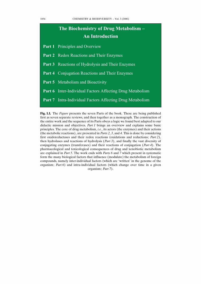
Fig. 1.1. The Figure presents the seven Parts of the book. These are being publishedfirst as seven separate reviews, and then together as a monograph. The construction ofthe entire work and the sequence of its Parts obeys a logic we found best adapted to ourdidactic mission and objectives. Part 1 brings an overview and explains some basicprinciples. The core of drug metabolism, i.e., its actors (the enzymes) and their actions(the metabolic reactions), are presented in Parts 2, 3, and 4. This is done by consideringfirst oxidoreductases and their redox reactions (oxidations and reductions; Part 2),then hydrolases and reactions of hydrolysis (Part 3), and finally the vast diversity ofconjugating enzymes (transferases) and their reactions of conjugation (Part 4). Thepharmacological and toxicological consequences of drug and xenobiotic metabolismare explained in Part 5. The work ends with Parts 6 and 7 which present in systematicform the many biological factors that influence (modulate) the metabolism of foreigncompounds, namely inter-individual factors (which are GwrittenH in the genome of theorganism; Part 6) and intra-individual factors (which change over time in a given
organism; Part 7).
CHEMISTRY & BIODIVERSITY – Vol. 3 (2006)1054
CHEMISTRY & BIODIVERSITY – Vol. 3 (2006) 1055
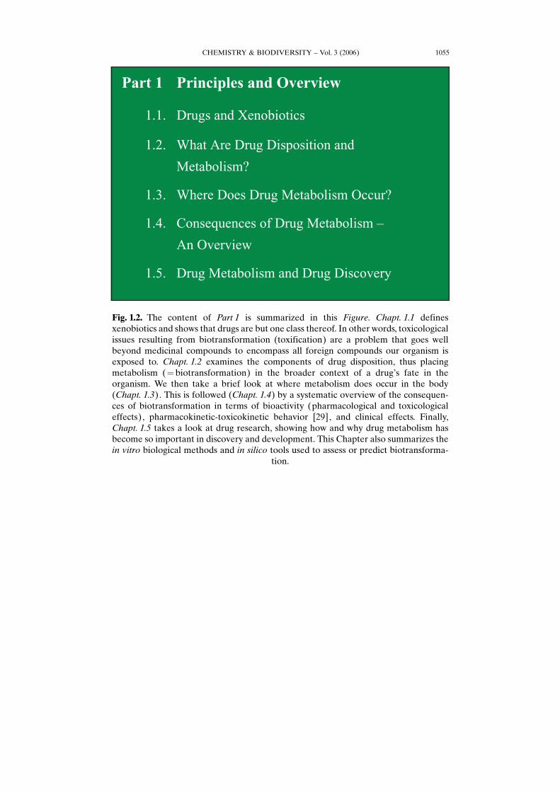
Fig. 1.2. The content of Part 1 is summarized in this Figure. Chapt. 1.1 definesxenobiotics and shows that drugs are but one class thereof. In other words, toxicologicalissues resulting from biotransformation (toxification) are a problem that goes wellbeyond medicinal compounds to encompass all foreign compounds our organism isexposed to. Chapt. 1.2 examines the components of drug disposition, thus placingmetabolism (¼ biotransformation) in the broader context of a drugHs fate in theorganism. We then take a brief look at where metabolism does occur in the body(Chapt. 1.3). This is followed (Chapt. 1.4) by a systematic overview of the consequen-ces of biotransformation in terms of bioactivity (pharmacological and toxicologicaleffects), pharmacokinetic-toxicokinetic behavior [29], and clinical effects. Finally,Chapt. 1.5 takes a look at drug research, showing how and why drug metabolism hasbecome so important in discovery and development. This Chapter also summarizes thein vitro biological methods and in silico tools used to assess or predict biotransforma-
tion.
CHEMISTRY & BIODIVERSITY – Vol. 3 (2006)1056
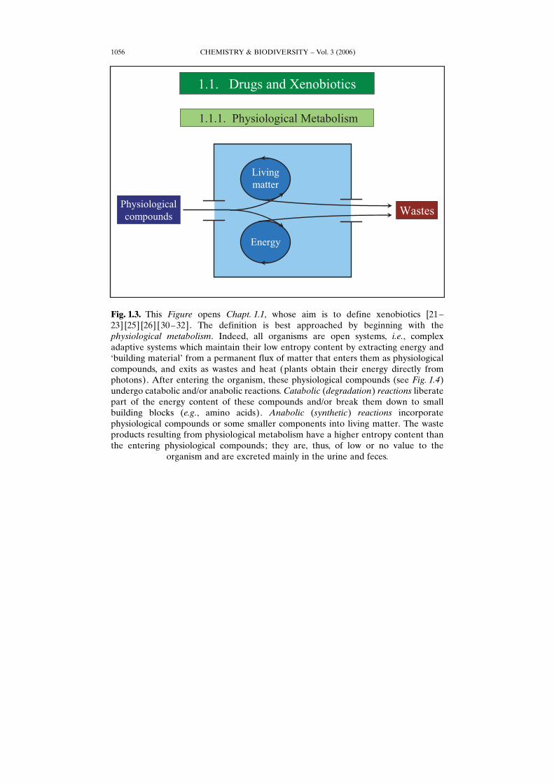
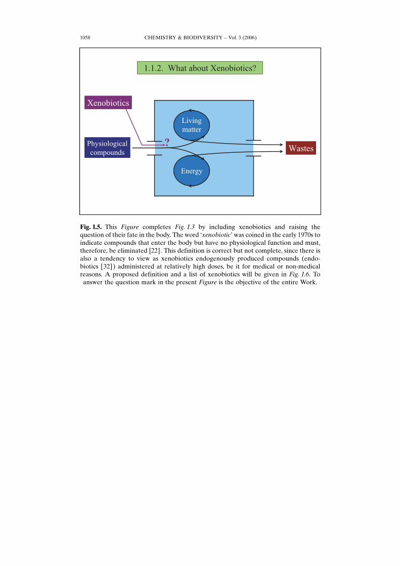
Fig. 1.3. This Figure opens Chapt. 1.1, whose aim is to define xenobiotics [21–23] [25] [26] [30–32]. The definition is best approached by beginning with thephysiological metabolism. Indeed, all organisms are open systems, i.e., complexadaptive systems which maintain their low entropy content by extracting energy andGbuilding materialH from a permanent flux of matter that enters them as physiologicalcompounds, and exits as wastes and heat (plants obtain their energy directly fromphotons). After entering the organism, these physiological compounds (see Fig. 1.4)undergo catabolic and/or anabolic reactions. Catabolic (degradation) reactions liberatepart of the energy content of these compounds and/or break them down to smallbuilding blocks (e.g., amino acids). Anabolic (synthetic) reactions incorporatephysiological compounds or some smaller components into living matter. The wasteproducts resulting from physiological metabolism have a higher entropy content thanthe entering physiological compounds; they are, thus, of low or no value to the
organism and are excreted mainly in the urine and feces.
CHEMISTRY & BIODIVERSITY – Vol. 3 (2006) 1057
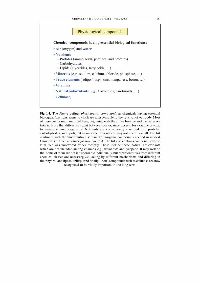
Fig. 1.4. The Figure defines physiological compounds as chemicals having essentialbiological functions, namely, which are indispensable to the survival of our body. Mostof these compounds are listed here, beginning with the air we breathe and the water wetake in. Note that differences exist between species, since oxygen, for example, is toxicto anaerobic microorganisms. Nutrients are conveniently classified into protides,carbohydrates, and lipids, but again some prokaryotes may not need them all. The listcontinues with the GmicronutrientsH, namely, inorganic compounds needed in modest(minerals) or trace amounts (oligo-elements). The list also contains compounds whosevital role was uncovered rather recently. These include those natural antioxidantswhich are not included among vitamins, e.g., flavonoids and lycopene. It may well bethat some of them are not indispensable individually, but representatives from differentchemical classes are necessary, i.e., acting by different mechanisms and differing intheir hydro- and liposolubility. And finally, GinertH compounds such as cellulose are now
recognized to be vitally important in the long term.
CHEMISTRY & BIODIVERSITY – Vol. 3 (2006)1058
Fig. 1.5. This Figure completes Fig. 1.3 by including xenobiotics and raising thequestion of their fate in the body. The word GxenobioticH was coined in the early 1970s toindicate compounds that enter the body but have no physiological function and must,therefore, be eliminated [22]. This definition is correct but not complete, since there isalso a tendency to view as xenobiotics endogenously produced compounds (endo-biotics [32]) administered at relatively high doses, be it for medical or non-medicalreasons. A proposed definition and a list of xenobiotics will be given in Fig. 1.6. Toanswer the question mark in the present Figure is the objective of the entire Work.
CHEMISTRY & BIODIVERSITY – Vol. 3 (2006) 1059
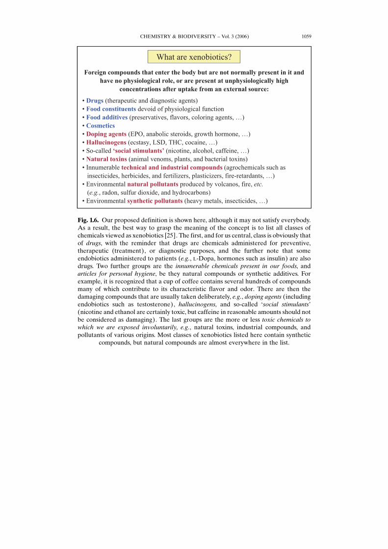
Fig. 1.6. Our proposed definition is shown here, although it may not satisfy everybody.As a result, the best way to grasp the meaning of the concept is to list all classes ofchemicals viewed as xenobiotics [25]. The first, and for us central, class is obviously thatof drugs, with the reminder that drugs are chemicals administered for preventive,therapeutic (treatment), or diagnostic purposes, and the further note that someendobiotics administered to patients (e.g., l-Dopa, hormones such as insulin) are alsodrugs. Two further groups are the innumerable chemicals present in our foods, andarticles for personal hygiene, be they natural compounds or synthetic additives. Forexample, it is recognized that a cup of coffee contains several hundreds of compoundsmany of which contribute to its characteristic flavor and odor. There are then thedamaging compounds that are usually taken deliberately, e.g., doping agents (includingendobiotics such as testosterone), hallucinogens, and so-called Gsocial stimulantsH(nicotine and ethanol are certainly toxic, but caffeine in reasonable amounts should notbe considered as damaging). The last groups are the more or less toxic chemicals towhich we are exposed involuntarily, e.g., natural toxins, industrial compounds, andpollutants of various origins. Most classes of xenobiotics listed here contain synthetic
compounds, but natural compounds are almost everywhere in the list.
CHEMISTRY & BIODIVERSITY – Vol. 3 (2006)1060
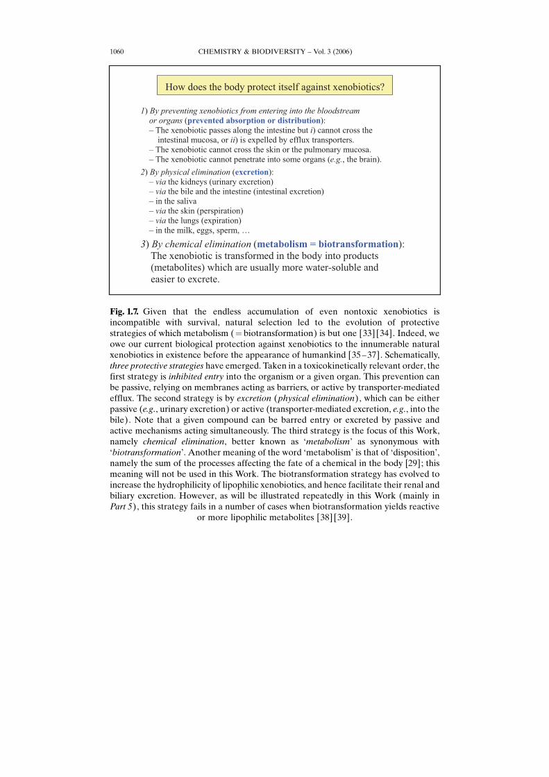
Fig. 1.7. Given that the endless accumulation of even nontoxic xenobiotics isincompatible with survival, natural selection led to the evolution of protectivestrategies of which metabolism (¼ biotransformation) is but one [33] [34]. Indeed, weowe our current biological protection against xenobiotics to the innumerable naturalxenobiotics in existence before the appearance of humankind [35–37]. Schematically,three protective strategies have emerged. Taken in a toxicokinetically relevant order, thefirst strategy is inhibited entry into the organism or a given organ. This prevention canbe passive, relying on membranes acting as barriers, or active by transporter-mediatedefflux. The second strategy is by excretion (physical elimination), which can be eitherpassive (e.g., urinary excretion) or active (transporter-mediated excretion, e.g., into thebile). Note that a given compound can be barred entry or excreted by passive andactive mechanisms acting simultaneously. The third strategy is the focus of this Work,namely chemical elimination, better known as GmetabolismH as synonymous withGbiotransformationH. Another meaning of the word GmetabolismH is that of GdispositionH,namely the sum of the processes affecting the fate of a chemical in the body [29]; thismeaning will not be used in this Work. The biotransformation strategy has evolved toincrease the hydrophilicity of lipophilic xenobiotics, and hence facilitate their renal andbiliary excretion. However, as will be illustrated repeatedly in this Work (mainly inPart 5), this strategy fails in a number of cases when biotransformation yields reactive
or more lipophilic metabolites [38] [39].
CHEMISTRY & BIODIVERSITY – Vol. 3 (2006) 1061
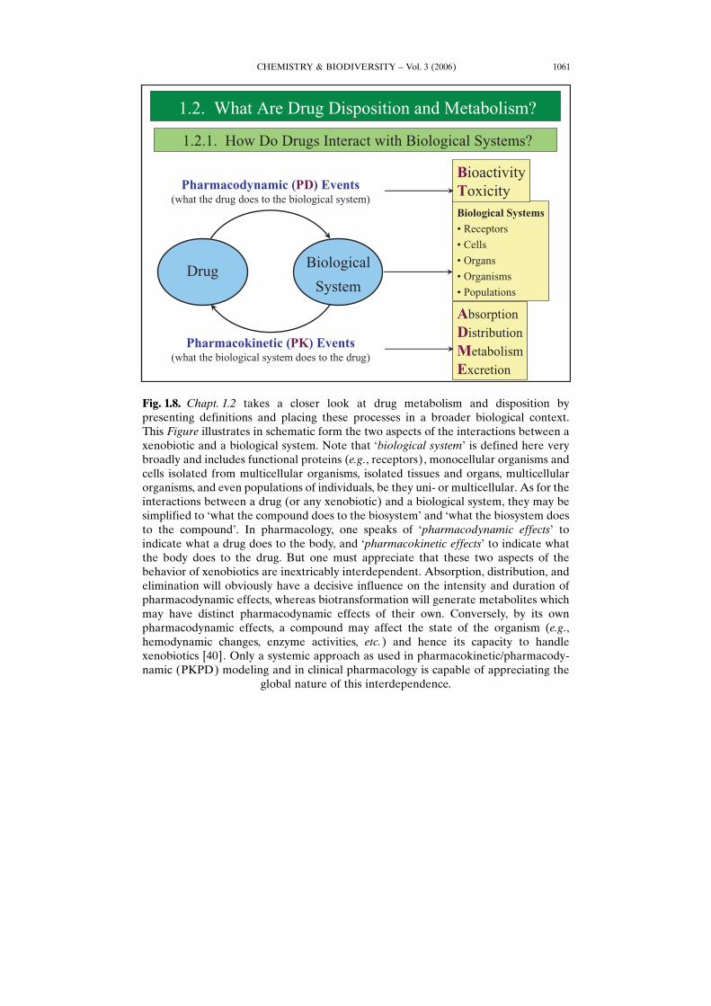
Fig. 1.8. Chapt. 1.2 takes a closer look at drug metabolism and disposition bypresenting definitions and placing these processes in a broader biological context.This Figure illustrates in schematic form the two aspects of the interactions between axenobiotic and a biological system. Note that Gbiological systemH is defined here verybroadly and includes functional proteins (e.g., receptors), monocellular organisms andcells isolated from multicellular organisms, isolated tissues and organs, multicellularorganisms, and even populations of individuals, be they uni- or multicellular. As for theinteractions between a drug (or any xenobiotic) and a biological system, they may besimplified to Gwhat the compound does to the biosystemH and Gwhat the biosystem doesto the compoundH. In pharmacology, one speaks of Gpharmacodynamic effectsH toindicate what a drug does to the body, and Gpharmacokinetic effectsH to indicate whatthe body does to the drug. But one must appreciate that these two aspects of thebehavior of xenobiotics are inextricably interdependent. Absorption, distribution, andelimination will obviously have a decisive influence on the intensity and duration ofpharmacodynamic effects, whereas biotransformation will generate metabolites whichmay have distinct pharmacodynamic effects of their own. Conversely, by its ownpharmacodynamic effects, a compound may affect the state of the organism (e.g.,hemodynamic changes, enzyme activities, etc.) and hence its capacity to handlexenobiotics [40]. Only a systemic approach as used in pharmacokinetic/pharmacody-namic (PKPD) modeling and in clinical pharmacology is capable of appreciating the
global nature of this interdependence.
CHEMISTRY & BIODIVERSITY – Vol. 3 (2006)1062
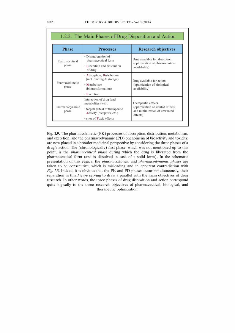
Fig. 1.9. The pharmacokinetic (PK) processes of absorption, distribution, metabolism,and excretion, and the pharmacodynamic (PD) phenomena of bioactivity and toxicity,are now placed in a broader medicinal perspective by considering the three phases of adrugHs action. The (chronologically) first phase, which was not mentioned up to thispoint, is the pharmaceutical phase during which the drug is liberated from thepharmaceutical form (and is dissolved in case of a solid form). In the schematicpresentation of this Figure, the pharmacokinetic and pharmacodynamic phases aretaken to be consecutive, which is misleading and in apparent contradiction withFig. 1.8. Indeed, it is obvious that the PK and PD phases occur simultaneously, theirseparation in this Figure serving to draw a parallel with the main objectives of drugresearch. In other words, the three phases of drug disposition and action correspondquite logically to the three research objectives of pharmaceutical, biological, and
therapeutic optimization.
CHEMISTRY & BIODIVERSITY – Vol. 3 (2006) 1063
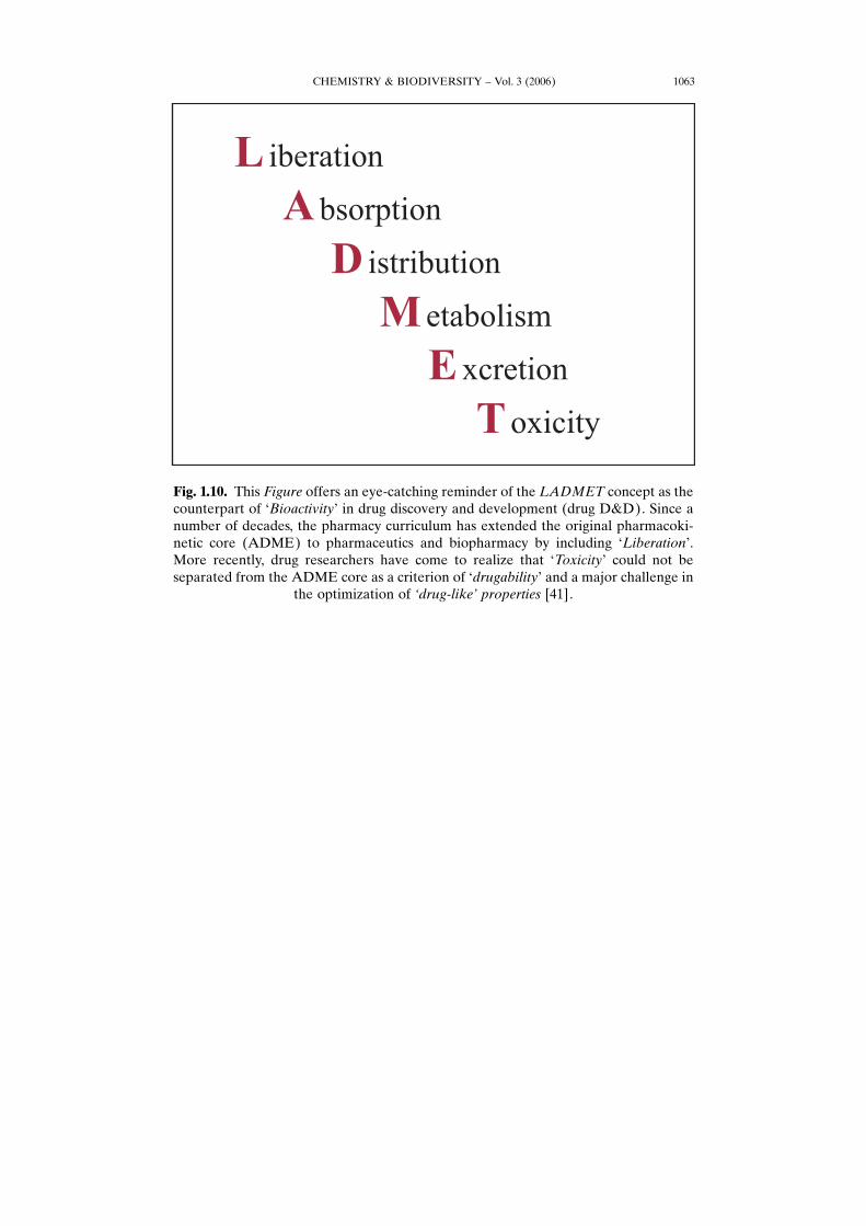
Fig. 1.10. This Figure offers an eye-catching reminder of the LADMET concept as thecounterpart of GBioactivityH in drug discovery and development (drug D&D). Since anumber of decades, the pharmacy curriculum has extended the original pharmacoki-netic core (ADME) to pharmaceutics and biopharmacy by including GLiberationH.More recently, drug researchers have come to realize that GToxicityH could not beseparated from the ADME core as a criterion of GdrugabilityH and a major challenge in
the optimization of ,drug-like. properties [41].
CHEMISTRY & BIODIVERSITY – Vol. 3 (2006)1064
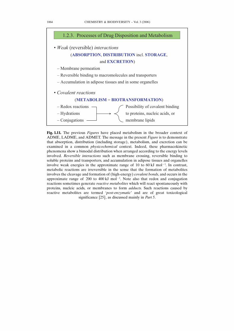
Fig. 1.11. The previous Figures have placed metabolism in the broader context ofADME, LADME, and ADMET. The message in the present Figure is to demonstratethat absorption, distribution (including storage), metabolism, and excretion can beexamined in a common physicochemical context. Indeed, these pharmacokineticphenomena show a bimodal distribution when arranged according to the energy levelsinvolved. Reversible interactions such as membrane crossing, reversible binding tosoluble proteins and transporters, and accumulation in adipose tissues and organellesinvolve weak energies in the approximate range of 10 to 60 kJ mol�1. In contrast,metabolic reactions are irreversible in the sense that the formation of metabolitesinvolves the cleavage and formation of (high-energy) covalent bonds, and occurs in theapproximate range of 200 to 400 kJ mol�1. Note also that redox and conjugationreactions sometimes generate reactive metabolites which will react spontaneously withproteins, nucleic acids, or membranes to form adducts. Such reactions caused byreactive metabolites are termed Gpost-enzymaticH and are of great toxicological
significance [25], as discussed mainly in Part 5.
CHEMISTRY & BIODIVERSITY – Vol. 3 (2006) 1065
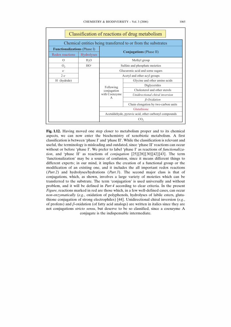
Fig. 1.12. Having moved one step closer to metabolism proper and to its chemicalaspects, we can now enter the biochemistry of xenobiotic metabolism. A firstclassification is between Gphase IH and Gphase IIH. While the classification is relevant anduseful, the terminology is misleading and outdated, since Gphase IIH reactions can occurwithout or before Gphase IH. We prefer to label Gphase IH as reactions of functionaliza-tion, and Gphase IIH as reactions of conjugation [25] [28] [30] [42] [43]. The termGfunctionalizationH may be a source of confusion, since it means different things todifferent experts; in our mind, it implies the creation of a functional group or themodification of an existing one, and it includes the all important redox reactions(Part 2) and hydrolyses/hydrations (Part 3). The second major class is that ofconjugations, which, as shown, involves a large variety of moieties which can betransferred to the substrate. The term GconjugationH is used universally and withoutproblem, and it will be defined in Part 4 according to clear criteria. In the presentFigure, reactions marked in red are those which, in a few well-defined cases, can occurnon-enzymatically (e.g., oxidation of polyphenols, hydrolyses of labile esters, gluta-thione conjugation of strong electrophiles) [44]. Unidirectional chiral inversion (e.g.,of profens) and b-oxidation (of fatty acid analogs) are written in italics since they arenot conjugations stricto sensu, but deserve to be so classified, since a coenzyme A
conjugate is the indispensable intermediate.
CHEMISTRY & BIODIVERSITY – Vol. 3 (2006)1066
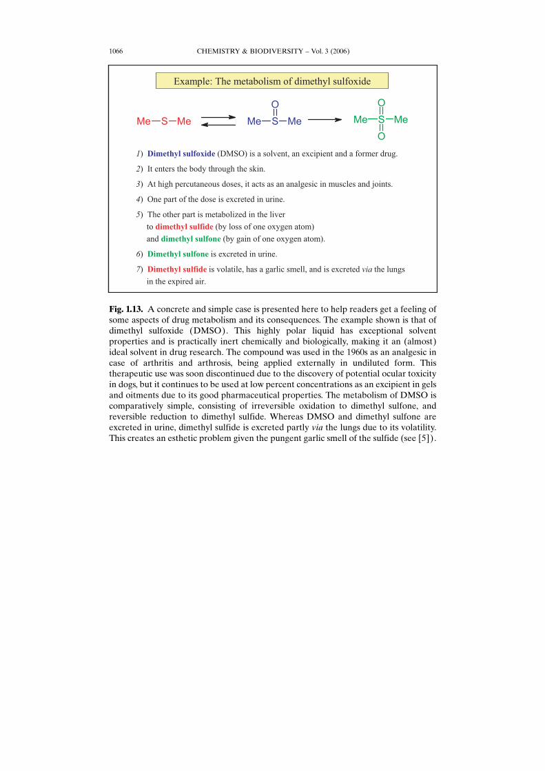
Fig. 1.13. A concrete and simple case is presented here to help readers get a feeling ofsome aspects of drug metabolism and its consequences. The example shown is that ofdimethyl sulfoxide (DMSO). This highly polar liquid has exceptional solventproperties and is practically inert chemically and biologically, making it an (almost)ideal solvent in drug research. The compound was used in the 1960s as an analgesic incase of arthritis and arthrosis, being applied externally in undiluted form. Thistherapeutic use was soon discontinued due to the discovery of potential ocular toxicityin dogs, but it continues to be used at low percent concentrations as an excipient in gelsand oitments due to its good pharmaceutical properties. The metabolism of DMSO iscomparatively simple, consisting of irreversible oxidation to dimethyl sulfone, andreversible reduction to dimethyl sulfide. Whereas DMSO and dimethyl sulfone areexcreted in urine, dimethyl sulfide is excreted partly via the lungs due to its volatility.This creates an esthetic problem given the pungent garlic smell of the sulfide (see [5]).
CHEMISTRY & BIODIVERSITY – Vol. 3 (2006) 1067
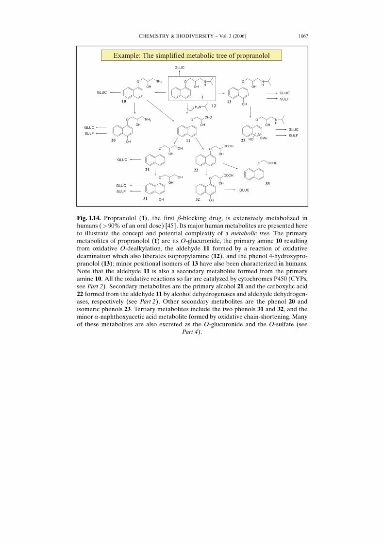
Fig. 1.14. Propranolol (1), the first b-blocking drug, is extensively metabolized inhumans (> 90% of an oral dose) [45]. Its major human metabolites are presented hereto illustrate the concept and potential complexity of a metabolic tree. The primarymetabolites of propranolol (1) are its O-glucuronide, the primary amine 10 resultingfrom oxidative O-dealkylation, the aldehyde 11 formed by a reaction of oxidativedeamination which also liberates isopropylamine (12), and the phenol 4-hydroxypro-pranolol (13); minor positional isomers of 13 have also been characterized in humans.Note that the aldehyde 11 is also a secondary metabolite formed from the primaryamine 10. All the oxidative reactions so far are catalyzed by cytochromes P450 (CYPs,see Part 2). Secondary metabolites are the primary alcohol 21 and the carboxylic acid22 formed from the aldehyde 11 by alcohol dehydrogenases and aldehyde dehydrogen-ases, respectively (see Part 2). Other secondary metabolites are the phenol 20 andisomeric phenols 23. Tertiary metabolites include the two phenols 31 and 32, and theminor a-naphthoxyacetic acid metabolite formed by oxidative chain-shortening. Manyof these metabolites are also excreted as the O-glucuronide and the O-sulfate (see
Part 4).

CHEMISTRY & BIODIVERSITY – Vol. 3 (2006)1068
Fig. 1.15. In this work, the specificity of an enzyme will be taken to mean an ensembleof properties, the description of which makes it possible to specify the enzymeHsbehavior. In contrast, the present Work will apply the term selectivity to metabolicprocesses, indicating that a given metabolic reaction or pathway is able to select somesubstrates or metabolites from a larger set. In other words, the selectivity of a metabolicreaction is the expression of the specificity of an enzyme. Having clarified thesedefinitions, we turn our attention to the various types of selectivities a metabolicreaction can show. When two or more substrates are metabolized at different ratesunder identical conditions, substrate selectivity is observed (left side of the Figure).Substrate selectivity is distinct from product selectivity (right side of the Figure), whichis observed when two or more metabolites are formed at different rates from a singlesubstrate under identical conditions. In other words, substrate-selective reactionsdiscriminate between different compounds, while product-selective reactions discrim-inate between different groups or positions in a given compound. The substrates beingmetabolized at different rates may share various types of relationships. They may bechemically very or slightly different (e.g., analogs, resulting in substrate selectivity in anarrow sense). Alternatively, the substrates may be isomers such as positional isomers(regioisomers, resulting in substrate regioselectivity), stereoisomers (diastereoisomersor enantiomers, resulting in substrate stereoselectivity, substrate diastereoselectivity(seldom used) or substrate enantioselectivity). Products formed at different rates inproduct-selective reactions may also share various types of relationships. Thus, theymay be analogs (product selectivity in a narrow sense), regioisomers (productregioselectivity), or stereoisomers (i.e., diastereoisomers or enantiomers, resulting inproduct stereoselectivity, product diastereoselectivity (seldom used) or product
CHEMISTRY & BIODIVERSITY – Vol. 3 (2006) 1069
enantioselectivity). And since Nature is never as simple as we would like it, the productselectivity displayed by two distinct substrates in a given metabolic reaction may bedifferent, implying that product selectivity itself may be substrate-selective. The termsubstrate–product selectivity is used to describe such complex cases, which have beenreported mainly for stereoselectivity. As presented here, these concepts are quiteabstract and not straightforward to grasp. But their repeated application in Parts 2 to 4
will reveal their usefulness [25] [46].
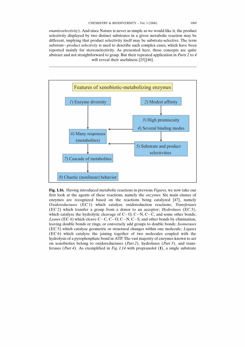
Fig. 1.16. Having introduced metabolic reactions in previous Figures, we now take ourfirst look at the agents of these reactions, namely the enzymes. Six main classes ofenzymes are recognized based on the reactions being catalyzed [47], namelyOxidoreductases (EC 1) which catalyze oxidoreduction reactions; Transferases(EC 2) which transfer a group from a donor to an acceptor; Hydrolases (EC 3),which catalyze the hydrolytic cleavage of C�O, C�N, C�C, and some other bonds;Lyases (EC 4) which cleave C�C, C�O, C�N, C�S, and other bonds by elimination,leaving double bonds or rings, or conversely add groups to double bonds; Isomerases(EC 5) which catalyze geometric or structural changes within one molecule; Ligases(EC 6) which catalyze the joining together of two molecules coupled with thehydrolysis of a pyrophosphate bond in ATP. The vast majority of enzymes known to acton xenobiotics belong to oxidoreductases (Part 2), hydrolases (Part 3), and trans-ferases (Part 4). As exemplified in Fig. 1.14 with propranolol (1), a single substrate
CHEMISTRY & BIODIVERSITY – Vol. 3 (2006)1070
usually yields several (often many) metabolites which are produced Gparallel-wise andseries-wiseH. Such a cascade of metabolites allows for nonlinear responses (chaoticbehavior) in the sense that small causes can have large effects, and large cause can havesmall effects (see Caption to Fig. 1.42). As for the production of many metabolites froma given substrate, this is caused by two factors, namely a) the variety and diversity ofenzymes that act on a given substrate, and b) the product selectivity of a given reaction.In turn, these two factors can be explained as a consequence of the low affinity and thepromiscuity (i.e., the capacity to recognize and metabolize a large structural variety ofsubstrates) of xenobiotic-metabolizing enzymes toward their substrates. However, thecore factor is the property of promiscuity shown by xenobiotic-metabolizing enzymes.Indeed, this property has been favored by Evolution, since it broadens the chemicalspace of potential substrates; but promiscuity comes at a cost, the trade-off being
reduced turnover.
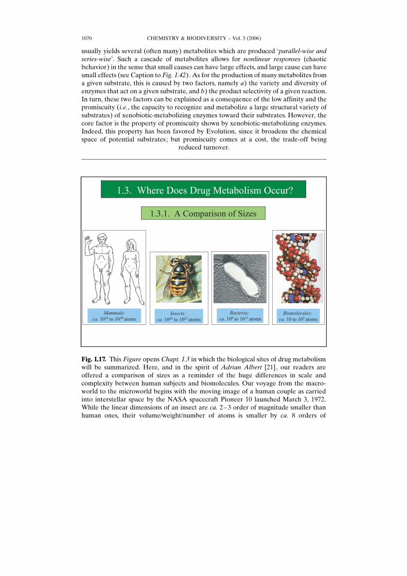
Fig. 1.17. This Figure opens Chapt. 1.3 in which the biological sites of drug metabolismwill be summarized. Here, and in the spirit of Adrian Albert [21], our readers areoffered a comparison of sizes as a reminder of the huge differences in scale andcomplexity between human subjects and biomolecules. Our voyage from the macro-world to the microworld begins with the moving image of a human couple as carriedinto interstellar space by the NASA spacecraft Pioneer 10 launched March 3, 1972.While the linear dimensions of an insect are ca. 2–3 order of magnitude smaller thanhuman ones, their volume/weight/number of atoms is smaller by ca. 8 orders of
CHEMISTRY & BIODIVERSITY – Vol. 3 (2006) 1071
magnitude. About 10–15 orders of magnitude are lost when comparing the volume ofinsects and bacteria. And biomolecules (micro- as well as macromolecules) are 5–19orders of magnitude lighter than bacteria. The point we want to make here is thatbiological phenomena at the macroscopic-medical level are often explainable (if onlyin part) by underlying biochemical processes at the microlevel, but cannot be deducedfrom them with acceptable certainty. Given the biochemical focus of the Work, caution
is urged when trying to infer macroscopic consequences.
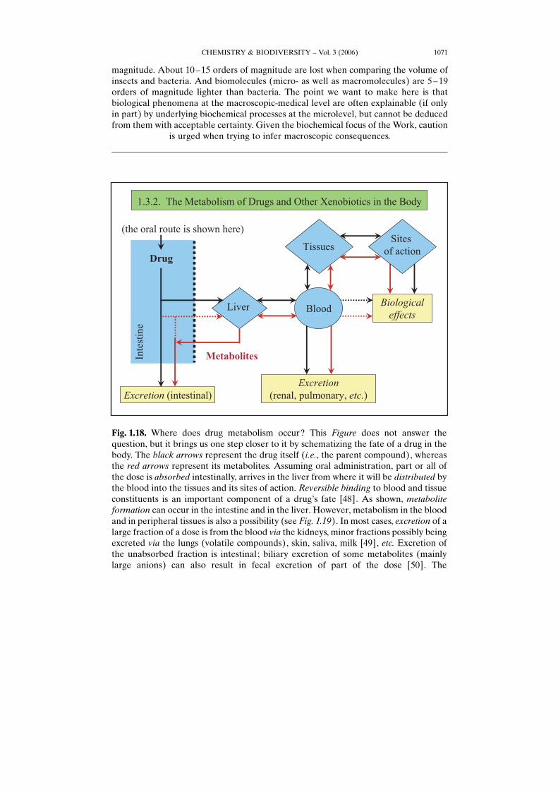
Fig. 1.18. Where does drug metabolism occur? This Figure does not answer thequestion, but it brings us one step closer to it by schematizing the fate of a drug in thebody. The black arrows represent the drug itself (i.e., the parent compound), whereasthe red arrows represent its metabolites. Assuming oral administration, part or all ofthe dose is absorbed intestinally, arrives in the liver from where it will be distributed bythe blood into the tissues and its sites of action. Reversible binding to blood and tissueconstituents is an important component of a drugHs fate [48]. As shown, metaboliteformation can occur in the intestine and in the liver. However, metabolism in the bloodand in peripheral tissues is also a possibility (see Fig. 1.19). In most cases, excretion of alarge fraction of a dose is from the blood via the kidneys, minor fractions possibly beingexcreted via the lungs (volatile compounds), skin, saliva, milk [49], etc. Excretion ofthe unabsorbed fraction is intestinal; biliary excretion of some metabolites (mainlylarge anions) can also result in fecal excretion of part of the dose [50]. The
CHEMISTRY & BIODIVERSITY – Vol. 3 (2006)1072
phenomenon of enterohepatic cycling is worth a mention. Glucuronides of sufficientmolecular weight (in humans, >ca. 500) undergo biliary excretion. When hydrolysis bythe intestinal bacteria is possible (this is the case for O-glucuronides; see Part 4), thephenol or alcohol so liberated can be re-absorbed, reach the liver and circulate again.
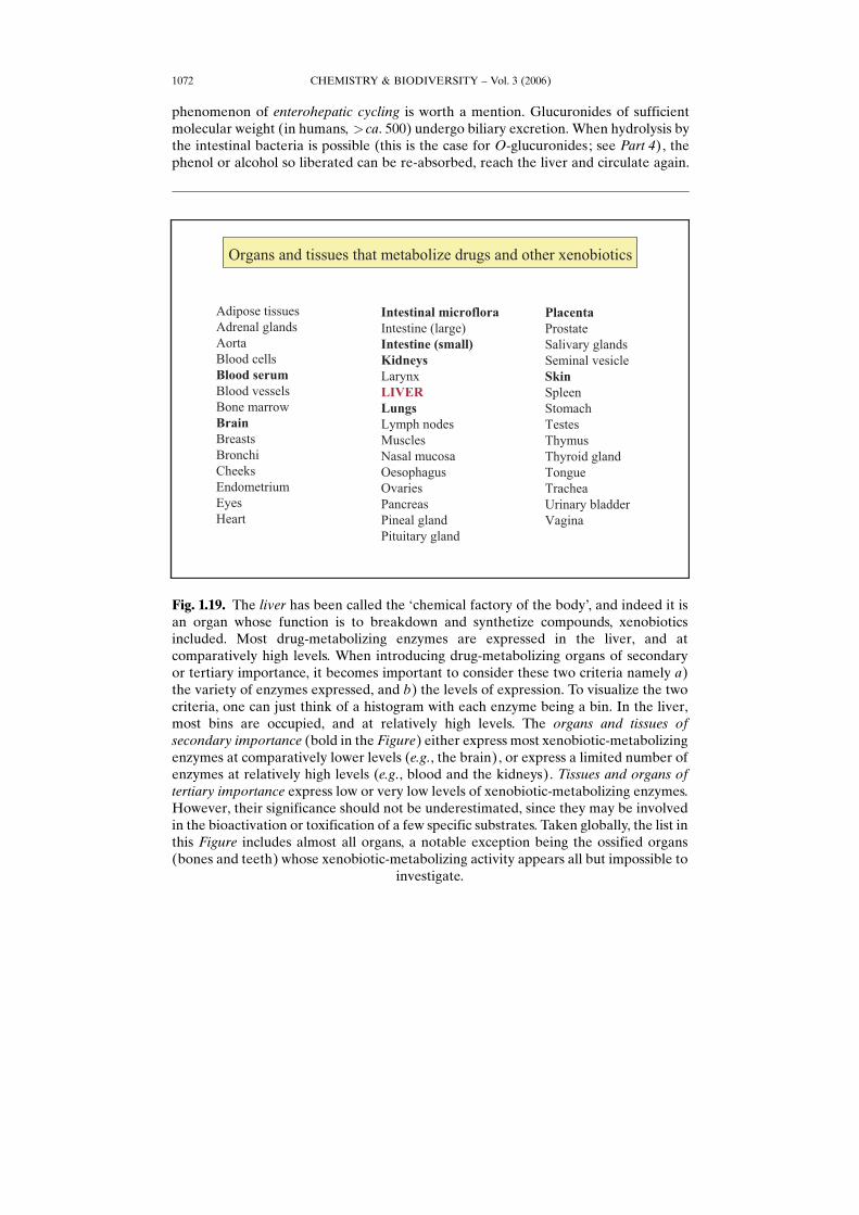
Fig. 1.19. The liver has been called the Gchemical factory of the bodyH, and indeed it isan organ whose function is to breakdown and synthetize compounds, xenobioticsincluded. Most drug-metabolizing enzymes are expressed in the liver, and atcomparatively high levels. When introducing drug-metabolizing organs of secondaryor tertiary importance, it becomes important to consider these two criteria namely a)the variety of enzymes expressed, and b) the levels of expression. To visualize the twocriteria, one can just think of a histogram with each enzyme being a bin. In the liver,most bins are occupied, and at relatively high levels. The organs and tissues ofsecondary importance (bold in the Figure) either express most xenobiotic-metabolizingenzymes at comparatively lower levels (e.g., the brain), or express a limited number ofenzymes at relatively high levels (e.g., blood and the kidneys). Tissues and organs oftertiary importance express low or very low levels of xenobiotic-metabolizing enzymes.However, their significance should not be underestimated, since they may be involvedin the bioactivation or toxification of a few specific substrates. Taken globally, the list inthis Figure includes almost all organs, a notable exception being the ossified organs(bones and teeth) whose xenobiotic-metabolizing activity appears all but impossible to
investigate.
CHEMISTRY & BIODIVERSITY – Vol. 3 (2006) 1073
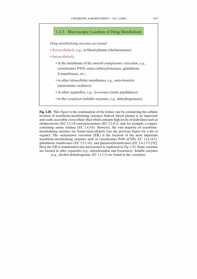
Fig. 1.20. This Figure is the continuation of the former one by considering the cellularlocation of xenobiotic-metabolizing enzymes. Indeed, blood plasma is an importantand easily accessible extracellular fluid which contains high levels of hydrolases such ascholinesterase (EC 3.1.1.8) and paraoxonase (EC 3.1.8.1), and, for example, a copper-containing amine oxidase (EC 1.4.3.6). However, the vast majority of xenobiotic-metabolizing enzymes are found intracellularly (see the previous Figure for a list oforgans). The endoplasmic reticulum (ER) is the location of the most importantxenobiotic-metabolizing enzymes such as cytochromes P450 (CYPs, EC 1.14.14.1),glutathione transferases (EC 2.5.1.18), and glucuronyltransferases (EC 2.4.1.17) [51].How the ER is transformed into microsomes is explained in Fig. 1.22. Some enzymesare located in other organelles (e.g., mitochrondria and lysosomes). Soluble enzymes
(e.g., alcohol dehydrogenase, EC 1.1.1.1) are found in the cytoplasm.
CHEMISTRY & BIODIVERSITY – Vol. 3 (2006)1074
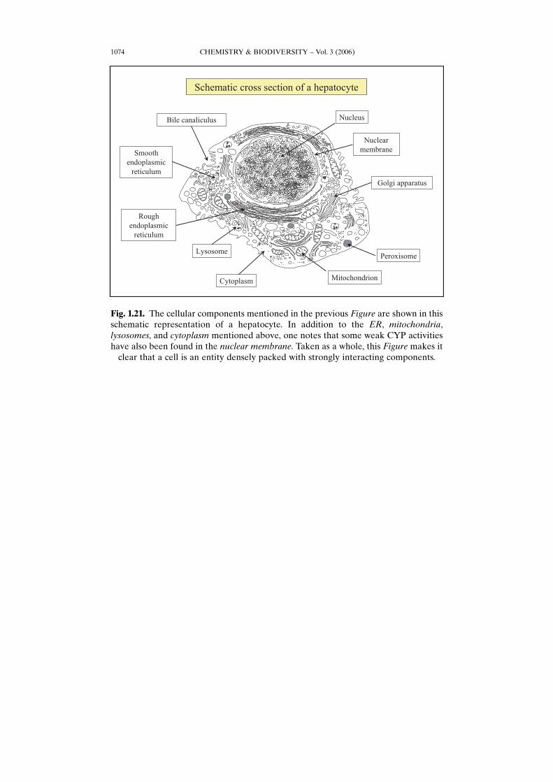
Fig. 1.21. The cellular components mentioned in the previous Figure are shown in thisschematic representation of a hepatocyte. In addition to the ER, mitochondria,lysosomes, and cytoplasm mentioned above, one notes that some weak CYP activitieshave also been found in the nuclear membrane. Taken as a whole, this Figure makes it
clear that a cell is an entity densely packed with strongly interacting components.
CHEMISTRY & BIODIVERSITY – Vol. 3 (2006) 1075
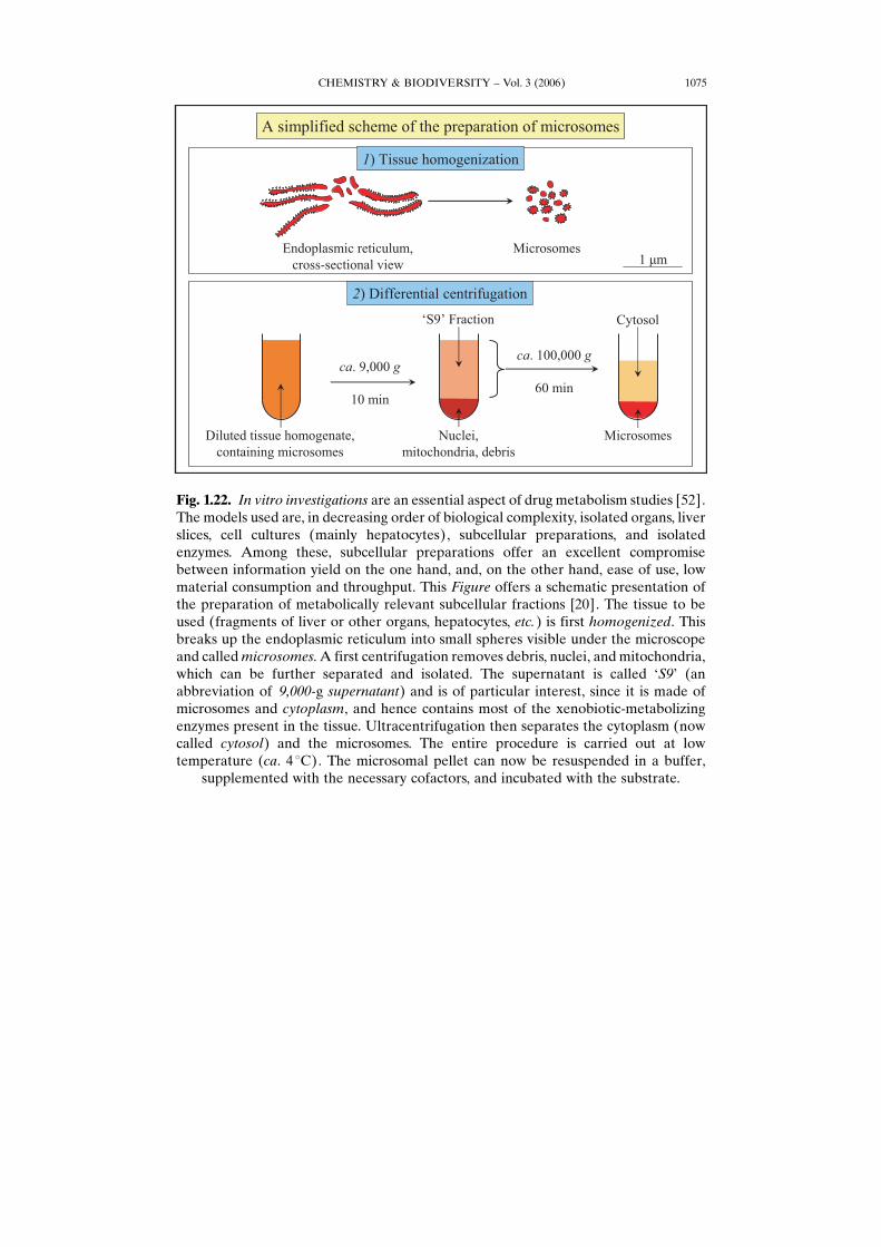
Fig. 1.22. In vitro investigations are an essential aspect of drug metabolism studies [52].The models used are, in decreasing order of biological complexity, isolated organs, liverslices, cell cultures (mainly hepatocytes), subcellular preparations, and isolatedenzymes. Among these, subcellular preparations offer an excellent compromisebetween information yield on the one hand, and, on the other hand, ease of use, lowmaterial consumption and throughput. This Figure offers a schematic presentation ofthe preparation of metabolically relevant subcellular fractions [20]. The tissue to beused (fragments of liver or other organs, hepatocytes, etc.) is first homogenized. Thisbreaks up the endoplasmic reticulum into small spheres visible under the microscopeand calledmicrosomes. A first centrifugation removes debris, nuclei, and mitochondria,which can be further separated and isolated. The supernatant is called GS9H (anabbreviation of 9,000-g supernatant) and is of particular interest, since it is made ofmicrosomes and cytoplasm, and hence contains most of the xenobiotic-metabolizingenzymes present in the tissue. Ultracentrifugation then separates the cytoplasm (nowcalled cytosol) and the microsomes. The entire procedure is carried out at lowtemperature (ca. 4 8C). The microsomal pellet can now be resuspended in a buffer,
supplemented with the necessary cofactors, and incubated with the substrate.
CHEMISTRY & BIODIVERSITY – Vol. 3 (2006)1076
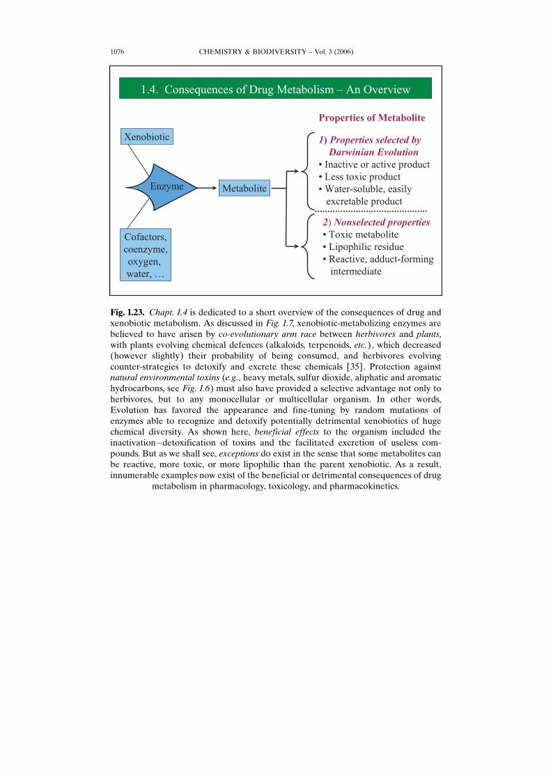
Fig. 1.23. Chapt. 1.4 is dedicated to a short overview of the consequences of drug andxenobiotic metabolism. As discussed in Fig. 1.7, xenobiotic-metabolizing enzymes arebelieved to have arisen by co-evolutionary arm race between herbivores and plants,with plants evolving chemical defences (alkaloids, terpenoids, etc.), which decreased(however slightly) their probability of being consumed, and herbivores evolvingcounter-strategies to detoxify and excrete these chemicals [35]. Protection againstnatural environmental toxins (e.g., heavy metals, sulfur dioxide, aliphatic and aromatichydrocarbons, see Fig. 1.6) must also have provided a selective advantage not only toherbivores, but to any monocellular or multicellular organism. In other words,Evolution has favored the appearance and fine-tuning by random mutations ofenzymes able to recognize and detoxify potentially detrimental xenobiotics of hugechemical diversity. As shown here, beneficial effects to the organism included theinactivation–detoxification of toxins and the facilitated excretion of useless com-pounds. But as we shall see, exceptions do exist in the sense that some metabolites canbe reactive, more toxic, or more lipophilic than the parent xenobiotic. As a result,innumerable examples now exist of the beneficial or detrimental consequences of drug
metabolism in pharmacology, toxicology, and pharmacokinetics.
CHEMISTRY & BIODIVERSITY – Vol. 3 (2006) 1077
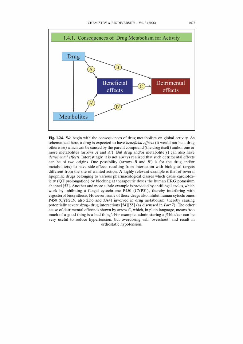
Fig. 1.24. We begin with the consequences of drug metabolism on global activity. Asschematized here, a drug is expected to have beneficial effects (it would not be a drugotherwise) which can be caused by the parent compound (the drug itself) and/or one ormore metabolites (arrows A and A’). But drug and/or metabolite(s) can also havedetrimental effects. Interestingly, it is not always realized that such detrimental effectscan be of two origins. One possibility (arrows B and B’) is for the drug and/ormetabolite(s) to have side-effects resulting from interaction with biological targetsdifferent from the site of wanted action. A highly relevant example is that of severallipophilic drugs belonging to various pharmacological classes which cause cardiotox-icity (QT prolongation) by blocking at therapeutic doses the human ERG potassiumchannel [53]. Another and more subtle example is provided by antifungal azoles, whichwork by inhibiting a fungal cytochrome P450 (CYP51), thereby interfering withergosterol biosynthesis. However, some of these drugs also inhibit human cytochromesP450 (CYP2C9, also 2D6 and 3A4) involved in drug metabolism, thereby causingpotentially severe drug–drug interactions [54] [55] (as discussed in Part 7). The othercause of detrimental effects is shown by arrow C, which, in plain language, means Gtoomuch of a good thing is a bad thingH. For example, administering a b-blocker can bevery useful to reduce hypertension, but overdosing will GovershootH and result in
orthostatic hypotension.
CHEMISTRY & BIODIVERSITY – Vol. 3 (2006)1078
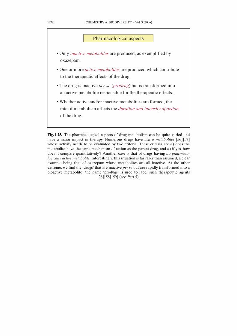
Fig. 1.25. The pharmacological aspects of drug metabolism can be quite varied andhave a major impact in therapy. Numerous drugs have active metabolites [56] [57]whose activity needs to be evaluated by two criteria. These criteria are a) does themetabolite have the same mechanism of action as the parent drug, and b) if yes, howdoes it compare quantitatively? Another case is that of drugs having no pharmaco-logically active metabolite. Interestingly, this situation is far rarer than assumed, a clearexample being that of oxazepam whose metabolites are all inactive. At the otherextreme, we find the GdrugsH that are inactive per se but are rapidly transformed into abioactive metabolite; the name GprodugsH is used to label such therapeutic agents
[28] [58] [59] (see Part 5).
CHEMISTRY & BIODIVERSITY – Vol. 3 (2006) 1079
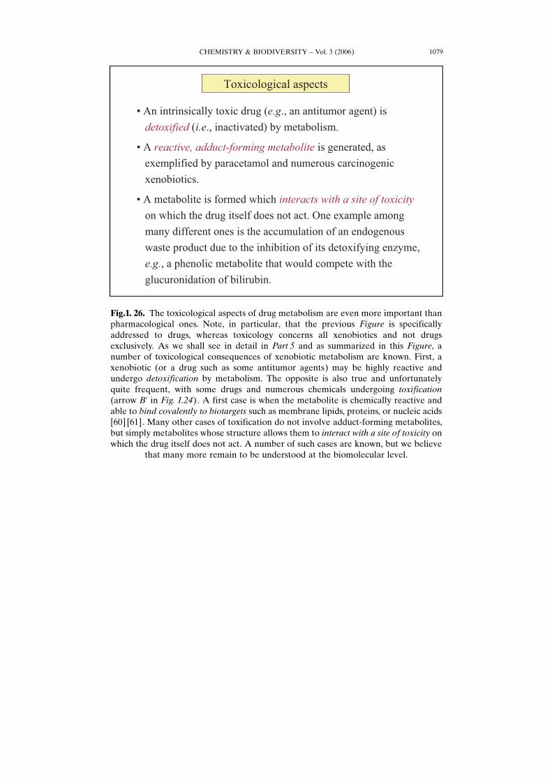
Fig.1. 26. The toxicological aspects of drug metabolism are even more important thanpharmacological ones. Note, in particular, that the previous Figure is specificallyaddressed to drugs, whereas toxicology concerns all xenobiotics and not drugsexclusively. As we shall see in detail in Part 5 and as summarized in this Figure, anumber of toxicological consequences of xenobiotic metabolism are known. First, axenobiotic (or a drug such as some antitumor agents) may be highly reactive andundergo detoxification by metabolism. The opposite is also true and unfortunatelyquite frequent, with some drugs and numerous chemicals undergoing toxification(arrow B’ in Fig. 1.24). A first case is when the metabolite is chemically reactive andable to bind covalently to biotargets such as membrane lipids, proteins, or nucleic acids[60] [61]. Many other cases of toxification do not involve adduct-forming metabolites,but simply metabolites whose structure allows them to interact with a site of toxicity onwhich the drug itself does not act. A number of such cases are known, but we believe
that many more remain to be understood at the biomolecular level.
CHEMISTRY & BIODIVERSITY – Vol. 3 (2006)1080
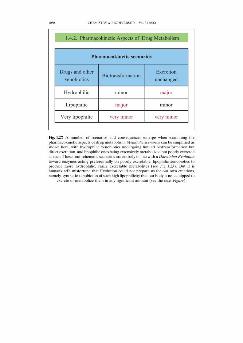
Fig. 1.27. A number of scenarios and consequences emerge when examining thepharmacokinetic aspects of drug metabolism. Metabolic scenarios can be simplified asshown here, with hydrophilic xenobiotics undergoing limited biotransformation butdirect excretion, and lipophilic ones being extensively metabolized but poorly excretedas such. These four schematic scenarios are entirely in line with a Darwinian Evolutiontoward enzymes acting preferentially on poorly excretable, lipophilic xenobiotics toproduce more hydrophilic, easily excretable metabolites (see Fig. 1.23). But it ishumankindHs misfortune that Evolution could not prepare us for our own creations,namely, synthetic xenobiotics of such high lipophilicity that our body is not equipped to
excrete or metabolize them in any significant amount (see the next Figure).
CHEMISTRY & BIODIVERSITY – Vol. 3 (2006) 1081
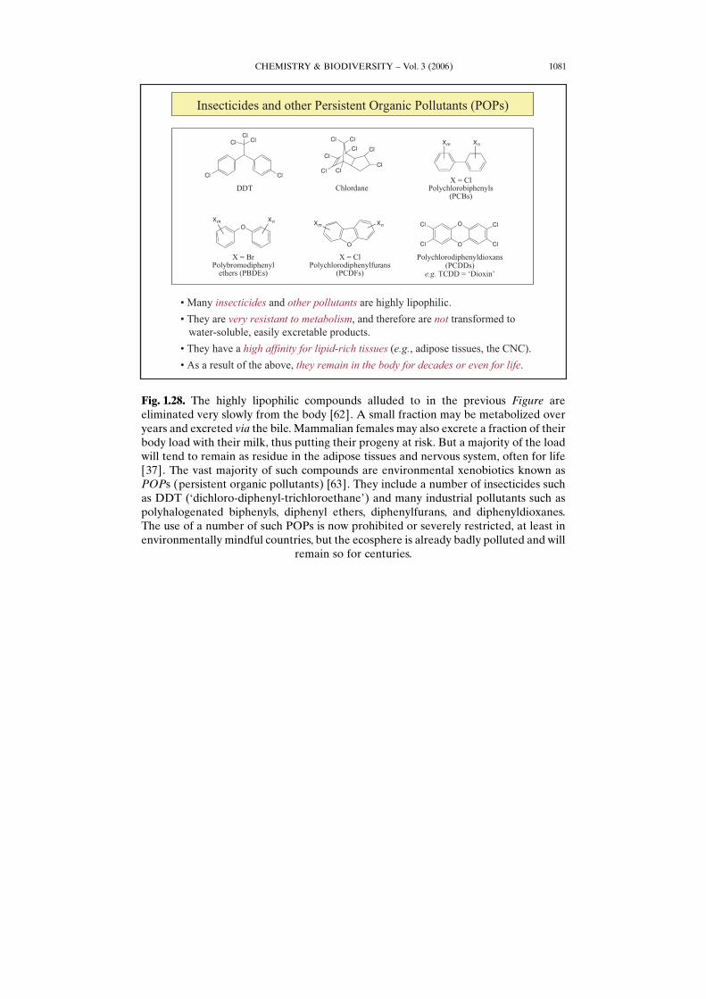
Fig. 1.28. The highly lipophilic compounds alluded to in the previous Figure areeliminated very slowly from the body [62]. A small fraction may be metabolized overyears and excreted via the bile. Mammalian females may also excrete a fraction of theirbody load with their milk, thus putting their progeny at risk. But a majority of the loadwill tend to remain as residue in the adipose tissues and nervous system, often for life[37]. The vast majority of such compounds are environmental xenobiotics known asPOPs (persistent organic pollutants) [63]. They include a number of insecticides suchas DDT (Gdichloro-diphenyl-trichloroethaneH) and many industrial pollutants such aspolyhalogenated biphenyls, diphenyl ethers, diphenylfurans, and diphenyldioxanes.The use of a number of such POPs is now prohibited or severely restricted, at least inenvironmentally mindful countries, but the ecosphere is already badly polluted and will
remain so for centuries.
CHEMISTRY & BIODIVERSITY – Vol. 3 (2006)1082
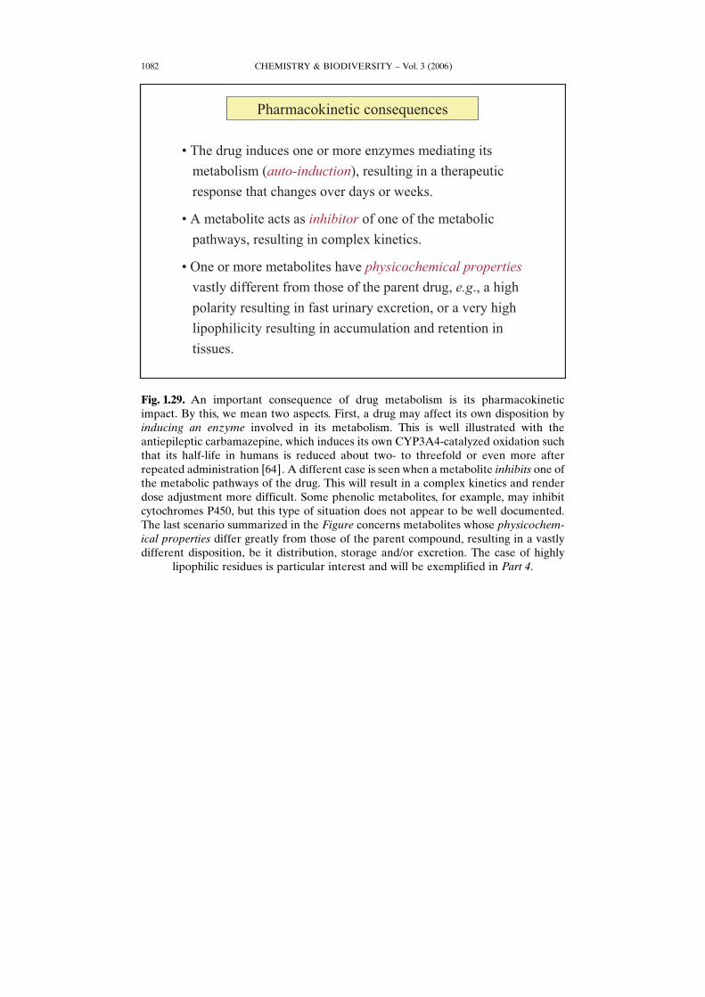
Fig. 1.29. An important consequence of drug metabolism is its pharmacokineticimpact. By this, we mean two aspects. First, a drug may affect its own disposition byinducing an enzyme involved in its metabolism. This is well illustrated with theantiepileptic carbamazepine, which induces its own CYP3A4-catalyzed oxidation suchthat its half-life in humans is reduced about two- to threefold or even more afterrepeated administration [64]. A different case is seen when a metabolite inhibits one ofthe metabolic pathways of the drug. This will result in a complex kinetics and renderdose adjustment more difficult. Some phenolic metabolites, for example, may inhibitcytochromes P450, but this type of situation does not appear to be well documented.The last scenario summarized in the Figure concerns metabolites whose physicochem-ical properties differ greatly from those of the parent compound, resulting in a vastlydifferent disposition, be it distribution, storage and/or excretion. The case of highly
lipophilic residues is particular interest and will be exemplified in Part 4.
CHEMISTRY & BIODIVERSITY – Vol. 3 (2006) 1083
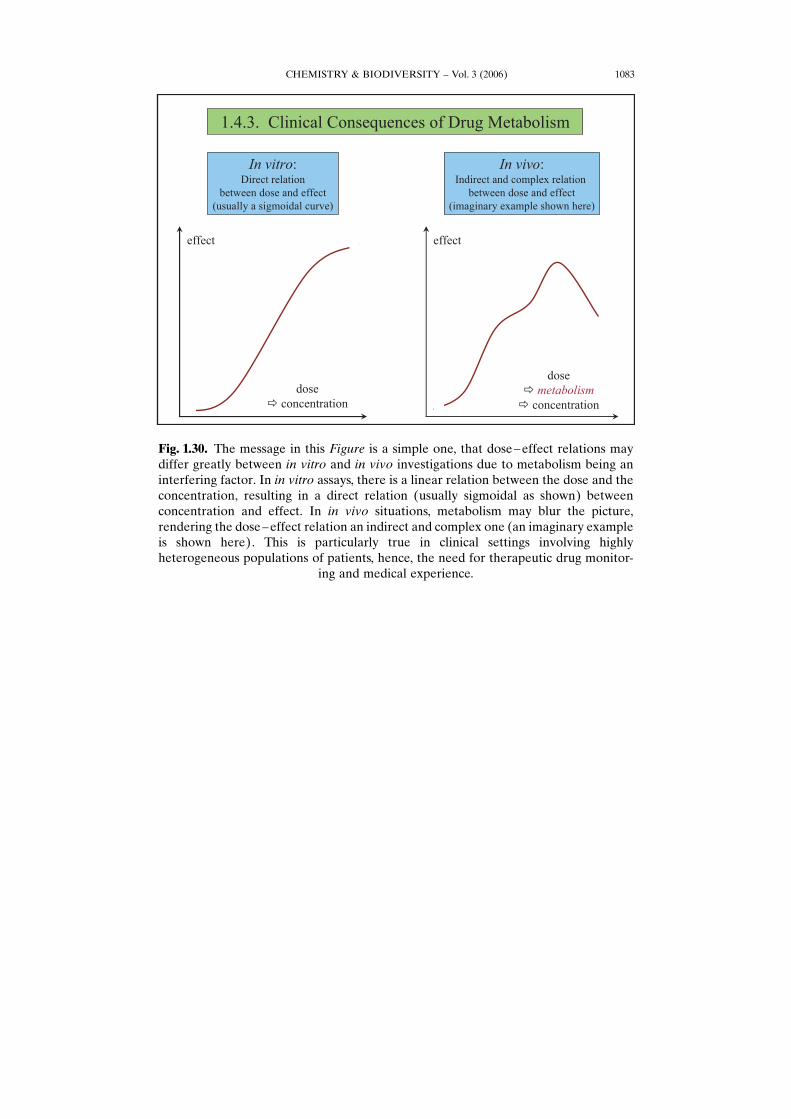
Fig. 1.30. The message in this Figure is a simple one, that dose–effect relations maydiffer greatly between in vitro and in vivo investigations due to metabolism being aninterfering factor. In in vitro assays, there is a linear relation between the dose and theconcentration, resulting in a direct relation (usually sigmoidal as shown) betweenconcentration and effect. In in vivo situations, metabolism may blur the picture,rendering the dose–effect relation an indirect and complex one (an imaginary exampleis shown here). This is particularly true in clinical settings involving highlyheterogeneous populations of patients, hence, the need for therapeutic drug monitor-
ing and medical experience.
CHEMISTRY & BIODIVERSITY – Vol. 3 (2006)1084
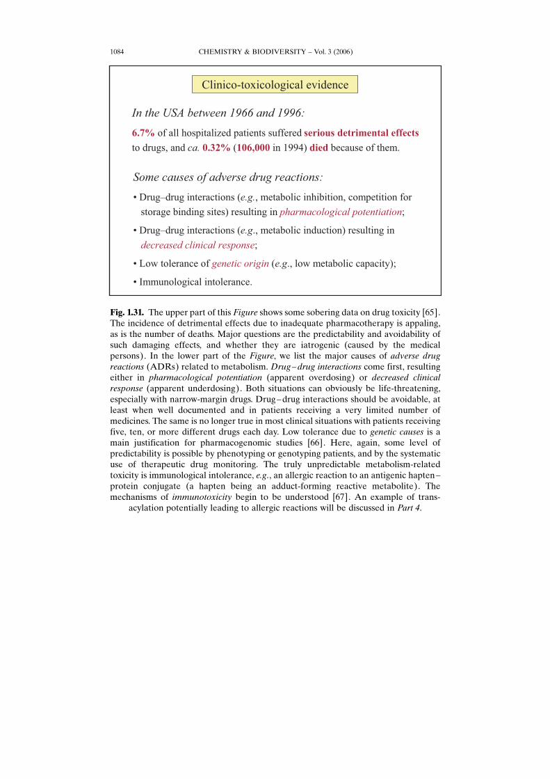
Fig. 1.31. The upper part of this Figure shows some sobering data on drug toxicity [65].The incidence of detrimental effects due to inadequate pharmacotherapy is appaling,as is the number of deaths. Major questions are the predictability and avoidability ofsuch damaging effects, and whether they are iatrogenic (caused by the medicalpersons). In the lower part of the Figure, we list the major causes of adverse drugreactions (ADRs) related to metabolism. Drug–drug interactions come first, resultingeither in pharmacological potentiation (apparent overdosing) or decreased clinicalresponse (apparent underdosing). Both situations can obviously be life-threatening,especially with narrow-margin drugs. Drug–drug interactions should be avoidable, atleast when well documented and in patients receiving a very limited number ofmedicines. The same is no longer true in most clinical situations with patients receivingfive, ten, or more different drugs each day. Low tolerance due to genetic causes is amain justification for pharmacogenomic studies [66]. Here, again, some level ofpredictability is possible by phenotyping or genotyping patients, and by the systematicuse of therapeutic drug monitoring. The truly unpredictable metabolism-relatedtoxicity is immunological intolerance, e.g., an allergic reaction to an antigenic hapten–protein conjugate (a hapten being an adduct-forming reactive metabolite). Themechanisms of immunotoxicity begin to be understood [67]. An example of trans-
acylation potentially leading to allergic reactions will be discussed in Part 4.
CHEMISTRY & BIODIVERSITY – Vol. 3 (2006) 1085
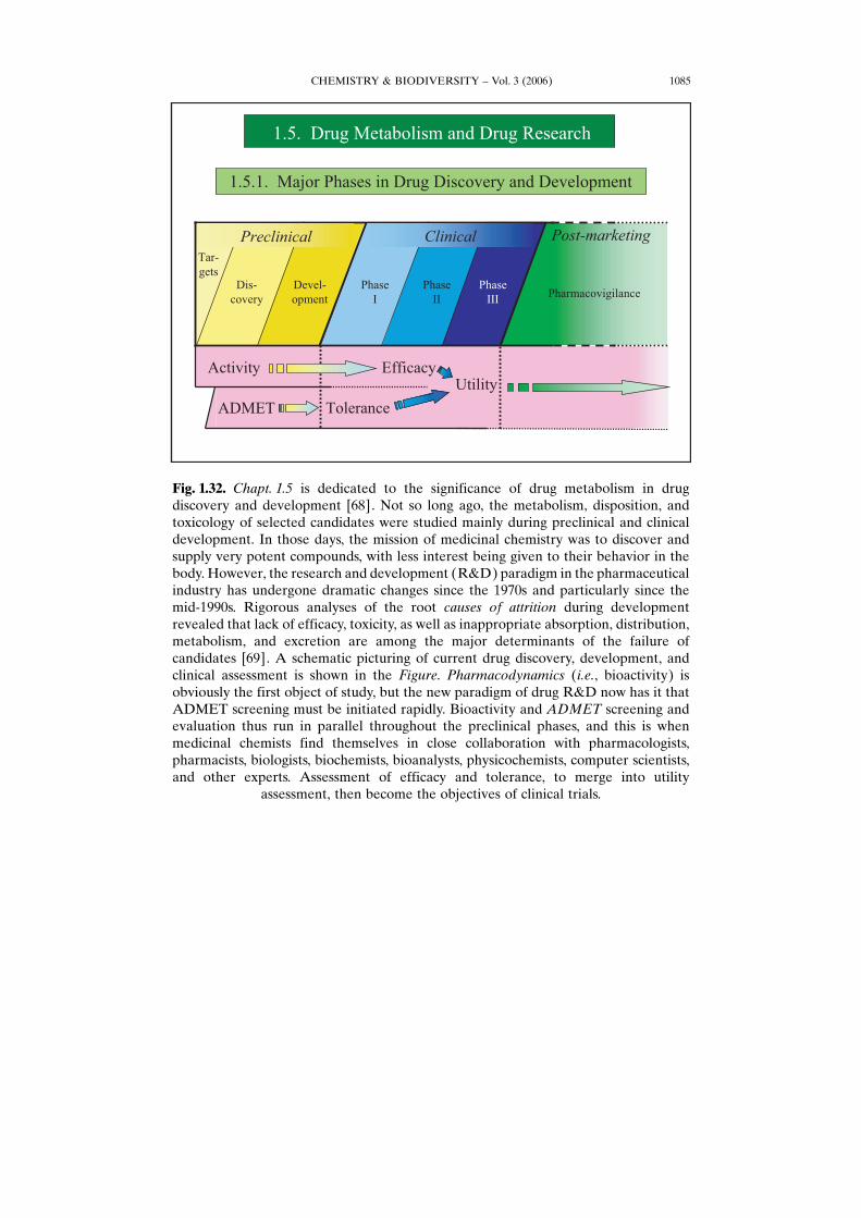
Fig. 1.32. Chapt. 1.5 is dedicated to the significance of drug metabolism in drugdiscovery and development [68]. Not so long ago, the metabolism, disposition, andtoxicology of selected candidates were studied mainly during preclinical and clinicaldevelopment. In those days, the mission of medicinal chemistry was to discover andsupply very potent compounds, with less interest being given to their behavior in thebody. However, the research and development (R&D) paradigm in the pharmaceuticalindustry has undergone dramatic changes since the 1970s and particularly since themid-1990s. Rigorous analyses of the root causes of attrition during developmentrevealed that lack of efficacy, toxicity, as well as inappropriate absorption, distribution,metabolism, and excretion are among the major determinants of the failure ofcandidates [69]. A schematic picturing of current drug discovery, development, andclinical assessment is shown in the Figure. Pharmacodynamics (i.e., bioactivity) isobviously the first object of study, but the new paradigm of drug R&D now has it thatADMET screening must be initiated rapidly. Bioactivity and ADMET screening andevaluation thus run in parallel throughout the preclinical phases, and this is whenmedicinal chemists find themselves in close collaboration with pharmacologists,pharmacists, biologists, biochemists, bioanalysts, physicochemists, computer scientists,and other experts. Assessment of efficacy and tolerance, to merge into utility
assessment, then become the objectives of clinical trials.
CHEMISTRY & BIODIVERSITY – Vol. 3 (2006)1086
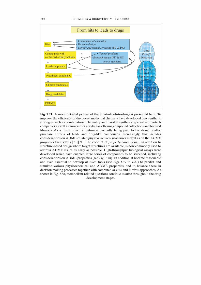
Fig. 1.33. A more detailed picture of the hits-to-leads-to-drugs is presented here. Toimprove the efficiency of discovery, medicinal chemists have developed new syntheticstrategies such as combinatorial chemistry and parallel synthesis. Specialized biotechcompanies as well as universities also began offering compound collections and focusedlibraries. As a result, much attention is currently being paid to the design and/orpurchase criteria of lead- and drug-like compounds. Increasingly, this includesconsiderations on ADME-related physicochemical properties as well as on the ADMEproperties themselves [70] [71]. The concept of property-based design, in addition tostructure-based design where target structures are available, is now commonly used toaddress ADME issues as early as possible. High-throughput biological assays weredeveloped which have enabled large series of compounds to be screened, includingconsiderations on ADME properties (see Fig. 1.38). In addition, it became reasonableand even essential to develop in silico tools (see Figs. 1.39 to 1.42) to predict andsimulate various physicochemical and ADME properties, and to balance these indecision making processes together with combined in vivo and in vitro approaches. Asshown in Fig. 1.36, metabolism-related questions continue to arise throughout the drug
development stages.
CHEMISTRY & BIODIVERSITY – Vol. 3 (2006) 1087
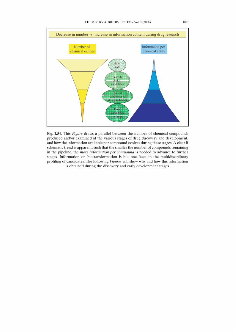
Fig. 1.34. This Figure draws a parallel between the number of chemical compoundsproduced and/or examined at the various stages of drug discovery and development,and how the information available per compound evolves during these stages. A clear ifschematic trend is apparent, such that the smaller the number of compounds remainingin the pipeline, the more information per compound is needed to advance to furtherstages. Information on biotransformation is but one facet in the multidisciplinaryprofiling of candidates. The following Figures will show why and how this information
is obtained during the discovery and early development stages.
CHEMISTRY & BIODIVERSITY – Vol. 3 (2006)1088
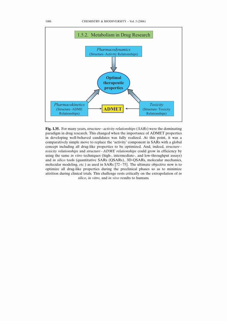
Fig. 1.35. For many years, structure–activity relationships (SARs) were the dominatingparadigm in drug research. This changed when the importance of ADMET propertiesin developing well-behaved candidates was fully realized. At this point, it was acomparatively simple move to replace the GactivityH component in SARs with a globalconcept including all drug-like properties to be optimized. And, indeed, structure–toxicity relationships and structure–ADME relationships could grow in efficiency byusing the same in vitro techniques (high-, intermediate-, and low-throughput assays)and in silico tools (quantitative SARs (QSARs), 3D-QSARs, molecular mechanics,molecular modeling, etc.) as used in SARs [72–75]. The ultimate objective now is tooptimize all drug-like properties during the preclinical phases so as to minimizeattrition during clinical trials. This challenge rests critically on the extrapolation of in
silico, in vitro, and in vivo results to humans.
CHEMISTRY & BIODIVERSITY – Vol. 3 (2006) 1089
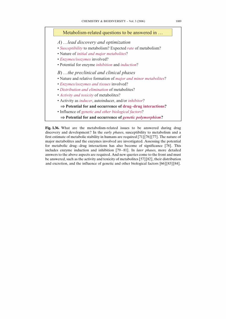
Fig. 1.36. What are the metabolism-related issues to be answered during drugdiscovery and development? In the early phases, susceptibility to metabolism and afirst estimate of metabolic stability in humans are required [71] [76] [77]. The nature ofmajor metabolites and the enzymes involved are investigated. Assessing the potentialfor metabolic drug–drug interactions has also become of significance [78]. Thisincludes enzyme induction and inhibition [79–81]. In later phases, more detailedanswers to the above aspects are required. And new queries come to the front and mustbe answered, such as the activity and toxicity of metabolites [57] [82], their distributionand excretion, and the influence of genetic and other biological factors [66] [83] [84].
CHEMISTRY & BIODIVERSITY – Vol. 3 (2006)1090
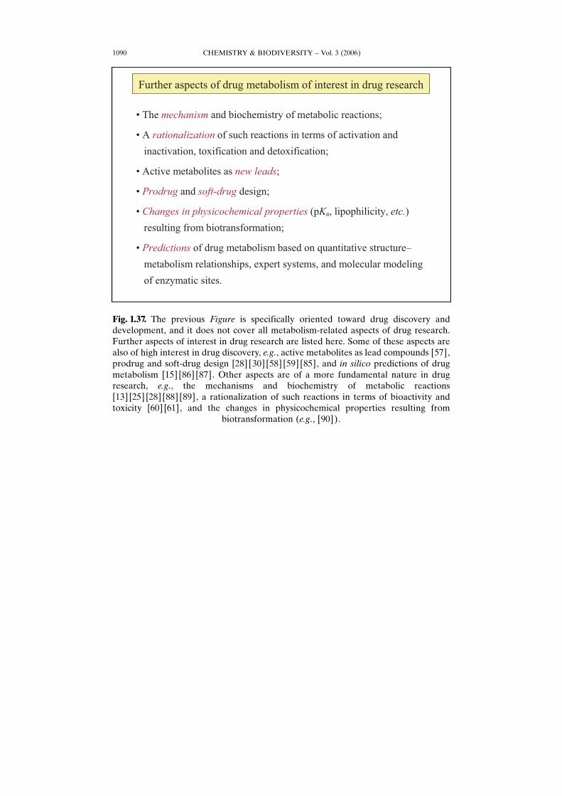
Fig. 1.37. The previous Figure is specifically oriented toward drug discovery anddevelopment, and it does not cover all metabolism-related aspects of drug research.Further aspects of interest in drug research are listed here. Some of these aspects arealso of high interest in drug discovery, e.g., active metabolites as lead compounds [57],prodrug and soft-drug design [28] [30] [58] [59] [85], and in silico predictions of drugmetabolism [15] [86] [87]. Other aspects are of a more fundamental nature in drugresearch, e.g., the mechanisms and biochemistry of metabolic reactions[13] [25] [28] [88] [89], a rationalization of such reactions in terms of bioactivity andtoxicity [60] [61], and the changes in physicochemical properties resulting from
biotransformation (e.g., [90]).
CHEMISTRY & BIODIVERSITY – Vol. 3 (2006) 1091
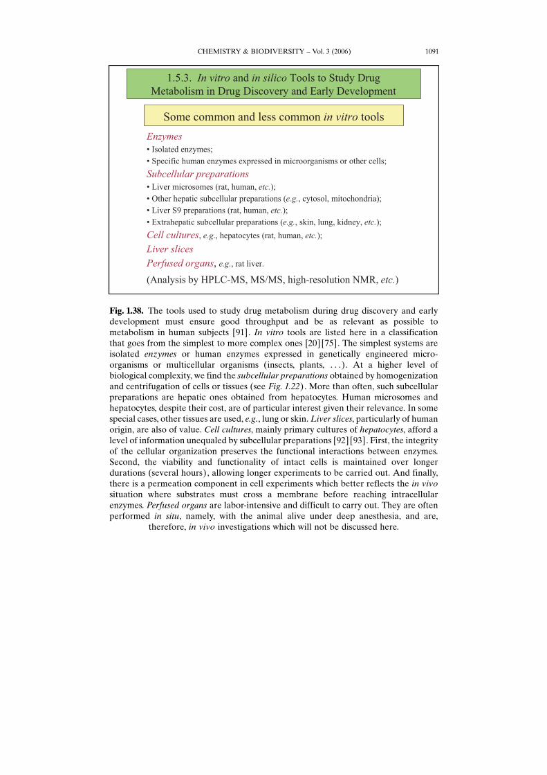
Fig. 1.38. The tools used to study drug metabolism during drug discovery and earlydevelopment must ensure good throughput and be as relevant as possible tometabolism in human subjects [91]. In vitro tools are listed here in a classificationthat goes from the simplest to more complex ones [20] [75]. The simplest systems areisolated enzymes or human enzymes expressed in genetically engineered micro-organisms or multicellular organisms (insects, plants, . . .). At a higher level ofbiological complexity, we find the subcellular preparations obtained by homogenizationand centrifugation of cells or tissues (see Fig. 1.22). More than often, such subcellularpreparations are hepatic ones obtained from hepatocytes. Human microsomes andhepatocytes, despite their cost, are of particular interest given their relevance. In somespecial cases, other tissues are used, e.g., lung or skin.Liver slices, particularly of humanorigin, are also of value. Cell cultures, mainly primary cultures of hepatocytes, afford alevel of information unequaled by subcellular preparations [92] [93]. First, the integrityof the cellular organization preserves the functional interactions between enzymes.Second, the viability and functionality of intact cells is maintained over longerdurations (several hours), allowing longer experiments to be carried out. And finally,there is a permeation component in cell experiments which better reflects the in vivosituation where substrates must cross a membrane before reaching intracellularenzymes. Perfused organs are labor-intensive and difficult to carry out. They are oftenperformed in situ, namely, with the animal alive under deep anesthesia, and are,
therefore, in vivo investigations which will not be discussed here.
CHEMISTRY & BIODIVERSITY – Vol. 3 (2006)1092
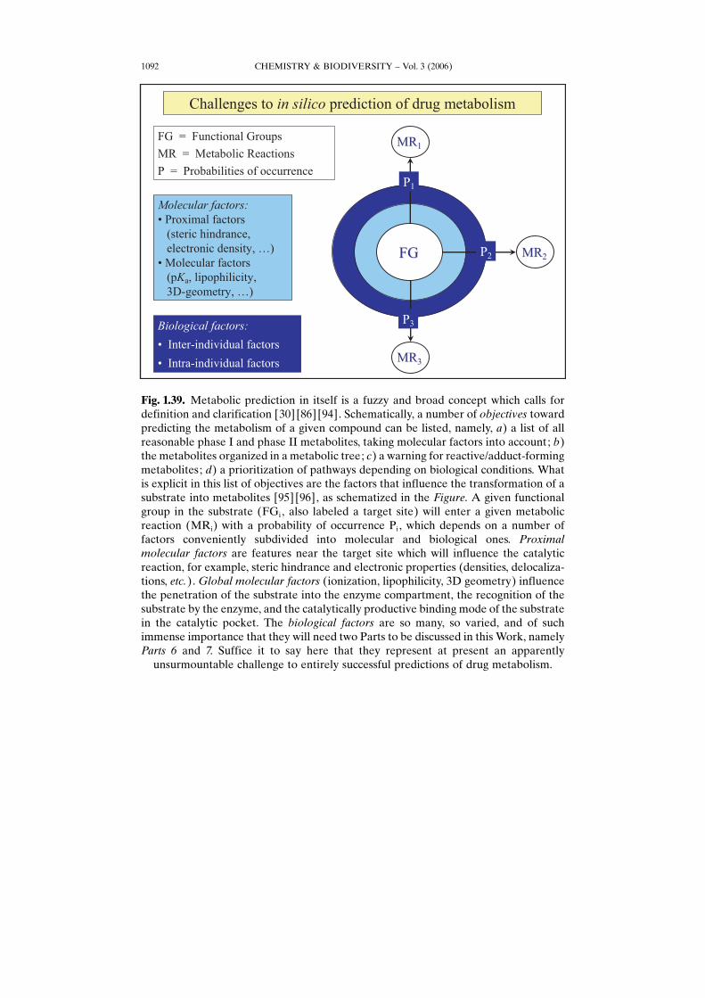
Fig. 1.39. Metabolic prediction in itself is a fuzzy and broad concept which calls fordefinition and clarification [30] [86] [94]. Schematically, a number of objectives towardpredicting the metabolism of a given compound can be listed, namely, a) a list of allreasonable phase I and phase II metabolites, taking molecular factors into account; b)the metabolites organized in a metabolic tree; c) a warning for reactive/adduct-formingmetabolites; d) a prioritization of pathways depending on biological conditions. Whatis explicit in this list of objectives are the factors that influence the transformation of asubstrate into metabolites [95] [96], as schematized in the Figure. A given functionalgroup in the substrate (FGi, also labeled a target site) will enter a given metabolicreaction (MRi) with a probability of occurrence Pi, which depends on a number offactors conveniently subdivided into molecular and biological ones. Proximalmolecular factors are features near the target site which will influence the catalyticreaction, for example, steric hindrance and electronic properties (densities, delocaliza-tions, etc.). Global molecular factors (ionization, lipophilicity, 3D geometry) influencethe penetration of the substrate into the enzyme compartment, the recognition of thesubstrate by the enzyme, and the catalytically productive binding mode of the substratein the catalytic pocket. The biological factors are so many, so varied, and of suchimmense importance that they will need two Parts to be discussed in this Work, namelyParts 6 and 7. Suffice it to say here that they represent at present an apparently
unsurmountable challenge to entirely successful predictions of drug metabolism.

CHEMISTRY & BIODIVERSITY – Vol. 3 (2006) 1093
Fig. 1.40. In a simplified manner, one can distinguish between two types of algorithmsto predict drug and xenobiotic metabolism, namely, specific (GlocalH) systems andcomprehensive (GglobalH) systems. Specific systems apply to simple biological (e.g.,single enzymes) and/or to single metabolic reactions, and they may or may not berestricted to rather narrow chemical series. Such systems include quantitative structure�metabolism relationships (QSMRs) based on structural and physicochemical properties[96] [97]. Quantum mechanical calculations may also shed light on SMRs and generateparameters to be used as independent variables in QSMRs, revealing, for example,correlations between rates of metabolic oxidation and energy barrier in H-atomabstraction [98] [99]. Three-dimensional QSMRs (3D-QSMRs) methods yield a partialview of the binding/catalytic site of a given enzyme as derived from the 3D-molecularfields of a series of substrates or inhibitors (the training set). In other words, they yield aGphotographic negativeH of such sites, and will allow a quantitative prediction for novelcompounds structurally related to the training set [100] [101]. The molecular modelingof xenobiotic-metabolizing enzymes affords another approach to rationalize and predictdrug–enzyme interactions [102] [103]. Its application to drug metabolism was madepossible by the crystallization and X-ray structural determination of cytochromes P450,first bacterial, and now human ones. While such pharmacophoric models cannot yetgive highly accurate quantitative affinity predictions, they nevertheless afford fairlyreliable answers as to the relative accessibility of target sites in the substrate molecules.The 3D models of a large number of mammalian and mostly human CYPs are nowavailable, as well as other xenobiotic-metabolizing enzymes such as DT-diaphorase andvarious transferases. The last approaches mentioned in this Figure are expert systems
CHEMISTRY & BIODIVERSITY – Vol. 3 (2006)1094
combining several methods, for example, pharmacophore models (obtained by 3D-QSAR), protein models (obtained by molecular modeling), and docking [104] [105].Another powerful combination are a) 3D models obtained by molecular modeling, andb) sophisticated QSAR approaches based on multivariate analyses of parametersobtained from molecular interaction fields (MIFs), as found in the MetaSite algorithm[106].MetaSite is a specific system in the sense that it is currently restricted to the majorhuman cytochromes P450. At the end of the procedure, the atoms of the substrate areranked according to their accessibility and reactivity. In other words, Metasite takes the3D stereoelectronic structure of both the enzyme and the ligand into account to
prioritize the potential target sites in the molecule.
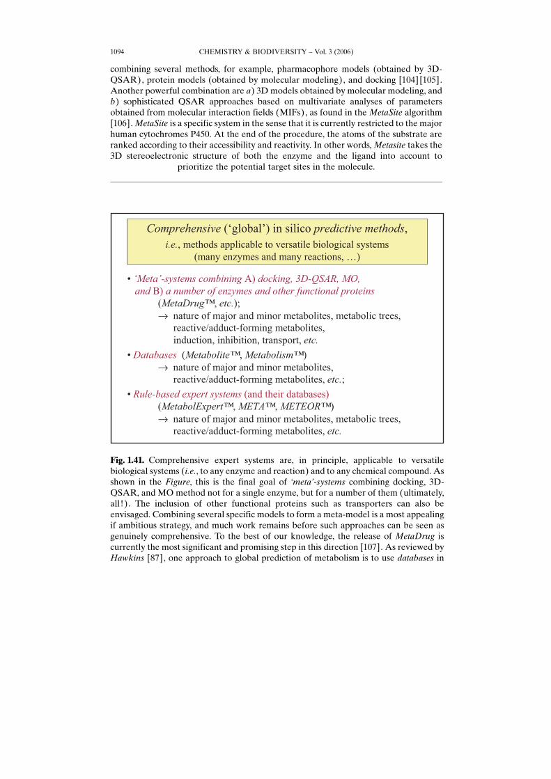
Fig. 1.41. Comprehensive expert systems are, in principle, applicable to versatilebiological systems (i.e., to any enzyme and reaction) and to any chemical compound. Asshown in the Figure, this is the final goal of ,meta.-systems combining docking, 3D-QSAR, and MO method not for a single enzyme, but for a number of them (ultimately,all!). The inclusion of other functional proteins such as transporters can also beenvisaged. Combining several specific models to form a meta-model is a most appealingif ambitious strategy, and much work remains before such approaches can be seen asgenuinely comprehensive. To the best of our knowledge, the release of MetaDrug iscurrently the most significant and promising step in this direction [107]. As reviewed byHawkins [87], one approach to global prediction of metabolism is to use databases in
CHEMISTRY & BIODIVERSITY – Vol. 3 (2006) 1095
the form of either knowledge-based systems or predictive, rule-based systems [15].Existing knowledge-based systems include the MDLMetabolite Database [108] and theAccelrys Metabolism database [109] originally established using data compiled in thebook series Biotransformations [110]. These databases can be searched to retrieveinformation on the known metabolism of compounds with similar structures orcontaining specific moieties. Predictive, rule-based systems attempt to portray themetabolites of a compound based on knowledge rules, defining the most likely products[111]. Existing systems of this type are MetabolExpert [112], META [113], and
METEOR [114].
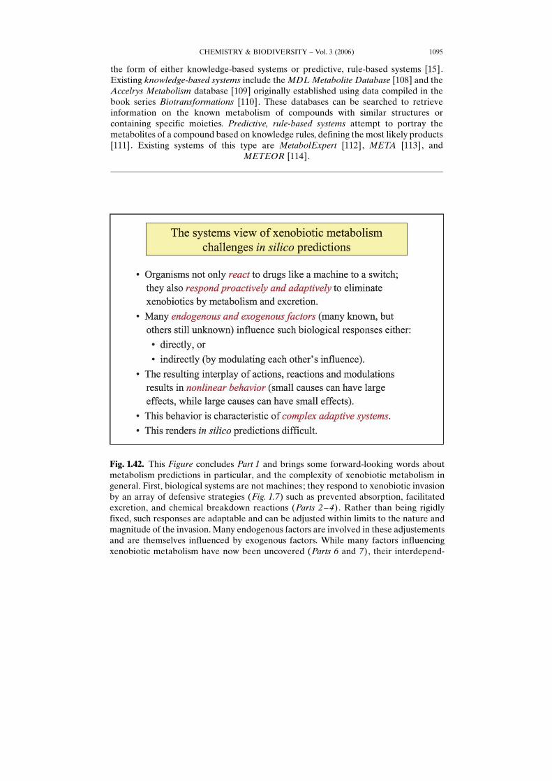
Fig. 1.42. This Figure concludes Part 1 and brings some forward-looking words aboutmetabolism predictions in particular, and the complexity of xenobiotic metabolism ingeneral. First, biological systems are not machines; they respond to xenobiotic invasionby an array of defensive strategies (Fig. 1.7) such as prevented absorption, facilitatedexcretion, and chemical breakdown reactions (Parts 2–4). Rather than being rigidlyfixed, such responses are adaptable and can be adjusted within limits to the nature andmagnitude of the invasion. Many endogenous factors are involved in these adjustementsand are themselves influenced by exogenous factors. While many factors influencingxenobiotic metabolism have now been uncovered (Parts 6 and 7), their interdepend-
REFERENCES
[1] A. Conti, M. H. Bickel, GHistory of drug metabolism: discoveries of the major pathways in the 19thcenturyH, Drug Metab. Rev. 1977, 6, 1–50; C. Bachmann, M. H. Bickel, GHistory of drug metabolism:the first half of the 20th centuryH, Drug Metab. Rev. 1986, 16, 185–253.
[2] International Society for the Study of Xenobiotics, www.issx.org.[3] R. T. Williams, GDetoxication MechanismsH, Chapman & Hall, London, 1947.[4] R. T. Williams, GDetoxication MechanismsH, 2nd edn., Chapman & Hall, London, 1959.[5] B. Testa, P. Jenner, GDrug Metabolism: Chemical and Biochemical AspectsH, Dekker, New York,
1976.[6] B. Testa, P. Jenner, GThe coming of age of drug metabolismH, Curr. Contents Life Sci. 1990, 33, 17.[7] GConcepts in Drug MetabolismH, Eds. P. Jenner, B. Testa, Dekker, New York, 1980 and 1981, Part A
and Part B.[8] GDrug Metabolism – from Molecules to ManH, Eds. D. J. Benford, J. W. Bridges, G. G. Gibson, Taylor
& Francis, London, 1987.[9] GConjugation Reactions in Drug MetabolismH, Ed. G. J. Mulder, Taylor & Francis, London, 1990.
[10] GPharmacokinetics of DrugsH, Eds. P. G. Welling, L. P. Balant, Springer, Heidelberg, 1994.[11] GConjugation-Deconjugation Reactions in Drug Metabolism and ToxicityH, Ed. F. C. Kauffman,
Springer Verlag, Berlin, 1994.[12] GConjugation-Dependent Carcinogenicity and Toxicity of Foreign CompoundsH, Eds. M. W. Anders,
W. Dekant, Academic Press, San Diego, 1994.[13] GCytochrome P450. Structure, Mechanism, and BiochemistryH, 2nd edn., Ed. P. R. Ortiz de
Montellano, Plenum Press, New York, 1996.[14] GHandbook of Drug MetabolismH, Ed. T. F. Woolf, Dekker, New York, 1999.[15] GDrug Metabolism: Databases and High-Throughput Testing during Drug Design and Develop-
mentH, Ed. P. W. Erhardt, International Union of Pure and Applied Chemistry and BlackwellScience, London, 1999.
[16] GEnzyme Systems that Metabolise Drugs and Other XenobioticsH, Ed. C. Ioannides, John Wiley &Sons, Chichester, 2002.
[17] GDrug-Drug InteractionsH, Ed. A. D. Rodrigues, Dekker, New York, 2002.[18] GHandbook of Drug-Nutrient InteractionsH, Eds. J. Boullata, V. T. Armenti, Humana Press, Totowa,
2004.[19] GDrug Metabolism and TransportH, Ed. L. H. Lash, Humana Press, Totowa, 2005.[20] GCytochrome P450 ProtocolsH, Eds. I. R. Phillips, E. A. Shephard, Humana Press, Totowa, 2006.[21] A. Albert, GSelective ToxicityH, Chapman & Hall, London, 1985.[22] A. Albert, GXenobiosisH, Chapman & Hall, London, 1987.[23] J. A. Timbrell, GPrinciples of Biochemical ToxicologyH, 2nd edn., Taylor & Francis, London, 1991.[24] R. B. Silverman, GThe Organic Chemistry of Drug Design and Drug ActionH, Academic Press, San
Diego, 1992.[25] B. Testa, GThe Metabolism of Drugs and Other Xenobiotics – Biochemistry of Redox ReactionsH,
Academic Press, London, 1995.[26] W. N. Aldridge, GMechanisms and Concepts in ToxicologyH, Taylor & Francis, London, 1996.
CHEMISTRY & BIODIVERSITY – Vol. 3 (2006)1096
character of the responses, such that small causes can have small or large effects, andlarge causes can have large or small effects. This chaotic behavior is the characteristic ofcomplex adaptive systems; it is also the source of the apparently unsurmountabledifficulty of making close-to-perfect in silico predictions, and above all it is the source of
the endless satisfactions one obtains when studying drug metabolism.
[27] G. G. Gibson, P. Skett, GIntroduction to Drug MetabolismH, 3rd edn., Nelson Thornes, CheltenhamUK, 2001.
[28] B. Testa, J. M. Mayer, GHydrolysis in Drug and Prodrug Metabolism – Chemistry, Biochemistry, andEnzymologyH, Verlag Helvetica Chimica Acta, Zurich, and Wiley-VCH, Weinheim, 2003.
[29] F. J. Di Carlo, GMetabolism, pharmacokinetics, and toxicokinetics definedH, Drug Metab. Rev. 1982,13, 1–4.
[30] B. Testa, W. Soine, GPrinciples of drug metabolismH, in GBurgerHs Medicinal Chemistry and DrugDiscoveryH, 6th edn., Vol. 2, Ed. D. J. Abraham, Wiley-Interscience, Hoboken, 2003, p. 431–498.
[31] A. Albert, GThe behaviour of foreign substances in the human bodyH, Trends Pharmacol. Sci. 1987, 8,258–261.
[32] P. Jenner, B. Testa, F. J. Di Carlo, GXenobiotic and endobiotic metabolizing enzymes: anoverstretched discrimination?H, Trends Pharmacol. Sci. 1981, 2, 135–137.
[33] W. B. Jakoby, D. M. Ziegler, GThe enzymes of detoxicationH, J. Biol. Chem. 1990, 265, 20715–20718.[34] D. W. Nebert, GProposed role of drug-metabolizing enzymes: Regulation of steady state levels of the
ligands that effect growth, homeostasis, differentiation, and neuroendocrine functionsH, Mol.Endocrinol. 1991, 5, 1203–1214.
[35] F. J. Gonzalez, D. W. Nebert, GEvolution of the P450 gene superfamily: animal-plant GwarfareH,molecular drive and human genetic differences in drug oxidationH, Trends Genet. 1990, 6, 182–186.
[36] B. N. Ames, M. Profet, GNatureHs pesticidesH, Nat. Toxins 1992, 1, 2–3.[37] W. R. Jondorf, GDrug metabolism and drug toxicity: Some evolutionary considerationsH, in GConcepts
in Drug MetabolismH, Part B, Eds. P. Jenner, B. Testa, Dekker, New York, 1981, p. 305–376.[38] J. R. Gillette, GThe use of theoretical pharmacokinetic concepts in studies of the mechanisms of
formation of chemically reactive metabolites in vitro and in vivoH, Drug Metab. Rev. 1983, 14, 9–33.[39] T. J. Monks, S. S. Lau, GReactive intermediates and their toxicological significanceH, Toxicology 1988,
52, 1–54.[40] B. Testa, GPharmacokinetic and pharmacodynamic events: can they always be distinguished?H,
Trends Pharmacol. Sci. 1987, 8, 381–383.[41] GADME-Tox: The Fate of Drugs in the BodyH, Eds. B. Testa, H. van de Waterbeemd, in
GComprehensive Medicinal ChemistryH, 2nd edn., Eds. D. J. Triggle, J. Taylor, Elsevier, Oxford,Vol. 5, in press.
[42] B. Testa, P. Jenner, GNovel metabolites produced by functionalization reactions: Chemistry andtoxicologyH, Drug Metab. Rev. 1978, 7, 325–369.
[43] J. Caldwell, GConjugation reactions in foreign-compound metabolism: definition, consequences, andspecies variationsH, Drug Metab. Rev. 1982, 13, 745–777.
[44] B. Testa, GNonenzymatic contributions to xenobiotic metabolismH,DrugMetab. Rev. 1982, 13, 25–50.[45] T. Walle, U. K. Walle, L. S. Olanoff, GQuantitative account of propranolol metabolism in urine of
normal manH, Drug Metab. Dispos. 1985, 13, 204–209.[46] B. Testa, P. Jenner, GThe concept of regioselectivity in drug metabolismH, J. Pharm. Pharmacol. 1976,
28, 731–744.[47] GEnzyme NomenclatureH, Nomenclature Committee of the International Union of Biochemistry and
Molecular Biology (IUBMB), www.chem.qmul.ac.uk/iubmb/enzyme.[48] T. Rodgers, D. Leahy, M. Rowland, GTissue distribution of basic drugs: Accounting for enantiomeric,
compound and regional differences amongst b-blocking drugs in ratH, J. Pharm. Sci. 2005, 94, 1237–1248; L. Z. Benet, B. Y. T. Perotti, L. Hardy, GDrug absorption, distribution, and eliminationH, inGBurgerHs Medicinal Chemistry and Drug DiscoveryH, 6th edn., Vol. 2, Ed. D. J. Abraham, Wiley-Interscience, Hoboken, 2003, p. 633–647; B. Fichtl, A. von Nieciecki, K. Walter, GTissue bindingversus plasma binding of drugs: General principles and pharmacokinetic consequencesH, inGAdvances in Drug ResearchH, Vol. 20, Ed. B. Testa, Academic Press, London, 1991, p. 117–166.
[49] P. J. McNamara, M. Abbassi, GNeonatal exposure to drugs in breast milkH, Pharm. Res. 2004, 21, 555–566.
[50] I. Mahmood, GInterspecies scaling of biliary excreted drugs: A comparison of several methodsH, J.Pharm. Sci. 2005, 94, 883–892.
CHEMISTRY & BIODIVERSITY – Vol. 3 (2006) 1097
[51] A. E. Cribb, M. Peyrou, S. Muruganandan, GThe endoplasmic reticulum in xenobiotic toxicityH, DrugMetab. Rev. 2005, 37, 405–442.
[52] S. A. Robert, GHigh-throughput screening approaches for investigating drug metabolism andpharmacokineticsH, Xenobiotics 2001, 31, 557–589.
[53] M. Recanatini, E. Poluzzi, M. Masetti, A. Cavalli, F. De Ponti, GQT prolongation through hERG Kþ
channel blockade: Current knowledge and strategies for the early prediction during drugdevelopmentH, Med. Res. Rev. 2005, 25, 133–166.
[54] W. J. Watkins, T. E. Renau, GAntifungal agentsH, in GBurgerHs Medicinal Chemistry and DrugDiscoveryH, 6th edn., Vol. 5, Ed. D. J. Abraham, Wiley-Interscience, Hoboken, 2003, p. 881–918.
[55] M. Strolin Benedetti, M. Bani, GMetabolism-based drug interactions involving oral azole antifungalsin humansH, Drug Metab. Rev. 1999, 31, 665–717.
[56] D. E. Drayer, GPharmacologically active metabolites of drugs and other foreign compounds. Clinical,pharmacological, therapeutic and toxicological considerationsH, Drugs 1982, 24, 519–542.
[57] A. Fura, Y. Z. Shu, M. Zhu, R. L. Hanson, V. Roongta, W. G. Humphreys, GDiscovering drugs thoughbiological transformation: Role of pharmacologically active metabolites in drug discoveryH, J. Med.Chem. 2004, 47, 4339–4351.
[58] P. Ettmayer, G. Amidon, B. Clement, B. Testa, GLessons learned from marketed and investigationalprodrugsH, J. Med. Chem. 2004, 47, 2393–2404.
[59] B. Testa, GProdrug research: Futile or fertile?H, Biochem. Pharmacol. 2004, 68, 2097–2106.[60] D. P. Williams, D. J. Naisbitt, GToxicophores: Groups and metabolic routes associated with increased
safety riskH, Curr. Opin. Drug Discov. Dev. 2002, 5, 104–115.[61] A. S. Kalgutkar, I. Gardner, R. S. Obach, C. L. Schaffer, E. Callegari, K. R. Henne, A. E. Mutlib,
D. K. Dalvie, J. S. Lee, Y. Nakai, J. P. OHDonnell, J. Boer, S. P. Harriman, GA comprehensive listing ofbioactivation pathways of organic functional groupsH, Curr. Drug Metab. 2005, 6, 161–225.
[62] M. H. Bickel, GFactors affecting the storage of drugs and other xenobiotics in adipose tissueH, inGAdvances in Drug ResearchH, Vol. 25, Eds. B. Testa, U. A. Meyer, Academic Press, London, 1994,p. 56–86.
[63] E. Stokstad, GPollution gets personalH, Science 2004, 304, 1892–1894; P. Webster, GExposure to flameretardants on the riseH, Science 2004, 304, 1730.
[64] K. R. Scott, GAnticonvulsantsH, in GBurgerHs Medicinal Chemistry and Drug DiscoveryH, 6th edn.,Vol. 6, Ed. D. J. Abraham, Wiley-Interscience, Hoboken, 2003, p. 263–328.
[65] J. Lazarou, B. H. Pomeranz, P. N. Corey, GIncidence of adverse drug reactions in hospitalizedpatients: a meta-analysis of prospective studiesH, J. Am. Med. Assoc. 1998, 279, 1200–1205.
[66] R. Bullingham, GPharmacogenomics: how gene variants can ruin good drugsH, Curr. Drug Discov.2001 (March), 17–20.
[67] C. Esser, GImmunotoxicologyH, in GADME-Tox: The Fate of Drugs in the BodyH, Eds. B. Testa, H. vande Waterbeemd, in GComprehensive Medicinal ChemistryH, 2nd edn., Eds. D. J. Triggle, J. Taylor,Elsevier, Oxford, Vol. 5, in press.
[68] H. van de Waterbeemd, B. Testa, GThe Why and How of ADMET ResearchH, in GADME-Tox: TheFate of Drugs in the BodyH, Eds. B. Testa, H. van de Waterbeemd, in GComprehensive MedicinalChemistryH, 2nd edn., Eds. D. J. Triggle, J. Taylor, Elsevier, Oxford, Vol. 5, in press; GPharmacokineticOptimization in Drug Research: Biological, Physicochemical, and Computational StrategiesH, Eds.B. Testa, H. van de Waterbeemd, G. Folkers, R. Guy, Verlag Helvetica Chimica Acta, Zurich, andWiley-VCH, Weinheim, 2001; GPharmacokinetic Profiling in Drug Research: Biological, Physico-chemical, and Computational StrategiesH, Eds. B. Testa, S. KrSmer, H. Wunderli-Allenspach, G.Folkers, Verlag Helvetica Chimica Acta, Zurich, and Wiley-VCH, Weinheim, 2006.
[69] D. A. Smith, E. F. Schmid, GDrug withdrawals and the lessons withinH, Curr. Opin. Drug Discov. Dev.2006, 9, 38–46.
[70] D. A. Smith, E. F. Schmid, B. Jones, GDo drug metabolism and pharmacokinetic departments makeany contribution to drug discovery?H, Clin. Pharmacokin. 2002, 41, 1005–1019.
[71] T. N. Thompson, GOptimization of metabolic stability as a goal of modern drug designH, Med. Res.Rev. 2001, 21, 412–449.
CHEMISTRY & BIODIVERSITY – Vol. 3 (2006)1098
[72] C. Helma, GIn silico predictive toxicology: The state-of-the-art and strategies to predict humanhealth effectsH, Curr. Opin. Drug Discov. Dev. 2005, 8, 27–31.
[73] N. Greene, GComputational models to predict toxicityH, in GADME-Tox: The Fate of Drugs in theBodyH, Eds. B. Testa, H. van de Waterbeemd, in GComprehensive Medicinal ChemistryH, 2nd edn.,Eds. D. J. Triggle, J. Taylor, Elsevier, Oxford, Vol. 5, in press.
[74] M. Cronin, GIn silico models to predict passage through the skin and other biological barriersH, inGADME-Tox: The Fate of Drugs in the BodyH, Eds. B. Testa, H. van de Waterbeemd, inGComprehensive Medicinal ChemistryH, 2nd edn., Eds. D. J. Triggle, J. Taylor, Elsevier, Oxford,Vol. 5, in press.
[75] Y. Parmentier, M. J. Bossant, M. Bertrand, B. Walther, GIn vitro studies of drug metabolismH, inGADME-Tox: The Fate of Drugs in the BodyH, Eds. B. Testa, H. van de Waterbeemd, inGComprehensive Medicinal ChemistryH, 2nd edn., Eds. D. J. Triggle, J. Taylor, Elsevier, Oxford,Vol. 5, in press.
[76] I. A. M. De Graaf, C. E. Van Meijeren, F. Pektas, H. J. Koster, GComparison of in vitro preparationsfor semi-quantitative prediction of in vivo drug metabolismH,DrugMetab. Dispos. 2002, 30, 1129–1136.
[77] H. C. Rawden, D. J. Carlile, A. Tindall, D. Hallifax, A. Galetin, K. Ito, J. B. Houston, GMicrosomalprediction of in vivo clearance and associated interindividual variability of six benzodiazepines inhumansH, Xenobiotica 2005, 35, 603–625.
[78] R. J. Weaver, GAssessment of drug-drug interactions: concepts and approachesH, Xenobiotica 2001,31, 499–538.
[79] D. C. Evans, D. P. Hartley, R. Evers, GChapter 31. Enzyme Induction – Mechanisms, Assays, andRelevance to Drug Discovery and DevelopmentH, Annu. Rep. Med. Chem. 2003, 38, 315–331.
[80] D. A. Smith, GInduction and drug developmentH, Eur. J. Pharm. Sci. 2000, 11, 185–189.[81] C. Yao, R. H. Levy, GInhibition-based metabolic drug-drug interactions: predictions from in vitro
dataH, J. Pharm. Sci. 2002, 91, 1923–1935.[82] B. Oesch-Bartlomowicz, F. Oesch, GMechanisms of toxification and detoxification which challenge
drug candidates and drugsH, in GADME-Tox: The Fate of Drugs in the BodyH, Eds. B. Testa, H. van deWaterbeemd, in GComprehensive Medicinal ChemistryH, 2nd edn., Eds. D. J. Triggle, J. Taylor,Elsevier, Oxford, Vol. 5, in press.
[83] M. Ingelman-Sundberg, GImplications of polymorphic cytochrome P450-dependent drug metabolismfor drug developmentH, Drug Metab. Dispos. 2001, 29, 570–573.
[84] H. Reiser, GPharmacogenetics and drug developmentH, Annu. Rep. Med. Chem. 2005, 40, 414–427.[85] B. Testa, GProdrug objectives and designH, in GADME-Tox: The Fate of Drugs in the BodyH, Eds. B.
Testa, H. van de Waterbeemd, in GComprehensive Medicinal ChemistryH, 2nd edn., Eds. D. J. Triggle,J. Taylor, Elsevier, Oxford, Vol. 5, in press.
[86] B. Testa, A.-L. Balmat, A. Long, GPredicting drug metabolism – Concepts and challengesH, PureAppl. Chem. 2004, 76, 907–914.
[87] D. R. Hawkins, GComprehensive expert systems to predict drug metabolismH, in GADME-Tox: TheFate of Drugs in the BodyH, Eds. B. Testa, H. van de Waterbeemd, in GComprehensive MedicinalChemistryH, 2nd edn., Eds. D. J. Triggle, J. Taylor, Elsevier, Oxford, Vol. 5, in press.
[88] W. F. Trager, GPrinciples of drug metabolism 1: Redox reactionsH, in GADME-Tox: The Fate of Drugsin the BodyH, Eds. B. Testa, H. van de Waterbeemd, in GComprehensive Medicinal ChemistryH, 2ndedn., Eds. D. J. Triggle, J. Taylor, Elsevier, Oxford, Vol. 5, in press.
[89] B. Testa, GPrinciples of drug metabolism 2: Hydrolysis and conjugation reactionsH, in GADME-Tox:The Fate of Drugs in the BodyH, Eds. B. Testa, H. van de Waterbeemd, in GComprehensive MedicinalChemistryH, 2nd edn., Eds. D. J. Triggle, J. Taylor, Elsevier, Oxford, Vol. 5, in press.
[90] Y. Giroud, P. A. Carrupt, A. Pagliara, B. Testa, R. G. Dickinson, GIntrinsic and intramolecularlipophilicity effects in O-glucuronidesH, Helv. Chim. Acta 1998, 81, 330–341.
[91] R. DeWitte, GOvercoming the quantity-quality trade-off in preclinical profilingH, DrugPlus Intern.2006 (April/May), 16–19.
[92] K. P. Persson, S. Ekehed, C. Otter, E. S. M. Lutz, J. McPheat, C. M. Masimirembwa, T. B.Andersson, GEvaluation of human liver slices and reporter gene assays as systems for predicting thecytochrome P450 induction potential of drugs in vivo in humansH, Pharm. Res. 2006, 23, 56–69.
CHEMISTRY & BIODIVERSITY – Vol. 3 (2006) 1099
[93] D. M. Cross, M. K. Bayliss, GA commentary on the use of hepatocytes in drug metabolism studiesduring drug discovery and developmentH, Drug Metab. Rev. 2000, 32, 219–240.
[94] D. S. Wishart, GBioinformatics in drug development and assessmentH, Drug Metab. Rev. 2005, 37,279–310.
[95] B. Testa, G. Cruciani, GStructure-metabolism relations, and the challenge of predicting biotransfor-mationH, in GPharmacokinetic Optimization in Drug Research: Biological, Physicochemical, andComputational StrategiesH, Eds. B. Testa, H. van de Waterbeemd, G. Folkers, R. Guy, VerlagHelvetica Chimica Acta, Zurich, and Wiley-VCH, Weinheim, 2001, p. 65–84.
[96] B. Testa, P. Crivori, M. Reist, P. A. Carrupt, GThe influence of lipophilicity on the pharmacokineticbehavior of drugs: Concepts and examplesH, Perspect. Drug Discov. Des. 2000, 19, 179–211.
[97] C. Hansch, S. B. Mekapati, A. Kurup, R. P. Verma, GQSAR of cytochrome P450H, Drug Metab. Rev.2004, 36, 105–156.
[98] J. P. Jones, M. Mysinger, K. R. Korzekwa, GComputational models for cytochrome P450: Apredictive electronic model for aromatic oxidation and hydrogen atom abstractionH, Drug Metab.Dispos. 2002, 30, 7–12.
[99] D. L. Harris, GIn silico predictive metabolism: A structural/electronic filter methodH, Curr. Opin.Drug Discov. Dev. 2004, 7, 43–48.
[100] L. E. Korhonen, M. Rahnasto, N. J. MShçnen, C. Wittekindt, A. Poso, R. O. Juvonen, H. Raunio,GPredictive three-dimensional quantitative structure-activity relationship of cytochrome P450 1A2inhibitorsH, J. Med. Chem. 2005, 48, 3808–3815.
[101] M. J. Sorich, J. O. Miners, R. A. McKinnon, P. A. Smith, GMultiple pharmacophores for theinvestigation of human UDP-glucuronosyltransferase isoform substrate selectivityH, Mol. Pharma-col. 2004, 65, 301–308.
[102] D. F. V. Lewis, GMolecular modeling of human cytochrome P450-substrate interactionsH,DrugMetab.Rev. 2002, 34, 55–67.
[103] D. F. V. Lewis, GHomology modelling of human CYP2 family enzymes based on the CYP2C5 crystalstructureH, Xenobiotica 2002, 32, 305–323.
[104] M. J. de Groot, A. A. Alex, B. C. Jones, GDevelopment of a combined protein and pharmacophoremodel for cytochrome P450 2C9H, J. Med. Chem. 2002, 45, 1983–1993.
[105] C. de Graaf, N. P. E. Vermeulen, K. A. Feenstra, GCytochrome P450 in silico: An integrativemodeling approachH, J. Med. Chem. 2005, 48, 2725–2755.
[106] L. Afzelius, I. Zamora, C. M. Misimirembwa, A. Karlen, T. B. Andersson, S. Mecucci, M. Baroni, G.Cruciani, GConformer- and alignment-independent model for predicting structurally diversecompetitive CYP2C9 inhibitorsH, J. Med. Chem 2004, 47, 907–914; G. Cruciani, E. Carosati, B. DeBoeck, K. Ethirajulu, C. Mackie, T. Howe, R. Vianello, GMetaSite: Understanding Metabolism inHuman Cytochromes from the Perspective of the ChemistH, J. Med. Chem. 2005, 48, 6970–6979;www.moldiscovery.com.
[107] S. Ekins, S. Andreyev, A. Ryabov, E. Kirillov, E. A. Rakhmatulin, A. Bugrim, T. Nikolskaya,GComputational prediction of human drug metabolismH, Exp. Opin. Drug Metab. Toxicol. 2005, 1,303–324; www.geneco.com.
[108] MDL Information Systems Inc., www.mdl.com.[109] Accelyrs Ltd., www.accelrys.com.[110] GBiotransformations: A Survey of the Biotransformations of Drugs and Chemicals in AnimalsH, Vols.
1–7, Ed. D. R. Hawkins, Royal Society of Chemistry, Cambridge, UK, 1988–1996.[111] S. A. Kulkarni, J. Zhu, S. Blechinger, GIn silico techniques for the study and prediction of xenobiotic
metabolism: A reviewH, Xenobiotica 2005, 35, 955–973.[112] P. Darvas, S. Marokhazi, P. Kormos, G. Kulkarni, H. Kalasz, A. Papp, GMetabolExpert: Its use in
metabolism research and in combinatorial chemistryH, in GDrug Metabolism: Databases and High-Throughput Testing During Drug Design and DevelopmentH, Ed. P. W. Erhardt, International Unionof Pure and Applied Chemistry and Blackwell Science, London, 1999, p. 237–270; www.compu-drug.com.
[113] G. Klopman, M. Tu, GMETA: A program for the prediction of the products of mammal metabolismof xenobioticsH, in GDrug Metabolism: Databases and High-Throughput Testing During Drug Design
CHEMISTRY & BIODIVERSITY – Vol. 3 (2006)1100

and DevelopmentH, Ed. P. W. Erhardt, International Union of Pure and Applied Chemistry andBlackwell Science, London, 1999, p. 271–276; www.multicase.com.
[114] J. J. Langowski, A. Long, GComputer systems for the prediction of xenobiotic metabolismH, Adv.Drug Delivery Rev. 2002, 54, 407–415; B. Testa, A. L. Balmat, A. Long, P. Judson, GPredicting drugmetabolism – An evaluation of the expert system METEORH, Chem. Biodiv. 2005, 2, 872–885;www.lhasalimited.org.
Received June 10, 2006
CHEMISTRY & BIODIVERSITY – Vol. 3 (2006) 1101