(Microsoft PowerPoint - Basic biochemical examination in ...
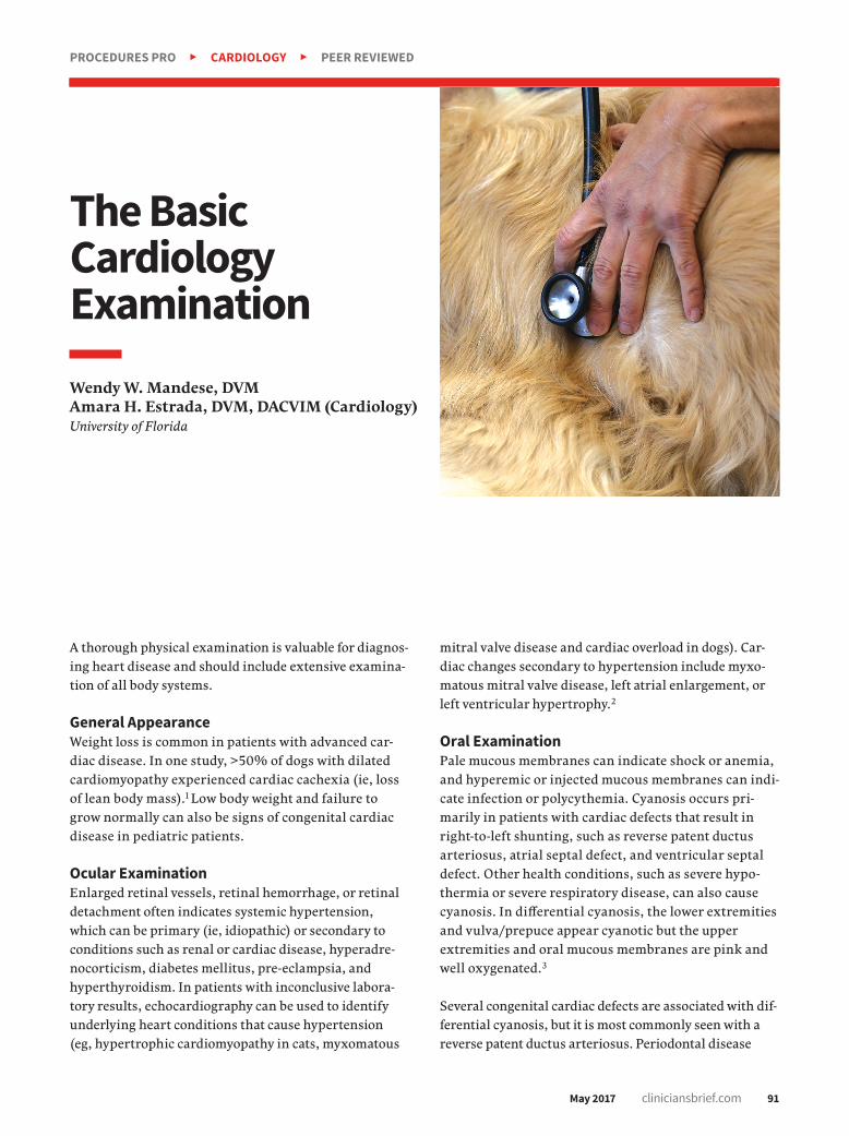
The Basic Cardiology ExaminationWendy W. Mandese, DVMAmara H. Estrada, DVM, DACVIM (Cardiology)University of Florida
PROCEDURES PRO h CARDIOLOGY h PEER REVIEWED
A thorough physical examination is valuable for diagnos-ing heart disease and should include extensive examina-tion of all body systems.
General AppearanceWeight loss is common in patients with advanced car-diac disease. In one study, >50% of dogs with dilated cardiomyopathy experienced cardiac cachexia (ie, loss of lean body mass).1 Low body weight and failure to grow normally can also be signs of congenital cardiac disease in pediatric patients.
Ocular ExaminationEnlarged retinal vessels, retinal hemorrhage, or retinal detachment often indicates systemic hypertension, which can be primary (ie, idiopathic) or secondary to conditions such as renal or cardiac disease, hyperadre-nocorticism, diabetes mellitus, pre-eclampsia, and hyperthyroidism. In patients with inconclusive labora-tory results, echocardiography can be used to identify underlying heart conditions that cause hypertension (eg, hypertrophic cardiomyopathy in cats, myxomatous
mitral valve disease and cardiac overload in dogs). Car-diac changes secondary to hypertension include myxo-matous mitral valve disease, left atrial enlargement, or left ventricular hypertrophy.2
Oral ExaminationPale mucous membranes can indicate shock or anemia, and hyperemic or injected mucous membranes can indi-cate infection or polycythemia. Cyanosis occurs pri-marily in patients with cardiac defects that result in right-to-left shunting, such as reverse patent ductus arteriosus, atrial septal defect, and ventricular septal defect. Other health conditions, such as severe hypo-thermia or severe respiratory disease, can also cause cyanosis. In differential cyanosis, the lower extremities and vulva/prepuce appear cyanotic but the upper extremities and oral mucous membranes are pink and well oxygenated.3
Several congenital cardiac defects are associated with dif-ferential cyanosis, but it is most commonly seen with a reverse patent ductus arteriosus. Periodontal disease
May 2017 cliniciansbrief.com 91
92 cliniciansbrief.com May 2017
should be assessed and noted, as secondary cardiac complications (eg, endocarditis) are possible if severe periodontal disease is not addressed.4
Tracheal PalpationTracheal palpation should be performed on all coughing dogs. Small-breed dogs more prone to myxomatous mitral valve disease are also more prone to a collapsing trachea. In both of these conditions, cough can occur during exercise or excitement5 and may be caused by tracheal disease, even if a murmur is present. Coughing can be heard with tracheitis or tracheobronchitis and can be caused by bacterial or viral infection or environmental allergens.
Respiratory Rate/EffortRespiration should be evaluated when the patient is calm. Clients can be trained to take respiratory rates at home, especially while the patient is sleeping. Several phone apps are available to make it easy for clients to obtain accurate readings. One study suggested that most dogs and cats without cardiac disease or with well controlled heart disease will have a resting respiratory rate of <30 breaths/min at home.6 Increased respiratory effort can indi-cate upper airway disease (effort occurs during inspiration) or lower airway disease (effort occurs during expiration).
Lung SoundsAbnormal lung sounds are most common in patients with primary respiratory disease. Lung sounds also may be abnormal in patients with secondary respiratory conditions such as pulmonary edema and pleural effusion. Aus-cultation of the lungs is not a sensitive means of detecting pulmonary edema or pleural effu-sion in dogs and cats, and many patients have pulmonary edema with no auscultatory abnor-malities other than increased bronchovesicu-lar sounds.7 Patients with severe pulmonary edema resulting in free fluid in the airways are
more likely to have audible crackles; crackles can also be auscultated in patients with pul-monary hypertension, bronchitis, and pneu-monia. Muffled lung sounds can indicate pleural effusion. Wheezes are associated with allergic airway disease, bronchitis, and col-lapsing trachea.
Heart RateStress and anxiety associated with the veteri-nary environment can markedly increase a patient’s heart rate. Waiting for a patient’s ini-tial arrival excitement to subside and asking the client to be present during examination can help the clinician obtain a normal heart rate. The client can also be asked to obtain the patient’s resting heart rate at home. If an arrhythmia is present, the type (eg, tachyar-rhythmia, bradyarrhythmia) should be noted and the nature characterized.
PulsePulse pressure can be decreased or increased or have an altered configuration. Decreased pulse pressure may be seen in patients with dilated cardiomyopathy, aortic or pulmonic stenosis, heart failure, hypovolemia, or shock. Increased pulse pressure can occur because of excitement and/or pain or hyper-trophic cardiomyopathy.8 Dogs with aortic regurgitation commonly have a bounding pulse. Bounding pulses can also be felt in patients with patent ductus arteriosus, severe bradycardia, hyperthyroidism, fever, or anemia.
Alterations in pulse conformation also may occur. Dogs with severe subaortic stenosis can have a weak pulse or a pulse pressure that increases more slowly and peaks later during systole (ie, pulsus parvus et tardus). Conversely, dogs with mitral regurgitation commonly have a brisk pulse that rises more rapidly in systole and lasts a shorter time. Other pulse abnormalities include pulsus par-adoxus and pulse deficits. Pulsus paradoxus is
PROCEDURES PRO h CARDIOLOGY h PEER REVIEWED

May 2017 cliniciansbrief.com 93
an increase in pulse pressure on expiration and a decrease on inspiration. This occurs normally but is exaggerated in cardiac tam-ponade. Pulse deficits can occur with cardiac tachyarrhythmias in which beats occur so rapidly that the ventricle does not have time to fill with an adequate amount of blood before ejection (eg, fast atrial fibrillation, ventricular premature beats).7 Pulse should always be monitored while performing car-diac auscultation to detect pulse deficits.
Blood PressureObtaining a systolic blood pressure using a Doppler is a relatively simple procedure. To obtain the most accurate reading, blood pres-sure should be measured in a quiet, calm envi-ronment with the client present, if possible. Proper technique, including appropriate cuff width (ie, 40% of the circumference of the limb or tail) is integral to obtaining an accu-rate measurement. A minimum of 3 measure-ments should be obtained, and the variability between measurements should be <20%.9
Heart SoundsThe first heart sound (S1) is produced by clos-ing of the mitral and tricuspid valves. S1 is loudest over the mitral valve area and is louder, longer, and lower pitched than the second heart sound (S2). S2 is produced by closing of the pulmonic and aortic valves. S2 is shorter and higher pitched than S1.
The third heart sound (S3) is not usually heard during auscultation of healthy small animals, and its presence indicates myocardial failure. The sound is generated during the period of rapid filling in early diastole when the ventri-cles suddenly resist expansion.
The fourth heart sound (S4) originates from the vibration generated by cardiac structures when the atria contract. It can be a normal finding in giant-breed dogs and large animals or be asso-ciated with advanced hypertrophic cardiomy-
opathy. The presence of an S3 or S4 is referred to as a diastolic gallop. When a diastolic gallop is present, further evaluation is warranted.
Systolic clicks are found in dogs with chronic valvular disease and originate from vibra-tions that occur when chordae tendineae and the mitral leaflets suddenly resist further stretching and protrude into the left atrium.10 Muffled heart sounds may indicate the pres-ence of pericardial or pleural effusion.
ArrhythmiasSinus arrhythmia is common in dogs, espe-cially in patients who are relaxed and have a lower heart rate. The heart rhythm is faster on inspiration and slower on expiration. When the patient becomes more excited or active, the sinus arrhythmia is often no lon-ger heard. Sinus arrhythmia does not always correspond to respiration and can occur with other causes of increased vagal tone (eg, GI disease).11 Pulses should be palpated in con-junction with auscultation to determine if pulse deficits are present. Sinus arrhythmia was previously thought to be uncommon in cats, but a 2009 study determined that relaxed cats in their home environment can have frequent sinus arrhythmia.12
Common abnormal rhythms include: h Premature beats with pulse deficits: Asso-
ciated with atrial and ventricular prema-ture complexes. Can occur in bursts or be sustained
h Irregularly irregular rhythm: Associated with atrial fibrillation. Has been described as “shoes in a dryer”
h Slow arrhythmia with intermittent pauses: Heard in patients with AV block or sinus arrhythmia
h Persistent tachycardia or persistent bra-dycardia: Clients should be informed that an abnormality in heart rate and/or rhythm requires additional testing to determine the cause.
h Murmurs: Caused by turbulence disturbing the normal laminar flow of blood, which most often is caused by dysfunctional valves or septal defects. Can also have physiologic causes because of patient size, athletic abil-ity, or underlying noncardiac disease pro-cesses (eg, fever, anemia)13
Characterization of murmurs is based on several criteria: h Timing in the cycle: A systolic murmur
occurs between S1 and S2 and is common in small animals. A diastolic murmur occurs between S2 of one beat and S1 of the follow-ing beat and is uncommon in small ani-mals. A continuous murmur can be heard throughout the cardiac cycle (Table 1).
h Location: Location or point of maximal intensity (PMI) refers to the valve area at which the murmur is heard loudest. Recog-nition of the PMI can help identify the spe-cific cardiac abnormality (Table 2; Figures 1-5, page 96).
h Intensity • Murmurs are graded on a scale of 1 to 6.
This scale can be subjective and may vary between observers.
– Grade 1/6: Softest murmur audible. May only be heard in a quiet room after listen-ing for several minutes. May not be audi-ble to all observers, and may be transient
– Grade 2/6: Soft but more easily heard. Often focal over one valve only
– Grade 3/6: Prominent and easily heard. May radiate to other areas
– Grade 4/6: Loud and radiates widely. Not accompanied by a palpable thrill
– Grade 5/6: Loud and accompanied by a palpable thrill
– Grade 6/6: Loud, accompanied by a pal-pable thrill, and can be heard with the stethoscope barely touching the thorax
• Innocent murmurs are soft, systolic, heard best at the mitral or aortic valves, and often low-grade (grade 1). They do not radiate. They are often auscultated in young puppies and kittens and usually disappear by 3 to 4 months of age.
• Physiologic murmurs are soft and usually of low intensity (grade 1-2). PMI is at the heart base in the area of the outflow tracts (aortic and pulmonic area). These murmurs typically resolve with resolution of the underlying disease. They may also be heard in otherwise healthy animals that are deep chested and/or athletic. They do not indi-cate any underlying cardiac disorder that can be identified by chest radiography or echocardiography and are common in ani-mals with anemia and occurs as a result of changes in blood viscosity.
h Character: Most murmurs have character-istic sounds on auscultation:7
• Harsh or regurgitant: Mimicked by placing the back of the tongue close to the roof of the mouth and blowing out forcefully. Heard in patients with ventricular septal defects and atrioventricular valve insufficiency
• Blowing: Mimicked by blowing air with
TABLE 1
COMMON PATHOLOGIC CAUSES OF MURMURS BASED ON TIMING7
Murmur Type Pathologic Cause
Systolic murmur Atrioventricular valve regurgitation Left-to-right shunt Increased flow (hyperthyroidism) Aortic or pulmonic stenosis Ventricular septal defect/atrial septal defect (often not audible)
Diastolic murmur Aortic regurgitationPulmonary regurgitationMitral stenosisEndocarditis
Continuous murmur Patent ductus arteriosus
PROCEDURES PRO h CARDIOLOGY h PEER REVIEWED
94 cliniciansbrief.com May 2017

moderate force through slightly parted lips. Often heard in patients with aortic or pulmonic insufficiency
• Machinery: Sounds like the wind blowing through a tunnel. Most often heard in patients with patent ductus arteriosus
• Systolic clicks: High frequency sounds heard over the left apex that may indicate mitral valve disease
• Crescendo-decrescendo: An ejection mur-mur most common in patients with atrial septal defects and aortic or pulmonic stenosis
Heart murmurs are present in approximately one-third of apparently healthy adult cats.14-16 The intensity of these murmurs can vary with
sympathetic tone and usually increases as heart rate increases. The murmur can disap-pear entirely when sympathetic stimulation abates and heart rate slows. A murmur may be audible on initial auscultation and may soften or disappear as the patient relaxes during the examination.
Hypertrophic cardiomyopathy and systolic anterior motion of the mitral valve are the most common diagnoses in cats with mur-murs caused by heart disease.14-16 Because benign murmurs and murmurs caused by car-diac disease are dynamic and audibly indis-tinguishable, further evaluation is warranted when a murmur is detected.17
Abdominal PalpationAscites and/or liver enlargement may be present in patients with right-sided heart failure.
TABLE 2
LOCALIZATION OF MURMURS IN COMMON CARDIAC LESIONS7
Condition Left Base Right Base Left Apex Right Apex Right Sternum
Mitral regurgitation Systolic PMI (+/- Systolic referred)
Tricuspid regurgitation Systolic PMI
Aortic regurgitation Diastolic PMI +/- Diastolic (Diastolic referred)
Aortic stenosis Systolic PMI +/- Systolic +/- Systolic
Pulmonic stenosis Systolic PMI
Ventricular septal defect +/- Systolic (often not heard) Systolic PMI
Atrial septal defect +/- Systolic (often not heard)
Tetralogy of Fallot Systolic PMI +/- Systolic
Patent ductus arteriosus Continuous PMI
Visit cliniciansbrief.com/cardiac-library to hear a collection of heart sounds associated with clinical diagnoses.
PMI = point of maximal intensity
May 2017 cliniciansbrief.com 95

Place the stethoscope over the left cardiac apex (location of ventricles) and base (location of pul-monary and aortic outflow tracts), right cardiac apex and base, the length of the sternum, and the thoracic inlet.
STEP-BY-STEPCARDIAC AUSCULTATION
STEP 1
Cardiac auscultation is best performed in a quiet room with a standing patient. In anxious dogs, pant-ing sounds can be mistaken for murmurs. Manually close the dog’s mouth for a short time during auscul-tation and give panting breaks as needed. Purring in cats can also be mistaken for a murmur. Distract the cat with a toy or change the position or location of the patient to stop purring.
d FIGURE 1 Position of cardiac apex and base in a left canine thorax. Illustrations by Ally Mandese and used with permission
d FIGURE 2 Canine right thorax. A = aortic valve, RA = right atrium, RV = right ventricle, T = tricuspid valve
d FIGURE 3 Postmortem photograph of the right side of the heart in the thoracic cavity. A = aortic valve, RA = right atrium, RV = right ventricle, T = tricuspid valve. Photo courtesy of Dr. Lisa Farrina, University of Florida College of Veterinary Medicine Department of Pathology
d FIGURE 4 Canine left thorax. A = aortic valve, LA = left atrium, LV = left ventricle, M = mitral valve, P = pulmonic valve
d FIGURE 5 Postmortem photograph of the left side of the heart in the thoracic cavity. A = aortic valve, LA = left atrium, M = mitral valve, P = pulmonic valve
PROCEDURES PRO h CARDIOLOGY h PEER REVIEWED
96 cliniciansbrief.com May 2017

Auscultate each heart valve at its PMI (Table 3).
STEP 2 ConclusionCardiac abnormalities can be identified during a basic cardiac examination that includes obtaining a complete history and performing a thorough physical examination and simple diagnostics. Creating a list of dif-ferential diagnoses based on the information gathered during the cardiac examination can help the clinician make informed decisions about the appropriate next diagnostic or therapeutic step. n
TABLE 3
STETHOSCOPE PLACEMENT FOR CARDIAC EXAMINATION
Valve Point of Maximal Intensity
Mitral Left 5th intercostal space at costochondral junction
Tricuspid Between right intercostal spaces 3-5 just above costochondral junction
Pulmonic Between left intercostal spaces 2-4 just above sternum
Aortic Within left intercostal space 4-5 just above costrochondral junction
PMI = point of maximal intensity
Purchase Requirements (single invoice) FREE UNITS*
Purchase any of these Bayer Supplements.
No mixing and matching.
9 units 3
9 units 3
9 units
9 units
9 units
6
6
6
Volume Discount Pricing also eligible: Receive 15% OFF when 12 or more units of the SAME SKU are purchased on a single invoice.**
*Per ship-to account
Purchase Requirements (single invoice)Purchase Requirements (single invoice)
See page 98 for references.

98 cliniciansbrief.com May 2017
PROCEDURES PRO h CARDIOLOGY h CONTINUED FROM PAGE 97
TALKING POINTS h CONTINUED FROM PAGE 85
References 1. Freeman LM, Rush JE, Kehayias JJ, et al. Nutrition alterations
and the eff ect of fish oil supplementation in dogs with heart failure. J Vet Intern Med. 1998;12(6):440-448.
2. Davies C, Shell L. Systemic hypertension. In: Davies C, Shell L, eds. Common Small Animal Medical Diagnoses: An Algorithmic Approach. Saunders; 2002. Accessed on Veterinary Information Network, April 18, 2017.
3. Kittleson MD. Diagnosis. In: Kittleson MD, Kienle RD, eds. Small Animal Cardiovascular Medicine. Maryland Heights, MO: Mosby Elsevier; 2005. Accessed on Veterinary Information Network, April 18, 2017.
4. Glickman LT, Glickman NW, Moore GE, Goldstein GS, Lewis HB. Evaluation of the risk of endocarditis and other cardiovascular events on the basis of the severity of periodontal disease in dogs. J Am Vet Med Assoc. 2009;234(4):486-494.
5. Tappin SW. Canine tracheal collapse. J Small Anim Pract. 2016;57(1):9-17.
6. Porciello F, Rishniw M, Ljungvall I, Ferasin L, Haggstrom J, Ohad DG. Sleeping and resting respiratory rates in dogs and cats with medically-controlled left -sided congestive heart failure. Vet J. 2016;207:164-168.
7. Kittleson MD, Kienle RD. Physical exam. In: Kittleson MD, Kienle RD, eds. Small Animal Cardiovascular Medicine. Maryland Heights, MO: Mosby Elsevier; 2005. Accessed on Veterinary Information Network, April 18, 2017.
8. Ware WA. Clinical manifestations of cardiac disease. In: Nelson RW, Couto CG, eds. Small Animal Internal Medicine. 5th ed. St. Louis, MO: Elsevier; 2014:6.
9. Ferasin L. How to measure blood pressure. Presented at: British Small Animal Veterinary Congress; 2011. Accessed on Veterinary Information Network, April 18, 2017.
10. Kvart C, Häggström. Heart sounds and murmurs in dogs and cats. In: Kvart C, Häggström J. Cardiac Auscultation & Phonocardiography in Dogs, Horses and Cats; 2002. Accessed on Veterinary Information Network, April 18, 2017.
11. Kittleson MD. Specific arrhythmias. In: Kittleson MD, Kienle RD, eds. Small Animal Cardiovascular Medicine. Maryland Heights, MO: Mosby Elsevier; 2005. Accessed on Veterinary Information Network, April 18, 2017.
12. Hanås S, Tidholm A, Egenvall A, Holst BS. Twenty-four hour Holter monitoring of unsedated healthy cats in the home environment. J Vet Cardiol. 2009;11(1):17-22.
13. Kvart C, Häggström J. Heart sounds and murmurs in dogs and cats: physiological flow murmurs. In: Kvart C, Häggström J. Cardiac Auscultation & Phonocardiography in Dogs, Horses and Cats; 2002. Accessed on Veterinary Information Network, April 18, 2017.
14. Paige CF, Abbott JA, Elvinger F, Pyle RL. Prevalence of cardiomyopathy in apparently healthy cats. J Am Vet Med Assoc. 2009;234(11):1398-1403.
15. Wagner T, Fuentes VL, Payne JR, McDermott N, Brodbelt D. Comparison of auscultatory and echocardiographic findings in healthy adult cats. J Vet Cardiol. 2010;12(3):171-182.
16. Nakumura RK, Rishniw M, King MK, Sammarco CD. Prevalence of echocardiographic evidence of cardiac disease in apparently healthy cats with murmurs. J Feline Med Surg. 2011;13(4):266-271.
17. Côté E, Manning AM, Emerson D, Laste NJ, Malakoff RL, Harpster NK. Assessment of the prevalence of heart murmurs in overtly healthy cats. J Am Vet Med Assoc. 2004;225(3):384-388.
References1. Freeman LM, Chandler ML, Hamper BA, Weeth LP. Current
knowledge about the risks and benefits of raw meat-based diets for dogs and cats. J Vet Intern Med. 2013;243(11):1549-1558.
2. Weese JS. Worms & Germs Blog. http://www.wormsandgermsblog.com
3. Chengappa MM, Staats J, Oberst RD, Gabbert NH, McVey S. Prevalence of Salmonella in raw meat used in diets of racing greyhounds. J Vet Diagn Invest. 1993;5(3):372-377.
4. Joff DJ, Schlesinger DP. Preliminary assessment of the risk of Salmonella infection in dogs fed raw chicken diets. Can Vet J. 2002;43(6):441-442.
5. Finley R, Reid-Smith R, Ribble C, Popa M, Vandermeer M, Aramini J. The occurrence and antimicrobial susceptibility of salmonellae isolated from commercially available canine raw food diets in three Canadian cities. Zoonoses Public Health. 2008;55(8-10):462-469.
6. Nemser SM, Doran T, Grabenstein M, et al. Investigation of Listeria, Salmonella, and Toxigenic Escherichia coli in various pet foods. Foodborne Pathog Dis. 2014;11(9):706-709.
7. Strohmeyer RA, Morley PS, Hyatt DR, Dargatz DA, Scorza AV, Lappin MR. Evaluation of bacterial and protozoal contamination of commercially available raw meat diets for dogs. J Am Vet Med Assoc. 2006;228(4):537-542.
8. Weese JS, Rousseau J, Arroyo L. Bacteriological evaluation of commercial canine and feline raw diets. Can Vet J. 2005;46(6):513-516.
9. Bojanić K, Midwinter AC, Marshall JC, Rogers LE, Biggs PJ, Acke E. Isolation of Campylobacter spp. from client-owned
dogs and cats, and retail raw meat pet food in the Manawatu, New Zealand. Zoonoses Public Health. 2016. doi: 10.1111/zph.12323
10. Stiver L, Frazier KS, Mauel MJ, Styer EL. Septicemic salmonellosis in two cats fed a raw meat diet. J Am Anim Hosp Assoc. 2003;39(6):538-542.
11. Morley PS, Strohmeyer RA, Tankson JD, et al. Evaluation of the association between feeding raw meat and Salmonella enterica infections at a Greyhound breeding facility. J Am Vet Med Assoc. 2006;228(10):1524-1532.
12. Centers for Disease Control and Prevention. Human salmonellosis associated with animal-derived pet treats—United States and Canada, 2005. MMWR Morb Mortal Wkly Rep. 2006;55(25):702-705.
13. Finley R, Reid-Smith R, Weese JS. Human health implications of Salmonella-contaminated natural pet treats and raw pet food. Clin Infect Dis. 2006;42(5):686-691.
14. Pitout JD, Reisbig MD, Mulvey M, et al. Association between handling of pet treats and infection with Salmonella enterica serotype newport expressing the AmpC beta-lactamase, CMY-2. J Clin Microbiol. 2003;41(10):4578-4582.
15. Cavallo SJ, Daly ER, Seiferth J, et al. Human outbreak of Salmonella typhimurium associated with exposure to locally made chicken jerky pet treats, New Hampshire, 2013. Foodborne Pathog Dis. 2015;12(5):441-446.
16. Fauth E, Freeman LM, Cornjeo L, Markovich JE, Janecko N, Weese JS. Salmonella bacteriuria in a cat fed a Salmonella-contaminated diet. J Am Vet Med Assoc. 2015;247(5):525-530.