The Autonomic Nervous System 1. The ANS and Visceral Sensory Neurons 2.
58
The Autonomic Nervous System 1
-
Upload
alanna-hilger -
Category
Documents
-
view
231 -
download
2
Transcript of The Autonomic Nervous System 1. The ANS and Visceral Sensory Neurons 2.

The Autonomic Nervous System
1
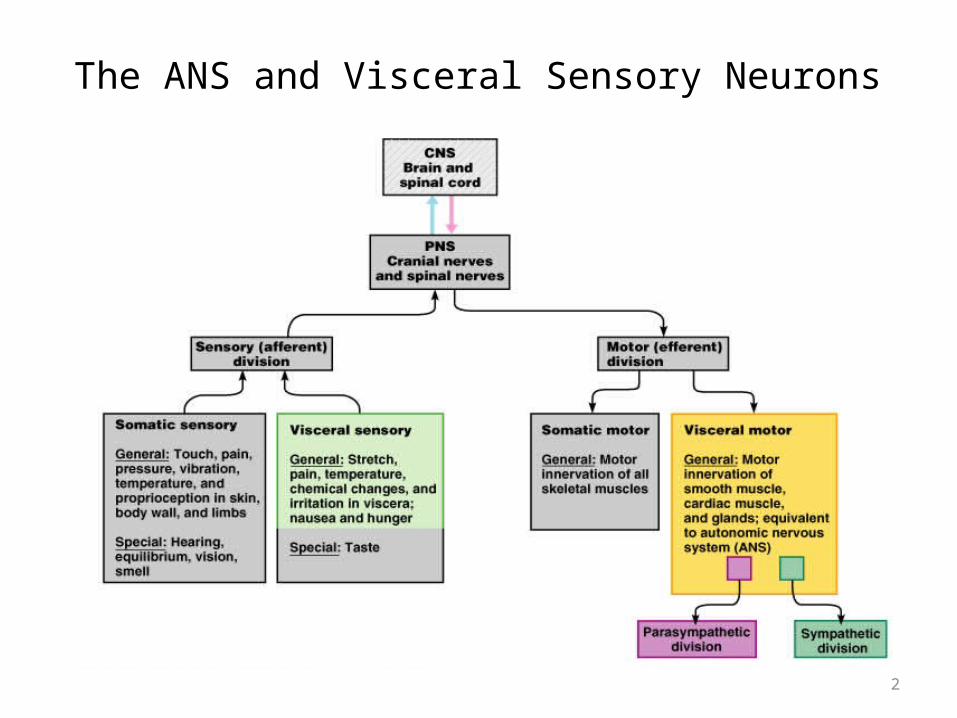
The ANS and Visceral Sensory Neurons
2

Sympathetic Nervous System
3

Anatomy of Autonomic Motor Pathways
• Preganglionic neuron• Postganglionic neuron
• Two divisions: • Sympathetic• Parasympathetic
4

Structure of the Sympathetic Division
5
Copyright © 2005 Pearson Education, Inc., publishing as Benjamin Cummings
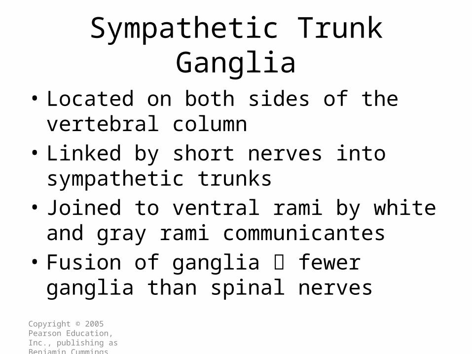
Sympathetic Trunk Ganglia
• Located on both sides of the vertebral column• Linked by short nerves into sympathetic trunks• Joined to ventral rami by white and gray rami
communicantes • Fusion of ganglia fewer ganglia than spinal
nerves
Copyright © 2005 Pearson Education, Inc., publishing as Benjamin Cummings
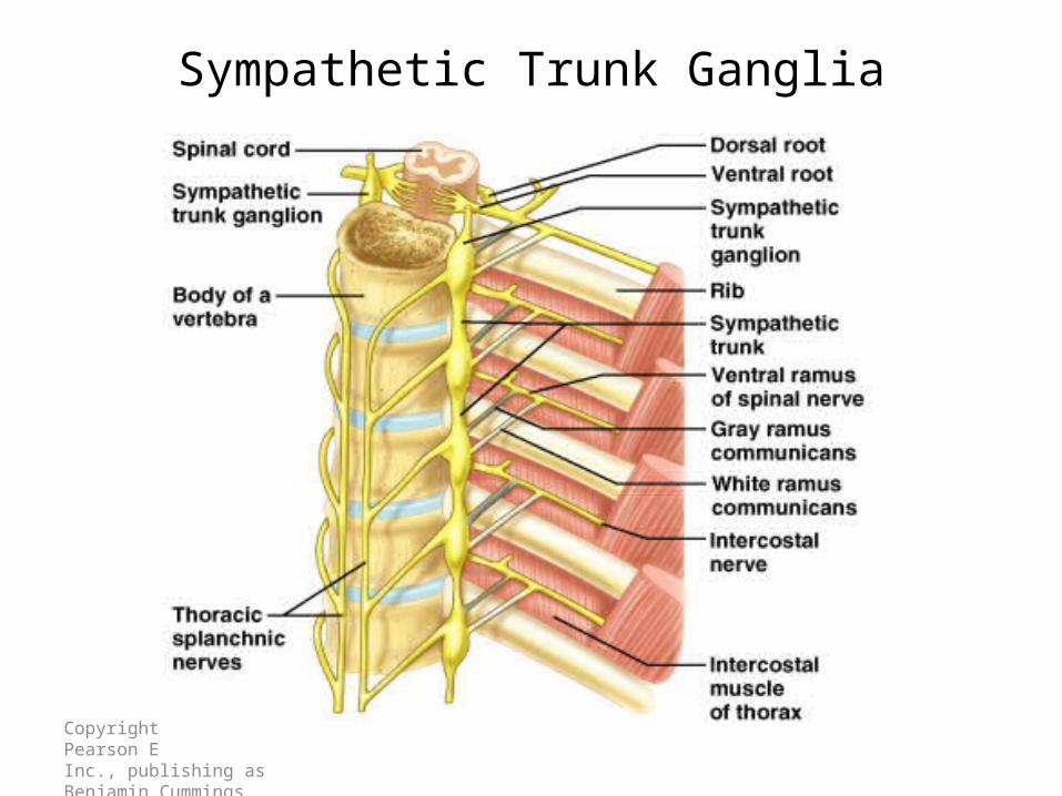
Sympathetic Trunk Ganglia

Copyright © 2005 Pearson Education, Inc., publishing as Benjamin Cummings
Prevertebral Ganglia
• Unpaired, not segmentally arranged• Occur only in abdomen and pelvis• Lie anterior to the vertebral column• Main ganglia
– Celiac, superior mesenteric, inferior mesenteric, inferior hypogastric ganglia

Sympathetic Division
• A single sympathetic preganglionic fiber has many axon collaterals and may synapse with 20 or more postganglionic neurons.
• The postganglionic axons typically terminate in several visceral effectors and therefore the effects of sympathetic stimulation are more widespread than the effects of parasympathetic stimulation.
9
Copyright © 2005 Pearson Education, Inc., publishing as Benjamin Cummings
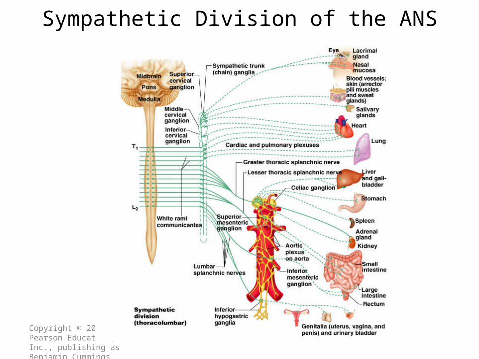
Sympathetic Division of the ANS

Copyright © 2005 Pearson Education, Inc., publishing as Benjamin Cummings
Sympathetic Pathways to Periphery
Figure 15.9

Copyright © 2005 Pearson Education, Inc., publishing as Benjamin Cummings
Sympathetic Pathways to the Head

Sympathetic Division
• Thoracolumbar division- Preganglionic neurons originate from the thoracic and lumbar levels of the spinal cord (T1-L2).
• Sympathetic ganglia:Sympathetic trunk (vertebral chain) ganglia.Prevertebral (collateral) ganglia: celiac, superior mesenteric, inferior mesenteric, aorticorenal and renal.
13

Copyright © 2005 Pearson Education, Inc., publishing as Benjamin Cummings
Sympathetic Pathways to Thoracic Organs

Copyright © 2005 Pearson Education, Inc., publishing as Benjamin Cummings
Sympathetic Pathways to the Abdominal Organs
Copyright © 2005 Pearson Education, Inc., publishing as Benjamin Cummings
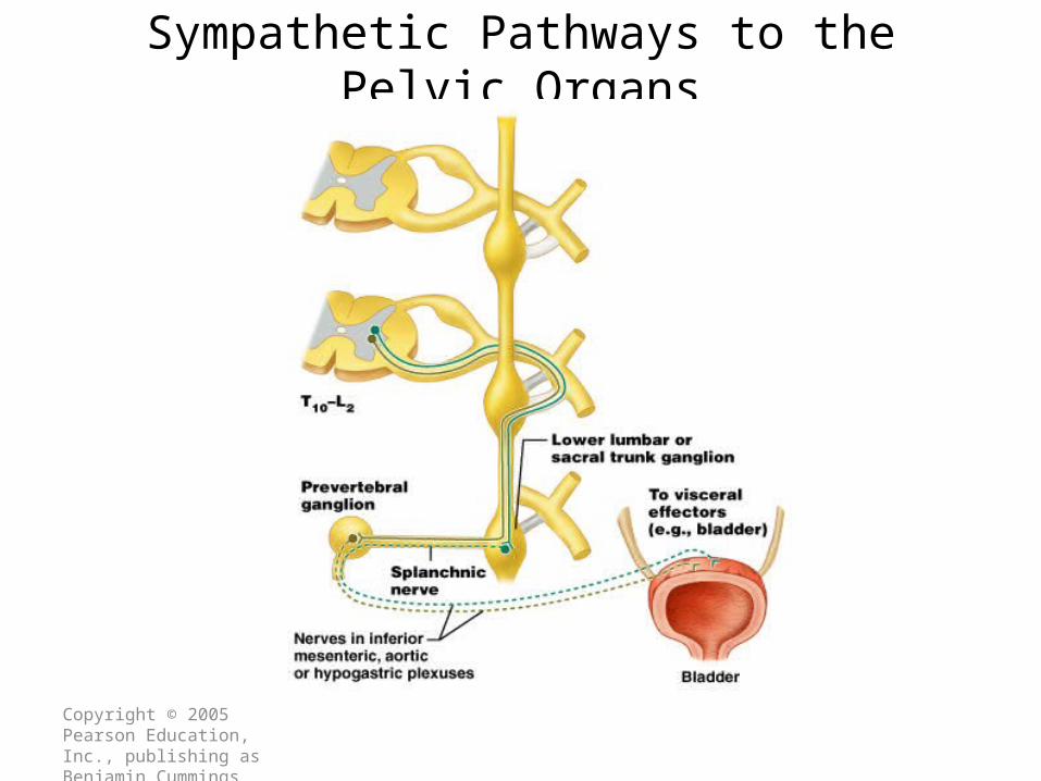
Sympathetic Pathways to the Pelvic Organs

Copyright © 2005 Pearson Education, Inc., publishing as Benjamin Cummings
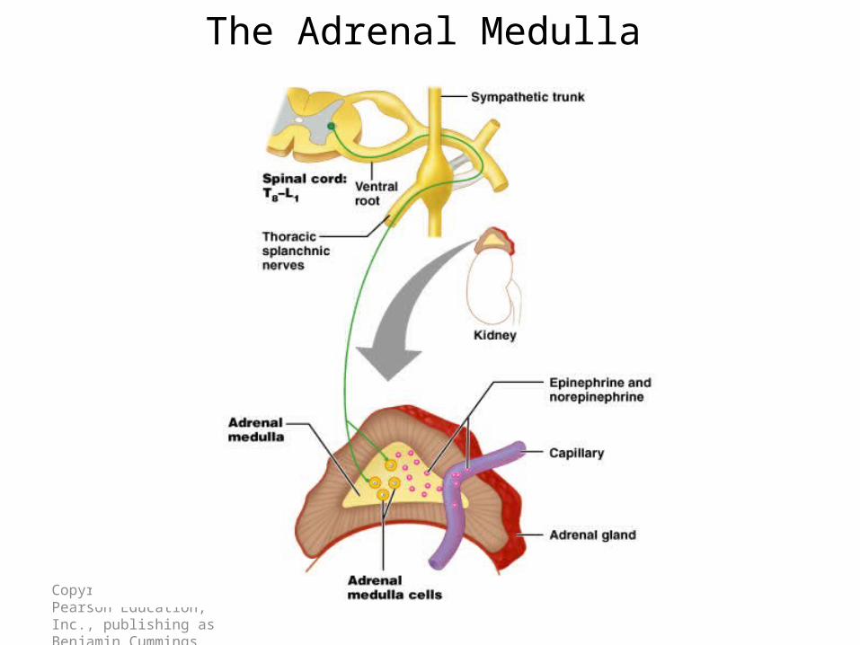
The Role of the Adrenal Medulla in the Sympathetic Division
• Major organ of the sympathetic nervous system
• Secretes great quantities epinephrine (a little norepinephrine)
• Stimulated to secrete by preganglionic sympathetic fibers
Copyright © 2005 Pearson Education, Inc., publishing as Benjamin Cummings
The Adrenal Medulla

Postganglionic Neurons in the Sympathetic Division
• An axon may synapse with postganglionic neurons in the ganglion it first reaches or
• Sympathetic chains or• An axon may continue, without synapsing, through
the sympathetic trunk ganglion to end at a prevertebral ganglion and synapse with postganglionic neurons there or
• An axon may pass through the sympathetic trunk ganglion and a prevertebral ganglion and then to the adrenal medulla.
19

Postganglionic neurons in the Sympathetic Division
20

Parasympathetic Nervous System
21
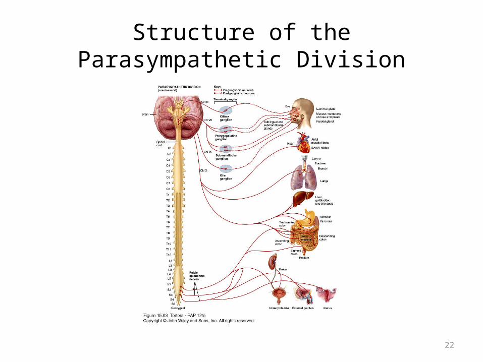
Structure of the Parasympathetic Division
22

Copyright © 2005 Pearson Education, Inc., publishing as Benjamin Cummings
The Parasympathetic Division

Copyright © 2005 Pearson Education, Inc., publishing as Benjamin Cummings
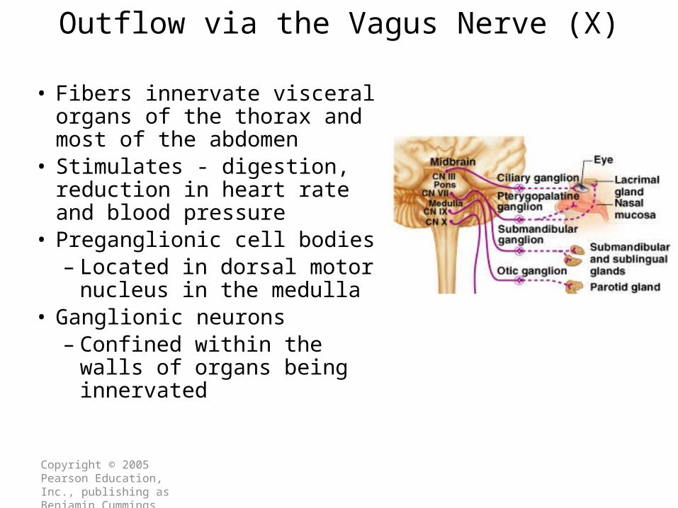
Cranial Outflow
• Preganglionic fibers run via:– Oculomotor nerve (III)– Facial nerve (VII)– Glossopharyngeal nerve (IX)– Vagus nerve (X)
• Cell bodies located in cranial nerve nuclei in the brain stem
Copyright © 2005 Pearson Education, Inc., publishing as Benjamin Cummings
Outflow via the Vagus Nerve (X)
• Fibers innervate visceral organs of the thorax and most of the abdomen
• Stimulates - digestion, reduction in heart rate and blood pressure
• Preganglionic cell bodies– Located in dorsal motor
nucleus in the medulla• Ganglionic neurons
– Confined within the walls of organs being innervated

Copyright © 2005 Pearson Education, Inc., publishing as Benjamin Cummings
Parasympathetic Nervous System: Sacral Outflow
• Emerges from S2-S4
• Innervates organs of the pelvis and lower abdomen
• Preganglionic cell bodies– Located in visceral motor region of spinal gray
matter• Form splanchnic nerves

Parasympathetic Division
• Craniosacral division: Preganglionic neurons originate from the cranial nerves III, VII, IX and X and sacral spinal nerves S2-S4.
• Parasympathetic ganglia: terminal ganglia.• Presynaptic neuron usually synapses with 4-5
postsynaptic neurons all of which supply a single visceral effector.
27

Putting the Two Together
To Compare and Functional Cooperation - Reciprocity
28

Comparison of Somatic and Autonomic Nervous Systems
Copyright © 2003 Pearson Education, Inc. publishing as Benjamin Cummings

Copyright © 2005 Pearson Education, Inc., publishing as Benjamin Cummings
Anatomical Differences in Sympathetic and Parasympathetic Divisions (Recall)
• Issue from different regions of the CNS– Sympathetic – also
called the thoracolumbar division
– Parasympathetic – also called the craniosacral division

Copyright © 2005 Pearson Education, Inc., publishing as Benjamin Cummings
Anatomical Differences in Sympatheticand Parasympathetic Divisions
• Length of postganglionic fibers– Sympathetic – long postganglionic fibers– Parasympathetic – short postganglionic fibers
• Branching of axons– Sympathetic axons – highly branched
• Influences many organs
– Parasympathetic axons – few branches • Localized effect

Copyright © 2005 Pearson Education, Inc., publishing as Benjamin Cummings
Anatomical Differences in Sympatheticand Parasympathetic Divisions

Copyright © 2005 Pearson Education, Inc., publishing as Benjamin Cummings
Anatomical Differences in Sympatheticand Parasympathetic Divisions

Copyright © 2005 Pearson Education, Inc., publishing as Benjamin Cummings
Neurotransmitters of Autonomic Nervous System
• Neurotransmitter released by preganglionic axons– Acetylcholine for both branches (cholinergic)
• Neurotransmitter released by postganglionic axons– Sympathetic – most release norepinephrine
(adrenergic)– Parasympathetic – release acetylcholine

Cholinergic and Adrenergic Neurons in the Autonomic Nervous System
35
Cholinergic Neurons
Cholinergic neurons → acetylcholine (ACh).Cholinergic neurons include-1. All sympathetic and parasympathetic
preganglionic neurons.2. Sympathetic postganglionic neurons that
innervate most sweat glands.3. All parasympathetic postganglionic neurons.
36

Cholinergic Receptors
Cholinergic receptors release acetylcholine.
• Two types: Nicotinic receptorsMuscarinic receptors
37

Adrenergic Neurons and Receptors
• Release norepinephrine (noradrenaline).• Most sympathetic postganglionic neurons are
adrenergic.• Two types of receptors: Alpha receptors-Beta receptors-
38

Comparison of Somatic and Autonomic Nervous Systems
39

Comparison of Somatic and Autonomic Nervous Systems
40

Autonomic Plexuses in the Thorax, Abdomen and Pelvis
41

Autonomic Plexuses
• A network of sympathetic and parasympathetic axons.
• Cardiac plexus- heart.• Pulmonary plexus- the bronchial tree.• Celiac plexus- largest. Supplies the stomach,
spleen, pancreas, liver, gallbladder, and adrenal medullae.
42

Autonomic Plexuses (Cont’d)..
• Superior mesenteric plexus- small intestine and proximal colon.
• Inferior mesenteric plexus- distal colon and rectum.
• Hypogastric plexus- urinary bladder and genital organs.
• Renal plexus- kidneys and ureters.
43

Pathway from Spinal Cord to Sympathetic Trunk Ganglia:
• Preganglionic axons → anterior root of a spinal nerve → white ramus → sympathetic trunk ganglion.
• White rami communicantes: structures containing sympathetic preganglionic axons that connect the anterior ramus of the spinal nerve with the ganglia of the sympathetic trunk.
44

Organization of Sympathetic Trunk Ganglia
• Sympathetic trunk ganglia: 3 cervical, 11 or 12 thoracic, 4 or 5 lumbar, 4 or 5 sacral and 1 coccygeal.
• Postganglionic neurons from the superior cervical region-head and heart.middle cervical ganglion and the inferior cervical ganglion-heart.
• Thoracic sympathetic trunk- heart, lungs, and bronchi.
45

Pathways from Sympathetic Trunk Ganglia to Visceral Effectors
• Axons leave the sympathetic trunk in 4 possible ways:- spinal nerves- cephalic periarterial nerves- sympathetic nerves- splanchnic nerves
46

Spinal nerves
• Gray ramus: Axons of some postganglionic neurons leave the sympathetic trunk by entering a short pathway called a gray ramus and merge with the anterior ramus of a spinal nerve.
• Gray rami communicantes: structures containing sympathetic postganglionic axons that connect the ganglia of the sympathetic trunk to spinal nerves.
47

Cephalic Periarterial Nerves
• Some sympathetic preganglionic neurons that enter the sympathetic trunk ascend to the superior cervical ganglion where they synapse with postganglionic neurons. Some of these leave the sympathetic trunk by forming cephalic periarterial nerves.
• Serve visceral effectors in the skin of the face and head.
48

Sympathetic Nerves
• Some axons of the postganglionic neurons leave the trunk by forming sympathetic nerves.
• Innervate the heart and lungs.
49
Splanchnic Nerves continued..
• Some sympathetic preganglionic axons pass through the sympathetic trunk without terminating in it. Beyond the trunk they form nerves called splanchnic nerves which extend to prevertebral ganglia.
• T5-T9 or T10- Greater splanchnic nerve.• T10-T11- Lesser splanchnic nerve.• L1-L4- Lumbar splanchnic nerve.
50
Splanchnic Nerves to the Adrenal Medulla
• Some sympathetic preganglionic axons pass, without synapsing, through the sympathetic trunk, greater splanchnic nerves and celiac ganglion into the adrenal medulla (modified sympathetic ganglia).
• Release hormones into blood- 80% epinephrine, 20% norepinephrine.
51

Cranial Parasympathetic Outflow
The cranial outflow has four pairs of ganglia and are associated with the vagus nerve.1. Ciliary ganglia-2. Pterygopalatine ganglia-3. Submandibular ganglia-4. Otic ganglia-
Vagus nerve carries nearly 80% of the total craniosacral flow.
52

Sacral Parasympathetic Outflow
• Consists of S2-S4.• Pelvic splanchnic nerves
53

Physiology of the ANS
• Autonomic tone- a balance between the sympathetic and parasympathetic activity.
• Regulated by the hypothalamus.
54

Sympathetic Responses
• Stress ↑ sympathetic system ↑ fight-or-flight response.
• ↑ production of ATP.• Dilation of the pupils.• ↑ heart rate and blood pressure.• Dilation of the airways.• Constriction of blood vessels that supply the
kidneys and gastrointestinal tract.
55
Sympathetic Responses continued..
• ↑ blood supply to the skeletal muscles, cardiac muscle, liver and adipose tissue
• ↑ glycogenolysis ↑ blood glucose.• ↑ lipolysis.
56

Parasympathetic Responses
• Rest-and-digest response.• Conserve and restore body energy.• ↑ digestive and urinary function.• ↓ body functions that support physical
activity.
57

Integration and Control of Autonomic Functions
• Direct innervation- brain stem and spinal cord.• Hypothalamus is the major control and
integration center of the ANS.• It receives input from the limbic system.
58