The Autographa californica Multicapsid Nucleopolyhedrovirus GP64 Protein
15
JOURNAL OF VIROLOGY, May 2009, p. 4447–4461 Vol. 83, No. 9 0022-538X/09/$08.000 doi:10.1128/JVI.02252-08 Copyright © 2009, American Society for Microbiology. All Rights Reserved. The Autographa californica Multicapsid Nucleopolyhedrovirus GP64 Protein: Analysis of Transmembrane Domain Length and Sequence Requirements Zhaofei Li and Gary W. Blissard* Boyce Thompson Institute, Cornell University, Ithaca, New York 14853 Received 27 October 2008/Accepted 12 February 2009 GP64, the major envelope glycoprotein of the Autographa californica multicapsid nucleopolyhedrovirus budded virion, is important for host cell receptor binding and mediates low-pH-triggered membrane fusion during entry by endocytosis. Previous transmembrane (TM) domain replacement studies showed that the TM domain serves a critical role in GP64 function. To extend the prior studies and examine specific sequence requirements of the TM domain, we generated a variety of GP64 TM domain mutations. The mutations included 4- to 8-amino-acid deletions, as well as single and multiple point mutations. While most TM domain deletion constructs remained fusion competent, those containing deletions of eight amino acids from the C terminus did not mediate detectable fusion. The addition of a hydrophobic amino acid (A, L, or V) to the C terminus of construct C8 (a construct that contains a TM domain deletion of eight amino acids from the C terminus) restored fusion activity. These data suggest that the membrane fusion function of GP64 is dependent on a critical length of the hydrophobic TM domain. All GP64 proteins with a truncated TM domain mediated detectable virion budding with dramatically lower levels of efficiency than wild-type GP64. The effects of deletions of various lengths and positions in the TM domain were also examined for their effects on viral infectivity. Further analysis of the TM domain by single amino acid substitutions and 3-alanine scanning mutations identified important but not essential amino acid positions. These studies showed that amino acids at positions 485 to 487 and 503 to 505 are important for cell surface expression of GP64, while amino acids at positions 483 to 484 and 494 to 496 are important for virus budding. Overall, our results show that specific features and amino acid sequences, particularly the length of the hydrophobic TM domain, play critical roles in membrane anchoring, membrane fusion, virus budding, and infectivity. In typical infections of eukaryotic cells by enveloped viruses, viral entry is mediated by the fusion of viral and cellular mem- branes in a process that is directed by membrane-associated viral fusion proteins. In the best models of membrane fusion, two hydrophobic domains of the viral fusion protein are criti- cally important in fusion: the fusion peptide and the transmem- brane (TM) domain. The fusion peptide is a hydrophobic do- main that inserts into the target cellular membrane, thereby attaching the fusion protein to the target membrane (14). By insertion of the fusion peptide into the target membrane and anchoring of the envelope protein in the viral envelope via the TM domain, a bridge is formed between the two membranes. Subsequent structural rearrangements in the envelope fusion protein bring the viral and cellular membranes into close prox- imity and culminate in the merger of two bilayers and the subsequent opening of a fusion pore. The TM domain may serve several roles in this overall process. In addition to an- choring the envelope protein in the membrane, the TM do- main may play a crucial role in the transition from the initial merger of the outer leaflets of the two membranes (hemifu- sion) to complete membrane merger and pore formation. Ev- idence of a more-direct role of the TM domain in this process comes from studies in which the proteinaceous TM domains of viral fusion proteins were replaced by lipid (glycosylphosphati- dylinositol [GPI]) anchors or protein sequences that altered their structures. Such modifications of the TM domain may lead to partial or full arrest of fusion at the hemifusion step (22). In addition, it was recently proposed that a direct inter- action between the fusion and TM peptides of hemagglutinin (HA) may be required to open the fusion pore (50). The virus Autographa californica multicapsid nucleopolyhe- drovirus (AcMNPV) is a member of the group I NPVs within the Alphabaculovirus genus and is the type species for the family Baculoviridae (17). Budded virions of AcMNPV appear to enter cells via a clathrin-mediated, low-pH-dependent en- docytic pathway (25). During entry by endocytosis, the major envelope glycoprotein GP64 mediates low-pH-triggered mem- brane fusion (5). The GP64 proteins are very highly conserved in the group I NPVs and appear to be closely similar in struc- ture and function only to GP75 proteins from thogotoviruses, a subgroup of the Orthomyxoviridae. Recent structural studies also indicate that GP64 should be classified as a class III fusion protein, along with proteins such as vesicular stomatitis virus glycoprotein (VSV G) and herpesvirus gB (21). GP64 is a type I integral membrane protein that is present on the infected cell surface and in the virion as a disulfide-linked homotrimer (38). GP64 has host cell receptor-binding activity (16), and a domain associated with receptor binding was recently mapped to an N-terminal region of the ectodomain (60). GP64 is both nec- essary and sufficient for mediating pH-dependent membrane fusion during viral entry (5, 55, 61). In addition to its essential * Corresponding author. Mailing address: Boyce Thompson Institute at Cornell University, Tower Road, Ithaca, NY 14853-1801. Phone: (607) 254-1366. Fax: (607) 254-1242. E-mail: [email protected]. Published ahead of print on 25 February 2009. 4447 Downloaded from https://journals.asm.org/journal/jvi on 21 November 2021 by 14.47.44.157.
Transcript of The Autographa californica Multicapsid Nucleopolyhedrovirus GP64 Protein

JOURNAL OF VIROLOGY, May 2009, p. 4447–4461 Vol. 83, No. 90022-538X/09/$08.00�0 doi:10.1128/JVI.02252-08Copyright © 2009, American Society for Microbiology. All Rights Reserved.
The Autographa californica Multicapsid NucleopolyhedrovirusGP64 Protein: Analysis of Transmembrane Domain Length
and Sequence Requirements�
Zhaofei Li and Gary W. Blissard*Boyce Thompson Institute, Cornell University, Ithaca, New York 14853
Received 27 October 2008/Accepted 12 February 2009
GP64, the major envelope glycoprotein of the Autographa californica multicapsid nucleopolyhedrovirusbudded virion, is important for host cell receptor binding and mediates low-pH-triggered membrane fusionduring entry by endocytosis. Previous transmembrane (TM) domain replacement studies showed that the TMdomain serves a critical role in GP64 function. To extend the prior studies and examine specific sequencerequirements of the TM domain, we generated a variety of GP64 TM domain mutations. The mutationsincluded 4- to 8-amino-acid deletions, as well as single and multiple point mutations. While most TMdomain deletion constructs remained fusion competent, those containing deletions of eight amino acids fromthe C terminus did not mediate detectable fusion. The addition of a hydrophobic amino acid (A, L, or V) to theC terminus of construct C8 (a construct that contains a TM domain deletion of eight amino acids from the Cterminus) restored fusion activity. These data suggest that the membrane fusion function of GP64 is dependenton a critical length of the hydrophobic TM domain. All GP64 proteins with a truncated TM domain mediateddetectable virion budding with dramatically lower levels of efficiency than wild-type GP64. The effects ofdeletions of various lengths and positions in the TM domain were also examined for their effects on viralinfectivity. Further analysis of the TM domain by single amino acid substitutions and 3-alanine scanningmutations identified important but not essential amino acid positions. These studies showed that amino acidsat positions 485 to 487 and 503 to 505 are important for cell surface expression of GP64, while amino acids atpositions 483 to 484 and 494 to 496 are important for virus budding. Overall, our results show that specificfeatures and amino acid sequences, particularly the length of the hydrophobic TM domain, play critical rolesin membrane anchoring, membrane fusion, virus budding, and infectivity.
In typical infections of eukaryotic cells by enveloped viruses,viral entry is mediated by the fusion of viral and cellular mem-branes in a process that is directed by membrane-associatedviral fusion proteins. In the best models of membrane fusion,two hydrophobic domains of the viral fusion protein are criti-cally important in fusion: the fusion peptide and the transmem-brane (TM) domain. The fusion peptide is a hydrophobic do-main that inserts into the target cellular membrane, therebyattaching the fusion protein to the target membrane (14). Byinsertion of the fusion peptide into the target membrane andanchoring of the envelope protein in the viral envelope via theTM domain, a bridge is formed between the two membranes.Subsequent structural rearrangements in the envelope fusionprotein bring the viral and cellular membranes into close prox-imity and culminate in the merger of two bilayers and thesubsequent opening of a fusion pore. The TM domain mayserve several roles in this overall process. In addition to an-choring the envelope protein in the membrane, the TM do-main may play a crucial role in the transition from the initialmerger of the outer leaflets of the two membranes (hemifu-sion) to complete membrane merger and pore formation. Ev-idence of a more-direct role of the TM domain in this processcomes from studies in which the proteinaceous TM domains of
viral fusion proteins were replaced by lipid (glycosylphosphati-dylinositol [GPI]) anchors or protein sequences that alteredtheir structures. Such modifications of the TM domain maylead to partial or full arrest of fusion at the hemifusion step(22). In addition, it was recently proposed that a direct inter-action between the fusion and TM peptides of hemagglutinin(HA) may be required to open the fusion pore (50).
The virus Autographa californica multicapsid nucleopolyhe-drovirus (AcMNPV) is a member of the group I NPVs withinthe Alphabaculovirus genus and is the type species for thefamily Baculoviridae (17). Budded virions of AcMNPV appearto enter cells via a clathrin-mediated, low-pH-dependent en-docytic pathway (25). During entry by endocytosis, the majorenvelope glycoprotein GP64 mediates low-pH-triggered mem-brane fusion (5). The GP64 proteins are very highly conservedin the group I NPVs and appear to be closely similar in struc-ture and function only to GP75 proteins from thogotoviruses,a subgroup of the Orthomyxoviridae. Recent structural studiesalso indicate that GP64 should be classified as a class III fusionprotein, along with proteins such as vesicular stomatitis virusglycoprotein (VSV G) and herpesvirus gB (21). GP64 is a typeI integral membrane protein that is present on the infected cellsurface and in the virion as a disulfide-linked homotrimer (38).GP64 has host cell receptor-binding activity (16), and a domainassociated with receptor binding was recently mapped to anN-terminal region of the ectodomain (60). GP64 is both nec-essary and sufficient for mediating pH-dependent membranefusion during viral entry (5, 55, 61). In addition to its essential
* Corresponding author. Mailing address: Boyce Thompson Institute atCornell University, Tower Road, Ithaca, NY 14853-1801. Phone: (607)254-1366. Fax: (607) 254-1242. E-mail: [email protected].
� Published ahead of print on 25 February 2009.
4447
Dow
nloa
ded
from
http
s://j
ourn
als.
asm
.org
/jour
nal/j
vi o
n 21
Nov
embe
r 20
21 b
y 14
.47.
44.1
57.

role in virus entry, GP64 is also necessary for efficient buddingand production of infectious virions (36, 37). The AcMNPVGP64 protein is posttranslationally modified by palmitoylationand by glycosylation at multiple sites. However, neither palmi-toylation nor any single glycosylation site is necessary for GP64synthesis, transport, budded virus (BV) production, infectivity,or membrane fusion activity (20, 59). GP64 is also posttrans-lationally modified by phosphorylation (30, 54), and little isknown of the structural or functional implications of this mod-ification. Two important hydrophobic regions were identifiedin the GP64 ectodomain and examined by mutagenesis. Oneregion is located between amino acids (aa) 220 and 230 of theclosely related Orgyia pseudotsugata (OpMNPV) GP64, andsubstitutions in that region disrupted normal membrane fusionactivity. The second region is located between aa 327 and 335and is important for oligomerization and cell surface expres-sion (35). The latter region lies within a highly conserved 4-3heptad repeat (a leucine zipper from residues 299 to 346) thatis predicted to form an amphipathic alpha helix (35) and nearthe top of a long central helix in the postfusion structure (21).Mutations that disrupted the predicted helix or reduced thehydrophobicity along the hydrophobic face of the predictedOpMNPV GP64 leucine zipper motif were sufficient to disruptfusion activity (35).
GP64 proteins have very short predicted cytoplasmic tail do-mains (CTD), ranging from approximately 3 to 8 aa in length.Earlier experimental analysis showed that the 7-aa CTD ofAcMNPV GP64 is not essential for production of infectiousBV, but removal of the CTD results in a measurable reductionin budding efficiency (37). Baculovirus GP64 proteins have apredicted TM domain ranging from approximately 16 to 23amino acids. Unlike the highly conserved ectodomain aminoacid sequences (which are approximately 80% identical amongbaculovirus GP64 proteins), sequences of the TM domains aremore variable in sequence conservation (13 to 100%), and theextent of the biological function(s) of the GP64 TM domainremains unknown. In previous studies, we demonstrated thatreplacing the 23-aa AcMNPV GP64 TM domain with corre-sponding sequences from a range of viral or cellular type Imembrane proteins or with a GPI addition sequence had, inmany cases, severe effects on fusion activity and virus infectiv-ity (23). This suggested that the specific amino acid sequenceof the GP64 TM domain is critical for the function of GP64. Inthe current study, we further investigated the function of theGP64 TM domain by examining a series of GP64 proteins withmodified TM domains. TM domain modifications includedtruncations, deletions, 3-alanine scanning mutations, and sin-gle and multiple amino acid substitutions. These studies re-vealed that (i) the hydrophobic length of the GP64 TM domainis critical for GP64-mediated membrane fusion activity, (ii) nospecific residue of the TM domain was required for the mem-brane fusion activity, and (iii) several regions within the TMdomain are important for cell surface localization or virusbudding.
MATERIALS AND METHODS
Cells, transfections, and infections. Spodoptera frugiperda (Sf9) cells and thecell line Sf9Op1D (that constitutively expresses the OpMNPV GP64 protein) (42)were cultured at 27°C in TNMFH medium (18) containing 10% fetal bovineserum (FBS). Transfections were carried out using CaPO4 precipitation as de-
scribed earlier (4). For viral infection, BV was incubated on cells (multiplicity ofinfection of 5) for 1 h, and then cells were washed once in TNMFH. Timespostinfection (p.i.) were calculated from the time the viral inoculum was added.As controls in transfection and infection experiments, mock transfections wereperformed in the absence of plasmid DNA and mock infections were performedwith no virus.
Mutagenesis and construction of plasmids and bacmids. The plasmidpGEM3ZGP64 (23), which contains the promoter region and open readingframe (ORF) of AcMNPV GP64, was used as a target for mutagenesis. TheC-terminal truncation mutants were generated by PCR. The N-terminal andcentral region TM deletion mutations, as well as the substitution mutations andalanine scanning mutations, were generated by using site-directed, ligase-inde-pendent mutagenesis (9). The sequences of the primers used are available uponrequest. The modified GP64 ORFs were subcloned into the XbaI/EcoRI sites ofthe pBiepA vector, which contains the promoter region of AcMNPV ie1 and thepoly(A) signal of AcMNPV gp64 (23). Plasmids were prepared for transfectionsby using a DNA Maxiprep kit (Marligen Biosciences, Inc.). In order to constructrecombinant baculoviruses expressing the modified GP64 proteins, GP64 con-structs in pGEM3ZGP64 were digested with KpnI and EcoRI to excise thefragment containing the promoter and GP64 ORF and the fragments weresubcloned into the KpnI and EcoRI sites of the pFastBac1 plasmid (Invitrogen),resulting in removal of the AcMNPV polyhedrin promoter. The modified gp64genes were then inserted into the polyhedrin locus of an AcMNPV gp64-nullbacmid (vAc64�) by Tn7-mediated transposition as described previously (27).Constructs were confirmed by restriction enzyme digestion and DNA sequenc-ing.
cELISA, syncytium formation, and fusion assays. For analysis and compari-sons of relative levels of cell surface-localized GP64 proteins, we used a cellsurface enzyme-linked immunosorbent assay (cELISA). Sf9 cells were trans-fected with plasmids and incubated for 24 h to permit expression and cell surfacelocalization, and then cells were fixed in glutaraldehyde so that cells were notpermeabilized. Cell surface-localized GP64 was then detected in a cELISA assayby using monoclonal antibody (mAb) AcV5 as described previously (59). Briefly,transfected cells in 24-well plates (Corning, Inc.) were rinsed once in phosphate-buffered saline (PBS, pH 7.4) and fixed in 0.5% glutaraldehyde for 10 min atroom temperature. Fixed cells were washed once with PBS and blocked byincubation for 2 h in PBS containing 1% gelatin at 27°C. Cells were thenincubated in mAb AcV5 (hybridoma cell culture supernatant diluted 1:25 in PBScontaining 0.5% gelatin) for 45 min at 27°C. Cells were washed three times inPBS and then incubated in a secondary goat anti-mouse antibody conjugated tobeta-galactosidase (GAM-Beta-gal; diluted 1:750 in PBS containing 0.5% gela-tin) for 45 min at 27°C. Cells were washed five times in PBS and then incubatedat 37°C in 2 mg/ml chlorophenol red–beta-D-galactopyranoside (CPRG) in asolution of PBS containing 2 mM MgCl2 and 1% BSA. After addition of thesubstrate, the absorbance of the supernatant (optical density, 570 nm) wasdetermined at various intervals using an ELISA plate reader.
For membrane fusion activity assays, Sf9 cells in 24-well plates were trans-fected with plasmids encoding wild-type (WT) or modified forms of GP64 asdescribed above. At 24 h posttransfection (p.t.), TNMFH medium was removed,and cells were washed once with PBS at pH 7.4. The PBS at pH 7.4 was thenreplaced with PBS at pH 5.0. After a 3-min incubation period, cells were washedagain with PBS at pH 7.4 and then returned to TNMFH. After 4 h of incubationat 27°C, cells were fixed with methanol for 10 min and stained using a HEMA3stain kit (Fisher Scientific Company L.L.C.), and the number of nuclei found insyncytia was scored. The criterion for identification of a syncytial mass was thepresence of at least five nuclei in a fused cell mass. Five randomly selectedrepresentative fields were evaluated for each construct. For calculations of rel-ative levels of fusion activity, the number of nuclei in syncytia was divided by thetotal number of cells in a field. Those percentages were then normalized toparallel syncytium formation data from WT GP64 that was localized to the cellsurface at equivalent levels.
Labeling of RBCs with R18 and calcein-AM. Sheep red blood cells (RBCs;HemoState Laboratories) were colabeled with the lipid probe octadecyl rhoda-mine B chloride (R18) and the aqueous dye calcein-AM (Molecular Probes,Invitrogen) as described previously (52), with a minor modification (23). In brief,10 �l of R18 (2 mM in ethanol) was added to RBCs (1% packed cell volume in20 ml PBS) while gently shaking. The mixture was incubated for 30 min at roomtemperature in the dark, and then 20 ml of 7.5% FBS–Dulbecco’s modifiedEagle’s medium (DMEM) was added to the suspension to absorb unboundprobe. After a 20-min incubation at room temperature in the dark, the RBCsuspension was washed five times in 40 ml of PBS and resuspended in 1.25 mlPBS. A 5-�l aliquot of calcein-AM (4 mM in dimethyl sulfoxide) was added to 1ml R18-labeled RBCs, and the suspension was incubated for 1 h at 37°C in the
4448 LI AND BLISSARD J. VIROL.
Dow
nloa
ded
from
http
s://j
ourn
als.
asm
.org
/jour
nal/j
vi o
n 21
Nov
embe
r 20
21 b
y 14
.47.
44.1
57.

dark. The unbound calcein-AM was also absorbed using 20 ml of 7.5% FBS–DMEM for 20 min, and the colabeled cells washed four times with PBS asdescribed above. The double-labeled RBCs were suspended in 200 ml of PBSand used within 1 h.
Hemifusion and pore formation assay. To analyze hemifusion and pore for-mation by WT GP64 and GP64 constructs containing TM domain mutations, anR18 and calcein transfer assay was performed. At 24 h p.t., transfected Sf9 cells(8 � 104 cells/well) were washed once with PBS (pH 7.4) and then incubated withR18- and calcein-labeled RBCs (0.4 ml 0.1% packed cell volume/well) for 20 minat room temperature for binding. Unbound RBCs were removed by three washeswith PBS (pH 7.4), and the Sf9 cells were then incubated with PBS at pH 5.0 for3 min at room temperature. Sf9 cells were then washed in PBS at pH 7.4 andtransferred into TNMFH medium. After incubation for 20 min at 27°C,hemadsorption and the transfer of fluorescence were observed by phase-contrast and epifluorescence microscopy, respectively. Five randomly se-lected fields were scored for dye transfer. For each dye-labeling experiment,the efficiency of dye (R18 or calcein-AM) transfer was estimated by dividingthe number of dye-containing Sf9 cells displaying mutant GP64 proteinsby the number of dye-containing Sf9 cells displaying WT GP64.
Immunofluorescence analysis of cell surface GP64. To confirm cell surfacelocalization of GP64, Sf9 cells were plated in 24-well plates (8 � 104 cells/well)and transfected with plasmids expressing either WT or modified GP64 proteins.After 24 h p.t., cells were fixed with 4% paraformaldehyde in PBS (pH 7.4). Cellswere washed with PBS and then incubated with a blocking buffer (1% gelatin inPBS, pH 7.4) at 27°C for 2 h. After being washed with PBS, the cells wereincubated with a primary anti-GP64 mAb (AcV1, 1:25 dilution in PBS) at 27°Cfor 1 h. Cells were washed three times with PBS (pH 7.4) and incubated for 1 hat 27°C with Alexa Fluor 488–goat anti-mouse antibody (Molecular Probes,Invitrogen) diluted 1:750 in PBS. After cells were washed five times with PBS(pH 7.4), fluorescence was observed with an Olympus IX70 epifluorescencemicroscope.
Budding assay and measurement of GP64 incorporation into virions. Foranalysis of virus budding efficiency and the relative amounts of GP64 incorpo-ration into BV, we performed the following assays. Viruses expressing the TM-truncated and TM-deleted GP64 were amplified and their titer determined inSf9Op1D cells. Viruses containing the alanine scanning mutants were amplifiedand their titers determined in Sf9 cells. The titered viruses were used to infect Sf9cells (5 � 106) at a multiplicity of infection of 5. After inoculation and incubationfor 1 h, the virus inoculum was removed and cells were washed once withTNMFH and then incubated at 27°C. At 15 h p.i., cells were washed once andstarved by incubation in 2 ml methionine-free Grace’s medium (Grace�met;Invitrogen) for 1 h. At 17 h p.i., the medium was replaced with 2.2 ml ofGrace�met containing 200 �Ci of 35S-EasyTag Express protein labeling mix(1,175.0 Ci/mmol; Perkin-Elmer Life Sciences). At 30 h p.i., 0.8 ml of TNMFHwas added. The supernatants were harvested at 40 h p.i. and cleared of cell debrisby brief centrifugation (10 min at 3,000 � g at 4°C) and then loaded onto a 25%sucrose cushion and centrifuged at 80,000 � g for 90 min at 4°C in an SW60rotor. Virus pellets were resuspended in 200 �l Laemmli buffer (4% sodiumdodecyl sulfate [SDS], 20% glycerol, 10% 2-mercaptoethanol, 0.04% bromophe-nol blue, 0.125 M Tris, pH 6.8) containing a cocktail of protease inhibitors(complete; Roche Applied Science) and electrophoresed on 10% SDS-poly-acrylamide gel electrophoresis (PAGE) gels. Dried gels were exposed on phos-phorimager screens, and screens were scanned on a Molecular Dynamics phos-phorimager. Quantification of individual bands was performed by using theImageQuant TL software package (Amersham, GE).
Western blot analysis. Reducing and nonreducing SDS-PAGE was performedin 6% or 10% polyacrylamide gels as described previously (37). Following trans-fer to polyvinylidene difluoride membrane (Millipore), blots were blocked in a4% milk Tris-buffered saline–Tween 20 solution as previously described (59). Fordetection of GP64, mAb AcV5 was used at a dilution of 1:1,000. Immunoreactiveproteins were visualized by using alkaline phosphatase-conjugated goat anti-mouse immunoglobulin G antibody and nitroblue tetrazolium chloride-5-bromo-4-chloro-3-indolyl phosphate (NBT/BCIP) (Promega) as described previously(59).
RESULTS
Expression and intracellular processing of GP64 deletionmutants. In a prior study (23), we found that substitution ofheterologous TM domains from other viral and cellular mem-brane proteins for the GP64 TM domain had substantial neg-
ative effects on GP64 function, indicating an important role forthe TM domain beyond simple anchoring of GP64 in the viralenvelope. To examine the requirements of the GP64 TM do-main for membrane fusion activity and viral infectivity, wegenerated and examined a series of truncation, deletion, andamino acid substitution mutations in the GP64 TM domain.For the initial analysis, N-terminal, central, and C-terminalportions of the hydrophobic TM domain were truncated (Fig.1A). Although the very short (7 aa) CTD was previously shownto be nonessential, several of the larger deletion constructswere generated in both the presence and absence of the CTD(Fig. 1A). The GP64 proteins with modified TM and CTDdomains were transiently expressed from plasmids in Sf9 cells.At 24 h p.t., GP64 proteins from cell lysates were examined byWestern blot analysis using either reducing or nonreducingconditions for SDS-PAGE (Fig. 1B). Because GP64 trimersare associated by disulfide bonds, nonreducing conditions wereused to assess GP64 oligomerization (16, 38). Oligomericforms typical of trimer I, trimer II, and dimer were detectedfrom most or all GP64 mutants. However, electrophoretic mi-gration patterns of most constructs were altered from those ofWT GP64. Band migration patterns were retarded for trimersof constructs with C-terminal truncations (C4, C5, C7, C8, andC8CT), while the constructs with truncations of the N-terminaland central portion of the TM domain had trimer migrationpatterns similar to that of WT GP64 (Fig. 1B). Examination ofprotein from each culture supernatant for evidence of proteinshedding from the cell surface revealed that reduction of theTM domain length resulted in GP64 shedding. While the WTand control (C0) GP64 proteins were detected in the superna-tant in only trace amounts, GP64 constructs containing TMdomains of reduced length were detected at substantiallyhigher levels in the supernatant (Fig. 1B, bottom). Also, inclu-sion of the short hydrophilic CTD at the C terminus of con-structs N8, M8, and C8 did not eliminate or appear to sub-stantially reduce the shedding of GP64 proteins with truncatedTM domains (Fig. 1B, bottom, lanes 7 versus 8, 10 versus 11,and 13 versus 14, super).
Cell surface localization and conformation. Next, we askedwhether the TM-truncated GP64 proteins were transported toand localized at the cell surface. Using a cELISA protocol, wemeasured the cell surface level of each protein construct rel-ative to that detected from WT GP64. Deletion of the short,7-aa GP64 cytoplasmic tail dramatically increased the level ofthe GP64 C0 construct detected at the cell surface. In contrast,most constructs containing truncations of the TM domain re-sulted in decreased surface levels of GP64 (Fig. 2A). For ex-ample, when the TM domain was truncated by removing fouror five amino acids from the C terminus, surface expressionlevels decreased to approximately 25 to 30% of that from WTGP64. Further truncations of seven or eight amino acids fromthe C terminus resulted in further reductions in cell surfacelevels, with constructs C7 and C8 detected at approximately9.7% and 7.4% of the level from WT GP64. Deletion of sevenor eight amino acids from the N terminus or central region ofthe TM domain also resulted in substantially decreased cellsurface levels of the modified GP64 proteins relative to WTGP64 levels. Addition of the CTD to the C terminus of trun-cated protein constructs (denoted as “CT” in construct names)increased cell surface levels in most cases. Constructs C8CT
VOL. 83, 2009 AcMNPV GP64 TRANSMEMBRANE DOMAIN 4449
Dow
nloa
ded
from
http
s://j
ourn
als.
asm
.org
/jour
nal/j
vi o
n 21
Nov
embe
r 20
21 b
y 14
.47.
44.1
57.

and N8CT were expressed at the cell surface at substantiallyhigher levels than C8 and N8, respectively (Fig. 2A). To con-firm the presence of GP64 constructs at the cell surface and toalso examine the conformation of the surface-localized GP64constructs, we used indirect immunofluorescence with a con-formation-specific mAb (AcV1) that recognizes the native,neutral pH, prefusion conformation of GP64 (19, 61). Theresults indicated that, like WT GP64, the TM-truncated GP64constructs were surface localized and were in the native pre-fusion conformation (Fig. 2B).
Fusion activity of GP64 truncation mutants. We evaluatedthe membrane fusion activity of each TM-truncated GP64 con-struct by measuring fusion efficiency in a semiquantitative syn-cytium formation assay. Because surface levels of different
GP64 constructs varied, the fusion activity of each modifiedGP64 construct was compared with the activity from WT GP64that was localized to the cell surface at the same level. Whilesurface levels of some short C-terminal truncations of the TMwere reduced (Fig. 2A, C4 and C5), the normalized membranefusion activities for those constructs were only marginally de-creased. Fusion activities detected for constructs C4 and C5were approximately 70 to 80% of that from WT GP64 (Fig.2D). The further deletion of seven or eight amino acids fromthe C terminus of the TM domain resulted in substantiallyreduced or no detectable fusion activity (Fig. 2D, C7 and C8).We note that in a prior study (37), fusion activity was notdetected from a construct equivalent to construct C7 from thecurrent study. This difference results from differences in the
FIG. 1. Construction and expression of GP64 proteins with truncated TM domains. (A) Schematic representation of the WT GP64 protein withthe truncations of the GP64 TM domain illustrated below. The schematic at the top shows the positions of several major features of the GP64protein, including the signal peptide (SP), predicted glycosylation sites (fork symbols), mAb epitopes (AcV1, B12D5, and AcV5), an amphipathicalpha-helical domain (Helix), an acylation site (wave symbol), and the TM domain and CTD. The name of each GP64 construct is shown to theright of the sequence of the TM domain region. Results from trimerization, surface localization, membrane fusion (syncytium formation), andinfectivity assays are summarized on the left: �, positive; �, negative; ND, not done. (B) Analysis of expression and oligomerization ofTM-truncated GP64 constructs. Sf9 cells were transfected with plasmids encoding WT and modified GP64 constructs. Mock represents transfectionwith no plasmid DNA. Cells were lysed at 24 h p.t. under reducing (top) or nonreducing (middle) conditions. GP64 proteins were resolved bySDS-PAGE and detected by Western blotting using mAb AcV5. The positions of oligomeric forms of GP64 (Trimer I and II, Dimer) are identifiedon the right. The supernatants from transfected cells were also analyzed under reducing conditions by Western blot analysis (bottom).
4450 LI AND BLISSARD J. VIROL.
Dow
nloa
ded
from
http
s://j
ourn
als.
asm
.org
/jour
nal/j
vi o
n 21
Nov
embe
r 20
21 b
y 14
.47.
44.1
57.

FIG. 2. Analysis of cell surface levels and membrane fusion activity of TM-truncated GP64 constructs. (A) Relative cell surface levels of GP64constructs were measured by cELISA, using mAb AcV5. Each mutant GP64 construct was expressed by transfecting cells with 0.5 �g of theappropriate plasmid DNA. Values represent the mean results from triplicate transfections and are normalized to that of cells transfected with 0.5�g of the plasmid DNA expressing WT GP64. Error bars represent the standard deviations from the means, and the amount of WT DNA pertransfection is indicated below the graph. (B) Immunofluorescence analysis of cell surface levels of GP64 mutants using mAb AcV1. Cell surfaceGP64 was detected on transfected Sf9 cells fixed with 4% paraformaldehyde. (C) Analysis of membrane fusion by syncytium formation assays. Sf9cells were transfected with plasmids expressing WT or TM-truncated GP64 proteins. At 24 h p.t., cells were exposed to a pH of 5.0 for 3 min andthen incubated for 4 h and observed and photographed using phase-contrast microscopy (�200). Arrows indicate syncytial masses. WT0.5 andWT0.005 represent cells transfected with 0.5 and 0.005 �g DNA, respectively, expressing WT GP64. (D) Analysis of syncytium formation efficiency.For calculations of relative levels of fusion activity, the number of nuclei in syncytia was divided by the total number of cells in a field. Thosepercentages were normalized to parallel syncytium formation data from WT GP64 that was localized to the cell surface at equivalent levels. Foreach sample, five fields were examined. The means and standard deviations of the results of triplicate transfections are shown.
VOL. 83, 2009 AcMNPV GP64 TRANSMEMBRANE DOMAIN 4451
Dow
nloa
ded
from
http
s://j
ourn
als.
asm
.org
/jour
nal/j
vi o
n 21
Nov
embe
r 20
21 b
y 14
.47.
44.1
57.

pH used for triggering. We found that while no fusion activitywas detected from construct C7 exposed to pH 5.2 (data notshown), as in the prior study, exposure to pH 5.0 resulted in alow level of detectable syncytium formation or fusion activity.The absence of detectable fusion activity from construct C8may have resulted from poor anchoring in the membrane. Toexamine that possibility, we included the hydrophilic cytoplas-mic tail in construct C8CT (Fig. 1A). While inclusion of theshort hydrophilic CTD resulted in higher levels of this con-struct at the cell surface (Fig. 2A, C8CT versus C8), fusionactivity was not restored (Fig. 2D, C8CT versus C8). Overall,GP64 constructs that contained 4- or 5-aa truncations of the Cterminus of the TM domain remained highly functional formembrane fusion, but a larger TM truncation (C7) substan-tially reduced fusion activity, and further truncation of the TMdomain (C8) completely abolished detectable fusion activity.Addition of the hydrophilic CTD to the C8 deletion resulted ingreater accumulation of GP64 at the cell surface, but fusionactivity was not restored.
A similar analysis was performed by generating deletions ofseven or eight amino acids from the N terminus of the TMdomain. Similar to the 7-aa deletion from the C terminus ofthe TM domain, a 7-aa deletion from the N terminus resultedin only approximately 14% of the fusion activity of WT GP64(Fig. 2D, N7). Unlike the C-terminal deletions, a deletion ofeight amino acids from the N terminus of the TM domain (N8)resulted in extremely low but detectable fusion activity (Fig.2D, N8). And in the case of the N8 construct, addition of thehydrophilic CTD resulted in an increase in fusion activity (Fig.1A and 2D, N8CT). Similar results were obtained when sevenor eight amino acids were deleted from the central or middleportion of the TM domain (Fig. 1 and 2, M7, M8). As with theN8 construct described above, addition of the CTD to the M8construct (M8CT) resulted in increased fusion activity (Fig.2D, compare M8 and M8CT). In total, these results suggestthat when the length of the 23-aa hydrophobic TM domain wasreduced to less than 16 amino acids, the fusion activity of themutant protein construct was either lost (C8) or very low (N8and M8). Overall, adding the hydrophilic cytoplasmic tail in-creased the levels of protein at the cell surface (Fig. 2A, C8,N8, and M8 versus C8CT, N8CT, and M8CT). When fusionactivity was detected, it appears that addition of the CTDresulted in an increase in the normalized membrane fusionactivity (Fig. 2D, N8 and M8 versus N8CT and M8CT). How-ever, addition of the CTD did not restore activity of the fusion-deficient C8 construct.
Since control comparisons with WT GP64 indicated that thefusion defects of C8 and C8CT did not result from low levels ofthese proteins at the cell surface, we asked whether fusion wasblocked prior to the hemifusion intermediate or pore forma-tion steps. RBCs were colabeled with a membrane dye (R18)and a soluble cytosolic dye (calcein-AM) and bound to Sf9 cellsexpressing various GP64 constructs. Fusion was then inducedby low-pH treatment (pH 5.0 for 3 min) (see Materials andMethods). Although we observed R18 dye transfer in cellsdisplaying construct C8 (Fig. 3), the R18 transfer efficiencieswere very low (less than 1% of that from WT GP64) (data notshown). We did not observe transfer of R18 or calcein-AMfrom cells expressing C8CT (Fig. 3). Thus, by comparison withWT GP64, constructs containing deletions of eight amino acids
from the C terminus of the TM domain appear to be severelycompromised or defective in the first step in membrane fusion,the merger of the outer leaflet of the lipid bilayer.
Extending the hydrophobic length of a truncated TM do-main restores fusion activity of GP64. The C8 truncation of theGP64 TM domain was defective for membrane fusion, andaddition of the hydrophilic cytoplasmic tail did not restorefusion activity to the resulting GP64 construct (C8CT). There-fore, we next asked whether addition of a single hydrophobicamino acid to the C-terminal end of the C8 TM domain wassufficient to restore fusion activity (Fig. 4A). For this analysis,we selected hydrophobic amino acids (A, V, and L), and ascontrols, we added hydrophilic amino acids (N and R) (Fig.4A). GP64 constructs containing these TM modifications weretransiently expressed in Sf9 cells and detected by Western blotanalysis of cell lysates (Fig. 4B). Under both reducing andnonreducing conditions, the profiles of the modified GP64constructs were similar but not identical to that of WT GP64.The cell surface levels of all constructs except C8A were sim-ilar and were less than 10% of that from WT GP64 (trans-fected and expressed under the same conditions, Fig. 4C). Incontrast to the other constructs, C8A was detected at the cellsurface at a higher level, corresponding to approximately 22%of that from WT GP64 (Fig. 4C, C8A, upper graph). Analysisof membrane fusion by these constructs showed that all con-structs containing an additional (heterologous) hydrophobicamino acid (C8A, C8L, and C8V) induced syncytium forma-tion. However, the fusion activity detected was low and in allcases fusion activity was less than 10% of that from WT GP64(Fig. 4C, lower graph). No fusion activity was detected forconstructs C8, C8N, and C8R by syncytium formation assay, so
FIG. 3. Analysis of hemifusion and fusion pore formation by fu-sion-deficient GP64 proteins. RBCs that were dual labeled with mem-brane-restricted (R18) and cytosolic (Calcein-AM) dyes were bound toSf9 cells transiently expressing mutant GP64 proteins. The bound cellswere exposed to acidic PBS (pH 5.0) for 3 min to induce fusion andexamined after a 20-min incubation period. Membrane dye transfer(R18) or cytosolic dye transfer (Calcein-AM) was monitored by fluo-rescence microscopy. Each mutant GP64 construct (or mock control)used for transfection is indicated at the left.
4452 LI AND BLISSARD J. VIROL.
Dow
nloa
ded
from
http
s://j
ourn
als.
asm
.org
/jour
nal/j
vi o
n 21
Nov
embe
r 20
21 b
y 14
.47.
44.1
57.

we examined these fusion-negative constructs for membranemerger (hemifusion) and pore formation, as described above.Constructs C8N and C8R showed detectable but low-efficiencyR18 transfer that appeared similar to that of C8 and was lessthan 1% of that detected with WT GP64 (Fig. 4D and data notshown). No cytosolic dye transfer was observed for these sameconstructs. These results combined with prior data (Fig. 3)
indicate that the length of the hydrophobic TM domain (�16aa) is critically important for fusion activity of AcMNPV GP64.
Effects of TM domain deletions on infectious BV production.To examine the effects of GP64 TM truncations on virus in-fectivity, each gp64 construct (under the transcriptional controlof the WT GP64 promoter) was inserted into the polyhedrinlocus of a gp64-null bacmid using standard Tn7-based trans-
FIG. 4. Analysis of the fusion requirements for GP64 construct C8. A single hydrophobic (A, L, or V) or hydrophilic (R or N) amino acid wasadded to the C terminus of the TM domain of the fusion-defective GP64 construct C8, and each resulting construct was then analyzed forrestoration of membrane fusion activity. (A) Construction of GP64 constructs. The TM domain sequences of the fusion-defective C8 construct andthe other constructs are shown below that of the WT GP64; amino acids added to the C terminus of the TM domain of C8 are indicated inunderlined bold, and the construct name is indicated on the left. (B) Expression and trimerization of modified GP64 proteins. Sf9 cells weretransfected with plasmids expressing either WT GP64 or modified GP64 construct C8A, C8L, C8V, C8N, or C8R, and expression and trimerizationof GP64 were examined by Western blot analysis of cell extracts on reducing (top) and nonreducing SDS-PAGE gels (bottom). The positions ofoligomeric forms (Trimer I and II and Dimer) are indicated on the right. (C) Analysis of relative cell surface levels and fusion activities of GP64constructs. Relative cell surface levels of GP64 constructs were measured by cELISA (as described in the Fig. 2 legend). Each mutant GP64construct was expressed by transfecting 0.5 �g plasmid DNA encoding the modified GP64 construct. Relative fusion activity was evaluated by usingefficiency of syncytium formation, as described earlier (Fig. 2 legend). For each sample, five fields were examined. The means and standarddeviations of the results of triplicate transfections are shown. (D) Analysis of hemifusion and fusion pore formation by GP64 protein constructs.Fluorescence micrographs show either membrane dye transfer (R18) or cytosolic dye transfer (Calcein-AM). Each mutant GP64 construct (ormock control) used for transfection is indicated at the left, and assays were performed as described in Materials and Methods and the Fig. 3 legend.
VOL. 83, 2009 AcMNPV GP64 TRANSMEMBRANE DOMAIN 4453
Dow
nloa
ded
from
http
s://j
ourn
als.
asm
.org
/jour
nal/j
vi o
n 21
Nov
embe
r 20
21 b
y 14
.47.
44.1
57.
position (27, 28). In addition to the gp64 gene construct, thedonor plasmid used for transposition also encoded a GUSreporter gene under the control of the AcMNPV p6.9 pro-moter. As a positive control, the WT AcMNPV gp64 gene wasinserted into the same donor plasmid and used to rescue thegp64-null bacmid. A similar donor plasmid containing no gp64gene was used to generate a negative control bacmid. Follow-ing the generation of a bacmid encoding each gp64 gene con-struct, bacmid DNA was used to transfect Sf9 cells in a trans-fection-infection assay (28). Transfected and infected cellswere scored for GUS activity, which is (i) an indirect indicatorof viral replication when detected after transfection and (ii) anindicator of infectious virion production when detected afterinfection. Transfection of Sf9 cells with gp64-null bacmids car-rying the gp64 TM deletion constructs C0, C4, C5, and C7resulted in rescue of gp64-null AcMNPV and production ofinfectious virions (Fig. 1). In contrast, the gp64-null bacmidscarrying the other TM-truncated gp64 genes did not rescueviral infectivity (Fig. 1, C8, C8CT, N7, N8, N8CT, M7, andM8CT). Interestingly, our previous analysis showed that gp64protein constructs N7, N8, N8CT, M7, and M8CT were capa-ble of mediating membrane fusion (Fig. 2C and D) but they didnot rescue viral infectivity (Fig. 1). These results suggest thatthe TM amino acid sequence requirements for membrane fu-sion and infectivity are distinct. To ensure that all the gp64protein constructs were expressed in Sf9 cells after transfectionof the bacmids, lysates of the transfected Sf9 cells were ana-lyzed by Western blot analysis. We detected expression of all ofthe gp64 protein constructs in the bacmid-transfected cells(data not shown). In addition, to confirm that any defectsdetected were not due to other lethal mutations in the bacmid,all bacmids encoding modified gp64 genes were transfectedinto Sf9Op1D cells, which constitutively express the OpMNPVGP64 protein. In each case, infectious virions were produced(data not shown), indicating that the lack of virion productionin Sf9 cells was due to lack of a functional GP64 protein andnot to a second-site mutation.
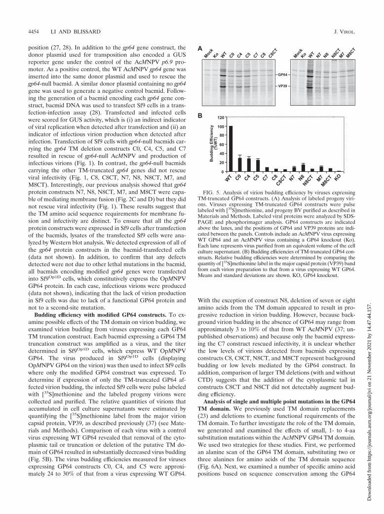
Budding efficiency with modified GP64 constructs. To ex-amine possible effects of the TM domain on virion budding, weexamined virion budding from viruses expressing each GP64TM truncation construct. Each bacmid expressing a GP64 TMtruncation construct was amplified as a virus, and the titerdetermined in Sf9Op1D cells, which express WT OpMNPVGP64. The virus produced in Sf9Op1D cells (displayingOpMNPV GP64 on the virion) was then used to infect Sf9 cellswhere only the modified GP64 construct was expressed. Todetermine if expression of only the TM-truncated GP64 af-fected virion budding, the infected Sf9 cells were pulse labeledwith [35S]methionine and the labeled progeny virions werecollected and purified. The relative quantities of virions thataccumulated in cell culture supernatants were estimated byquantifying the [35S]methionine label from the major virioncapsid protein, VP39, as described previously (37) (see Mate-rials and Methods). Comparison of each virus with a controlvirus expressing WT GP64 revealed that removal of the cyto-plasmic tail or truncation or deletion of the putative TM do-main of GP64 resulted in substantially decreased virus budding(Fig. 5B). The virus budding efficiencies measured for virusesexpressing GP64 constructs C0, C4, and C5 were approxi-mately 24 to 30% of that from a virus expressing WT GP64.
With the exception of construct N8, deletion of seven or eightamino acids from the TM domain appeared to result in pro-gressive reduction in virion budding. However, because back-ground virion budding in the absence of GP64 may range fromapproximately 3 to 10% of that from WT AcMNPV (37; un-published observations) and because only the bacmid express-ing the C7 construct rescued infectivity, it is unclear whetherthe low levels of virions detected from bacmids expressingconstructs C8, C8CT, N8CT, and M8CT represent backgroundbudding or low levels mediated by the GP64 construct. Inaddition, comparison of larger TM deletions (with and withoutCTD) suggests that the addition of the cytoplasmic tail inconstructs C8CT and N8CT did not detectably augment bud-ding efficiency.
Analysis of single and multiple point mutations in the GP64TM domain. We previously used TM domain replacements(23) and deletions to examine functional requirements of theTM domain. To further investigate the role of the TM domain,we generated and examined the effects of small, 1- to 4-aasubstitution mutations within the AcMNPV GP64 TM domain.We used two strategies for these studies. First, we performedan alanine scan of the GP64 TM domain, substituting two orthree alanines for amino acids of the TM domain sequence(Fig. 6A). Next, we examined a number of specific amino acidpositions based on sequence conservation among the GP64
FIG. 5. Analysis of virion budding efficiency by viruses expressingTM-truncated GP64 constructs. (A) Analysis of labeled progeny viri-ons. Viruses expressing TM-truncated GP64 constructs were pulselabeled with [35S]methionine, and progeny BV purified as described inMaterials and Methods. Labeled viral proteins were analyzed by SDS-PAGE and phosphorimager analysis. GP64 constructs are indicatedabove the lanes, and the positions of GP64 and VP39 proteins are indi-cated between the panels. Controls include an AcMNPV virus expressingWT GP64 and an AcMNPV virus containing a GP64 knockout (Ko).Each lane represents virus purified from an equivalent volume of the cellculture supernatant. (B) Budding efficiencies of TM-truncated GP64 con-structs. Relative budding efficiencies were determined by comparing thequantity of [35S]methionine label in the major capsid protein (VP39) bandfrom each virion preparation to that from a virus expressing WT GP64.Means and standard deviations are shown. KO, GP64 knockout.
4454 LI AND BLISSARD J. VIROL.
Dow
nloa
ded
from
http
s://j
ourn
als.
asm
.org
/jour
nal/j
vi o
n 21
Nov
embe
r 20
21 b
y 14
.47.
44.1
57.
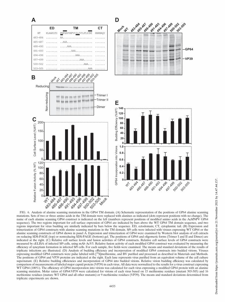
FIG. 6. Analysis of alanine scanning mutations in the GP64 TM domain. (A) Schematic representation of the positions of GP64 alanine scanningmutations. Sets of two or three amino acids in the TM domain were replaced with alanines as indicated (dots represent positions with no change). Thename of each alanine scanning GP64 construct is indicated on the left (numbers represent positions of modified amino acids in the AcMNPV GP64sequence). The two regions important for cell surface expression of GP64 are indicated by bars above the WT GP64 TM domain sequence, and tworegions important for virus budding are similarly indicated by bars below the sequence. ED, ectodomain; CT, cytoplasmic tail. (B) Expression andtrimerization of GP64 constructs with alanine scanning mutations in the TM domain. Sf9 cells were infected with viruses expressing WT GP64 or thealanine scanning constructs of GP64 shown in panel A. Expression and trimerization of GP64 were examined by Western blot analysis of cell extractson reducing SDS-PAGE (top) or nonreducing SDS-PAGE (bottom) gel. The positions of GP64 and oligomeric forms (Trimer I and II and Dimer) areindicated at the right. (C) Relative cell surface levels and fusion activities of GP64 constructs. Relative cell surface levels of GP64 constructs weremeasured by cELISA of infected Sf9 cells, using mAb AcV5. Relative fusion activity of each modified GP64 construct was evaluated by measuring theefficiency of syncytium formation in infected Sf9 cells. For each sample, five fields were examined. The means and standard deviations of the results oftriplicate infections are illustrated. (D) Analysis of budding efficiency and incorporation of modified GP64 constructs into budded virions. Virusesexpressing modified GP64 constructs were pulse labeled with [35S]methionine, and BV purified and processed as described in Materials and Methods.The positions of GP64 and VP39 proteins are indicated at the right. Each lane represents virus purified from an equivalent volume of the cell culturesupernatant. (E) Relative budding efficiencies and incorporation of GP64 into budded virions. Relative virion budding efficiency was calculated bycomparison of measurements of labeled major capsid protein (VP39) in each virus. All data were normalized to the results for a virus construct expressingWT GP64 (100%). The efficiency of GP64 incorporation into virions was calculated for each virus expressing a modified GP64 protein with an alaninescanning mutation. Molar ratios of GP64:VP39 were calculated for virions of each virus based on 15 methionine residues (mutant 503-505) and 16methionine residues (mature WT GP64 and all other mutants) or 9 methionine residues (VP39). The means and standard deviations determined fromtriplicate experiments are shown.
4455
Dow
nloa
ded
from
http
s://j
ourn
als.
asm
.org
/jour
nal/j
vi o
n 21
Nov
embe
r 20
21 b
y 14
.47.
44.1
57.

and related proteins and on current models of membranefusion protein function.
For alanine scanning mutagenesis of the GP64 TM domain,we replaced each two or three amino acids in the TM domainwith alanine residues (Fig. 6A) and inserted each constructinto a recombinant GP64-null virus of AcMNPV as describedabove. All GP64 constructs were initially examined by reducingand nonreducing gel electrophoresis and Western blot analysesto confirm that the substitution constructs were expressed andtrimerized in a manner similar to that of WT GP64 in virus-infected Sf9 cells (Fig. 6B). Analysis of cell surface localizationfor these alanine scanning constructs showed that all constructswere expressed and localized at the cell surface, with surfacelevels ranging between approximately 45 and 185% of thatmeasured from WT GP64 (Fig. 6C, upper panel). Two con-structs (485-487 and 503-505) resulted in reduced surface lev-els of GP64, while the surface levels of all others were similarto or even greater than that of WT GP64. To determine ifspecific TM amino acid positions were required for the fusionfunction of GP64, the alanine scanning constructs of GP64were examined in a syncytium formation assay. All alaninescanning constructs retained fusion activity, and the fusionactivity mediated by these constructs was very similar to that ofWT GP64, ranging from approximately 70 to 110% of thatfrom WT GP64 (Fig. 6C, lower panel). In addition, analysis ofone-step growth curves from viruses expressing the alaninescanning substitution constructions revealed that the infectiousvirus production from all viruses was similar to that from thecontrol virus expressing the WT GP64 protein, with no dra-matic differences from 6 h to 120 h p.i. (data not shown). Tofurther examine possible effects of the TM domain alaninescanning substitutions on virion budding or incorporation ofGP64 into virions, infected Sf9 cells were pulse labeled with[35S]methionine, progeny virions were purified, and relativelevels of labeled progeny virions were estimated by measuringrelative quantities of label in the major capsid protein, VP39,as described earlier. As shown in Fig. 6D, all of the GP64constructs containing alanine scanning mutations in the TMdomain were assembled into virions. The budding efficienciesof mutants 483-484 and 494-496 were decreased to around50% of that of from WT virus, while the budding efficienciesmeasured for other constructs were similar to that of WT virus(Fig. 6E, top panel). Analysis of the incorporation of GP64constructs into virions showed that GP64 incorporation wassimilar to (or in one case only slightly less) that observed forWT GP64. Thus, analysis of alanine scanning mutations iden-tified no amino acid positions (or small regions) in the GP64TM domain that were absolutely required for expression, tri-merization, cell surface localization, membrane fusion, virionbudding, targeting to virions, or viral infectivity. However, weidentified two regions that affected cell surface localization andtwo regions that affected virion budding efficiency (Fig. 6Cand E).
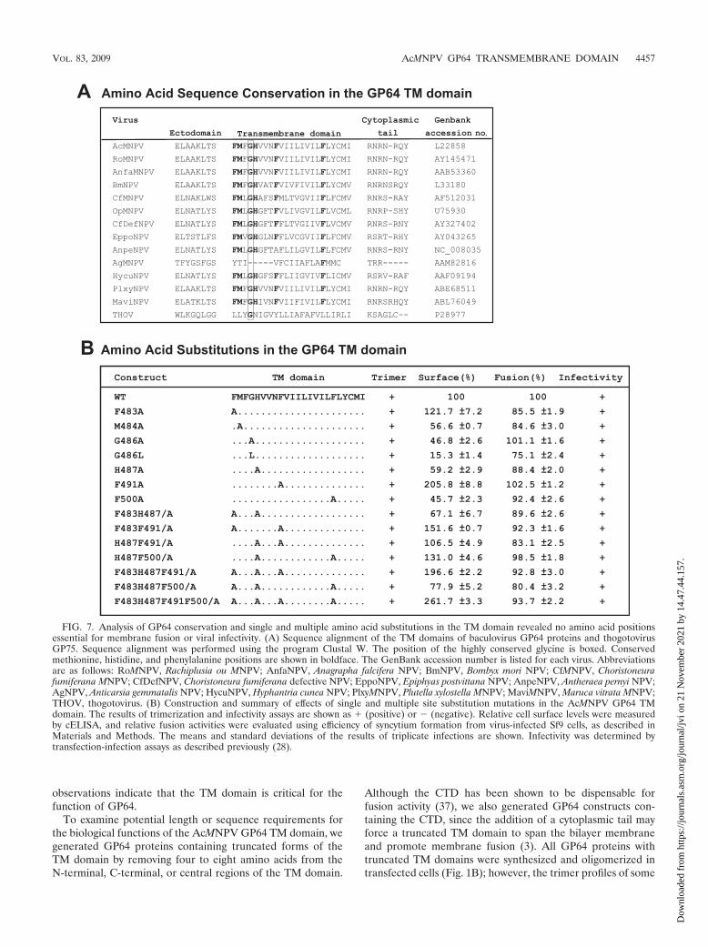
The GP64 protein is very highly conserved within the groupI NPVs of the Baculoviridae, and this high level of conservationincludes the TM domain (Fig. 7A). Because our initial studiesshowed that the GP64 TM domain could not be functionallyreplaced by TM domains from other (nonbaculovirus or unre-lated) viral or cellular-membrane proteins, we concluded thatthe TM domain sequence had important functions beyond that
of simply anchoring the protein in the membrane. Previousstudies revealed that glycine residues within the TM domain ofthe VSV G protein play a critical role in membrane fusion andappear to function in the transition from hemifusion to com-plete membrane fusion (10). Indeed, glycine residues appear tobe overrepresented in the TM domains of viral fusion proteins(2, 46). Where glycines are not present in such TM domains, ithas been proposed that methionine residues may functionallysubstitute. In addition, more recently it was reported that phe-nylalanine residues in combination with glycine residues con-tribute to TM domain interactions (53). Because the GP64 TMdomain contains only a single conserved glycine residue andmultiple phenylalanine residues, we generated alanine substi-tution mutations in single or multiple positions to examine thepotential function of the glycine or phenylalanine residues inGP64 function (Fig. 7). Substitutions for the Met residue at theN terminus of the TM domain and the single charged histidineresidue were also examined either alone or in combinationwith other substitutions. The single and multiple substitutionmutations are shown in Fig. 7B. All modified GP64 constructswere inserted into recombinant viruses (replacing native GP64)and examined for trimer formation, localization at the cellsurface, fusion activity (syncytium formation efficiency), andvirus infectivity (Fig. 7B). In viruses containing the GP64 TMdomain substitutions of single and multiple amino acids (Fig.7B), the GP64 proteins behaved similarly to WT GP64, with nosubstantial differences observed. A minor difference observedwas that levels of cell surface GP64 were moderately reducedfor some of the substitution mutations. Replacement of Met484, Gly 486, His 487, or Phe 500 resulted in decreased cellsurface levels (40 to 75% of the level with WT GP64) (Fig. 7B),although the fusion activity detected remained high. Thus,consistent with the alanine scanning analysis, our analysis ofsingle and multiple amino acid substitutions at conserved res-idues or those identified as important in other viral membranefusion proteins did not identify amino acids that were abso-lutely required for GP64 function in membrane fusion or othercritical functions.
DISCUSSION
For baculoviruses that encode a GP64 protein, GP64 is es-sential for the entry of the budded form of the virus. Comparedwith the highly conserved ectodomains of GP64 proteins, theTM domains are more variable in length and amino acid se-quence similarity. This suggests the possibility that TM do-mains of GP64 proteins have conserved structural characteris-tics that are not apparent from an analysis of the primaryamino acid sequence. The recently described structure of thepostfusion form of GP64 (21) does not include the TM do-main, and TM domain structures are generally rare. Recently,we found that replacement of the predicted 23-aa GP64 TMdomain with corresponding TM domain sequences from arange of viral or cellular type I membrane proteins or with aGPI anchor addition sequence resulted in defects in transport,membrane fusion, virion budding, and virus infectivity (23).Only TM domains from two related viral membrane proteins(OpMNPV GP64 and thogotovirus GP75) functionally substi-tuted for the AcMNPV GP64 TM domain sequence. These
4456 LI AND BLISSARD J. VIROL.
Dow
nloa
ded
from
http
s://j
ourn
als.
asm
.org
/jour
nal/j
vi o
n 21
Nov
embe
r 20
21 b
y 14
.47.
44.1
57.
observations indicate that the TM domain is critical for thefunction of GP64.
To examine potential length or sequence requirements forthe biological functions of the AcMNPV GP64 TM domain, wegenerated GP64 proteins containing truncated forms of theTM domain by removing four to eight amino acids from theN-terminal, C-terminal, or central regions of the TM domain.
Although the CTD has been shown to be dispensable forfusion activity (37), we also generated GP64 constructs con-taining the CTD, since the addition of a cytoplasmic tail mayforce a truncated TM domain to span the bilayer membraneand promote membrane fusion (3). All GP64 proteins withtruncated TM domains were synthesized and oligomerized intransfected cells (Fig. 1B); however, the trimer profiles of some
FIG. 7. Analysis of GP64 conservation and single and multiple amino acid substitutions in the TM domain revealed no amino acid positionsessential for membrane fusion or viral infectivity. (A) Sequence alignment of the TM domains of baculovirus GP64 proteins and thogotovirusGP75. Sequence alignment was performed using the program Clustal W. The position of the highly conserved glycine is boxed. Conservedmethionine, histidine, and phenylalanine positions are shown in boldface. The GenBank accession number is listed for each virus. Abbreviationsare as follows: RoMNPV, Rachiplusia ou MNPV; AnfaNPV, Anagrapha falcifera NPV; BmNPV, Bombyx mori NPV; CfMNPV, Choristoneurafumiferana MNPV; CfDefNPV, Choristoneura fumiferana defective NPV; EppoNPV, Epiphyas postvittana NPV; AnpeNPV, Antheraea pernyi NPV;AgNPV, Anticarsia gemmatalis NPV; HycuNPV, Hyphantria cunea NPV; PlxyMNPV, Plutella xylostella MNPV; MaviMNPV, Maruca vitrata MNPV;THOV, thogotovirus. (B) Construction and summary of effects of single and multiple site substitution mutations in the AcMNPV GP64 TMdomain. The results of trimerization and infectivity assays are shown as � (positive) or � (negative). Relative cell surface levels were measuredby cELISA, and relative fusion activities were evaluated using efficiency of syncytium formation from virus-infected Sf9 cells, as described inMaterials and Methods. The means and standard deviations of the results of triplicate infections are shown. Infectivity was determined bytransfection-infection assays as described previously (28).
VOL. 83, 2009 AcMNPV GP64 TRANSMEMBRANE DOMAIN 4457
Dow
nloa
ded
from
http
s://j
ourn
als.
asm
.org
/jour
nal/j
vi o
n 21
Nov
embe
r 20
21 b
y 14
.47.
44.1
57.
constructs containing C-terminal deletions of the TM domainappeared to differ from that of WT GP64. The GP64 trimer istypically observed as two distinct electrophoretic forms. Whilethe two trimer forms (I and II) have different migration ratesin nonreducing SDS-PAGE, the two forms appear to have thesame mass, as determined by mass spectrometry (38). It hasbeen suggested that the two trimer forms may represent disul-fide isomers (26), but it is not known whether they differ in anyfunctional properties. The cell surface levels of TM-truncatedGP64 constructs were dramatically reduced (�67%) and ap-peared to decrease with successive reductions in length of the TMdomain (Fig. 2). Similar results were previously reported for hu-man immunodeficiency virus envelope glycoprotein and VSV Gconstructs that contained truncated TM domains (1, 39).
Recent studies of TM model peptides suggest that peptide-membrane interactions depend on peptide hydrophobicity andon the matching of hydrophobic peptide length with mem-brane thickness. A negative hydrophobic mismatch occurswhen the hydrophobic stretch of a peptide is too short withrespect to the membrane thickness (11, 13, 45, 49, 57). Theobserved shedding of GP64 proteins with truncated TM do-mains (Fig. 1B) may result from such a negative hydrophobicmismatch. Addition of the cytoplasmic tail significantly in-creased the cell surface levels of C8CT and N8CT and alsomoderately increased surface levels of M8CT (Fig. 2A), sug-gesting that addition of the CTD may force the truncated TMdomain to adapt its structure or orientation to overcome themismatch, as suggested for some model TM peptides (12, 15,47). The length of the GP64 TM domain also had an effect onits fusogenicity. With successive truncations, the level of mem-brane fusion activity also decreased successively compared tothat for WT GP64 (normalized activity). Compared with nor-malized WT GP64, we found that the fusion activity decreasedmoderately (15 to 20%) for GP64 constructs C4 and C5 anddramatically for C7 and C8 (Fig. 2D). No fusion activity wasdetected for C8, a construct containing only 15 amino acids ofthe TM domain. The N-terminal and middle regions of the TMdomain were similarly sensitive, since deletion of seven oreight amino acids from those regions also resulted in dramat-ically reduced fusion activity (77 to 90% reduction). Whileaddition of the CTD increased the fusion activities of N8CTand M8CT in comparison to those of N8 and M8, the fusionactivities of N8CT and M8CT were still reduced by more than90% compared with that of WT GP64. Addition of the CTDdid not rescue fusion activity of C8, which was fusion negative.Because the C-terminal portion of the TM domain is some-what more hydrophobic than the N-terminal region (as mea-sured by hydrophobicity plots), removing the most-hydropho-bic portion of the TM domain may account for both the lack ofdetectable fusion activity in C8 and the failure of C8CT torescue fusion activity. Thus, these data suggested that GP64-mediated fusion requires a critical TM domain length of 15 or16 amino acids. The critical nature of the hydrophobic lengthwas confirmed by experiments in which a single hydrophobicamino acid (A, L, or V) was added to the C terminus of mutantC8, resulting in restored fusion activity (Fig. 4C). The additionof hydrophilic or charged amino acids did not restore activity.Interestingly, the fusion activities of the C8 mutants extendedwith L or V were considerably less than that of the C7 mutantthat contains an I at the same position. It is of interest that in
the case of the WT C7 sequence or C8L, C8V, and C8A (whichrestored fusion), the relative level of fusion correlates with thedegree of hydrophobicity of the respective C-terminal aminoacid, I, L, V, or A.
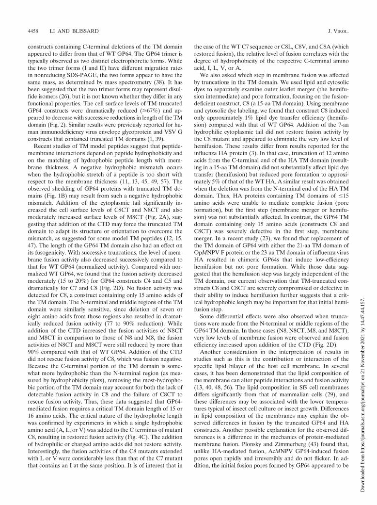
We also asked which step in membrane fusion was affectedby truncations in the TM domain. We used lipid and cytosolicdyes to separately examine outer leaflet merger (the hemifu-sion intermediate) and pore formation, focusing on the fusion-deficient construct, C8 (a 15-aa TM domain). Using membraneand cytosolic dye labeling, we found that construct C8 inducedonly approximately 1% lipid dye transfer efficiency (hemifu-sion) compared with that of WT GP64. Addition of the 7-aahydrophilic cytoplasmic tail did not restore fusion activity bythe C8 mutant and appeared to eliminate the very low level ofhemifusion. These results differ from results reported for theinfluenza HA protein (3). In that case, truncation of 12 aminoacids from the C-terminal end of the HA TM domain (result-ing in a 15-aa TM domain) did not substantially affect lipid dyetransfer (hemifusion) but reduced pore formation to approxi-mately 5% of that of the WT HA. A similar result was obtainedwhen the deletion was from the N-terminal end of the HA TMdomain. Thus, HA proteins containing TM domains of �15amino acids were unable to mediate complete fusion (poreformation), but the first step (membrane merger or hemifu-sion) was not substantially affected. In contrast, the GP64 TMdomain containing only 15 amino acids (constructs C8 andC8CT) was severely defective in the first step, membranemerger. In a recent study (23), we found that replacement ofthe TM domain of GP64 with either the 21-aa TM domain ofOpMNPV F protein or the 23-aa TM domain of influenza virusHA resulted in chimeric GP64s that induce low-efficiencyhemifusion but not pore formation. While those data sug-gested that the hemifusion step was largely independent of theTM domain, our current observation that TM-truncated con-structs C8 and C8CT are severely compromised or defective intheir ability to induce hemifusion further suggests that a crit-ical hydrophobic length may be important for that initial hemi-fusion step.
Some differential effects were also observed when trunca-tions were made from the N-terminal or middle regions of theGP64 TM domain. In those cases (N8, N8CT, M8, and M8CT),very low levels of membrane fusion were observed and fusionefficiency increased upon addition of the CTD (Fig. 2D).
Another consideration in the interpretation of results instudies such as this is the contribution or interaction of thespecific lipid bilayer of the host cell membrane. In severalcases, it has been demonstrated that the lipid composition ofthe membrane can alter peptide interactions and fusion activity(13, 40, 48, 56). The lipid composition in Sf9 cell membranesdiffers significantly from that of mammalian cells (29), andthese differences may be associated with the lower tempera-tures typical of insect cell culture or insect growth. Differencesin lipid composition of the membranes may explain the ob-served differences in fusion by the truncated GP64 and HAconstructs. Another possible explanation for the observed dif-ferences is a difference in the mechanics of protein-mediatedmembrane fusion. Plonsky and Zimmerberg (43) found that,unlike HA-mediated fusion, AcMNPV GP64-induced fusionpores open rapidly and irreversibly and do not flicker. In ad-dition, the initial fusion pores formed by GP64 appeared to be
4458 LI AND BLISSARD J. VIROL.
Dow
nloa
ded
from
http
s://j
ourn
als.
asm
.org
/jour
nal/j
vi o
n 21
Nov
embe
r 20
21 b
y 14
.47.
44.1
57.

larger than those of HA (43). Interestingly, the minimal TMdomain length for fusogenicity is in the same range for differ-ent TM peptides: 17 residues for HA (3), 16 residues for theGP64 TM domain, and between 14 and 18 residues for TMmodel peptides (26). Effects of the TM domain length on thefusion process were also described previously for other fusionproteins, such as those from human immunodeficiency virus(39), foamy virus (41), murine coronavirus (6), and murineleukemia virus (44), and for SNARE proteins (58).
In addition to membrane fusion, we also examined the po-tential roles of the TM domain in virion assembly during bud-ding. An analysis of virus budding efficiency revealed that virusbudding efficiency decreased 70 to 75% upon deletion of thecytoplasmic tail. Truncation of an additional four or five aminoacids from the C-terminal end of the TM domain (constructsC4 and C5) resulted in a similarly reduced budding efficiency.Further truncation or deletion of seven or eight amino acids ofthe TM domain resulted in even more dramatically reducedvirus budding efficiency (reductions of 86 to 96% compared tothat with WT GP64) (Fig. 5B). While the reduced buddingefficiency of truncated GP64 constructs appeared to correlatewith reduced surface levels of the GP64 constructs, the addi-tion of the CTD (which restored surface levels and fusionactivity) (Fig. 2A and D) did not rescue the budding defects.We also examined the capacity of GP64 proteins with trun-cated TM domains to substitute for WT GP64, using a trans-fection-infection assay in Sf9 cells. Infectivity was rescued byconstructs containing 16 amino acids from the N-terminal por-tion of the TM domain but not by constructs containing 16amino acids from other parts of the TM domain (i.e., con-structs containing deletions from the N-terminal or middleregions were not able to substitute for WT GP64) (Fig. 1, C7versus N7 and M7). By comparing fusion assay results to re-sults from virus infectivity studies, we can conclude that thesequence requirements for infectivity are more stringent thanthose for membrane fusion, which in turn are more stringentthan the requirements for membrane anchoring and intracel-lular transport.
To further examine the GP64 TM domain and identify spe-cific residues important for GP64 function, we generated andanalyzed single and multiple amino acid substitution mutationsin the TM domain. We specifically focused on conserved me-thionine (M484), glycine (G486), histidine (H487), and phe-nylalanine (F483, F487, F491, and F500) residues of the TMdomain of GP64 (Fig. 7B). We also examined small regions oftwo to three amino acids by using an alanine scan (Fig. 6A).The glycine at 486 is conserved among almost all known bac-ulovirus GP64 proteins and the more-distantly related thogoto-virus GP75. The results of previous studies (10) suggested thatat least one of the two glycine residues of VSV G protein playsa significant role in the transition from hemifusion to completefusion. Furthermore, a study of influenza virus HA, whichcontains two glycines within the TM domain, demonstratedthat substitution of a leucine (G250L) for the more-N-terminalglycine caused a restricted hemifusion phenotype (33). It hasbeen proposed that these important glycine residues may func-tion as helix breakers and thereby distort the bilayer, promot-ing membrane fusion. This concept is supported by the factthat a conserved proline residue (also a helix breaker) is foundin the TM domains of foamy virus and murine leukemia virus
envelope proteins and is essential for fusion function in bothinstances (41, 51). In the current study, substitution of alanineor leucine for the conserved glycine resulted in reduced cellsurface levels of those GP64 constructs (Fig. 6, 485-487 and7B), but the fusion activities of those constructs (485-487 andG486A and G486L mutants) were similar to or only slightlydecreased from that of WT GP64. Similar results were foundfor Semliki Forest virus E1 protein. Indeed, neither of the twoconserved glycine residues nor any of the five total glycineresidues within the TM domain of E1 are required for fusionactivity (24).
Since many or most of the Gly-less TM domains of fusionproteins possess internal methionine residues (10), we alsoexamined the conserved methionine at position 484. Substitu-tion of alanine for Met 484 resulted in similar or decreasedlevels of the GP64 construct at the cell surface but had onlysubtle effects on membrane fusion (Fig. 6, 483-484, and 7,M484A). We also examined the histidine (H487), based on thereported role of a charged residue within the TM domain offoamy virus envelope protein (41). Pietschmann et al. (41)found that an evolutionarily conserved positively chargedamino acid, K959, within the putative TM domain of foamyvirus appears to regulate fusion activity. We found no substan-tial effect from two substitution approaches for the GP64 TMdomain histidine residue (Fig. 6, 485-487, and 7B, H487A).Recently, Unterreitmeier et al. (53) used a randomized libraryto biologically select TM domains that self-interact and foundthat higher-affinity TM domain sequences were enriched inphenylalanine. In addition, phenylalanine is frequently foundassociated with GxxxG motifs in TM domains of self-interact-ing proteins. Notably, disruption of the FxxGxxxG motif of theVSV G protein by substitution for phenylalanine or glycineresidues resulted in reduced TM-TM associations (53). Substi-tutions for the conserved phenylalanines within the TM do-main of GP64 (singly or in various combinations with H487)did not cause dramatic effects on fusion activity, although thecell surface levels were somewhat reduced for GP64 substitu-tion construct F500A (Fig. 7B).
Using the 2- or 3-alanine scanning substitutions (Fig. 6), wefound two regions, residues 485 to 487 and 503 to 505, thatwere important for cell surface localization of GP64. Substitu-tion of alanines for either of these two regions resulted in cellsurface levels that were decreased by more than 50% (Fig. 6C).In two additional regions, positions 483 to 484 and 494 to 496,substitution mutations resulted in virus budding that was re-duced by more than 50%.
While many reports describe critical roles for the TM do-mains of viral fusion proteins, no uniform theme has emergedfrom these studies. In the case of influenza virus HA, it is clearthat a TM anchor is required for full fusion activity (32, 34),but a variety of TM sequences will substitute for the native TMdomain (33). In contrast, specific sequence requirements ap-pear to be encoded within the TM domains of other fusionproteins, such as the envelope proteins from human immuno-deficiency virus type I (39), murine leukemia virus (51), foamyviruses (41), coronavirus (6, 8), VSV (10), Newcastle diseasevirus (31), and measles virus (7). In the case of the AcMNPVGP64 protein, the results of prior studies indicated that, likethe later cases, the TM domain sequence includes critical func-tions (23). In the present more-detailed study of the TM do-
VOL. 83, 2009 AcMNPV GP64 TRANSMEMBRANE DOMAIN 4459
Dow
nloa
ded
from
http
s://j
ourn
als.
asm
.org
/jour
nal/j
vi o
n 21
Nov
embe
r 20
21 b
y 14
.47.
44.1
57.
main sequence, we reached two broad conclusions. First, wefound that the TM domain of the GP64 protein requires acritical minimum hydrophobic length of 16 amino acids. Therequirement for this length is not only related to protein an-choring but, more importantly, represents a requirement forthe function of GP64 as a fusion protein. Second, our analysisof TM domain sequences by substitution mutations in con-served positions and alanine scanning show that no singleamino acid in the TM domain is absolutely required for GP64function in any of its critical roles in the virus. Combined withthe results of prior studies of substitutions for the entire TMdomain, the results of these studies suggest that either (i) theGP64 TM domain may include critical amino acids or se-quence elements that are separate but redundant or (ii) theoverall sequence forms a higher-order structure that is notdisrupted by single substitutions or our alanine scanning ap-proach, yet cannot be replaced by an unrelated heterologousTM domain. Future studies aimed at examining the higher-order structure of the GP64 TM domain may provide insightinto the relationship between TM domain structure and thecomplex and critical process of protein-mediated membranefusion.
ACKNOWLEDGMENTS
We thank Gerrit Heetderks and Jian Zhou for expert technicalassistance.
This work was supported by NIH grant AI33657 and BTI projectsG01707-R06-1255 and B00103-R06-1255.
REFERENCES
1. Adams, G. A., and J. K. Rose. 1985. Structural requirements of a membrane-spanning domain for protein anchoring and cell surface transport. Cell41:1007–1015.
2. Arkin, I. T., and A. T. Brunger. 1998. Statistical analysis of predicted trans-membrane alpha-helices. Biochim. Biophys. Acta 1429:113–128.
3. Armstrong, R. T., A. S. Kushnir, and J. M. White. 2000. The transmembranedomain of influenza hemagglutinin exhibits a stringent length requirement tosupport the hemifusion to fusion transition. J. Cell Biol. 151:425–437.
4. Blissard, G. W., and G. F. Rohrmann. 1991. Baculovirus gp64 gene expres-sion: analysis of sequences modulating early transcription and transactiva-tion by IE1. J. Virol. 65:5820–5827.
5. Blissard, G. W., and J. R. Wenz. 1992. Baculovirus gp64 envelope glyco-protein is sufficient to mediate pH-dependent membrane fusion. J. Virol.66:6829–6835.
6. Bos, E. C., L. Heijnen, W. Luytjes, and W. J. Spaan. 1995. Mutationalanalysis of the murine coronavirus spike protein: effect on cell-to-cell fusion.Virology 214:453–463.
7. Caballero, M., J. Carabana, J. Ortego, R. Fernandez-Munoz, and M. L.Celma. 1998. Measles virus fusion protein is palmitoylated on transmem-brane-intracytoplasmic cysteine residues which participate in cell fusion.J. Virol. 72:8198–8204.
8. Chang, K. W., Y. Sheng, and J. L. Gombold. 2000. Coronavirus-inducedmembrane fusion requires the cysteine-rich domain in the spike protein.Virology 269:212–224.
9. Chiu, J., P. E. March, R. Lee, and D. Tillett. 2004. Site-directed, ligase-independent mutagenesis (SLIM): a single-tube methodology approaching100% efficiency in 4 h. Nucleic Acids Res. 32:e174.
10. Cleverley, D. Z., and J. Lenard. 1998. The transmembrane domain in viralfusion: essential role for a conserved glycine residue in vesicular stomatitisvirus G protein. Proc. Natl. Acad. Sci. USA 95:3425–3430.
11. de Planque, M. R., J. W. Boots, D. T. Rijkers, R. M. Liskamp, D. V.Greathouse, and J. A. Killian. 2002. The effects of hydrophobic mismatchbetween phosphatidylcholine bilayers and transmembrane alpha-helical pep-tides depend on the nature of interfacially exposed aromatic and chargedresidues. Biochemistry 41:8396–8404.
12. de Planque, M. R., E. Goormaghtigh, D. V. Greathouse, R. E. Koeppe II,J. A. Kruijtzer, R. M. Liskamp, B. de Kruijff, and J. A. Killian. 2001.Sensitivity of single membrane-spanning alpha-helical peptides to hydropho-bic mismatch with a lipid bilayer: effects on backbone structure, orientation,and extent of membrane incorporation. Biochemistry 40:5000–5010.
13. de Planque, M. R., and J. A. Killian. 2003. Protein-lipid interactions studied
with designed transmembrane peptides: role of hydrophobic matching andinterfacial anchoring. Mol. Membr. Biol. 20:271–284.
14. Epand, R. M. 2003. Fusion peptides and the mechanism of viral fusion.Biochim. Biophys. Acta 1614:116–121.
15. Harzer, U., and B. Bechinger. 2000. Alignment of lysine-anchored mem-brane peptides under conditions of hydrophobic mismatch: a CD, 15N and31P solid-state NMR spectroscopy investigation. Biochemistry 39:13106–13114.
16. Hefferon, K., A. Oomens, S. Monsma, C. Finnerty, and G. Blissard. 1999.Host cell receptor binding by baculovirus GP64 and kinetics of virion entry.Virology 258:455–468.
17. Herniou, E. A., J. A. Olszewski, J. S. Cory, and D. R. O’Reilly. 2003. Thegenome sequence and evolution of baculoviruses. Annu. Rev. Entomol.48:211–234.
18. Hink, W. F. 1970. Established insect cell line from the cabbage looper,Trichoplusia ni. Nature 226:466–467.
19. Hohmann, A. W., and P. Faulkner. 1983. Monoclonal antibodies to baculo-virus structural proteins: determination of specificities by Western blot anal-ysis. Virology 125:432–444.
20. Jarvis, D. L., L. Wills, G. Burow, and D. A. Bohlmeyer. 1998. Mutationalanalysis of the N-linked glycans on Autographa californica nucleopolyhedro-virus gp64. J. Virol. 72:9459–9469.
21. Kadlec, J., S. Loureiro, N. G. Abrescia, D. I. Stuart, and I. M. Jones. 2008.The postfusion structure of baculovirus gp64 supports a unified view of viralfusion machines. Nat. Struct. Mol. Biol. 15:1024–1030.
22. Langosch, D., M. Hofmann, and C. Ungermann. 2007. The role of trans-membrane domains in membrane fusion. Cell. Mol. Life Sci. 64:850–864.
23. Li, Z., and G. W. Blissard. 2008. Functional analysis of the transmembrane(TM) domain of the Autographa californica multicapsid nucleopolyhedrovi-rus GP64 protein: substitution of heterologous TM domains. J. Virol. 82:3329–3341.
24. Liao, M., and M. Kielian. 2005. The conserved glycine residues in thetransmembrane domain of the Semliki Forest virus fusion protein are notrequired for assembly and fusion. Virology 332:430–437.
25. Long, G., X. Pan, R. Kormelink, and J. M. Vlak. 2006. Functional entry ofbaculovirus into insect and mammalian cells is dependent on clathrin-medi-ated endocytosis. J. Virol. 80:8830–8833.
26. Lorin, A., B. Charloteaux, Y. Fridmann-Sirkis, A. Thomas, Y. Shai, and R.Brasseur. 2007. Mode of membrane interaction and fusogenic properties ofa de novo transmembrane model peptide depend on the length of thehydrophobic core. J. Biol. Chem. 282:18388–18396.
27. Luckow, V. A., S. C. Lee, G. F. Barry, and P. O. Olins. 1993. Efficientgeneration of infectious recombinant baculoviruses by site- specific transpo-son-mediated insertion of foreign genes into a baculovirus genome propa-gated in Escherichia coli. J. Virol. 67:4566–4579.
28. Lung, O., M. Westenberg, J. M. Vlak, D. Zuidema, and G. W. Blissard. 2002.Pseudotyping Autographa californica multicapsid nucleopolyhedrovirus(AcMNPV): F proteins from group II NPVs are functionally analogous toAcMNPV GP64. J. Virol. 76:5729–5736.
29. Marheineke, K., S. Grunewald, W. Christie, and H. Reilander. 1998. Lipidcomposition of Spodoptera frugiperda (Sf9) and Trichoplusia ni (Tn) insectcells used for baculovirus infection. FEBS Lett. 441:49–52.
30. Maruniak, J. W., and M. D. Summers. 1981. Autographa californica nuclearpolyhedrosis virus phosphoproteins and synthesis of intracellular proteinsafter virus infection. Virology 109:25–34.
31. McGinnes, L., T. Sergel, and T. Morrison. 1993. Mutations in the transmem-brane domain of the HN protein of Newcastle disease virus affect the struc-ture and activity of the protein. Virology 196:101–110.
32. Melikyan, G. B., S. A. Brener, D. C. Ok, and F. S. Cohen. 1997. Inner but notouter membrane leaflets control the transition from glycosylphosphatidyl-inositol-anchored influenza hemagglutinin-induced hemifusion to full fusion.J. Cell Biol. 136:995–1005.
33. Melikyan, G. B., S. Lin, M. G. Roth, and F. S. Cohen. 1999. Amino acidsequence requirements of the transmembrane and cytoplasmic domains ofinfluenza virus hemagglutinin for viable membrane fusion. Mol. Biol. Cell10:1821–1836.
34. Melikyan, G. B., J. M. White, and F. S. Cohen. 1995. GPI-anchored influenzahemagglutinin induces hemifusion to both red blood cell and planar bilayermembranes. J. Cell Biol. 131:679–691.
35. Monsma, S. A., and G. W. Blissard. 1995. Identification of a membranefusion domain and an oligomerization domain in the baculovirus GP64envelope fusion protein. J. Virol. 69:2583–2595.
36. Monsma, S. A., A. G. Oomens, and G. W. Blissard. 1996. The GP64 envelopefusion protein is an essential baculovirus protein required for cell-to-celltransmission of infection. J. Virol. 70:4607–4616.
37. Oomens, A. G. P., and G. W. Blissard. 1999. Requirement for GP64 to driveefficient budding of Autographa californica multicapsid nucleopolyhedrovi-rus. Virology 254:297–314.
38. Oomens, A. G. P., S. A. Monsma, and G. W. Blissard. 1995. The baculovirusGP64 envelope fusion protein: synthesis, oligomerization, and processing.Virology 209:592–603.
39. Owens, R. J., C. Burke, and J. K. Rose. 1994. Mutations in the membrane-
4460 LI AND BLISSARD J. VIROL.
Dow
nloa
ded
from
http
s://j
ourn
als.
asm
.org
/jour
nal/j
vi o
n 21
Nov
embe
r 20
21 b
y 14
.47.
44.1
57.

spanning domain of the human immunodeficiency virus envelope glycopro-tein that affect fusion activity. J. Virol. 68:570–574.
40. Ozdirekcan, S., D. T. Rijkers, R. M. Liskamp, and J. A. Killian. 2005.Influence of flanking residues on tilt and rotation angles of transmembranepeptides in lipid bilayers. A solid-state 2H NMR study. Biochemistry 44:1004–1012.
41. Pietschmann, T., H. Zentgraf, A. Rethwilm, and D. Lindemann. 2000. Anevolutionarily conserved positively charged amino acid in the putative mem-brane-spanning domain of the foamy virus envelope protein controls fusionactivity. J. Virol. 74:4474–4482.
42. Plonsky, I., M. S. Cho, A. G. P. Oomens, G. W. Blissard, and J. Zimmerberg.1999. An analysis of the role of the target membrane on the gp64-inducedfusion pore. Virology 253:65–76.
43. Plonsky, I., and J. Zimmerberg. 1996. The initial fusion pore induced bybaculovirus GP64 is large and forms quickly. J. Cell Biol. 135:1831–1839.
44. Ragheb, J. A., and W. F. Anderson. 1994. Uncoupled expression of Moloneymurine leukemia virus envelope polypeptides SU and TM: a functionalanalysis of the role of TM domains in viral entry. J. Virol. 68:3207–3219.
45. Ren, J., S. Lew, Z. Wang, and E. London. 1997. Transmembrane orientationof hydrophobic alpha-helices is regulated both by the relationship of helixlength to bilayer thickness and by the cholesterol concentration. Biochem-istry 36:10213–10220.
46. Senes, A., M. Gerstein, and D. M. Engelman. 2000. Statistical analysis ofamino acid patterns in transmembrane helices: the GxxxG motif occursfrequently and in association with beta-branched residues at neighboringpositions. J. Mol. Biol. 296:921–936.
47. Sharpe, S., K. R. Barber, C. W. Grant, D. Goodyear, and M. R. Morrow.2002. Organization of model helical peptides in lipid bilayers: insight into thebehavior of single-span protein transmembrane domains. Biophys. J. 83:345–358.
48. Sparr, E., W. L. Ash, P. V. Nazarov, D. T. Rijkers, M. A. Hemminga, D. P.Tieleman, and J. A. Killian. 2005. Self-association of transmembrane alpha-helices in model membranes: importance of helix orientation and role ofhydrophobic mismatch. J. Biol. Chem. 280:39324–39331.
49. Strandberg, E., S. Ozdirekcan, D. T. Rijkers, P. C. van der Wel, R. E. KoeppeII, R. M. Liskamp, and J. A. Killian. 2004. Tilt angles of transmembranemodel peptides in oriented and non-oriented lipid bilayers as determined by2H solid-state NMR. Biophys. J. 86:3709–3721.
50. Tamm, L. K., J. Crane, and V. Kiessling. 2003. Membrane fusion: a struc-tural perspective on the interplay of lipids and proteins. Curr. Opin. Struct.Biol. 13:453–466.
51. Taylor, G. M., and D. A. Sanders. 1999. The role of the membrane-spanningdomain sequence in glycoprotein-mediated membrane fusion. Mol. Biol.Cell 10:2803–2815.
52. Ujike, M., K. Nakajima, and E. Nobusawa. 2004. Influence of acylation sitesof influenza B virus hemagglutinin on fusion pore formation and dilation.J. Virol. 78:11536–11543.
53. Unterreitmeier, S., A. Fuchs, T. Schaffler, R. G. Heym, D. Frishman, and D.Langosch. 2007. Phenylalanine promotes interaction of transmembrane do-mains via GxxxG motifs. J. Mol. Biol. 374:705–718.
54. Volkman, L. E., and P. A. Goldsmith. 1984. Budded Autographa californicaNPV 64K protein: further biochemical analysis and effects of postimmuno-precipitation sample preparation conditions. Virology 139:295–302.
55. Volkman, L. E., and P. A. Goldsmith. 1985. Mechanism of neutralization ofbudded Autographa californica nuclear polyhedrosis virus by a monoclonalantibody: inhibition of entry by adsorptive endocytosis. Virology 143:185–195.
56. Webb, R. J., J. M. East, R. P. Sharma, and A. G. Lee. 1998. Hydrophobicmismatch and the incorporation of peptides into lipid bilayers: a possiblemechanism for retention in the Golgi. Biochemistry 37:673–679.
57. Weiss, T. M., P. C. van der Wel, J. A. Killian, R. E. Koeppe II, and H. W.Huang. 2003. Hydrophobic mismatch between helices and lipid bilayers.Biophys. J. 84:379–385.
58. Xu, Y., F. Zhang, Z. Su, J. A. McNew, and Y. K. Shin. 2005. Hemifusion inSNARE-mediated membrane fusion. Nat. Struct. Mol. Biol. 12:417–422.
59. Zhang, S. X., Y. Han, and G. W. Blissard. 2003. Palmitoylation of theAutographa californica multicapsid nucleopolyhedrovirus envelope glycopro-tein GP64: mapping, functional studies, and lipid rafts. J. Virol. 77:6265–6273.
60. Zhou, J., and G. W. Blissard. 2008. Identification of a GP64 subdomaininvolved in receptor binding by budded virions of the baculovirus AcMNPV.J. Virol. 82:4449–4460.
61. Zhou, J., and G. W. Blissard. 2006. Mapping the conformational epitope ofa neutralizing antibody (AcV1) directed against the AcMNPV GP64 protein.Virology 352:427–437.
VOL. 83, 2009 AcMNPV GP64 TRANSMEMBRANE DOMAIN 4461
Dow
nloa
ded
from
http
s://j
ourn
als.
asm
.org
/jour
nal/j
vi o
n 21
Nov
embe
r 20
21 b
y 14
.47.
44.1
57.