The author responsible for distribution of materials ...
37
RESEARCH ARTICLE COR27 and COR28 are Novel Regulators of the COP1–HY5 Regulatory Hub and Photomorphogenesis in Arabidopsis Xu Li 1,3, Cuicui Liu 1,2,3, Zhiwei Zhao 1,2, Dingbang Ma 1,2, Jinyu Zhang 1,2, Yu Yang 1,2, Yawen Liu 1, Hongtao Liu 1* 1 National Key Laboratory of Plant Molecular Genetics (NKLPMG), CAS Center for Excellence in Molecular Plant Sciences, Shanghai Institutes for Biological Sciences (SIBS), Chinese Academy of Sciences. Shanghai 200032, P. R. China 2 University of Chinese Academy of Sciences, Shanghai 200032, P. R. China 3 These authors contributed equally to this work * Correspondence: Hongtao Liu Tel: +86-21-54924291 E-mail: [email protected] Short title: COR27 and COR28 Regulate Photomorphogenesis One-sentence summary: COR27 and COR28 act as key regulators in the COP1–HY5 regulatory hub by regulating HY5 activity to ensure proper skotomorphogenic growth in the dark and photomorphogenic development in the light. The author responsible for distribution of materials integral to the findings presented in this article in accordance with the policy described in the Instructions for Authors (www.plantcell.org) is: Hongtao Liu ([email protected]). Key words: COR27/COR28, photomorphogenesis, COP1, HY5, light signaling, skotomorphogenesis. Plant Cell Advance Publication. Published on August 7, 2020, doi:10.1105/tpc.20.00195 ©2020 American Society of Plant Biologists. All Rights Reserved
Transcript of The author responsible for distribution of materials ...
RESEARCH ARTICLE
COR27 and COR28 are Novel Regulators of the COP1–HY5 Regulatory Hub and
Photomorphogenesis in Arabidopsis
Xu Li 1,3, Cuicui Liu 1,2,3, Zhiwei Zhao 1,2, Dingbang Ma 1,2, Jinyu Zhang 1,2, Yu Yang 1,2,
Yawen Liu 1, Hongtao Liu 1*
1 National Key Laboratory of Plant Molecular Genetics (NKLPMG), CAS Center for
Excellence in Molecular Plant Sciences, Shanghai Institutes for Biological Sciences (SIBS),
Chinese Academy of Sciences. Shanghai 200032, P. R. China
2 University of Chinese Academy of Sciences, Shanghai 200032, P. R. China
3 These authors contributed equally to this work
* Correspondence: Hongtao Liu
Tel: +86-21-54924291
E-mail: [email protected]
Short title: COR27 and COR28 Regulate Photomorphogenesis
One-sentence summary: COR27 and COR28 act as key regulators in the COP1–HY5
regulatory hub by regulating HY5 activity to ensure proper skotomorphogenic growth in the
dark and photomorphogenic development in the light.
The author responsible for distribution of materials integral to the findings presented in
this article in accordance with the policy described in the Instructions for Authors
(www.plantcell.org) is: Hongtao Liu ([email protected]).
Key words: COR27/COR28, photomorphogenesis, COP1, HY5, light signaling,
skotomorphogenesis.
Plant Cell Advance Publication. Published on August 7, 2020, doi:10.1105/tpc.20.00195
©2020 American Society of Plant Biologists. All Rights Reserved
ABSTRACT
Plants have evolved sensitive signaling systems to fine-tune photomorphogenesis in response
to changing light environments. Light and low temperatures are known to regulate the
expression of the COLD REGULATED (COR) genes COR27 and COR28, which influence the
circadian clock, freezing tolerance, and flowering time. Blue light stabilizes the COR27 and
COR28 proteins, but the underlying mechanism is unknown. We therefore performed a yeast
two-hybrid screen using COR27- and COR28 as bait, and identified the E3 ubiquitin ligase
CONSTITUTIVE PHOTOMORPHOGENIC 1 (COP1) as an interactor. COR27 and COR28
physically interact with COP1, which is in turn responsible for their degradation in the dark.
Furthermore, COR27 and COR28 promote hypocotyl elongation and act as negative regulators
of photomorphogenesis in Arabidopsis. Genome-wide gene expression analysis showed that
HY5, COR27, and COR28 co-regulate many common genes. COR27 interacts directly with
HY5 and associate with the promoters of the HY5 target genes HY5 and PIF4, and regulates
their transcription together with HY5. Our results demonstrate that COR27 and COR28 act as
key regulators in the COP1–HY5 regulatory hub, by regulating the transcription of HY5 target
genes together with HY5 to ensure proper skotomorphogenic growth in the dark and
photomorphogenic development in the light.
INTRODUCTION
Light is critical for plants, not only as an energy source for photosynthesis, but also because it
regulates the plant development program known as photomorphogenesis. In Arabidopsis
(Arabidopsis thaliana), at least five types of photoreceptors are involved in the regulation of
overlapping physiological functions essential to plant development, such as de-etiolation and
photoperiodic flowering. The main photoreceptors include blue light photoreceptors, known as
cryptochromes (CRYs) (Lin, 2002); the red/far-red light photoreceptors, called phytochromes
(Phys) (Quail, 2002); the blue light/UV-A photoreceptors phototropin (PHOTs) (Briggs and
Christie, 2002); the LOV-domain/F-box proteins ZEITLUPE (ZTL), FLAVIN BINDING,
KELCH REPEAT, F-BOX PROTEIN 1 (FKF), and LOV KELCH PROTEIN2 (LKP2)
(Demarsy and Fankhauser, 2009); and the UV-B photoreceptor UV RESISTANCE LOCUS 8
(UVR8) (Rizzini et al., 2011).
Photomorphogenesis is critical for seedling development. Buried seeds will germinated into
etiolated seedlings that develop long hypocotyls, keep their cotyledons closed, and maintain
curved apical hooks to emerge from the soil unscathed. However, once they reach the soil
surface, hypocotyl elongation is inhibited while cotyledons quickly expand in response to light
(Sullivan and Deng, 2003). Mnay key factors have been reported to be involved in de-etiolation.
CONSTITUTIVE PHOTOMORPHOGENIC 1 (COP1) is a RING finger E3 ubiquitin ligase
that acts downstream of the Phys, CRYs, and UVR8 (Ang and Deng, 1994; Christie et al., 2012).
COP1 is responsible for the degradation of various photomorphogenesis-promoting
transcription factors in the dark, including the bHLH transcription factor LONG HYPOCOTYL
IN FAR RED 1 (HFR1) and the basic leucine-zipper (bZIP) factor ELONGATED
HYPOCOTYL 5 (HY5) (Yi and Deng, 2005; Jiao et al., 2007; Foreman et al., 2011; Liu et al.,
2011), thus promoting skotomorphogenesis (seedling development in the dark) (Lau and Deng,
2012). Arabidopsis cop1 mutant seedlings exhibit a constitutive photomorphogenic phenotype,
with expanded cotyledons and short hypocotyls even when grown in constant darkness (Deng
et al., 1991). The COP1-related protein SUPPRESSOR OF PHYTOCHROME A (SPA1)
interacts with COP1 to positively regulate COP1 activity, whereas CRYs and Phys suppress the
E3 ubiquitin ligase activity of COP1 by forming a complex with SPA1 and COP1 in a light–
dependent manner (Deng et al., 1991; Lian et al., 2011; Liu et al., 2011; Zuo et al., 2011).

The perception of light by the photoreceptors results in the regulation of the activity of many
transcription factors. For example, CRYs interact with the transcription factors
CRYPTOCHROME-INTERACTING BASIC-HELIX-LOOP-HELIX 1 (CIB1) and
PHYTOCHROME-INTERACTING FACTOR (PIF) PIF4/ and PIF5 to regulate transcription
(Liu et al., 2008; Liu et al., 2013a; Liu et al., 2013b; Ma et al., 2016; Pedmale et al., 2016).
Similarly, Phys also interact with PIFs to regulate transcription (Ni et al., 1998; Leivar and
Quail, 2011). HY5 promotes photomorphogenesis downstream of CRYs, Phys, and UVR8, and
plays a critical role during de-etiolation (Jiao et al., 2007). In the dark, HY5 is a target of COP1
and is degraded via the proteasome, but remains highly abundant in the light, as COP1 is
repressed by the photoreceptors (Osterlund et al., 2000; Hoecker, 2017; Podolec and Ulm,
2018). HY5 positively or negatively regulates the expression of over 3,000 genes, a large
fraction of which are involved in photomorphogenesis (Zhang et al., 2011). HY5 is also
involved in a positive feedback loop promoting COP1 transcription by binding to its promoter
(Huang et al., 2012). HY5 and the related HY5-HOMOLOGY (HYH) proteins interact directly
with a T/G-box cis-acting element within the HY5 promoter, activating its transcription in
response to visible light and UV-B (Binkert et al., 2014). It was reported very recently that the
primary activity of HY5 is to promote transcription and that this function relies on other, likely
light-regulated, factors (Bischof, 2020; Burko et al., 2020).
COLD-REGULATED GENE 27 (COR27) and COR28 were identified as cold-responsive
genes in Arabidopsis transcriptome profiling studies (Fowler and Thomashow, 2002; Mikkelsen
and Thomashow, 2009). The expression of COR27 and COR28 is regulated by both low
temperatures and light, and represents a trade-off between flowering and freezing tolerance, as
the cor27 and cor28 mutants show delayed flowering and increased resistance to cold stress (Li
et al., 2016). Furthermore, COR27 and COR28 are involved in regulating the period length of
the circadian clock and associate with the chromatin regions surrounding the clock genes
PSEUDO-RESPONSE REGULATOR 5 (PRR5) and TIMING OF CAB2 EXPRESSION 1 (TOC1)
to regulate their transcription (Li et al., 2016). Here, we show that COR27 and COR28
physically interact with COP1 and undergo COP1-mediated degradation in the dark. COR27
and COR28 negatively regulate photomorphogenesis by repressing the transcriptional activity
of HY5, thereby fine-tuning the COP1–HY5 regulatory hub to ensure proper

photomorphogenic development in the light. COR27, COR28, and HY5 are all degraded in the
dark, and COR27 inhibits the transcriptional activity of HY5 even in the dark to promote
skotomorphogenic growth.
RESULTS
COR27 and COR28 Physically Interact with COP1
We previously showed that COR27 and COR28 protein levels were low in plants kept in the
dark (Li et al., 2016). Here, we used transgenic lines overexpressing COR27 fused to the Yellow
Fluorescent Protein (YFP) from the construct 35S:YFP-COR27 and determined that COR27
protein levels increased significantly within 1 h of a white-light treatment or 2 h of a blue-light
treatment (Supplemental Figure 1A-D). Furthermore, COR27 was highly expressed in seedlings
pretreated with blue light, but COR27 protein levels decreased markedly within 3 h after the
seedlings were transferred from white or blue light to darkness (Supplemental Figure 1A-D).We
next examined the effects of blue light on the levels of the COR27 protein in the presence or
absence of the 26S proteasome inhibitor MG132. The abundance of COR27 decreased
markedly in darkness in the absence of MG132; by contrast, COR27 levels remained constant
in darkness in the presence of MG132 (Supplemental Figure 1E-H), suggesting that the
decrease of COR27 protein in the absence of blue light is due to its proteolysis by the 26S
proteasome. The increase in COR27 in response to blue light and its decrease in the absence of
blue light in the cry1 cry2 double mutant were not as pronounced as in the wild type
(Supplemental Figure 1I-L). Therefore, the CRY1 and CRY2 photoreceptors at least partially
mediate the blue light suppression of COR27 degradation.
To elucidate the mechanism by which blue light stabilizes the COR27 and COR28 proteins,
we performed a yeast two-hybrid screen on an Arabidopsis cDNA library to identify COR27-
and COR28-interacting proteins. We identified COP1 and SPA1 (a COP1 interacting protein)
in screens using COR27 or COR28, respectively, as the bait. The full list of identified COR27
and COR28 interactors is shown in Supplemental Table 1. In yeast cells, both COR27 and
COR28 interacted with COP1 (Figure 1A). Bimolecular luminescence complementation (BiLC)
assays also indicated that COP1 interacts with COR27 and COR28 in plant cells. Indeed, we
detected strong luciferase activity in Nicotiana benthamiana leaves co-infiltrated with cLUC-
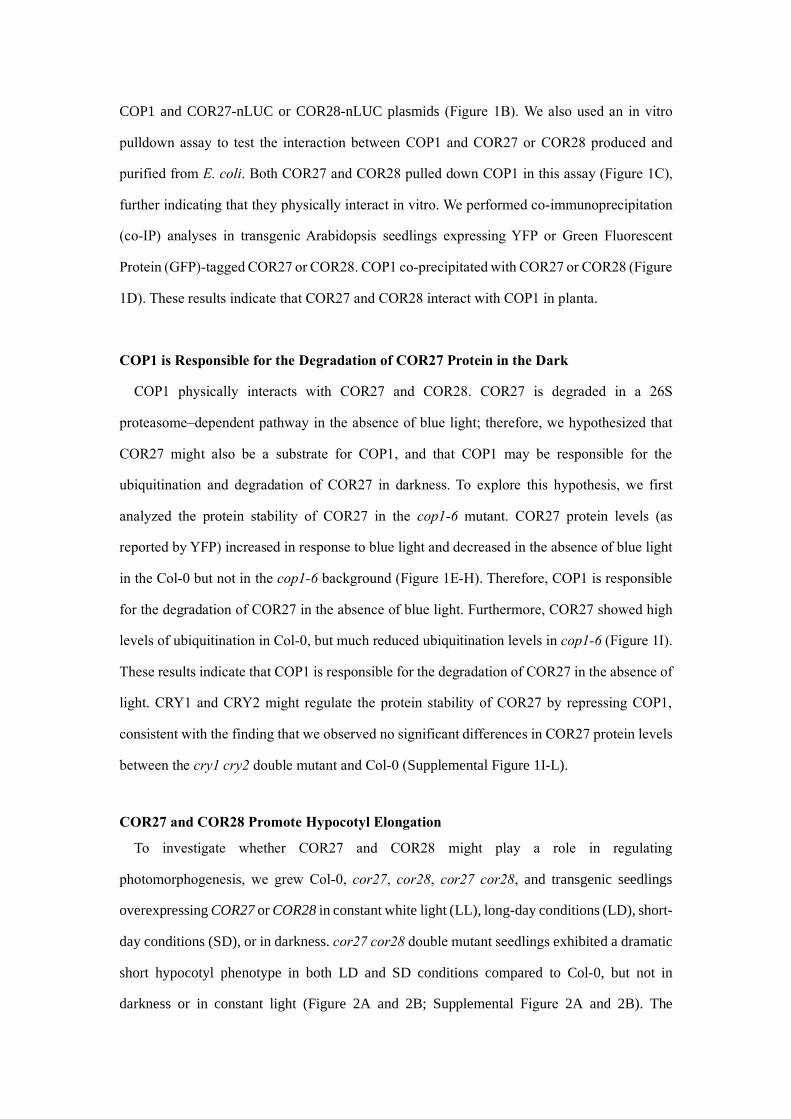
COP1 and COR27-nLUC or COR28-nLUC plasmids (Figure 1B). We also used an in vitro
pulldown assay to test the interaction between COP1 and COR27 or COR28 produced and
purified from E. coli. Both COR27 and COR28 pulled down COP1 in this assay (Figure 1C),
further indicating that they physically interact in vitro. We performed co-immunoprecipitation
(co-IP) analyses in transgenic Arabidopsis seedlings expressing YFP or Green Fluorescent
Protein (GFP)-tagged COR27 or COR28. COP1 co-precipitated with COR27 or COR28 (Figure
1D). These results indicate that COR27 and COR28 interact with COP1 in planta.
COP1 is Responsible for the Degradation of COR27 Protein in the Dark
COP1 physically interacts with COR27 and COR28. COR27 is degraded in a 26S
proteasome–dependent pathway in the absence of blue light; therefore, we hypothesized that
COR27 might also be a substrate for COP1, and that COP1 may be responsible for the
ubiquitination and degradation of COR27 in darkness. To explore this hypothesis, we first
analyzed the protein stability of COR27 in the cop1-6 mutant. COR27 protein levels (as
reported by YFP) increased in response to blue light and decreased in the absence of blue light
in the Col-0 but not in the cop1-6 background (Figure 1E-H). Therefore, COP1 is responsible
for the degradation of COR27 in the absence of blue light. Furthermore, COR27 showed high
levels of ubiquitination in Col-0, but much reduced ubiquitination levels in cop1-6 (Figure 1I).
These results indicate that COP1 is responsible for the degradation of COR27 in the absence of
light. CRY1 and CRY2 might regulate the protein stability of COR27 by repressing COP1,
consistent with the finding that we observed no significant differences in COR27 protein levels
between the cry1 cry2 double mutant and Col-0 (Supplemental Figure 1I-L).
COR27 and COR28 Promote Hypocotyl Elongation
To investigate whether COR27 and COR28 might play a role in regulating
photomorphogenesis, we grew Col-0, cor27, cor28, cor27 cor28, and transgenic seedlings
overexpressing COR27 or COR28 in constant white light (LL), long-day conditions (LD), short-
day conditions (SD), or in darkness. cor27 cor28 double mutant seedlings exhibited a dramatic
short hypocotyl phenotype in both LD and SD conditions compared to Col-0, but not in
darkness or in constant light (Figure 2A and 2B; Supplemental Figure 2A and 2B). The

hypocotyl lengths of cor27 and cor28 single mutant seedlings were slightly shorter than those
of Col-0 in SD conditions (Supplemental Figure 2C and 2D), consistent with the partial
redundancy of COR27 and COR28 in the regulation of photomorphogenesis. The COR27- and
COR28-overexpression lines showed longer hypocotyls than Col-0 under both LD and SD
conditions, but not in constant darkness or in constant light (Figure 2A and 2B; Supplemental
Figure 1A and 2B, 2E). All genotypes tested (Col-0, cor27 cor28, and transgenic lines
overexpressing COR27 or COR28) showed similar hypocotyl lengths when grown in constant
darkness; therefore, we next grew them in SD conditions with white, blue, red, and far-red light
for 7 d to analyze which aspect of light regulated hypocotyl growth. cor27 cor28 double mutant
seedlings developed shorter hypocotyls than Col-0 when grown under blue, red, or far-red light
in SD conditions, whereas the overexpression lines produced significantly longer hypocotyls,
similar to their respective patterns under white light (Figure 2C and 2D). These results indicate
that COR27 and COR28 promote hypocotyl elongation and act as negative regulators of the
blue-, red-, and far-red light–mediated repression of hypocotyl elongation.
COR27 and COR28 Regulate the Expression of COP1-Regulated Genes
To elucidate the mechanism by which COR27 and COR28 regulate photomorphogenesis, we
performed RT-qPCR to analyze the expression of HY5 and PACLOBUTRAZOL RESISTANCE1
(PRE1) in LD-grown Col-0 and cor27 cor28 seedlings. HY5 loss of function leads to
dramatically elongated hypocotyls under all light conditions (Ang et al., 1998), while PRE1
was reported to regulate cell elongation (Lee et al., 2006). HY5 transcript levels were higher in
the cor27 cor28 double mutant relative to Col-0, especially at night, while PRE1 mRNA levels
were lower in cor27 cor28 compared to Col-0 during both the morning and night (Supplemental
Figure 2F and 2G).
We then performed deep sequencing of the transcriptome (RNA-seq) to identify downstream
genes affected in the cor27 cor28 double mutant at night when grown in SD. We collected
samples at Zeitgeber Time (ZT) 20, that is 12 h after lights off, late into the dark period. We
identified 1,440 differentially expressed genes between cor27 cor28 and Col-0 (Figure 3A,
Supplemental Data Set 1). As shown above, COP1 is responsible for the degradation of COR27
in darkness, and COR27 is involved in photomorphogenesis. COR27 should therefore influence
the expression of genes downstream of COP1. Indeed, our RNA-seq datasets of the genes
differentially expressed in cor27 cor28 significantly overlapped with RNA-seq datasets of
genes differentially expressed in the cop1-6 mutant (Figure 3A–D, Supplemental Data Set 2).
For 87% of these genes, the effects of the cor27 cor28 double mutant on gene expression were
the same as in cop1-6 (compare cor27 cor28 vs. Col-0 and cop1-6 vs. Col-0 in Figure 3B and
3C). These results indicate that COP1, COR27 and COR28 exert similar effects on many
commonly regulated genes. A Gene Ontology (GO) analysis revealed a marked enrichment in
light-responsive, growth-related functions in the cop1-6 and cor27 cor28 co-regulated genes,
including PIF4 and HYH (Figure 3D). These genomic data thus provide direct evidence for the
important roles of COR27 and COR28 in photomorphogenesis, particularly in their regulation
of genes involved in cell elongation.
We verified the transcriptomic data using RT-qPCR. We selected PIF4, HYH, PRE1,
INDOLE-3-ACETIC ACID INDUCIBLE 19 (IAA29), and ARABIDOPSIS COLUMBIA SAUR
GENE23 (SAUR23) from the list of genes commonly regulated by COR27, COR28 and COP1
(Figure 3E–J). HY5 and its homologue HYH were both up-regulated in cor27 cor28 and cop1-
6 mutants, but down-regulated in the transgenic lines overexpressing COR27 or COR28.
Conversely, PIF4, PRE1, IAA29, and SAUR23 were all up-regulated in seedlings
overexpressing COR27, COR28, or COP1 compared to Col-0, but down-regulated in the cor27
cor28 and cop1-6 mutants.
COR27 and COR28 Promote Hypocotyl Elongation in a COP1-Dependent Manner
To further determine the relationship between COP1 and COR27, we investigated the genetic
interactions between the COR27 and COP1 loci. We crossed the 35S:YFP-COR27 transgenic
line with the cop1-6 mutant, resulting in the YFP-COR27 cop1-6 line. The long-hypocotyl
phenotype of seedlings overexpressing YFP-COR27 was mostly suppressed in the cop1-6
background (Figure 4A and 4B), which suggested that COR27-promoted hypocotyl elongation
is dependent on COP1. The hypocotyl phenotype of GFP-COR28 cop1-6 was also similar to
that of cop1-6 (Supplemental Figure 3A and 3B). Consistent with these observations, HYH and
HY5 transcript levels were markedly higher in YFP-COR27 cop1-6 seedlings than in YFP-
COR27 in the Col-0 background, while the transcript levels of PIF4, IAA29, PRE1, and

SAUR23 were significantly lower in YFP-COR27 cop1-6 seedlings than in YFP-COR27 in the
Col-0 background (Figure 4C–H). These results indicate that COR27 regulates the transcription
of these elongation-related genes in a COP1-dependent manner.
COR27 Physically Interacts with HY5
COR27 and COR28 indirectly bind to the chromatin surrounding PRR5 and TOC1 to repress
their expression and regulate the circadian clock (Li et al., 2016). We previously proposed that
COR27 and COR28 might form a protein complex with other transcription factors to associate
with the chromatin at these regions (Li et al., 2016). To determine the mechanism by which
COR27 and COR28 regulate photomorphogenesis and the expression of downstream
elongation-related genes, we tested the interaction between COR27, COR28, and various
proteins known to be involved in regulating hypocotyl elongation using a yeast two-hybrid
assay. Among the 32 proteins we tested (gene names and accession numbers are given in
Supplemental Table 2), HY5 interacted with COR27 and COR28 in yeast cells (Figure 5A). We
also pulled down HY5 with both COR27 and COR28 in an in vitro pull-down assay (Figure
5B), indicating that the proteins physically interact in vitro. We further confirmed the interaction
between HY5 and COR27 and COR28 in plant cells by BiLC assay. Indeed, we detected strong
luciferase activity in N. benthamiana leaves co-infiltrated with cLUC-HY5 and COR27-nLUC
or COR28-nLUC plasmids (Figure 5C). Finally, we performed co-IP analyses in transgenic
Arabidopsis seedlings expressing YFP-tagged COR27 and GFP-tagged COR28 in the Col-0
background, which revealed that HY5 co-precipitated with COR27 and COR28 (Figure 5D).
COR27 and COR28 Influence the Expression of HY5-Regulated Genes
COR27 physically interacts with HY5, suggesting they may act together to regulate
transcription and photomorphogenesis. To explore how COR27 and COR28 might influence
the expression of HY5-target genes, we performed RNA-seq to identify downstream genes
affected in the hy5-215 mutant at night (SD, ZT20). Our RNA-seq datasets of differentially
expressed genes in cor27 cor28 relative to Col-0 significantly overlapped with RNA-seq
datasets of differentially expressed genes in hy5-215 (Figure 5E-G, Supplemental Data Set 3).
There were 235 genes co-regulated by both COR27 and COR28 and HY5. For 53.2% of these
genes, the cor27 cor28 double mutant had the opposite effect from that in hy5, with 17.9%

promoted by HY5 but repressed by COR27 and COR28, including HYH and ELF4; the
remaining 35.3% were repressed by HY5 but promoted by COR27 and COR28, including PIF4,
IAA29 and SAUR23. These results indicated that COR27, COR28 and HY5 act in an opposite
manner on the regulation of a large number of common genes. At the same time, we compared
the list of COR27- and COR28-regulated genes with the genes whose promoters are bound by
HY5, previously identified by Lee et al. (2007) (Figure 5D): 20% of the genes affected by the
loss of COR27 and COR28 were direct targets of HY5 (Supplemental Figure 4E and 4F).
Collectively, these results indicate that HY5 and COR27 and COR28 regulate a large number
of common genes. Gene Ontology analysis showed that the molecular function categories
stimulus and absence of light response genes were markedly changed in the list of genes
commonly regulated by COR27, COR28 and HY5, including HYH and PIF4 (Figure 5H;
Supplemental Figure 4F). These genomic data thus provide direct evidence for the important
role of COR27 in HY5-mediated regulation of gene expression.
Similarly, HY5 and PIFs co-regulated photomorphogenesis in both LD and SD conditiosn.
When we grew Col-0, hy5-215, and pifq (a quadruple pif mutant) seedlings in LD and SD
conditions, pifq exhibited a dramatically short hypocotyl phenotype relative to the wild type,
and in sharp contrast to the long hypocotyl phenotype displayed by hy5-215 (Supplemental
Figure 4A–C). Consistent with these phenotypes, PRE1 and IAA29 transcript levels were higher
in hy5-215 compared to Col-0, but lower in pifq seedlings. HY5 also affects PIF4 expression,
hy5-215 caused the up-regulation of PIF4 (Supplemental Figure 4D).
COR27 Associates with the Promoters of HY5 Targets to Regulate their Expression
COR27 physically interacts with HY5 and regulates the expression of HY5-target genes. To
achieve this role, we hypothesized that COR27 might physically associate with the same
genomic regions to which HY5 binds. To test this hypothesis, we performed chromatin
immunoprecipitation (ChIP)-qPCR assays, which revealed that COR27 did associate with the
same chromatin region of the HY5 and PIF4 promoters as HY5 does in vivo (Figure 6A–F). We
used the same PCR primer pairs to detect chromatin binding by HY5 and COR27. A yeast one-
hybrid assay revealed that HY5, but not COR27, bound to the PIF4 promoter (Supplemental
Figure 5A and 5B), indicating that COR27 physically interacts with HY5 to then associate with

the genomic regions to which HY5 binds.
We then analyzed whether COR27 affected the transcriptional activity of HY5 on its target
promoters using a transient transcription assay in Arabidopsis protoplasts and N. benthamiana.
We used a dual-LUC reporter plasmid encoding the firefly luciferase (LUC) gene driven by the
HY5 or PIF4 promoter and a Renilla luciferase (REN) gene driven by the constitutive
cauliflower Mosaic Virus (CaMV) 35S promoter (Figure 6G; Supplemental Figure 5C). We
transiently expressed the HY5pro:LUC reporter in hy5-215 protoplasts together with either
35S:HY5-GFP or 35S:COR27-GFP or both. The LUC activity level from the HY5pro-LUC
reporter was about 2.5-fold higher when we co-expressed HY5 in the protoplasts relative to co-
expressed GFP, while the combination of HY5 and COR27 induced the reporter very modestly
(Figure 6H). We also infiltrated N. benthamiana leaves with an Agrobacterium strain containing
the reporter construct (pGreen-PIF4pro:LUC) alone or mixed with an Agrobacterium strain
containing the indicated effector plasmid (pCambia1300-HY5-YFP, pEGAD-MYC-COR27, or
pCambia1306-VP16-HY5-Flag). We fused HY5 to the VP16 activation domain (from Herpes
simplex virus protein vmw65) to make it a strong constitutive activator of PIF4 transcription
rather than its typical function as a repressor (Supplemental Figure 4D), allowing us to more
easily analyze the effect of COR27 on HY5 transcriptional activity. COR27 repressed the
transcriptional activity of VP16-HY5 on the PIF4pro:LUC reporter gene (Supplemental Figure
5D). COR27 itself did not significantly affect the transcription of PIF4pro:LUC or
HY5pro:LUC (Figure 6H; Supplemental Figure 5D); however, it did inhibit the transcriptional
activity of HY5.
To further study the relationship between COR27, COR28, and HY5, we investigated the
genetic interactions between the corresponding genes. We crossed the cor27 cor28 double
mutant to hy5-215, to generate the hy5 cor27 cor28 triple mutant (Supplemental Fig 6A).
Bringing the hy5-215 mutation into the cor27 cor28 double mutant background dramatically
suppressed its short hypocotyl phenotype, resulting in hypocotyls that were almost as long in
the triple mutant as in the hy5-215 single mutant (Figure 7A and 7B). Transcript levels of HYH
were also markedly lower in hy5 cor27 cor28 compared to cor27 cor28, and were similar to
those seen in hy5-215 (Figure 7C), indicating that COR27 and COR28 regulate hypocotyl
elongation at least partially via HY5, and that COR27 and COR28 also regulate hypocotyl

growth through HY5-independent pathways. Consistent with this, YFP-COR27 hy5-215
showed longer hypocotyls than YFP-COR27 in the Col-0 background, and slightly longer
hypocotyls than in the hy5-215 mutant. HYH transcript levels were also lower in YFP-COR27
hy5-215 seedlings than in YFP-COR27 seedlings, and comparable to those seen in hy5-215,
indicating that COR27 and COR28 regulate the transcription of HYH through HY5 (Figure 7D-
F).
DISCUSSION
Dark growth (skotomorphogenesis) and light-mediated growth (photomorphogenesis) are
critical for germination and seedling development. Buried seeds use elongated hypocotyls,
closed cotyledons, and curved apical hooks (skotomorphogenic growth) to limit mechanical
damage after germination as they grow through soil particles, until they reach the soil surface.
Once top-side, seedlings are exposed to light, which then inhibits hypocotyl elongation and
promotes cotyledon expansion (photomorphogenesis). COP1 is a central repressor of light
signaling, acting downstream of multiple photoreceptors (Ang and Deng, 1994; Christie et al.,
2012). COP1 negatively regulates photomorphogenesis by mediating the degradation of various
photomorphogenesis-promoting transcription factors in the dark, including HY5 (Yi and Deng,
2005; Jiao et al., 2007; Foreman et al., 2011; Liu et al., 2011). Light represses COP1 by
activating multiple photoreceptors, stabilizing the HY5 protein (Ang and Deng, 1994; Christie
et al., 2012). The COP1–HY5 module plays a critical role in regulating photomorphogenesis.
COR27 and COR28 were identified as cold-responsive genes during Arabidopsis transcriptome
analysis, and the expression of COR27 was shown to be clock- and light-regulated (Fowler and
Thomashow, 2002; Mikkelsen and Thomashow, 2009; Li et al., 2016). Light- and low
temperature–regulated COR27 and COR28 are involved in the regulation of the circadian clock,
freezing tolerance, and flowering time, representing a trade-off between flowering and freezing
tolerance (Li et al., 2016).
Here, we showed that COR27 and COR28 are novel regulators of photomorphogenesis, and
are involved in the COP1–HY5-mediated elongation of Arabidopsis seedlings in response to
light. Like the HY5 protein, COR27 and COR28 are degraded in the dark via the 26S
proteasome pathway. We also showed that COP1 physically interacts with COR27 and COR28,

that it is responsible for their degradation, and that their protein levels are relatively low in
etiolated seedlings. Light promotes the accumulation of HY5, COR27, and COR28, most likely
due to the light-mediated inactivation of COP1 (Ang and Deng, 1994; Christie et al., 2012).
COP1 regulates the protein stability of HY5, COR27, and COR28, while COR27 or COR28
form a protein complex with HY5 on the chromatin of HY5 target genes to modulate their
transcription and ultimately photomorphogenesis. COR27 inhibits the transcriptional activity
of HY5 even in the dark to promote skotomorphogenic growth, and COR27 and COR28 fine-
tune photomorphogenesis by modulating the COP1–HY5 module in response to changing light
environments (Figure 6F).
Multiple HY5-interacting proteins have been identified and characterized, such as HYH, G-
BOX BINDING FACTOR1 (GBF1), CALMODULIN7 (CAM7), B-BOX DOMAIN
PROTEIN21 (BBX21), BBX22, BBX24, BBX25, BBX28, and BBX32. These factors interact
with HY5 to positively or negatively regulate its transcriptional activity (Holm et al., 2002;
Datta et al., 2006; Datta et al., 2008; Holtan et al., 2011; Singh et al., 2012; Gangappa et al.,
2013; Abbas et al., 2014; Zhang et al., 2017; Lin et al., 2018). HY5 is a key transcription factor
in photomorphogenesis, functioning alongside multiple factors to fine-tune HY5 biochemical
activity and gene expression in response to the changing light environment. COP1 also
regulates the protein stability of BBX28, which represses photomorphogenesis by inhibiting
the DNA-binding activity of HY5 (Lin et al., 2018). Different proteins thus regulate HY5 via
different mechanisms, with some regulating its DNA-binding activity while others form
complexes with HY5 at its target chromatin sites to regulate its transcriptional activity. The
expression of COR27 and COR28 is also regulated by cold temperatures and the circadian clock;
they may integrate multiple signals to fine-tune skotomorphogenesis and photomorphogenesis.
The transcriptome data indicates that cold-responsive genes are enriched in the COP1, HY5,
COR27 and COR28 co-regulated genes, suggesting that these proteins may also be involved in
the joint regulation of cold tolerance.
COR27 and COR28 are both small proteins with unknown biochemical functions. They do
not have any known DNA-binding domains, and cannot bind DNA themselves in vitro,
indicating that they are unlikely to be transcription factors (Supplemental Figure 5A and 5B).
We previously showed that they physically associate with the genomic regions of the clock

genes TOC1 and PRR5 to directly regulate their transcription, and hypothesized that they may
form a protein complex with other DNA-binding transcription factors to associate with
chromatin and regulate the transcription of these and other clock genes (Li et al., 2016). Here,
we showed that COR27 interacts with HY5 and physically associates with the genomic regions
to which HY5 binds, regulating the transcriptional activity of HY5 and its gene expression. Our
results confirm that COR27 and COR28 work as transcriptional regulators and interact with
other transcription factors to regulate the expression of at least 1,840 genes. When compared
with Col-0, about evenly split between up-regulation (53%) and down-regulation (47%) in the
cor27 cor28 mutant. Only a fraction of these genes (12.8 %) were also regulated by HY5,
indicating that COR27 and COR28 may interact with multiple transcription factors to regulate
transcription.
HY5 protein also accumulated to higher levels in cor27 cor28 than in Col-0, consistent with
the fact that COR27 and COR28 inhibited the transcription of HY5 (Supplemental Fig 6B and
6C). The long-hypocotyl phenotype of seedlings overexpressing YFP-COR27 was mostly
suppressed in the cop1-6 background. It is interesting that COP1 is responsible for the
degradation of COR27 in the dark but is also required for its function. One possible explanation
for the suppression of the long hypocotyl phenotype of YFP-COR27 seedlings by cop1-6 would
invoke an accumulation of HY5 protein. To test this possibility, we checked the protein levels
of HY5 in Col-0, cop1-6, YFP-COR27 and YFP-COR27 cop1-6 (Supplemental Fig 6D and 6E).
Our results indicated that indeed, there was more HY5 protein in cop1-6 and also in YFP-
COR27 cop1-6 relative to Col-0 and YFP-COR27.
In summary, we showed that COR27 and COR28 are negative regulators of
photomorphogenesis. COR27 and COR28 undergo COP1-mediated degradation in the dark.
Light promotes the accumulation of HY5 and COR27, while COR27 physically interacts with
HY5 to inhibit its transcriptional activity and fine-tune skotomorphogenesis development in the
dark and photomorphogenic development in the light (Supplemental Fig 6F).
MATERIALS AND METHODS
Plant Materials and Growth Conditions

We used the Arabidopsis Columbia (Col-0) accession as wild type. The cor27 (CS834545),
cor28 (SALK_137155), cor27 cor28, 35S:YFP-COR27, 35S:GFP-COR28, cop1-6,
pro35S:MYC-COP1, hy5-215, hy5 (SALK_096651), and pifq (pif1 pif3 pif4 pif5) lines have
been described previously (Ang and Deng, 1994; McNellis et al., 1994; Leivar et al., 2008; Li
et al., 2016; Lin et al., 2016). Additional lines (YFP-COR27 cop1-6, GFP-COR28 cop1-6, and
YFP-COR27 hy5-215) were generated by genetic crosses and combined phenotyping and
genotyping of F2 progeny.
All seeds were surface-sterilized in 75% ethanol for 10 min, washed four times with sterile
water, then sown on half-strength Murashige and Skoog (MS) medium supplemented with 0.8%
agar and 1% sucrose. Plates were then stratified for 4 d in the dark at 4°C before transfer to a
Percival growth chamber (AR-22L for white light,E-30LEDL3 for blue, red or far-red light,
Percival Scientific, Perry, IA, USA) under either constant light, LD (long days, 16 h light/8 h
dark), SD (short days, 8 h light/16 h dark), or constant darkness conditions at 22°C. The lights
used were white light (Fluorescent light, Philips F17T8/TL841, 90 μmol/m2/s), blue light (LED
light 40 μmol/m2/s), red light (LED light 40 μmol/m2/s), or far-red light (LED light 8 μmol/m2/s).
Immunoblot Analysis
Seeds were grown for 7 d in SD conditions (white light) before being moved to blue or white
light or darkness for 24 h, after we exposed seedlings to a defined light treatment (light to dark
or dark to light for the indicated times). After treatment, we collected seedlings and prepared
whole protein extracts with extraction buffer (25.2g Glycerol, 0.02g Bromophenol Blue, 4g
SDS, 20 mL 1M Tris-HCl, pH6.8, 3.1g DTT, to 50mL ddH2O). Equal protein amounts were
loaded and separated on a 10% or 12% SDS-PAGE gel and transferred to Nitrocellulose
Blotting Membrane (P/N66485, PALL, USA)VDF membrane. We then probed the membrane
with an anti-GFP antibody (AE012, Abclonal, China, 1:3,000 dilution), stripped the blot, and
re-probed with the anti-ACTIN antibody (AC009, Abclonal, China, 1:3,000 dilution) as loading
control.
Transcriptome Analysis
We extracted total RNA from 7-d-old Col-0, cor27 cor28, cop1-6 and hy5-215 seedlings
grown in SD conditions and collected at ZT20 (12 h after lights off) under dim green light,
using the mirVana miRNA Isolation Kit (Ambion-1561). We then generated mRNA sequencing
libraries (with three independent biological replicates), which were sequenced by OE Biotech
Co., Ltd. (Shanghai, China). We evaluated RNA integrity on an Agilent 2100 Bioanalyzer
(Agilent Technologies, Santa Clara, CA, USA). The samples with RNA Integrity Number (RIN)
≥ 7 were subjected to further analysis. We generated libraries using the TruSeq Stranded
mRNA LT Sample Prep Kit (Illumina, San Diego, CA, USA) according to the manufacturer’s
instructions. Then these libraries were sequenced on an Illumina sequencing platform (Illumina
HiSeq X Ten) as 125bp/150bp paired-end reads. Sequenced reads were processed using
Trimmomatic (Version 0.36). Reads containing multiple Ns and low-quality reads were
removed to obtain clean reads. We then mapped the clean reads to the Arabidopsis reference
genome (TAIR10.1_NCBI) using hisat2 (Version 2.2.1.0). We then quantified read counts per
gene and Fragments Per Kilobase exon per Million reads mapped (FPKM) values using
Cufflinks (version:2.2.1), while read count values per transcript (protein_coding) were
calculated using bowtie2 and eXpress. We identified differentially expressed genes (DEGs)
using DESeq (Version1.18.0) (Anders et al., 2013). The R package functions ‘Estimate Size
Factors’ and ‘nbinom Test’, Padjvalues<0.05 and Fold Changes >1.5 were set as the threshold
for significantly different expression. We downloaded the ChIP-chip (Chromatin
immunoprecipitation (ChIP) followed by microarray hybridization) data for HY5 from the
Gene Expression Omnibus database (accession number GSE6510). We performed a GO
enrichment analysis (Biological processes) using the Gene Ontology website
(http://geneontology.org) with the released version 2019-07-01.
RT-qPCR Analysis
We performed all RT-qPCR expression analyses as described previously (Ma et al., 2016).
Briefly, we isolated total RNAs using a RNAiso Plus kit (Takara Bio, Shiga, Japan). We
synthesized first-strand cDNAs from 500 ng of starting total RNA using a Prime Script RT
Reagent Kit with a genomic DNA Eraser (Takara Bio). We then used SYBR Premix Ex Tag
(Takara Bio) and the MX3000 Real Time PCR System (Stratagene, San Diego, CA, USA) for
the qPCR reactions. We used the ISOPENTENYL DIPHOSPHATE ISOMERASE 2 (IPP2,

At3g02780) and ACTIN 7 (At5g09810) genes as internal controls. Following initial
denaturation of the first-strand cDNAs, we executed the following PCR program: denaturation
at 95°C for 30 s, 2 step PCR for 40 cycles (95°C for 5 s, 60°C for 20 s per cycle) with
fluorescence read at the end of each cycle, a dissociation program was performed after the
reaction. The biological replicates represent three independent experiments involving about 20
seedlings per experiment. Three technical replicates were performed for each PCR reaction.
The primers used are listed in Supplemental Data Set 4.
Yeast Two-Hybrid Analysis
We cloned the coding sequences of COR27 and COR28 in-frame with the GAL4 DNA-
binding domain (BD) sequence in the bait vector pBridge (Takara Bio). We obtained the
Arabidopsis cDNA library clones in the prey vector pACT from Dr. Joe Ecker (Salk Institute,
CA, USA). We co-transformed the bait plasmid pBridge-COR27/COR28 and the prey plasmid
library DNA into yeast strain Y190. Yeast cells were then grown on synthetic dropout medium
(SD –Trp –Leu) plates for 4 d. We tested the interactions using -galactosidase assays (168
μg/mL substrate), checking the activity every h for eight h, and verified positive yeast clones
by PCR and sequencing. The full list of identified COR27 and COR28 interactors is given in
Supplemental Table 1. We fused the coding sequence of COP1 in-frame with the sequence of
the GAL4 activation domain (AD) in the prey vector pGADT7 (Takara Bio).We cloned the
coding sequence of HY5 into the pGADT7 or pDEST22 (Takara Bio) vectors to verify the
interaction in yeast strain AH109 against pBridge-COR27, pBridge-COR28 or pDEST32-
COR28.
Bi-Luminescence Complementation (BiLC) Assays
We fused COR27/COR28 or COP1/HY5 to the N- or C-terminus of firefly luciferase, then
transformed the constructs into Agrobacterium (Agrobacterium tumefaciens) strain GV3101.
We collected overnight Agrobacterium cultures by centrifugation at 3200g for 20 min and
resuspended the cells in MES buffer (10 mM MES,10 mM MgCl2, and 100 mM acetosyringone)
to a final OD600 = 0.8~1 before infiltration of Nicotiana benthamiana leaves. We then returned
infiltrated plants to LD conditions for 3 d. Before luciferase activity observation, we infiltrated
leaves with luciferin solution (1 mM luciferin and 0.01%Triton X-100) and captured images
immediately using a CCD camera (Tanon-5200, BioTanon, China).
in vitro Pull-downs
We performed in vitro pulldown protein–protein interaction assays as previously described
(Liu et al., 2008; Liu et al., 2013b; Ma et al., 2016; Liang et al., 2018; Yang et al., 2018), with
the following modifications. We cloned the full-length coding sequences of COR27 or COR28
into the pGEX4T-1 vector, and the full-length coding sequences of COP1 or HY5 into the
pCold-TF vector. We then produced and purified the proteins in Escherichia coli BL21 strain
by using Glutathione Sepharose 4B (17-0756-01, GE Healthcare, USA) or Ni-NTA Agarose
(R901-15, invitrogen, USA). We then incubated equal amounts of eluted TF-His-COP1 or TF-
His-HY5 with GST-COR27 or GST-COR28 in XB buffer (50 mM Tris (pH7.8), 500 mM NaCl,
0.5% Triton X-100, 1 mM PMSF, 4.7mM β-ME), used to pull down the protein complexes with
glutathione beads. We removed unbound proteins by washing with WB buffer (50 mM Tris
(pH7.8), 500 mM NaCl,0.1% Triton X-100, 1 mM PMSF), after which we eluted bound proteins
and analyzed the resulting eluates by immunoblot analysis an anti-TF antibody (M201; Takara
Bio, Japan, 1:3,000 dilution) or anti-GST antibody (G018, Abcam, China, 1:3,000 dilution).
Co-Immunoprecipitations (Co-IPs)
The co-IP procedure has been described previously (Liu et al., 2008; Liu et al., 2013b; Ma et
al., 2016; Liang et al., 2018; Yang et al., 2018). Briefly, we grew seedlings for 10 d from the
genotypes Col-0, YFP-COR27 (in Col-0), GFP-COR28 (in Col-0) in SD conditions, at which
point we treated them with 50 mM MG132 at ZT 7.5 (30 min before lights off). We then moved
seedlings into darkness at ZT8 for 4 h, collected and ground tissue in liquid nitrogen, which we
then homogenized in binding buffer (20 mM HEPES (pH 7.5), 40 mM KCl, 1 mM EDTA, 1%
Triton X-100, 1 mM PMSF). We incubated protein extracts at 4°C for 10 min, before
centrifugation at 14,000g for 10 min. We mixed the supernatant with 35 μL of anti-GFP-IgG-
coupled protein A Sepharose (KTSM1301, Alpalife, Shenzhen, China), incubated the mixtures
at 4°C for 30 min, and washed the beads three times with washing buffer (20 mM HEPES (pH
7.5), 40 mM KCl, 1 mM EDTA, 0.1% Triton X-100). We eluted bound proteins from the affinity

beads with 4× SDS/PAGE sample buffer and analyzed the eluates by immunoblotting with anti-
COP1 (YKCP938; Youke Biotech, China, 1:2,000 dilution) and anti-HY5 (PHY1908, QWBIO,
China, 1:2,000 dilution) antibodies to detect the target proteins.
Yeast One-Hybrid Analysis
We cloned PIF4 promoter fragments (–1,703bp to –1,231bp, –1,286bp to –785bp, –870bp to
–435bp, –545bp to –1bp) into the pLacZi destination vector and transformed the resulting
constructs into yeast strain YM4271. We also cloned the COR27 and HY5 coding sequences
into the pDEST22 vector. We then transformed the resulting constructs into YM4271 cells
containing the various promoter reporter plasmids. We grew cells in synthetic dropout medium
(SD –Trp –Ura), and analyzed protein-promoter interactions using a β-galactosidase assay (with
168 μg/mL substrate added).
Chromatin Immunoprecipitation (ChIP) Assays
We performed ChIP experiments as described previously (Liu et al., 2008; Liu et al., 2013b;
Ma et al., 2016; Liang et al., 2018; Yang et al., 2018), using 10-d-old Col-0, YFP-COR27 (in
Col-0), and HY5-HA (in the hy5-215 background) seedlings grown in SD conditions. We
harvested 2 g plant material, which we then cross-linked with 1% formaldehyde (Sigma-Aldrich,
St. Louis, MO, USA) for 15 min under a vacuum. We stopped cross-linking by the addition of
glycine to the solution, to a final concentration of 0.125 M. We rinsed seedlings with water,
froze them in liquid nitrogen, and ground them into a fine powder. We sonicated chromatin
fragments (~500 bp) with a bioruptor (Bioruptor Plus,Diagenode SA, Belgium, at program 30
sec on and 30 sec off for 15 min) before immunoprecipitation with anti-GFP (AE012, Abclonal,
China) or anti-HA (clone3F10, Roche, USA) antibodies, and analyzed the precipitated DNA by
qPCR with the indicated primer pairs (Supplemental Data Set 4). The level of binding was
calculated as the ratio between the IP and Input proteins.
Transient Transcription Dual-LUC Assays
We performed transient transcription dual-LUC assays using N. benthamiana plants as
previously described (Liu et al., 2008). We used Agrobacterium cultures containing both the
reporter construct (pGreen-PIF4pro:LUC) and the helper construct pSoup-P19, alone or mixed
with Agrobacterium cultures containing the effector plasmids (pCambia1300-HY5-YFP,
pEGAD-MYC-COR27, or pCambia1306-VP16HY5-Flag). We cloned the full length coding
sequences of COR27 and HY5 into pEGAD (ABRC) at the EcoRI and XhoI restriction sites,
resulting in pEGAD-MYC-COR27, into pCambia1300 and pCambia1306 (CAMBIA, Canberra,
Australia) at the BamHI and SalI sites, resulting in pCambia1300-HY5-YFP and pCambia1306-
VP16HY5-Flag.. We collected overnight Agrobacterium cultures by centrifugation at 3200g for
20 min and resuspended pellets in infiltration buffer to a final OD600 = 0.8~1, and infiltrated
cell suspensions into healthy N.benthamiana leaves.
We carried out protoplast isolation and PEG-mediated transformation as previously
described (Liang et al., 2018; Yang et al., 2018). We isolated protoplasts from three-week-old
hy5-215 plants grown in SD conditions. We transfected protoplasts with a total of 20 μg DNA
(effector constructs 35S:COR27-GFP and 35S:HY5-GFP, and pGreen-HY5pro:LUC reporter)
and incubated overnight. We measured luciferase activity using a luminometer (GloMax 20/20;
Promega, Madison, WI, USA) with Dual-Luciferase Reporter Assay System (E1910, Promega,
USA), according to the manufacturer’s instructions.
ACCESSION NUMBERS
Sequence data for genes described in this article can be found in The Arabidopsis Information
Resource (TAIR) under the following accession numbers:
COR27 (At5g42900), COR28 (At4g33980), COP1 (At2g32950), HY5 (At5g11260), CRY1
(At4g08920), CRY2 (At1g04400), HYH (At3g17609), PIF4 (At2g43010), PRE1 (At5g39860),
IAA29 (At4g32280), SAUR23 (At5g18060).
Accession numbers for 32 photomorphogenesis-related genes tested for interaction with
COR27 are listed in Supplemental Table 2.
RNA-seq data have been deposited into the Gene Expression Omnibus with the following
accession numbers: cor27 cor28 and cop1_RNA-seq GSE154409, hy5_RNA-seq GSE154416.
AUTHOR CONTRIBUTIONS

X.L. and H.L. conceived the project. X.L. performed most of the experiments, C. L. performed
the genomic expression analysis. Z.Z and Y.L performed the RNA-seq analysis. D.M. J.Z and
Y.Y made some of the constructs. X.L and H.L analyzed the data and wrote the manuscript.
ACKNOWLEDGEMENTS
The authors thank Drs Jigang Li and Jingbo Jing for materials and technical assistance. This
work was supported in part by the National Key Research and Development Program of China
(2017YFA0503800), the Foundation of Youth Innovation Promotion Association of the
Chinese Academy of Sciences (to Li Xu), National Natural Science Foundation of China
(31670307, 31825004, 31721001, 31730009, 31670282, 31701231), the Strategic Priority
Research Program of the Chinese Academy of Sciences (XDB27030000), and the Program of
Shanghai Academic Research Leader (19XD1404400).
SUPPLEMENTAL DATA
Supplemental Figure 1. The COR27 protein is degraded in the dark by the 26S
proteasome and in CRY1 and COP1-dependent manner.
Supplemental Figure 2. COR27 and COR28 inhibit photomorphogenesis in
photoperiod condition.
Supplemental Figure 3. COR28 inhibits photomorphogenesis in a COP1 dependent
manner.
Supplemental Figure 4. HY5 and PIFs regulate hypocotyl elongation in both LD and
SD.
Supplemental Figure 5. HY5, but not COR27, binds the PIF4 promoter.
Supplemental Table 1. Full list of identified COR27 or COR28 interactors.
Supplemental Table 2. Accession numbers for 32 photomorphogenesis-related genes
tested for interaction with COR27.
Supplemental Data Set 1. List of 1,440 COR27 and COR28 regulated genes.
Supplemental Data Set 2. List of 3,854 COP1 regulated genes.
Supplemental Data Set 3. List of 2,157 HY5 regulated genes.
Supplemental Data Set 4. Primers list used in this study.
Supplemental Data Set 5. Statistical analysis of t-test and ANOVA results for the data
shown in figures.

References
Abbas, N., Maurya, J.P., Senapati, D., Gangappa, S.N., and Chattopadhyay, S. (2014). Arabidopsis CAM7
and HY5 physically interact and directly bind to the HY5 promoter to regulate its expression
and thereby promote photomorphogenesis. The Plant cell 26, 1036-1052.
Anders, S., and Hubre W. (2013). Differential expression of RNA-Seq data at the gene level-the DESeq
package. European Molecular Biology Laboratory (EMBL)
Ang, L.H., and Deng, X.W. (1994). Regulatory hierarchy of photomorphogenic loci: allele-specific and
light-dependent interaction between the HY5 and COP1 loci. The Plant cell 6, 613-628.
Ang, L.H., Chattopadhyay, S., Wei, N., Oyama, T., Okada, K., Batschauer, A., and Deng, X.W. (1998).
Molecular interaction between COP1 and HY5 defines a regulatory switch for light control of
Arabidopsis development. Mol Cell 1, 213-222.
Binkert, M., Kozma-Bognar, L., Terecskei, K., De Veylder, L., Nagy, F., and Ulm, R. (2014). UV-B-
responsive association of the Arabidopsis bZIP transcription factor ELONGATED HYPOCOTYL5
with target genes, including its own promoter. Plant Cell 26, 4200-4213.
Bischof, S. (2020). Chimeric Activators and Repressors Define HY5 Activity. The Plant cell 32, 793-794.
Briggs, W.R., and Christie, J.M. (2002). Phototropins 1 and 2: versatile plant blue-light receptors. Trends
in plant science 7, 204-210.
Burko, Y., Seluzicki, A., Zander, M., Pedmale, U.V., Ecker, J.R., and Chory, J. (2020). Chimeric Activators
and Repressors Define HY5 Activity and Reveal a Light-Regulated Feedback Mechanism. The
Plant cell 32, 967-983.
Christie, J.M., Arvai, A.S., Baxter, K.J., Heilmann, M., Pratt, A.J., O'Hara, A., Kelly, S.M., Hothorn, M.,
Smith, B.O., Hitomi, K., Jenkins, G.I., and Getzoff, E.D. (2012). Plant UVR8 photoreceptor
senses UV-B by tryptophan-mediated disruption of cross-dimer salt bridges. Science 335, 1492-
1496.
Datta, S., Hettiarachchi, G.H., Deng, X.W., and Holm, M. (2006). Arabidopsis CONSTANS-LIKE3 is a
positive regulator of red light signaling and root growth. The Plant cell 18, 70-84.
Datta, S., Johansson, H., Hettiarachchi, C., Irigoyen, M.L., Desai, M., Rubio, V., and Holm, M. (2008).
LZF1/SALT TOLERANCE HOMOLOG3, an Arabidopsis B-box protein involved in light-dependent
development and gene expression, undergoes COP1-mediated ubiquitination. The Plant cell
20, 2324-2338.
Demarsy, E., and Fankhauser, C. (2009). Higher plants use LOV to perceive blue light. Curr Opin Plant
Biol 12, 69-74.
Deng, X.W., Caspar, T., and Quail, P.H. (1991). cop1: a regulatory locus involved in light-controlled
development and gene expression in Arabidopsis. Genes & development 5, 1172-1182.
Foreman, J., Johansson, H., Hornitschek, P., Josse, E.M., Fankhauser, C., and Halliday, K.J. (2011). Light
receptor action is critical for maintaining plant biomass at warm ambient temperatures. Plant
J 65, 441-452.
Fowler, S., and Thomashow, M.F. (2002). Arabidopsis transcriptome profiling indicates that multiple
regulatory pathways are activated during cold acclimation in addition to the CBF cold response
pathway. The Plant cell 14, 1675-1690.
Gangappa, S.N., Crocco, C.D., Johansson, H., Datta, S., Hettiarachchi, C., Holm, M., and Botto, J.F.
(2013). The Arabidopsis B-BOX protein BBX25 interacts with HY5, negatively regulating BBX22
expression to suppress seedling photomorphogenesis. The Plant cell 25, 1243-1257.
Hoecker, U. (2017). The activities of the E3 ubiquitin ligase COP1/SPA, a key repressor in light signaling.

Curr Opin Plant Biol 37, 63-69.
Holm, M., Ma, L.G., Qu, L.J., and Deng, X.W. (2002). Two interacting bZIP proteins are direct targets of
COP1-mediated control of light-dependent gene expression in Arabidopsis. Genes &
development 16, 1247-1259.
Holtan, H.E., Bandong, S., Marion, C.M., Adam, L., Tiwari, S., Shen, Y., Maloof, J.N., Maszle, D.R., Ohto,
M.A., Preuss, S., Meister, R., Petracek, M., Repetti, P.P., Reuber, T.L., Ratcliffe, O.J., and
Khanna, R. (2011). BBX32, an Arabidopsis B-Box protein, functions in light signaling by
suppressing HY5-regulated gene expression and interacting with STH2/BBX21. Plant Physiol
156, 2109-2123.
Huang, X., Ouyang, X., Yang, P., Lau, O.S., Li, G., Li, J., Chen, H., and Deng, X.W. (2012). Arabidopsis
FHY3 and HY5 positively mediate induction of COP1 transcription in response to
photomorphogenic UV-B light. Plant Cell 24, 4590-4606.
Jiao, Y., Lau, O.S., and Deng, X.W. (2007). Light-regulated transcriptional networks in higher plants. Nat
Rev Genet 8, 217-230.
Lau, O.S., and Deng, X.W. (2012). The photomorphogenic repressors COP1 and DET1: 20 years later.
Trends in plant science 17, 584-593.
Lee, S., Lee, S., Yang, K.Y., Kim, Y.M., Park, S.Y., Kim, S.Y., and Soh, M.S. (2006). Overexpression of PRE1
and its homologous genes activates Gibberellin-dependent responses in Arabidopsis thaliana.
Plant Cell Physiol 47, 591-600.
Leivar, P., and Quail, P.H. (2011). PIFs: pivotal components in a cellular signaling hub. Trends in plant
science 16, 19-28.
Leivar, P., Monte, E., Oka, Y., Liu, T., Carle, C., Castillon, A., Huq, E., and Quail, P.H. (2008). Multiple
phytochrome-interacting bHLH transcription factors repress premature seedling
photomorphogenesis in darkness. Curr Biol 18, 1815-1823.
Li, X., Ma, D., Lu, S.X., Hu, X., Huang, R., Liang, T., Xu, T., Tobin, E.M., and Liu, H. (2016). Blue Light- and
Low Temperature-Regulated COR27 and COR28 Play Roles in the Arabidopsis Circadian Clock.
The Plant cell 28, 2755-2769.
Lian, H.L., He, S.B., Zhang, Y.C., Zhu, D.M., Zhang, J.Y., Jia, K.P., Sun, S.X., Li, L., and Yang, H.Q. (2011).
Blue-light-dependent interaction of cryptochrome 1 with SPA1 defines a dynamic signaling
mechanism. Genes & development 25, 1023-1028.
Liang, T., Mei, S., Shi, C., Yang, Y., Peng, Y., Ma, L., Wang, F., Li, X., Huang, X., Yin, Y., and Liu, H. (2018).
UVR8 Interacts with BES1 and BIM1 to Regulate Transcription and Photomorphogenesis in
Arabidopsis. Dev Cell 44, 512-523 e515.
Lin, C. (2002). Blue light receptors and signal transduction. The Plant cell 2002S, S207-S225.
Lin, F., Jiang, Y., Li, J., Yan, T., Fan, L., Liang, J., Chen, Z.J., Xu, D., and Deng, X.W. (2018). B-BOX DOMAIN
PROTEIN28 Negatively Regulates Photomorphogenesis by Repressing the Activity of
Transcription Factor HY5 and Undergoes COP1-Mediated Degradation. The Plant cell 30, 2006-
2019.
Lin, X.L., Niu, D., Hu, Z.L., Kim, D.H., Jin, Y.H., Cai, B., Liu, P., Miura, K., Yun, D.J., Kim, W.Y., Lin, R., and
Jin, J.B. (2016). An Arabidopsis SUMO E3 Ligase, SIZ1, Negatively Regulates
Photomorphogenesis by Promoting COP1 Activity. PLoS Genet 12, e1006016.
Liu, B., Zuo, Z., Liu, H., Liu, X., and Lin, C. (2011). Arabidopsis cryptochrome 1 interacts with SPA1 to
suppress COP1 activity in response to blue light. Genes & development 25, 1029-1034.
Liu, H., Yu, X., Li, K., Klejnot, J., Yang, H., Lisiero, D., and Lin, C. (2008). Photoexcited CRY2 interacts with
CIB1 to regulate transcription and floral initiation in Arabidopsis. Science 322, 1535-1539.
Liu, H., Wang, Q., Liu, Y., Zhao, X., Imaizumi, T., Somers, D.E., Tobin, E.M., and Lin, C. (2013a).
Arabidopsis CRY2 and ZTL mediate blue-light regulation of the transcription factor CIB1 by
distinct mechanisms. Proc Natl Acad Sci U S A 110, 17582-17587.
Liu, Y., Li, X., Li, K., Liu, H., and Lin, C. (2013b). Multiple bHLH proteins form heterodimers to mediate
CRY2-dependent regulation of flowering-time in Arabidopsis. PLoS Genet 9, e1003861.
Ma, D., Li, X., Guo, Y., Chu, J., Fang, S., Yan, C., Noel, J.P., and Liu, H. (2016). Cryptochrome 1 interacts
with PIF4 to regulate high temperature-mediated hypocotyl elongation in response to blue
light. Proc Natl Acad Sci U S A 113, 224-229.
McNellis, T.W., von Arnim, A.G., and Deng, X.W. (1994). Overexpression of Arabidopsis COP1 results in
partial suppression of light-mediated development: evidence for a light-inactivable repressor
of photomorphogenesis. The Plant cell 6, 1391-1400.
Mikkelsen, M.D., and Thomashow, M.F. (2009). A role for circadian evening elements in cold-regulated
gene expression in Arabidopsis. Plant J 60, 328-339.
Ni, M., Tepperman, J.M., and Quail, P.H. (1998). PIF3, a phytochrome-interacting factor necessary for
normal photoinduced signal transduction, is a novel basic helix-loop-helix protein. Cell 95, 657-
667.
Osterlund, M.T., Hardtke, C.S., Wei, N., and Deng, X.W. (2000). Targeted destabilization of HY5 during
light-regulated development of Arabidopsis. Nature 405, 462-466.
Pedmale, U.V., Huang, S.S., Zander, M., Cole, B.J., Hetzel, J., Ljung, K., Reis, P.A., Sridevi, P., Nito, K.,
Nery, J.R., Ecker, J.R., and Chory, J. (2016). Cryptochromes Interact Directly with PIFs to Control
Plant Growth in Limiting Blue Light. Cell 164, 233-245.
Podolec, R., and Ulm, R. (2018). Photoreceptor-mediated regulation of the COP1/SPA E3 ubiquitin ligase.
Curr Opin Plant Biol 45, 18-25.
Quail, P.H. (2002). Photosensory perception and signalling in plant cells: new paradigms? Curr Opin Cell
Biol 14, 180-188.
Rizzini, L., Favory, J.J., Cloix, C., Faggionato, D., O'Hara, A., Kaiserli, E., Baumeister, R., Schafer, E., Nagy,
F., Jenkins, G.I., and Ulm, R. (2011). Perception of UV-B by the Arabidopsis UVR8 protein.
Science 332, 103-106.
Singh, A., Ram, H., Abbas, N., and Chattopadhyay, S. (2012). Molecular interactions of GBF1 with HY5
and HYH proteins during light-mediated seedling development in Arabidopsis thaliana. J Biol
Chem 287, 25995-26009.
Sullivan, J.A., and Deng, X.W. (2003). From seed to seed: the role of photoreceptors in Arabidopsis
development. Dev Biol 260, 289-297.
Yang, Y., Liang, T., Zhang, L., Shao, K., Gu, X., Shang, R., Shi, N., Li, X., Zhang, P., and Liu, H. (2018).
UVR8 interacts with WRKY36 to regulate HY5 transcription and hypocotyl elongation in
Arabidopsis. Nat Plants 4, 98-107.
Yi, C., and Deng, X.W. (2005). COP1 - from plant photomorphogenesis to mammalian tumorigenesis.
Trends Cell Biol 15, 618-625.
Zhang, H., He, H., Wang, X., Wang, X., Yang, X., Li, L., and Deng, X.W. (2011). Genome-wide mapping
of the HY5-mediated gene networks in Arabidopsis that involve both transcriptional and post-
transcriptional regulation. Plant J 65, 346-358.
Zhang, X., Huai, J., Shang, F., Xu, G., Tang, W., Jing, Y., and Lin, R. (2017). A PIF1/PIF3-HY5-BBX23
Transcription Factor Cascade Affects Photomorphogenesis. Plant Physiol 174, 2487-2500.

Zuo, Z., Liu, H., Liu, B., Liu, X., and Lin, C. (2011). Blue light-dependent interaction of CRY2 with SPA1
regulates COP1 activity and floral initiation in Arabidopsis. Curr Biol 21, 841-847.

Figure 1. COR27 and COR28 Interact with COP1.
(A) Histidine auxotrophy assays showing the interaction between COR27 or COR28 and COP1. Yeast
cells (strain AH109) containing plasmids encoding the indicated proteins were grown on medium in the
presence (+) or absence (–) of histidine (His) in the dark for 3 d.
(B) Bimolecular luminescence complementation assays showing COR27 and COR28 interacting with
COP1. Leaf epidermal cells of Nicotiana benthamiana were co-infiltrated with nLuc, COR27-nLuc, or
COR28-nLuc and cLuc or cLuc-COP1 as indicated.
(C) In vitro pulldown assays showing the interaction of COR27 or COR28 with COP1. GST or GST-tagged
COR27 or COR28 bound to glutathione-agarose beads was mixed with His-TF or His-TF-COP1 purified
from Escherichia coli, as shown in the Coomassie-stained SDS-PAGE gel (left): (M) size marker, (1) His-
TF tag, (2) His-TF-tagged COP1, (3) GST tag, (4) GST-tagged COR27, (5) GST-tagged COR28. The
asterisk indicates the respective target protein. The pulldown products were analyzed using immunoblots
probed with anti-GST or anti-TF antibodies (right).
(D) Co-immunoprecipitation assays showing in vivo protein interactions. Proteins were extracted from 10-
d-old SD-grown YFP-COR27, GFP-COR28, or Col-0 seedlings. Input: immunoblots showing the level of
YFP-COR27, GFP-COR28, COP1 in total protein extracts. α-GFP-IP: immunoprecipitation products
precipitated with anti-GFP antibody. Total proteins (input) or immunoprecipitation products were probed in
immunoblots using antibodies against GFP or COP1.
(E) Immunoblots showing that the cop1-6 mutation blocks the degradation of COR27 in the dark. “B to D”

stand for blue light to darkness.
(F) Quantified protein levels in (E) with three biological repeats are shown. The relative level of YFP-
COR27 accumulation as presented in relative expression units (REU) is calculated based on the formula
[YFP-COR27t/ACTINt]/[YFP-COR270/ACTIN0 in YFP-COR27 (in Col-0)], in which “YFP-COR27” and
“ACTIN” denote the digitized band intensities of YFP-COR27 or ACTIN in the respective samples collected
at time 0 or at the indicated time (t) after dark treatment. Error bars represent standard deviation (n=3).
Asterisks indicate a significant difference compared with the protein level at time 0 (*P<0.05, paired
samples t-test).
(G) Immunoblots showing that a lack of light-induced COR27 accumulation in cop1-6 mutant seedlings.
“D to B” stand for darkness to blue light.
(H) Quantified protein levels in (G) are shown. The relative level of YFP-COR27 accumulation as
presented in relative expression units (REU) is calculated by the formula [YFP-COR27t/ACTINt]/[ YFP-
COR270/ACTIN0in YFP-COR27 (in Col-0)], in which “YFP-COR27” and “ACTIN” denote the digitized band
intensities of YFP-COR27 or ACTIN in the respective samples collected at time 0 or at the indicated time(t)
after blue light exposure. Error bars represent standard deviation (n=3). Asterisks indicate a significant
difference compared with the protein level at time 0 (*P<0.05, paired samples t-test).
(I) SD-grown 10-d-old YFP-COR27 (in Col-0) and YFP-COR27 cop1-6 transgenic lines treated with 50
μM MG132 (26S proteasome inhibitor) at Zeitgeber Time (ZT) 7.5, then moved into darkness at ZT8 for 4
h.YFP-COR27 protein was extracted by immunoprecipitation with anti-GFP-conjugated sepharose, then
separated on a pre-poured gradient gel. The ubiquitinated COR27 molecules between 75 kDa and 180
kDa in size were detected using an anti-ubiquitin antibody(α-Ubi). Anti-GFP antibody was used to detect
non-ubiquitinated YFP-COR27 to ensure equal loading.

Figure 2. COR27 and COR28 Promote Hypocotyl Elongation.
(A) Phenotypes of 7-d-old Col-0, cor27 cor28, 35S:YFP-COR27 (YFP-COR27), 35S:GFP-COR28 (GFP-
COR28), 35S:MYC-COP1 (MYC-COP1), and cop1-6 seedlings grown in constant light (LL), long-day
(16h/8h; LD), and short-day (8h/16h; SD) conditions. Bars = 5 mm.
(B) Quantification of hypocotyl lengths of the genotypes indicated in (A). Error bars represent standard
deviation (n=15). Asterisks indicate a significant difference compared with the wild type under the same
treatment conditions (*P<0.05; **P<0.01, paired samples t-test).
(C) Phenotypes of 7-d-old Col-0, cor27 cor28, YFP-COR27, GFP-COR28, cop1-6, and MYC-COP1
seedlings grown under SD conditions with white (90 μmol/m2/s), blue (40μmol/m2/s), or red light (40
μmol/m2/s), or far-red light (8 μmol/m2/s). Bars = 5 mm.
(D) Quantification of hypocotyl lengths indicated in (C). Error bars represent standard deviation (n=15).
Asterisks indicate a significant difference compared with the wild type under the same treatment conditions
(**P<0.01paired samples t-test).

Figure 3. Genome-Wide Transcriptomic Analysis of Genes Coregulated by COR27, COR28, and
COP1.
(A) Venn diagram showing the overlap between the sets of differentially expressed genes in Col-0 vs.
cor27 cor28 and Col-0 vs. cop1-6.
(B) Heatmap of COR27 and COR28 and COP1-regulated genes. The scale bar shows fold changes (log2
value).
(C) Distribution of genes co-regulated by COR27, COR28 and COP1. Reverse 1: COP1-repressed and
COR27 and COR28-induced gene. Reverse 2: COP1-induced and COR27 and COR28-repressed gene.
(D) Gene Ontology analysis of the coregulated genes in (B).
(E) to (J) COR27, COR28, and COP1 co-regulate the expression of the cell elongation genes PIF4, HYH,
HY5, PRE1, IAA29, and SAUR23. RT-qPCR analysis of gene expression of 7-d-old Col-0, cor27 cor28,
35S:YFP-COR27 (YFP-COR27), 35S:GFP-COR28 (GFP-COR28), cop1-6, and 35S:MYC-COP1 (MYC-
COP1) seedlings grown in SD conditions. Samples were collected at ZT 20. The IPP2 gene was used as
internal control. Gene expression in different genotypes were normalized to Col-0 (gene/IPP2 in Col-0
was set to 1). Error bars represent standard deviation of three biological replicates. Asterisks indicate a
significant difference compared with Col-0 (*P<0.05, **P<0.01, paired samples t-test).

Figure 4. COR27 Inhibits Photomorphogenesis in a COP1-Dependent Manner.
(A) Phenotypes of 7-d-old Col-0, 35S:YFP-COR27 (YFP-COR27), cop1-6, YFP-COR27 cop1-6 seedlings
grown in SD conditions. Bars = 5 mm.
(B) Quantification of hypocotyl lengths of the genotypes indicated in (A). Error bars represent standard

deviation (n=15). The letters “a” to “d” indicate statistically significant differences between the indicated
genotypes, as determined by Turkey’s HSD test (p<0.05).
(C) to (F) The function of COR27 in regulating elongation-related gene expression depends on COP1. RT-
qPCR analysis of gene expression of 7-d-old Col-0, YFP-COR27, cop1-6, and YFP-COR27 cop1-6
seedlings grown in SD conditions at ZT 20. The IPP2 gene was used as internal control. Gene expression
in different genotypes was normalized to Col-0 (gene/IPP2 in Col-0 was set to 1). Error bars represent the
standard deviation of three biological replicates.

Figure 5. COR27 and COR28 Interact with HY5 to Regulate the Expression of Various Genes.
(A) Histidine auxotrophy assays showing the interaction between COR27 or COR28 and HY5. Yeast cells
(strain AH109) containing plasmids encoding the indicated proteins were grown on medium in the
presence (+) or absence (–) of histidine (His) in the dark for 3 d.
(B) In vitro pulldown assays showing that COR27 and COR28 interact with HY5. GST or GST-tagged
COR27 or COR28 bound to glutathione-agarose beads were mixed with His-TF or His-TF-HY5 purified
from Escherichia coli, as shown on the Coomassie-stained SDS-PAGE gel (left): (M) marker, (1) GST tag,
(2) GST-tagged COR27, (3) GST-tagged COR28, (4) His-TF tag, and (5) His-TF-tagged HY5. The
pulldown products were analyzed by immunoblot with anti-GST or anti-HY5 antibodies (right). NS: Non-
specific band.
(C) Bimolecular luminescence complementation assays showing that COR27 and COR28 interact with
HY5. Leaf epidermal cells of Nicotiana benthamiana were co-infiltrated with nLuc, COR27-nLuc, or
COR28-nLuc and cLuc or cLuc-HY5, as indicated.
(D) Co-immunoprecipitation assays showing that COR27 and COR28 interact with HY5 in vivo. Ten-day-
old SD-grown YFP-COR27, GFP-COR28, or Col-0 seedlings were collected and treated with MG132 (50
μM) at ZT 8, then moved to the dark for 4 h, after which the proteins were extracted for co-
immunoprecipitation. Input: immunoblots showing the level of YFP-COR27, GFP-COR28, or HY5 in total
protein extracts. α-GFP-IP: immunoprecipitation products precipitated using anti-GFP antibody. Total
proteins (input) or immunoprecipitation products were probed in immunoblots with antibodies against GFP
or HY5.
(E) Venn diagram showing the overlap between the sets of differentially expressed genes in Col-0 vs.
cor27 cor28 and Col-0 vs. hy5-215.
(F) Heatmap of COR27 and COR28 and HY5-regulated genes. The scale bar shows fold changes (log2
value).

(G) Distribution of genes co-regulated by COR27, COR28 and HY5.
(H) Gene Ontology analysis of the co-regulated genes indicated in (F).

Figure 6. COR27 Associates with HY5 to Regulate Gene Expression During Photomorphogenesis.
(A) Diagram depicting the HY5 promoter, including the two promoter sites studied (P1 and P2).
(B) and (C) ChIP-qPCR results showing that (B) HY5-HA and (C) YFP-COR27 bind to the HY5 promoter.
The P1 region contains various binding sites of the HY5 promoter, including the ACG box, T/G box, and
E box. The error bars represent the standard deviation of three biological replicates. Asterisks indicate a
significant difference compared in the comparisons shown (*P<0.05, **P<0.01, paired samples t-test).
(D) Diagram depicting the PIF4 promoter, including the five promoter sites studied (P1–5).
(E) and (F) ChIP-qPCR results showing that (E) HY5-HA and (F) YFP-COR27 bind to the PIF4 promoter.
The error bars represent the standard deviation of three biological replicates. Asterisks indicate a
significant difference compared in the comparisons shown (*P<0.05, **P<0.01, paired samples t-test).
(G) Structure of the HY5 promoter-driven dual-luciferase (LUC) reporter gene and two effector genes
(35S:COR27-GFP, 35S:HY5-GFP). For the reporter constructs: 35S promoter, HY5 promoter (–700 bp to
–1 bp), REN luciferase (REN), firefly luciferase (LUC) are indicated.

(H) Arabidopsis protoplasts isolated from the hy5-215 mutant were transfected with reporter DNA together
with empty effector DNA (GFP), COR27, HY5 or COR27+HY5.After transfection, the protoplasts were
kept in low light for 12 h. LUC activity was normalized to REN activity. Error bars represent the standard
deviation of three biological replicates. Letters “a” to “c” indicate statistically significant differences for the
indicated values, as determined by a one-way analysis of variance (ANOVA), followed by a Tukey’s HSD
test (P < 0.05).
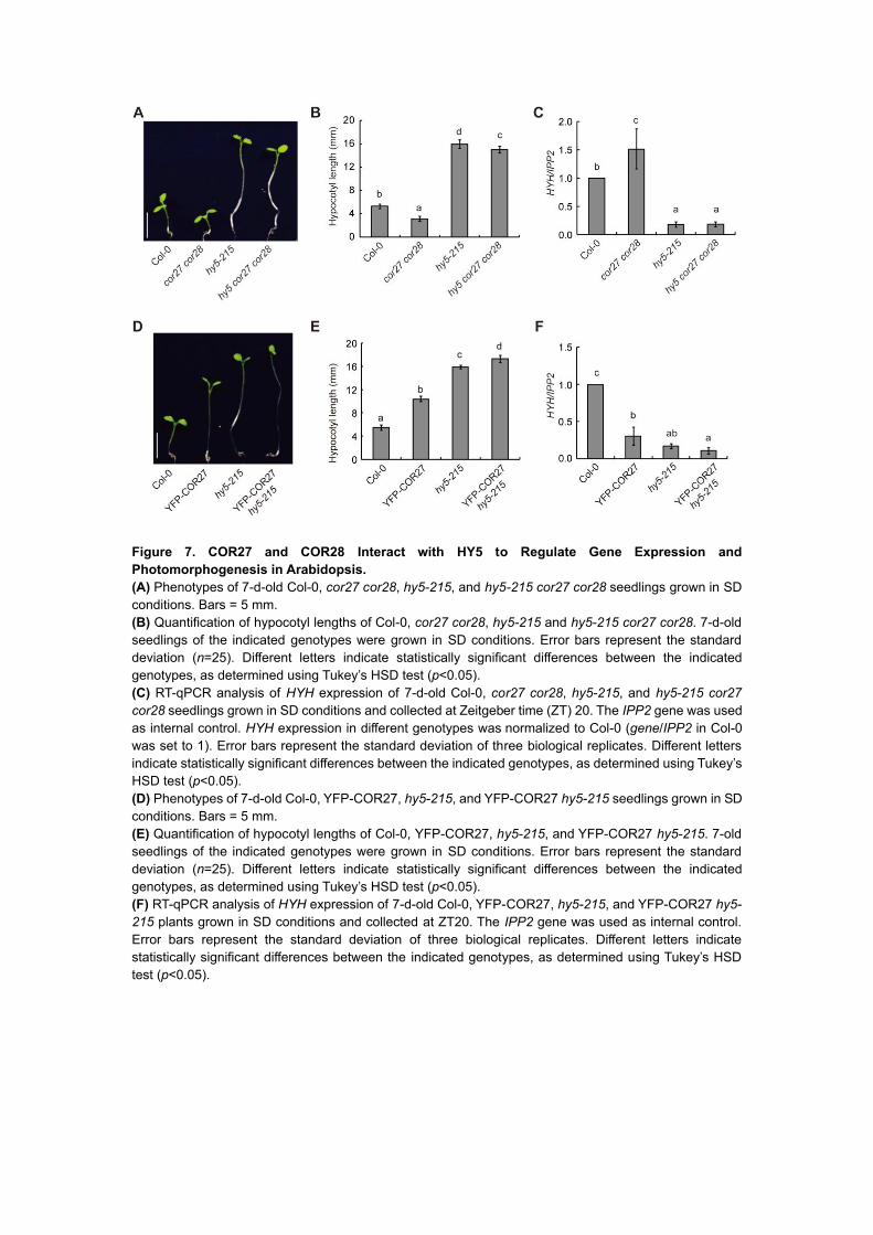
Figure 7. COR27 and COR28 Interact with HY5 to Regulate Gene Expression and
Photomorphogenesis in Arabidopsis.
(A) Phenotypes of 7-d-old Col-0, cor27 cor28, hy5-215, and hy5-215 cor27 cor28 seedlings grown in SD
conditions. Bars = 5 mm.
(B) Quantification of hypocotyl lengths of Col-0, cor27 cor28, hy5-215 and hy5-215 cor27 cor28. 7-d-old
seedlings of the indicated genotypes were grown in SD conditions. Error bars represent the standard
deviation (n=25). Different letters indicate statistically significant differences between the indicated
genotypes, as determined using Tukey’s HSD test (p<0.05).
(C) RT-qPCR analysis of HYH expression of 7-d-old Col-0, cor27 cor28, hy5-215, and hy5-215 cor27
cor28 seedlings grown in SD conditions and collected at Zeitgeber time (ZT) 20. The IPP2 gene was used
as internal control. HYH expression in different genotypes was normalized to Col-0 (gene/IPP2 in Col-0
was set to 1). Error bars represent the standard deviation of three biological replicates. Different letters
indicate statistically significant differences between the indicated genotypes, as determined using Tukey’s
HSD test (p<0.05).
(D) Phenotypes of 7-d-old Col-0, YFP-COR27, hy5-215, and YFP-COR27 hy5-215 seedlings grown in SD
conditions. Bars = 5 mm.
(E) Quantification of hypocotyl lengths of Col-0, YFP-COR27, hy5-215, and YFP-COR27 hy5-215. 7-old
seedlings of the indicated genotypes were grown in SD conditions. Error bars represent the standard
deviation (n=25). Different letters indicate statistically significant differences between the indicated
genotypes, as determined using Tukey’s HSD test (p<0.05).
(F) RT-qPCR analysis of HYH expression of 7-d-old Col-0, YFP-COR27, hy5-215, and YFP-COR27 hy5-
215 plants grown in SD conditions and collected at ZT20. The IPP2 gene was used as internal control.
Error bars represent the standard deviation of three biological replicates. Different letters indicate
statistically significant differences between the indicated genotypes, as determined using Tukey’s HSD
test (p<0.05).

DOI 10.1105/tpc.20.00195; originally published online August 7, 2020;Plant Cell
Xu Li, Cuicui Liu, Zhiwei Zhao, Dingbang Ma, Jinyu Zhang, Yu Yang, Yawen Liu and Hongtao LiuPhotomorphogenesis in Arabidopsis
COR27 and COR28 are Novel Regulators of the COP1-HY5 Regulatory Hub and
This information is current as of August 10, 2020
Permissions X
https://www.copyright.com/ccc/openurl.do?sid=pd_hw1532298X&issn=1532298X&WT.mc_id=pd_hw1532298
eTOCs http://www.plantcell.org/cgi/alerts/ctmain
Sign up for eTOCs at:
CiteTrack Alerts http://www.plantcell.org/cgi/alerts/ctmain
Sign up for CiteTrack Alerts at:
Subscription Information http://www.aspb.org/publications/subscriptions.cfm
is available at:Plant Physiology and The Plant CellSubscription Information for
ADVANCING THE SCIENCE OF PLANT BIOLOGY © American Society of Plant Biologists