THE AURICULOTEMPORAL -...
8
THE AURICULOTEMPORAL SYNDROME A CLINICAL AND PHARMACOLOGIC STUDY By A. S. FREEDBERG, ROBERT S. SHAW AND M. J. McMANUS (From the Department of Medicine, Harvard Medical School, and the Medical Service and Research Laboratories, Beth Israel Hospital, Boston) (Received for publication May 26, 1948) The auriculotemporal syndrome which develops after injury to the parotid gland, consists of flushing and profuse sweating, upon eating, over the cutaneous distribution of the auriculotemporal nerve on the injured side. Although there have been few cases reported since the original descrip- tion by Frey (1) in 1923, the syndrome is not rare, having been noted repeatedly, for-example, in clinics observing patients after operation on a parotid gland (2, 3). The syndrome is of interest to the physiologist because it affords an oppor- tunity to study the consequences of denervation of cholinergic end organs. It is our purpose to pre- sent three instances of this syndrome with some observations relating to its mechanism. CASE HISTORIES Case 1: J. B., a 68 year old man, BIH No. 78777, entered the hospital on 9/29/44 complaining of transient weakness and dysarthria. These symptoms were believed to be consequent to a recent cerebrovascular accident. Forty-one years ago, while in the Russian army, the patient had had typhoid fever, complicated by right paro- titis. Two attempts to drain the parotid gland by vertical incisions were unsuccessful. Two incisions, 2 cm. lateral to the right upper border of the thyroid gland, resulted in drainage for approximately 50 days. Approximately one month after the successful drainage, and two months after the onset of the parotitis, the patient noticed the de- velopment of profuse sweating over the right side of the face while eating. Although ingestion of many foods was followed by sweating, the phenomenon was particularly marked when apples were eaten. On each occasion just prior to the onset of sweating the patient experienced a sensation of warmth over the involved area. Redness, swelling and pain, however, were absent; at no time was numbness noted over the involved area. The amount of sweating had become less pronounced during the previous three years. On physical examination, scars on the face and neck were noted. The opening of the right Stenson's duct was protuberant; saliva could not be expressed from it. The orifice of the left duct appeared normal and saliva was easily expressed. Perception of heat and cold was normal and equal on both sides of the face. There was cutaneous hypaesthesia in the distribution of the right auriculo- temporal nerve. There was a right facial weakness and ptosis of the right upper eyelid; the latter was said to have been present since the patient's recent cerebral vas- cular accident. The cranial nerves were otherwise normal. Case 2: T. H., a 63 year old man, MGH No. 484270, with a history of recent sinusitis, had had bilateral sup- purative parotitis, of unknown etiology, twenty-five years previously; this had been drained through incisions over the glands on both sides. Several weeks after operation the patient noted the occurrence of profuse sweating and a feeling of warmth on the left side of his face when eat- ing. The phenomenon had continued until admission. It was brought on by eating any food, especially apples. At no time had the patient noticed any numbness or paralysis of his face. Physical examination was not remarkable except for the old drainage scars over both parotid glands. The parotid glands were normal to palpation and saliva was easily expressed from both ducts. Sensation over the face was normal and the function of other cranial nerves was apparently intact. Case 3: D. E. S., a 49 year old married woman (re- ferred by Dr. I. T. Nathanson), had had a tumor of the right parotid gland removed in 1942. Approximately onc. year later she noted the onset of sweating following the eating of various foods. The sweating involved the right side of the face in the region of the incision. Rarely a sense of heat was noted but visible flushing was never observed. No specific food was noted to be particularly effective in producing the sweating reaction. Although on rare occasions a whole meal did not produce sweating, hot tea by itself occasionally produced profuse perspira- tion. She had had mumps as a child, but with no sequelae. OBSERVATIONS Observations in Case 1: The observations on sweating were made by means of the Minor starch-iodine test (4) which consists of applying a solution of iodine (15 cc. 1 per cent iodine, 5 cc. of castor oil and 80 cc. 95 per cent alcohol) and after drying, lightly dusting the skin with starch and noting the onset of sweating which is signal- ized as black spots. Ten seconds after taking a bite of apple and while chewing, the patient noticed a feeling of warmth over the right face, and a slight flush was noted on this side. Five seconds later, sweating appeared in part of the area in- 669
Transcript of THE AURICULOTEMPORAL -...

THE AURICULOTEMPORALSYNDROMEA CLINICAL ANDPHARMACOLOGICSTUDY
By A. S. FREEDBERG, ROBERTS. SHAWAND M. J. McMANUS
(From the Department of Medicine, Harvard Medical School, and the Medical Service andResearch Laboratories, Beth Israel Hospital, Boston)
(Received for publication May 26, 1948)
The auriculotemporal syndrome which developsafter injury to the parotid gland, consists offlushing and profuse sweating, upon eating, overthe cutaneous distribution of the auriculotemporalnerve on the injured side. Although there havebeen few cases reported since the original descrip-tion by Frey (1) in 1923, the syndrome is notrare, having been noted repeatedly, for-example,in clinics observing patients after operation on aparotid gland (2, 3). The syndrome is of interestto the physiologist because it affords an oppor-tunity to study the consequences of denervation ofcholinergic end organs. It is our purpose to pre-sent three instances of this syndrome with someobservations relating to its mechanism.
CASE HISTORIES
Case 1: J. B., a 68 year old man, BIH No. 78777,entered the hospital on 9/29/44 complaining of transientweakness and dysarthria. These symptoms were believedto be consequent to a recent cerebrovascular accident.
Forty-one years ago, while in the Russian army, thepatient had had typhoid fever, complicated by right paro-titis. Two attempts to drain the parotid gland by verticalincisions were unsuccessful. Two incisions, 2 cm. lateralto the right upper border of the thyroid gland, resultedin drainage for approximately 50 days. Approximatelyone month after the successful drainage, and two monthsafter the onset of the parotitis, the patient noticed the de-velopment of profuse sweating over the right side of theface while eating. Although ingestion of many foods wasfollowed by sweating, the phenomenon was particularlymarked when apples were eaten. On each occasion justprior to the onset of sweating the patient experienced asensation of warmth over the involved area. Redness,swelling and pain, however, were absent; at no time wasnumbness noted over the involved area. The amount ofsweating had become less pronounced during the previousthree years.
On physical examination, scars on the face and neckwere noted. The opening of the right Stenson's duct wasprotuberant; saliva could not be expressed from it. Theorifice of the left duct appeared normal and saliva waseasily expressed. Perception of heat and cold was normaland equal on both sides of the face. There was cutaneoushypaesthesia in the distribution of the right auriculo-
temporal nerve. There was a right facial weakness andptosis of the right upper eyelid; the latter was said tohave been present since the patient's recent cerebral vas-cular accident. The cranial nerves were otherwise normal.
Case 2: T. H., a 63 year old man, MGHNo. 484270,with a history of recent sinusitis, had had bilateral sup-purative parotitis, of unknown etiology, twenty-five yearspreviously; this had been drained through incisions overthe glands on both sides. Several weeks after operationthe patient noted the occurrence of profuse sweating anda feeling of warmth on the left side of his face when eat-ing. The phenomenon had continued until admission. Itwas brought on by eating any food, especially apples. Atno time had the patient noticed any numbness or paralysisof his face. Physical examination was not remarkableexcept for the old drainage scars over both parotid glands.The parotid glands were normal to palpation and salivawas easily expressed from both ducts. Sensation over theface was normal and the function of other cranial nerveswas apparently intact.
Case 3: D. E. S., a 49 year old married woman (re-ferred by Dr. I. T. Nathanson), had had a tumor of theright parotid gland removed in 1942. Approximately onc.year later she noted the onset of sweating following theeating of various foods. The sweating involved the rightside of the face in the region of the incision. Rarely asense of heat was noted but visible flushing was neverobserved. No specific food was noted to be particularlyeffective in producing the sweating reaction. Althoughon rare occasions a whole meal did not produce sweating,hot tea by itself occasionally produced profuse perspira-tion. She had had mumpsas a child, but with no sequelae.
OBSERVATIONS
Observations in Case 1: The observations onsweating were made by means of the Minorstarch-iodine test (4) which consists of applying asolution of iodine (15 cc. 1 per cent iodine, 5 cc.of castor oil and 80 cc. 95 per cent alcohol) andafter drying, lightly dusting the skin with starchand noting the onset of sweating which is signal-ized as black spots. Ten seconds after taking abite of apple and while chewing, the patient noticeda feeling of warmth over the right face, and aslight flush was noted on this side. Five secondslater, sweating appeared in part of the area in-
669

A. S. VREDIBERG, ROBERTS. SHAWAND M. J. MCMANUS
FIG. 1. THE DISTRIBUTION OF SWEATING OBSERVEDIN
CASE 1, REPRESENTEDBY THE CROSS-HATCHEDAREA
cluded in the distribution of the auriculotemporalnerve (Figure 1). The sweating could be inducedby chewing any food, but was absent or slightwhen the patient chewed paraffin. The applica-tion of ice to the right face before chewing foodresulted in a decreased amount of sweating in thechilled area. Sweating was absent when the pa-
tient held but did not chew food placed in hismouth. When the tongue was swabbed withvinegar, a small amount of sweating appeared.The response was independent of the region of thetongue swabbed. When the patient chewed a
pledget of cotton soaked in vinegar, the amountof sweating, as judged by the extent and depth ofthe starch-iodine reaction, was more marked.Psychic salivation and sweating could not be in-duced. Sweating induced by the general appli-cation of heat to the body was irregularly dimin-ished over the right side of the face.
Procainization of the right auriculotemporalnerve resulted in anesthesia over part of the dis-tribution of the nerve. In the area of anesthesiasweating failed to appear after the usual stimula-tion. Novocaine injection 1 of the right superiorcervical ganglion resulted in a Horner's syndrome,suffusion of the conjunctiva, increased warmth,and dilation of the right hand veins. While thecervical sympathetics were blocked, eating an appleinduced sweating in 45 seconds and 20 seconds intwo successive trials. The patient was apprehen-
1 The authors are indebted to Dr. Reginald Smithwickfor the procaine blocks in Cases 1 and 2.
sive during the whole period of injection and fora short time afterward.
A series of experiments was performed to de-termine whether there was hypersensitivity of thesweating mechanism in the involved area. Innormal controls, local sweating was observed overand around intradermal wheals made by the in-jection of 0.1 cc. of various concentrations ofacetylcholine bromide dissolved in isotonic salinesolution. By using increasing dilutions, a meas-ure of the sensitivity of the local sweating appa-ratus to acetylcholine might be obtained. Whilesensitivity was found to vary in different individu-als and in different regions of the body, the endpoints were consistent. In this patient, intra-dermal wheals in the involved area on the rightside of the face produced with 0.1 cc. of isotonicsaline solution containing 0.0001-0.1 'gamma ofacetylcholine bromide resulted in local sweating.Similar concentrations of acetylcholine in sym-metrical positions on the left face of the patient didnot produce sweating. A wheal on the left sideof the face which contained 0.5 gammaacetylcho-line per 0.1 cc. produced sweating.
Iontophoresis (3.4 milliamperes for ten minutesthrough a 1 sq. cm. pad) of a 1: 12,500 solutionof acetylcholine bromide in isotonic saline solu-tion, produced sweating on the involved right sideof the face and no sweating on the uninvolved leftside on two attempts, with no sweating on eitherside on the third attempt. Ten mgm. of acetylcho-line bromide injected subcutaneously did not re-sult in facial sweating.
The possibility was examined that disturbancesin vasomotor nervous function are present in theauriculotemporal syndrome. The variation in skintemperature of the involved areas in response tocooling and heating the body and extremities wasmeasured and compared to symmetrical unin-volved areas. To determine the time of occur-rence of the maximal response to heating and cool-ing, measurements of the skin temperature of afinger tip were made. The measurements of skintemperature were made at frequent intervals witha thermocouple. The experiment was performedin a constant temperature room, 200 C. The pa-tient was seated and the legs, arms and chest wereuncovered. Within 20 minutes a distinct fall' infinger tip temperature was observed (Figure 2)which 'reached a maximum in 70 minutes. The
670

THE AURICULOTEMPORALSYNDROME
right ear skin temperature fell from 29.50 C to25.50 C in this interval of time. At the arrow(70 minutes) one leg and one arm were immersedin running warm water (T 420); the patient wascovered with blankets and hot water bottles wereapplied to the chest and back. Approximately 15minutes later, a distinct and sustained rise infinger and left ear temperature occurred. Theinvolved right ear, however, showed no rise intemperature in response to heating (Figure 2);furthermore, the increase in right facial skin tem-perature in response to heat was less than that ob-served in a symmetrical point on the uninvolvedleft side.
An attempt was made to measure and comparethe secretion from the parotid glands in this pa-tient. No saliva could be collected from the rightparotid duct. Secretion from the left duct ap-peared normal.
Observations in Case 2: In this subject flushingwas observed over the left side of the face tenseconds after he began to eat an apple, and in 15seconds sweating was observed over the entiredistribution of the auriculotemporal nerve on theleft. A small area on the right cheek was also ob-
36
35
34
z 330
32e)LwJ 31wW- 30LLwa 29
W 28cr-D 27
C 26O
25wI 24
23
served to sweat, although this had never beennoted by the patient. Psychic salivation was eas-ily induced, but the syndrome of flushing andsweating did not occur.
Sweating, distributed usually over the upperlip and nares, may be induced in normal subjectsby the application to the tongue and chewing ofred pepper or ginger. In this patient these foodsresulted in sweating on -the upper lip but did notinduce sweating in the distribution of the auriculo-temporal nerve.
Novocaine injection of the superior cervicalsympathetic ganglion on the left resulted in aHorner's syndrome and an absence of sweatingin response to heat. In the presence of this block,the auriculotemporal syndrome was observed asbefore, after the usual stimulus.
Intradermal wheals produced with .05 gammaacetylcholine bromide contained in 0.1 cc. isotonicsaline solution produced sweating in the involvedarea on the left face, but none in the correspondinglocation on the right.
Saliva was collected from the right and leftparotid ducts by means of silver cups attached bysuction to the parotid gland duct orifices. No
0 10 20 30 40 50 60 70 80 90 100 10 120 130 140
TJME - MINUTESFIG. 2. VARIATIONS IN SKIN TEMPERATUREON COOLING AND HEATING THE BODY
See text for description of figure.
671
A. S. FREEDBERG, ROBERTS. SHAWAND M. J. MCMANUS
UOLI-w
zU0
llJ
CL
UU
0U
a-U5I-
110
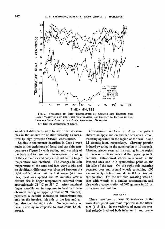
TIME- MINUTESFIG. 3. VARIATION IN SKIN TEMPERATUREON COOLING AND HEATING THE
BODY; VARIATIONS OF THE SKIN TEMPERATURECONSEQUENTTO EATING IN THEINVOLVED SKIN AREA IN THE AURICULOTEMPORALSYNDROME
See text for description of figure.
significant differences were found in the two sam-ples in the amount or relative viscosity as meas-ured by high pressure Ostwald viscosimeter.
Studies in the manner described in Case 1 weremade of the variations of facial and ear skin tem-perature (Figure 3) with cooling and warming ofthe body and extremities. In response to coolingof the extremities and body a distinct fall in fingertemperature was obtained. The changes in skintemperature of the ears and face were slight andno significant difference was observed between theright and left sides. At the first arrow (48 min-utes) heat was applied and 25 minutes later adistinct rise in finger temperature occurred fromapproximately 210 C to 310 C. After maximalfinger vasodilation in response to heat had beenobtained, eating an apple (arrow at 91 minutes)produced a definite increase in temperature notonly on the involved left side of the face and earbut also on the right side. No asymmetry offacial sweating in response to heat could be ob-served.
Observations in Case 3: After the patientchewed an apple and on another occasion a lemon,sweating appeared in the region of the scar 16 and12 seconds later, respectively. Chewing paraffininduced sweating in the same region in 16 seconds.Chewing ginger resulted in sweating in the regionof the scar in 14 seconds and the upper lip in 30seconds. Intradermal wheals were made in theinvolved area and in a symmetrical point on theleft side of the face. On the right side sweatingoccurred over and around wheals containing .005gamma acetylcholine bromide in 0.1 cc. isotonicsalt solution. On the left side sweating was ab-sent with wheals of a similar concentration andalso with a concentration of 0.05 gammain 0.1 cc.of isotonic salt solution.
COMMENT
There have been at least 35 instances of theauriculotemporal syndrome reported in the litera-ture (1, 5-15). In the majority of cases the orig-inal episode involved both infection in and opera-
672
THE AURICULOTEMPORALSYNDROME
tion on the parotid gland. The most common in-citing circumstance was a parotitis, complicatingtyphus or typhoid fever, which had been drainedsurgically. The syndrome has also followed paro-titis complicating syringomyelia, mumps, opera-tion on the parotid without infection (2, 16), andparotitis of unknown etiology in the absence ofsuppuration and operation. The sweating makesits appearance from a few days (14) to threeyears (9) after the original episode, and lasts therest of the patient's life. One case is reportedwith gradual disappearance of the syndrome overa three-year period (11). Uniformly the sweat-ing appears over part or all of the distribution ofthe auriculotemporal nerve on the injured side.It is usually accompanied by visible flushing, andpatients without visible flushing describe a sen-sation of warmth over the involved area. Minordisturbances in sensation and decreased sweatingresponse to heat over the distribution of the auric-ulotemporal nerve of the involved side are fre-quently noted.
The severity of sweating varies from patientto patient, being slight in some (Case 3) and pro-fuse in others (Cases 1 and 2). The studies de-scribed above suggest that relief may be obtainedin instances of severe flushing and sweating by in-terruption of the efferent arc by alcohol injectionor surgical section of the auriculotemporal nerve.
Mechanism of auriculotemporal syndrome-anatomic pathway
It appears that the disorder in the auriculo-temporal syndrome lies in the efferent arc. Thepathway of the afferent arc is variable since sweat-ing may be initiated by stimulation of the seventhor ninth nerves and has been reported to resultfrom psychic stimulation. The efferent pathwayinvolves fibers in the region of the auriculo-temporal nerve as demonstrated by the effect ofprocainization (Case 1). Since procainization ofthe superior cervical ganglion resulted in the disap-pearance of sweating in response to heat over theface but did not affect the occurrence of the auricu-lotemporal syndrome, it is clear that the fibers inthe efferent arc do not pass through the cervicalsympathetics.
Proposed theories:
I. It has been suggested (17) that the sweatingphenomenon results from the diffusion of acetyl-choline or cholinergic substances from the injuredparotid gland. The speed of the reaction, thedistribution in the course of the entire auriculo-temporal nerve and the occurrence of the phe-nomenon in patients in whom the involved paro-tid gland is in all likelihood completely destroyed(Case 1) are against this interpretation.
II. Needles (12) noted the appearance of theauriculotemporal syndrome coincident with theclosure of a salivary fistula, and felt that the syn-drome might be caused by the irritation of su-domotor nerve fibers passing through or near theparotid gland by the swelling of the gland onmastication. This appears unlikely, since thesyndrome may appear in the presence of an atro-'phied and functionless parotid gland (Case 1).
III. Ford has suggested (8) that the auriculo-temporal syndrome may be explained by injuryto the auriculotemporal nerve and subsequentaberrant regeneration of parotid secretary fibersalong'sudomotor and vasomotor pathways. Theauriculotemporal nerve contains, in addition tosensory fibers, secretary fibers to the parotidgland, sudomotor fibers, and vasodilator fibers tothe area of sensory distribution of the nerve (18);the parotid secretary fibers, sudomotor and va-sodilator fibers are all cholinergic. The followingfacts are in support of this hypothesis:
1. The time interval between the original injuryand the appearance of the syndrome, usually onemonth or longer, is consistent with a period ofnerve degeneration and subsequent regeneration.One case, however, is reported (14) where thesyndrome appeared two to three days followinginjury. 2. The frequently noted abnormalitiesin sensation and decreased sweating response toheat over the distribution of the auriculotemporalnerve, and, as in one of our cases, defective va-somotor response over the distribution of the au-riculotemporal nerve, are consistent with injury toall components of the auriculotemporal nerve.Although hypersensitivity of the sweat glands overthe affected region is consistent with the inter-pretation that the involved sweat glands have beenpartially denervated, it is difficult to explain the
673

A. S. FREEDBERG, ROMERTS. SHAWAND M. J. MCMANUS
evident hypersensitivity after regeneration hastaken place.
The resemblance of the case described byUprus, Gaylor and Carmichael (16) to those ex-hibiting the auriculotemporal syndrome is so strik-ing as to suggest a common mechanism. Theydescribed an instance of flushing and gustatorysweating over the distribution of the right cu-taneous colli nerve appearing one to two yearsfollowing the removal of cervical lymph glandsthrough a horizontal incision over the mid rightsternocleidomastoid muscle. In the involved area,sensations to pin prick and light touch were di-minished. There was diminished sweating re-sponse to heat and an impaired vasomotor re-sponse after thermal stimuli. Procainization ofthe cutaneous colli nerve (sensory and vasomotorto the area) had no effect upon the flushing and thesweating reaction induced by food. Blocking thelingual nerve proximal to the origin of the chordatympani resulted in cessation of salivation and thedisappearance of the phenomena. In this in-stance the primary injury was to the cutaneouscolli nerve and injury to. the secretary fibres inthe lingual nerve appears unlikely because of thelevel of the incision. With absence of injury tosubmaxillary secretary fibers, aberrant regenera-tion is unlikely in this case.
IV. The explanation offered here is that theauriculotemporal syndrome may represent theresponse of denervated and hypersensitive sweatglands to previously unapparent cranial sudomotorimpulses. List and Peet observed (18) markedfacial gustatory sweating following postganglioniccervical sympathectomy. In these patients, thesweating appeared following the eating of foodswhich were similar to those foods causing sweat-ing in patients with the auriculotemporal syn-drome. List and Peet (18) suggested that thistype of sweating was an "exaggeration of nor-mal gustatory sweating" caused by denervationhypersensitivity of the sweat glands, but theyfelt that denervation hypersensitivity was not theexplanation of the auriculotemporal syndrome, be-cause the diminution of heat sweating over thearea involved in the auriculotemporal syndromewas so slight as to suggest that the denervation ofthe sweat glands was negligible. However, in the
reported cases of the auriculotemporal syndrome,hypersensitivity to pilocarpine as manifested byearly sweating in the involved area, and our ob-servations of local hypersensitivity to acetylcholinein the involved regions as demonstrated by intra-cutaneous wheals or iontophoresis of acetylcholine,clearly show that there is a hypersensitivity of thesweat glands in the involved region. That hyper-sensitivity to cholinergic substances may occur inorgans following denervation has been demon-strated in the submaxillary gland (19), skeletalmuscle (20) and iris (21). In these latter studieshypersensitivity developed in from one to twoweeks. Observations on the time relationship ofthe development of hypersensitivity following de-nervation of sweat glands are necessary.
Other data are available consistent with the in-terpretation that a cranial supply of sudomotorfibers exists. It is a common observation thatfacial sweating occurs in normal individuals withthe eating of hot spicy foods. More unusually, itmay occur with the eating of chocolate (9) andthe drinking of vinegar (22). Gustatory sweat-ing in normal individuals, as produced by the eat-ing of ginger, has been studied by us. The sweat-ing occurs maximally about the mouth and nose.Observations were also made in a patient who hada unilateral preganglionic cervical sympathectomyfor Raynaud's syndrome. In this patient who hadno Horner syndrome, there was absence of heatsweating on the denervated side. Gustatory sweat-ing induced by eating ginger was marked on thenormal side of the face but scarcely perceptibleon the denervated side. This would suggest thatthe nervous pathways involved in gustatory sweat-ing consequent upon the eating of ginger passedthrough the cervical sympathetics. The pathwaysconcerned in the auriculotemporal syndrome donot, as demonstrated by the fact that the syndromeis not affected by cervical sympathetic block.Furthermore in Case 2 sweating induced by gingerwas observed to occur independently from theauriculotemporal syndrome sweating. The failureto block the syndrome by procainization of thesuperior cervical ganglion (Cases 1 and 2), al-though heat sweating was abolished, is also con-sistent with the presence of cranial sudomotorfibers. That the facial sweat glands may have a
674

THE AURICULOTEMPORALSYNDROME
dual innervation from the brain and cervicalsympathetics was suggested by Takino (23) whoobserved both medullated and non-medullatednerves around the sweat glands of the face. Thelatter evidence is not decisive and further studieson this point are necessary.
It would seem, therefore, that the auriculotem-poral syndrome, post-sympathectomy gustatorysweating and the case of Uprus, Gaylor and Car-michael represent sweating responses to nervousimpulses in sweat glands made hypersensitive bydenervation. Although as noted above, in thediscussion of Ford's-hypothesis, these nervous im-pulses may be from abnormally regenerated sali-vary secretary fibers, if cranial sudomotor fibersexist, they might be the efferent pathway in thethree types of gustatory sweating under considera-tion. The three syndromes would then representcomplete or partial severance of the sudomotorand vasomotor sympathetics to the involved re-gions leading to hypersensitivity of the sweatglands and exaggeration of normal and hithertonot obvious cranial sudomotor and vasomotor im-pulses.
SUMMARYANDCONCLUSIONS
1. The auriculotemporal syndrome is charac-terized by gustatory sweating and flushing overthe cutaneous distribution of the auriculotemporalnerve. Three patients have been studied who ex-hibited this syndrome.
2. The gustatory sweating and flushing seen inthe auriculotemporal syndrome is a manifestationof a reflex in which the efferent arc is throughthe auriculotemporal nerve or through adjacentautonomic fibers and not through the cervicalsympathetic nerves. The afferent arc is non-specific; the syndrome may be induced by stimu-lating the seventh and ninth nerves.
3. There may be a deficiency in sudomotor,thermo-regulatory and sensory innervation overthe involved area.
4. A local hypersensitivity of the sweat glandsin the involved region to acetylcholine bromide ispresent.
5. The mechanism of this syndrome is ap-parently related to denervation hypersensitivity ofthe sweat glands analogous to the hypersensitivity
to adrenalin which occurs after post-ganglionicdivision of sympathetic fibers.
6. In instances of severe flushing and sweating,the described studies suggest that relief may beobtained by interruption of the efferent arc by al-cohol injection or surgical section of the auriculo-temporal nerve.
BIBLIOGRAPHY
1. Frey, L., Le Syndrome du Nerf Auriculo-temporal.Rev. neurol., 1923, 2, 97.
2. Nathanson, I. T., Personal Communication.3. Bailey, H., Parotidectomy: Indications and Results.
Brit. M. J., 1947, 1, 404.4. Minor, V., Eines neues Verfahren zu der Klinischen
Untersuchung der Schweissabsonderung. DeutschesZtschr. f. Nervenh., 1928, 101, 302.
5. Kaminsky, S. D., Das "auriculo-temporale (Parotitis)Syndrom" bie Syringomyelie. Deutsches Ztschr. f.Nervenh., 1929, 109, 296.
6. Thomas, A., Le double reflex vaso-dilatateur etsudoral de la face consecutif aux blessures de laloge parotidienne; les parareflexes. Rev. neurol.,1927, 1, 447.
7. Fridberg, D., Das Auriculo-temporale Syndrom.Deutsches Ztschr. f. Nervenh., 1931, 121, 225.
8. Ford, F. R., Paroxysmal lacrimation during eatingas a sequel of facial palsy (syndrome of crocodiletears): report of four cases with a possible inter-pretation and comparison with the auriculotemporalsyndrome. Arch. Neurol. & Psychiat., 1933, 29,1279.
9. Bassoe, P. N., Auriculotemporal syndrome and othervasomotor disturbances about the head: "auriculo-temporal syndrome" complicating diseases of paro-tid gland, angioneurotic edema of the brain. M.Clin. of North America, 1932, 16, 405.
10. Noica and Bagdasar, Syndrome du nerf auriculo-temporal. Rev. neurol., 1926, 1, 225.
11. Trioumphoff, A., Une forme particuliere de l'hyper-hidrose locale de la face. Presse med., 1926, II,86, 1350.
12. Needles, W., The auriculotemporal syndrome, withsuggestion regarding therapy. Arch. Neurol. &Psychiat., 1936, 35, 357.
13. Karnosh, L. J., The syndrome of the auriculotemporalnerve. Cleveland Clin. Quart., 1946, 13, 194.
14. Souques, A., Hyperhidrose unilaterale de la face con-s~cutive a un traumatisme de la region sourciliereet provoquee par les excitations gustatives et parla chaleur. Des hemihyperhidroses d'origine cere-bro-spinale. Rev. neurol., 1927, 2, 145.
15. Guttmann, L., and List, C. F., Zur Topik und Patho-physiologie der Schweissekretion. Ztschr. f. d.ges. Neurol. u. Psychiat., 1928, 116, 504.
16. Uprus, V., Gaylor, J. B., and Carmichael, E. A.,Localized abnormal flushing and sweating on eat-ing. Brain, 1934, 57, 443.
675

A. S. FREEDBERG, ROBERTS. SHAWAND M. J. MCMANUS
17. List, C. F., and Peet, M. M., Sweat secretion in man.
III. Clinical observation on sweating produced bypilocarpine and mecholyl. Arch. Neurol. &Psychiat., 1938, 40, 269.
18. List, C. F., and Peet, M. M., Sweat secretion in man.
IV. Sweat secretion of the face and its disturb-ances. Arch. Neurol. & Psychiat., 1938, 40, 443.
19. Simeone, F. A., and Maes, J. P., Sensitization of thesubmaxillary gland by sympathetic denervation.Am. J. Physiol., 1939, 125, 674.
20. (annon, W. B., Haimovici, H., Sensitization of moto-neurones by partial "denervation." Anm J. ofPhys., 1939, 126, 731.
21. Cannon, S., cited by Cannon, W. B., and Rosen-blueth, A., Autonomic Neuro-Effector Systems.Macmillan Co., New York, 1937, p. 229.
22. Luchsinger, B., Die Schweissabsonderung und einigeverwandte Secretionen bei thieren. Hermann Hand-buch, phys. Leipzig, 1883, 1, 421.
23. Takino, cited by Kuno, Y., The Physiology of HumanPerspiration. Churchill, Ltd., London, 1934, p. 9.
676