The Arabidopsis DELAYED DEHISCENCE1 Gene Encodes … · The Arabidopsis DELAYED DEHISCENCE1 Gene...
22
The Plant Cell, Vol. 12, 1041–1061, July 2000, www.plantcell.org © 2000 American Society of Plant Physiologists The Arabidopsis DELAYED DEHISCENCE1 Gene Encodes an Enzyme in the Jasmonic Acid Synthesis Pathway Paul M. Sanders, a,1 Pei Yun Lee, a,2 Christian Biesgen, b,3 James D. Boone, a,4 Thomas P. Beals, a,5 Elmar W. Weiler, b and Robert B. Goldberg a,6 a Department of Molecular, Cell, and Developmental Biology, University of California, Los Angeles, California 90095-1606 b Lehrstuhl für Pflanzenphysiologie, Ruhr-Universität, D-44780 Bochum, Germany delayed dehiscence1 is an Arabidopsis T-DNA mutant in which anthers release pollen grains too late for pollination to occur. The delayed dehiscence1 defect is caused by a delay in the stomium degeneration program. The gene disrupted in delayed dehiscence1 encodes 12-oxophytodienoate reductase, an enzyme in the jasmonic acid biosynthesis path- way. We rescued the mutant phenotype by exogenous application of jasmonic acid and obtained seed set from previ- ously male-sterile plants. In situ hybridization studies showed that during the early stages of floral development, DELAYED DEHISCENCE1 mRNA accumulated within all floral organs. Later, DELAYED DEHISCENCE1 mRNA accumu- lated specifically within the pistil, petals, and stamen filaments. DELAYED DEHISCENCE1 mRNA was not detected in the stomium and septum cells of the anther that are involved in pollen release. The T-DNA insertion in delayed dehiscence1 eliminated both DELAYED DEHISCENCE1 mRNA accumulation and 12-oxophytodienoate reductase activity. These ex- periments suggest that jasmonic acid signaling plays a role in controlling the time of anther dehiscence within the flower. INTRODUCTION Dehiscence is the terminal function of the anther and results in the release of pollen grains to enable pollination, fertiliza- tion, and seed set to occur (Goldberg et al., 1993, 1995). Dehiscence involves the breakage of the anther wall at a specific site that runs the length of the anther (Keijzer, 1987; Bonner and Dickinson, 1989; Goldberg et al., 1993, 1995; Beals and Goldberg, 1997). This site forms an indentation, or “notch,” between the locules of each theca. The stomium, which is derived from the outer L1 layer of the stamen pri- mordium, and the septum, which is derived from the primor- dium L2 layer, are two specialized cell types present within the notch region (Goldberg et al., 1993). In solanaceous plants, the septum is designated as either the circular cell cluster (e.g., tobacco; Koltunow et al., 1990), the interspo- rangial septum (e.g., tomato; Bonner and Dickinson, 1989), the hypodermal stomium (e.g., sweet pepper; Horner and Wagner, 1992), or the oxalate package (D’Arcy et al., 1996). The mechanisms responsible for the differentiation of the stomium and septum from L1 and L2 cells positioned be- tween each pair of locules are not known. The program of events that leads to the breakage of the stomium and pollen release begins early in anther develop- ment and involves a number of processes. First, septum and stomium cells must differentiate from precursor cells be- tween the locules of the anther. Second, septum and sto- mium cells must undergo a developmentally timed cell- degeneration program (tobacco [Koltunow et al., 1990; Beals and Goldberg, 1997] and Arabidopsis [Sanders et al., 1999]). The septum degenerates before the stomium, thereby creating a bilocular anther that establishes the sto- mium as the future site of anther wall breakage and pollen release. Targeted ablation of the stomium results in anthers that fail to dehisce, indicating that active processes must occur within these cells to enable pollen release (Beals and Goldberg, 1997). Third, expansion of the endothecial layer and deposition of fibrous lignified bands in the walls of the endothecial cells are required for dehiscence (Sanders et al., 1999). Finally, events leading to anther dehiscence must be coordinated precisely with pollen differentiation, floral devel- opment, and flower opening. The mechanisms that control and coordinate all of these developmental activities within the flower are not known. 1 Current address: Genesis Research and Development Corporation, Ltd., One Fox St., Auckland, New Zealand. 2 Current address: Division of Biology, California Institute of Tech- nology, Pasadena, CA 91125. 3 Current address: Sungene GmbH and Co., KGaA, Corrensstrasse 3, 06466 Gatersleben, Germany. 4 Current address: School of Medicine, University of California, San Diego, CA 92093. 5 Current address: Cereon Genomics, One Kendall Square, Cam- bridge, MA 02139. 6 To whom correspondence should be addressed. E-mail bobg@ ucla.edu; fax 310-825-8201.
Transcript of The Arabidopsis DELAYED DEHISCENCE1 Gene Encodes … · The Arabidopsis DELAYED DEHISCENCE1 Gene...
The Plant Cell, Vol. 12, 1041–1061, July 2000, www.plantcell.org © 2000 American Society of Plant Physiologists
The Arabidopsis
DELAYED DEHISCENCE1
Gene Encodes an Enzyme in the Jasmonic Acid Synthesis Pathway
Paul M. Sanders,
a,1
Pei Yun Lee,
a,2
Christian Biesgen,
b,3
James D. Boone,
a,4
Thomas P. Beals,
a,5
Elmar W. Weiler,
b
and Robert B. Goldberg
a,6
a
Department of Molecular, Cell, and Developmental Biology, University of California, Los Angeles, California 90095-1606
b
Lehrstuhl für Pflanzenphysiologie, Ruhr-Universität, D-44780 Bochum, Germany
delayed dehiscence1
is an Arabidopsis T-DNA mutant in which anthers release pollen grains too late for pollination tooccur. The
delayed dehiscence1
defect is caused by a delay in the stomium degeneration program. The gene disruptedin
delayed dehiscence1
encodes 12-oxophytodienoate reductase, an enzyme in the jasmonic acid biosynthesis path-way. We rescued the mutant phenotype by exogenous application of jasmonic acid and obtained seed set from previ-ously male-sterile plants. In situ hybridization studies showed that during the early stages of floral development,DELAYED DEHISCENCE1 mRNA accumulated within all floral organs. Later, DELAYED DEHISCENCE1 mRNA accumu-lated specifically within the pistil, petals, and stamen filaments. DELAYED DEHISCENCE1 mRNA was not detected in thestomium and septum cells of the anther that are involved in pollen release. The T-DNA insertion in
delayed dehiscence1
eliminated both DELAYED DEHISCENCE1 mRNA accumulation and 12-oxophytodienoate reductase activity. These ex-periments suggest that jasmonic acid signaling plays a role in controlling the time of anther dehiscence within the flower.
INTRODUCTION
Dehiscence is the terminal function of the anther and resultsin the release of pollen grains to enable pollination, fertiliza-tion, and seed set to occur (Goldberg et al., 1993, 1995).Dehiscence involves the breakage of the anther wall at aspecific site that runs the length of the anther (Keijzer, 1987;Bonner and Dickinson, 1989; Goldberg et al., 1993, 1995;Beals and Goldberg, 1997). This site forms an indentation,or “notch,” between the locules of each theca. The stomium,which is derived from the outer L1 layer of the stamen pri-mordium, and the septum, which is derived from the primor-dium L2 layer, are two specialized cell types present withinthe notch region (Goldberg et al., 1993). In solanaceousplants, the septum is designated as either the circular cellcluster (e.g., tobacco; Koltunow et al., 1990), the interspo-
rangial septum (e.g., tomato; Bonner and Dickinson, 1989),the hypodermal stomium (e.g., sweet pepper; Horner andWagner, 1992), or the oxalate package (D’Arcy et al., 1996).The mechanisms responsible for the differentiation of thestomium and septum from L1 and L2 cells positioned be-tween each pair of locules are not known.
The program of events that leads to the breakage of thestomium and pollen release begins early in anther develop-ment and involves a number of processes. First, septum andstomium cells must differentiate from precursor cells be-tween the locules of the anther. Second, septum and sto-mium cells must undergo a developmentally timed cell-degeneration program (tobacco [Koltunow et al., 1990;Beals and Goldberg, 1997] and Arabidopsis [Sanders et al.,1999]). The septum degenerates before the stomium,thereby creating a bilocular anther that establishes the sto-mium as the future site of anther wall breakage and pollenrelease. Targeted ablation of the stomium results in anthersthat fail to dehisce, indicating that active processes mustoccur within these cells to enable pollen release (Beals andGoldberg, 1997). Third, expansion of the endothecial layerand deposition of fibrous lignified bands in the walls of theendothecial cells are required for dehiscence (Sanders et al.,1999). Finally, events leading to anther dehiscence must becoordinated precisely with pollen differentiation, floral devel-opment, and flower opening. The mechanisms that controland coordinate all of these developmental activities withinthe flower are not known.
1
Current address: Genesis Research and Development Corporation,Ltd., One Fox St., Auckland, New Zealand.
2
Current address: Division of Biology, California Institute of Tech-nology, Pasadena, CA 91125.
3
Current address: Sungene GmbH and Co., KGaA, Corrensstrasse3, 06466 Gatersleben, Germany.
4
Current address: School of Medicine, University of California, SanDiego, CA 92093.
5
Current address: Cereon Genomics, One Kendall Square, Cam-bridge, MA 02139.
6
To whom correspondence should be addressed. E-mail [email protected]; fax 310-825-8201.
1042 The Plant Cell
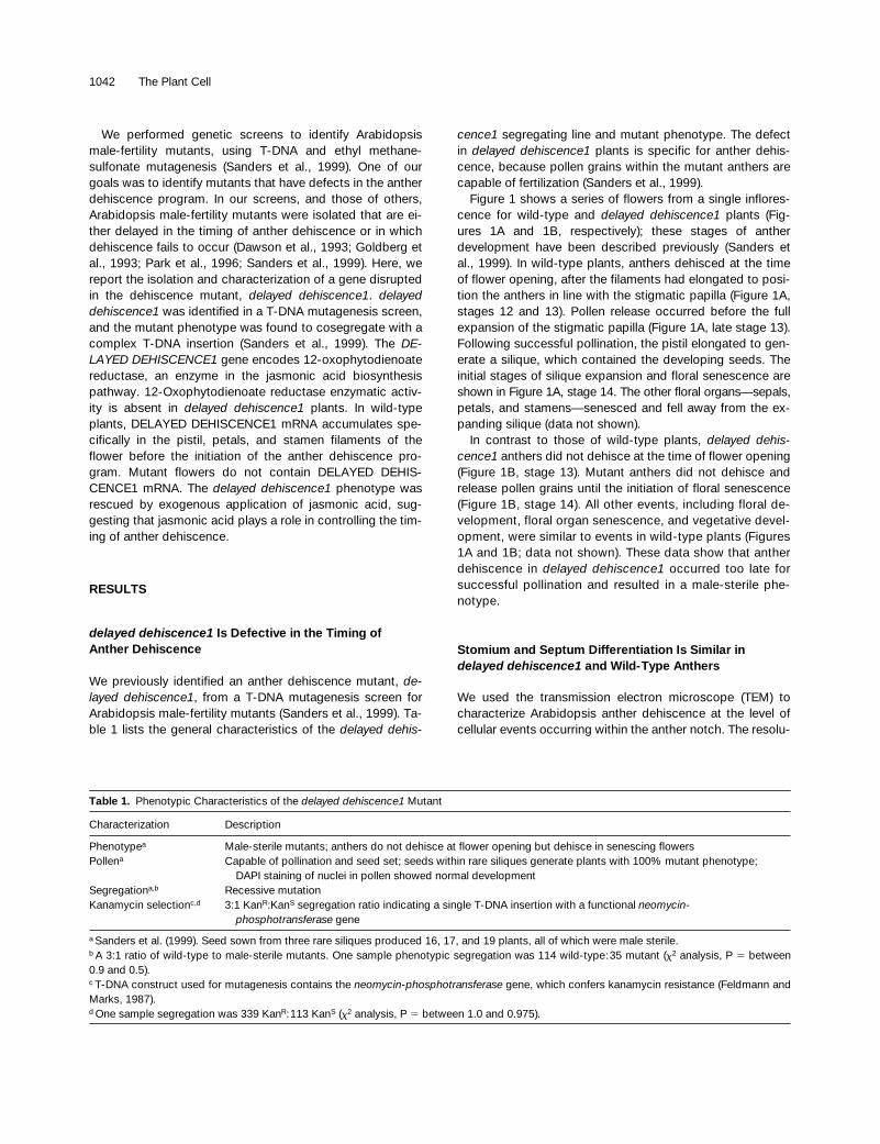
We performed genetic screens to identify Arabidopsismale-fertility mutants, using T-DNA and ethyl methane-sulfonate mutagenesis (Sanders et al., 1999). One of ourgoals was to identify mutants that have defects in the antherdehiscence program. In our screens, and those of others,Arabidopsis male-fertility mutants were isolated that are ei-ther delayed in the timing of anther dehiscence or in whichdehiscence fails to occur (Dawson et al., 1993; Goldberg etal., 1993; Park et al., 1996; Sanders et al., 1999). Here, wereport the isolation and characterization of a gene disruptedin the dehiscence mutant,
delayed dehiscence1
.
delayeddehiscence1
was identified in a T-DNA mutagenesis screen,and the mutant phenotype was found to cosegregate with acomplex T-DNA insertion (Sanders et al., 1999). The
DE-LAYED DEHISCENCE1
gene encodes 12-oxophytodienoatereductase, an enzyme in the jasmonic acid biosynthesispathway. 12-Oxophytodienoate reductase enzymatic activ-ity is absent in
delayed dehiscence1
plants. In wild-typeplants, DELAYED DEHISCENCE1 mRNA accumulates spe-cifically in the pistil, petals, and stamen filaments of theflower before the initiation of the anther dehiscence pro-gram. Mutant flowers do not contain DELAYED DEHIS-CENCE1 mRNA. The
delayed dehiscence1
phenotype wasrescued by exogenous application of jasmonic acid, sug-gesting that jasmonic acid plays a role in controlling the tim-ing of anther dehiscence.
RESULTS
delayed dehiscence1
Is Defective in the Timing ofAnther Dehiscence
We previously identified an anther dehiscence mutant,
de-layed dehiscence1
, from a T-DNA mutagenesis screen forArabidopsis male-fertility mutants (Sanders et al., 1999). Ta-ble 1 lists the general characteristics of the
delayed dehis-
cence1
segregating line and mutant phenotype. The defectin
delayed dehiscence1
plants is specific for anther dehis-cence, because pollen grains within the mutant anthers arecapable of fertilization (Sanders et al., 1999).
Figure 1 shows a series of flowers from a single inflores-cence for wild-type and
delayed dehiscence1
plants (Fig-ures 1A and 1B, respectively); these stages of antherdevelopment have been described previously (Sanders etal., 1999). In wild-type plants, anthers dehisced at the timeof flower opening, after the filaments had elongated to posi-tion the anthers in line with the stigmatic papilla (Figure 1A,stages 12 and 13). Pollen release occurred before the fullexpansion of the stigmatic papilla (Figure 1A, late stage 13).Following successful pollination, the pistil elongated to gen-erate a silique, which contained the developing seeds. Theinitial stages of silique expansion and floral senescence areshown in Figure 1A, stage 14. The other floral organs—sepals,petals, and stamens—senesced and fell away from the ex-panding silique (data not shown).
In contrast to those of wild-type plants,
delayed dehis-cence1
anthers did not dehisce at the time of flower opening(Figure 1B, stage 13). Mutant anthers did not dehisce andrelease pollen grains until the initiation of floral senescence(Figure 1B, stage 14). All other events, including floral de-velopment, floral organ senescence, and vegetative devel-opment, were similar to events in wild-type plants (Figures1A and 1B; data not shown). These data show that antherdehiscence in
delayed dehiscence1
occurred too late forsuccessful pollination and resulted in a male-sterile phe-notype.
Stomium and Septum Differentiation Is Similar in
delayed dehiscence1
and Wild-Type Anthers
We used the transmission electron microscope (TEM) tocharacterize Arabidopsis anther dehiscence at the level ofcellular events occurring within the anther notch. The resolu-
Table 1.
Phenotypic Characteristics of the
delayed dehiscence1
Mutant
Characterization Description
Phenotype
a
Male-sterile mutants; anthers do not dehisce at flower opening but dehisce in senescing flowersPollen
a
Capable of pollination and seed set; seeds within rare siliques generate plants with 100% mutant phenotype; DAPI staining of nuclei in pollen showed normal development
Segregation
a,b
Recessive mutationKanamycin selection
c,d
3:1 Kan
R
:Kan
S
segregation ratio indicating a single T-DNA insertion with a functional
neomycin-phosphotransferase
gene
a
Sanders et al. (1999). Seed sown from three rare siliques produced 16, 17, and 19 plants, all of which were male sterile.
b
A 3:1 ratio of wild-type to male-sterile mutants. One sample phenotypic segregation was 114 wild-type:35 mutant (
x
2
analysis, P
5
between0.9 and 0.5).
c
T-DNA construct used for mutagenesis contains the
neomycin-phosphotransferase
gene, which confers kanamycin resistance (Feldmann andMarks, 1987).
d
One sample segregation was 339 Kan
R
:113 Kan
S
(
x
2
analysis, P
5
between 1.0 and 0.975).

Jasmonic Acid Dehiscence Mutant 1043
tion of the TEM enabled us to identify the Arabidopsis sto-mium and septum cell types. Figure 2 shows TEMmicrographs of anther dehiscence in wild-type and
delayeddehiscence1
anthers.At stage 10 of development in wild-type anthers (Sanders
et al., 1999), all cell types destined to become the stomiumand septum were identified within the notch region (Figure
2A). At this stage, the anther has a four-locule structure andthe locules contain developing microspores or male gameto-phytes (Sanders et al., 1999). The prestomium cell was smallerand lacked the large vacuole present in the contiguous epi-dermal cells (Figure 2A). The septum cells, adjacent andsubepidermal to the stomium, were also small and cytoplas-mically dense, in contrast to the contiguous endothecium
Figure 1. Dehiscence and Senescence of Wild-Type and delayed dehiscence1 Anthers.
A developmental series of flowers from young to old within a single inflorescence were photographed using a dissecting microscope. Stages 12,13, late-13 (13L), and 14 of anther development are shown. These stages were defined in Sanders et al. (1999). Inserts of stage 14 anthers weremagnified 32.25 and enhanced digitally. The photographs represent a series of six or seven flowers from wild-type or delayed dehiscence1 in-florescences, respectively.(A) Wild-type flowers. Floral bud (anther stage 12). Flower at anthesis (anther stage 13). Older open flower (late anther stage 13L). Initial stagesof senescence in floral structures after seed set (anther stage 14).(B) delayed dehiscence1 flowers, representing the same stages shown for the wild type in (A).A, anther; F, filament; Ov, ovary; Pa, stigmatic papilla; Sg, stigma; Sy, style. Bar in (B) 5 250 mm for (A) and (B).
1044 The Plant Cell
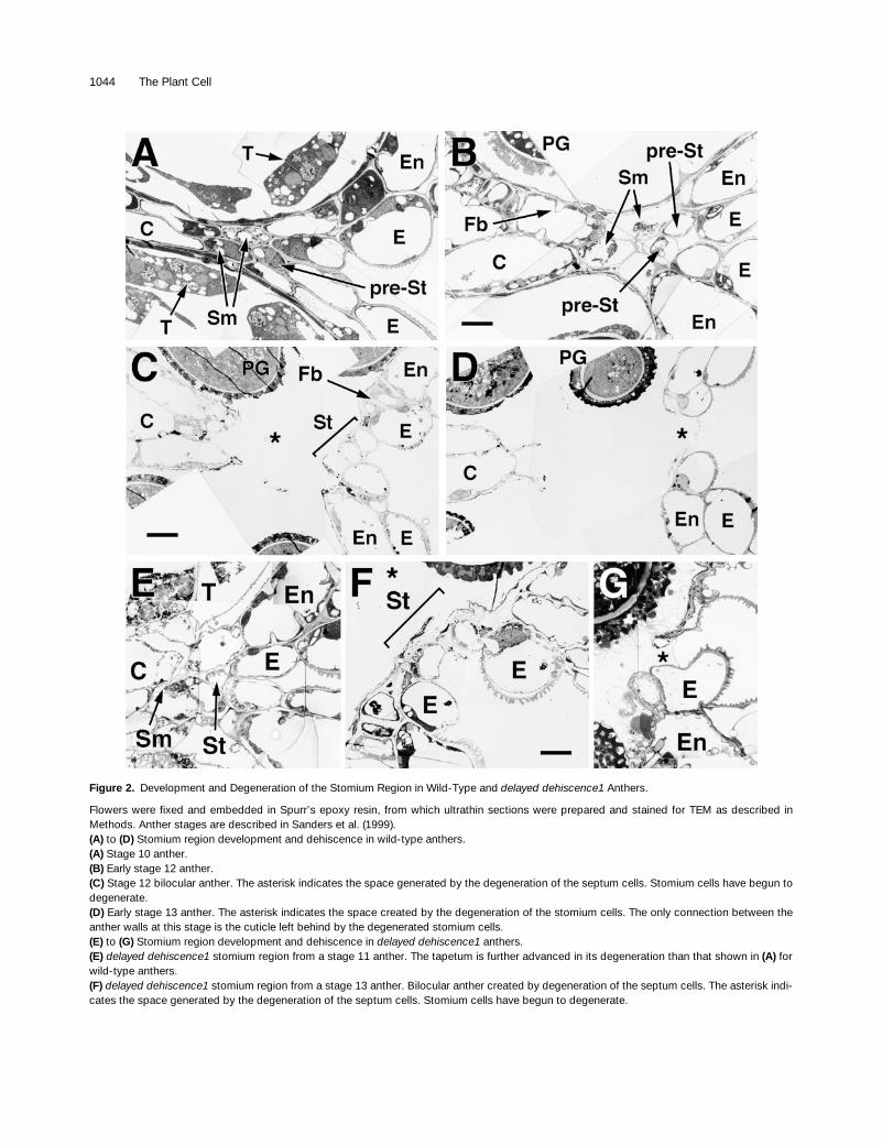
Figure 2.
Development and Degeneration of the Stomium Region in Wild-Type and
delayed dehiscence1
Anthers.
Flowers were fixed and embedded in Spurr’s epoxy resin, from which ultrathin sections were prepared and stained for TEM as described inMethods. Anther stages are described in Sanders et al. (1999).
(A)
to
(D)
Stomium region development and dehiscence in wild-type anthers.
(A)
Stage 10 anther.
(B)
Early stage 12 anther.
(C)
Stage 12 bilocular anther. The asterisk indicates the space generated by the degeneration of the septum cells. Stomium cells have begun todegenerate.
(D)
Early stage 13 anther. The asterisk indicates the space created by the degeneration of the stomium cells. The only connection between theanther walls at this stage is the cuticle left behind by the degenerated stomium cells.
(E)
to
(G)
Stomium region development and dehiscence in
delayed dehiscence1
anthers.
(E)
delayed dehiscence1
stomium region from a stage 11 anther. The tapetum is further advanced in its degeneration than that shown in
(A)
forwild-type anthers.
(F)
delayed dehiscence1
stomium region from a stage 13 anther. Bilocular anther created by degeneration of the septum cells. The asterisk indi-cates the space generated by the degeneration of the septum cells. Stomium cells have begun to degenerate.

Jasmonic Acid Dehiscence Mutant 1045
and the connective cells that contained large vacuoles.Tapetal cells, although degenerating, were still presentwithin the anther locules (Figure 2A).
From stages 10 to 12 in wild-type anthers, the dehiscenceprogram occurred and resulted in pollen release (Figure 1).The tapetum degenerated and, by stage 12, was no longerpresent within the locules (Figure 2B). The cells of the endo-thecium and connective became enlarged, and lignified fi-brous bands were deposited within the walls of these cells(Figure 2B). In addition, the septum cells degenerated, re-sulting in a bilocular anther (Figures 2B and 2C). At stage 12,the mature stomium was observed as three cells that werenoticeably smaller than neighboring epidermal cells (Figure2C, stage 12). Degeneration of the stomium cells was similarto that observed for septum cells, and by stage 13, only acuticle remnant was left bridging the site of anther wallbreakage and pollen release (Figures 2C and 2D).
The TEM analysis of the
delayed dehiscence1
notch region,shown in Figures 2E to 2G, indicated that at the cellular level,stomium and septum cell differentiation and degenerationevents were not detectably different from those of wild-typeanthers (Figures 2C and 2F). However,
delayed dehiscence1
stomium cells degenerated at a later time in developmentthan stomium cells in wild-type anthers (cf. Figures 2C and2D to Figures 2F and 2G, respectively). Together, these datashow that dehiscence involves the differentiation and de-generation of septum and stomium cells within the anthernotch and that stomium degeneration in
delayed dehis-cence1
is delayed relative to that of the wild type.
A Complex T-DNA Insertion Cosegregates with the
delayed dehiscence1
Mutant Phenotype
We performed polymerase chain reaction (PCR) amplifica-tion with T-DNA–specific primers and analyzed genomicblots with T-DNA probes to determine whether a T-DNA in-sert was linked with the
delayed dehiscence1
phenotype(Figure 3). Table 1 shows that the
delayed dehiscence1
phe-notype segregated in a 3:1 ratio of wild-type to mutantplants, indicating that it was a recessive mutation. In addi-tion, the
delayed dehiscence1
line segregated in a 3:1Kan
R
:Kan
S
ratio, which suggested a single T-DNA insertionwith an active
neomycin-phosphotransferase
gene (Table 1).T-DNA–specific PCR primers were used to show that theT-DNA cosegregated with the mutant phenotype. Figure 3A
shows the expected 336-bp product in 10
delayed dehis-cence1
mutants. The 336-bp product was not detected innontransgenic wild-type DNA but was observed in some
ofthe plants with a wild-type phenotype in the
delayeddehiscence1
segregating population. These plants werehemizygous for the T-DNA insertion (data not shown). AT-DNA insert was detected in all 106
delayed dehiscence1
mutants tested using primers that generated either the 336-bpfragment (Figure 3A) or a 823-bp product (data not shown;see Methods), indicating close linkage between the T-DNAinsertion and the mutant phenotype.
DNA gel blots were hybridized with T-DNA–specificprobes to determine the structure of the T-DNA insert in the
delayed dehiscence1
segregating population. Figure 3Bshows the results obtained by hybridizing the T-DNA rightborder probe to mutant and wild-type plant DNAs from a
de-layed dehiscence1
segregating population.
delayed dehis-cence1
mutants contained three EcoRI T-DNA fragmentsthat were 7, 8.5, and 14 kb in length (Figure 3B). Hybridiza-tion of mutant DNA with the T-DNA left border showedseven SalI T-DNA fragments that were 15, 13, 10, 9, 7, 5.5,and 1.25 kb in length (data not shown). A single T-DNA in-sert would be expected to produce one EcoRI band andone SalI band when hybridized with right border and leftborder T-DNA probes, respectively (Behringer and Medford,1992). Wild-type plants showed either the same complexEcoRI or SalI fragments as
delayed dehiscence1
mutants orno hybridization, indicating that some wild-type plants werehemizygous for the T-DNA insertion. Twenty-five
delayeddehiscence1
mutant plants showed the same EcoRI andSaLI T-DNA fragment patterns (data not shown). Taken to-gether, these results indicated the presence of multipleT-DNAs in the
delayed dehiscence1
mutant within a singlelocus.
Isolation of Mutant and Wild-Type Genomic Clones Containing the T-DNA Insertion Site
To characterize the gene disrupted by T-DNA, a genomic li-brary was constructed using
delayed dehiscence1
DNA andscreened for clones that contained the T-DNA left and rightborder termini (see Methods). Eighty-one phage cloneswere identified and purified through three successiverounds of hybridization. Individual clones were screened by
Figure 2.
(continued).
(G)
delayed dehiscence1
stomium region from a stage 14 anther. The asterisk indicates the space created by the degeneration of the stomiumcells. The only connection between the anther walls at this stage is the cuticle.C, connective; E, epidermis; En, endothecium; Fb, fibrous bands; PG, pollen grain; pre-St, pre-stomium cell; Sm, septum; St, stomium; T, tape-tum. Bar in
(B)
5
3
m
m for
(A)
and
(B)
; bar in
(C)
5
5
m
m for
(C)
and
(D)
; bar in
(F)
5
3
m
m for
(E)
to
(G)
.

1046 The Plant Cell
hybridization to wild-type Arabidopsis genomic DNA gelblots to determine whether they contained plant DNA se-quences (data not shown). One lambda clone isolated fromthe
delayed dehiscence1
genomic library contained a 974-bp plant DNA sequence that flanked a T-DNA left border.This region was cloned, labeled, and hybridized to a ge-nomic blot that contained both wild-type and delayeddehiscence1 genomic DNAs. Figure 3C shows that a restric-tion fragment length polymorphism (RFLP) between wild-type DNA and the delayed dehiscence1 mutant was identi-fied by using two different restriction enzymes. For example,a 2.4-kb BamHI fragment in wild-type DNA was increased to18 kb after insertion of the T-DNA in the delayed dehiscence1locus. The RFLP in the EcoRI DNA gel blot (Figure 3C)
caused the wild-type DNA EcoRI fragment size to decreasefrom 2.6 to 2.3 kb because of an EcoRI site present withinthe T-DNA left border (Behringer and Medford, 1992). Takentogether, these results indicate that we isolated a plant DNAsequence flanking the T-DNA insertion in delayed dehis-cence1 plants.
The 974-bp delayed dehiscence1 plant sequence flankingthe T-DNA was used to screen a wild-type Arabidopsis ge-nomic library to isolate the corresponding wild-type DNA re-gion and identify the gene disrupted by the T-DNA insertion.Twenty-four clones were identified and purified to single iso-lates (data not shown). One 18-kb clone, designated F7,contained the 974-bp plant DNA sequence and the com-plete DELAYED DEHISCENCE1 gene (data not shown).
Figure 3. Detection of T-DNA and Plant Flanking Sequence in delayed dehiscence1 and Wild-Type Plants.
(A) PCR amplification of T-DNA–specific products in delayed dehiscence1 mutants. Genomic DNA was isolated from individual delayeddehiscence1 plants and used as a template to determine the presence of a T-DNA insertion. PCR amplification with primers specific to theT-DNA generated a 336-bp product in all 106 mutant plants studied (see Methods). The results from 10 individual delayed dehiscence1 mutantsare shown here.(B) Detection of T-DNA insert in plants that segregate for the delayed dehiscence1 phenotype. Genomic DNA (1 mg) was isolated from individualwild-type and mutant plants, digested with EcoRI, fractionated on a 0.7% agarose gel, and blotted to a Nytran filter. The filter was hybridizedwith a right border T-DNA probe (see Methods). Exposure time of autoradiogram was 14 hr. Bands at 7, 8.5, and 14 kb were observed in all mu-tant plants and in hemizygous wild-type plants but not in homozygous wild-type plants. A single copy T-DNA would be expected to produce onehybridizing fragment in this DNA gel blot (Behringer and Medford, 1992).(C) Identification of an RFLP between wild-type and delayed dehiscence1 plants. Genomic DNA (2 mg) was isolated from wild-type and delayeddehiscence1 plants by using a CsCl purification procedure (see Methods), digested with BamHI or EcoRI, fractionated on a 0.6% agarose gel,and blotted to a Nytran filter. The filters were hybridized with the 974-bp plant sequence that flanked the T-DNA left border (see Methods). Posi-tions of the BamHI and EcoRI sites and of the 974-bp fragment in the DELAYED DEHISCENCE1 region are shown in Figure 4. Exposure timewas 12 hr.(D) and (E) Comparison of plant genomic DNA flanking the T-DNA insertion site in delayed dehiscence1 and wild-type plants.(D) Genomic DNA (2 mg) was isolated from wild-type and delayed dehiscence1 plants (see Methods), digested with NcoI, fractionated on a 0.6%agarose gel, and blotted to a Nytran filter. The filter was hybridized with a genomic DNA sequence probe that contained exons 1 and 2 of de-layed dehiscence1 (see Methods and Figure 4). Exposure time of autoradiogram was 21 hr.(E) Genomic DNA (2 mg) was isolated from wild-type and delayed dehiscence1 plants (see Methods), digested with AatII, fractionated on a 0.6%agarose gel, and blotted to a Nytran filter. The filter was hybridized with a cDNA sequence probe that contained exons 3 and 4 of delayeddehiscence1 (see Methods and Figure 4). Exposure time of autoradiogram was 21 hr.L, marker lane; M, delayed dehiscence1 mutant; W, wild-type plant.
Jasmonic Acid Dehiscence Mutant 1047
A Single Gene Is Disrupted by the T-DNA Insertion in delayed dehiscence1
We sequenced both the 974-bp DNA fragment from the de-layed dehiscence1 genomic library and the entire 18-kbwild-type genomic DNA insert to identify genes in the regionof the T-DNA insertion (GenBank accession numberAF218257). Sequence information from the 974-bp frag-ment identified the insertion site of the T-DNA left borderwithin the delayed dehiscence1 mutant allele and positionedthe T-DNA insertion within the 18-kb sequence, as illus-trated in Figures 4A and 4B. DNA sequence analysis alsosuggested several possible exons at the region of the T-DNAinsertion site and a putative structure for the gene disruptedby the T-DNA. We designated the putative wild-type genedisrupted by the T-DNA as DELAYED DEHISCENCE1. PCRprimers were designed using sequence information from theexons surrounding the T-DNA insertion site to amplify a par-tial cDNA (Figure 4A, primer 3R; see Methods). cDNAs wereamplified from inflorescence mRNA, cloned, and sequenced(see Methods). Figures 4A and 4B show the DELAYEDDEHISCENCE1 gene structure determined by comparisonof the genomic and cDNA sequences. The DELAYED DEHIS-CENCE1 gene consists of four exons with a coding se-quence (ATG→STOP) of 1176 bp that specifies a 391–aminoacid protein sequence (shown in Figure 5). The 974-bpflanking sequence contained exons 1 and 2, and sequenceanalysis showed that the delayed dehiscence1 T-DNA inser-tion was in the second intron (Figure 4A). Two other putativegenes, PG1 and PG2, were identified within the 18-kb wild-type DNA sequence (GenScan; see Methods) and are illus-trated in Figure 4B. PG1 and PG2 are upstream of theT-DNA insertion site in the delayed dehiscence1 mutant.
Analysis of the delayed dehiscence1 974-bp plant flankingsequence showed that the DNA sequence immediately 59 tothe T-DNA insertion was the same as in wild-type DNA (datanot shown). To determine whether the T-DNA insertion haddisrupted plant sequences more distally 59 to the insertionsite, nontransgenic wild-type and mutant DNAs were di-gested with NcoI, gel blotted, and hybridized with a 59 DE-LAYED DEHISCENCE1 probe containing exons 1 and 2(Figures 3D and 4). As Figure 3D shows, the wild-type 8.4-kbNcoI DNA fragment upstream of the T-DNA insertion site indelayed dehiscence1 had not been altered.
To address the possibility of a sequence alteration 39 tothe T-DNA insertion site, PCR primers were designed spe-cifically for the DELAYED DEHISCENCE1 genomic se-quence downstream of the T-DNA left border. A PCR primerpair, 1P and 2P, in which primer 1P was located 243 bpdownstream of the left border insertion (illustrated in Figure4B), was successful in generating the corresponding wild-type 2.7-kb fragment from the delayed dehiscence1 mutantDNA (data not shown). This result demonstrated that theT-DNA insertion did not generate a 39 deletion in the delayeddehiscence1 gene. We also confirmed that genomic DNAfurther 39 to the mutated delayed dehiscence1 gene was not
disrupted. DNA gel blots containing nontransgenic wild-typeand mutant DNA digested with AatII were hybridized with a39 DELAYED DEHISCENCE1 probe containing exons 3 and4 (Figures 3E and 4). The same .23-kb AatII fragment wasdetected in both delayed dehiscence1 mutant and nontrans-genic wild-type plants (Figure 3E).
Taken together, these results indicate that no rearrange-ments or deletions were detected upstream or downstreamof the T-DNA insertion in delayed dehiscence1 mutants andsuggest that the T-DNA in delayed dehiscence1 affects onlythe DELAYED DEHISCENCE1 gene at its site of insertion.
The Gene Disrupted in delayed dehiscence1 Encodes an Enzyme in the Jasmonic Acid Biosynthesis Pathway
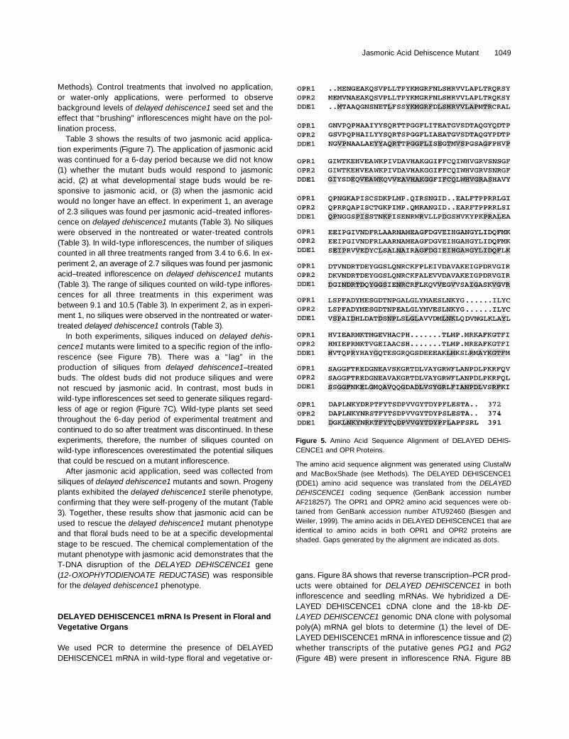
Homology searches with DELAYED DEHISCENCE1 nucle-otide and translated amino acid sequences identified the Ar-abidopsis 12-OXOPHYTODIENOATE REDUCTASE genes,OPR1 and OPR2, as a strong match (Biesgen and Weiler,1999). The amino acid alignment of the three OPR familymembers is shown in Figure 5. The structures and the ho-mology relationships of the OPR1, OPR2, and DELAYEDDEHISCENCE1 genes are shown in Table 2. 12-Oxophyto-dienoate reductase (OPR) is an enzyme in the jasmonic acidbiosynthesis pathway, as shown in Figure 6A. The OPRenzymatic step converts 12-oxophytodienoic acid (OPDA)into 3-oxo-2-(29-Z-pentenyl)-cyclopentane-1-octanoic acid(OPC-8:0) (Figure 6A; Schaller and Weiler, 1997a). Theconserved structure of DELAYED DEHISCENCE1 and theOPR genes indicated a gene-family relationship in whichDELAYED DEHISCENCE1 was the more divergent member(Figure 5 and Table 2). The DELAYED DEHISCENCE1 pro-tein has z50% identity (67% similarity) with the OPR1 andOPR2 peptides. The region of conservation was spreadthroughout the protein (Figure 5). The DELAYED DEHIS-CENCE1 gene was also identified as being related to OPR1and OPR2 in an Arabidopsis Genome Project DNA se-quence release (chromosome 2, 106,716 bp, BAC F5K7, ac-cession number AC006413). These results suggest that theDELAYED DEHISCENCE1 gene is a divergent member ofthe Arabidopsis 12-OXOPHYTODIENOATE REDUCTASEgene family and encodes an enzyme in the jasmonic acidbiosynthesis pathway.
Exogenous Jasmonic Acid Restores Fertility to delayed dehiscence1 Plants
We attempted to rescue the delayed dehiscence1 male-sterile phenotype by exogenous application of jasmonicacid to determine whether a defect in jasmonic acid synthe-sis was responsible for the delay in anther dehiscence. Fig-ures 7A and 7B show in a schematic diagram how theexogenous jasmonic acid (2 mmol/mL) was applied to devel-oping inflorescences of delayed dehiscence1 plants (see
1048 The Plant Cell
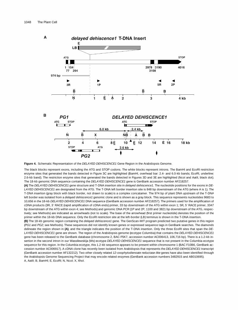
Figure 4. Schematic Representation of the DELAYED DEHISCENCE1 Gene Region in the Arabidopsis Genome.
The black blocks represent exons, including the ATG and STOP codons. The white blocks represent introns. The BamHI and EcoRI restrictionenzyme sites that generated the bands detected in Figure 3C are highlighted (BamHI, overhead bar: 2.4- and 6.0-kb bands; EcoRI, underline:2.6-kb band). The restriction enzyme sites that generated the bands detected in Figures 3D and 3E are highlighted (NcoI and AatII, black dot).The 18-kb genomic DNA sequence containing the DELAYED DEHISCENCE1 gene is GenBank accession number AF218257.(A) The DELAYED DEHISCENCE1 gene structure and T-DNA insertion site in delayed dehiscence1. The nucleotide positions for the exons in DE-LAYED DEHISCENCE1 are designated from the ATG. The T-DNA left border insertion site is 848 bp downstream of the ATG (where A is 1). TheT-DNA insertion (gray block with black border, not drawn to scale) is a complex concatamer. The 974 bp of plant DNA upstream of the T-DNAleft border was isolated from a delayed dehiscence1 genomic clone and is shown as a gray block. This sequence represents nucleotides 9683 to10,656 in the 18-kb DELAYED DEHISCENCE1 DNA sequence (GenBank accession number AF218257). The primers used for the amplification ofcDNA products (3R, 39 RACE (rapid amplification of cDNA ends) primer, 33 bp downstream of the ATG within exon 1; 5R, 59 RACE primer, 3347bp downstream of the ATG within exon 4; see Methods) and genomic DNA PCR (1P and 2P, 1100 and 3821 bp downstream of the ATG, respec-tively; see Methods) are indicated as arrowheads (not to scale). The base of the arrowhead (first primer nucleotide) denotes the position of theprimer within the 18-kb DNA sequence. Only the EcoRI restriction site at the left-border (LB) terminus is shown in the T-DNA insertion.(B) The 18-kb genomic region containing the delayed dehiscence1 gene. The GenScan-MIT program predicted two putative genes in this region(PG1 and PG2; see Methods). These sequences did not identify known genes or expressed sequence tags in GenBank searches. The diamondsdelineate the region shown in (A), and the triangle indicates the position of the T-DNA insertion. Only the three EcoRI sites that span the DE-LAYED DEHISCENCE1 gene are shown. The region of the Arabidopsis genome (ecotype Columbia) that contains the DELAYED DEHISCENCE1gene has been released to the GenBank database (chromosome 2, BAC F5K7, accession number AC006413, 106,716 bp). There is a 1.2-kb in-sertion in the second intron in our Wassilewskija (Ws) ecotype DELAYED DEHISCENCE1 sequence that is not present in the Columbia ecotypesequence for this region. In the Columbia ecotype, this 1.2-kb sequence appears to be present within chromosome 1 (BAC F10B6, GenBank ac-cession number AC006917). A cDNA clone has recently been isolated from Arabidopsis that represents the DELAYED DEHISCENCE1 transcript(GenBank accession number AF132212). Two other closely related 12-oxophytodienoate reductase-like genes have also been identified throughthe Arabidopsis Genome Sequencing Project that may encode related enzymes (GenBank accession numbers 3482915 and AB010695).A, AatII; B, BamHI; E, EcoRI; N, NcoI; X, XhoI.
Jasmonic Acid Dehiscence Mutant 1049
Methods). Control treatments that involved no application,or water-only applications, were performed to observebackground levels of delayed dehiscence1 seed set and theeffect that “brushing” inflorescences might have on the pol-lination process.
Table 3 shows the results of two jasmonic acid applica-tion experiments (Figure 7). The application of jasmonic acidwas continued for a 6-day period because we did not know(1) whether the mutant buds would respond to jasmonicacid, (2) at what developmental stage buds would be re-sponsive to jasmonic acid, or (3) when the jasmonic acidwould no longer have an effect. In experiment 1, an averageof 2.3 siliques was found per jasmonic acid–treated inflores-cence on delayed dehiscence1 mutants (Table 3). No siliqueswere observed in the nontreated or water-treated controls(Table 3). In wild-type inflorescences, the number of siliquescounted in all three treatments ranged from 3.4 to 6.6. In ex-periment 2, an average of 2.7 siliques was found per jasmonicacid–treated inflorescence on delayed dehiscence1 mutants(Table 3). The range of siliques counted on wild-type inflores-cences for all three treatments in this experiment wasbetween 9.1 and 10.5 (Table 3). In experiment 2, as in experi-ment 1, no siliques were observed in the nontreated or water-treated delayed dehiscence1 controls (Table 3).
In both experiments, siliques induced on delayed dehis-cence1 mutants were limited to a specific region of the inflo-rescence (see Figure 7B). There was a “lag” in theproduction of siliques from delayed dehiscence1–treatedbuds. The oldest buds did not produce siliques and werenot rescued by jasmonic acid. In contrast, most buds inwild-type inflorescences set seed to generate siliques regard-less of age or region (Figure 7C). Wild-type plants set seedthroughout the 6-day period of experimental treatment andcontinued to do so after treatment was discontinued. In theseexperiments, therefore, the number of siliques counted onwild-type inflorescences overestimated the potential siliquesthat could be rescued on a mutant inflorescence.
After jasmonic acid application, seed was collected fromsiliques of delayed dehiscence1 mutants and sown. Progenyplants exhibited the delayed dehiscence1 sterile phenotype,confirming that they were self-progeny of the mutant (Table3). Together, these results show that jasmonic acid can beused to rescue the delayed dehiscence1 mutant phenotypeand that floral buds need to be at a specific developmentalstage to be rescued. The chemical complementation of themutant phenotype with jasmonic acid demonstrates that theT-DNA disruption of the DELAYED DEHISCENCE1 gene(12-OXOPHYTODIENOATE REDUCTASE) was responsiblefor the delayed dehiscence1 phenotype.
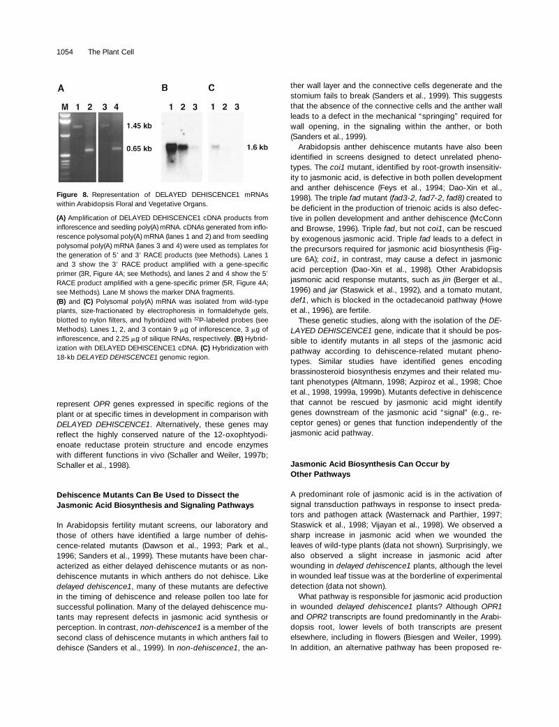
DELAYED DEHISCENCE1 mRNA Is Present in Floral and Vegetative Organs
We used PCR to determine the presence of DELAYEDDEHISCENCE1 mRNA in wild-type floral and vegetative or-
gans. Figure 8A shows that reverse transcription–PCR prod-ucts were obtained for DELAYED DEHISCENCE1 in bothinflorescence and seedling mRNAs. We hybridized a DE-LAYED DEHISCENCE1 cDNA clone and the 18-kb DE-LAYED DEHISCENCE1 genomic DNA clone with polysomalpoly(A) mRNA gel blots to determine (1) the level of DE-LAYED DEHISCENCE1 mRNA in inflorescence tissue and (2)whether transcripts of the putative genes PG1 and PG2(Figure 4B) were present in inflorescence RNA. Figure 8B
Figure 5. Amino Acid Sequence Alignment of DELAYED DEHIS-CENCE1 and OPR Proteins.
The amino acid sequence alignment was generated using ClustalWand MacBoxShade (see Methods). The DELAYED DEHISCENCE1(DDE1) amino acid sequence was translated from the DELAYEDDEHISCENCE1 coding sequence (GenBank accession numberAF218257). The OPR1 and OPR2 amino acid sequences were ob-tained from GenBank accession number ATU92460 (Biesgen andWeiler, 1999). The amino acids in DELAYED DEHISCENCE1 that areidentical to amino acids in both OPR1 and OPR2 proteins areshaded. Gaps generated by the alignment are indicated as dots.

1050 The Plant Cell
ther dehiscence program (Figures 9B and 9C), and at stage12 (Figure 9D) the anther becomes bilocular after the degen-eration of the septum. After stage 12, the stomium degener-ates to allow pollen release (stage 13; Figures 1A and 2D).
In wild-type floral sections at stage 5, the DELAYEDDEHISCENCE1 mRNA was localized within all floral organs(Figures 9E and 9I). Later in anther development, DELAYEDDEHISCENCE1 mRNA was localized specifically within thepistil, petal, and stamen filament (Figures 9F to 9H and 9J to9L). Serial anther sections showed that DELAYED DEHIS-CENCE1 mRNA was present within the vascular region clos-est to the stamen filament but decreased in sections fartherfrom the base of the anther (data not shown). At stage 10,the level of DELAYED DEHISCENCE1 mRNA increasedwithin the pistil, petal, and stamen filament (cf. Figures 9Fand 9G to Figures 9J and 9K, respectively). However, afterstage 10, the DELAYED DEHISCENCE1 mRNA at thesesites decreased to a low level (anther development stage 12;Figures 9H and 9L). The highest level of DELAYED DEHIS-CENCE1 mRNA was observed to correspond to the antherstage preceding the expansion of the endothecium and theappearance of fibrous bands (Figure 9; Sanders et al., 1999).During late anther development and after the initiation of de-hiscence program events, DELAYED DEHISCENCE1 mRNAwas not detected above background levels in cells of theanther, except within the vascular region contiguous to thestamen filament (Figures 9F to 9H and 9J to 9L).
DELAYED DEHISCENCE1 anti-mRNA probes were hy-bridized with delayed dehiscence1 floral sections to deter-mine the level of DELAYED DEHISCENCE1 mRNA within themutant. DELAYED DEHISCENCE1 mRNA was not detectedwithin the cross-sections of delayed dehiscence1 floralbuds, as shown in Figures 9M to 9P. As a positive controlwe hybridized anti-rRNA to the anther developmental stages5, 10, and 12 (see Methods), the results of which are shownin Figures 9Q to 9T. rRNA was localized to all cells and or-gans of the flower and was clearly present within the cells of
shows the presence of the 1.6-kb DELAYED DEHISCENCE1mRNA in inflorescence polysomal poly(A) mRNA. DELAYEDDEHISCENCE1 transcripts were also detected at a reducedlevel in silique polysomal poly(A) mRNA (Figure 8B). Underour RNA gel blot conditions, the DELAYED DEHISCENCE1cDNA would not have hybridized with related OPR1 andOPR2 mRNAs (Table 2). As shown in Figure 8C, only the DE-LAYED DEHISCENCE1 mRNA was detected on RNA gelblots hybridized with the 18-kb DELAYED DEHISCENCE1genomic region. At a longer autoradiogram exposure (6days), the hybridization signal for the 18-kb genomic regionprobe was equivalent to that obtained with the cDNA probe(data not shown), and no PG1 and PG2 transcripts were de-tected (data not shown). Taken together, these results indi-cate that DELAYED DEHISCENCE1 mRNA is present onpolysomes in developing inflorescences.
DELAYED DEHISCENCE1 mRNA Is Regulated Spatially and Temporally during Floral Development
We hybridized a DELAYED DEHISCENCE1 anti-mRNAprobe with transverse wild-type inflorescence sections to lo-calize DELAYED DEHISCENCE1 mRNA within developingfloral buds (see Methods). Under our in situ hybridizationconditions, the DELAYED DEHISCENCE1 anti-mRNA probedoes not cross-hybridize with OPR1 and OPR2 mRNAs (Ta-ble 2; data not shown).
Figure 9 shows the results of the in situ hybridization ex-periment. A ribosomal gene was used as a positive controlfor this experiment. We focused the in situ hybridizationstudies on stages of floral development that contained an-thers undergoing dehiscence program events. Figures 9A to9D show bright-field photographs of stages 5, 10 (early andlate), and 12 of wild-type anther development (Sanders etal., 1999). Stage 5 of anther development precedes meiosis(Figure 9A), stage 10 occurs before the initiation of the an-
Table 2. Comparison of Arabidopsis DELAYED DEHISCENCE1 and 12-OXOPHYTODIENOATE REDUCTASE Gene Family Membersa
Coding Sequence (ATG to STOP)b
Gene Number of Exons Number of Amino Acids Exon Intron Exon Intron Exon Intron Exon % Identity to OPR1c
OPR1 3 372 — — 218 96 308 75 593 100OPR2 4 374 83 79 141 93 308 83 593 89DDE1 4 391 77 76 141 2683 131 81 827 50
a GenBank accession number AF218257 for the DELAYED DEHISCENCE1 18-kb gene region. GenBank accession no. ATU92460 for the 12-OXOPHYTODIENOATE REDUCTASE genes, OPR1 and OPR2 (Biesgen and Weiler, 1999). The DELAYED DEHISCENCE1 gene was recentlyidentified in the Arabidopsis sequencing project. It is present on the chromosome 2 BAC F5K7 (GenBank accession no. AC006413) and was an-notated in a conceptual translation as a putative OPR enzyme (GenBank accession no. AAD19764).b As defined in this table, the first exon begins at the ATG codon and the last exon ends at the STOP codon. Nucleotide lengths of introns are initalics.c Sequence comparison by GenBank BlastP (Blast 2 Sequences at NCBI; http://www.ncbi.nlm.nih.gov). Percent identity to OPR1 at amino acidlevel.
Jasmonic Acid Dehiscence Mutant 1051
the anthers in our floral sections. These results show thatanther sections at all developmental stages contained cellsin which a positive in situ hybridization could be obtained.
Taken together, our results indicate that no DELAYEDDEHISCENCE1 mRNA is present in delayed dehiscence1mutant plants. In wild-type plants, the DELAYED DEHIS-CENCE1 mRNA accumulation is regulated in a spatial andtemporal pattern during floral development. During the an-ther dehiscence program, DELAYED DEHISCENCE1 mRNAis detectable only within the vascular region of the anther.
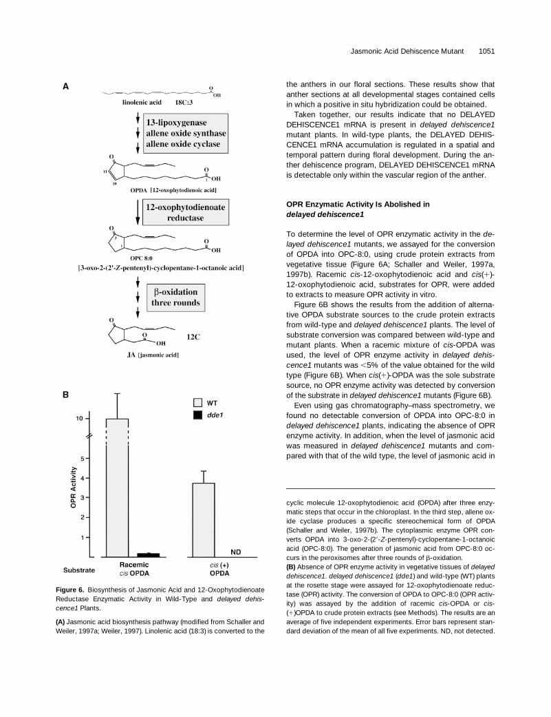
OPR Enzymatic Activity Is Abolished indelayed dehiscence1
To determine the level of OPR enzymatic activity in the de-layed dehiscence1 mutants, we assayed for the conversionof OPDA into OPC-8:0, using crude protein extracts fromvegetative tissue (Figure 6A; Schaller and Weiler, 1997a,1997b). Racemic cis-12-oxophytodienoic acid and cis(1)-12-oxophytodienoic acid, substrates for OPR, were addedto extracts to measure OPR activity in vitro.
Figure 6B shows the results from the addition of alterna-tive OPDA substrate sources to the crude protein extractsfrom wild-type and delayed dehiscence1 plants. The level ofsubstrate conversion was compared between wild-type andmutant plants. When a racemic mixture of cis-OPDA wasused, the level of OPR enzyme activity in delayed dehis-cence1 mutants was ,5% of the value obtained for the wildtype (Figure 6B). When cis(1)-OPDA was the sole substratesource, no OPR enzyme activity was detected by conversionof the substrate in delayed dehiscence1 mutants (Figure 6B).
Even using gas chromatography–mass spectrometry, wefound no detectable conversion of OPDA into OPC-8:0 indelayed dehiscence1 plants, indicating the absence of OPRenzyme activity. In addition, when the level of jasmonic acidwas measured in delayed dehiscence1 mutants and com-pared with that of the wild type, the level of jasmonic acid in
Figure 6. Biosynthesis of Jasmonic Acid and 12-OxophytodienoateReductase Enzymatic Activity in Wild-Type and delayed dehis-cence1 Plants.
(A) Jasmonic acid biosynthesis pathway (modified from Schaller andWeiler, 1997a; Weiler, 1997). Linolenic acid (18:3) is converted to the
cyclic molecule 12-oxophytodienoic acid (OPDA) after three enzy-matic steps that occur in the chloroplast. In the third step, allene ox-ide cyclase produces a specific stereochemical form of OPDA(Schaller and Weiler, 1997b). The cytoplasmic enzyme OPR con-verts OPDA into 3-oxo-2-(29-Z-pentenyl)-cyclopentane-1-octanoicacid (OPC-8:0). The generation of jasmonic acid from OPC-8:0 oc-curs in the peroxisomes after three rounds of b-oxidation.(B) Absence of OPR enzyme activity in vegetative tissues of delayeddehiscence1. delayed dehiscence1 (dde1) and wild-type (WT) plantsat the rosette stage were assayed for 12-oxophytodienoate reduc-tase (OPR) activity. The conversion of OPDA to OPC-8:0 (OPR activ-ity) was assayed by the addition of racemic cis-OPDA or cis-(1)OPDA to crude protein extracts (see Methods). The results are anaverage of five independent experiments. Error bars represent stan-dard deviation of the mean of all five experiments. ND, not detected.

1052 The Plant Cell
the delayed dehiscence1 mutants was not detectable (datanot shown). Taken together, these results indicate that thedelayed dehiscence1 mutants have no detectable 12-oxo-phytodienoate reductase activity and no detectable level ofjasmonic acid. In addition, these results show that the T-DNAinsertion eliminated 12-oxophytodienoate reductase enzymeactivity in delayed dehiscence1 mutants and demonstratethat in the delayed dehiscence1 mutant, the T-DNA insertiondisrupted a gene encoding 12-oxophytodienoate reductase,an enzyme in the octadecanoid pathway (Figure 6A).
DISCUSSION
We isolated an Arabidopsis gene, DELAYED DEHIS-CENCE1, that encodes 12-oxophytodienoate reductase, anenzyme in the jasmonic acid biosynthesis pathway (Figure6A). This gene is disrupted by a T-DNA insertion in delayed
dehiscence1 plants, which causes a delay in anther de-hiscence and male sterility. In mutant plants, DELAYEDDEHISCENCE1 mRNA is not detected, and no 12-oxophy-todienoate reductase enzyme activity is observed. Applica-tion of exogenous jasmonic acid to floral buds of delayeddehiscence1 plants rescues the male sterility defect, an indi-cation that jasmonic acid signaling, either directly or indi-rectly, plays a role in the anther dehiscence process.
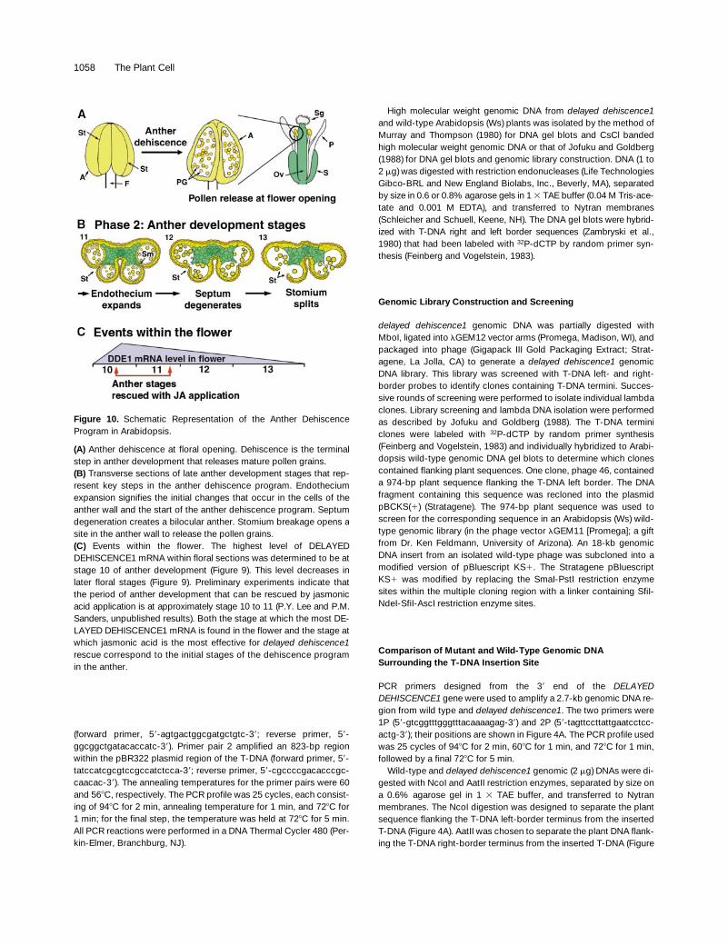
Anther Dehiscence Requires a Coordinated Programof Events
Anther dehiscence leads to breakage of the anther wall andthe release of pollen grains from the locules at flower open-ing (Figures 1 and 2). Figure 10 summarizes the events in thedehiscence program of the Arabidopsis anther. Two special-ized cell types—the septum and the stomium—undergo se-quential degeneration events that lead to development of abilocular anther (Figures 2A to 2C and 10) and breakage ofthe anther wall (Figures 2C, 2D, and 10). Previously, we ab-lated the stomium of tobacco anthers, which generatedmale-sterile plants (Beals and Goldberg, 1997). These find-ings, together with the TEM studies in Figure 2, suggest thatcell degeneration of the stomium and breakage of the antherwall are genetically programmed events that are required fordehiscence to occur. The molecular processes that controlthe cell degeneration events during anther dehiscence areprobably initiated before this degeneration can be visualizedin the septum and stomium cells.
The events that determine the timing of anther dehiscencemust be coordinated with other developmental processesthat occur within both the anther (e.g., pollen development)and the flower (e.g., flower opening, pistil receptivity, andovary development) to ensure that successful fertilizationand seed set take place (Figures 10A and 10B). The flowersof delayed dehiscence1 mutants open, are receptive to polli-nation, and senesce just as those on wild-type plants do(Figure 1). In delayed dehiscence1, anther dehiscenceevents appear similar to those in the wild type; however, achange in the timing of stomium cell degeneration causesthe mutant defect to occur (Figure 2 and Sanders et al.,1999). These results show that degeneration of the stomium indelayed dehiscence1 mutant anthers is uncoupled from otherdehiscence program events (e.g., septum degeneration).
The complexity of the anther dehiscence program sug-gests that many genes are required for pollen release. Thisprediction has been confirmed in our recent Arabidopsisethyl methanesulfonate screen for sterility mutants by thefinding that a large number of mutants affect the dehiscenceprocess (Sanders et al., 1999). Many of these mutants aredelayed in dehiscence with phenotypes similar to delayeddehiscence1. These data indicate that many genes partici-pate in the dehiscence process, several of which may en-code enzymes in biosynthesis (e.g., jasmonic acid) and celldegeneration pathways.
Figure 7. Schematic Representation of the Jasmonic Acid RescueExperiment.
Inflorescences of delayed dehiscence1 (dde1) and wild-type plantswere tagged after removal of all open flowers. Exogenous jasmonicacid was applied to the floral buds of the tagged inflorescences overa 6-day period (see Methods). After 6 to 8 days, the inflorescenceswere examined for siliques.(A) delayed dehiscence1 plant tagged at start of experiment.(B) delayed dehiscence1 plant at end of experiment. After jasmonicacid treatment, seed set was observed, as demonstrated by thepresence of siliques. Siliques were found at specific positions withinthe developing inflorescence.(C) Wild-type control plants at end of the experiment. Wild-typeplants set seed under all experimental conditions (see Methods).Floral buds on wild-type inflorescences continued to set seed aftercompletion of the jasmonic acid treatment.

Jasmonic Acid Dehiscence Mutant 1053
DELAYED DEHISCENCE1 Encodes an Enzyme in the Jasmonic Acid Biosynthesis Pathway
The demonstration that DELAYED DEHISCENCE1 encodesan enzyme in the octadecanoid pathway suggests a role forjasmonic acid in anther dehiscence. DELAYED DEHIS-CENCE1 encodes 12-oxophytodienoate reductase, a cyto-plasmic enzyme that catalyzes the removal of a double bondin the first cyclic intermediate, 12-oxophytodienoic acid, inthe octadecanoic pathway (Figure 6A; Vick and Zimmerman,1984; Schaller and Weiler, 1997a). The octadecanoid path-way results in the synthesis of jasmonic acid (Figure 6A; Vickand Zimmerman, 1984; Weiler, 1997). The DELAYEDDEHISCENCE1 gene is the third gene identified to encodean Arabidopsis 12-oxophytodienoate reductase–like en-zyme. The related OPR1 and OPR2 genes were previouslyidentified by Schaller et al. (1998) and Biesgen and Weiler(1999). In delayed dehiscence1 plants, we did not detect
mRNA encoded by the DELAYED DEHISCENCE1 gene (Fig-ure 9), nor did we detect 12-oxophytodienoate reductaseactivity (Figure 6B). Together, these results indicate that theDELAYED DEHISCENCE1–encoded enzyme is the predomi-nant enzymatic activity responsible for conversion of OPDAin vivo (Figure 6) and, therefore, that DELAYED DEHIS-CENCE1 encodes the major 12-oxophytodienoate reduc-tase enzyme in Arabidopsis.
The role that the other 12-oxophytodienoate reductaseenzymes have in Arabidopsis is not known. The OPR1 andOPR2 enzymes have been reported to exhibit different sub-strate specificities, and Schaller et al. (1998) have suggestedthat OPR1 is not an enzyme in the octadecanoid pathwaybut may serve some unknown enzymatic function in vivo.OPR1 and OPR2 (Schaller et al., 1998; Biesgen and Weiler,1999) and two other candidate genes recently identified inthe Arabidopsis Genome Sequencing Project (GenBank ac-cession numbers 3,482,915 and AB010695) therefore, may
Table 3. Rescue of delayed dehiscence1 Phenotype after Exogenous Application of Jasmonic Acida
Experimental Treatment No. of Inflorescences No. of Siliquesb No. of Unexpanded PistilsAverage No. of Budsper Inflorescence
Average No. of Siliquesb
per Inflorescence
Experiment 1c
delayed dehiscence1No treatment 13 0 94 7.2 0Water 15 0 105 7.0 0Jasmonic acid 15 35 110 9.7 2.3d
Wild typeNo treatment 15 99 0 6.6 6.6Water 14 48 0 3.4 3.4Jasmonic acid 15 68 0 4.5 4.5
Experiment 2c
delayed dehiscence1No treatment 14 0 292 20.8 0Water 14 0 256 18.3 0Jasmonic acid 15 41 273 20.9 2.7d
Wild typeNo treatment 15 136 15 10.6 9.1Water 15 201 28 15.2 13.4Jasmonic acid 15 157 98 17.0 10.5
a Application of 2 mmol/mL jasmonic acid or water to inflorescences of delayed dehiscence1 and wild-type plants as described in Methods. Theprocedure of McConn and Browse (1996) was followed.b The application of jasmonic acid was limited to a 6-day period. In our experiments, we did not know whether the mutant buds would respond tojasmonic acid or at what developmental stage buds would be responsive to jasmonic acid. Rescue of mutant buds could be limited to the periodof jasmonic acid application or restricted to a responsive stage of development. The delayed dehiscence1 mutants responded to jasmonic acidtreatment and set seed, as observed by the presence of siliques. Siliques on delayed dehiscence1 mutants were produced within a specific re-gion of the inflorescence (see Figure 7B). By contrast, most buds set seed to generate siliques on wild-type inflorescences. Wild-type plants setseed throughout the period of experimental treatment and continued to do so after the 6-day treatment was discontinued. Therefore, in these ex-periments the number of siliques counted on wild-type inflorescences will overestimate the potential siliques that can be rescued on a mutant in-florescence.c Two jasmonic acid rescue experiments were performed under different conditions with plants grown in our greenhouse. Experiment 1 (August1998) was a 6-day treatment, 10 applications, with siliques tallied after 6 days. Experiment 2 (November 1998) was a 6-day treatment, 11 appli-cations, with siliques tallied after 8 days.d In a trial sowing, we obtained 151 progeny from jasmonic acid–rescued siliques on mutant plants. All except one showed the delayeddehiscence1 phenotype.
1054 The Plant Cell
represent OPR genes expressed in specific regions of theplant or at specific times in development in comparison withDELAYED DEHISCENCE1. Alternatively, these genes mayreflect the highly conserved nature of the 12-oxophtyodi-enoate reductase protein structure and encode enzymeswith different functions in vivo (Schaller and Weiler, 1997b;Schaller et al., 1998).
Dehiscence Mutants Can Be Used to Dissect the Jasmonic Acid Biosynthesis and Signaling Pathways
In Arabidopsis fertility mutant screens, our laboratory andthose of others have identified a large number of dehis-cence-related mutants (Dawson et al., 1993; Park et al.,1996; Sanders et al., 1999). These mutants have been char-acterized as either delayed dehiscence mutants or as non-dehiscence mutants in which anthers do not dehisce. Likedelayed dehiscence1, many of these mutants are defectivein the timing of dehiscence and release pollen too late forsuccessful pollination. Many of the delayed dehiscence mu-tants may represent defects in jasmonic acid synthesis orperception. In contrast, non-dehiscence1 is a member of thesecond class of dehiscence mutants in which anthers fail todehisce (Sanders et al., 1999). In non-dehiscence1, the an-
ther wall layer and the connective cells degenerate and thestomium fails to break (Sanders et al., 1999). This suggeststhat the absence of the connective cells and the anther wallleads to a defect in the mechanical “springing” required forwall opening, in the signaling within the anther, or both(Sanders et al., 1999).
Arabidopsis anther dehiscence mutants have also beenidentified in screens designed to detect unrelated pheno-types. The coi1 mutant, identified by root-growth insensitiv-ity to jasmonic acid, is defective in both pollen developmentand anther dehiscence (Feys et al., 1994; Dao-Xin et al.,1998). The triple fad mutant (fad3-2, fad7-2, fad8) created tobe deficient in the production of trienoic acids is also defec-tive in pollen development and anther dehiscence (McConnand Browse, 1996). Triple fad, but not coi1, can be rescuedby exogenous jasmonic acid. Triple fad leads to a defect inthe precursors required for jasmonic acid biosynthesis (Fig-ure 6A); coi1, in contrast, may cause a defect in jasmonicacid perception (Dao-Xin et al., 1998). Other Arabidopsisjasmonic acid response mutants, such as jin (Berger et al.,1996) and jar (Staswick et al., 1992), and a tomato mutant,def1, which is blocked in the octadecanoid pathway (Howeet al., 1996), are fertile.
These genetic studies, along with the isolation of the DE-LAYED DEHISCENCE1 gene, indicate that it should be pos-sible to identify mutants in all steps of the jasmonic acidpathway according to dehiscence-related mutant pheno-types. Similar studies have identified genes encodingbrassinosteroid biosynthesis enzymes and their related mu-tant phenotypes (Altmann, 1998; Azpiroz et al., 1998; Choeet al., 1998, 1999a, 1999b). Mutants defective in dehiscencethat cannot be rescued by jasmonic acid might identifygenes downstream of the jasmonic acid “signal” (e.g., re-ceptor genes) or genes that function independently of thejasmonic acid pathway.
Jasmonic Acid Biosynthesis Can Occur byOther Pathways
A predominant role of jasmonic acid is in the activation ofsignal transduction pathways in response to insect preda-tors and pathogen attack (Wasternack and Parthier, 1997;Staswick et al., 1998; Vijayan et al., 1998). We observed asharp increase in jasmonic acid when we wounded theleaves of wild-type plants (data not shown). Surprisingly, wealso observed a slight increase in jasmonic acid afterwounding in delayed dehiscence1 plants, although the levelin wounded leaf tissue was at the borderline of experimentaldetection (data not shown).
What pathway is responsible for jasmonic acid productionin wounded delayed dehiscence1 plants? Although OPR1and OPR2 transcripts are found predominantly in the Arabi-dopsis root, lower levels of both transcripts are presentelsewhere, including in flowers (Biesgen and Weiler, 1999).In addition, an alternative pathway has been proposed re-
Figure 8. Representation of DELAYED DEHISCENCE1 mRNAswithin Arabidopsis Floral and Vegetative Organs.
(A) Amplification of DELAYED DEHISCENCE1 cDNA products frominflorescence and seedling poly(A) mRNA. cDNAs generated from inflo-rescence polysomal poly(A) mRNA (lanes 1 and 2) and from seedlingpolysomal poly(A) mRNA (lanes 3 and 4) were used as templates forthe generation of 59 and 39 RACE products (see Methods). Lanes 1and 3 show the 39 RACE product amplified with a gene-specificprimer (3R, Figure 4A; see Methods), and lanes 2 and 4 show the 59
RACE product amplified with a gene-specific primer (5R, Figure 4A;see Methods). Lane M shows the marker DNA fragments.(B) and (C) Polysomal poly(A) mRNA was isolated from wild-typeplants, size-fractionated by electrophoresis in formaldehyde gels,blotted to nylon filters, and hybridized with 32P-labeled probes (seeMethods). Lanes 1, 2, and 3 contain 9 mg of inflorescence, 3 mg ofinflorescence, and 2.25 mg of silique RNAs, respectively. (B) Hybrid-ization with DELAYED DEHISCENCE1 cDNA. (C) Hybridization with18-kb DELAYED DEHISCENCE1 genomic region.
Jasmonic Acid Dehiscence Mutant 1055
cently that may generate jasmonic acid using 16:3 trienoicacids as substrate (Weber et al., 1997; Farmer et al., 1998).These data suggest that there is an alternative source of jas-monic acid synthesis in Arabidopsis that does not rely onthe 12-oxophytodienoate reductase activity of the DELAYEDDEHISCENCE1–encoded protein.
Jasmonic Acid May Control the Timing ofAnther Dehiscence
The chemical complementation of the delayed dehiscence1male-sterility phenotype indicates that jasmonic acid (or aderivative), directly or indirectly, plays a role in the timing ofanther dehiscence (Table 3 and Figure 7). The defect in de-layed dehiscence1 anthers delays stomium degeneration,the terminal step in the anther dehiscence program. Surpris-ingly, late-stage anthers were not rescued by exogenousjasmonic acid application in our experiments (Figure 7B).The lag in rescue observed in our experiments suggests thatexogenous jasmonic acid exerts its effect before the de-layed dehiscence1 defect is apparent and that a jasmonicacid “signal” is required at a specific developmental periodin the anther before anther dehiscence. Preliminary experi-ments treating individual, developmentally staged delayeddehiscence1 floral buds with jasmonic acid suggest thatonly buds containing anthers at approximately stages 10and 11 (stage 10 is shown in Figures 9B and 9C) can be res-cued and produce expanded siliques (P.Y. Lee, P.M. Sanders,and R.B.Goldberg, unpublished results). In contrast, budswith anthers at either later or earlier stages fail to respond tojasmonic acid (P.Y. Lee, P.M. Sanders, and R.B. Goldberg,unpublished results). Assuming that jasmonic acid is equallypermeable at all developmental stages, these preliminary re-sults suggest the existence of a developmental “window” inwhich jasmonic acid is perceived by the anther, such thattarget cells for the jasmonic acid signal are competent torecognize the “signal” only during this time period. This hy-pothesis might explain why ,100% of the jasmonic acid–treated delayed dehiscence1 bud clusters (Figure 7B) wererescued in the experiments presented in Table 3. That is,only a subset of the treated buds contained anthers atstages competent to be rescued. The question remains asto how the jasmonic acid signal, when perceived at onestage in anther development, is transferred to the terminalstep in anther development and results in stomium degener-ation (Figures 10B and 10C).
What Is the Source of Jasmonic Acid within the Arabidopsis Flower?
If jasmonic acid is a “signal” that initiates or coordinatesevents during the anther dehiscence program, what is theendogenous source of jasmonic acid within the flower? Oneclue to answering this question can be obtained from our in
situ hybridization experiments (Figure 9). In early anther de-velopment, before the differentiation of the stomium andseptum and preceding the expansion of the endothecium(Sanders et al., 1999), we detected DELAYED DEHIS-CENCE1 mRNA throughout premeiotic anthers and in otherorgans of the developing floral buds (Figures 9E and 9I). Bycontrast, during late stages of anther development, whenthe dehiscence program occurs, we did not detect DE-LAYED DEHISCENCE1 mRNA in the stomium, septum, con-nective, wall layers, or epidermis of the anther (Figures 9F to9H and 9J to 9L). Assuming that DELAYED DEHISCENCE1mRNA is not present below the detection limit of our in situhybridization experiments, these results suggest that theDELAYED DEHISCENCE1 gene is not expressed in the cellsof the anther that play a role in dehiscence (Figures 9J to9L). If the jasmonic acid “signal” is produced within the an-ther, then it must originate from jasmonic acid synthesizedduring the early stages of anther development.
We also observed that during the later stages of Arabi-dopsis flower development, DELAYED DEHISCENCE1 mRNAwas present in substantial levels in the pistils, petals, andstamen filaments and that DELAYED DEHISCENCE1 mRNAlevels were highest in stage 10 anthers (Figures 9F to 9Hand 10C), that is, before the initiation of dehiscence programevents (Figure 2). The level of DELAYED DEHISCENCE1mRNA decreased after stage 10 and by stage 12 wasalso decreased within the pistil, petals, and stamen filament(Figures 9, 10B, and 10C). The accumulation profile of DE-LAYED DEHISCENCE1 mRNA during late floral develop-ment corresponds to the stage of anther development thatcan be rescued by jasmonic acid (Figure 7; data not shown).Methyl-jasmonic acid is known to exert its effect as a vola-tile signal in plant defense responses (Farmer and Ryan,1990). If the DELAYED DEHISCENCE1 mRNA localizationpattern predicts the expression pattern for the DELAYEDDEHISCENCE1 gene (i.e., the DELAYED DEHISCENCE1–encoded 12-oxophytodienoate reductase enzyme activity),then perhaps jasmonic acid is synthesized primarily withinthe pistil and petals. If so, then these organs might providethe major source of “signal” for the anther within the en-closed floral bud. Alternatively, because DELAYED DEHIS-CENCE1 mRNA is present in stamen filaments, the filamentscould also be a site of jasmonic acid synthesis and a sourceof the “signal” within the anther.
Exogenous jasmonic acid application is capable of restor-ing fertility in the delayed dehiscence1 mutant flowers duringa specific period of anther development (Figure 7B). Late-stage delayed dehiscence1 floral buds were not rescued.Together, the stage of anther development responsive to theexogenous jasmonic acid and the mRNA localization patternsuggest that the jasmonic acid “signal” is required beforethe initiation of the anther dehiscence program (Figures 10Band 10C). Are the stomium cells the target for jasmonic acid,or is stomium cell degeneration the last step in a cascade ofevents initiated earlier? Our results indicate that jasmonicacid acts to time the degeneration of the stomium, but it is

1056 The Plant Cell
Figure 9. Localization of DELAYED DEHISCENCE1 mRNA within Floral Tissues of Arabidopsis Wild-Type and delayed dehiscence1 Plants.
Inflorescences were fixed, embedded in paraffin, sliced into 10-mm-thick sections, and hybridized with DELAYED DEHISCENCE1 anti-mRNAand ribosomal anti-RNA probes as outlined in Methods. Photographs were taken by using bright-field and dark-field microscopy.(A) to (D) Bright-field photographs of wild-type anther development stages. (A) Stage 5. Premeiotic microspore mother cells. (B) Early stage 10.(C) Late stage 10, before endothecium expansion. (D) Early stage 12. The tapetum has almost degenerated and the endothecium has ex-panded, but the anther still has four locules and an intact septum. These bright-field anther photographs are close-ups of anthers shown in(E) to (H).(E) to (L) Hybridization of DELAYED DEHISCENCE1 anti-mRNA probes to sections of wild-type floral buds corresponding to anther stagesshown in (A) to (D). (I) to (L) are close-ups of anthers shown in (E) to (H). Slide emulsions were exposed for 8 days, and the photographs weretaken by using dark-field photography.(M) to (P) Hybridization of DELAYED DEHISCENCE1 anti-mRNA probes to sections of delayed dehiscence1 floral buds corresponding to antherstages shown in (A) to (D). Slide emulsions were exposed for 8 days, except (N), which was exposed for 18 days.

Jasmonic Acid Dehiscence Mutant 1057
not known what other steps or molecules might play a partin the series of events leading to stomium degeneration.
Why Is There a Delay in Dehiscence in delayed dehiscence1 Mutants?
What causes dehiscence to be “delayed” in delayeddehiscence1 anthers? Because the T-DNA insertion knocksout the predominant 12-oxophytodienoate reductase en-zyme, the level of jasmonic acid may be insufficient to “sig-nal” the start of stomium cell degeneration at the time ofwild-type anther dehiscence. In delayed dehiscence1 flow-ers, perhaps the threshold level of jasmonic acid is reachedlater in floral development. Alternative 12-oxophytodienoatereductase enzymes encoded by the other members of theOPR gene family (e.g., OPR1 and OPR2) might produce suf-ficient levels of jasmonic acid to allow this buildup to occur.In addition, different pathways for generating jasmonic acidin delayed dehiscence1 mutants (e.g., the hexadecanoidpathway) could also account for the “delay” observed in an-ther dehiscence.
The isolation of the DELAYED DEHISCENCE1 geneshows that jasmonic acid acts as a signal to control the tim-ing of dehiscence. Our conclusion is based on the followingfindings: (1) a single T-DNA insertion generating a male-ster-ile phenotype, (2) the absence of DELAYED DEHISCENCE1mRNA transcripts in delayed dehiscence1 inflorescence tis-sue, (3) the absence of DELAYED DEHISCENCE1 12-oxo-phytodienoate reductase enzymatic activity, and (4) thedirect complementation of the delayed dehiscence1 pheno-type with jasmonic acid. The anther perceives the jasmonicacid signal before the initiation of the major dehiscence pro-gram events. The precise mechanism by which jasmonicacid signaling controls the timing of anther wall breakage atthe stomium remains to be determined.
METHODS
Mutant Isolation and Genetic Analysis
The male-sterile delayed dehiscence1 mutant was identified in a screenof T-DNA mutagenized Arabidopsis lines (ecotype Wassilewskija[Ws]) generated by Dr. Ken Feldmann (University of Arizona) (Feldmannand Marks, 1987; Feldmann, 1991; Forsthoefel et al., 1992). The
male-sterility screen and our initial characterization of the delayeddehiscence1 phenotype were described in Sanders et al. (1999). TheFeldmann line 1926 segregates for the delayed dehiscence1 pheno-type and is available through the Arabidopsis Biological ResourceCenter (ABRC) seed catalog (http://aims.cps.msu.edu/aims).
Light Microscopy
Bright-field photographs of individual flowers were taken using a dis-secting microscope (Olympus model SZH; Olympus, Lake Success,NY). Mutant and wild-type flowers were fixed overnight in FAA (3.7%formaldehyde, 50% ethanol, and 5.0% glacial acetic acid), dehy-drated in a graded ethanol series (twice at 50%, 60%, 70%, 85%,and 95%; three times at 100%), embedded in Spurr’s epoxy resin(Spurr, 1969; TedPella, Inc., Redding, CA), and sectioned into 1-mm-thick sections with an ultramicrotome (Sorvall model MT-600; DuPont,Wilmington, DE). Anther transverse sections were stained in 1% tolu-idine blue at 428C for 1 to 2 hr. Bright-field photographs of the anthercross-sections were taken using a compound microscope (Olympusmodel BH2). All photographs were taken with Kodak Gold 100 film(ISO 100/21).
Transmission Electron Microscopy
Inflorescences from wild-type and delayed dehiscence1 plants werefixed, processed, and embedded in Spurr’s epoxy resin as describedby Owen and Makaroff (1995). Flowers were placed in individualmolds before the resin blocks were cured. The floral buds were sec-tioned (1-mm-thick sections) with an ultramicrotome (model MT-600;Sorval) and stained in 1% toluidine blue as described above, and theregion of the stomium within the sections was visualized in the lightmicroscope. Ultrathin anther sections were then generated andplaced on formavar-coated copper grids, where they were stainedwith lead citrate and uranyl acetate. The anther sections were ob-served in a JEOL electron microscope (model 100CX II; JEOL USA,Inc., Peabody, MA) at 80 kV, and pictures of the anther notch regionswere taken using Kodak Electron Microscope film No. 4489.
Genomic DNA Isolation and T-DNA Insert Analysis
Leaves from individual Arabidopsis (Ws) plants were ground in liquidnitrogen, and the genomic DNA was isolated for amplification bypolymerase chain reaction (PCR) (Edwards et al., 1991). The ge-nomic DNA was amplified by PCR with T-DNA–specific primers andTaq DNA polymerase (Life Technology Gibco-BRL, Gaithersburg,MD). Two PCR primer pairs for the T-DNA construct (Feldmann andMarks, 1987) were used. Primer pair 1 amplified a 336-bp regionspanning the pBR322 plasmid and right-border regions of the T-DNA
Figure 9. (continued).
(Q) to (T) Hybridization of ribosomal anti-RNA probes to sections of wild-type and delayed dehiscence1 floral buds corresponding to antherstages shown in (A) to (D). (Q) Wild type; (R) to (T) delayed dehiscence1. Slide emulsions were exposed for 1 hr.A, anther; P, petal; Pi, pistil; S, sepal; St, stomium. Bar in (A) 5 25 mm; bar in (B) 5 50 mm for (B), (C), (I) to (K), (R), and (S); bar in (D) 5 50 mmfor (D), (L), and (T); bar in (M) 5 100 mm for (E) to (G), (M) to (O), and (Q); bar in (P) 5 100 mm for (H) and (P).
1058 The Plant Cell
(forward primer, 59-agtgactggcgatgctgtc-39; reverse primer, 59-ggcggctgatacaccatc-39). Primer pair 2 amplified an 823-bp regionwithin the pBR322 plasmid region of the T-DNA (forward primer, 59-tatccatcgcgtccgccatctcca-39; reverse primer, 59-cgccccgacacccgc-caacac-39). The annealing temperatures for the primer pairs were 60and 568C, respectively. The PCR profile was 25 cycles, each consist-ing of 948C for 2 min, annealing temperature for 1 min, and 728C for1 min; for the final step, the temperature was held at 728C for 5 min.All PCR reactions were performed in a DNA Thermal Cycler 480 (Per-kin-Elmer, Branchburg, NJ).
High molecular weight genomic DNA from delayed dehiscence1and wild-type Arabidopsis (Ws) plants was isolated by the method ofMurray and Thompson (1980) for DNA gel blots and CsCl bandedhigh molecular weight genomic DNA or that of Jofuku and Goldberg(1988) for DNA gel blots and genomic library construction. DNA (1 to2 mg) was digested with restriction endonucleases (Life TechnologiesGibco-BRL and New England Biolabs, Inc., Beverly, MA), separatedby size in 0.6 or 0.8% agarose gels in 1 3 TAE buffer (0.04 M Tris-ace-tate and 0.001 M EDTA), and transferred to Nytran membranes(Schleicher and Schuell, Keene, NH). The DNA gel blots were hybrid-ized with T-DNA right and left border sequences (Zambryski et al.,1980) that had been labeled with 32P-dCTP by random primer syn-thesis (Feinberg and Vogelstein, 1983).
Genomic Library Construction and Screening
delayed dehiscence1 genomic DNA was partially digested withMboI, ligated into lGEM12 vector arms (Promega, Madison, WI), andpackaged into phage (Gigapack III Gold Packaging Extract; Strat-agene, La Jolla, CA) to generate a delayed dehiscence1 genomicDNA library. This library was screened with T-DNA left- and right-border probes to identify clones containing T-DNA termini. Succes-sive rounds of screening were performed to isolate individual lambdaclones. Library screening and lambda DNA isolation were performedas described by Jofuku and Goldberg (1988). The T-DNA terminiclones were labeled with 32P-dCTP by random primer synthesis(Feinberg and Vogelstein, 1983) and individually hybridized to Arabi-dopsis wild-type genomic DNA gel blots to determine which clonescontained flanking plant sequences. One clone, phage 46, containeda 974-bp plant sequence flanking the T-DNA left border. The DNAfragment containing this sequence was recloned into the plasmidpBCKS(1) (Stratagene). The 974-bp plant sequence was used toscreen for the corresponding sequence in an Arabidopsis (Ws) wild-type genomic library (in the phage vector lGEM11 [Promega]; a giftfrom Dr. Ken Feldmann, University of Arizona). An 18-kb genomicDNA insert from an isolated wild-type phage was subcloned into amodified version of pBluescript KS1. The Stratagene pBluescriptKS1 was modified by replacing the SmaI-PstI restriction enzymesites within the multiple cloning region with a linker containing SfiI-NdeI-SfiI-AscI restriction enzyme sites.
Comparison of Mutant and Wild-Type Genomic DNA Surrounding the T-DNA Insertion Site
PCR primers designed from the 39 end of the DELAYEDDEHISCENCE1 gene were used to amplify a 2.7-kb genomic DNA re-gion from wild type and delayed dehiscence1. The two primers were1P (59-gtcggtttgggtttacaaaagag-39) and 2P (59-tagttccttattgaatcctcc-actg-39); their positions are shown in Figure 4A. The PCR profile usedwas 25 cycles of 948C for 2 min, 608C for 1 min, and 728C for 1 min,followed by a final 728C for 5 min.
Wild-type and delayed dehiscence1 genomic (2 mg) DNAs were di-gested with NcoI and AatII restriction enzymes, separated by size ona 0.6% agarose gel in 1 3 TAE buffer, and transferred to Nytranmembranes. The NcoI digestion was designed to separate the plantsequence flanking the T-DNA left-border terminus from the insertedT-DNA (Figure 4A). AatII was chosen to separate the plant DNA flank-ing the T-DNA right-border terminus from the inserted T-DNA (Figure
Figure 10. Schematic Representation of the Anther DehiscenceProgram in Arabidopsis.
(A) Anther dehiscence at floral opening. Dehiscence is the terminalstep in anther development that releases mature pollen grains.(B) Transverse sections of late anther development stages that rep-resent key steps in the anther dehiscence program. Endotheciumexpansion signifies the initial changes that occur in the cells of theanther wall and the start of the anther dehiscence program. Septumdegeneration creates a bilocular anther. Stomium breakage opens asite in the anther wall to release the pollen grains.(C) Events within the flower. The highest level of DELAYEDDEHISCENCE1 mRNA within floral sections was determined to be atstage 10 of anther development (Figure 9). This level decreases inlater floral stages (Figure 9). Preliminary experiments indicate thatthe period of anther development that can be rescued by jasmonicacid application is at approximately stage 10 to 11 (P.Y. Lee and P.M.Sanders, unpublished results). Both the stage at which the most DE-LAYED DEHISCENCE1 mRNA is found in the flower and the stage atwhich jasmonic acid is the most effective for delayed dehiscence1rescue correspond to the initial stages of the dehiscence programin the anther.

Jasmonic Acid Dehiscence Mutant 1059
4A). The NcoI DNA gel blot was hybridized with DELAYED DEHIS-CENCE1 exons 1 and 2, the AatII DNA gel blot with exons 3 and 4(Figure 4A). Both probes were labeled with 32P-dCTP by randomprimer synthesis (Feinberg and Vogelstein, 1983).
Poly(A) mRNA Isolation, RNA Gel Blots, andcDNA Synthesis
Polysomal RNA was isolated from a developmental pool of un-opened floral buds and young seedlings from wild-type Arabidopsis(Ws) plants (Cox and Goldberg, 1988). Poly(A) mRNA was isolated byusing PolyA Tract magnetic beads (Promega). Floral bud and youngseedling poly(A) mRNAs were used to generate double-strandedcDNAs (Marathon cDNA Kit; Clontech, Palo Alto, CA).
Poly(A) mRNA was size fractionated by electrophoresis in formal-dehyde gels and blotted to Nytran membranes as outlined inKoltunow et al. (1990). The RNA gel blots were hybridized with eitherlabeled DELAYED DEHISCENCE1 cDNA or the 18-kb genomic se-quence containing the DELAYED DEHISCENCE1 gene (Cox andGoldberg, 1988). Each probe was labeled with 32P-dCTP by randomprimer synthesis (Feinberg and Vogelstein, 1983).
Amplification of 59 and 39 Products of Rapid Amplification of cDNA Ends
Gene-specific primers were designed to identify 59 and 39 rapid am-plification of cDNA ends (RACE) products. cDNA products were gen-erated from wild-type Arabidopsis (Ws) inflorescence and youngseedling double-stranded cDNAs. Plant sequences flanking theT-DNA generated the 39 cDNA (39RACE) primer, 59-tctgttttcttctta-caagatgggaagattcg-39. Plant sequences in DELAYED DEHIS-CENCE1 exon 4 generated the 59 cDNA (59RACE) primer, 59-gca-ataatcctccaccacccgaggtatc-39. The RACE primers were designed tomatch the Marathon (Clontech) cDNA amplification protocol specifi-cations. The 39 cDNAs were reamplified, gel-purified (GeneClean;Bio101, Vista, CA), and cloned into the pCR2.1 vector (TA Cloning;Invitrogen, Carlsbad, CA).
In Situ Hybridization Studies
Inflorescences from wild-type Arabidopsis (Ws) and delayeddehiscence1 plants were fixed, processed, and sectioned as de-scribed previously (Cox and Goldberg, 1988; Yadegari et al., 1994;Beals and Goldberg, 1997). The synthesis of single-stranded labeledRNA probes, in situ hybridization, slide washing, and exposure of theKodak NTB-2 emulsion were performed as described by Yadegari etal. (1994) and Beals and Goldberg (1997). RNAs were labeled with33P-dUTP (Beals and Goldberg, 1997). In situ hybridization was per-formed with an anti-sense probe generated from the DELAYEDDEHISCENCE1 39 cDNA clone. Sense and anti-sense 33P-rRNAprobes were used as controls (Delsney et al., 1983). Bright-field anddark-field photographs were taken using an Olympus compound mi-croscope (model BH2) with Kodak Gold 100 ASA film (ISO 100/21).The photographs were digitized by using the Adobe Photoshop(Adobe Systems Inc., San Jose, CA). The KPT-Equilizer plug-in(Metacreations Corp., Carpinteria, CA) was used to adjust the digitalimage to obtain the best resolution of silver grains.
Assay of 12-Oxophytodienoate Reductase Activity
DELAYED DEHISCENCE1 and wild-type plants at the rosette stagewere assayed for 12-oxophytodienoate reductase activity. The DE-LAYED DEHISCENCE1 plant population was obtained from the sow-ing of self seed collected on mutant plants after jasmonic acidapplication and rescue. Crude protein extracts (250 mg) (extractedwith 50 mM potassium phosphate buffer, pH 7.5; 100,000g superna-tant) were incubated for 35 min with 5 mg of racemic cis-12-oxophy-todienoic acid (OPDA) (34 mM) or 5 mg of cis (1)-OPDA and 1 mMNADPH in 0.5 mL of 50 mM potassium phosphate buffer, pH 7.5, at308C. The reaction was stopped by adjusting the solution to pH ,3.0with 1 M HCl and extracted with 2 volumes of ethyl acetate. ExtractedOPDA and 3-oxo-2-(29-Z-pentenyl)-cyclopentane-1-octanoic acid(OPC-8:0) were quantified as described by Schaller and Weiler(1997b). The experiment was repeated five times; the data reportedare an average of the generated results. Racemic cis-OPDA was syn-thesized as described by Laudert et al. (1997). The (1)-enantiomer ofcis-OPDA, 9S,13S-OPDA, was prepared as described by Schaller etal. (1998). Enantiomeric excess (of the 9S,13S-enantiomer over the13R,13R-enantiomer) was 99%.
DNA Sequence Analysis
DNA was sequenced either at the UCLA Life Sciences SequencingFacility or at Ceres, Inc. (Malibu, CA). Sequencing was initiated bycommercial or gene-specific primers using ABI PCR protocols forthe sequencing of plasmid templates (Perkin-Elmer/Roche Molecu-lar, Branchburg, NJ). For the wild-type DELAYED DEHISCENCE1 18-kb genomic clone, templates were prepared using the Genome Prim-ing System (New England BioLabs Inc., Beverly, MA). Compilationand analysis of DNA sequence data were performed with the Genet-ics Computer Group Wisconsin programs. Open reading frames andexon–intron junctions were identified by using NetPlantGene(Hebsgaard et al., 1996; http://genome.cbs.dtu.dk/services/NetPGene)and GenScan-MIT (http://ccr-081.mit.edu/GENSCANW.html) andwere confirmed by comparing gene and cDNA sequences. DNA se-quence comparisons were performed using the NCBI GenBankBLAST programs (Altschul et al., 1990, 1997; http://www.ncbi.nlm.nih.gov/). DNA and protein alignments were performed using theClustalW program (http://bic.nus.edu.sg:8888/clustalw/clustalw.html).MacBoxShade was used to shade identical residues in the alignments(Figure 5; http://helix.nih.gov/science/boxshade.html). Primers weredesigned by use of Primer3 (http://www-genome.wi.mit.edu/cgi-bin/primer/primer3.cgi).
Whole Inflorescence Jasmonic Acid Rescue Experiments
Inflorescences of recently flowering delayed dehiscence1 and wild-type plants were tagged, and all flowers with emerged petals wereremoved. Inflorescences were given one of three treatments—notreatment, water, or 2.0 mmol/mL jasmonic acid (J-2500; Sigma, St.Louis, MO)—twice a day (morning and night) by applying water orjasmonic acid solution to floral buds from 15 inflorescences with asmall paint brush (capacity z800 mL). The plants were grown in agrowth cabinet (16-hr-light/8-hr-dark at 248C) for 6 days, after whichtreatment was discontinued. This procedure was modified from thatdescribed by McConn and Browse (1996). The inflorescences thatwere tagged but not treated acted as a control for the experiment.

1060 The Plant Cell
After 6 days (experiment 1) or 8 days (experiment 2), the number ofexpanded siliques was counted for each tagged inflorescence. Seedsfrom the siliques identified on delayed dehiscence1 and wild-typeplants were collected and sown for analysis of fertility phenotype.
ACKNOWLEDGMENTS
We thank Dr. Ken Feldmann (Ceres, Inc.) for allowing us to screen hisArabidopsis T-DNA lines. We thank Dr. Koen Weterings (UCLA) forhis help and interaction on this project. We greatly appreciate thehelp of Drs. Yu Ping Lu and Ya Ping Li (Ceres, Inc.) in sequencing the18-kb genomic clone containing the DELAYED DEHISCENCE1 gene,and we are grateful to Dr. Jon Donson (Ceres, Inc.) for a set of T-DNAprimers. We also thank Margaret Kowalczyk and Brandon Le (UCLA)for help in preparing figures for this paper. P.Y.L. was supported by aHoward Hughes Medical Institute grant through the UCLA Under-graduate Biological Sciences Education Program. This research wasfunded by a National Science Foundation grant to R.B.G.
Received March 17, 2000; accepted May 19, 2000.
REFERENCES
Altmann, T. (1998). Recent advances in brassinosteroid moleculargenetics. Curr. Opin. Plant Biol. 1, 378–383.
Altschul, S.F., Gish, W., Miller, W., Myers, E.W., and Lipman, D.J.(1990). Basic local alignment search tool. J. Mol. Biol. 215, 403–410.
Altschul, S.F., Madden, T.L., Schaffer, A.A., Zhang, J., Zhang, Z.,Miller, W., and Lipman, D.J. (1997). Gapped BLAST and PSI-BLAST: A new generation of protein database search programs.Nucleic Acids Res. 25, 3389–3402.
Azpiroz, R., Wu, Y., LoCascio, J.C., and Feldmann, K.A. (1998).An Arabidopsis brassinosteroid-dependent mutant is blocked incell elongation. Plant Cell 10, 219–230.
Beals, T.P., and Goldberg, R.B. (1997). A novel cell ablation strat-egy blocks tobacco anther dehiscence. Plant Cell 9, 1527–1545.
Behringer, F.J., and Medford, J.I. (1992). A plasmid rescue tech-nique for the recovery of plant DNA disrupted by T-DNA insertion.Plant Mol. Biol. Rep. 10, 190–194.
Berger, S., Bell, E., and Mullet, J.E. (1996). Two methyl jasmonate–insensitive mutants show altered expression of AtVsp in responseto methyl jasmonate and wounding. Plant Physiol. 111, 525–531.
Biesgen, C., and Weiler, E.W. (1999). Structure and regulation ofOPR1 and OPR2, two closely related genes encoding 12-oxophy-todienoic acid-10,11-reductases. Planta 208, 155–165.
Bonner, L.J., and Dickinson, H.G. (1989). Anther dehiscence inLycopersicon esculentum Mill. I. Structural aspects. New Phytol.113, 97–115.
Choe, S., Dilkes, B.P., Fujioka, S., Takatsuto, S., Sakurai, A., andFeldmann, K.A. (1998). The DWF4 gene of Arabidopsis encodesa cytochrome P450 that mediates multiple 22a-hydroxylationsteps in brassinosteroid biosynthesis. Plant Cell 10, 231–243.
Choe, S., Noguchi, T., Fujioka, S., Takatsuto, S., Tissier, C.P.,Gregory, B.D., Ross, A.S., Tanaka, A., Yoshida, S., Tax, F.E.,and Feldmann, K.A. (1999a). The Arabidopsis dwf7/ste1 mutantis defective in the delta7 sterol C-5 desaturation step leading tobrassinosteroid biosynthesis. Plant Cell 11, 207–221.
Choe, S., Dilkes, B.P., Gregory, B.D., Ross, A.S., Yuan, H., Noguchi,T., Fujioka, S., Takatsuto, S., Tanaka, A., Yoshida, S., Tax, F.E.,and Feldmann, K.A. (1999b). The Arabidopsis dwarf1 mutant isdefective in the conversion of 24-methylenecholesterol tocampesterol in brassinosteroid biosynthesis. Plant Physiol. 119,897–907.
Cox, K.H., and Goldberg, R.B. (1988). Analysis of plant geneexpression. In Plant Molecular Biology: A Practical Approach,C.H. Shaw, ed (Oxford, UK: IRL Press), pp. 1–34.
Dao-Xin, X., Feys, B.F., James, S., Nieto-Rostro, M., and Turner,J.G. (1998). COI1: An Arabidopsis gene required for jasmonate-regulated defense and fertility. Science 280, 1091–1094.
D’Arcy, W.G., Keating, R.C., and Buchmann, S.L. (1996). The cal-cium oxalate package or so-called resorption tissue in someangiosperm anthers. In The Anther: Form, Function and Phylog-eny, W.G. D’Arcy and R.C. Keating, eds (Cambridge, UK: Cam-bridge University Press), pp. 159–191.
Dawson, J., Wilson, Z.A., Aarts, M.G.M., Braithwaite, A.F.,Briarty, L.G., and Mulligan, B.J. (1993). Microspore and pollendevelopment in six male-sterile mutants of Arabidopsis thaliana.Can. J. Bot. 71, 629–638.
Delsney, M., Cooke, R., and Penon, P. (1983). Sequence heteroge-neity in radish nuclear ribosomal RNA genes. Plant Sci. Lett. 30,107–119.
Edwards, K., Johnstone, C., and Thompson, C. (1991). A simpleand rapid method for the preparation of plant genomic DNA forPCR analysis. Nucleic Acids Res. 19, 1349.
Farmer, E.E., and Ryan, C.A. (1990). Interplant communication: Air-borne methyl jasmonate induces synthesis of proteinase inhibitorsin plant leaves. Proc. Natl. Acad. Sci. USA 87, 7713–7716.
Farmer, E.E., Weber, H., and Vollenweider, S. (1998). Fatty acidsignaling in Arabidopsis. Planta 206, 167–174.
Feinberg, A.P., and Vogelstein, B. (1983). A technique for radiola-beling DNA restriction endonuclease fragments to high specificactivity. Anal. Biochem. 132, 6–13.
Feldmann, K.A. (1991). T-DNA insertion mutagenesis in Arabidop-sis: Mutational spectrum. Plant J. 1, 71–82.
Feldmann, K.A., and Marks, M.D. (1987). Agrobacterium-mediatedtransformation of germinating seeds of Arabidopsis thaliana: Anon-tissue culture approach. Mol. Gen. Genet. 208, 1–9.
Feys, B.J., Benedetti, C.E., Penfold, C.N., and Turner, J.G. (1994).Arabidopsis mutants selected for resistance to the phytotoxincoronatine are male sterile, insensitive to methyl jasmonate, andresistant to a bacterial pathogen. Plant Cell 6, 751–759.
Forsthoefel, N.R., Wu, Y., Schulz, B., Bennett, M.J., andFeldmann, K.A. (1992). T-DNA insertion mutagenesis in Arabi-dopsis: Prospects and perspectives. Aust. J. Plant Physiol. 19,353–366.
Goldberg, R.B., Beals, T.P., and Sanders, P.M. (1993). Antherdevelopment: Basic principles and practical applications. PlantCell 5, 1217–1229.
Jasmonic Acid Dehiscence Mutant 1061
Goldberg, R.B., Sanders, P.M., and Beals, T.P. (1995). A novelcell-ablation strategy for studying plant development. Philos.Trans. R. Soc. London B 350, 5–17.
Hebsgaard, S.M., Korning, P.G., Tostrup, N., Engelbrecht, J.,Rouze, P., and Brunak, S. (1996). Splice site prediction in Arabi-dopsis thaliana pre-mRNA by combining local and globalsequence information. Nucleic Acids Res. 24, 3439–3452.
Horner, H.T., and Wagner, B.L. (1992). Association of four differentcalcium crystals in the anther connective tissue and hypodermalstomium of Capsicum annuum (Solanaceae) during microsporo-genesis. Am. J. Bot. 79, 531–541.
Howe, G.A., Lightner, J., Browse, J., and Ryan, C.A. (1996). Anoctadecanoid pathway mutant (JL5) of tomato is compromised insignaling for defense against insect attack. Plant Cell 8, 2067–2077.
Jofuku, K.D., and Goldberg, R.B. (1988). Analysis of plant genestructure. In Plant Molecular Biology: A Practical Approach, C.H.Shaw, ed (Oxford, UK: IRL Press), pp. 37–66.
Keijzer, C.J. (1987). The process of anther dehiscence and pollendispersal: 1. The opening mechanism of longitudinally dehiscinganthers. New Phytol. 105, 487–498.
Koltunow, A.M., Truettner, J., Cox, K.H., Wallroth, M., and Goldberg,R.B. (1990). Different temporal and spatial gene expression pat-terns occur during anther development. Plant Cell 2, 1201–1224.
Laudert, D., Hennig, P., Stelmach, B.A., Müller, A., Andert, L.,and Weiler, E.W. (1997). Analysis of 12-oxophytodienoic acidenantiomers in biological samples by capillary chromatography–mass spectrometry using cyclodextrin stationary phases. Anal.Biochem. 246, 211–217.
McConn, M., and Browse, J. (1996). The critical requirement forlinolenic acid is pollen development, not photosynthesis, in anArabidopsis mutant. Plant Cell 8, 403–416.
Murray, M.G., and Thompson, W.F. (1980). Rapid isolation of high-molecular-weight plant DNA. Nucleic Acids Res. 8, 4321–4325.
Owen, H.A, and Makaroff, C.A. (1995). Ultrastructure of micro-sporogenesis and microgametogenesis in Arabidopsis thaliana(L.) Heynh. ecotype Wassilewskija (Brassicaceae). Protoplasma185, 7–21.
Park, S.K., Yoon, Y.H., Kim, B.C., Hwang, Y.H., Chung, I.K., Nam,H.G., and Kim, D.U. (1996). Pollen of a male-sterile mutant ofArabidopsis thaliana isolated from a T-DNA insertion pool is noteffectively released from the anther locule. Plant Cell Physiol. 37,580–585.
Sanders, P.M., Bui, A.Q., Weterings, K., McIntire, K.N., Hsu, Y.C.,
Lee, P.Y., Truong, M.T., Beals, T.P., and Goldberg, R.B. (1999).Anther developmental defects in Arabidopsis thaliana male-sterilemutants. Sex. Plant. Reprod. 11, 297–322.
Schaller, F., and Weiler, E.W. (1997a). Enzymes of octadecanoidbiosythesis in plants: 12-Oxo-phytodienoate 10,11-reductase.Eur. J. Biochem. 245, 294–299.
Schaller, F., and Weiler, E.W. (1997b). Molecular cloning and char-acterization of 12-oxophytodienoate reductase, an enzyme of theoctadecanoid signaling pathway from Arabidopsis thaliana: Struc-tural and functional relationship to yeast old yellow enzyme. J.Biol. Chem. 272, 28066–28072.
Schaller, F., Hennig, P., and Weiler, E.W. (1998). 12-Oxophytodi-enoate-10,11 reductase: Occurrence of two isoenzymes of differ-ent specificity against stereoisomers of 12-oxophytodienoic acid.Plant Physiol. 118, 1345–1351.
Spurr, A.H. (1969). A low-viscosity epoxy resin embedding mediumfor electron microscopy. J. Ultrastruct. Res. 26, 31–43.
Staswick, P.E., Su, W., and Howell, S.H. (1992). Methyl jasmonateinhibition of root growth and induction of a leaf protein aredecreased in an Arabidopsis thaliana mutant. Proc. Natl. Acad.Sci. USA 89, 6837–6840.
Staswick, P.E., Yuen, G.Y., and Lehman, C.C. (1998). Jasmonatesignaling mutants of Arabidopsis are susceptible to the soil fun-gus Pythium irregulare. Plant J. 15, 747–754.
Vick, B.A., and Zimmerman, D.C. (1984). Biosynthesis of jasmonicacid by several plant species. Plant Physiol. 75, 458–461.
Vijayan, P., Shockey, J., Levesque, C.A., Cook, R.J., and Browse,J. (1998). A role for jasmonate in pathogen defense of Arabidop-sis. Proc. Natl. Acad. Sci. USA 95, 7209–7214.
Wasternack, C., and Parthier, B. (1997). Jasmonate-signalled plantgene expression. Trends Plant Sci. 2, 302–307.
Weber, H., Vick, B.A., and Farmer, E.E. (1997). Dinor-oxo-phytodi-enoic acid: A new hexadecanoid signal in the jasmonate family.Proc. Natl. Acad. Sci. USA 94, 10473–10478.
Weiler, E.W. (1997). Octadecanoid-mediated signal transduction inhigher plants. Naturwissenschaften 84, 340–349.
Yadegari, R., de Paiva, G.R., Laux, T., Koltunow, A.M., Apuya, N.,Zimmerman, J.L., Fischer, R.L., Harada, J.J., and Goldberg,R.B. (1994). Cell differentiation and morphogenesis are uncoupledin Arabidopsis raspberry embryos. Plant Cell 6, 1713–1729.
Zambryski, P., Holsters, M., Kruger, K., Depicker, A., Schell, J.,Montagu, M.V., and Goodman, H.M. (1980). Tumor DNA struc-ture in plant cells transformed by A. tumefaciens. Science 209,1385–1391.
DOI 10.1105/tpc.12.7.1041 2000;12;1041-1061Plant Cell
and Robert B. GoldbergPaul M. Sanders, Pei Yun Lee, Christian Biesgen, James D. Boone, Thomas P. Beals, Elmar W. Weiler
Synthesis Pathway Gene Encodes an Enzyme in the Jasmonic AcidDELAYED DEHISCENCE1The Arabidopsis
This information is current as of July 16, 2018
References /content/12/7/1041.full.html#ref-list-1
This article cites 46 articles, 21 of which can be accessed free at:
Permissions https://www.copyright.com/ccc/openurl.do?sid=pd_hw1532298X&issn=1532298X&WT.mc_id=pd_hw1532298X
eTOCs http://www.plantcell.org/cgi/alerts/ctmain
Sign up for eTOCs at:
CiteTrack Alerts http://www.plantcell.org/cgi/alerts/ctmain
Sign up for CiteTrack Alerts at:
Subscription Information http://www.aspb.org/publications/subscriptions.cfm
is available at:Plant Physiology and The Plant CellSubscription Information for
ADVANCING THE SCIENCE OF PLANT BIOLOGY © American Society of Plant Biologists