the advance of personalized and stratified therapies in bronchial ...
13
SEBASTIAN LAUER, HARALD RENZ THE ADVANCE OF PERSONALIZED AND STRATIFIED THERAPIES IN BRONCHIAL ASTHMA: PHENOTYPES – ENDOTYPES – BIOMARKERS THE ADVANCE OF PERSONALIZED AND STRATIFIED THERAPIES IN BRONCHIAL ASTHMA: PHENOTYPES – ENDOTYPES – BIOMARKERS Sebasan Lauer, Harald Renz Instute of Laboratory Medicine, Philipps-Universität Marburg, Germany Corresponding Author: Harald Renz, MD Instute of Laboratory Medicine Philipps- Universität Marburg Baldinger Straße 35043 Marburg, Germany Telephone: +49-(0)6421-5866234 Fax: +49-(0)6421-5865594 e-mail: [email protected] Key words: bronchial asthma, TH2 genotype, TH2 endotype, biomarker, periosn. ABSTRACT Bronchial asthma (BA) is a chronic inflammatory condion with increasing incidence and prevalence worldwide. BA is currently the most prevalent chronic disease in pediatric paents. The majority of BA paents are therapeucally well controlled with guideline based an-inflammatory therapies; however, there is also a clinically recognized proporon of paents who do not benefit from currently available medicaon for several reasons. This is the starng point for further invesgaon into the complexity of the inflammatory phenotype of BA. Recently, the heterogeneity in terms of cellular and molecular pathways underlying BA has been recognized and established. These different pathogenic mechanisms are defined as ‘endotypes’. The best studied endotype so far is the associaon with T-helper type 2 (TH2) cell eosinophilic airway inflammaon. Recently, a number of different therapeuc strategies have been clinically explored which target certain mediators of this pathway, including the interleukins IL-4, IL-5 and IL-13. It is now clear that paents with the TH2-endotype largely benefit from novel biologicals in this area. However, the challenge for diagnoscs is to idenfy paents exhibing this endotype, and this is the starng point for the search for new biomarkers. One biomarker that has recently been selected based on differenal gene expression analysis, and which seems to be strongly associated with the TH2 endotype, is periosn. In this arcle we will provide a state of the art update on the definion of clinical phenotypes, pathogenec endotypes and biomarker development for improving BA treatment. INTRODUCTION In the middle of the 20th century, a significant increase in the prevalence of bronchial asthma (BA) was reported worldwide. This rise parcularly referred to BA of pediatric onset, therefore asthma became one of the most prevalent chronic inflammatory diseases in pediatrics (1–3). BA is described as a chronic inflammatory disorder of the airways with inflammatory symptoms causing a variable degree of airflow limitaon and is accompanied by an increased sensivity to mulple pharmacological and non-pharmacological smuli. The bronchial obstrucon seen is oſten reversible, either spontaneously or with treatment. 1 3-4 Copyright © 2013 Internaonal Federaon of Clinical Chemistry and Laboratory Medicine (IFCC). All rights reserved The Journal of the International Federation of Clinical Chemistry and Laboratory Medicine
Transcript of the advance of personalized and stratified therapies in bronchial ...

SEBASTIAN LAUER, HARALD RENZ THE ADVANCE OF PERSONALIZED AND STRATIFIED THERAPIESIN BRONCHIAL ASTHMA:
PHENOTYPES – ENDOTYPES – BIOMARKERS
THE ADVANCE OF PERSONALIZED AND STRATIFIED THERAPIES IN BRONCHIAL ASTHMA:PHENOTYPES – ENDOTYPES – BIOMARKERS
Sebastian Lauer, Harald RenzInstitute of Laboratory Medicine, Philipps-Universität Marburg, Germany
Corresponding Author:
Harald Renz, MDInstitute of Laboratory MedicinePhilipps- Universität MarburgBaldinger Straße35043 Marburg, GermanyTelephone: +49-(0)6421-5866234Fax: +49-(0)6421-5865594e-mail: [email protected]
Key words: bronchial asthma, TH2 genotype, TH2 endotype, biomarker, periostin.
ABSTRACT
Bronchial asthma (BA) is a chronic inflammatory condition with increasing incidence and prevalence worldwide. BA is currentlythe most prevalent chronic disease in pediatric patients. The majority of BA patients are therapeutically well controlled withguideline based anti-inflammatory therapies; however, there is also a clinically recognized proportion of patients who do notbenefit from currently available medication for several reasons. This is the starting point for further investigation into thecomplexity of the inflammatory phenotype of BA. Recently, the heterogeneity in terms of cellular and molecular pathwaysunderlying BA has been recognized and established. These different pathogenic mechanisms are defined as ‘endotypes’. Thebest studied endotype so far is the association with T-helper type 2 (TH2) cell eosinophilic airway inflammation. Recently, anumber of different therapeutic strategies have been clinically explored which target certain mediators of this pathway, includingthe interleukins IL-4, IL-5 and IL-13. It is now clear that patients with the TH2-endotype largely benefit from novel biologicals inthis area. However, the challenge for diagnostics is to identify patients exhibiting this endotype, and this is the starting point forthe search for new biomarkers. One biomarker that has recently been selected based on differential gene expression analysis,and which seems to be strongly associated with the TH2 endotype, is periostin. In this article we will provide a state of the artupdate on the definition of clinical phenotypes, pathogenetic endotypes and biomarker development for improving BAtreatment.
INTRODUCTION
In the middle of the 20th century, a significant increase in the prevalence of bronchial asthma (BA) was reported worldwide.This rise particularly referred to BA of pediatric onset, therefore asthma became one of the most prevalent chronic inflammatorydiseases in pediatrics (1–3). BA is described as a chronic inflammatory disorder of the airways with inflammatory symptomscausing a variable degree of airflow limitation and is accompanied by an increased sensitivity to multiple pharmacological andnon-pharmacological stimuli. The bronchial obstruction seen is often reversible, either spontaneously or with treatment.
1
3-4
Copyright © 2013 International Federation of Clinical Chemistry and Laboratory Medicine (IFCC). All rights reserved
The Journal of the International Federation of Clinical Chemistryand Laboratory Medicine

BA is considered a complex disorder based on a multifactorial interaction of genetic predisposition and diverse environmentalinfluences (4, 5). However, there are global differences in the frequency as well as the increase in prevalence. After a significantrise during the last centuries in Western industrial countries, a plateau seems to have been reached in some areas. Overall,Eastern European countries show a lower prevalence of BA than Western European countries. Asia and South America areemerging regions showing a lower prevalence of BA than industrialized regions despite the presence of particularly high levelsof environmental pollution. This rise cannot be explained by a change in the genetic predisposition of the population, but it caninstead be ascribed to changes in the environment. Changes in life style conditions contribute to disease susceptibility – theseinclude changes in qualitative and quantitative exposure to microbes (referred to as the hygiene hypothesis), dietary habits,lack of outdoor activities and exercise, psychological stress and others (4, 6–9).
ALLERGIC AND NON-ALLERGIC ASTHMA
The multifactorial syndrome of BA is caused by a diverse set of environmental triggers against the background of geneticpredisposition (4, 5). Historically it can be divided into extrinsic, IgE-mediated, allergic asthma caused by allergen contact, andnon IgE-mediated, non-allergic BA which is exacerbated by infections, toxic or chemical-irritative substances or gastro-esophagealreflux. Often, non-allergic or intrinsic BA develops from a bronchial infection of the upper or lower airways. It has also beenproposed that an autoimmune reaction may play a contributing role. As well as respiratory (viral) infections, a number of otherfactors including obesity, physical exercise, cold air, stress and emotion, noxious inhalations such as cigarette smoke, ozone,nitrous gas, sulfur dioxide, and other noxious substances (analgesics, beta-blockers) have been identified as triggers of BAexacerbations (10–19).
More recent data indicate that the categorization into allergic IgE-mediated and non-allergic non-IgE-mediated phenotypes isoversimplifying the complex situation in BA patients. Although the majority of asthma patients are well controlled with anti-inflammatory medications including inhaled corticosteroids (ICS) or leukotriene receptor antagonists together with short and/orlong acting beta-adrenergic drugs, there is a substantial need for additional and new therapeutic approaches. Several studiesindicate that about a third to a quarter of patients do not respond adequately to ICS, or are steroid resistant or develop sideeffects to conventional anti-inflammatory therapy, thus limiting their use (20, 21). This situation has stimulated a series ofinvestigations aiming to better understand the complex pathophysiology of the disease in order to target medication in a morespecific way. This approach requires the identification of distinct signaling pathways and, subsequently, biomarkers, to test newdrug developments which target and interfere in a highly specific and selective fashion with these cellular and molecularphenotypes. Here we describe the progress made in the area of BA research.
THE EXTENDED CONCEPT OF THE PATHOGENESIS OF ALLERGIC ASTHMA: THE TH1 AND TH2 CONCEPT
The chronic inflammatory reaction is based on a complex interaction of different cell types, all of them belonging to the adaptiveor innate immune system and including T-lymphocytes, mast cells, eosinophils and antigen-presenting cells and structural cellssuch as macrophages, dendritic cells, bronchial epithelium and myofibroblasts and their products.
As central cells in the regulation of allergic inflammation, eosinophils and T lymphocytes have the ability to differentiate fromnaïve TH0 cells to effector T cells under special conditions. The subsets of T-helper type 1 (TH1) and T-helper type 2 (TH2) cellsare prototypical. An enhanced production of proinflammatory cytokines such as the interleukins IL-4, IL-5 or IL-13 by the TH2subset mediates pulmonary allergic inflammation. These T cells control the effector response at the level of the innate immunesystem (granulocytes, mast cells). Furthermore, they control the level of antibody production by the B-lymphocytes – in thiscase especially the IgE isotype. The TH2 subset plays a central role in regulation of the allergen-driven phenotype of BA (22, 23).
The TH1–TH2 concept: differentiation of T lymphocytesActivation of epithelial cells leads to the secretion of cytokines IL-33 and IL-25, which subsequently promotes the release of IL-13 and IL-5 from the IL-25-receptor-positive natural helper cells, which themselves induce the differentiation of naïve T cellsinto TH2 cells (23–26). The induction of TH2 cells is prototypical for the development of atopic disorders; the preferentialdifferentiation into the TH2 endotype is mediated through the interaction of genetic predisposition and environmental influencessuch as the microbiological milieu (22, 90).
For the development of TH2 effector cells, the transcription factors STAT6 and GATA binding protein 3 (GATA3), which is inducedin the presence of IL-4 and IL-6, play a critical role. GATA3 itself activates genes which encode for IL-3, IL-4, IL-5, IL-9, IL-13 andGM-CSF (22). GATA3 inhibits STAT-4 as well T-bet (the transcription factor which regulates the TH1-endotype), which results inTH2 polarization. Reversely, T-bet can inhibit GATA3 expression (22, 90). The differentiation of T cells is essentially modulated bythe prevalent milieu of cytokines. At the other end of the spectrum, the differentiation of TH0 cells into TH1 cells takes place
2
THE ADVANCE OF PERSONALIZED AND STRATIFIED THERAPIESIN BRONCHIAL ASTHMA:
PHENOTYPES – ENDOTYPES – BIOMARKERS
SEBASTIAN LAUER, HARALD RENZ
3-4

under different conditions. In the presence of IL-12 the TH1 machinery is started, which is activated through binding of thetranscription factors STAT-4 and subsequently T-bet (T-box expressed in T-cells) to their receptors on the naïve TH0 cell (9, 27–32).
Therefore IL-4 and IL-13 play a prominent role in IgE isotype switching in plasma cells. The allergen-specific IgE antibodies bindto high-affinity receptors expressed on basophils and mast cells, which degranulate after a second allergen contact followingcrosslinking of membrane-bound antibodies. This leads to a release of mediators of inflammation such as histamine,prostaglandins and leukotrienes from preformed granulae as well to the de novo synthesis of soluble mediators of inflammation.This represents the allergic early-phase reaction, which develops within just a few minutes of allergen contact and which presentsclinically as an acute bronchial constriction that returns back to baseline levels within the next few hours. In the late-phaseresponse, newly synthesized lipid mediators such as prostaglandins and leukotrienes appear together with an influx of eosinophilsand T cells (33–35).
A further aspect in a TH2-dominated effector response is the development of goblet-cell metaplasia, increased production ofmucus and the development of fibrosis in the airways. Further consequences include hyper-responsiveness of the smooth musclefibers. All of these are part of so-called airway remodeling (36–42).
Eosinophils play an essential role in the TH2-inflammatory response. In contrast to the early-phase allergic reaction, the asthmaticlate-phase response is characterized by the dominance of this cell type. IL-5 causes the sustained development and activationof eosinophil granulocytes in the bone marrow. After accumulation in the bronchial tissue, eosinophils release a broad spectrumof inflammatory mediators. The inflammation is maintained by different lipid mediators, cytokines, and other highly toxicmediators such as oxygen-free radicals, major basic protein (MBP), eosinophil-derived neurotoxin (EDN), platelet-activatingfactor (PAF), prostaglandins and especially the highly toxic eosinophilic cationic protein (ECP). Following chronic activation thesemechanisms (Figure 1) form the basis for airway remodeling, which is characterized by the destruction of the bronchial epitheliumand hyperplasia of the smooth myocytes and glandular cells, as well as an enlargement of the bronchial basement membrane(35, 43). The consequence of these processes is the development of peribronchial fibrosis of the airways and thereby chronicconstriction of the airway profile, a reduction in airway resilience and a fixation of bronchial obstruction with persistingsymptoms. This process is essentially disease-limiting.
3
SEBASTIAN LAUER, HARALD RENZ THE ADVANCE OF PERSONALIZED AND STRATIFIED THERAPIESIN BRONCHIAL ASTHMA:
PHENOTYPES – ENDOTYPES – BIOMARKERS
3-4
Figure 1Cellular network of allergic asthma.

CLINICAL BRONCHIAL ASTHMA PHENOTYPES
Early-onset allergic asthmaIt is evident that most asthmatic patients have their first asthma episode before the age of 6 years. At the age of 11, theprevalence in male and female children is similar at 7.7% and 7.4%, respectively, whereas a gender difference has been describedat the age of 16, with female subjects more frequently being affected by asthma than males (6.2% versus 4.3%, respectively). Afamily history of atopic diseases and allergies, based on sensitization to allergens and smoking exposure in early life have beenshown to be a risk factor for childhood asthma. From a clinical perspective, the early-onset asthma phenotype is more prone torecurrent infections and eczema in early childhood compared with those children with later onset BA. The co-appearance ofeczema leads to the hypothesis that this phenotype may be TH2 driven, but cases with low IgE and limited response to ICSsuggest that there are forms of early asthma not related to TH2. Viral infections are co-modulators of early onset BA. Earlyinfection with the respiratory syncytial virus (RSV) modulates regulatory T-cell function and, via the IL-4 receptor pathway,increases susceptibility to allergic asthma (44–56).
Adult-onset (severe) asthmaThis phenotype shows an association with TH2 cytokines as well as TH2 inflammatory cells. Some patients often show severereactions after cyclooxygenase inhibitor intake. This phenotype is often severe from disease onset. Anti-IL-5 may be a promisingtherapeutic option for these patients (44, 57, 58)
Late-onset non-allergic asthma of the elderlyThe prevalence of asthma in individuals beyond 65 years is about 8–10%. This phenotype is often misdiagnosed. Atopy andelevated IgE levels are less frequent. The symptoms of cough, wheezing and dyspnea and the histopathology are comparable tothose seen in younger patients. Comorbidities (e.g. chronic obstructive pulmonary disease [COPD] or heart insufficiency) thatlead to polypharmacy might play an important role since some drugs may also cause asthma symptoms (59–64).
Virus-induced asthmaVirus-induced asthma is a phenotype which is characterized by a sudden onset and sometimes a severe clinical course. Eachviral infection may alter the course of preexisting asthma, or can affect the immune system and subsequently modify thesusceptibility to allergen sensitization and asthma in childhood. RSV, influenza and parainfluenza viruses may affect the airwaysand may cause inflammation, increased airway responsiveness and obstruction. A range of possible mechanisms have beensuggested: increased airway hyper-responsiveness, altered neural control mechanisms, disturbed small airway geometry causedby airwall thickening and mucus plugging and airway inflammation may also play a role. In terms of inflammation, it has beenshown that human rhinovirus (HRV) causes a neutrophilic inflammation. Patients with asthma exacerbations showed anapproximately 5-fold higher sputum neutrophil count compared with that seen in stable asthmatics (10–13, 64–68).
Exercise-induced asthmaIn exercise-induced asthma, the symptoms usually develop 10–15 minutes after starting exercise and diminish 30–45 minutesafter ending it. Symptoms occur more frequently under cold and dry conditions. A TH2 association with elevated sputumeosinophils has been described in a subgroup of athletes, although individuals with no marked eosinophilic inflammation werealso described (14–18).
Other phenotypes (obesity-related asthma, neutrophilic asthma, brittle asthma, menstrual-linked asthma, smoking-associatedasthma)Obesity-induced asthma is characterized by a lack of atopy, female predominance and late onset of the disease. The role of obesityitself in asthma is, however, controversial as the symptoms caused by obesity can be misdiagnosed as asthma. A link betweenobesity and BA seems to exist regarding non-TH2 asthma, but in TH2 asthma a correlation between disease duration and bodymass index (BMI) has also been shown. Higher BMI levels are usually linked to higher production levels of inflammatory mediators.
Asthmatics with elevated peripheral neutrophils and neutrophilic airway inflammation display another phenotype of asthma.Neutrophilia is often seen after steroid treatment and cigarette smoking. Thus a relationship between neutrophilic asthma andsmoking has also been discussed. Brittle asthma is characterized by recurrent severe asthma attacks (19, 69–81)
Cellular bronchial asthma phenotypesThe relative distribution of eosinophils and neutrophils in induced sputum has been proposed as a criterion for the differentiationof cellular phenotypes. Classification as eosinophilic (eosinophils in the sputum >2%) and non-eosinophilic (eosinophils in thesputum <2%) BA does not necessarily correlate with the degree of disease severity, but is associated with differences in qualitativeclinical characteristics, especially different responses to therapeutic intervention. Several cellular subgroups could be distinguished
4
THE ADVANCE OF PERSONALIZED AND STRATIFIED THERAPIESIN BRONCHIAL ASTHMA:
PHENOTYPES – ENDOTYPES – BIOMARKERS
SEBASTIAN LAUER, HARALD RENZ
3-4

based on the quantitative and qualitative assessment of eosinophils and neutrophils in induced sputum, in particular an eosinophilictype, a neutrophilic type, a mixed-cell type and a paucigranular type (absence of granulocytes) (82–84).
A comparison of patients with and without sputum eosinophilia showed that many non-eosinophilic BA patients respondedinadequately to treatment with ICS. With regard to a the degree of BA severity, in many cases eosinophilic BA presented with aworse forced expiratory volume in 1 minute (FEV1), a lower number of mast cells and less subendothelial fibrosis (85).Eosinophilia is often found in cases of classic atopic BA in association with allergen-mediated inflammation and usually shows agood response to ICS except for severe cases (82, 85–89).
In contrast, neutrophilic BA (neutrophil granulocytes >60%) is frequently associated with acute and chronic infection, obesity,smoking, a higher degree of asthma severity and more frequent exacerbations as well as exposure to environmental toxins.Neutrophilia is often associated with reduced lung function as measured by FEV1 (82, 85, 86).
Mixed-cell phenotypes are associated with refractory clinical courses in association with the poorest lung function and thehighest frequency of exacerbation. Several studies have shown that a low number of eosinophils and a high number ofneutrophils correlate with a reduced response to ICS (82, 83).
The key question is whether the classification of cellular phenotypes has a therapeutic impact on BA treatment. In the case ofeosinophilic BA, interference at the level of the TH2 signaling pathway may represent a potentially promising strategy. IL-5 playsa central role in the biology of eosinophil development, maturation, chemotaxis and survival. The scientific rationale fortreatment with anti-IL-5 antibodies is based on the central function of IL-5 with regard to the induction and activation ofeosinophilic granulocytes.
In several clinical studies stratification of patients was performed on the basis of their cellular phenotypes. These studies revealedthat focusing on this target (IL-5) in a selected patient population showed significant positive results (83, 90, 92). Patients withsevere asthma of the eosinophilic type (eosinophils in the sputum >3%) who were treated with ICS and sometimes requiredoral steroids, showed a significant improvement in their asthma following treatment with anti-IL-5 monoclonal antibodies (91,92) (Figure 2).
5
SEBASTIAN LAUER, HARALD RENZ THE ADVANCE OF PERSONALIZED AND STRATIFIED THERAPIESIN BRONCHIAL ASTHMA:
PHENOTYPES – ENDOTYPES – BIOMARKERS
3-4
Figure 2Differential cell typing in induced sputum, association with clinical features, and cell-type specific therapeutic approaches. Haldar et al. (82,91); Nair et al, (92).

Molecular immunological phenotypes / endotypesRecently, great progress has been made with regard to new insights into the heterogeneity of asthma on a molecular-biologicallevel using unsupervised hierarchical clustering. Through molecular-immunological endotyping, a change of gene expressionwas identified, with the gene products representing potential biomarkers (73).In a study by Baines et al. (73) gene expression analysis was used to perform endotyping at a transcriptional level. mRNA wasextracted from induced sputum of BA patients and gene expression was examined using whole-genome microarray analysis. Intotal, 330 genes were found to be differentially expressed in these patients as compared with healthy controls. Three subgroupscould be identified by the use of hierarchical clustering (Figure 3). According to clinical and cellular findings, the first translationalasthma phenotype 1 (TAP1) was associated with a lower FEV1/forced vital capacity (FVC) ratio (FEV1%), higher levels of fractionalexhaled nitric oxide (FeNO), and a high rate of eosinophils in the sputum. It seems to be associated with the eosinophilicphenotype. With regard to clinical and cytological information, TAP2 was also associated with a lower FEV1%, together with ahigher proportion of sputum-eosinophils compared to healthy controls. This phenotype was associated with a neutrophilicdominance, particularly with an up-regulation of genes of the IL-1 and TNF-alpha/NF-kB-signaling-pathway. In comparison to theother phenotypes, in terms of inflammatory parameters, TAP 3 lies in the range of healthy controls with a slightly higher amountof macrophages in the sputum. It approximately resembles the paucigranular and mixed-cell type and the healthy controls (73).
Woodruff et al. (93, 94) performed bronchoscopies in order to obtain epithelial cells from mild to moderate asthmatics, smokers,and healthy controls before and after a randomized, placebo-controlled, double-blind treatment with ICS. In particular, geneexpression profile was analyzed in the different subgroups through genome-wide profiling after isolation of mRNA and microarrayevaluation. The analysis led to the identification of a panel of differentially expressed genes, which were strongly over-expressedin subjects with asthma compared with healthy controls, smokers and ICS-treated asthmatics. The genes that were most strongly-induced in asthma were:- Chloride channel, calcium activated, family member 1 (CLCA1), (6.2 fold)- Periostin (POSTN), (4.4 fold)- Serine peptidase inhibitor, clade B (ovalbumin), member 2 (SerpinB2) also known as plasminogen activator inhibitor-2 (PAI
2), (3.5 fold)
Validation of gene-expression was performed by PCR, and validation of protein expression was performed by immunostainingand enzyme-linked immunosorbent assay (ELISA). In order to obtain further information regarding the regulation of these genes, additional invitro experiments were carried outon human respiratory epithelium, which showed that IL-13 induced the identified genes while dexamethasone prevented thiseffect or inhibited the gene expression (93–96).
6
THE ADVANCE OF PERSONALIZED AND STRATIFIED THERAPIESIN BRONCHIAL ASTHMA:
PHENOTYPES – ENDOTYPES – BIOMARKERS
SEBASTIAN LAUER, HARALD RENZ
3-4
Figure 3Clustering of asthmatic patients based on differential gene expression profiling from bronchial brushings and induced sputum samples revealsat least three distinct (clinical) phenotypes. Baines et al. (73).

TH2-high and TH2-low gene signatures and endotypesBased on the above findings suggesting that the products of the POSTN, CLCA1 and SERPINB2 genes could serve as a surrogatemarker for a TH2-driven immune-response, Woodruff et al. (93, 94) examined whether this gene signature can contribute to theidentification of different endotypes within the whole population of BA patients. The experiments and studies revealed that thesethree genes were over-expressed only in a subset of asthmatics. To define this subset precisely, unsupervised hierarchical clusteringwas applied to the findings of the gene array analysis based on the expression level of the genes POSTN, CLCA1 and SERPINB2.
Almost half of the subjects showed high expression level TH2-induced genes, while the other half exhibited a normal expressionlevel. A comparison of all BA subjects and healthy controls revealed a general over-expression of the three genes. However afterhierarchical clustering about half of the asthma-patients remained undistinguished from the healthy controls using thesesurrogate markers. As a result the following two clusters could be separated: in cluster 1 the markers POSTN, CLCA1 and SERPINB2were highly up-regulated, which was not the case in cluster 2, representing the other half of the participants and the healthycontrols (Table 1). These studies together with additional analysis of the characteristics of airway inflammation helped to definethe TH2-high and the TH2-low endotypes. At one end of the spectrum it is possible to define an endotype with an up-regulatedTH2 response mediated by the typical TH2 cytokines IL-5, IL-13 and IL-4 measured by the surrogate-markers POSTN, CLCA1 andSERPINB2; and at the other end resides the endotype which is not associated with a TH2 response. This provides further evidencethat the population of asthmatics is heterogeneous on a molecular level (94). It has been suggested that the TH2 endotypeshows a higher degree of bronchial hyper-reactiveness, higher serum IgE concentrations and a higher eosinophilia in blood andbronchoalveolar lavage (BAL). There is also a higher number of mast cells in the TH2 endotype (82). Patients with the TH2-endotype benefit from ICS treatment as measured by improvement of the FEV1, while the TH2-low subset is more resistant tosteorid treatment (23).Molecular endotyping obviously has important therapeutic implications. While the TH2 endotype, in particular the eosinophilicsubtype, largely contains responders to steroid-treatment, TH2-low endotype asthma, which probably is consistent with theneutrophilic phenotype, is less understood with regard to the molecular pathogenesis. Based on this concept, Corren et al. (97)undertook a randomized placebo-controlled study using a monoclonal antibody against IL-13 (lebrikizumab), a prototypic TH2cytokine, which is pathogenetically important in the TH2 endotype. A total of 219 asthmatics were stratified according to TH2endotype (high versus low) using serum periostin level as a surrogate marker of IL-13 (and hence TH2 status). The primaryendpoint was FEV1. Treatment with anti-IL-13 monoclonal antibody was given once a month for 6 months, with the result thatwhen compared with placebo, there was a significant improvement in FEV1 only in the TH2-high endotype (p=0.03) which wasnot seen in the TH2-low endotype (p=0.61) (97) (Figure 4).
7
SEBASTIAN LAUER, HARALD RENZ THE ADVANCE OF PERSONALIZED AND STRATIFIED THERAPIESIN BRONCHIAL ASTHMA:
PHENOTYPES – ENDOTYPES – BIOMARKERS
3-4
Figure 4Molecular target-based therapies significantly improve outcomes in patients stratified on the basis of the TH2-endotype. Bhatka et al. (23);Corren et al. (97).
PERIOSTIN AS A POTENTIAL NOVEL BIOMARKER OF A TH2 INFLAMMATION /AS BIOMARKER OF A TH2 ENDOTYPE
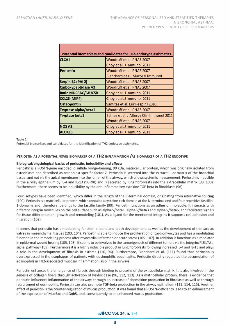
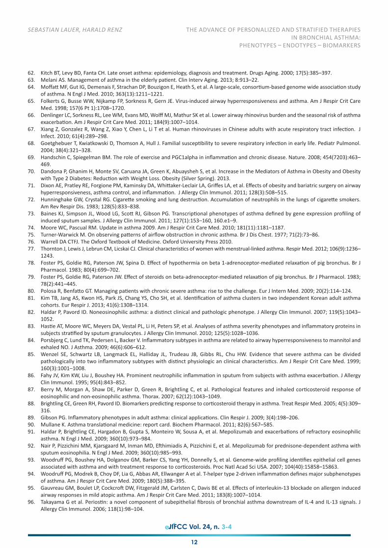
Biological/physiological basics of periostin, inducibility and effectsPeriostin is a POSTN-gene encoded, disulfide bridge-bearing, 90 kDa, matricellular protein, which was originally isolated fromosteoblasts and described as osteoblast-specific factor 2. Periostin is secreted into the extracellular matrix of the bronchialtissue, and not via the apical membrane into the lumen of the airway, which allows systemic measurement. Periostin is induciblein the airway epithelium by IL-4 and IL-13 (96–98) and is secreted by lung fibroblasts into the extracellular matrix (99, 100).Furthermore, there seems to be inducibility by the anti-inflammatory cytokine TGF-beta in fibroblasts (96).
Four isotypes have been identified, which differ in the length of the C-terminal domain, originating from alternative splicing(100). Periostin is a matricellular protein, which contains a cysteine-rich domain at the N-terminal end and four repetitive fascillin-1-domains and, therefore, belongs to the fascilin family (99). Periostin functions as an adhesion molecule. It interacts withdifferent integrin molecules on the cell surface such as alpha-V/beta1, alpha-V/beta3 and alpha-V/beta5, and facilitates signalsfor tissue differentiation, growth and remodeling (101). As a ligand for the mentioned integrins it supports cell adhesion andmigration (102).
It seems that periostin has a modulating function in bone and teeth development, as well as the development of the cardiacvalves in mesenchymal tissues (103, 104). Periostin is able to induce the proliferation of cardiomyocytes and has a modulatingfunction in the remodeling process after myocardial infarction or acute stress (105–107). In addition it functions as a mediatorin epidermal wound healing (105, 108). It seems to be involved in the tumorigenesis of different tumors via the integrin/PI3K/Akt-signal pathway (109). Furthermore it is a highly inducible product in lung fibroblasts following increased IL-4 and IL-13 and playsa role in the development of fibrosis in asthma (110, 96). Furthermore, Blanchard et al. (111) found that periostin isoverexpressed in the esophagus of patients with eosinophilic esophagitis. Periostin directly regulates the accumulation ofeosinophils in TH2-associated mucosal inflammation, also in the airways.
Periostin enhances the emergence of fibrosis through binding to proteins of the extracellular matrix. It is also involved in thegenesis of collagen fibers through activation of lysyloxidase (96, 112, 113). As a matricellular protein, there is evidence thatperiostin influences inflammation of the airways through an increase of chemokine production in fibrolasts as well as throughrecruitment of eosinophils. Periostin can also promote TGF-beta production in the airway epithelium (111, 114, 115). Anothereffect of periostin is the counter-regulation of mucus production. It was found that a POSTN-deficiency leads to an enhancementof the expression of Muc5ac and Gob5, and, consequently to an enhanced mucus production.
8
THE ADVANCE OF PERSONALIZED AND STRATIFIED THERAPIESIN BRONCHIAL ASTHMA:
PHENOTYPES – ENDOTYPES – BIOMARKERS
SEBASTIAN LAUER, HARALD RENZ
3-4
Table 1Potential biomarkers and candidates for the identification of TH2 endotype asthmatics.
These novel observations suggest that periostin not only promotes inflammation. This is further supported by the observationthat periostin can increase the production of IFN-gamma (116). How these pro- and anti-inflammatory effects of periostin areregulated and controlled requires further study and whether periostin is important in the pathogenesis of other allergicconditions, or even in other chronic inflammatory diseases also needs to be investigated.
Periostin as biomarker of TH2 inflammationIs it possible to use markers of TH2 inflammation as biomarkers for stratification prior to therapeutic interventions? A distincteosinophilia is a reliable predictor for response to ICS. Particularly with regard to molecular-based therapies, differentiation inTH2-high and TH2-low profiles is required and studies of mepulizumab and reslizumab (two monoclonal antibodies against theTH2 cytokine IL-5) demonstrate the importance and the benefit of selection of patients to obtain a clinically significant effect oftreatment with targeted therapeutics (117).There is recent evidence (84, 118, 119) that periostin levels in serum are significantly increased in patients with asthma. Thiswas found particularly for the eosinophilic type of airway inflammation compared with patients with only minimal eosinophilicairway inflammation. Periostin levels seem to correlate well with the TH2-high gene signature, and with the TH2 endotype. Thisleads to the question of whether serum periostin levels may serve as a good predictive parameter for stratifying patients priorto asthma treatment.The next important step is the development of reliable assay systems for such new biomarkers. For this purpose Jia et al. (117)established an ELISA for serum periostin. The assay was tested in a variety of asthmatic patients, and significantly elevated serumperiostin levels were found in a subset of asthmatics together with a correlation with eosinophilic airway inflammation. Although treatment with ICS has been found to reduce periostin levels as well as the airway eosinophilia in patients with mild-to-moderate asthma (93, 120), a subset of asthmatics showed persistent eosinophilic airway inflammation and symptoms despitehigh doses of ICS. This subset could be divided into a subpopulation with sputum- or tissue-eosinophilia (ensured by biopsy) and asubpopulation without these criteria (sputum eosinophils <3% and tissue eosinophils <22/mm²). The median periostin levels weresignificantly higher in individuals with high sputum- or tissue-eosinophilia as compared with individuals with lower eosinophilcounts. Individuals with a periostin level above a cut-off of 25 ng/mL had a greater than 85% risk of having an increased amount ofeosinophils either in sputum or in bronchial tissue. These eosinophil-low and eosinophil-high subpopulations could be distinguishedby means of a periostin cut-off level of 25 ng/mL with a positive predictive value of 93%. These analyses may serve as a startingpoint for further investigation in order to establish periostin as a systemic biomarker of eosinophilic airway inflammation.
Of all previously examined non-invasive biomarkers such as blood eosinophils, FeNO or IgE, periostin shows the highestprobability as a predicator of a TH2-driven inflammation (117) (Table 2). The ability of periostin to serve as a suitable biomarker
9
SEBASTIAN LAUER, HARALD RENZ THE ADVANCE OF PERSONALIZED AND STRATIFIED THERAPIESIN BRONCHIAL ASTHMA:
PHENOTYPES – ENDOTYPES – BIOMARKERS
3-4
Table 2Periostin: biological characterization.

for the identification of theTH2-endotype was illustrated in the study by Corren et al. (97) in which 219 BA patients were stratifiedinto a TH2-high and a TH2-low-endotype as measured by their peripheral periostin levels. After treatment with lebrikizumab,an anti-IL-13 monoclonal antibody, subjects with a high peripheral periostin level showed a significant improvement in FEV1compared with placebo that was not observed in subjects with low periostin levels.
CONCLUSIONS
Recognition of the heterogeneity of BA together with the definition of clinical phenotypes, pathogenetic endotypes and thedevelopment of novel biomarkers has opened a new chapter in BA clinical care. Although the vast majority of BA patients arewell controlled with guideline based anti-inflammatory therapies, there is a strong medical need for improved therapies in theremaining portion of BA patients. This goes hand in hand with recognition of the complexity of BA pathogenesis. Certain well-defined pathogenetic cellular molecular networks underlying the disease in a defined group of patients are defined as‘endotypes’. The best studied endotype so far is the TH2-eosinophilic endotype of airway inflammation. Patients suffering fromthis endotype seem to benefit from targeted biological anti-inflammatory therapies. Such biologicals interfere, for example,with the cytokine network IL-4, IL-5 and IL-13. Whether periostin is a suitable biomarker for identifying this particular endotypein clinical practice requires further studies. Additional research is also required to develop further biomarkers, not only for theTH2 endotype, but also for the other endotypes. Therefore, the field of allergy and asthma research has reached a new chapterin the area of personalized, individualized therapeutic strategies. Many more studies are needed to improve this situation, butthe right avenue has been taken so far.
REFERENCES
1. Asher MI, Weiland SK. The International Study of Asthma and Allergies in Childhood (ISAAC). ISAAC Steering Committee. Clin Exp Allergy.1998; 28 Suppl 5:52–66.
2. Von Mutius E. Prävalenz und Determinanten des Asthma bronchiale. Monatsschr Kinderheilkd. 2010; 158:121–128.3. Von Mutius E. Epidemiologie des Asthma bronchiale. Monatsschr Kinderheilkd. 2001; 149:86–93.4. Garn H, Renz H. Epidemiological and immunological evidence for the hygiene hypothesis. Immunobiology. 2007; 212(6):441–452.5. Murphy KM , Travers P, Walport M, Janeway CA, Seidler I, Ehrenstein M. (2009). Janeway Immunologie. Spektrum Akad. Verl, Heidelberg,
7. Aufl.6. Strachan DP. Hay fever, hygiene, and household size. BMJ. 1989; 299(6710):1259–1260.7. Strachan DP. Allergy and family size: a riddle worth solving. Clin Exp Allergy. 1997; 27(3):235–256.8. Celedon JC, Wright RJ, Litonjua AA, Sredl D, Ryan L, Weiss ST, Gold DR. Day care attendance in early life, maternal history of asthma, and
asthma at the age of 6 years. Am J Respir Crit Care Med. 2003; 167(9):1239–1243.9. Frei R, Lauener RP, Crameri R, O'Mahony L. Microbiota and dietary interactions: an update to the hygiene hypothesis. Allergy. 2012;
67(4):451–461.10. Shaheen SO. Changing patterns of childhood infection and the rise in allergic disease. Clin Exp Allergy. 1995; 25 (11):1034–1037.11. Jackson DJ, Gangnon RE, Evans MD, Roberg KA, Anderson EL, Pappas TE, et al. Wheezing rhinovirus illnesses in early life predict asthma
development in high-risk children. Am J Respir Crit Care Med. 2008; 178(7):667–672.12. Weinberger M. Consensus statement from a conference on treatment of viral respiratory infection induced asthma in young children. J
Pediatr. 2003; 142(Suppl 2):45–46.13. Weinberger M. Treatment strategies for viral respiratory infection-induced asthma. J Pediatr. 2003; 142(Suppl 2):38–39.14. Hallstrand TS, Moody MW, Aitken ML, Henderson WR Jr. Airway immunopathology of asthma with exercise-induced bronchoconstriction.
J Allergy Clin Immunol. 2005; 116(3):586–593.15. Hallstrand TS, Moody MW, Wurfel MM, Schwartz LB, Henderson WR Jr, Aitken ML. Inflammatory basis of exercise-induced
bronchoconstriction. Am J Respir Crit care Med. 2005; 172(6):679–686.16. Helenius IJ, Tikkanen HO, Sarna S, Haahtela T. Asthma and increased bronchial responsiveness in elite athletes: atopy and sport event as
risk factors. J Allergy Clin Immunol. 1998; 101(5):646–652.17. Helenius IJ, Rytilä P, Metso T, Haahtela T, Venge P, Tikkanen HO. Respiratory symptoms, bronchial responsiveness, and cellular
characteristics of induced sputum in elite swimmers. Allergy. 1998; 53(4):346–352.18. Karjalainen EM, Laitinen A, Sue-Chu M, Altraja A, Bjermer L, Laitinen LA. Evidence of airway inflammation and remodeling in ski athletes
with and without bronchial hyperresponsiveness to methacholine. Am J Respir Crit Care Med. 2000; 161(6):2086–2091.19. Farzan S. The asthma phenotype in the obese: distinct or otherwise? J Allergy (Cairo). 2013; 2013:602908.20. Szefler SJ, Martin RJ, King TS, Boushey HA, Cherniack RM, Chinchilli VM, et al. Significant variability in response to inhaled corticosteroids
for persistent asthma. J Allergy Clin Immunol. 2002; 109(3):410–418.21. Martin RJ, Szefler SJ, King TS, Kraft M, Boushey HA, Chinchilli VM, et al. The Predicting Response to Inhaled Corticosteroid Efficacy (PRICE)
trial. J Allergy Clin Immunol. 2007; 119(1):73–80.22. Holgate ST, Polosa R. Treatment strategies for allergy and asthma. Nat Rev Immunol. 2008; 8(3):218–230.23. Bhakta NR, Woodruff PG. Human asthma phenotypes: from the clinic, to cytokines, and back again. Immunol Rev. 2011; 242(1):220–
232.24. Fort MM, Cheung J, Yen D, Li J, Zurawski SM, Lo S, et al. IL-25 induces IL-4, IL-5, and IL-13 and Th2-associated pathologies in vivo. Immunity.
10
THE ADVANCE OF PERSONALIZED AND STRATIFIED THERAPIESIN BRONCHIAL ASTHMA:
PHENOTYPES – ENDOTYPES – BIOMARKERS
SEBASTIAN LAUER, HARALD RENZ
3-4

2001; 15(6):985–995.25. Moro K, Yamada T, Tanabe M, Takeuchi T, Ikawa T, Kawamoto H, et al. Innate production of T(H)2 cytokines by adipose tissue-associated
c-Kit(+)Sca-1(+) lymphoid cells. Nature. 2009; 463(7280):540–544.26. Price AE, Liang HE, Sullivan BM, Reinhardt RL, Eisley CJ, Erle DJ, Locksley RM. Systemically dispersed innate IL-13-expressing cells in type
2 immunity. Proc Natl Acad Sci USA. 2010; 107(25):11489–11494.27. Romagnani S. Immunologic influences on allergy and the TH1/TH2 balance. J Allergy Clin Immunol. 2004; 113(3):395–400.28. Averbeck M, Gebhardt C, Emmrich F, Treudler R, Simon JC. Immunologic principles of allergic disease. J Dtsch Dermatol Ges. 2007;
5(11):1015–1028.29. O'Garra A, Arai N. The molecular basis of T helper 1 and T helper 2 cell differentiation. Trends Cell Biol. 2000; 10(12):542–550.30. Grünig G, Warnock M, Wakil AE, Venkayya R, Brombacher F, Rennick DM, et al. Requirement for IL-13 independently of IL-4 in experimental
asthma. Science. 1998; 282(5397):2261–2263.31. Humbert M, Durham SR, Kimmitt P, Powell N, Assoufi B, Pfister R, et al. Elevated expression of messenger ribonucleic acid encoding IL-
13 in the bronchial mucosa of atopic and nonatopic subjects with asthma. J Allergy Clin Immunol. 1997; 99(5):657–665.32. Wills-Karp M, Luyimbazi J, Xu X, Schofield B, Neben TY, Karp CL, Donaldson DD. Interleukin-13: central mediator of allergic asthma.
Science. 1998; 282(5397):2258–2261.33. Kroegel C. Asthmatherapie Leitfaden einer pathogenetisch begründeten Therapie. Steinen: ZETT-Verlag 2001.34. Beeh KM, Buhl R. Asthma pathogenesis – implications for novel therapies. Medizinische Klinik 2001; 96:15–25.35. Kroegel C. Asthma bronchiale. Pathogenetische Grundlagen, Diagnostik, Therapie. Stuttgart, New York: Georg Thieme Verlag 2002.36. Shim JJ, Dabbagh K, Ueki IF, Dao-Pick T, Burgel PR, Takeyama K, et al. IL-13 induces mucin production by stimulating epidermal growth
factor receptors and by activating neutrophils. Am J Physiol Lung Cell Mol Physiol. 2001; 280(1):L134–140.37. Zhu Z, Homer RJ, Wang Z, Chen Q, Geba GP, Wang J, et al. Pulmonary expression of interleukin-13 causes inflammation, mucus
hypersecretion, subepithelial fibrosis, physiologic abnormalities, and eotaxin production. J Clin Invest. 1999; 103(6):779–788.38. Akiho H, Blennerhassett P, Deng Y, Collins SM. Role of IL-4, IL-13, and STAT6 in inflammation-induced hypercontractility of murine smooth
muscle cells. Am J Physiol Gastrointest Liver Physiol. 2002; 282(2):G226–232.39. Eum SY, Maghni K, Tolloczko B, Eidelman DH, Martin JG. IL-13 may mediate allergen-induced hyperresponsiveness independently of IL-
5 or eotaxin by effects on airway smooth muscle. Am J Physiol Lung Cell Mol Physiol. 2005; 288(3):L576–584.40. Grunstein MM, Hakonarson H, Leiter J, Chen M, Whelan R, Grunstein JS, Chuang S. IL-13-dependent autocrine signaling mediates altered
responsiveness of IgE-sensitized airway smooth muscle. Am J Physiol Lung Cell Mol Physiol. 2002; 282(3):L520–528.41. Tliba O, Deshpande D, Chen H, Van Besien C, Kannan M, Panettieri RA Jr, Amrani Y. IL-13 enhances agonist-evoked calcium signals and
contractile responses in airway smooth muscle. Br J Pharmacol. 2003; 140(7):1159–1162.42. Espinosa K, Bosse Y, Stankova J, Rola-Pleszczynski M. CysLT1 receptor upregulation by TGF-beta and IL-13 is associated with bronchial
smooth muscle cell proliferation in response to LTD4. J Allergy Clin Immunol. 2003; 111(5):1032–1040.43. Bousquet J, Jeffery PK, Busse WW, Johnson M, Vignola AM. Asthma. From bronchoconstriction to airways inflammation and remodeling.
Am J Respir Crit Care Med. 2000; 161(5):1720–1745.44. Wenzel SE. Asthma phenotypes: the evolution from clinical to molecular approaches. Nat Med. 2012; 18(5):716–725.45. Masoli M, Fabian D, Holt S, Beasley R. The global burden of asthma: executive summary of the GINA Dissemination Committee report.
Allergy. 2004; 59(5):469–478.46. Vink NM, Postma DS, Schouten JP, Rosmalen JG, Boezen HM. Gender differences in asthma development and remission during transition
through puberty: the TRacking Adolescents' Individual Lives Survey (TRAILS) study. J Allergy Clin Immunol. 2010; 126(3):498–504.47. Bray GW. The hereditary factor in asthma and other allergies. Br Med J. 1930; 1(3608):384–387.48. Lau S, Illi S, Sommerfeld C, Niggemann B, Bergmann R, von Mutius E, Wahn U. Early exposure to house-dust mite and cat allergens and
development of childhood asthma: a cohort study. Multicentre Allergy Study Group. Lancet. 2000; 356(9239):1392–1397.49. Bouzigon E, Corda E, Aschard H, Dizier MH, Boland A, Bousquet J, et al. Effect of 17q21 variants and smoking exposure in early-onset
asthma. N Engl J Med. 2008; 359(19):1985–1994.50. de Nijs SB, Venekamp LN, Bel EH. Adult-onset asthma: is it really different? Eur Respir Rev. 2013; 22(127):44–52.51. Hesselmar B, Enelund AC, Eriksson B, Padyukov L, Hanson LA, Aberg N. The heterogeneity of asthma phenotypes in children and young
adults. J Allergy (Cairo). 2012; 2012:163089.52. Wenzel S. Severe asthma: from characteristics to phenotypes to endotypes. Clin Exp Allergy. 2012; 42(5):650–658.53. Hayashi T, Gong X, Rossetto C, Shen C, Takabayashi K, Redecke V, et al. Induction and inhibition of the Th2 phenotype spread: implications
for childhood asthma. J Immunol. 2005; 174(9):5864–5873.54. Szefler SJ, Phillips BR, Martinez FD, Chinchilli VM, Lemanske RF, Strunk RC, et al. Characterization of within-subject responses to fluticasone
and montelukast in childhood asthma. J Allergy Clin Immunol. 2005; 115(2):233–242.55. Martinez FD, Wright AL, Taussig LM, Holberg CJ, Halonen M, Morgan WJ. Asthma and wheezing in the first six years of life. The Group
Health Medical Associates. N Engl J Med. 1995; 332(3):133–138.56. Krishnamoorthy N, Khare A, Oriss TB, Raundhal M, Morse C, Yarlagadda M, et al. Early infection with respiratory syncytial virus impairs
regulatory T cell function and increases susceptibility to allergic asthma. Nat Med. 2012; 18(10):1525–1530.57. Samter M, Beers RF, Jr. Concerning the nature of intolerance to aspirin. J Allergy. 1967; 40(5):281–293.58. Pavord ID, Korn S, Howarth P, Bleecker ER, Buhl R, Keene ON, et al. Mepolizumab for severe eosinophilic asthma (DREAM): a multicentre,
double-blind, placebo-controlled trial. Lancet. 2012; 380(9842):651–659.59. Gibson PG, McDonald VM, Marks GB. Asthma in older adults. Lancet. 2010; 376(9743):803–813.60. Radenne F, Verkindre C, Tonnel AB. [Asthma in the elderly]. Rev Mal Respir. 2004; 21(5 Pt 3):8S117–125.61. Radenne F, Verkindre C, Tonnel AB. [Asthma in the elderly]. Rev Mal Respir. 2003; 20(1 Pt 1):95–103.
11
SEBASTIAN LAUER, HARALD RENZ THE ADVANCE OF PERSONALIZED AND STRATIFIED THERAPIESIN BRONCHIAL ASTHMA:
PHENOTYPES – ENDOTYPES – BIOMARKERS
3-4
62. Kitch BT, Levy BD, Fanta CH. Late onset asthma: epidemiology, diagnosis and treatment. Drugs Aging. 2000; 17(5):385–397.63. Melani AS. Management of asthma in the elderly patient. Clin Interv Aging. 2013; 8:913–22.64. Moffatt MF, Gut IG, Demenais F, Strachan DP, Bouzigon E, Heath S, et al. A large-scale, consortium-based genome wide association study
of asthma. N Engl J Med. 2010; 363(13):1211–1221.65. Folkerts G, Busse WW, Nijkamp FP, Sorkness R, Gern JE. Virus-induced airway hyperresponsiveness and asthma. Am J Respir Crit Care
Med. 1998; 157(6 Pt 1):1708–1720.66. Denlinger LC, Sorkness RL, Lee WM, Evans MD, Wolff MJ, Mathur SK et al. Lower airway rhinovirus burden and the seasonal risk of asthma
exacerbation. Am J Respir Crit Care Med. 2011; 184(9):1007–1014.67. Xiang Z, Gonzalez R, Wang Z, Xiao Y, Chen L, Li T et al. Human rhinoviruses in Chinese adults with acute respiratory tract infection. J
Infect. 2010; 61(4):289–298.68. Goetghebuer T, Kwiatkowski D, Thomson A, Hull J. Familial susceptibility to severe respiratory infection in early life. Pediatr Pulmonol.
2004; 38(4):321–328.69. Handschin C, Spiegelman BM. The role of exercise and PGC1alpha in inflammation and chronic disease. Nature. 2008; 454(7203):463–
469.70. Dandona P, Ghanim H, Monte SV, Caruana JA, Green K, Abuaysheh S, et al. Increase in the Mediators of Asthma in Obesity and Obesity
with Type 2 Diabetes: Reduction with Weight Loss. Obesity (Silver Spring). 2013.71. Dixon AE, Pratley RE, Forgione PM, Kaminsky DA, Whittaker-Leclair LA, Griffes LA, et al. Effects of obesity and bariatric surgery on airway
hyperresponsiveness, asthma control, and inflammation. J Allergy Clin Immunol. 2011; 128(3):508–515.72. Hunninghake GW, Crystal RG. Cigarette smoking and lung destruction. Accumulation of neutrophils in the lungs of cigarette smokers.
Am Rev Respir Dis. 1983; 128(5):833–838.73. Baines KJ, Simpson JL, Wood LG, Scott RJ, Gibson PG. Transcriptional phenotypes of asthma defined by gene expression profiling of
induced sputum samples. J Allergy Clin Immunol. 2011; 127(1):153–160, 160.e1–9.74. Moore WC, Pascual RM. Update in asthma 2009. Am J Respir Crit Care Med. 2010; 181(11):1181–1187.75. Turner-Warwick M. On observing patterns of airflow obstruction in chronic asthma. Br J Dis Chest. 1977; 71(2):73–86.76. Warrell DA CTFJ. The Oxford Textbook of Medicine. Oxford University Press 2010.77. Thornton J, Lewis J, Lebrun CM, Licskai CJ. Clinical characteristics of women with menstrual-linked asthma. Respir Med. 2012; 106(9):1236–
1243.78. Foster PS, Goldie RG, Paterson JW, Spina D. Effect of hypothermia on beta 1-adrenoceptor-mediated relaxation of pig bronchus. Br J
Pharmacol. 1983; 80(4):699–702.79. Foster PS, Goldie RG, Paterson JW. Effect of steroids on beta-adrenoceptor-mediated relaxation of pig bronchus. Br J Pharmacol. 1983;
78(2):441–445.80. Polosa R, Benfatto GT. Managing patients with chronic severe asthma: rise to the challenge. Eur J Intern Med. 2009; 20(2):114–124.81. Kim TB, Jang AS, Kwon HS, Park JS, Chang YS, Cho SH, et al. Identification of asthma clusters in two independent Korean adult asthma
cohorts. Eur Respir J. 2013; 41(6):1308–1314.82. Haldar P, Pavord ID. Noneosinophilic asthma: a distinct clinical and pathologic phenotype. J Allergy Clin Immunol. 2007; 119(5):1043–
1052.83. Hastie AT, Moore WC, Meyers DA, Vestal PL, Li H, Peters SP, et al. Analyses of asthma severity phenotypes and inflammatory proteins in
subjects stratified by sputum granulocytes. J Allergy Clin Immunol. 2010; 125(5):1028–1036.84. Porsbjerg C, Lund TK, Pedersen L, Backer V. Inflammatory subtypes in asthma are related to airway hyperresponsiveness to mannitol and
exhaled NO. J Asthma. 2009; 46(6):606–612.85. Wenzel SE, Schwartz LB, Langmack EL, Halliday JL, Trudeau JB, Gibbs RL, Chu HW. Evidence that severe asthma can be divided
pathologically into two inflammatory subtypes with distinct physiologic an clinical characteristics. Am J Respir Crit Care Med. 1999;160(3):1001–1008.
86. Fahy JV, Kim KW, Liu J, Boushey HA. Prominent neutrophilic inflammation in sputum from subjects with asthma exacerbation. J AllergyClin Immunol. 1995; 95(4):843–852.
87. Berry M, Morgan A, Shaw DE, Parker D, Green R, Brightling C, et al. Pathological features and inhaled corticosteroid response ofeosinophilic and non-eosinophilic asthma. Thorax. 2007; 62(12):1043–1049.
88. Brightling CE, Green RH, Pavord ID. Biomarkers predicting response to corticosteroid therapy in asthma. Treat Respir Med. 2005; 4(5):309–316.
89. Gibson PG. Inflammatory phenotypes in adult asthma: clinical applications. Clin Respir J. 2009; 3(4):198–206.90. Mullane K. Asthma translational medicine: report card. Biochem Pharmacol. 2011; 82(6):567–585.91. Haldar P, Brightling CE, Hargadon B, Gupta S, Monteiro W, Sousa A, et al. Mepolizumab and exacerbations of refractory eosinophilic
asthma. N Engl J Med. 2009; 360(10):973–984.92. Nair P, Pizzichini MM, Kjarsgaard M, Inman MD, Efthimiadis A, Pizzichini E, et al. Mepolizumab for prednisone-dependent asthma with
sputum eosinophilia. N Engl J Med. 2009; 360(10):985–993.93. Woodruff PG, Boushey HA, Dolganov GM, Barker CS, Yang YH, Donnelly S, et al. Genome-wide profiling identifies epithelial cell genes
associated with asthma and with treatment response to corticosteroids. Proc Natl Acad Sci USA. 2007; 104(40):15858–15863.94. Woodruff PG, Modrek B, Choy DF, Lia G, Abbas AR, Ellwanger A et al. T-helper type 2-driven inflammation defines major subphenotypes
of asthma. Am J Respir Crit Care Med. 2009; 180(5):388–395.95. Gauvreau GM, Boulet LP, Cockcroft DW, Fitzgerald JM, Carlsten C, Davis BE et al. Effects of interleukin-13 blockade on allergen induced
airway responses in mild atopic asthma. Am J Respir Crit Care Med. 2011; 183(8):1007–1014.96. Takayama G et al. Periostin: a novel component of subepithelial fibrosis of bronchial asthma downstream of IL-4 and IL-13 signals. J
Allergy Clin Immunol. 2006; 118(1):98–104.
12
THE ADVANCE OF PERSONALIZED AND STRATIFIED THERAPIESIN BRONCHIAL ASTHMA:
PHENOTYPES – ENDOTYPES – BIOMARKERS
SEBASTIAN LAUER, HARALD RENZ
3-4

97. Corren J, Lemanske RF, Hanania NA, Korenblat PE, Parsey MV, Arron JR et al. Lebrikizumab treatment in adults with asthma. N Engl JMed. 2011; 365(12):1088–1098.
98. Yuyama N, Davies DE, Akaiwa M, Matsui K, Hamasaki Y, Suminami Y, et al. Analysis of novel disease-related genes in bronchial asthma.Cytokine. 2002; 19(6):287–296.
99. Takeshita S, Kikuno R, Tezuka K, Amann E. Osteoblast-specific factor 2: cloning of a putative bone adhesion protein with homology withthe insect protein fasciclin I. Biochem J. 1993; 294(Pt 1):271–278.
100. Horiuchi K, Amizuka N, Takeshita S, Takamatsu H, Katsuura M, Ozawa H, et al. Identification and characterization of a novel protein,periostin, with restricted expression to periosteum and periodontal ligament and increased expression by transforming growth factorbeta. J Bone Miner Res. 1999; 14(7):1239–1249.
101. Masuoka M, Shiraishi H, Ohta S, Suzuki S, Arima K, Aoki S, et al. Periostin promotes chronic allergic inflammation in response to Th2cytokines. J Clin Invest. 2012; 122(7):2590–2600.
102. Gillan L, Matei D, Fishman DA, Gerbin CS, Karlan BY, Chang DD. Periostin secreted by epithelial ovarian carcinoma is a ligand foralpha(V)beta(3) and alpha(V)beta(5) integrins and promotes cell motility. Cancer Res. 2002; 62(18):5358–5364.
103. Rios H, Koushik SV, Wang H, Wang J, Zhou HM, Lindsley A, et al. Periostin null mice exhibit dwarfism, incisor enamel defects, and anearly-onset peridontal disease-like phenotype. Mol Cell Biol. 2005; 25(24):11131–11144.
104. Snider P, Hinton RB, Moreno-Rodriguez RA, Wang J, Rogers R, Lindsley A, et al. Periostin is required for maturation and extracellularmatrix stabilization of noncardiomyocyte lineages of the heart. Circ Res. 2008; 102(7):752–760.
105. Kühn B, del Monte F, Hajjar RJ, Chang YS, Lebeche D, Arab S, Keating MT. Periostin induces proliferation of differentiated cardiomyocytesand promotes cardiac repair. Nat Med. 2007; 13(8):962–969.
106. Katsuragi N, Morishita R, Nakamura N, Ochiai T, Taniyama Y, Hasegawa Y, et al. Periostin as a novel factor responsible for ventriculardilation. Circulation. 2004; 110(13):1806–1813.
107. Kruzynska-Frejtag A, Machnicki M, Rogers R, Markwald RR, Conway SJ. Periostin (an osteoblast-specific factor) is expressed within theembryonic mouse heart during valve formation. Mech Dev. 2001; 103(1–2):183–188.
108. Shimazaki M, Nakamura K, Kii I, Kashima T, Amizuka N, Li M, et al. Periostin is essential for cardiac healing after acute myocardial infarction.J Exp Med. 2008; 205(2):295–303.
109. Ruan K, Bao S, Ouyang G. The multifaceted role of periostin in tumorigenesis. Cell Common Signal. 2008; 2(1–2):9–17.110. Hayashi N, Yoshimoto T, Izuhara K, Matsui K, Tanaka T, Nakanishi K. T helper 1 cells stimulated with ovalbumin and IL-18 induce airway
hyperresponsiveness and lung fibrosis by IFN-gamma and IL-13 production. Proc Natl Acad Sci USA. 2007; 104(37):14765–14770.111. Blanchard C, Mingler MK, McBride M, Putnam PE, Collins MH, Chang G, et al. Periostin facilitates eosinophil tissue infiltration in allergic
lung and esophageal responses. Mucosal Immunol. 2008; 1(4):289–296.112. Kii I, Nishiyama T, Li M, Matsumoto K, Saito M, Amizuka N, Kudo A. Incorporation of tenascin-C into the extracellular matrix by periostin
underlies an extracellular meshwork architecture. J Biol Chem. 2010; 285(3):2028–2039.113. Maruhashi T, Kii I, Saito M, Kudo A. Interaction between periostin and BMP-1 promotes proteolytic activation of lysyl oxidase. J Biol
Chem. 2010; 285(17):13294–13303.114. Uchida M, Shiraishi H, Ohta S, Arima K, Taniguchi K, Suzuki S, et al. Periostin, a matricellular protein, plays a role in the induction of
chemokines in pulmonary fibrosis. Am J Respir Cell Mol Biol. 2012; 46(5):677–686.115. Sidhu SS, Yuan S, Innes AL, Kerr S, Woodruff PG, Hou L, et al. Roles of epithelial cell-derived periostin in TGF-β activation, collagen
production, and collagen gel elasticity in asthma. Proc Natl Acad Sci U S A. 2010; 107(32):14170–14175.116. Sehra S, Yao W, Nguyen ET, Ahyi AN, Tuana FM, Ahlfeld SK, et al. Periostin regulates goblet cell metaplasia in a model of allergic airway
inflammation. J Immunol. 2011; 186(8):4959–4966.117. Jia G, Erickson RW, Choy DF, Mosesova S, Wu LC, Solberg OD, et al. Periostin is a systemic biomarker of eosinophilic aiway inflammation
in asthmatic patients. J Allergy Clin Immunol. 2012; 130(3):647–654.118. Choy DF, Modrek B, Abbas AR, Kummerfeld S, Clark HF, Wu LC, et al. Gene expression patterns of Th2 inflammation and intercellular
communication in asthmatic airways. J Immunol. 2011; 186(3):1861–1869.119. Arron JR, Jia G-Q, Erickson RW, Choy DF, Mosesova S, Solberg OD, et al. Periostin is a systemic biomarker of eosinophilic airway
inflammation in asthma. Am J Respir Crit Care Med. 2011; 183:A4455.120. Boushey HA, Sorkness CA,. King TS, Sullivan SD, Fahy JV, Lazarus SC et al. Daily versus as-needed corticosteroids for mild persistent asthma.
N Engl J Med. 2005; 352(15): 1519-1528.
13
SEBASTIAN LAUER, HARALD RENZ THE ADVANCE OF PERSONALIZED AND STRATIFIED THERAPIESIN BRONCHIAL ASTHMA:
PHENOTYPES – ENDOTYPES – BIOMARKERS
3-4