The Adrenal Incidentaloma - Monster Headphones Incidentaloma Banner.pdf · The Adrenal...
67
The Adrenal Incidentaloma Beejal Shah, MD Phoenix VA
Transcript of The Adrenal Incidentaloma - Monster Headphones Incidentaloma Banner.pdf · The Adrenal...

The Adrenal Incidentaloma
Beejal Shah, MD Phoenix VA


Cortex consists of 3 different zones Zona Glomerulosa- mineralocortidoids Zona Fasciculata- cortisol Zona Reticularis- sex hormones
Medulla Chromaffin cells- metanephrines and catecholamines

Definition
• “An adrenal “incidentaloma” is an adrenal mass, generally 1 cm or more in diameter, that is discovered serendipitously during a radiologic examination performed for indications other than an evaluation for adrenal disease.”
Young WF Jr. Management approaches to adrenal incidentalomas: a view from Rochester, Minnesota. Endocrinol Metab Clin North Am 2000;29:159-85.

Statistics
• No population based studies • Exact prevalence unknown • Autopsy series frequency 1.4-8.7% • CT series frequency 0.6-4.4% • Most of the tumors are small (diameter <1cm) • Large CT series 79% of adenomas < 2cm • Autopsy series 0.025% adenomas >6cm • Prevalence increase with age & M=F
– Probability of adenoma 20-29 yo 0.2% – Probability of adenoma >70yo 7%
Pooled Data Prevalence 1%

Pathogenesis
• Largely unknown – Monoclonal expansion – Unregulated trophic stimulus from hypersecretion
of neuropeptides, neurotransmitters, growth factors, cytokines
– Association with metabolic syndrome

Normal Adrenal Anatomy on CT

Case 1: 67 y/o male c/o LLQ pain. CT ab/pelvis with contrast done in October 2014
Indeterminate density 1.3 cm right adrenal nodule.

Differential Diagnosis • Benign Non-functioning Adrenal
Adenoma – 41% • Metastasis – 19% • Hormone Producing Adenoma
16% – Pheochromocytoma – 8% – Subclinical Cushings 5% – Primary Hyperaldosteronism
3% • ACC (any adrenal hormone) -10%
*** most AI are > 6cm • Myolipoma 9%
• Malignant Pheochromocytoma • Congenital adrenal hyperplasia • Massive macronodular adrenal
disease • Nodular variant of adrenal
Cushing • Neuroblastoma • Ganglioneuroma • Cyst • Hemorrhage • Granuloma • Amyloidosis • Extraadrenal: infiltrative • Bilateral etiologies 10-15%

What should you do next?
1. Is the adrenal mass incidentally found or does the patient show symptoms or comorbidities (e. g. arterial hypertension) which might be caused by the adrenal mass?
2. Does the adrenal mass represent a metastasis of an unknown or known primary tumor?
3. Is the adrenal mass hormonally active? 4. Is there evidence of adrenocortical carcinoma? 5. What is the recommended long term f/u?
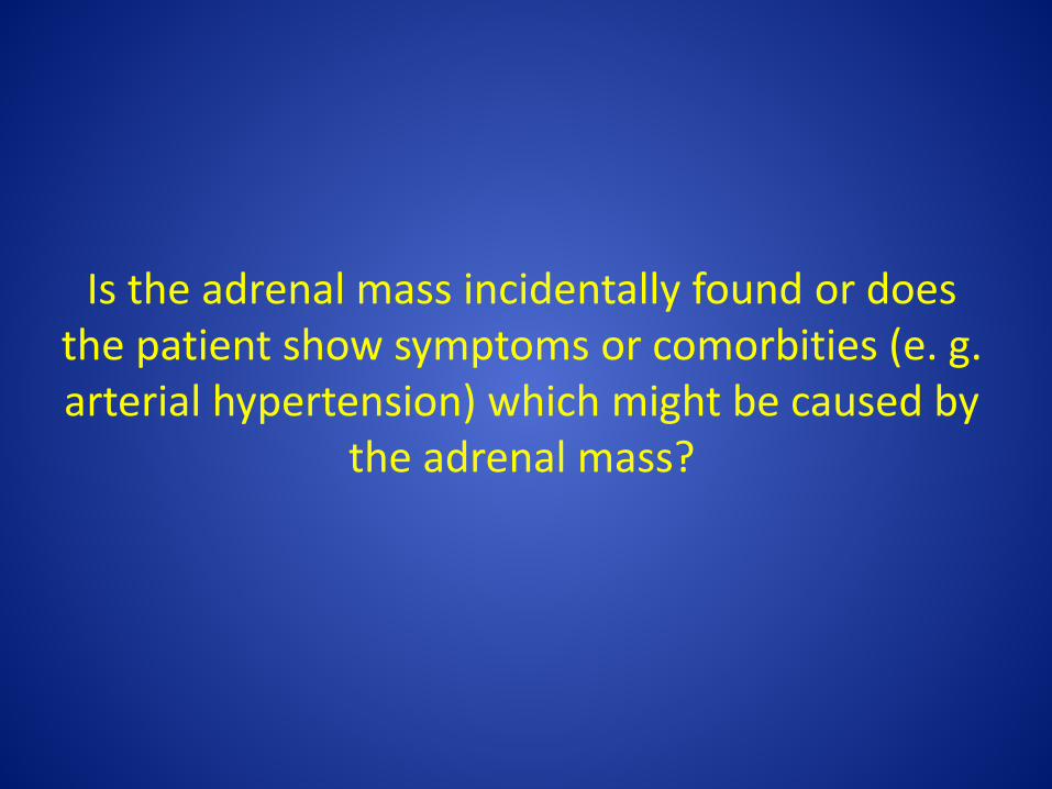
Is the adrenal mass incidentally found or does the patient show symptoms or comorbities (e. g. arterial hypertension) which might be caused by
the adrenal mass?

Prevalence of Endocrine Activity in Incidentally Detected Adrenal Masses
At the time of diagnosis up to 20 % of all incidentalomas are significantly hormonal active and little is known on the long term course of remaining 80%
Subclinical Cushing’s Syndrome

Symptoms and Signs Suggestive of Adrenal Hyperfunction or Malignant Disease
Young W. N Engl J Med 2007;356:601-610

Our Patient
• Patient has hx of HTN and has been on lisinopril and HCTZ for a few years.
• He denies difficulty with BP control but BP readings in CPRS shows SBP in 140-170s range and DBP in 90-100 range. Also noted to have mild hypokalemia with potassium level of 3.4 on recent BMP.
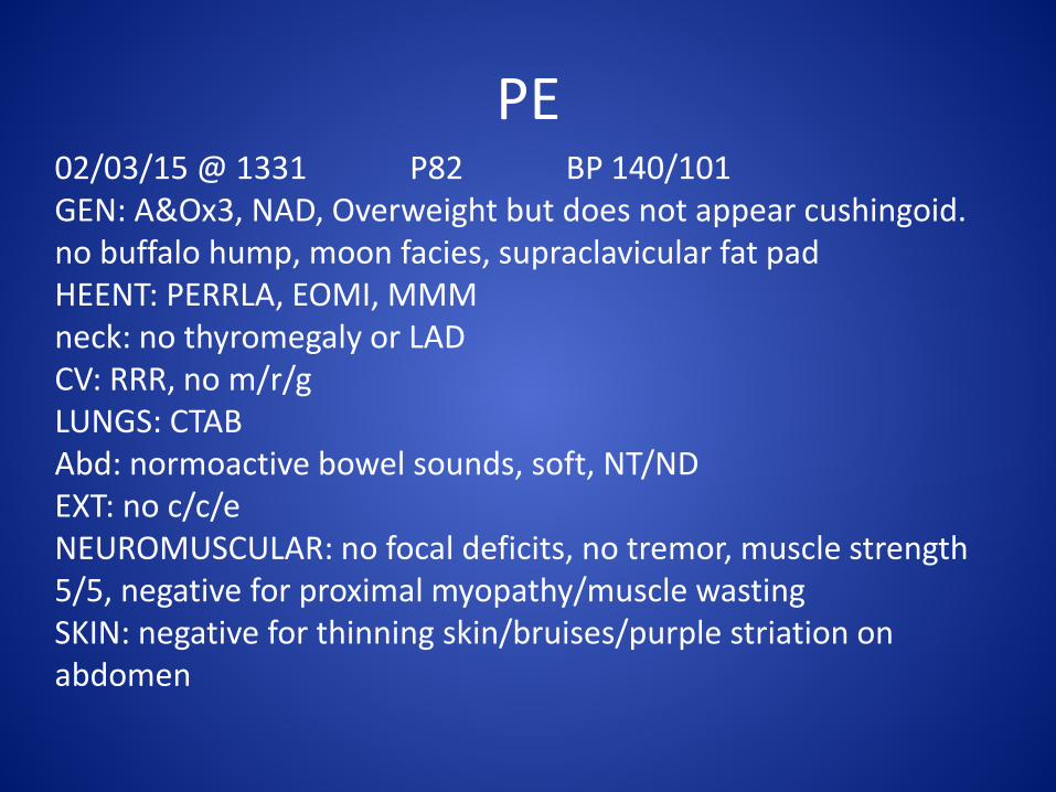
PE 02/03/15 @ 1331 P82 BP 140/101 GEN: A&Ox3, NAD, Overweight but does not appear cushingoid. no buffalo hump, moon facies, supraclavicular fat pad HEENT: PERRLA, EOMI, MMM neck: no thyromegaly or LAD CV: RRR, no m/r/g LUNGS: CTAB Abd: normoactive bowel sounds, soft, NT/ND EXT: no c/c/e NEUROMUSCULAR: no focal deficits, no tremor, muscle strength 5/5, negative for proximal myopathy/muscle wasting SKIN: negative for thinning skin/bruises/purple striation on abdomen

2. Does the adrenal mass represent a metastasis of an unknown or known primary tumor? • Per patient hx no history of other cancers and
imaging does not demonstrate other concerning masses or lymph nodes
• SIZE of mass and APPEARANCE on imaging are two major predictors of malignant disease
• Series of 2005 patients with AI, metastatic lesion found in 2.5% of patients

Size of Adrenal Mass
• Diameter >4cm 90% sensitivity for detecting ACC but low specificity only 24% of lesions >4cm are malignant
• Smaller the lesion at diagnosislower tumor stagebetter overall prognosis
• Large series, 70% of ACC were >7cm • Most recommend surgical resection of AI if tumor
>6cm but take into account other features (age, HU, comorbidities etc)
• Size of AI does not affect recommendation for hormonal testing

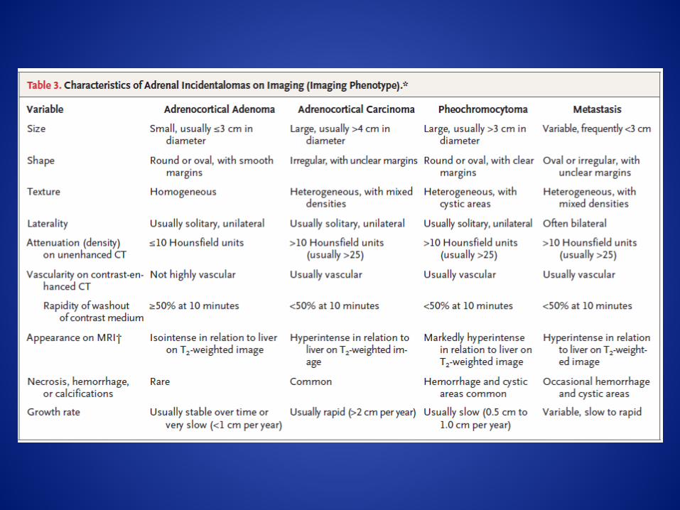
Imaging Phenotype
• CT comparable to MRI, thus CT is preferred imaging modality for cost/access etc
• CT features distinguish adenoma from nonadenomas – Lipid content – Rapidity of washout of the contrast medium

CT - Benign Adenoma
• Lots of Fat, low attenuation on CT precontrast <10 Hounsfield Units
• Delayed contrast-enhanced CT (CT adrenal protocol) – 10 minutes after the
administration of contrast, an absolute of >50% sensitivity and specificity of 100% for a benign lesion
CT- Nonadenoma (pheo, metastatic, ACC) *pheo are mostly benign but high HU and characteristic imaging phenotype c/w nonadenoma
• Lower fat content, higher attenuation on CT precontrast >10 HU
• Higher the precontrast HU the more concerning the lesion
• <40-50% washout of contrast on delayed imaging
• Heterogeneous, may be cystic component, irregular borders, vascular
**30% of adenomas do not contain large amounts of fat therefore precontrast HU cannot distinguish adenoma from nonadenoma

Always document the precontrast HU of an adrenal lesion and when possible
obtain the % washout

3. Is there Evidence for Adrenal Cortical Carcinoma
• Series of 2005 patients with AI, ACC found in 4.7% of patients • Rare but highly lethal malignancy • Often larger, heterogeneous, high HU, PET + (but 16% of
benign adrenal lesions can have background uptake) • PET not recommended for routine evaluation of AI who do not
have hx of malignancy • Adrenalectomy for tumors >6cm • 4-6cm tumor observe vs adrenalectomy (including other
clinical factors such as age, imaging phenotype) • <4cm unlikely ACC and can observe (including other clinical
factors such as age, imaging phenotype) • NEVER FNA adrenal if SUSPECT ACC • NEVER send for laparoscopic surgery if SUSPECT ACC- always
do open procedure
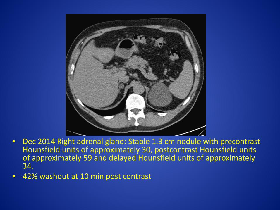
• Dec 2014 Right adrenal gland: Stable 1.3 cm nodule with precontrast Hounsfield units of approximately 30, postcontrast Hounsfield units of approximately 59 and delayed Hounsfield units of approximately 34.
• 42% washout at 10 min post contrast

Our Patient
• Nodule is small which is leans towards a benign lesion but the precontrast HU is mildly elevated and the washout is <50% – This does not mean it is malignant it simply
suggests that the lesion not definitely benign

3. Is the adrenal mass hormonally active? • Pt’s history suggestive of what endocrine
disorder? • What endocrine disorders do we screen for? • What screening tests do we order?

Laboratory Evaluation of AI
Young W. N Engl J Med 2007;356:601-610
24hr UFC
Plasma free metanephrines
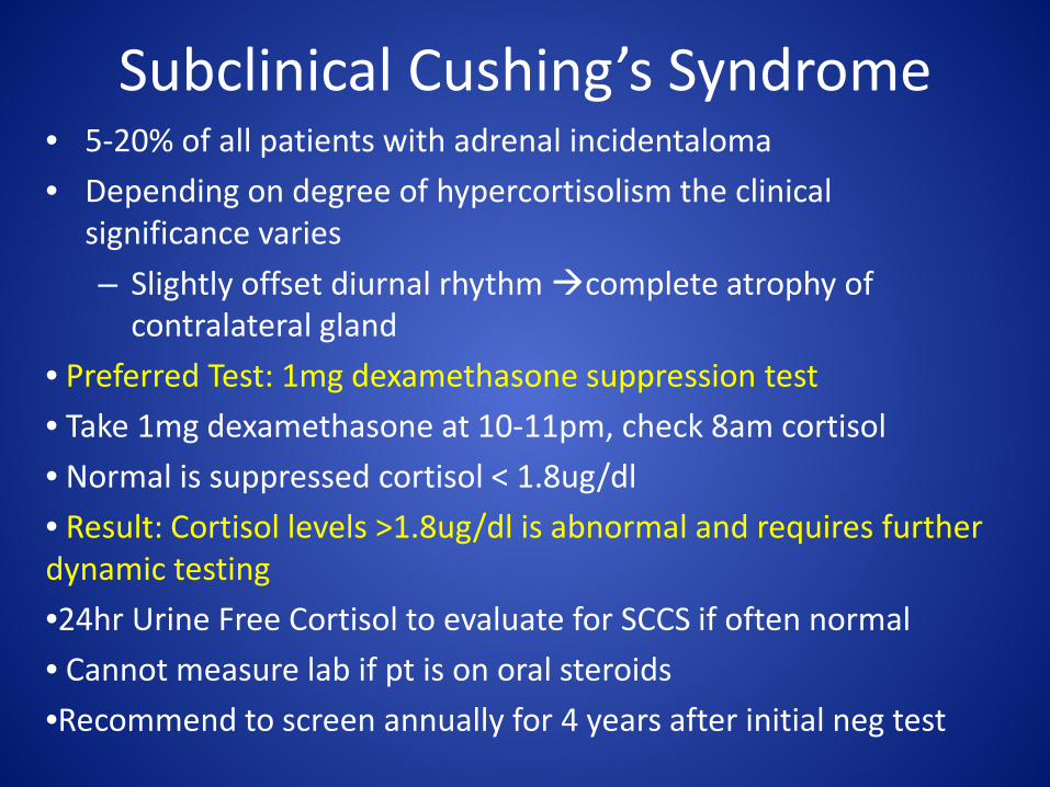
Subclinical Cushing’s Syndrome • 5-20% of all patients with adrenal incidentaloma • Depending on degree of hypercortisolism the clinical
significance varies – Slightly offset diurnal rhythm complete atrophy of
contralateral gland • Preferred Test: 1mg dexamethasone suppression test • Take 1mg dexamethasone at 10-11pm, check 8am cortisol • Normal is suppressed cortisol < 1.8ug/dl • Result: Cortisol levels >1.8ug/dl is abnormal and requires further dynamic testing •24hr Urine Free Cortisol to evaluate for SCCS if often normal • Cannot measure lab if pt is on oral steroids •Recommend to screen annually for 4 years after initial neg test

Pheochromocytoma • 0-11% patient with adrenal incidentalomas • Most no typical symptoms (sweating, headache,
hypertension, tachycardia) • 50% of pheochromocytoma are asymptomatic • Must do careful biochemical evaluation • Test: Plasma free fractionated metanephrines or 24hr
Urine free metanephrines and catecholamines • Should know which test is better at your institution • No medications need to be held for the lab test • Multiple reasons for false positive results (above the upper
limit of normal) • Result: True Positive test is lab value 3-4x ULN & needs
further evaluation • Must r/o pheochromocytoma prior to surgery or biopsy

Primary Hyperaldosteronism/Conn’s Syndrome
• 1% of hypertensive patients with adrenal incidentalomas
• HTN and hypokalemia <3.9mm/l should raise suspicion
• Not obligatory to screen in normotensive/normokalemic individual however most pre-emptively screen since it is a potentially curable form of hypertension and often otherwise underdiagnosed

Primary Hyperaldosteronism • Test: Morning ambulatory plasma aldosterone
concentration (PAC) and plasma renin activity (PRA ) to give the PAC/PRA ratio
• Test Protocol: For screening only hold mineralocorticoid antagonists and high dose amiloride for 4-6 weeks all other BP agents can be continued for a screening test
• The plasma aldosterone concentration and plasma renin activity should be taken between 8-10am (since aldosterone has a diurnal variation) with the patient ambulatory just prior to venipucture and recumbent for the blood draw.
• Result: Positive screening test is PAC/PRA ratio ≥ 20 AND a PAC ≥ 15ng/dl (PRA should be low or suppressed)
• Further testing then needed, pay attention to the assay cut-offs which are lab dependent

Proportion of endocrine hormone hypersecretion in indicidentalomas
increases with tumor size • Endocrine activity
and tumor size in 68 patients with incidentally detected adrenal
Allolio, B., Adrenal Incidentalomas. Adrenal Disorders, ed. C.G. Margioris AN. 2001, Totowa: Humana Press Inc. 249 – 261

Our Patient -Labs • 24 HR. URINE. TOTAL 24HR URINE VOLUME 1900 mL • Test name Result units Ref. range • Epinephrine 6 mcg/24 h 2 - 24 • Norepinephrine 56 mcg/24 h 15 - 100 • Catecholamines Total (E+NE) 62 mcg/24 h 26 - 121 • Dopamine 158 mcg/24 h 52 - 480 • CREATININE 24 Hr URINE 1.72 g/24 h
• 24 HR. URINE. TOTAL 24HR URINE VOLUME 1900 mL • METANEPHRINES, TOTAL 684 mcg/24 h 224 - 832 • METANEPHRINE 165 mcg/24 h 90 - 315 • NORMETANEPHRINE 519 mcg/24 h 122 – 676
• 24 HR. URINE. TOTAL 24HR URINE VOLUME 1900 mL • CORTISOL, FREE, URINE 21.0 mcg/24 h 4.0 - 50.0 • CREATININE 24 Hr URINE 2.03 g/24 h • ALDO/PRA RATIO 8.3 Ratio 0.9 - 28.9 • ALDOSTERONE,SERUM 13 ng/dL • PRA-MS 2.19 ng/mL/h
• Negative Screening for pheochromocytoma and subclinical cushings, primary hyperaldosteronism
Follow up?
• Most lesions of adrenal gland are benign and hormonally silent (80-90) so important to limit costs and risks to the patient by avoiding unnecessary tests
• However, always keep in the back of your mind that cancers do start small
• Our patient: F/U CT adrenal (can be done with our without contrast) in 6 months with repeat 1mg dex and possible PAC/PRA ratio in 1 year
• 1mg dex suppression testing annually for total of 5 years from diagnosis

Case # 2 Nov 2014 MRI Lumbar Spine for Radicuolopathy - Partially evaluated approximately
2.7 cm right adrenal mass.

Right adrenal gland: 2.9 x 2.6 cm low attenuation mass with Hounsfield units of approximately -2 to -6 compatible with an adrenal adenoma.

1. Is the adrenal mass incidentally found or does the patient show symptoms or comorbidities (e. g. arterial hypertension) which might be caused by the adrenal mass? Incidentally found no signs or symptoms except obesity
2. Does the adrenal mass represent a metastasis of an unknown or known primary tumor? Very low HU, small, unilateral no hx of cancer therefore not a metastasis
3. Is the adrenal mass hormonally active? 4. Is there evidence of adrenocortical carcinoma? No
Small lesion and very low HU 5. Follow-up?

Hormonally Active?
• Plasma • ALDO/PRA RATIO 5.7 Ratio 0.9 - 28.9 • PRA-MS 0.88 ng/mL/h
ALDOSTERONE 5ng/dL • Negative for Primary Hyperaldosteronism
• 24 Hr Urine Volume 1400ml • Catecholamines • Epinephrine 5mcg/24 h 2 - 24
mcg/24h • Norepinephrine 79 mcg/24 h 15 - 100 • Catecholamines Total (E+NE) 84 mcg/24 h
26 - 121 • Dopamine 333 mcg/24 h 52 –
480 • 24 Hr Urine Volume 1400ml • METANEPHRINES, TOTAL 703mcg/24 h 224 -
832 • METANEPHRINE 142mcg/24 h 90 -
315 • NORMETANEPHRINE 561mcg/24 h 122
- 676 • NEGATIVE for PHEOCHROMOCYTOMA
• Total Volume 1400 mL [7911]
• CORTISOL, FREE, URINE 102.7 H mcg/24 h Ref 4.0 - 50.0
• CREATININE 24 Hr URINE 2.89 H g/24 h
• 24HR UFC is 2x ULN (cushing’s screen + if 3-4x ULN)
• BMI 30- obese • Fasting Plasma Glucose is 118 mg/dl, hba1c
5.2?
• Does he have subclinical cushings?
• Possibly 24hr Urine is equivocal, we ordered a 1mg dex suppression test to confirm diagnosis

Follow-up
• No repeat imaging since very low HU so almost 100% benign lesion
• 1 mg dexamethasone suppression test ordered to evaluate subclinical cushing
• Annual screening with 1mg dex suppression for 4 years if initial test negative
• Follow for other hormone activity clinically

Case # 3 72 y/o male c/o chest pain…CT thorax without contrast done
A 1.9 cm left adrenal mass has average pre contrast HU of 26
CT chest: Adrenal lesion incompletely characterized on this non-IV contrast examination dedicated to the chest. Recommend abdominal CT dedicated to the adrenal glands with and without IV contrast material, if renal function permits.

1. Is the adrenal mass incidentally found or does the patient show symptoms or comorbities (e. g. arterial hypertension) which might be caused by the adrenal mass? Incidentally found no signs or symptoms except obesity
2. Does the adrenal mass represent a metastasis of an unknown or known primary tumor? Possibly since high precontrast HU but no hx of prior malignancy and no other lesions identified on CT
3. Is the adrenal mass hormonally active? 4. Is there evidence of adrenocortical carcinoma? 5. Follow-up?

CT adrenal protocol done soon after: Left adrenal gland 1.8 cm nodule. Enhancement characteristics show washout of 86% which is most characteristic for an adrenal adenoma (greater than 50% washout). …BENIGN ADRENAL ADENOMA and VERY unlikely to be METS OR ACC

Hormonally active? • ALDO/PRA RATIO 1.6 Ratio 0.9 - 28.9 • PRA-MS 2.48 ng/mL/h • ALDOSTERONE 4 ng/dL Specimen: 24 HR. URINE. Total Volume 1550 mL • METANEPHRINES, TOTAL 432 mcg/24 h 224 - 832 • METANEPHRINE 89 L mcg/24 h 90 - 315 • NORMETANEPHRINE 343 mcg/24 h 122 - 676
• 1 mg dex suppression • DEXCORT <1.0 ug/dL 0 - 2 (nl <1.8 ug/dl)
• Tumor is biochemically non-functioning

Follow-up
• Repeat imaging in 6-12 months (no right or wrong answer)
• Annual 1mg dex suppression test for total of 5 years, monitor for pheo and primary hyperaldo clinically

Adrenal glands: Lipid rich Right-sided adenoma measuring approximately 4.4 x 3.6 x 3.6 cm.
Case # 4 October 28, 2015 Ct ab/pelvis done for aortic aneurysm

Labs • Dec 2014 24 hr Urine collection
3600ml • METANEPHRINES, TOTAL 1078 H
mcg/24 h 224 - 832 • METANEPHRINE 178mcg/24 h
90 - 315 • NORMETANEPHRINE 900 H
mcg/24 h 122 - 676 Levels are about 1 ½ x ULN not diagnostic of pheo which is 3-4x ULN CORTISOL, FREE, URINE 119.5 H mcg/24 h Ref 4.0 - 50.0 CREATININE 24 Hr URINE 1.95 gr/24h
• ALDO/PRA RATIO 3.1 Ratio 0.9 - 28.9
• PRA-MS 1.30 ng/mL/h • Range: 0.25-5.82 • ALDOSTERONE 4 ng/dL
• False positive results for
subclinical cushing and pheo? pt under a lot stress, not sleeping and taking lots of Tylenol.
• Again tumor size typically correlates to hormonal activity

Labs • 1mg dex suppression • ACTH <5 L pg/mL Ref 6 - 50 • DEXCORT 3.0 H ug/dL Ref 0 - 2 • • Specimen: 24 HR. URINE. Total
Volume 3150 mL METANEPHRINES, TOTAL 355 mcg/24 h 224 - 832
• METANEPHRINE 77 L mcg/24 h 90 - 315
• NORMETANEPHRINE 278 mcg/24 h 122 - 676
• Specimen: 24 HR. URINE. • Total Volume 3150 mL • CORTISOL, FREE, URINE 84.6 H mcg/24
h 4.0 - 50.0 • CREATININE 24 Hr URINE 2.20 g/24 h
• 24 hr urine metanephrines
normalized • Failed 1mg dex suppression
and has suppressed ACTH suggesting adrenal subclinical cushing’s
• Also 24hr UFC remains high

More labs
• Repeat 1mg dex suppression • ACTH (RANDOM) <5 L pg/mL 6 - 50 [7911] • • DEXCORT 3.3 H ug/dL 0 - 2 • • Dexamethasone ARUP#2003248 SERUM FROZEN • DEXAMETHASONE, SERUM = 202.0 ng/dL • Adults baseline: Less than 50 ng/dL
• Again failed the 1mg dex suppression
• Need f/u testing– salivary cortisol for diurnal
rhythm since pt is a truck driver and then 2mg low dose dexamethasone suppression test

What are the issues with this adenoma?
• Size >4cm – Lipid rich which is 99%
benign – Do I follow up or resect?
• Hypercortisolism – Labs suggest adrenal
subclinical cushings – Asymptomatic – Pre-diabetes since 2011 – Normotensive – Normal weight – Active, healthy

F/u CT/Ab pelvis post AAA repair
• Stable 4.6cm lipid rich adenoma Nov 2014 • I do not recommend adrenalectomy and pt
does not want it either • Given size f/u Ct ab/pelvis in 6 months if no
change then likely no f/u imaging • Further testing for Cushings syndrome

Case #5 CT Ab/pel for renal calculi found on Lumbar CT for back pain
1.2 cm right adrenal nodule has Hounsfield units of 2 on Precontrast image

Patient History
• 74 y/o M Hx of CAD s/p PCI and recent NSTEMI s/p CABG
• History of HTN on lisinopril 40 qd, metoprolol 25bid, HCTZ/triamterene ½ tab daily, amlodipine 10qd, KCl 20meq daily
• BMP on 8/7: Na 140, K3.3 low • PE unremarkable

Labs • 24 HR. URINE Total Volume: 1200 mL • METANEPHRINES, TOTAL: 364 mcg/24 h Ref 224 - 832 • METANEPHRINE: 96 mcg/24 h Ref 90 - 315 • NORMETANEPHRINE: 268 mcg/24 h Ref 122 – 676 • NEGATIVE FOR PHEOCHROMOCYTOMA
• 24 HR. URINE Total Volume: 1200 mL • CREATININE 24 Hr URINE: 1.54 g/24 h Ref Range: 0.63-2.50 • CORTISOL, FREE, URINE: 5.9 mcg/24 h Ref: 4.0 - 50.0 • NEGATIVE FOR SUBCLINICAL CUSHINGS
• Oct 29, 2013@12:54 PLASMA ALDO/PRA RATIO: 41.8 H Ratio 0.9 - 28.9 • Oct 29, 2013@12:54 PLASMA PRA-MS: 0.55 ng/mL/h • Oct 29, 2013@12:54 PLASMA ALDOSTERONE : 23 ng/dL
PRA,LC/MS/MS Reference Range: 0.25-5.82
• VERY POSITIVE SCREEN FOR PRIMARY HYPERALDOSTERONISM

Management • Suppression testing relative contraindication due to
recent cardiac events • Due to CAD, CABG x 5 vessels in 5/2013 req LVAD
deemed not optimal surgical candidate • BP 114/71 Started spirinolactone 25mg BID • In f/u HCTZ/triamterene, Amlodipine, K d’cd • Remains on spirinolactone 25bid, metoprolol 25mg bid
and lisinopril 20mg daily BP 108/64 • NO suspicion for mets or ACC based on imaging
phenotype • Repeat CT Ab/Pelvis 6-12 months adrenal protocol • **try to get 1 CT under adrenal protocol

EXCEPT TO EVALUATE FOR METASTATIC DISEASE OR INFECTION
FINE NEEDLE BIOPSY
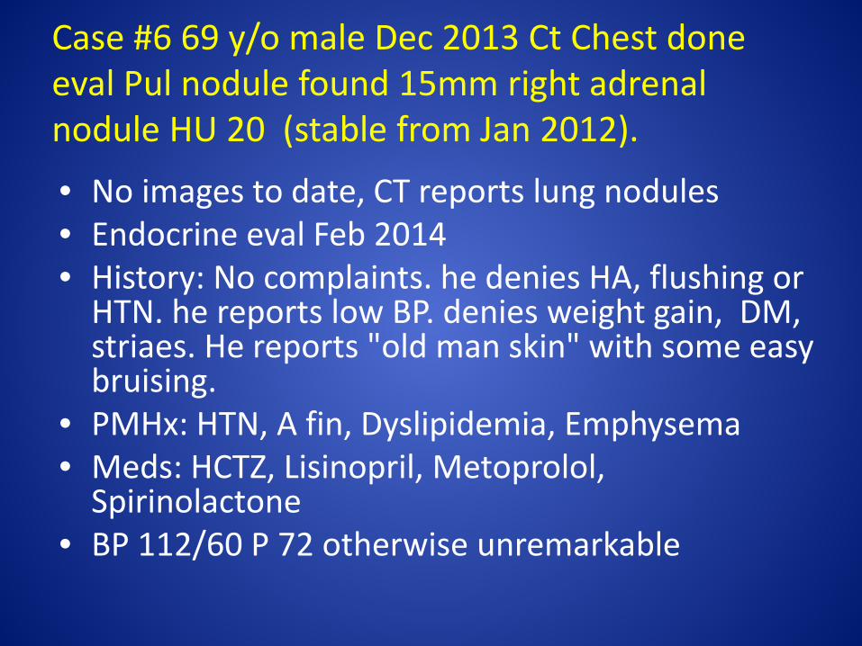
Case #6 69 y/o male Dec 2013 Ct Chest done eval Pul nodule found 15mm right adrenal nodule HU 20 (stable from Jan 2012).
• No images to date, CT reports lung nodules • Endocrine eval Feb 2014 • History: No complaints. he denies HA, flushing or
HTN. he reports low BP. denies weight gain, DM, striaes. He reports "old man skin" with some easy bruising.
• PMHx: HTN, A fin, Dyslipidemia, Emphysema • Meds: HCTZ, Lisinopril, Metoprolol,
Spirinolactone • BP 112/60 P 72 otherwise unremarkable

1. Is the adrenal mass incidentally found or does the patient show symptoms or comorbities (e. g. arterial hypertension) which might be caused by the adrenal mass? Incidentally found no signs or symptoms except fatigue due pul disease and hx of lung nodules
2. Does the adrenal mass represent a metastasis of an unknown or known primary tumor? Possibly since high precontrast HU hx of lung nodules.
3. Is the adrenal mass hormonally active? 4. Is there evidence of adrenocortical carcinoma?
Unlikely since lesion stable from almost 2 years 5. Follow-up?

Labs
• ALDO/PRA RATIO 0.7 L Ratio 0.9 - 28.9 • PRA-MS 22.72 H ng/mL/h • ALDOSTERONE 16 ng/dL • CORTISOL, FREE, URINE 7.8 mcg/24 h
4.0 - 50.0 • **labs done on spirinolactone therefore aldo
not interpretable (renin appropriately elevated due to spirinolactone and Acei)

Labs • BMP wnl • Specimen: PLASMA Specimen Collection Date: Feb 20, 2014@09:30 • Test name Result units Ref. range • Normetanephrine (ARUP) 2.67 H nmol/L 0.0 - 0.89 • Metanephrine (ARUP) 0.65 H nmol/L 0.0 - 0.49
• The majority of patients with pheochromocytoma have a plasma normetanephrine
concentration in excess of 2.2 nmol/L and/or a metanephrine concentration in excess of 1.1 nmol/L.
• Specimen: 24 HR. URINE. 2000 mL • Test name Result units Ref. range Site Code • Epinephrine 13 mcg/24 h 2 - 24 • Eval: Reference range prior to 6/20/02 was 0-20 ug/24h • Norepinephrine 120 H mcg/24 h 15 - 100 • Catecholamines Total (E+NE) 133 H mcg/24 h 26 - 121 • Dopamine 155 mcg/24 h 52 - 480 • CREATININE 24 Hr URINE 1.57 g/24 h

Repeated the test • Collection time: Mar 31, 2014@11:31:40 • Test Name Result Units Range • Normetanephrine (ARUP) 2.43 H nmol/L 0.0 - 0.89 • Metanephrine (ARUP) 0.32 nmol/L 0.0 - 0.49
• Again elevated metanephrines. Pt is on metoprolol, know
to interfere with urine catecholamines, does not interferes with plasma catecholamines.
• Puzzling that only normetanephrine is elevated, as pheo's are more associated with increased metanephrines.

May 2014 Ct ab/pelvis done for indeterminate adrenal nodule
• An approximately 18 mm in greatest dimension nodular mass lesion is seen in the right adrenal gland. Unenhanced images demonstrate Hounsfield units averaging approximately 18 with a
• standard deviation of approximately 19. Initial postenhanced imaging shows Hounsfield units averaging approximately in the range of 71- 78 with a standard deviation of approximately 27. Delayed images demonstrate Hounsfield units in the range of 77 to 81, with standard deviation of approximately 21.
• Washout <50% (in fact close to 0%)
• Small, stable in size HETEROGENOUS NODULE with high HU NOT C/W a benign adenoma

Does the patient have a pheochromocytoma?
• MIBG scan done- NEGATIVE • Clonidine suppression test done-
catecholamines decreased by 40% of baseline therefore pheo effectively ruled out
• October 2014 PET/CT ordered by pulmonologist showed PET + and growing lung nodules and 1.9cm metabolically active R adrenal gland intense FDG uptake, SUV max 6.6.

• Nov 2014 Diagnosed with adenocarcinoma of lung
• R adrenal ?metastatic lesion ?ACC (unlikely)
• Jan 2015 Biopsy done R adrenal NEGATIVE for Malignancy
• Jan 2015 R LL lobectomy
Bilateral Adrenal Masses
• Occurs in up to 15% of incidentalomas • Same hormonal work-up but consider
evaluation for adrenal insufficiency • More likely to suggest metastasis, congenital
adrenal hyperplasia, bilateral adenomas, infiltrative disease


Long Term Follow-up
• No consensus on optimal imaging follow-up – 0 (lipid rich not hormonally active) 3 (suspicious) 6,9,12 months
– All adenomas can grow but ACC grows the fastest • Consider surgery for nodules grow >1cm, or become biochemically active • Biochemical follow-up
– Repeat hormonal evaluation annually for at least 4 years when initial testing is negative…highest risk (20%) for development of subclinical cushings therefore annually recommend 1mg dex suppression and follow clinically for other hormonal function

References
1. The Incidentally Discovered Adrenal Mass William Young, N Engl J Med 2007;356:601-10.
2. Best Practice & Research Clinical Endocrinology & Metabolism 26 (2012) 69–82
3. EndoText.org


![adrenal cbc.ppt [Somente leitura] - cbcsp.org.br · 0 - 2: Comportamento benigno 3 ou +: Comportamento maligno ... Cisto de Adrenal / mielolipoma 4. Sd . Cushing 5. Incidentaloma](https://static.fdocuments.net/doc/165x107/5be3b64609d3f219598bb6dc/adrenal-cbcppt-somente-leitura-cbcsporgbr-0-2-comportamento-benigno.jpg)


![Seminario Incidentaloma Adrenal[1] - UED-HAM - … · Após 1-2 sem: Beta-bloqueador para controle de taquiarritmias; Metirosina: inibidor de tirosina hidroxilase, usado em pacientes](https://static.fdocuments.net/doc/165x107/5baf935309d3f2e27b8c8139/seminario-incidentaloma-adrenal1-ued-ham-apos-1-2-sem-beta-bloqueador.jpg)