The ACC/AHA Pocket Management Guidelines of Patients ... · of Patients WithAcute Myocardial...
21
The Management of Patients With Acute Myocardial Infarction (A Report of the American College of Cardiology/American Heart Association Task Force on Practice Guidelines) April, 2000 ACC/AHA Pocket Guidelines
Transcript of The ACC/AHA Pocket Management Guidelines of Patients ... · of Patients WithAcute Myocardial...
TheManagementof PatientsWith AcuteMyocardialInfarction(A Report of the American College
of Cardiology/American Heart Association
Task Force on Practice Guidelines)
April, 2000
ACC/AHA
Guidelines
ACC/AHA Pocket Guidelines for
The Managementof Patients withAcute MyocardialInfarction(A Report of the American College of Cardiology/American Heart Association Task Force on PracticeGuidelines)
Writing Committee
Thomas J. Ryan, MD, FACC, ChairElliott M. Antman, MD, FACC Neil H. Brooks, MD, FAAFP Robert M. Califf, MD, FACC L. David Hillis, MD, FACC Loren F. Hiratzka, MD, FACCElliot Rapaport, MD, FACCBarbara Riegel, DNSc, FAANRichard O. Russell, MD, FACC Earl E. Smith, III, MD, FACEP W. Douglas Weaver, MD, FACC
April, 2000
Distributed through an educational grant from
Genentech, Inc.
Genentech, Inc. was not involved in the development of this publication and in no way
influenced its contents.
Special thanks to
3
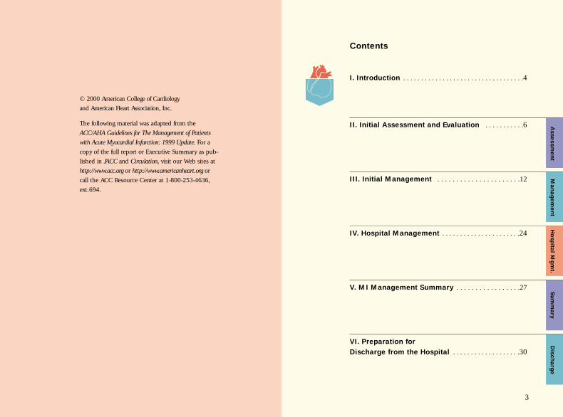
© 2000 American College of Cardiology and American Heart Association, Inc.
The following material was adapted from theACC/AHA Guidelines for The Management of Patientswith Acute Myocardial Infarction: 1999 Update. For acopy of the full report or Executive Summary as pub-lished in JACC and Circulation, visit our Web sites athttp://www.acc.org or http://www.americanheart.org orcall the ACC Resource Center at 1-800-253-4636,ext.694.
Contents
II. Initial Assessment and Evaluation . . . . . . . . . . .6
III. Initial Management . . . . . . . . . . . . . . . . . . . . . .12
IV. Hospital Management . . . . . . . . . . . . . . . . . . . . . .24
V. MI Management Summary . . . . . . . . . . . . . . . . .27
VI. Preparation for
Discharge from the Hospital . . . . . . . . . . . . . . . . . . .30
I. Introduction . . . . . . . . . . . . . . . . . . . . . . . . . . . . . . . . . .4
Ho
sp
ital M
gm
t.S
um
ma
ryD
isch
arg
eA
sse
ssm
en
tM
an
ag
em
en
t
54
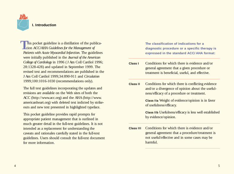
The classification of indications for a
diagnostic procedure or a specific therapy is
expressed in the standard ACC/AHA format:
Class I Conditions for which there is evidence and/or general agreement that a given procedure or treatment is beneficial, useful, and effective.
Class II Conditions for which there is conflicting evidence and/or a divergence of opinion about the useful-ness/efficacy of a procedure or treatment.
Class IIa Weight of evidence/opinion is in favor of usefulness/efficacy.
Class IIb Usefulness/efficacy is less well establishedby evidence/opinion.
Class III Conditions for which there is evidence and/or general agreement that a procedure/treatment is not useful/effective and in some cases may be harmful.
I. Introduction
his pocket guideline is a distillation of the publica-tion ACC/AHA Guidelines for the Management of
Patients with Acute Myocardial Infarction. The guidelineswere initially published in the Journal of the AmericanCollege of Cardiology in 1996 ( J Am Coll Cardiol 1996;28:1328-428) and updated in September 1999. Therevised text and recommendations are published in theJ Am Coll Cardiol 1999;34:890-911 and Circulation1999;100:1016-1030 (recommendations only).
The full text guidelines incorporating the updates andrevisions are available on the Web sites of both theACC (http://www.acc.org) and the AHA (http://www.americanheart.org) with deleted text indicted by strike-outs and new text presented in highlighted typeface.
This pocket guideline provides rapid prompts forappropriate patient management that is outlined inmuch greater detail in the full-text guidelines. It is notintended as a replacement for understanding thecaveats and rationales carefully stated in the full-textguidelines. Users should consult the full-text documentfor more information.
T
6 7
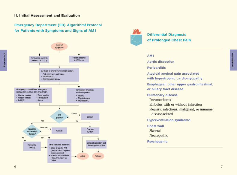
Differential Diagnosis
of Prolonged Chest Pain
AMI
Aortic dissection
Pericarditis
Atypical anginal pain associated
with hypertrophic cardiomyopathy
Esophageal, other upper gastrointestinal,
or biliary tract disease
Pulmonary disease
PneumothoraxEmbolus with or without infarctionPleurisy: infectious, malignant, or immune
disease-related
Hyperventilation syndrome
Chest wall
SkeletalNeuropathic
Psychogenic
Emergency Department (ED) Algorithm/Protocol
for Patients with Symptoms and Signs of AMI
II. Initial Assessment and Evaluation
Fibrinolytictherapy
Release
Ambulance presents patient to ED lobby
Patient presentsto ED lobby
Onset ofsymptoms
ED triage or charge nurse triages patient
• AMI symptoms and signs• 12-lead ECG• Brief, targeted history
Emergency nurse initiates emergency nursing care in acute care area of ED
• Cardiac monitor• Oxygen therapy• IV D5W
• Blood studies• Nitroglycerin• Aspirin
Emergency physician evaluates patient
• History• Physical exam• Interpret ECG
Consult
Uncertain
Uncertain
Consult
Yes
Yes
No
AMI patient?
Candidate for fibrinolytic
therapy?
Evaluate further
No
Admit
Other indicated treatment:
• Other drugs for AMI (beta-blockers, heparin, aspirin, nitrates)
• Transfer to cath lab for PTCA or surgery for CABG
Conduct education and follow-up instruction
Asse
ssm
en
tAsse
ssm
en
t
8 9
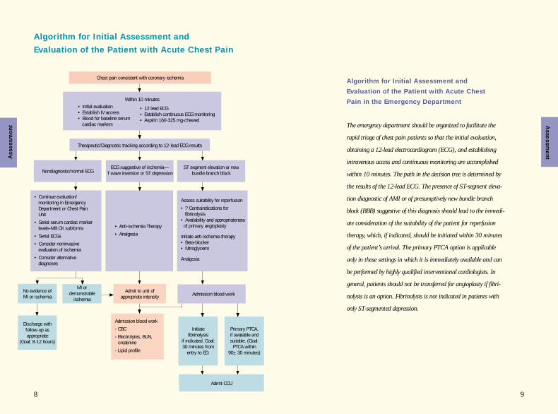
Algorithm for Initial Assessment and
Evaluation of the Patient with Acute Chest Pain
Algorithm for Initial Assessment and
Evaluation of the Patient with Acute Chest
Pain in the Emergency Department
The emergency department should be organized to facilitate the
rapid triage of chest pain patients so that the initial evaluation,
obtaining a 12-lead electrocardiogram (ECG), and establishing
intravenous access and continuous monitoring are accomplished
within 10 minutes. The path in the decision tree is determined by
the results of the 12-lead ECG. The presence of ST-segment eleva-
tion diagnostic of AMI or of presumptively new bundle branch
block (BBB) suggestive of this diagnosis should lead to the immedi-
ate consideration of the suitability of the patient for reperfusion
therapy, which, if indicated, should be initiated within 30 minutes
of the patient’s arrival. The primary PTCA option is applicable
only in those settings in which it is immediately available and can
be performed by highly qualified interventional cardiologists. In
general, patients should not be transferred for angioplasty if fibri-
nolysis is an option. Fibrinolysis is not indicated in patients with
only ST-segmented depression.
Chest pain consistent with coronary ischemia
Within 10 minutes• Initial evaluation• Establish IV access• Blood for baseline serum
cardiac markers
• 12 lead ECG• Establish continuous ECG monitoring• Aspirin 160-325 mg-chewed
ECG suggestive of ischemia—T wave inversion or ST depression
Therapeutic/Diagnostic tracking according to 12-lead ECG results
Nondiagnostic/normal ECGST segment elevation or new
bundle branch block
• Continue evaluation/ monitoring in EmergencyDepartment or Chest Pain Unit
• Serial serum cardiac marker levels-MB CK subforms
• Serial ECGs
• Consider noninvasive evaluation of ischemia
• Consider alternative diagnoses
Assess suitability for reperfusion
• ? Contraindications for fibrinolysis
• Availability and appropriatenessof primary angioplasty
Initiate anti-ischemia therapy• Beta-blocker• Nitroglycerin
Analgesia
• Anti-ischemia Therapy
• Analgesia
Admit to unit ofappropriate intensity
Admission blood work
- CBC
- Electrolytes, BUN, creatinine
- Lipid profile
No evidence of MI or ischemia
MI or demonstrable
ischemia
Initiate fibrinolysis
if indicated. Goal: 30 minutes from
entry to ED.
Primary PTCA, if available and suitable. (Goal:
PTCA within 90±30 minutes)
Discharge with follow-up as appropriate
(Goal: 8-12 hours)
Admit-CCU
Admission blood work
Asse
ssm
en
t Asse
ssm
en
t
10 11
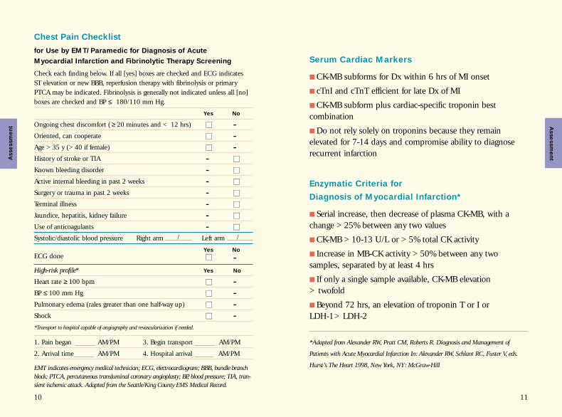
Chest Pain Checklist
for Use by EMT/Paramedic for Diagnosis of Acute
Myocardial Infarction and Fibrinolytic Therapy Screening
Check each finding below. If all [yes] boxes are checked and ECG indicates ST elevation or new BBB, reperfusion therapy with fibrinolysis or primaryPTCA may be indicated. Fibrinolysis is generally not indicated unless all [no]boxes are checked and BP ≤ 180/110 mm Hg.
Yes No
Ongoing chest discomfort ( ≥ 20 minutes and < 12 hrs) ■■ -Oriented, can cooperate ■■ -Age >35 y (>40 if female) ■■ -History of stroke or TIA - ■■
Known bleeding disorder - ■■
Active internal bleeding in past 2 weeks - ■■
Surgery or trauma in past 2 weeks - ■■
Terminal illness - ■■
Jaundice, hepatitis, kidney failure - ■■
Use of anticoagulants - ■■
Systolic/diastolic blood pressure Right arm / Left arm /
Yes No
ECG done ■■ -High-risk profile* Yes No
Heart rate ≥ 100 bpm ■■ -BP ≤ 100 mm Hg ■■ -Pulmonary edema (rales greater than one half-way up) ■■ -Shock ■■ -*Transport to hospital capable of angiography and revascularization if needed.
1. Pain began AM/PM 3. Begin transport AM/PM
2. Arrival time AM/PM 4. Hospital arrival AM/PM
EMT indicates emergency medical technician; ECG, electrocardiogram; BBB, bundle branchblock; PTCA, percutaneous transluminal coronary angioplasty; BP, blood pressure; TIA, tran-sient ischemic attack. Adapted from the Seattle/King County EMS Medical Record.
Serum Cardiac Markers
■ CK-MB subforms for Dx within 6 hrs of MI onset
■ cTnI and cTnT efficient for late Dx of MI
■ CK-MB subform plus cardiac-specific troponin best combination
■ Do not rely solely on troponins because they remain elevated for 7-14 days and compromise ability to diagnoserecurrent infarction
Enzymatic Criteria for
Diagnosis of Myocardial Infarction*
■ Serial increase, then decrease of plasma CK-MB, with achange >25% between any two values
■ CK-MB >10-13 U/L or >5% total CK activity
■ Increase in MB-CK activity >50% between any two samples, separated by at least 4 hrs
■ If only a single sample available, CK-MB elevation > twofold
■ Beyond 72 hrs, an elevation of troponin T or I or LDH-1>LDH-2
*Adapted from Alexander RW, Pratt CM, Roberts R. Diagnosis and Management of
Patients with Acute Myocardial Infarction In: Alexander RW, Schlant RC, Fuster V, eds.
Hurst’s The Heart 1998, New York, NY: McGraw-Hill
Asse
ssm
en
t Asse
ssm
en
t
12 13
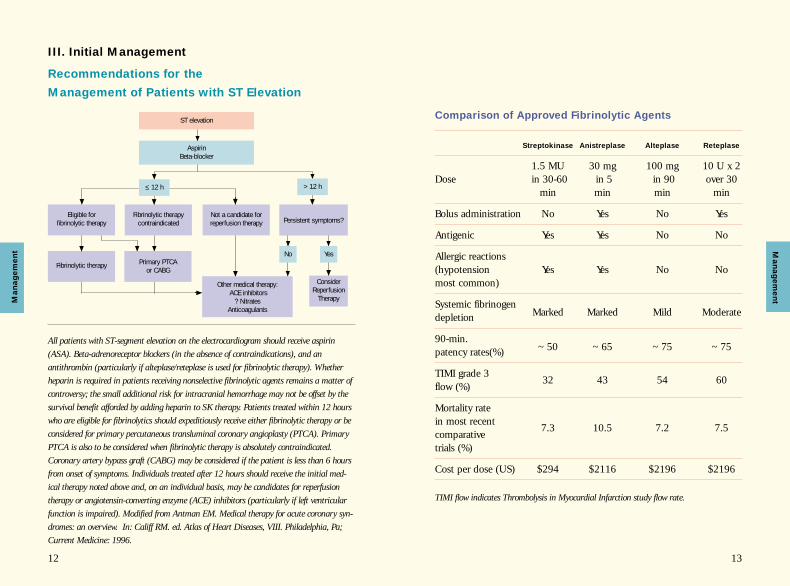
Comparison of Approved Fibrinolytic Agents
Streptokinase Anistreplase Alteplase Reteplase
1.5 MU 30 mg 100 mg 10 U x 2Dose in 30-60 in 5 in 90 over 30
min min min min
Bolus administration No Yes No Yes
Antigenic Yes Yes No No
Allergic reactions(hypotension Yes Yes No Nomost common)
Systemic fibrinogen depletion Marked Marked Mild Moderate
90-min. patency rates(%) ~50 ~65 ~75 ~75
TIMI grade 3 flow (%)
32 43 54 60
Mortality rate in most recent
7.3 10.5 7.2 7.5comparative trials (%)
Cost per dose (US) $294 $2116 $2196 $2196
TIMI flow indicates Thrombolysis in Myocardial Infarction study flow rate.
Recommendations for the
Management of Patients with ST Elevation
III. Initial Management
All patients with ST-segment elevation on the electrocardiogram should receive aspirin(ASA). Beta-adrenoreceptor blockers (in the absence of contraindications), and anantithrombin (particularly if alteplase/reteplase is used for fibrinolytic therapy). Whetherheparin is required in patients receiving nonselective fibrinolytic agents remains a matter ofcontroversy; the small additional risk for intracranial hemorrhage may not be offset by thesurvival benefit afforded by adding heparin to SK therapy. Patients treated within 12 hourswho are eligible for fibrinolytics should expeditiously receive either fibrinolytic therapy or beconsidered for primary percutaneous transluminal coronary angioplasty (PTCA). PrimaryPTCA is also to be considered when fibrinolytic therapy is absolutely contraindicated.Coronary artery bypass graft (CABG) may be considered if the patient is less than 6 hoursfrom onset of symptoms. Individuals treated after 12 hours should receive the initial med-ical therapy noted above and, on an individual basis, may be candidates for reperfusiontherapy or angiotensin-converting enzyme (ACE) inhibitors (particularly if left ventricularfunction is impaired). Modified from Antman EM. Medical therapy for acute coronary syn-dromes: an overview. In: Califf RM. ed. Atlas of Heart Diseases, VIII. Philadelphia, Pa;Current Medicine: 1996.
ST elevation
AspirinBeta-blocker
>12 h
Fibrinolytic therapy contraindicated
Not a candidate forreperfusion therapy
≤ 12 h
Fibrinolytic therapy
Eligible for fibrinolytic therapy
Primary PTCA or CABG
Yes
ConsiderReperfusion
Therapy
Persistent symptoms?
No
Other medical therapy:ACE inhibitors
? NitratesAnticoagulants
Ma
na
ge
me
ntM
an
ag
em
en
t

14 15
Primary Percutaneous Transluminal
Coronary Angioplasty Recommendations
Class I Recommendations
1. As an alternative to fibrinolytic therapy if:
■ ST-segment elevation or new or presumed new LBBB
■ Within 12 hrs of symptoms or >12 hrs of persistent pain
■ In a timely fashion (90±30 min)
■ By experienced operators
■ In appropriate laboratory environment
2. In cardiogenic shock patients <75 yrs who
are within 36 hrs of AMI and revascularization
can be performed within 18 hrs of onset of shock
Class IIa Recommendations
1. As a reperfusion strategy in candidates for
reperfusion who have a contraindication to
fibrinolytic therapy.
Contraindications and Cautions
for Fibrinolytic Use in Myocardial Infarction*
Absolute Contraindications
■ Previous hemorrhagic stroke at any time: other strokes or cerebrovascular events within 1 yr■ Known intracranial neoplasm■ Active internal bleeding (does not include menses)■ Suspected aortic dissection
Cautions/Relative Contraindications
■ Severe uncontrolled hypertension on presentation (blood pressure >180/110 mm Hg)†
■ History of prior cerebrovascular accident or known intracerebral pathology not covered in contraindications■ Current use of anticoagulants in therapeutic doses (INR ≥ 2-3); known bleeding diathesis■ Recent trauma (within 2-4 wks), including head trauma■ Noncompressible vascular punctures■ Recent (within 2-4 wks) internal bleeding■ For streptokinase/anistreplase: prior exposure (especially within 5d-2y) or prior allergic reaction■ Pregnancy■ Active peptic ulcer■ History of chronic hypertension
INR indicates International Normalized Ratio.
Viewed as advisory for clinical decision making and may not be all-inclusive or definitive.
Could be an absolute contraindication in low-risk patients with myocardial infarction.†
*
Ma
na
ge
me
nt M
an
ag
em
en
t
16 17
Advantages of Fibrinolytic Therapy
■ More universal access
■ Shorter time to treatment
■ Greater clinical trial evidence of:■ reduction in infarct size■ improvement of LV function
■ Results less dependent on physician experience
■ Lower system cost
Advantages of Primary PTCA
■ Higher initial reperfusion rates
■ Lower recurrence rates of ischemia/infarction
■ Less residual stenosis
■ Less intracranial bleeding
■ Defines coronary anatomy and LV function
■ Utility when fibrinolysis contraindicated
Class IIb Recommendations
1. In patients with AMI who do not present with
ST elevation but who have reduced [less than
TIMI (Thrombolysis in Myocardial Infarction)
grade 2] flow of the infarct-related artery and
when angioplasty can be performed within 12
hrs of onset of symptoms.
Class III Recommendations
1. This classification applies to patients with AMI
who
■ Undergo elective angioplasty in a noninfarct-related artery at the time of AMI
■ Are beyond 12 hrs after the onset of symptomsand have no evidence of myocardial ischemia
■ Have received fibrinolytic therapy and have nosymptoms of myocardial ischemia
■ Are fibrinolytic-eligible and are undergoing primary angioplasty by an unskilled operator in alaboratory that does not have surgical capability.
Ma
na
ge
me
nt M
an
ag
em
en
t
18 19
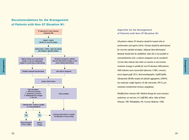
Recommendations for the Management
of Patients with Non-ST Elevation MI
ST depression/T-wave inversion:Suspected AMI
Heparin+Aspirin Nitrates for recurrent angina
Antithrombins: LMWH—high-risk patients Anti-Platelets: Gpllb/IIIa inhibitor
Medical Therapy
Yes
Revascularization(PTCA, CABG)
Patients without prior beta-blocker therapy or who are inadequately
treated on current dose of beta-blocker
Persistent symptoms in patients with prior beta-blocker therapy or who
cannot tolerate beta-blockers
Establish adequate beta-blockade Add calcium antagonist
Assess clinical status
Clinical stability
High-risk patient:1. Recurrent ischemia
2. Depressed LV function3. Widespread ECG changes
4. Prior MI
Catheterization: Anatomy suitablefor revascularization?
Continued observation in hospitalConsideration of stress testing
No
Algorithm for the Management
of Patients with Non-ST Elevation MI
All patients without ST elevation should be treated with an
antithrombin and aspirin (ASA). Nitrates should be administered
for recurrent episodes of angina. Adequate beta-adrenoceptor
blockade should then be established; when this is not possible or
contraindications exist, a calcium antagonist can be considered.
Current data indicate that either an invasive or non-invasive
treatment strategy is suitable for non-ST-elevation AMI patients.
AMI indicates acute myocardial infarction; CABG, coronary
artery bypass graft; ECG, electrocardiographic; GpIIb/GpIIIa,
Glycoprotein IIb/IIIa receptor for platelet aggregation; LMWH,
low molecular weight heparin; LV, left ventricular; PTCA, per-
cutaneous transluminal coronary angioplasty.
Modified from Antman EM. Medical therapy for acute coronary
syndromes: an overview. In Califf RM, editor. Atlas of Heart
Diseases, VIII. Philadelphia, PA: Current Medicine; 1996.
Ma
na
ge
me
nt M
an
ag
em
en
t
2120
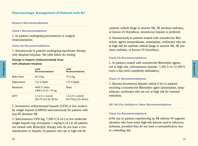
Pharmacologic Management of Patients with MI
Heparin Recommendations
Class I Recommendations
1. In patients undergoing percutaneous or surgical revascularization.
Class IIa Recommendations
1. Intravenously in patients undergoing reperfusion therapywith alteplase/reteplase. See table below for dosing:
Change in Heparin (Unfractionated) Dose
with alteplase/reteplase
1999 1996
Recommendations Recommendations
Bolus Dose 60 U/kg 70 U/kg
Maintenance ≈12 U/kg/hr ≈15 U/kg/hr
Maximum 4000 U bolus None1000 U/h if >70 kg
aPTT 1.5-2.0 x control 1.5-2.0 x control(50-70 sec) for 48 hrs (50-70sec) for 48 hrs
2. Intravenous unfractionated heparin (UFH) or low molecu-lar weight heparin (LMWH) subcutaneously for patients withnon-ST elevation MI.
3. Subcutaneous UFH (eg, 7,500 U b.i.d.) or low molecularweight heparin (eg, enoxaparin 1 mg/kg b.i.d.) in all patientsnot treated with fibrinolytic therapy who do not have a con-traindication to heparin. In patients who are at high risk for
systemic emboli (large or anterior MI, AF, previous embolus,or known LV thrombus), intravenous heparin is preferred.
4. Intravenously in patients treated with nonselective fibri-nolytic agents (streptokinase, anistreplase, urokinase) who areat high risk for systemic emboli (large or anterior MI, AF, pre-vious embolus, or known LV thrombus).
Class IIb Recommendations
1. In patients treated with nonselective fibrinolytic agents,not at high risk, subcutaneous heparin, 7,500 U to 12,500 Utwice a day until completely ambulatory.
Class III Recommendations
1. Routine intravenous heparin within 6 hrs to patientsreceiving a nonselective fibrinolytic agent (anistreplase, strep-tokinase, urokinase) who are not at high risk for systemicembolism.
GP IIb/IIIa Inhibitors—New Recommendations
Class IIa Recommendations
■ For use in patients experiencing an MI without ST segmentelevation who have some high-risk features and/or refractoryischemia, provided they do not have a contraindication dueto a bleeding risk.
Ma
na
ge
me
nt M
an
ag
em
en
t
22 23
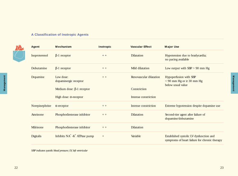
A Classification of Inotropic Agents
Agent Mechanism Inotropic Vascular Effect Major Use
Isoproterenol β-1 receptor ++ Dilatation Hypotension due to bradycardia; no pacing available
Dobutamine β-1 receptor ++ Mild dilatation Low output with SBP >90 mm Hg
Dopamine Low dose: ++ Renovascular dilatation Hypoperfusion with SBPdopaminergic receptor <90 mm Hg or ≥ 30 mm Hg
below usual valueMedium dose: β-1 receptor Constriction
High dose: α-receptor Intense constriction
Norepinephrine α-receptor ++ Intense constriction Extreme hypotension despite dopamine use
Amrinone Phosphodiesterase inhibitor ++ Dilatation Second-tier agent after failure of dopamine/dobutamine
Milrinone Phosphodiesterase inhibitor ++ Dilatation
Digitalis Inhibits NA+-K+ATPase pump + Variable Established systolic LV dysfunction and symptoms of heart failure for chronic therapy
SBP indicates systolic blood pressure; LV, left ventricular
Ma
na
ge
me
ntM
an
ag
em
en
t
24 25
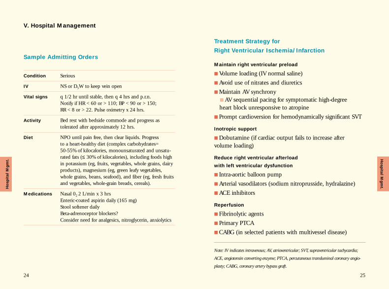
Sample Admitting Orders
Condition Serious
IV NS or D5W to keep vein open
Vital signs q 1/2 hr until stable, then q 4 hrs and p.r.n. Notify if HR <60 or >110; BP <90 or >150; RR <8 or >22. Pulse oximetry x 24 hrs.
Activity Bed rest with bedside commode and progress as tolerated after approximately 12 hrs.
Diet NPO until pain free, then clear liquids. Progress to a heart-healthy diet (complex carbohydrates=50-55% of kilocalories, monounsaturated and unsatu-rated fats (≤ 30% of kilocalories), including foods high in potassium (eg, fruits, vegetables, whole grains, dairy products), magnesium (eg, green leafy vegetables, whole grains, beans, seafood), and fiber (eg, fresh fruits and vegetables, whole-grain breads, cereals).
Medications Nasal 02 2 L/min x 3 hrsEnteric-coated aspirin daily (165 mg)Stool softener dailyBeta-adrenoceptor blockers?Consider need for analgesics, nitroglycerin, anxiolytics
Treatment Strategy for
Right Ventricular Ischemia/Infarction
Maintain right ventricular preload
■ Volume loading (IV normal saline)
■ Avoid use of nitrates and diuretics
■ Maintain AV synchrony■ AV sequential pacing for symptomatic high-degree heart block unresponsive to atropine
■ Prompt cardioversion for hemodynamically significant SVT
Inotropic support
■ Dobutamine (if cardiac output fails to increase after volume loading)
Reduce right ventricular afterload
with left ventricular dysfunction
■ Intra-aortic balloon pump
■ Arterial vasodilators (sodium nitroprusside, hydralazine)
■ ACE inhibitors
Reperfusion
■ Fibrinolytic agents
■ Primary PTCA
■ CABG (in selected patients with multivessel disease)
Note: IV indicates intravenous; AV, atrioventricular; SVT, supraventricular tachycardia;
ACE, angiotensin converting enzyme; PTCA, percutaneous transluminal coronary angio-
plasty; CABG, coronary artery bypass graft.
V. Hospital Management
Ho
sp
ital M
gm
t.Ho
sp
ita
l M
gm
t.
27
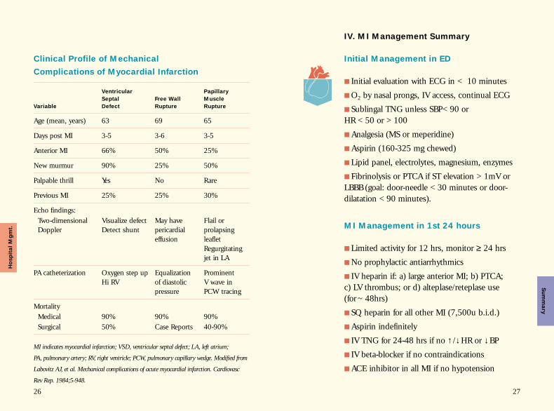
Initial Management in ED
■ Initial evaluation with ECG in < 10 minutes
■ O2 by nasal prongs, IV access, continual ECG
■ Sublingal TNG unless SBP<90 or HR <50 or >100
■ Analgesia (MS or meperidine)
■ Aspirin (160-325 mg chewed)
■ Lipid panel, electrolytes, magnesium, enzymes
■ Fibrinolysis or PTCA if ST elevation >1mV orLBBB (goal: door-needle <30 minutes or door-dilatation <90 minutes).
MI Management in 1st 24 hours
■ Limited activity for 12 hrs, monitor ≥ 24 hrs
■ No prophylactic antiarrhythmics
■ IV heparin if: a) large anterior MI; b) PTCA; c) LV thrombus; or d) alteplase/reteplase use(for ~48hrs)
■ SQ heparin for all other MI (7,500u b.i.d.)
■ Aspirin indefinitely
■ IV TNG for 24-48 hrs if no ↑ /↓HR or ↓BP
■ IV beta-blocker if no contraindications
■ ACE inhibitor in all MI if no hypotension
26
Clinical Profile of Mechanical
Complications of Myocardial Infarction
Ventricular Papillary
Septal Free Wall Muscle
Variable Defect Rupture Rupture
Age (mean, years) 63 69 65
Days post MI 3-5 3-6 3-5
Anterior MI 66% 50% 25%
New murmur 90% 25% 50%
Palpable thrill Yes No Rare
Previous MI 25% 25% 30%
Echo findings:Two-dimensional Visualize defect May have Flail or Doppler Detect shunt pericardial prolapsing
effusion leafletRegurgitating jet in LA
PA catheterization Oxygen step up Equalization Prominent Hi RV of diastolic V wave in
pressure PCW tracing
MortalityMedical 90% 90% 90%Surgical 50% Case Reports 40-90%
MI indicates myocardial infarction; VSD, ventricular septal defect; LA, left atrium;
PA, pulmonary artery; RV, right ventricle; PCW, pulmonary capillary wedge. Modified from
Labovitz AJ, et al. Mechanical complications of acute myocardial infarction. Cardiovasc
Rev Rep. 1984;5-948.
IV. MI Management Summary
Ho
sp
ita
l M
gm
t.S
um
ma
ry
2928
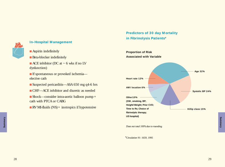
In-Hospital Management
■ Aspirin indefinitely
■ Beta-blocker indefinitely
■ ACE inhibitor (DC at ~6 wks if no LV dysfunction)
■ If spontaneous or provoked ischemia—elective cath
■ Suspected pericarditis—ASA 650 mg q4-6 hrs
■ CHF—ACE inhibitor and diuretic as needed
■ Shock—consider intra-aortic balloon pump+cath with PTCA or CABG
■ RV MI-fluids (NS)+ inotropics if hypotensive
Age 32%
Systolic BP 24%
Heart rate 12%
AMI location 6%
Other10%
(DM, smoking, BP;
Height/Weight; Prior CVD;
Time to Rx; Choice of
fibrinolytic therapy;
US hospital)
Killip class 15%
*Circulation 91: 1659, 1995
Does not total 100% due to rounding.
Su
mm
ary
Su
mm
ary
Predictors of 30 day Mortality
in Fibrinolysis Patients*
Proportion of Risk
Associated with Variable
30 31
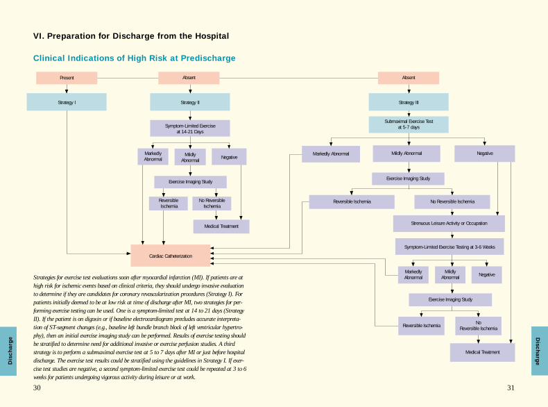
Clinical Indications of High Risk at Predischarge
VI. Preparation for Discharge from the Hospital
Strategy III
Submaximal Exercise Test at 5-7 days
Mildly Abnormal
Exercise Imaging Study
Reversible Ischemia No Reversible Ischemia
Absent
Strategy II
Symptom-Limited Exercise at 14-21 Days
MarkedlyAbnormal
Mildly Abnormal
Exercise Imaging Study
ReversibleIschemia
No ReversibleIschemia
Strategies for exercise test evaluations soon after myocardial infarction (MI). If patients are athigh risk for ischemic events based on clinical criteria, they should undergo invasive evaluationto determine if they are candidates for coronary revascularization procedures (Strategy I). Forpatients initially deemed to be at low risk at time of discharge after MI, two strategies for per-forming exercise testing can be used. One is a symptom-limited test at 14 to 21 days (StrategyII). If the patient is on digoxin or if baseline electrocardiogram precludes accurate interpreta-tion of ST-segment changes (e.g., baseline left bundle branch block of left ventricular hypertro-phy), then an initial exercise imaging study can be performed. Results of exercise testing shouldbe stratified to determine need for additional invasive or exercise perfusion studies. A thirdstrategy is to perform a submaximal exercise test at 5 to 7 days after MI or just before hospitaldischarge. The exercise test results could be stratified using the guidelines in Strategy I. If exer-cise test studies are negative, a second symptom-limited exercise test could be repeated at 3 to 6weeks for patients undergoing vigorous activity during leisure or at work.
Negative
Medical Treatment
Strategy I
Present Absent
Negative
No Reversible Ischemia
Strenuous Leisure Activity or Occupation
Symptom-Limited Exercise Testing at 3-6 Weeks
Mildly Abnormal Negative
Exercise Imaging Study
Medical Treatment
Markedly Abnormal
MarkedlyAbnormal
Reversible Ischemia
Cardiac Catheterization
Dis
ch
arg
eDis
ch
arg
e

33
Energy Levels Required to
Perform Some Common Activities
Self-Care WashingShaving DressingDesk workWashing dishesDriving autoLight housekeeping
Cleaning windowsRakingPower lawn mowingBedmaking/strippingCarrying objects (15-30 lb.)
Occupational Sitting (clerical/assembly)TypingDesk workStanding (store clerk)
Stocking shelves (light objects)Auto repairLight welding/carpentry
Recreational Golf (cart)KnittingHand sewing
Dancing (social)Golf (walking)SailingTennis (doubles)Volleyball (6 persons)
Physical conditioning Walking (2 mph)Stationary bikeVery light calisthenics
Level walking (3-4 mph)Level biking (6-8 mph)Light calisthenics
Easy digging in gardenHand lawn mowing (level)Climbing stairs (slowly)Carrying objects (30-60 lb.)Digging vigorously
Sawing woodHeavy shovelingClimbing stairs (moderate speed)Carrying objects (60-90 lb.)
Carrying loads upstairs(objects >90 lb.)Climbing stairs (quickly)Shoveling heavy snow
Carpentry (exterior)Shoveling dirtSawing woodOperating pneumatictools
Digging ditches (pick and shovel)
Lumber jackHeavy laborer
Badminton (competitive)Tennis (singles)Snow skiing (downhill)Light backpackingBasketballFootballStream fishing
CanoeingMountain climbingPaddle ball
HandballSquashSki touringVigorous basketball
Level walking (4.5 -5.0 mph)Bicycling (9 -10 mph)Swimming, breast stroke
Level jogging (5 mph)Swimming (crawl stroke)Rowing machineHeavy calisthenicsBicycling (12 mph)
Running (>6 mph)Bicycling (>13 mph)Rope jumpingWalking uphill (5 mph)
5-7 METs 7-9 METs > 9 METs<3 METs 3-5 METs
METs indicates metabolic equivalents. Adapted from Table 9.2, p 147. Rehabilitation of the
coronary patient (Wenger NI, Hellerstein HK, eds), Haskell WL, Design and Implementa-
tion of Cardiac Conditional Program. New York, NY: Churchill Livingstone: 1978.
32
Dis
ch
arg
eD
isch
arg
e
34 35
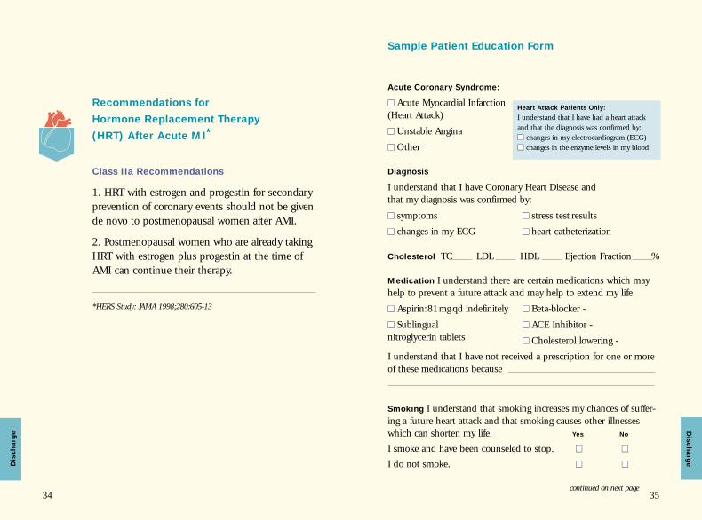
Recommendations for
Hormone Replacement Therapy
(HRT) After Acute MI*
Class IIa Recommendations
1. HRT with estrogen and progestin for secondaryprevention of coronary events should not be givende novo to postmenopausal women after AMI.
2. Postmenopausal women who are already takingHRT with estrogen plus progestin at the time ofAMI can continue their therapy.
*HERS Study: JAMA 1998;280:605-13
Dis
ch
arg
e
Sample Patient Education Form
Acute Coronary Syndrome:
■■ Acute Myocardial Infarction (Heart Attack)
■■ Unstable Angina
■■ Other
Diagnosis
I understand that I have Coronary Heart Disease and that my diagnosis was confirmed by:
■■ symptoms ■■ stress test results
■■ changes in my ECG ■■ heart catheterization
Cholesterol TC LDL HDL Ejection Fraction %
Medication I understand there are certain medications which mayhelp to prevent a future attack and may help to extend my life.
■■ Aspirin:81mg qd indefinitely ■■ Beta-blocker -
■■ Sublingual ■■ ACE Inhibitor -nitroglycerin tablets ■■ Cholesterol lowering -
I understand that I have not received a prescription for one or moreof these medications because
Smoking I understand that smoking increases my chances of suffer-ing a future heart attack and that smoking causes other illnesseswhich can shorten my life. Yes No
I smoke and have been counseled to stop. ■■ ■■
I do not smoke. ■■ ■■
Heart Attack Patients Only:
I understand that I have had a heart attackand that the diagnosis was confirmed by: ■■ changes in my electrocardiogram (ECG)■■ changes in the enzyme levels in my blood
continued on next page
Dis
ch
arg
e
36
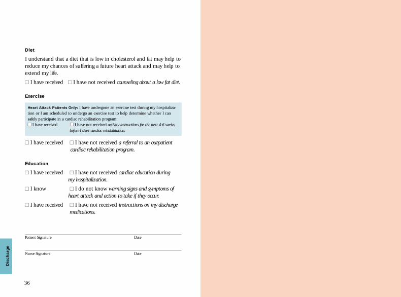
Diet
I understand that a diet that is low in cholesterol and fat may help toreduce my chances of suffering a future heart attack and may help toextend my life.
■■ I have received ■■ I have not received counseling about a low fat diet.
Exercise
Heart Attack Patients Only: I have undergone an exercise test during my hospitaliza-tion or I am scheduled to undergo an exercise test to help determine whether I can safely participate in a cardiac rehabilitation program.■■ I have received ■■ I have not received activity instructions for the next 4-6 weeks,
before I start cardiac rehabilitation.
■■ I have received ■■ I have not received a referral to an outpatient cardiac rehabilitation program.
Education
■■ I have received ■■ I have not received cardiac education during my hospitalization.
■■ I know ■■ I do not know warning signs and symptoms of heart attack and action to take if they occur.
■■ I have received ■■ I have not received instructions on my discharge medications.
Patient Signature Date
Nurse Signature Date
Dis
ch
arg
e