The 2016 WHO Classification of Tumours of ... -...
14
Platinum Priority – Guidelines Editorial by Rodolfo Montironi, Liang Cheng, Marina Scarpelli and Antonio Lopez-Beltran on pp. 120–123 of this issue The 2016 WHO Classification of Tumours of the Urinary System and Male Genital Organs—Part B: Prostate and Bladder Tumours Peter A. Humphrey a , Holger Moch b, *, Antonio L. Cubilla c , Thomas M. Ulbright d , Victor E. Reuter e a Department of Pathology, Yale University School of Medicine, New Haven, CT, USA; b Department of Pathology, University Hospital Zurich, Zurich, Switzerland; c Instituto de Patologı´a e Investigacio ´n, Facultad de Ciencias Me ´dicas, Universidad Nacional de Asuncio ´n, San Lorenzo, Paraguay; d Department of Pathology and Laboratory Medicine, Indiana University Health Partners, Indiana University School of Medicine, Indianapolis, IN, USA; e Department of Pathology, Memorial Sloan Kettering Cancer Center, New York, NY, USA EUROPEAN UROLOGY 70 (2016) 106–119 available at www.sciencedirect.com journal homepage: www.europeanurology.com Article info Article history: Accepted February 4, 2016 Associate Editor: James Catto Keywords: WHO classification Prostate Bladder Abstract It has been 12 yr since the publication of the last World Health Organization (WHO) classification of tumours of the prostate and bladder. During this time, significant new knowledge has been generated about the pathology and genetics of these tumours. Intraductal carcinoma of the prostate is a newly recognized entity in the 2016 WHO classification. In most cases, it represents intraductal spread of aggressive prostatic carcinoma and should be separated from high-grade prostatic intraepithelial neoplasia. New acinar adenocarcinoma variants are microcystic adenocarcinoma and pleomorphic giant cell adenocarcinoma. Modifications to the Gleason grading system are incorpo- rated into the 2016 WHO section on grading of prostate cancer, and it is recommended that the percentage of pattern 4 should be reported for Gleason score 7. The new WHO classification further recommends the recently developed prostate cancer grade group- ing with five grade groups. For bladder cancer, the 2016 WHO classification continues to recommend the 1997 International Society of Urological Pathology grading classifica- tion. Newly described or better defined noninvasive urothelial lesions include urothelial dysplasia and urothelial proliferation of uncertain malignant potential, which is fre- quently identified in patients with a prior history of urothelial carcinoma. Invasive urothelial carcinoma with divergent differentiation refers to tumours with some percent- age of ‘‘usual type’’ urothelial carcinoma combined with other morphologies. Pathol- ogists should mention the percentage of divergent histologies in the pathology report. Patient summary: Intraductal carcinoma of the prostate is a newly recognized entity in the 2016 World Health Organization classification. Better defined noninvasive urothelial lesions include urothelial dysplasia and urothelial proliferation of uncertain malignant potential. # 2016 European Association of Urology. Published by Elsevier B.V. All rights reserved. * Corresponding author. Department of Pathology, University Hospital Zurich, Schmelzbergstrasse 12, CH-8091 Zurich, Switzerland. Tel. +41 442552500; Fax: +41 442553194. E-mail address: [email protected] (H. Moch). http://dx.doi.org/10.1016/j.eururo.2016.02.028 0302-2838/# 2016 European Association of Urology. Published by Elsevier B.V. All rights reserved.
Transcript of The 2016 WHO Classification of Tumours of ... -...
E U R O P E A N U R O L O G Y 7 0 ( 2 0 1 6 ) 1 0 6 – 1 1 9
avai lable at www.sciencedirect .com
journal homepage: www.europeanurology.com
Platinum Priority – GuidelinesEditorial by Rodolfo Montironi, Liang Cheng, Marina Scarpelli and Antonio Lopez-Beltran on pp. 120–123 of
this issue
The 2016 WHO Classification of Tumours of the Urinary System
and Male Genital Organs—Part B: Prostate and Bladder Tumours
Peter A. Humphrey a, Holger Moch b,*, Antonio L. Cubilla c, Thomas M. Ulbright d,Victor E. Reuter e
a Department of Pathology, Yale University School of Medicine, New Haven, CT, USA; b Department of Pathology, University Hospital Zurich, Zurich,
Switzerland; c Instituto de Patologıa e Investigacion, Facultad de Ciencias Medicas, Universidad Nacional de Asuncion, San Lorenzo, Paraguay; d Department
of Pathology and Laboratory Medicine, Indiana University Health Partners, Indiana University School of Medicine, Indianapolis, IN, USA; e Department of
Pathology, Memorial Sloan Kettering Cancer Center, New York, NY, USA
Article info
Article history:Accepted February 4, 2016
Associate Editor:
James Catto
Keywords:
WHO classification
Prostate
Bladder
Abstract
It has been 12 yr since the publication of the last World Health Organization (WHO)classification of tumours of the prostate and bladder. During this time, significant newknowledge has been generated about the pathology and genetics of these tumours.Intraductal carcinoma of the prostate is a newly recognized entity in the 2016 WHOclassification. In most cases, it represents intraductal spread of aggressive prostaticcarcinoma and should be separated from high-grade prostatic intraepithelial neoplasia.New acinar adenocarcinoma variants are microcystic adenocarcinoma and pleomorphicgiant cell adenocarcinoma. Modifications to the Gleason grading system are incorpo-rated into the 2016 WHO section on grading of prostate cancer, and it is recommendedthat the percentage of pattern 4 should be reported for Gleason score 7. The new WHOclassification further recommends the recently developed prostate cancer grade group-ing with five grade groups. For bladder cancer, the 2016 WHO classification continues torecommend the 1997 International Society of Urological Pathology grading classifica-tion. Newly described or better defined noninvasive urothelial lesions include urothelialdysplasia and urothelial proliferation of uncertain malignant potential, which is fre-quently identified in patients with a prior history of urothelial carcinoma. Invasiveurothelial carcinoma with divergent differentiation refers to tumours with some percent-age of ‘‘usual type’’ urothelial carcinoma combined with other morphologies. Pathol-ogists should mention the percentage of divergent histologies in the pathology report.Patient summary: Intraductal carcinoma of the prostate is a newly recognized entity inthe 2016 World Health Organization classification. Better defined noninvasive urotheliallesions include urothelial dysplasia and urothelial proliferation of uncertain malignantpotential.
# 2016 European Association of Urology. Published by Elsevier B.V. All rights reserved.
* Corresponding author. Department of Pathology, University Hospital Zurich, Schmelzbergstrasse12, CH-8091 Zurich, Switzerland. Tel. +41 442552500; Fax: +41 442553194.
[email protected] (H. Moch).
E-mail address: holger.mhttp://dx.doi.org/10.1016/j.eururo.2016.02.0280302-2838/# 2016 European Association of Urology. Published by Elsevier B.V. All rights reserved.
E U R O P E A N U R O L O G Y 7 0 ( 2 0 1 6 ) 1 0 6 – 1 1 9 107
1. The new prostate tumour classification
The aim of this review is to summarize the new additions to
the 2016 World Health Organization (WHO) classification
(WHO ‘‘blue book’’) compared with the 2004 WHO classifi-
cation, with emphasis on a new entity, new variants of acinar
adenocarcinoma, and new imunohistochemical stains for
diagnosis, grading, risk stratification, and molecular genetics
of acinar adenocarcinoma of the prostate. The 2016 WHO
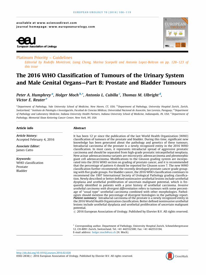
[(Fig._1)TD$FIG]Fig. 1 – World Health Organization (WHO) classification of tumours of the proWHO = World Health Organization.
classification of tumours of the prostate [1] is summarized
in Figure 1.
1.1. New entity: intraductal carcinoma
Intraductal carcinoma is newly recognized as an entity in
the 2016 WHO classification. This term has been used for
several decades, dating back to at least 1985 [2], and it has
been variably used to describe intraductal spread or in situ
state. Reproduced with permission from the WHO [1].

E U R O P E A N U R O L O G Y 7 0 ( 2 0 1 6 ) 1 0 6 – 1 1 9108
growth of acinar or ductal adenocarcinoma of the prostate
and intraductal proliferation of urothelial carcinoma
[3–11]. The 2016 WHO definition is as follows: ‘‘Intraductal
carcinoma of the prostate is intra-acinar and/or intraductal
neoplastic epithelial proliferation that has some features
of high-grade prostatic intraepithelial neoplasia (HGPIN)
but exhibits much greater architectural and/or cytological
atypia, typically associated with high-grade, high-stage
prostate carcinoma.’’
Intraductal carcinoma is thought to represent a late event
in prostate cancer evolution, with intraductal spread of
aggressive prostatic carcinoma and cancerization of pre-
existing ducts and acini by high-grade prostatic adenocarci-
noma. A minority of cases, however, may be precursors
to proliferation because in approximately 10% of radical
prostatectomy (RP) cases following a needle biopsy diagnosis
of intraductal carcinoma, the intraductal carcinoma in the
whole prostate gland is found in pure form, without
associated invasive adenocarcinoma [8].
Intraductal carcinoma is rare in isolated form in needle
biopsy tissue, being detected in 0.1–0.3% of needle core
cases [11,89], and is uncommon in the presence of invasive
adenocarcinoma in needle core tissue, being diagnosed in
2.8% of such cases [11]. In whole prostate glands, the
incidence is dependent on the grade and stage of the
prostatic adenocarcinoma in the series and ranges from 20%
to 40% of RP cases [5,10].
Diagnostic separation of intraductal carcinoma from
HGPIN is critical due to the association of intraductal
carcinoma with an average Gleason score of 8 and pT3
prostatic adenocarcinoma in the whole gland [8]. In contrast
to HGPIN, intraductal carcinoma exhibits a solid or dense
cribriform pattern or a loose cribriform or micropapillary
pattern with either marked nuclear atypia (ie, nuclear size
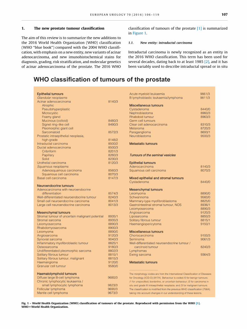
�6� normal) or comedonecrosis (Fig. 2) [11]. PTEN and ERG
[(Fig._2)TD$FIG]Fig. 2 – Intraductal carcinoma of the prostate with comedonecrosis, withsurrounding dense cribriform glands.
immunostaining may be a useful adjunctive method because
intraductal carcinoma commonly shows PTEN loss and ERG
expression, whereas PTEN loss is rare in HGPIN and ERG
expression is uncommon [12].
An important point is that intraductal carcinoma is not
assigned a Gleason grade [13].
Reporting of isolated intraductal carcinoma in needle
biopsy should include a comment stating that intraductal
carcinoma of the prostate is associated with high-grade and
high-volume prostate carcinoma and that therapy may be
indicated. Repeat biopsy may also be recommended.
1.2. New variants of acinar adenocarcinoma of the prostate
Variants of acinar adenocarcinoma of the prostate may be of
significance due to difficulty in pathologic diagnosis and to
prognostic and/or therapeutic differences compared with
usual acinar adenocarcinoma [14]. The acinar adenocarci-
noma variants that are difficult to diagnose look deceptively
benign and are highlighted in the WHO classification. These
include atrophic, pseudohyperplastic, foamy gland, and
microcystic adenocarcinomas. Variants of acinar adenocar-
cinoma with worse prognosis compared with usual acinar
adenocarcinoma include signet ring–like, sarcomatoid, and
pleomorphic giant cell adenocarcinoma. The newly recog-
nized acinar adenocarcinoma variants in the WHO
2016 classification are microcystic adenocarcinoma and
pleomorphic giant cell adenocarcinoma [1].
Microcystic adenocarcinoma: Microcystic carcinoma is a
deceptively benign-appearing variant of acinar adenocarci-
noma of the prostate [15]. Cystic change in prostatic
adenocarcinoma glands is unusual and may be confused
with cystic change in benign glands, which is common.
These dilated malignant microcystic glands are, on average,
10-fold larger than typical small gland adenocarcinoma of
the prostate. Alpha-methylacyl-CoA racemase (AMACR) is
expressed in almost all cases, and the glands uniformly lack
basal cells in immunohistochemistry using p63 and 34bE12
antibodies. The Gleason grade is pattern 3.
Pleomorphic giant cell adenocarcinoma: Pleomorphic
giant cell adenocarcinoma is a rare variant of acinar
adenocarcinoma with giant, bizarre, anaplastic cells har-
bouring pleomorphic nuclei. Fewer than 10 cases have been
reported [16,17]. Some patients have a history of hormonal
or radiation therapy of usual acinar adenocarcinoma before
the diagnosis of pleomorphic giant cell carcinoma is
rendered. This variant is unusual in the degree of nuclear
atypia because even the highest grade usual acinar
adenocarcinomas typically display relatively uniform nu-
clei. The clinical course is typically highly aggressive.
1.3. New variant of neuroendocrine tumours of the prostate:
large cell neuroendocrine carcinoma
Large cell neuroendocrine carcinoma of the prostate is a rare
neuroendocrine tumour variant. It was not recognized in the
2004 WHO classification. The largest series, of seven cases,
was published in 2006 [18]. Almost all cases arose after
hormonal treatment of adenocarcinoma of the prostate. The
[(Fig._3)TD$FIG]
E U R O P E A N U R O L O G Y 7 0 ( 2 0 1 6 ) 1 0 6 – 1 1 9 109
histologic features are identical to large cell neuroendocrine
carcinomas diagnosed in other anatomic sites such as the
lung. Outcome is poor, with mean survival of 7 mo after
platinum-based chemotherapy.
1.4. Immunophenotype
In 2004, the prostate tissue markers most commonly
targeted in diagnostic immunohistochemistry included
prostate-specific antigen (PSA), prostate-specific acid phos-
phatase (PAP), high-molecular-weight cytokeratins (using
monoclonal antibody 34bE12), p63, and AMACR. These
remain important immunostains in the diagnosis of
selected cases of acinar adenocarcinoma of the prostate.
In the 2016 WHO blue book, utilization of these immu-
nostains and others is presented in specific differential
diagnostic scenarios. New immunostains discussed include
the prostatic markers prostein (also known as P501S, a
plasma membrane protein) and NKX3.1 (a homeobox-
containing transcription factor) [19–22]. Immunohisto-
chemical detection of NKX3.1 can be particularly valuable
for confirmation of a PSA- and/or PAP-negative prostatic
carcinoma when urothelial carcinoma is in the differential
diagnosis and for the diagnosis of metastatic adenocarci-
noma of the prostate. PSA, PAP, prostein, and NKX3.1
immunostains are all highly sensitive for diagnosis of
metastatic prostatic adenocarcinoma, with each displaying
>94% sensitivity [19,21]. PSA and PAP expression can be
decreased after androgen deprivation therapy, and prostein
and NKX3.1 immunostains can be of use in such cases.
1.5. Grading of adenocarcinoma of the prostate
Gleason grading remains the standard approach to histo-
logic grading of adenocarcinoma of the prostate. Since the
2004 WHO classification, there have been modifications to
the Gleason grading system, and these were incorporated
into the 2016 WHO section on grading of prostate cancer. In
addition, for Gleason score 7 adenocarcinomas, reporting
percentage of adenocarcinoma that is pattern grade 4 is
recommended, and grade groups are introduced.
2014 International Society of Urological Pathology mod-
ifications of Gleason grading: Significant evidence-based
modifications of Gleason grading are presented, based on an
International Society of Urological Pathology (ISUP) meet-
ing in 2014 [23]. The major conclusions, which are rendered
in that publication [23] and in the 2016 WHO blue book [1],
are as follows:
� C
ribriform glands should be assigned Gleason pattern 4.� G
lomeruloid glands should be assigned Gleason pattern 4.� G
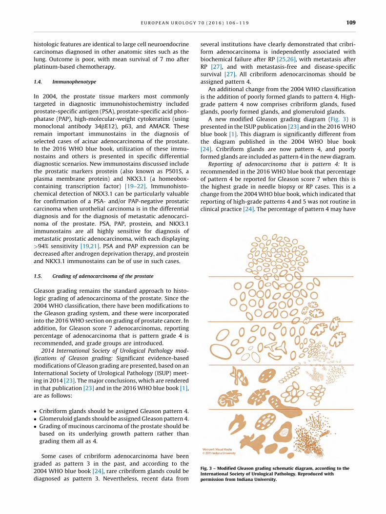
Fig. 3 – Modified Gleason grading schematic diagram, according to theInternational Society of Urological Pathology. Reproduced withpermission from Indiana University.
rading of mucinous carcinoma of the prostate should be
based on its underlying growth pattern rather than
grading them all as 4.
Some cases of cribriform adenocarcinoma have been
graded as pattern 3 in the past, and according to the
2004 WHO blue book [24], rare cribriform glands could be
diagnosed as pattern 3. Nevertheless, recent data from
several institutions have clearly demonstrated that cribri-
form adenocarcinoma is independently associated with
biochemical failure after RP [25,26], with metastasis after
RP [27], and with metastasis-free and disease-specific
survival [27]. All cribriform adenocarcinomas should be
assigned pattern 4.
An additional change from the 2004 WHO classification
is the addition of poorly formed glands to pattern 4. High-
grade pattern 4 now comprises cribriform glands, fused
glands, poorly formed glands, and glomeruloid glands.
A new modified Gleason grading diagram (Fig. 3) is
presented in the ISUP publication [23] and in the 2016 WHO
blue book [1]. This diagram is significantly different from
the diagram published in the 2004 WHO blue book
[24]. Cribriform glands are now pattern 4, and poorly
formed glands are included as pattern 4 in the new diagram.
Reporting of adenocarcinoma that is pattern 4: It is
recommended in the 2016 WHO blue book that percentage
of pattern 4 be reported for Gleason score 7 when this is
the highest grade in needle biopsy or RP cases. This is a
change from the 2004 WHO blue book, which indicated that
reporting of high-grade patterns 4 and 5 was not routine in
clinical practice [24]. The percentage of pattern 4 may have
E U R O P E A N U R O L O G Y 7 0 ( 2 0 1 6 ) 1 0 6 – 1 1 9110
implications for management strategies such as active
surveillance (AS) because some patients with Gleason
grade 3 + 4 = 7 with a low percentage of pattern 4 may be
considered for AS [28]. An abundance of data suggests that
percentage of adenocarcinoma that is high-grade pattern 4/5
is an important prognostic indicator [29–31]. The method
for determination of percentage of pattern 4 was not
specified.
Grade groups: A new set of grade groups was recently
developed [13,32], with a broad consensus for acceptance
by expert urologic pathologists and clinicians at the
2014 ISUP consensus conference on Gleason grading of
prostatic carcinoma [23]. These grade groups are as follows:
� Grade group 1: Gleason score �6
� G
rade group 2: Gleason score 3 + 4 = 7� G
rade group 3: Gleason score 4 + 3 = 7� G
rade group 4: Gleason score 4 + 4 = 8, 3 + 5 = 8, 5 + 3 = 8� G
rade group 5: Gleason scores 9–10There are several rationales for the generation of the grade
groups: Gleason scores 2–5 are rarely used, Gleason scores
have been grouped together in the past in arrangements that
do not accuratelyreflect prognosis, and grade group1 signifies
to the clinician and the patient that Gleason score 6 is the
lowest possible grade rather than an intermediate grade 6 of
10. The latter point is critical and informs all concerned that a
diagnosis of adenocarcinoma of the prostate, grade group 1,
carries an excellent prognosis [13,32]. Many patients with
grade group 1 tumours, in the correct clinical context with
consideration of other parameters (eg, serum PSA level,
clinical stage, and amount of cancer in needle core tissue),
could be candidates for AS. The prognostic impact of the five
grade groups has been validated in a large multi-
institutional study of >20 000 RP cases, >16 000 needle
biopsy cases, and >5000 biopsies followed by radiation
therapy [13]. Of interest, there are genomic correlates and
molecular support for the grade group system [33]. The
2016 WHO blue book states that the grade groups should
be reported in conjunction with the 2014 modified ISUP
Gleason scores.
1.6. Risk stratification and active surveillance for acinar
adenocarcinoma of prostate
In the 2016 WHO blue book, the vital importance of risk
stratification for patients with adenocarcinoma of the
prostate is highlighted in a section on prognosis and
predictive factors [1]. In particular, there is much detail on
pathologic prognostic factors for the different types of
tissue samples: needle biopsy, transurethral resection, and
RP tissues. In addition, the 2015 National Comprehensive
Cancer Network risk groups, which use clinical and
pathologic factors, are presented in a table. Because many
prostate cancers (especially many grade group 1 tumours)
are indolent and may be managed by AS, a new discussion
is provided on AS, along with a table on clinical and
pathologic inclusion criteria used by a number of large AS
programs.
1.7. Genetic profile of adenocarcinoma of the prostate
Since 2004, there has been a remarkable expansion of
knowledge about the genetics of prostate cancer. Advances
in sequencing technology have revealed complex rearran-
gements and marked heterogeneity [34–37]. Only a few
abnormalities in specific genes are highly recurrent, but
alterations in certain signalling pathways predominate,
such as PI3K/PTEN/AKT, cell cycle regulation, and chromatin
regulation [34]. The most common alterations, in both
primary and metastatic prostate cancer, are fusions of
androgen-regulated promoters with ERG and other mem-
bers of the ETS family of transcription factors [37],
particularly the TMPRSS2-ERG fusion, which is present in
approximately 50% of all prostate cancers. In primary
clinically localized prostate cancer, there are relatively few
recurrent nonsynonymous point mutations, including
mutations in the SPOP (11%) and FOXA1 (3%) genes
[37]. In comparison, in castration-resistant metastatic
prostate cancer, there are increased alteration rates in
many genes and pathways, including abnormalities in
androgen receptor (AR) signalling (usually due to AR gene
amplification or mutation), DNA repair, and PI3K pathways,
as well as mutations or deletions in the TP53, RB1, KMT2C,
and KMT2D genes [36,37]. This landscape of somatic genetic
abnormalities in adenocarcinoma of the prostate is dis-
cussed in depth in a genetic profile section, and a model for
molecular classification of prostate cancer is shown.
Although this molecular classification is not currently in
clinical use, the discovery of these genetic abnormalities has
led to greater understanding of the molecular pathogenesis
of prostate cancer and has demonstrated potentially
therapeutically actionable molecular defects. Such molecu-
lar classifications may be incorporated into WHO classifica-
tions of prostate cancer in the future.
2. The new bladder tumour classification
The fourth edition of the WHO classification of tumours of
the urothelial tract provides a contemporary review of the
morphology of urothelial neoplasms, emphasizing their
unique ability to exhibit divergent differentiation, multiple
morphologic variants, and a diverse genomic landscape
(Fig. 4) [1]. It is becoming clearer how both morphology and
genotype may be exploited to select therapy, and for the
latter, clinical protocols are in place to take advantage of
activated molecular pathways in specific tumours. What
follows is not a comprehensive summary of the entire WHO
narrative but rather a selected summary of new or evolving
concepts or entities. Mesenchymal, neuroendocrine, and
other types of nonurothelial lesions are beyond the scope of
this summary.
2.1. Grading of urothelial tumours
Grading of urothelial tumours has particular importance in
noninvasive disease, specifically papillary neoplasms.
Although a small percentage of invasive carcinomas are
low grade, usually limited to the lamina propria, >95% of

[(Fig._4)TD$FIG]
Fig. 4 – World Health Organization (WHO) classification of tumours of the urothelial tract. Reproduced with permission from the WHO [1].WHO = World Health Organization.
E U R O P E A N U R O L O G Y 7 0 ( 2 0 1 6 ) 1 0 6 – 1 1 9 111
invasive tumours are high grade. Exceptions exist, a good
example being the nested variant of urothelial carcinoma,
which, despite its deceptively bland cytomorphology, may
present as locally advanced disease and is associated with
poor outcome. Noninvasive tumours can be divided into
two categories: papillary or flat. Carcinoma devoid of
papillary structures is called carcinoma in situ (CIS) and is,
by definition, high grade. Importantly, flat urothelium can
exhibit a wide spectrum of atypia, from reactive to
preneoplastic to frankly malignant. Papillary tumours are
also quite varied, including reactive proliferations and
papilloma as well as papillary urothelial proliferation of
low malignant potential (PUNLMP) and low- and high-
grade papillary carcinoma [38]. Interobserver variability
is high, even among experienced pathologists, despite many
decades of efforts to develop pathologic classifications that
best reflect clinical behaviour [39–50]. As in 2004, the
2016 WHO classification continues to recommend the
application of the grading classification first put forth by
ISUP in 1997 (Table 1). In fact, this classification continues
to be endorsed by ISUP and all major contemporary
pathology textbooks and guidelines, including the AFIP
[US Armed Forces Institute of Pathology] Atlas of Tumor
Pathology, Series 4 fascicle on tumours of the kidney,
bladder, and related urinary structures and the latest
editions of the American Joint Committee on Cancer Cancer
Staging Manual and the International Collaboration on
Cancer Reporting. Multiple studies have been published
comparing this classification with others, particularly the
1973 WHO classification, in terms of reproducibility and
Table 1 – World Health Organization classification of tumours:tumours of the urothelial tract, differences between the third andfourth editions for noninvasive urothelial lesions
Third edition [51]: Fourth edition [1]:
Noninvasive urothelial lesions Noninvasive urothelial lesions
Urothelial carcinoma in situ Urothelial carcinoma in situ
Papillary urothelial
carcinoma, low grade
Papillary urothelial carcinoma, low
grade
Papillary urothelial
carcinoma, high grade
Papillary urothelial carcinoma, high
grade
Papillary urothelial
neoplasm of low
malignant potential
Papillary urothelial neoplasm of low
malignant potential
Urothelial papilloma Urothelial papilloma
Inverted urothelial
papilloma
Inverted urothelial papilloma
Urothelial proliferation of uncertain
malignant potential (hyperplasia)
Urothelial dysplasia
E U R O P E A N U R O L O G Y 7 0 ( 2 0 1 6 ) 1 0 6 – 1 1 9112
clinical impact. Results have been mixed but mostly
positive. It is recommended that this classification be
adopted worldwide because of its inherent advantages:
� Uniform terminology and definitions based on the level of
cytologic and architectural abnormalities (order and
disorder) and the establishment of detailed criteria for
various preneoplastic conditions and tumour grades
� D
efinition of a group of lesions (high grade) with a highrisk of progression that may be candidates for adjuvant
therapy
� E
limination of ambiguity in diagnostic categories in the1973 WHO system (grade 1–2, grade 2–3)
� In
Table 2 – World Health Organization classification of tumours:tumours of the urothelial tract, differences between the third andfourth editions, invasive urothelial tumors
Third edition [51]: Fourth edition [1]:
Invasive urothelial tumours Invasive urothelial tumours
Infiltrating urothelial carcinoma Infiltrating urothelial carcinoma
with divergent differentiation
with squamous differentiation Nested, including large nested
with glandular differentiation Microcystic
with trophoblastic differentiation Micropapillary
Nested Lymphoepithelioma-like
Microcystic Plasmacytoid/signet ring
cell/diffuse
Micropapillary Sarcomatoid
Lymphoepithelioma-like Giant cell
Lymphoma-like Poorly differentiated
Plasmacytoid Lipid rich
Sarcomatoid Clear cell
Giant cell Tumours of maullerian type
Undifferentiated Tumors arising in a bladder
diverticulum
clusion of a category of papillary neoplasm (ie,
PUNLMP) that is not associated with invasion at the
time of diagnosis and has a negligible risk of progression,
although the potential for recurrence requires clinical
surveillance
Admittedly, controversy remains, and the reasons are
multifactorial but mainly due to the fact that the clinical risk
of recurrence and progression are determined not solely by
growth pattern and grade but also by other factors such as
size, multifocality, time to recurrence, and prior intravesical
therapy. In addition, we must accept the fact that grading is
largely subjective and that, in the future, ancillary studies
(either immunohistochemical or molecular assays) will lead
to enhanced reproducibility and better correlation with
clinical outcome [52].
The term urothelial proliferation of uncertain malignant
potential has been introduced, supplanting the term
hyperplasia [53–58]. It describes a thickened urothelium
with minimal or no cytological atypia and no true papillary
fronds, although undulations are common. These entities
may be seen de novo, and in this setting, the clinical
relevance is unknown. More frequently they are seen in
patients who have a history of prior carcinoma or seen
adjacent to papillary lesions. It is likely that most represent
lateral extension (‘‘shoulder lesion’’) of a papillary neo-
plasm; this assumption is supported by high incidence of
chromosome 9 deletions and lesser but significant inci-
dence of FGFR3 abnormalities.
Urothelial dysplasia is defined as a flat lesion with
appreciable cytologic and architectural abnormalities that
are believed to be preneoplastic but that fall short of the
criteria required for urothelial CIS. It is rarely described de
novo, and for this reason, it is poorly studied. More important,
it is the most difficult category to define morphologically
because of significant interobserver variability and the total
absence of large clinical studies documenting its relationship
to the development of subsequent CIS. In patients with a prior
history of urothelial carcinoma, this diagnosis is particularly
challenging and fraught with variability in interpretation,
given the changes induced by prior instrumentation, biopsy
site changes, and intravesical therapy. It is of no surprise that
urologists rarely alter management based on this diagnosis
alone.
2.2. Invasive urothelial carcinoma with divergent
differentiation
By definition, urothelial carcinoma with divergent differen-
tiation refers to tumours arising within the urothelial tract, in
which some percentage of ‘‘usual type’’ urothelial carcinoma
is present along with other morphologies (Fig. 5a, Table 2).
Urothelial carcinoma has long been known to have a
remarkable propensity for divergent differentiation, which
is seen most commonly in association with high-grade and
locally advanced disease [59–62]. The incidence of divergent
differentiation in cystectomy specimens is as high as 33%. Its
presence is associated with established predictors of
aggressive behaviour, and although some studies have found
an association with adverse outcome on univariate analysis,
the effect does not remain significant on multivariable
analysis. The amount of divergent histology present does not
seem to have a bearing on outcome, although limited data are
available to this effect [61]; however, it is recommended that
pathologists report the percentage of divergent histologies in
the pathology report.
[(Fig._5)TD$FIG]
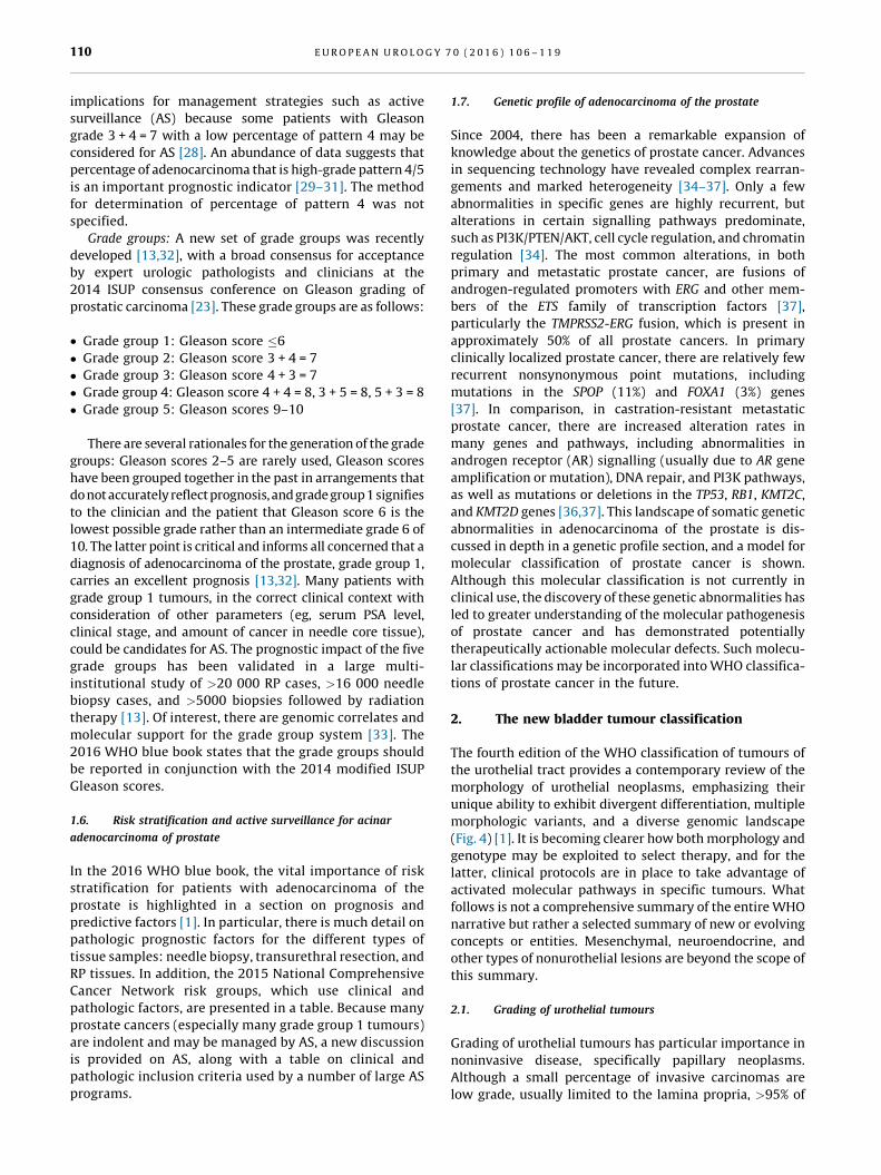
Fig. 5 – (a) Urothelial carcinoma with mixed histologic features (divergent differentiation), including a micropapillary component. Strong HER2expression is preferentially seen in the micropapillary component (insert). (b) Adenocarcinoma of the bladder. This case has enteric features andextensive mucin production. Notice signet ring cells within the mucin. (c) Plasmacytoid carcinoma of the bladder. Notice the presence of bothplasmacytoid and signet ring cells devoid of extracellular mucin. Insert demonstrates loss of e-cadherin expression within the invasive tumour,whereas it is retained in the surface urothelium. (d) Clear cell carcinoma. This tumour can arise from the surface urothelium or from mullerian restslocated within or adjacent to the urogenital tract.
E U R O P E A N U R O L O G Y 7 0 ( 2 0 1 6 ) 1 0 6 – 1 1 9 113
Common morphologic manifestations of divergent
differentiation appear along squamous, glandular, small
cell, and even trophoblastic lines. Squamous cell carcinoma
is defined by the presence of intercellular bridges or
keratinization and may be present in up to 40% of invasive
urothelial carcinomas [63–66]. It is almost never associated
with human papillomavirus infection, with the rare
exception of some cases with a basaloid morphology
[67,68]. Interestingly, recent genomic data have described
a basal/squamous-like molecular subtype that has squa-
moid morphology and immunophenotype and is associated
with poor survival and poor response to systemic therapy
[69,70]. Glandular neoplasms constitute the second most
common form of divergent differentiation, seen in up to 18%
of invasive tumours and defined by the presence of gland
formation [71–73]. These tumours commonly have enteric
features and, in isolation, can be easily confused with
colonic adenocarcinoma. These tumours can express an
identical immunophenotype such that site of origin is best
determined clinically. In this setting and in others in which
the tumour is composed exclusively of a variant morpholo-
gy, pathologists are encouraged to include a comment in the
pathology report, stating, ‘‘We would accept as primary at
this site if direct extension or a metastasis from another
organ can be ruled out clinically.’’ Some tumours are
associated with extravasated mucin (mucinous), with or
without signet ring cells [74]. Rare tumours exhibit
trophoblastic differentiation (syncytiotrophoblasts) with
human chorionic gonadotrophin production, and some may
even have an endodermal sinus, which expresses a-
fetoprotein [71,75].
Many other morphologic manifestations of divergent
differentiation may be encountered including nested,
micropapillary, and small cell. When present in a pure
E U R O P E A N U R O L O G Y 7 0 ( 2 0 1 6 ) 1 0 6 – 1 1 9114
form, these are considered variants of urothelial carcinoma.
As mentioned previously, their presence in a tumour with
mixed histology is of questionable clinical importance
compared with urothelial carcinomas of equal stage and
grade, although some exceptions exist, such as small cell
carcinoma and possibly micropapillary carcinoma.
2.3. Invasive variants of urothelial carcinoma
The variants of urothelial carcinoma are listed in Table 1.
This discussion will highlight only novel entities or novel
concepts within selected variants.
The morphologic types of glandular neoplasms arising in
the urothelial tract include enteric and mucinous types
[71,74]. The enteric type is morphologically identical to its
colonic counterpart, with which it can be easily confused. The
mucinous type is characterized by the presence of abundant
extravasated mucin with free-floating neoplastic cells,
including signet ring cells (Fig. 5b). Contemporary thinking
suggests that signet ring cell carcinoma, which by definition
is not associated with any extravasated mucin, should not be
included in this variant. Experience has taught us that
tumours previously classified as such were either of the
mucinous type or consisted of tumours with a variable
number of signet ring cells as well as a significant number of
cells with plasmacytoid features. In fact, plasmacytoid cells
almost always predominate. These facts and recent molecu-
lar studies suggest that such tumours fit best in the
plamacytoid variant category (described subsequently).
The nested variant of urothelial carcinoma is character-
ized by cytologically bland tumour cells, infiltrating as
disorderly arranged, discrete or confluent small nests or
tubules. A large nested variant of urothelial carcinoma has
been described recently and is composed of equally bland
tumour cells [76–79]. The importance of identifying this
variant cannot be overstated because it can mimic benign
urothelial proliferations, particularly in superficial trans-
urethral resections and cold-cup biopsies, but it presents
characteristically as locally advanced tumours and is
associated with poor clinical outcome. The traditional
grading scheme for urothelial carcinomas does not apply
to these deceptively bland variants. Although the micro-
cystic variant of urothelial carcinoma is considered a
distinct entity, some examples can also have nests and
tubules of neoplastic cells [80,81]. Importantly, what they
also share is a deceptively bland cytologic appearance that
can mimic benign conditions such as cystitis glandularis.
The micropapillary variant of urothelial carcinoma has
been well documented in the literature [82–89]. Morpho-
logically, it is defined as small nests and aggregates of
tumour cells within lacunae. Multiple small nests without
vascular cores are characteristic of this entity. The nuclei are
markedly atypical and oriented to the periphery of the cell
cluster. Cytoplasmic vacuoles with distortion of the nuclear
contour are common. These tumours are commonly
associated with lymphovascular invasion, present at a high
pathologic stage, and exhibit aggressive clinical behaviour.
Despite the early literature advocating early cystectomy in
all cases, it remains controversial whether these tumours
should be treated differently form other high-grade, locally
advanced bladder tumours, particularly regarding early
cystectomy or neoadjuvant therapy. Whether clinical
outcome is related to the morphology per se or to the
stage at presentation is unclear, as is whether the
proportion of the micropapillary component influences
outcome. At the molecular level, overexpression or ampli-
fication of ERBB2 is more common in this variant than in
conventional urothelial carcinoma (Fig. 5a) [89–91].
Plasmacytoid urothelial carcinoma was described sever-
al decades ago, but recent data have defined the morpho-
logic spectrum, clinical behaviour, and genotype in a more
comprehensive manner [92–98]. This rare tumour is
characterized by the presence of mononuclear tumour cells
with plasmacytoid, lymphoid, or even rhabdoid features. The
tumour will very commonly exhibit a variable percentage of
cells with cytoplasmic vacuoles, imparting the appearance of
signet ring cells, with or without intracellular mucin but
never associated with extracellular mucin (Fig. 5c). In fact,
virtually every case of signet ring cell carcinoma of the
urinary bladder that has been described in the literature
would now be placed into this category of tumour, assuming
absence of extracellular mucin. Of all the variants, this one is
most likely to be encountered in its pure form, although it can
also be seen in association with usual urothelial carcinoma
or other variants. It is invariably diagnosed at a locally
advanced stage and is associated with a dismal outcome. At
the molecular level, these tumours are characterized by
the presence of truncating mutations of CDH1 and loss of
e-cadherin expression (Fig. 5c) [97].
2.4. Tumours arising along the genitourinary tract but not of
urothelial origin
As described previously, the morphologic plasticity seen in
urothelial carcinoma is very broad and includes tumours
with clear cell features [99–103]. However, a series of
tumours is encountered predominantly in women and
appears to arise from mullerian precursors present within
the bladder wall or adjacent soft tissues, commonly
endometriosis but rarely mullerianosis [99]. Clear cell
carcinoma predominates, but occasional cases of endome-
trioid-type carcinoma have been described, only in women.
Clear cell carcinomas are characterized by the usual
tubulocystic, papillary, or diffuse growth patterns (Fig. 5d).
Hobnail cells are common, as are basophilic or eosinophilic
secretions. Although some cases may be confused with
nephrogenic adenoma, the level of nuclear enlargement and
hyperchromasia present in clear cell carcinoma should lead
to the proper diagnosis. As might be expected, this tumour is
immunoreactive for PAX8, HNF1B, CA125 and p53, similar to
its ovarian counterparts. The endometrioid variant is usually
PAX8 and p53 negative but positive for ER and PR.
2.5. Tumours arising in a bladder diverticulum
Epithelial neoplasms have been reported in up to 14% of
bladder diverticula, composing approximately 1% of blad-
der neoplasms [104–106]. Based on the unique clinical
E U R O P E A N U R O L O G Y 7 0 ( 2 0 1 6 ) 1 0 6 – 1 1 9 115
scenario and anatomy of diverticula, it is an important topic
that was not covered in the prior edition. The majority of
tumours arise in acquired diverticula, the wall of which is
composed of urothelium and lamina propria only; by
definition, no muscularis propria is present except in the
bladder wall immediately adjacent to the diverticulum
(diverticular os). As such, pathologic staging of these
tumours is different from those that arise within the
bladder because pT2 disease does not exist. Up to 50% of
cases are noninvasive, either papillary or flat. Of those
tumours that are invasive, most are of usual urothelial type,
whereas the rest may exhibit a variant morphology or
mixed histologic features (divergent differentiation). Simi-
lar to vesical primaries, pathologic stage is the most
important prognostic factor.
2.6. Genomics of urothelial carcinoma
Studies have suggested that invasive urothelial tumours
develop along at least two molecular pathways, via either
high-grade papillary tumours or CIS. Molecular alterations
differ markedly between low- and high-grade tumours and
between those that are invasive and those that are not.
Because it is likely that tumours develop from areas of
premalignant urothelial cells, it is not surprising that
multifocal and metachronous tumours show common as
well as novel uniquely acquired mutations [107–109]. Copy
number abnormalities, loss of heterozygosity, and in-
creased genomic instability have been associated with
increasing tumour grade and stage. Multiple tumour
suppressor genes and oncogenes have been described in
invasive urothelial carcinoma, but often it is difficult to
determine whether these are required for cancer develop-
ment [110,111]. Recurrent mutations occur in genes such as
TP53, FGFR3, PIK3CA, RB1 and HRAS, with TP53 and FGFR3
being the most common, together with promoter mutations
of TERT [112–114]. Although TERT mutations are present
in up to 79% of bladder neoplasms, they have no
association with clinical outcome; however, its presence
can be of great diagnostic utility, given the relative rarity
of this mutation in other tumours that may have over-
lapping histology. Next-generation sequencing efforts
have demonstrated that the mutational landscape of
urothelial tumours are quite complex, with >300 muta-
tions, >200 copy number alterations, and >20 rearrange-
ments per tumour [108,115–117]. Only lung cancer has
been shown to harbour a higher rate of mutations,
although most are certainly passenger mutations with
no functional consequence [118].
The most frequently altered pathways in bladder cancer
include the PI3K/AKT/mammalian target of rapamycin
pathway [96,119–121], the FGFR3/RAF/RAS pathway, the
TP53/RB1 pathway, immune response checkpoint modula-
tors [122,123], and chromatin-regulating and -remodelling
genes [124–126]. In general, mutations along a given
pathway are mutually exclusive. Some of the components of
these pathways are altered in low-risk disease, whereas
others are characteristic of high-risk disease. FGFR3
mutations, for example, are seen in up to 80% of papillary
noninvasive and low-grade carcinomas. Although these
mutations have been associated with a higher risk of
recurrence, they are not associated with disease progression
[11,127]. Mutations in chromatin-remodelling and histone-
modifying genes have been described in up to 89% of
muscularis propria invasive bladder tumours [116,128]. As
novel therapeutic agents are developed that target these
pathways, improvements in therapy will come. In addition,
emerging data show that immune-modulating agents may
have a promising role in the management of advanced
urothelial carcinoma.
The discovery of the molecular pathways involved in
urothelial cancer recurrence and progression has allowed for
the identification of potential prognostic and predictive
markers [116,129,130]. It has also permitted the develop-
ment of novel noninvasive detection and surveillance
strategies and revealed potential therapeutic targets [131–
136]. The absence of multi-institutional randomized pro-
spective trials, however, has delayed the validation of these
prognostic and predictive markers for routine clinical use. The
good news is that a significant number of these trials have
been launched or will be in the near future and will likely alter
the way we identify, risk-assess, and treat these tumours.
Author contributions: Holger Moch had full access to all the data in the
study and takes responsibility for the integrity of the data and the
accuracy of the data analysis.
Study concept and design: Humphrey, Moch, Cubilla, Ulbright, Reuter.
Acquisition of data: Humphrey, Moch, Cubilla, Ulbright, Reuter.
Analysis and interpretation of data: Humphrey, Moch, Cubilla, Ulbright,
Reuter.
Drafting of the manuscript: Humphrey, Moch, Cubilla, Ulbright, Reuter.
Critical revision of the manuscript for important intellectual content:
Humphrey, Moch, Cubilla, Ulbright, Reuter.
Statistical analysis: None.
Obtaining funding: None.
Administrative, technical, or material support: None.
Supervision: Humphrey, Moch, Cubilla, Ulbright, Reuter.
Other (specify): None.
Financial disclosures: Holger Moch certifies that all conflicts of interest,
including specific financial interests and relationships and affiliations
relevant to the subject matter or materials discussed in the manuscript
(eg, employment/affiliation, grants or funding, consultancies, honoraria,
stock ownership or options, expert testimony, royalties, or patents filed,
received, or pending), are the following: None.
Funding/Support and role of the sponsor: None.
References
[1] Moch H, Humphrey PA, Ulbright TM, Reuter V. WHO Classification
of Tumours of the Urinary System and Male Genital Organs. Lyon,
France: International Agency for Research on Cancer; 2016.
[2] Kovi J, Jackson MA, Heshmat MY. Ductal spread in prostatic
carcinoma. Cancer 1985;56:1566–73.
[3] Magers M, Kunju LP, Wu A. Intraductal carcinoma of the prostate:
morphologic features, differential diagnoses, significance, and
reporting practices. Arch Pathol Lab Med 2015;139:1234–41.
[4] Humphrey PA. Intraductal carcinoma of the prostate. J Urol
2015;194:1434–5.
E U R O P E A N U R O L O G Y 7 0 ( 2 0 1 6 ) 1 0 6 – 1 1 9116
[5] McNeal JE, Yemoto CE. Spread of adenocarcinoma within prostatic
ducts and acini. Morphologic and clinical correlations. Am J Surg
Pathol 1996;20:802–14.
[6] Tsuzuki T. Intraductal carcinoma of the prostate: a comprehensive
and updated review. Int J Urol 2015;22:140–5.
[7] Robinson B, Magi-Galluzzi C, Zhou M. Intraductal carcinoma of the
prostate. Arch Pathol Lab Med 2012;136:418–25.
[8] Robinson BD, Epstein JI. Intraductal carcinoma of the prostate
without invasive carcinoma on needle biopsy: emphasis on radical
prostatectomy findings. J Urol 2010;184:1328–33.
[9] Watts K, Li J, Magi-Galluzzi C, Zhou M. Incidence and clinicopatho-
logical characteristics of intraductal carcinoma detected in pros-
tate biopsies: a prospective cohort study. Histopathology 2013;63:
574–9.
[10] Miyai K, Divatia MK, Shen SS, Miles BJ, Ayala AG, Ro JY. Heteroge-
neous clinicopathological features of intraductal carcinoma of the
prostate: a comparison between ‘‘precursor-like’’ and ‘‘regular
type’’ lesions. Int J Clin Exp Pathol 2014;7:2518–26.
[11] Guo CC, Epstein JI. Intraductal carcinoma of the prostate on needle
biopsy: histologic features and clinical significance. Mod Pathol
2006;19:1528–35.
[12] Morais CL, Han JS, Gordetsky J, et al. Utility of PTEN and ERG
immunostaining for distinguishing high-grade PIN from intraduc-
tal carcinoma of the prostate on needle biopsy. Am J Surg Pathol
2015;39:169–78.
[13] Epstein JI, Zelefsky MJ, Sjoberg DD, et al. A contemporary prostate
cancer grading system: a validated alternative to the Gleason
score. Eur Urol 2016;69:428–35.
[14] Humphrey PA. Histological variants of prostatic carcinoma and
their significance. Histopathology 2012;60:59–74.
[15] Yaskiv O, Cao D, Humphrey PA. Microcystic adenocarcinoma of the
prostate: a variant of pseudohyperplastic and atrophic patterns.
Am J Surg Pathol 2010;34:556–61.
[16] Parwani AV, Herawi M, Epstein JI. Pleomorphic giant cell adeno-
carcinoma of the prostate: report of 6 cases. Am J Surg Pathol
2006;30:1254–9.
[17] Lopez-Beltran A, Eble JN, Bostwick DG. Pleomorphic giant cell
carcinoma of the prostate. Arch Pathol Lab Med 2005;129:683–5.
[18] Evans AJ, Humphrey PA, Belani J, van der Kwast TH, Srigley JR.
Large cell neuroendocrine carcinoma of prostate: a clinicopatho-
logic summary of 7 cases of a rare manifestation of advanced
prostate cancer. Am J Surg Pathol 2006;30:684–93.
[19] Epstein JI, Egevad L, Humphrey PA, Montironi R. Best practices
recommendations in the application of immunohistochemistry
in the prostate: report from the International Society of Urologic
Pathology consensus conference. Am J Surg Pathol 2014;38:
e6–19.
[20] Gelmann EP, Bowen C, Bubendorf L. Expression of NKX3.1 in
normal and malignant tissues. Prostate 2003;55:111–7.
[21] Gurel B, Ali TZ, Montgomery EA, et al. NKX3.1 as a marker of
prostatic origin in metastatic tumors. Am J Surg Pathol 2010;34:
1097–105.
[22] Sheridan T, Herawi M, Epstein JI, Illei PB. The role of P501S and PSA
in the diagnosis of metastatic adenocarcinoma of the prostate. Am
J Surg Pathol 2007;31:1351–5.
[23] Epstein JI, Egevad L, Amin MB, Delahunt B, Srigley JR, Humphrey
PA. The 2014 International Society of Urological Pathology (ISUP)
consensus conference on Gleason grading of prostatic carcinoma:
definition of grading patterns and proposal for a new grading
system. Am J Surg Pathol 2016;40:244–52.
[24] Epstein JI, Algaba F, Allsbrook Jr WC, et al. Acinar adenocarcinoma.
In: Eble JN, Sauter G, Epstein JI, Sesterhenn IA, editors. Pathology
and Genetics of Tumours of the Urinary System and Male Genital
Organs. Lyon, France: IARC Press; 2004. p. 162.
[25] Kir G, Sarbay BC, Gumus E, Topal CS. The association of the
cribriform pattern with outcome for prostatic adenocarcinomas.
Pathol Res Pract 2014;210:640–4.
[26] Dong F, Yang P, Wang C, et al. Architectural heterogeneity and
cribriform pattern predict adverse clinical outcome for Gleason
grade 4 prostatic adenocarcinoma. Am J Surg Pathol 2013;37:
1855–61.
[27] Kweldam CF, Wildhagen MF, Steyerberg EW, Bangma CH, van
der Kwast TH, van Leenders GJ. Cribriform growth is highly
predictive for postoperative metastasis and disease-specific
death in Gleason score 7 prostate cancer. Mod Pathol 2015;28:
457–64.
[28] Morash C, Tey R, Agbassi C, et al. Active surveillance for the
management of localized prostate cancer: guideline recommen-
dations. Can Urol Assoc J 2015;9:171–8.
[29] Stamey TA, McNeal JE, Yemoto CM, Sigal BM, Johnstone IM.
Biological determinants of cancer progression in men with pros-
tate cancer. JAMA 1999;281:1395–400.
[30] Cheng L, Davidson DD, Lin H, Koch MO. Percentage of Gleason
pattern 4 and 5 predicts survival after radical prostatectomy.
Cancer 2007;110:1967–72.
[31] Sauter G, Steurer S, Clauditz TS, et al. Clinical utility of quantitative
Gleason grading in prostate biopsies and prostatectomy speci-
mens. Eur Urol 2016;69:592–8.
[32] Pierorazio PM, Walsh PC, Partin AW, Epstein JI. Prognostic Gleason
grade grouping: data based on the modified Gleason scoring
system. BJU Int 2013;111:753–60.
[33] Rubin MA, Girelli G, Demichelis F. Genomic correlates to the newly
proposed grading prognostic groups for prostate cancer. Eur Urol
2016;69:557–60.
[34] Barbieri CE, Bangma CH, Bjartell A, et al. The mutational landscape
of prostate cancer. Eur Urol 2013;64:567–76.
[35] Barbieri CE, Rubin MA. Genomic rearrangements in prostate can-
cer. Curr Opin Urol 2015;25:71–6.
[36] Robinson D, Van Allen EM, Wu YM, et al. Integrative clinical
genomics of advanced prostate cancer. Cell 2015;161:1215–28.
[37] Cancer Genome Atlas Research Network. The molecular taxonomy
of primary prostate cancer. Cell 2015;163:1011–25.
[38] Epstein JI, Amin MB, Reuter VR, Mostofi FK. The World Health
Organization/International Society of Urological Pathology con-
sensus classification of urothelial (transitional cell) neoplasms of
the urinary bladder. Bladder Consensus Conference Committee.
Am J Surg Pathol 1998;22:1435–48.
[39] Bircan S, Candir O, Serel TA. Comparison of WHO 1973, WHO/ISUP
1998, WHO 1999 grade and combined scoring systems in evalua-
tion of bladder carcinoma. Urol Int 2004;73:201–8.
[40] Busch C, Algaba F. The WHO/ISUP 1998 and WHO 1999 systems for
malignancy grading of bladder cancer. Scientific foundation and
translation to one another and previous systems. Virchows Arch
2002;441:105–8.
[41] Billis A, Carvalho RB, Mattos AC, et al. Tumor grade heterogeneity
in urothelial bladder carcinoma–proposal of a system using com-
bined numbers. Scand J Urol Nephrol 2001;35:275–9.
[42] Epstein JI. The new World Health Organization/International So-
ciety of Urological Pathology (WHO/ISUP) classification for TA, T1
bladder tumors: is it an improvement? Crit Rev Oncol Hematol
2003;47:83–9.
[43] Jones TD, Cheng L. Papillary urothelial neoplasm of low malignant
potential: evolving terminology and concepts. J Urol 2006;175:
1995–2003.
E U R O P E A N U R O L O G Y 7 0 ( 2 0 1 6 ) 1 0 6 – 1 1 9 117
[44] Montironi R, Lopez-Beltran A, Mazzucchelli R, Bostwick DG. Clas-
sification and grading of the non-invasive urothelial neoplasms:
recent advances and controversies. J Clin Pathol 2003;56:91–5.
[45] Montironi R, Mazzucchelli R, Colanzi P, et al. Improving inter-
observer agreement and certainty level in diagnosing and grading
papillary urothelial neoplasms: usefulness of a Bayesian belief
network. Eur Urol 2002;41:449–57.
[46] Nishiyama N, Kitamura H, Maeda T, et al. Clinicopathological
analysis of patients with non-muscle-invasive bladder cancer:
prognostic value and clinical reliability of the 2004 WHO classifi-
cation system. Jpn J Clin Oncol 2013;43:1124–31.
[47] Yin H, Leong AS. Histologic grading of noninvasive papillary
urothelial tumors: validation of the 1998 WHO/ISUP system by
immunophenotyping and follow-up. Am J Clin Pathol 2004;121:
679–87.
[48] Amin MB, McKenney JK, Paner GP, et al. ICUD-EAU International
Consultation on Bladder Cancer 2012: pathology. Eur Urol
2013;63:16–35.
[49] Amin MB, Smith SC, Reuter VE, et al. Update for the practicing
pathologist: the International Consultation on Urologic Disease-
European Association of Urology consultation on bladder cancer.
Mod Pathol 2015;28:612–30.
[50] Amin MB, Trpkov K, Lopez-Beltran A, Grignon D. Best practices
recommendations in the application of immunohistochemistry in
the bladder lesions: report from the International Society of
Urologic Pathology consensus conference. Am J Surg Pathol
2014;38:e20–34.
[51] Eble JN, Sauter G, Epstein JI, Sesterhenn IA. Pathology and Genetics
of Tumours of the Urinary System and Male Genital Organs. Lyon,
France: IARC Press; 2004.
[52] van Rhijn BW, Vis AN, van der Kwast TH, et al. Molecular grading of
urothelial cell carcinoma with fibroblast growth factor receptor
3 and MIB-1 is superior to pathologic grade for the prediction of
clinical outcome. J Clin Oncol 2003;21:1912–21.
[53] Chow NH, Cairns P, Eisenberger CF, et al. Papillary urothelial
hyperplasia is a clonal precursor to papillary transitional cell
bladder cancer. Int J Cancer 2000;89:514–8.
[54] Hartmann A, Moser K, Kriegmair M, Hofstetter A, Hofstaedter F,
Knuechel R. Frequent genetic alterations in simple urothelial
hyperplasias of the bladder in patients with papillary urothelial
carcinoma. Am J Pathol 1999;154:721–7.
[55] Obermann EC, Junker K, Stoehr R, et al. Frequent genetic altera-
tions in flat urothelial hyperplasias and concomitant papillary
bladder cancer as detected by CGH, LOH, and FISH analyses.
J Pathol 2003;199:50–7.
[56] Readal N, Epstein JI. Papillary urothelial hyperplasia: relationship
to urothelial neoplasms. Pathology 2010;42:360–3.
[57] Taylor DC, Bhagavan BS, Larsen MP, Cox JA, Epstein JI. Papillary
urothelial hyperplasia. A precursor to papillary neoplasms. Am J
Surg Pathol 1996;20:1481–8.
[58] van Oers JM, Adam C, Denzinger S, et al. Chromosome 9 deletions
are more frequent than FGFR3 mutations in flat urothelial hyper-
plasias of the bladder. Int J Cancer 2006;119:1212–5.
[59] Dalbagni G, Genega E, Hashibe M, et al. Cystectomy for bladder
cancer: a contemporary series. J Urol 2001;165:1111–6.
[60] Linder BJ, Frank I, Cheville JC, et al. Outcomes following radical
cystectomy for nested variant of urothelial carcinoma: a matched
cohort analysis. J Urol 2013;189:1670–5.
[61] Kim SP, Frank I, Cheville JC, et al. The impact of squamous and
glandular differentiation on survival after radical cystectomy for
urothelial carcinoma. J Urol 2012;188:405–9.
[62] Xylinas E, Rink M, Robinson BD, et al. Impact of histological
variants on oncological outcomes of patients with urothelial
carcinoma of the bladder treated with radical cystectomy. Eur J
Cancer 2013;49:1889–97.
[63] Guo CC, Gomez E, Tamboli P, et al. Squamous cell carcinoma of the
urinary bladder: a clinicopathologic and immunohistochemical
study of 16 cases. Hum Pathol 2009;40:1448–52.
[64] Ikegami H, Iwasaki H, Ohjimi Y, Takeuchi T, Ariyoshi A, Kikuchi M.
Sarcomatoid carcinoma of the urinary bladder: a clinicopathologic
and immunohistochemical analysis of 14 patients. Hum Pathol
2000;31:332–40.
[65] Lagwinski N, Thomas A, Stephenson AJ, et al. Squamous cell
carcinoma of the bladder: a clinicopathologic analysis of 45 cases.
Am J Surg Pathol 2007;31:1777–87.
[66] Hansel DE, Zhang Z, Petillo D, Teh BT. Gene profiling suggests a
common evolution of bladder cancer subtypes. BMC Med Geno-
mics 2013;6:42.
[67] Blochin EB, Park KJ, Tickoo SK, Reuter VE, Al-Ahmadie H. Urothelial
carcinoma with prominent squamous differentiation in the set-
ting of neurogenic bladder: role of human papillomavirus infec-
tion. Mod Pathol 2012;25:1534–42.
[68] Schwartz LE, Khani F, Bishop JA, Vang R, Epstein JI. Carcinoma of
the uterine cervix unvolving the genitourinary tract: a potential
diagnostic dilemma. Am J Surg Pathol 2016;40:27–35.
[69] Choi W, Czerniak B, Ochoa A, et al. Intrinsic basal and luminal
subtypes of muscle-invasive bladder cancer. Nat Rev Urol
2014;11:400–10.
[70] Damrauer JS, Hoadley KA, Chism DD, et al. Intrinsic subtypes of
high-grade bladder cancer reflect the hallmarks of breast cancer
biology. Proc Natl Acad Sci U S A 2014;111:3110–5.
[71] Grignon DJ, Ro JY, Ayala AG, Johnson DE, Ordonez NG. Primary
adenocarcinoma of the urinary bladder. A clinicopathologic anal-
ysis of 72 cases. Cancer 1991;67:2165–72.
[72] el-Mekresh MM, el-Baz MA, Abol-Enein H, Ghoneim MA. Primary
adenocarcinoma of the urinary bladder: a report of 185 cases. Br J
Urol 1998;82:206–12.
[73] Zaghloul MS, Nouh A, Nazmy M, et al. Long-term results of primary
adenocarcinoma of the urinary bladder: A report on 192 patients.
Urol Oncol 2006;24:13–20.
[74] Grignon DJ, Ro JY, Ayala AG, Johnson DE. Primary signet-ring cell
carcinoma of the urinary bladder. Am J Clin Pathol 1991;95:13–20.
[75] Yamase HT, Wurzel RS, Nieh PT, Gondos B. Immunohistochemical
demonstration of human chorionic gonadotropin in tumors of the
urinary bladder. Ann Clin Lab Sci 1985;15:414–7.
[76] Cox R, Epstein JI. Large nested variant of urothelial carcinoma:
23 cases mimicking von brunn nests and inverted growth pattern
of noninvasive papillary urothelial carcinoma. Am J Surg Pathol
2011;35:1337–42.
[77] Holmang S, Johansson SL. The nested variant of transitional cell
carcinoma–a rare neoplasm with poor prognosis. Scand J Urol
Nephrol 2001;35:102–5.
[78] Drew PA, Furman J, Civantos F, Murphy WM. The nested variant of
transitional cell carcinoma: an aggressive neoplasm with innocu-
ous histology. Mod Pathol 1996;9:989–94.
[79] Lin O, Cardillo M, Dalbagni G, Linkov I, Hutchinson B, Reuter VE.
Nested variant of urothelial carcinoma: a clinicopathologic and
immunohistochemical study of 12 cases. Mod Pathol 2003;16:
1289–98.
[80] Young RH, Eble JN. Unusual forms of carcinoma of the urinary
bladder. Hum Pathol 1991;22:948–65.
[81] Young RH, Zukerberg LR. Microcystic transitional cell carcinomas
of the urinary bladder. A report of four cases. Am J Clin Pathol
1991;96:635–9.
[82] Amin MB, Ro JY, el-Sharkawy T, et al. Micropapillary variant of
transitional cell carcinoma of the urinary bladder. Histologic
E U R O P E A N U R O L O G Y 7 0 ( 2 0 1 6 ) 1 0 6 – 1 1 9118
pattern resembling ovarian papillary serous carcinoma. Am J Surg
Pathol 1994;18:1224–32.
[83] Comperat E, Roupret M, Yaxley J, et al. Micropapillary urothelial
carcinoma of the urinary bladder: a clinicopathological analysis of
72 cases. Pathology 2010;42:650–4.
[84] Guo CC, Tamboli P, Czerniak B. Micropapillary variant of urothelial
carcinoma in the upper urinary tract: a clinicopathologic study of
11 cases. Arch Pathol Lab Med 2009;133:62–6.
[85] Kamat AM, Dinney CP, Gee JR, et al. Micropapillary bladder cancer:
a review of the University of Texas M. D. Anderson Cancer Center
experience with 100 consecutive patients. Cancer 2007;110:62–7.
[86] Sangoi AR, Beck AH, Amin MB, et al. Interobserver reproducibility
in the diagnosis of invasive micropapillary carcinoma of the
urinary tract among urologic pathologists. Am J Surg Pathol
2010;34:1367–76.
[87] Samaratunga H, Khoo K. Micropapillary variant of urothelial car-
cinoma of the urinary bladder; a clinicopathological and immu-
nohistochemical study. Histopathology 2004;45:55–64.
[88] Spaliviero M, Dalbagni G, Bochner BH, et al. Clinical outcome of
patients with T1 micropapillary urothelial carcinoma of the blad-
der. J Urol 2014;192:702–7.
[89] Huang H, Jadallah S, Chen YB, et al. Her2 expression in urothelial
carcinoma with micropapillary morphology: preferential over-
expresssion by the micropapillary component. Lab Invest
2013;93, 219a-a.
[90] Ross JS, Wang K, Gay LM, et al. A high frequency of activating
extracellular domain ERBB2 (HER2) mutation in micropapillary
urothelial carcinoma. Clin Cancer Res 2014;20:68–75.
[91] Schneider SA, Sukov WR, Frank I, et al. Outcome of patients with
micropapillary urothelial carcinoma following radical cystec-
tomy: ERBB2 (HER2) amplification identifies patients with poor
outcome. Mod Pathol 2014;27:758–64.
[92] Kaimakliotis HZ, Monn MF, Cheng L, et al. Plasmacytoid bladder
cancer: variant histology with aggressive behavior and a new
mode of invasion along fascial planes. Urology 2014;83:1112–6.
[93] Ricardo-Gonzalez RR, Nguyen M, Gokden N, Sangoi AR, Presti Jr JC,
McKenney JK. Plasmacytoid carcinoma of the bladder: a urothelial
carcinoma variant with a predilection for intraperitoneal spread.
J Urol 2012;187:852–5.
[94] Ro JY, Shen SS, Lee HI, et al. Plasmacytoid transitional cell carci-
noma of urinary bladder: a clinicopathologic study of 9 cases. Am J
Surg Pathol 2008;32:752–7.
[95] Dayyani F, Czerniak BA, Sircar K, et al. Plasmacytoid urothelial
carcinoma, a chemosensitive cancer with poor prognosis, and
peritoneal carcinomatosis. J Urol 2013;189:1656–61.
[96] Gonzalez-Roibon ND, Chaux A, Al-Hussain T, et al. Dysregulation
of mammalian target of rapamycin pathway in plasmacytoid
variant of urothelial carcinoma of the urinary bladder. Hum Pathol
2013;44:612–22.
[97] Al-Ahmadie H, Iyer G, Mehra R, et al. E-cadherin (CDH1) is
frequently mutated in urothelial carcinoma with signet-ring cell
and plasmacytoid morphology. Mod Pathol 2013;26, 191A-A.
[98] Nigwekar P, Tamboli P, Amin MB, Osunkoya AO, Ben-Dor D.
Plasmacytoid urothelial carcinoma: detailed analysis of morphol-
ogy with clinicopathologic correlation in 17 cases. Am J Surg
Pathol 2009;33:417–24.
[99] Oliva E, Amin MB, Jimenez R, Young RH. Clear cell carcinoma of the
urinary bladder: a report and comparison of four tumors of
mullerian origin and nine of probable urothelial origin with
discussion of histogenesis and diagnostic problems. Am J Surg
Pathol 2002;26:190–7.
[100] Drew PA, Murphy WM, Civantos F, Speights VO. The histogen-
esis of clear cell adenocarcinoma of the lower urinary tract.
Case series and review of the literature. Hum Pathol 1996;27:
248–52.
[101] Lah K, Desai D, Hadway P, Perry-Keene J, Coughlin G. Primary
vesical clear cell adenocarcinoma arising in endometriosis: a rare
case of mullerian origin. Anticancer Res 2013;33:615–7.
[102] Young RH, Scully RE. Clear cell adenocarcinoma of the bladder and
urethra. A report of three cases and review of the literature. Am J
Surg Pathol 1985;9:816–26.
[103] Mai KT, Yazdi HM, Perkins DG, Morash C, Green J. Multicentric
clear cell adenocarcinoma in the urinary bladder and the urethral
diverticulum: evidence of origin of clear cell adenocarcinoma of
the female lower urinary tract from Mullerian duct remnants.
Histopathology 2000;36:380–2.
[104] Idrees MT, Alexander RE, Kum JB, Cheng L. The spectrum of
histopathologic findings in vesical diverticulum: implications
for pathogenesis and staging. Hum Pathol 2013;44:1223–32.
[105] Kong MX, Zhao X, Kheterpal E, et al. Histopathologic and clinical
features of vesical diverticula. Urology 2013;82:142–7.
[106] Walker NF, Gan C, Olsburgh J, Khan MS. Diagnosis and manage-
ment of intradiverticular bladder tumours. Nat Rev Urol 2014;11:
383–90.
[107] Hoglund M. On the origin of syn- and metachronous urothelial
carcinomas. Eur Urol 2007;51:1185–93, discussion 1193.
[108] Nordentoft I, Lamy P, Birkenkamp-Demtroder K, et al. Mutational
context and diverse clonal development in early and late bladder
cancer. Cell Rep 2014;7:1649–63.
[109] van Tilborg AA, de Vries A, de Bont M, Groenfeld LE, van der Kwast
TH, Zwarthoff EC. Molecular evolution of multiple recurrent can-
cers of the bladder. Hum Mol Genet 2000;9:2973–80.
[110] Brandli DW, Ulbright TM, Foster RS, et al. Stroma adjacent to
metastatic mature teratoma after chemotherapy for testicular
germ cell tumors is derived from the same progenitor cells as
the teratoma. Cancer Res 2003;63:6063–8.
[111] Knowles MA. Molecular subtypes of bladder cancer: Jekyll and
Hyde or chalk and cheese? Carcinogenesis 2006;27:361–73.
[112] Allory Y, Beukers W, Sagrera A, et al. Telomerase reverse tran-
scriptase promoter mutations in bladder cancer: high frequency
across stages, detection in urine, and lack of association with
outcome. Eur Urol 2014;65:360–6.
[113] Gunes C, Rudolph KL. The role of telomeres in stem cells and
cancer. Cell 2013;152:390–3.
[114] Groll RJ, Warde P, Jewett MA. A comprehensive systematic review
of testicular germ cell tumor surveillance. Crit Rev Oncol Hematol
2007;64:182–97.
[115] Balbas-Martinez C, Sagrera A, Carrillo-de-Santa-Pau E, et al.
Recurrent inactivation of STAG2 in bladder cancer is not associated
with aneuploidy. Nat Genet 2013;45:1464–9.
[116] Cancer Genome Atlas Research Network. Comprehensive molec-
ular characterization of urothelial bladder carcinoma. Nature
2014;507:315–22.
[117] Guo G, Sun X, Chen C, et al. Whole-genome and whole-exome
sequencing of bladder cancer identifies frequent alterations in
genes involved in sister chromatid cohesion and segregation. Nat
Genet 2013;45:1459–63.
[118] Kandoth C, McLellan MD, Vandin F, et al. Mutational landscape
and significance across 12 major cancer types. Nature 2013;502:
333–9.
[119] Slade I, Bacchelli C, Davies H, et al. DICER1 syndrome: clarifying
the diagnosis, clinical features and management implications of a
pleiotropic tumour predisposition syndrome. J Med Genet 2011;48:
273–8.
[120] Fahmy M, Mansure JJ, Brimo F, et al. Relevance of the mammalian
target of rapamycin pathway in the prognosis of patients with

E U R O P E A N U R O L O G Y 7 0 ( 2 0 1 6 ) 1 0 6 – 1 1 9 119
high-risk non-muscle invasive bladder cancer. Hum Pathol
2013;44:1766–72.
[121] Chaux A, Comperat E, Varinot J, et al. High levels of phosphatase and
tensin homolog expression are associated with tumor progression,
tumor recurrence, and systemic metastases in pT1 urothelial carci-
noma of the bladder: a tissue microarray study of 156 patients
treated by transurethral resection. Urology 2013;81:116–22.
[122] Inman BA, Sebo TJ, Frigola X, et al. PD-L1 (B7-H1) expression by
urothelial carcinoma of the bladder and BCG-induced granulomata:
associations with localized stage progression. Cancer 2007;109:
1499–505.
[123] Powles T, Eder JP, Fine GD, et al. MPDL3280A (anti-PD-L1) treat-
ment leads to clinical activity in metastatic bladder cancer. Nature
2014;515:558–62.
[124] Fedorov O, Lingard H, Wells C, et al. [1,2,4]triazolo[4,3-a]phthala-
zines: inhibitors of diverse bromodomains. J Med Chem 2014;57:
462–76.
[125] Filippakopoulos P, Qi J, Picaud S, et al. Selective inhibition of BET
bromodomains. Nature 2010;468:1067–73.
[126] Hay DA, Fedorov O, Martin S, et al. Discovery and optimization of
small-molecule ligands for the CBP/p300 bromodomains. J Am
Chem Soc 2014;136:9308–19.
[127] van Rhijn BW, van der Kwast TH, Liu L, et al. The FGFR3 mutation is
related to favorable pT1 bladder cancer. J Urol 2012;187:310–4.
[128] Gui Y, Guo G, Huang Y, et al. Frequent mutations of chromatin
remodeling genes in transitional cell carcinoma of the bladder.
Nat Genet 2011;43:875–8.
[129] Iyer G, Al-Ahmadie H, Schultz N, et al. Prevalence and co-occur-
rence of actionable genomic alterations in high-grade bladder
cancer. J Clin Oncol 2013;31:3133–40.
[130] Kim PH, Cha EK, Sfakianos JP, et al. Genomic Predictors of Survival
in Patients with High-grade Urothelial Carcinoma of the Bladder.
Eur Urol 2015;67:198–201.
[131] Birkhahn M, Mitra AP, Williams AJ, et al. Predicting recurrence and
progression of noninvasive papillary bladder cancer at initial
presentation based on quantitative gene expression profiles.
Eur Urol 2010;57:12–20.
[132] Lindgren D, Frigyesi A, Gudjonsson S, et al. Combined
gene expression and genomic profiling define two intrinsic
molecular subtypes of urothelial carcinoma and gene signa-
tures for molecular grading and outcome. Cancer Res 2010;70:
3463–72.
[133] Netto GJ. Molecular biomarkers in urothelial carcinoma of the
bladder: are we there yet? Nat Rev Urol 2012;9:41–51.
[134] Sanchez-Carbayo M, Socci ND, Lozano J, Saint F, Cordon-Cardo C.
Defining Molecular Profiles of Poor Outcome in Patients With
Invasive Bladder Cancer Using Oligonucleotide Microarrays. J Clin
Oncol 2006;24:778–89.
[135] Shariat SF, Ashfaq R, Sagalowsky AI, Lotan Y. Predictive value of
cell cycle biomarkers in nonmuscle invasive bladder transitional
cell carcinoma. J Urol 2007;177:481–7, discussion 487.
[136] van der Kwast TH, Bapat B. Predicting favourable prognosis of
urothelial carcinoma: gene expression and genome profiling. Curr
Opin Urol 2009;19:516–21.